Cardiac Imaging

Welcome message from author
This document is posted to help you gain knowledge. Please leave a comment to let me know what you think about it! Share it to your friends and learn new things together.
Transcript

Cardiac Imaging

Anatomy & imaging methods

Schema of heart silhouette in sagittal projection

Chest X-ray

Chest X-ray

Chest X-ray

Chest X-ray

CT axial anatomy – MSCT

CT MIP and MPR reconstructions
Coronary arteries

CT – VRT reconstruction

MR – different planes

Coronarography
Left coronary artery

Coronarography
Right coronary artery

Pathologic findings:
plain chest film

Lung vascular pattern
• Normal– Visible to 2/3 of lungs, the width of truncus intermedius a. pulm. to 14 mm– Craniocaudal Q quotient 0,8 A/B to 1,2 (till 1,4)
• Widened– Wide shadows visible to the periphery, craniocaudal index Q = 1
• Poor– Narrow shadows not visible to the periphery, narrow pulmonary artery,
increased transparency

Cardiomegaly
• Cardiomegaly vs. enlargement of the
heart chambers
1) Valvular diseases
2) Pericardial effusion
3) Atrial septal defect
4) Eisenmenger syndrom
5) Cardiomyopathy
6) Ebstein malformation
7) Myocarditis

Cardiomegaly
Measurement of the Cardiothoracic ratio:
A / B < 0.5 is normal (< 0.6 in infants).

Pericardial effusion
Cardiomegaly

Enlargement of the L atrium
• preload - mitral insufficency,
atrial and ventricular septal
defects, ductus arteriosus patens
• afterload - mitral stenosis
• secondary in the left
ventricle insufficiency
LA – dorsally

Enlargement of the L ventricle
• myocardial disease –ischemic heart disease, myocarditis, cardiomypathy
• preaload - aortic resp.
mitral insuf., atrial septal defect, ductus arteriorus patens
• afterload - aorticstenosis,coarctation, systemic hypertension
• enormous blood flow -anemia, arterio-venous fistula
LV -apex to the left and
laterally

Enlargement of the R atrium
• Preload (volume) –tricuspid insfuficency, atrial septal
defect, ebstain anomaly
• Afterload (pressure) –pulmonary hypertension
• Right ventricle
insufficiency
RA – right contour

Enlargement of the R ventricle
• secondary in the left heart
= postcapillary pulmonary
• hypertension - left ventricle
insufficiency, mitral disease
• pulmonary hypertension -PE, COPD, idiopathic
• afterload - pulmonary valvar
stenosis
• preload - ASD, VSD
RV – cranial displacement
of the apex

Small heart shadow
1) Lung emphysema
2) Dehydratation
3) Constrictive pericarditis

Small heart shadow
Lung emphysema:
• increased lung volumes
• flattened diaphragms
• wide separation of the ribs
• elongated narrow heart
shadow
• pulmonary vessels appear to
be diffusely decreased

Heart diseases

Heart diseases of adults
• Ischemic heart disease
– X-ray (lung congestion, cardiomegaly, Kerley lines, pleural effusion)
– AG (coronarography)• Atherosclerotic changes – stenosis, occlusion, thrombus
• Percutaneous Transluminal Angioplasty
– ECHO
– CT
– MR
– Nuclear medicine

Heart diseases of adults
• Ischemic heart disease
Coronary AG
CT MR
Stenosis of LAD
CT
Angiography
after stent implantation

Heart diseases of adults
• Valvular diseases – insufficiency, stenosis
– X-ray• Indirect changes (enlargment of chambers, pulmonary
vessels)
– Echocardiography• Morfology, flow
– CT• Morfology
– MR• Morfology, flow

Heart diseases of adults
• Valvular diseases
MRI: Mitral regurg.MRI: Mitral stenosis

Heart diseases of adults
• Valvular diseases
MRI: Mitral regurg. MRI: Aortic stenosis

Heart diseases of adults
• Myocardial diseases
– Myocarditis, Cardiomypathy
– (X-ray)
– Echocardiography
– CT
– MR

Heart diseases of adults
• Myocardial diseases
MRI: Myocarditis MRI: HOCM

Heart diseases of adults
• Pericardial diseases
– Paricarditis acute, Constrictive pericarditis
– with or without effusion
– X-ray• Effusion – widening of shadow, tent shape, change shape according
to the position
• Constrictive p. - calcification
– Echocardiography
– CT
– MR

Heart diseases of adults
• Pericardial diseases
X-ray and CT: Pericardial constriction MR: Effusion

Heart diseases of adults
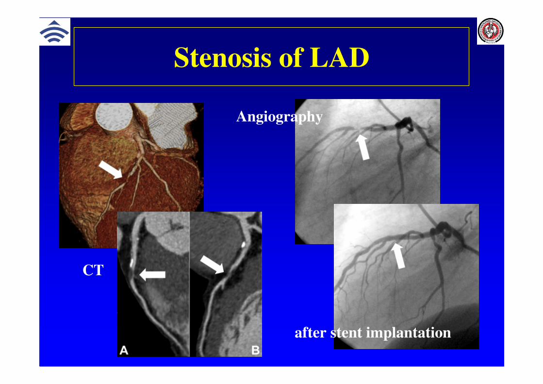
• Tumors and pseudotumors
– Localization: pericardial, myocardial, intracavital
– Thrombus, myxoma, fibroma, angiosarcoma, rhabdomyosarcoma, vegetation (infectiveendocarditis), metastasis, pericardial cyst
Heart diseases of adults
• Tumors and pseudotumors
CT: Myxoma of LA MRI: Myxoma of LA

Congenital heart disease

• Cardiac defects– Atrial septal defect
• L����P shunt: cardiomegaly, RA, RV and PA enlargement,
increased pulmonary vascularity
– Ventricular septal defect
• L����P shunt: cardiomegaly, LA enlargement, increased
pulmonary vascularity
– Tetralogy of Fallot
• VSD, overriding of aorta, pulm. stenosis, RV hypertrophy
• Coeur en sabot (wooden-shoe heart), pulmonary hypovascularity
– Ebstain's anomaly
• Displacement of tricuspid valve into the right ventricle,
cardiomegaly - globular heart
Congenital heart disease
Congenital heart diseases
• Vascular abnormalities– Persistent ductus arteriosus
• L����P shunt, PA, LA, LV and ascending aorta enlargement,
increased pulmonary vascularity
– Coarctation of the aorta
• Figure 3 sign, CM, LV hypertrophy, costal indentation
– Pulmonary stenosis
• Poststenotic dilatation of the trunk and left main pulmonary
artery, RV hypertrophy
– Transposition of the great arteries
• Ovoid heart configuration, narrow vascular pedicle,
increased pulmonary vascularity

Congenital heart diseasesDiagnostic algorithm
• ECHO (US)
Valves, pericardial fluid, intraluminal changes
• X-ray
Heart size and configuration, size of the heartchambers, pulmonary vascularity, effusion
• Angiography, Coronarography
size and shape of chambers, blood flow, coronary anomalies
• MR
Complex assessment of the intra- and extracardialfindings, myocardial perfusion

ASD-cardiomegaly,
-right atrial prominence,
-upturned apex,
- increased pulmonary
vascular markings
VSD-normal cardiac size
-main pulmonary artery
enlarged
-Peripheral vasculatur
small

Anomaly origin of
r. interventricularis ant.
left coronary artery from
lung artery

CT
RCx from ACD ACD from left
sinus
Myocardial
bridge

Fallot´s tetralogy
- Normal size heart
- Silhouette is normal or
prominent RV
- Concavity of in the main
pulm. artery
- Lifted apex
- Right aortic arch (25%)
Coeur en Sabot Sign

Fallot´s tetralogy

TOF, after correction

Severe coartation of aorta

Recoarctation
• Measuring of velocity
• Calculation pressure gradient
Velocity encoded
Related Documents


![Mobile left atrial mass-clot or left atrial myxoma....mass includes thrombus, myxoma, lipoma and non-myxomatous neoplasm [7,8]. Among them, cardiac myxoma is the most common benign](https://static.cupdf.com/doc/110x72/60fedab34ecd6d6c000feba7/mobile-left-atrial-mass-clot-or-left-atrial-mass-includes-thrombus-myxoma.jpg)