1 Advanced Cardiac Imaging for the General Practitioner Advanced Cardiac Imaging for the General Practitioner Jennifer Dickerson, MD, FACC Assistant Professor of Medicine Clinical Director of the Echocardiography Lab Assistant Director for CMR/CT Quality Assurance Division of Cardiovascular Medicine The Ohio State University Wexner Medical Center Outline Outline • Intro to cardiac imaging/stress testing. Advanced imaging modalities MRI/CT • Overview of indications and contraindications to cardiac MRI – Patient selection – Stress Testing with CMR – Video for treadmill CMR • Overview of indications and contraindications to cardiac CT – Difference between Calcium score and CTA – Patient selection for CTA/calcium score – Clinical case for calcium score Advanced Cardiac Imaging for the General Practitioner Advanced Cardiac Imaging for the General Practitioner Sharon Roble, MD Assistant Professor of Clinical Medicine Department of Cardiovascular Medicine Division of Cardiovascular Medicine The Ohio State University Wexner Medical Center Introduction to Cardiac MRI Introduction to Cardiac MRI • Allows for assessment of anatomical structures in any plane • Functional information (quantitative) – Ventricular function (left and right) Intracardiac shunt assessment – Intracardiac shunt assessment – Stenotic lesions • Infiltrative diseases/fibrosis – Viability – ARVD – Sarcoid, Amyloid • Vascular imaging (aorta)

Welcome message from author
This document is posted to help you gain knowledge. Please leave a comment to let me know what you think about it! Share it to your friends and learn new things together.
Transcript
1
Advanced Cardiac Imaging for the General Practitioner
Advanced Cardiac Imaging for the General Practitioner
Jennifer Dickerson, MD, FACCAssistant Professor of Medicine
Clinical Director of the Echocardiography LabAssistant Director for CMR/CT Quality Assurance
Division of Cardiovascular Medicine The Ohio State University Wexner Medical Center
OutlineOutline• Intro to cardiac imaging/stress testing. Advanced
imaging modalities MRI/CT
• Overview of indications and contraindications to cardiac MRI
– Patient selection
– Stress Testing with CMR
– Video for treadmill CMR
• Overview of indications and contraindications to cardiac CT
– Difference between Calcium score and CTA
– Patient selection for CTA/calcium score
– Clinical case for calcium score
Advanced Cardiac Imaging for the General Practitioner
Advanced Cardiac Imaging for the General Practitioner
Sharon Roble, MD Assistant Professor of Clinical Medicine
Department of Cardiovascular Medicine Division of Cardiovascular Medicine
The Ohio State University Wexner Medical Center
Introduction toCardiac MRI
Introduction toCardiac MRI
• Allows for assessment of anatomical structures in any plane
• Functional information (quantitative)
– Ventricular function (left and right)
Intracardiac shunt assessment– Intracardiac shunt assessment
– Stenotic lesions
• Infiltrative diseases/fibrosis
– Viability
– ARVD
– Sarcoid, Amyloid
• Vascular imaging (aorta)
2
Cardiac MRI Clinical Applications
Cardiac MRI Clinical Applications
• Ischemic Evaluation: Adenosine, dobutamine or treadmill stress testing
• Viability assessment: prior to revascularization
C di th t• Cardiomyopathy assessment
– Biventricular function assessment
– Ischemic/non-ischemic/infiltrative
– Risk for Sudden Cardiac Death
– Response to cardiac resynchronization therapy
Additional Clinical Applications
Additional Clinical Applications
• Congenital Heart Disease
• Aortic Evaluation
I t di M E l ti• Intracardiac Mass Evaluation
• Pericardial Disease
Advanced Cardiac Imaging for the General Practitioner
Advanced Cardiac Imaging for the General Practitioner
Jennifer Dickerson, MD, FACCAssistant Professor of Medicine
Clinical Director of the Echocardiography LabAssistant Director for CMR/CT Quality Assurance
Division of Cardiovascular Medicine The Ohio State University Wexner Medical Center
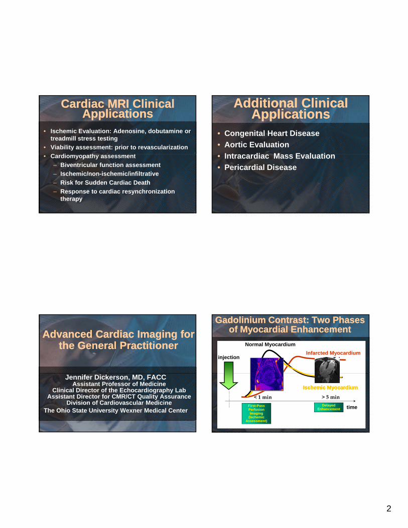
Normal Myocardium
injectionInfarcted Myocardium
Gadolinium Contrast: Two Phases of Myocardial Enhancement
Gadolinium Contrast: Two Phases of Myocardial Enhancement
time
Ischemic MyocardiumIschemic MyocardiumIschemic MyocardiumIschemic Myocardium
First-PassPerfusion Imaging
(Ischemic Assessment)
Delayed Enhancement
< 1 min > 5 min

3
Patterns of Hyperenhancement
Patterns of Hyperenhancement
• Transmural
– Involves entire wall
– Consistent with myocardial infarction/ischemic eventinfarction/ischemic event
– If more than 50% of wall involved, felt to be non-viable
• Non-transmural
– Endocardial, epicardial, mid-wall
– Non-ischemic myopathies, infiltrative diseases
DME: LAD-territory infarct scar
DME: LAD-territory infarct scar
Mid-Myocardial Hyperenhancement
Mid-Myocardial Hyperenhancement
4
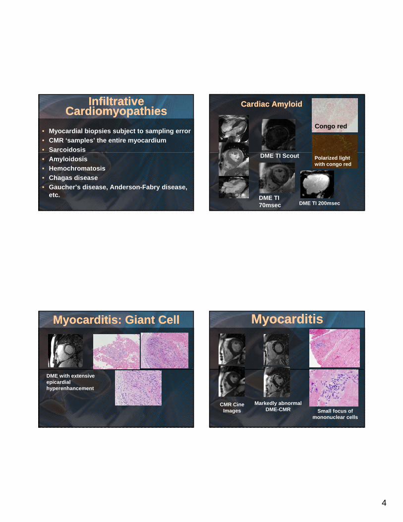
Infiltrative Cardiomyopathies
Infiltrative Cardiomyopathies
• Myocardial biopsies subject to sampling error
• CMR ‘samples’ the entire myocardium
• Sarcoidosis
• Amyloidosis
• Hemochromatosis
• Chagas disease
• Gaucher’s disease, Anderson-Fabry disease, etc.
Cardiac AmyloidCardiac Amyloid
Congo red
Polarized light with congo red
DME TI 70msec DME TI 200msec
DME TI Scout
Myocarditis: Giant CellMyocarditis: Giant Cell
DME with extensive epicardial hyperenhancement
MyocarditisMyocarditis
Small focus of mononuclear cells
Markedly abnormal DME-CMR
CMR Cine Images
5
Limitations of MRILimitations of MRI• Long acquisition times
– 45-60 min
• Most imaging sequences require breath holding
– 10-30 sec breath holds per image sequence
10 16 i i d t i ti h t– 10-16 images required to image entire heart
• Contraindications to MRI
– Pacemakers/ICDs
– Any ferrous material within body
– CKDNephrogenic systemic fibrosis (NSF)
Nephrogenic Systemic Fibrosis (NSF)
Nephrogenic Systemic Fibrosis (NSF)
• Diffuse systemic fibrosis involving skin, skeletal muscle, GI tract, cardiovascular system– Skin lesions symmetrical and extend distal to
proximal• After the administration of gadolinium in patients with
renal failure (GFR<60)renal failure (GFR<60)– No cases reported in patients with GFR >30
• Diagnosis: skin biopsy– Lab testing non-specific
• Treatment supportive– Restore renal function (HD not effective once patient
develops NSF)– Pain management
• For further questions, refer to OSU Radiology Departmental website on OneSource
Overview of Cardiac MRI Stress Testing
Overview of Cardiac MRI Stress Testing
• Pharmacologic– Adenosine/Regadenoson– DobutamineDobutamine
• Exercise (Treadmill)– Functional data– NIH supported research at Ohio State
6
Advanced Cardiac Imaging for the General Practitioner
Advanced Cardiac Imaging for the General Practitioner
Sharon Roble, MD Assistant Professor of Clinical Medicine
Department of Cardiovascular Medicine Division of Cardiovascular Medicine
The Ohio State University Wexner Medical Center
Clinical Case 1Clinical Case 1
• 16 year-old asymptomatic basketball player
• ROS: no syncope, palpitations, DOE, etc.
• PMH: negative• PMH: negative
• FH: unremarkable
Physical ExaminationPhysical Examination
• Height 182 cm, weight 71 kg
• BP 118/54, HR 45-60
• Symmetric pulses
• II/VI SEM at LUSB, no positional change
• Rest of PE unremarkable
7
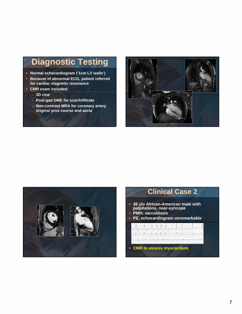
Diagnostic TestingDiagnostic Testing• Normal echocardiogram (‘1cm LV walls’)
• Because of abnormal ECG, patient referred for cardiac magnetic resonance
• CMR exam included:
– 3D cine
– Post-gad DME for scar/infiltrate
– Non-contrast MRA for coronary artery origins/ prox course and aorta
Clinical Case 2Clinical Case 2
• 36 y/o African-American male with palpitations, near-syncope
• PMH: sarcoidosis• PE, echocardiogram unremarkable
• CMR to assess myocardium
8
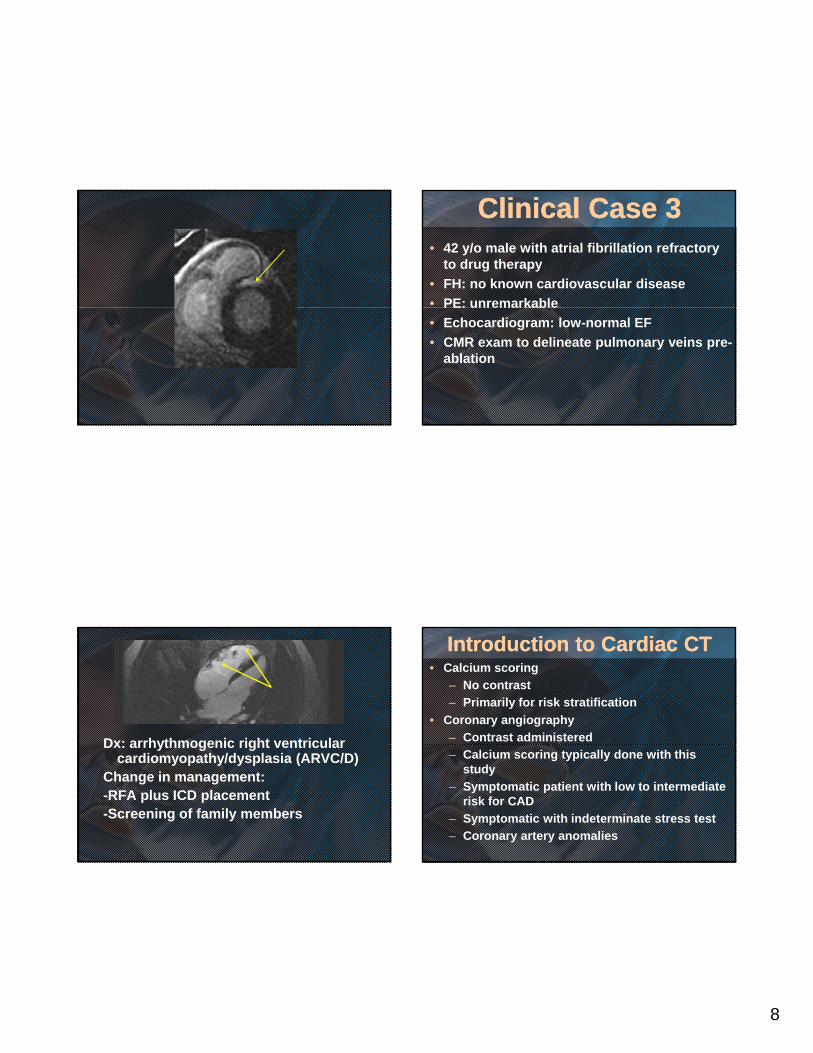
Clinical Case 3Clinical Case 3• 42 y/o male with atrial fibrillation refractory
to drug therapy
• FH: no known cardiovascular disease
• PE: unremarkablePE: unremarkable
• Echocardiogram: low-normal EF
• CMR exam to delineate pulmonary veins pre-ablation
Dx: arrhythmogenic right ventricularDx: arrhythmogenic right ventricular cardiomyopathy/dysplasia (ARVC/D)
Change in management: -RFA plus ICD placement-Screening of family members
Introduction to Cardiac CTIntroduction to Cardiac CT• Calcium scoring
– No contrast
– Primarily for risk stratification
• Coronary angiography
– Contrast administered
– Calcium scoring typically done with this study
– Symptomatic patient with low to intermediate risk for CAD
– Symptomatic with indeterminate stress test
– Coronary artery anomalies
9
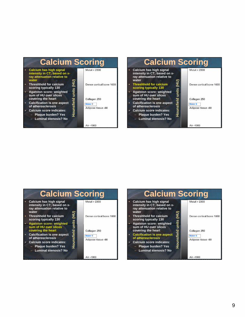
Calcium ScoringCalcium Scoringit
s (H
U)
• Calcium has high signal intensity in CT; based on x-ray attenuation relative to water
• Threshhold for calcium scoring typically 130
• Agatston score: weighted
Ho
un
sfie
ld u
n
Water 0
Agatston score: weighted sum of HU over slices covering the heart
• Calcification is one aspect of atherosclerosis
• Calcium score indicates:– Plaque burden? Yes– Luminal stenosis? No
Calcium ScoringCalcium Scoring
its
(HU
)
• Calcium has high signal intensity in CT; based on x-ray attenuation relative to water
• Threshhold for calcium scoring typically 130
• Agatston score: weighted
Ho
un
sfie
ld u
n
Water 0
Agatston score: weighted sum of HU over slices covering the heart
• Calcification is one aspect of atherosclerosis
• Calcium score indicates:– Plaque burden? Yes– Luminal stenosis? No

10
Calcium ScoringCalcium Scoringit
s (H
U)
• Calcium has high signal intensity in CT; based on x-ray attenuation relative to water
• Threshhold for calcium scoring typically 130
• Agatston score: weighted
Ho
un
sfie
ld u
n
Water 0
Agatston score: weighted sum of HU over slices covering the heart
• Calcification is one aspect of atherosclerosis
• Calcium score indicates:– Plaque burden? Yes– Luminal stenosis? No
Advanced Cardiac Imaging for the General Practitioner
Advanced Cardiac Imaging for the General Practitioner
Jennifer Dickerson, MD, FACCAssistant Professor of Medicine
Clinical Director of the Echocardiography LabAssistant Director for CMR/CT Quality Assurance
Division of Cardiovascular Medicine The Ohio State University Wexner Medical Center
Clinical Case 4Clinical Case 4• 65yo Male presents for an annual physical
– Exercises 5 days a week without any concerning symptoms
• PMHx: Hyperlipidemia
• Medications: 20mg Simvastatin, 325mg Aspirin
• SoHx: 2ppd tobacco x 20 years (quit in 2009)
–Cigar use 1-2 times a month
• FmHx: Father with MI age 53, PGM, PGF and mother with MI in their 60s.
Clinical Case 4 ContinuedClinical Case 4 Continued
• PE: BP 168/83 HR 65, BMI 29
– Unremarkable physical findings.
• Lipid• Lipid
– Total cholesterol 221
– LDL 145
– HDL 41
– Triglycerides 176

11
• “So Doc, how’s my heart doing? I don’t want to end up like my parents.”
ATPIII Executive summary JACC: vol 49, 3:2007
12
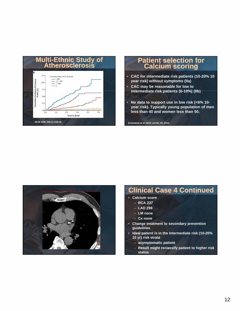
Multi-Ethnic Study of Atherosclerosis
Multi-Ethnic Study of Atherosclerosis
NEJM 2008; 358;13:1336-45
Patient selection for Calcium scoring
Patient selection for Calcium scoring
• CAC for intermediate risk patients (10-20% 10 year risk) without symptoms (IIa)
• CAC may be reasonable for low to intermediate risk patients (6 10%) (IIb)intermediate risk patients (6-10%) (IIb)
• No data to support use in low risk (<6% 10-year risk). Typically young population of men less than 40 and women less than 50.
Greenland et al JACC vol 56, 25, 2010
Clinical Case 4 ContinuedClinical Case 4 Continued• Calcium score
– RCA 237
– LAD 298
– LM none
– Cx none
• Change treatment to secondary prevention guidelines
• Ideal patient is in the Intermediate risk (10-20% 10 yr) risk strata
– asymptomatic patient
– Result might reclassify patient to higher risk status
13
Advanced Cardiac Imaging for the General Practitioner
Advanced Cardiac Imaging for the General Practitioner
Sharon Roble, MD Assistant Professor of Clinical Medicine
Department of Cardiovascular Medicine Division of Cardiovascular Medicine
The Ohio State University Wexner Medical Center
Contraindications for calcium scoring
Contraindications for calcium scoring
• Known CAD
• Symptomatic patient
• Cardiac “hardware”: pacemakers stents• Cardiac “hardware”: pacemakers, stents, prosthetic valves
Clinical Case 5Clinical Case 5
• 12 year old female with no significant past medical history had syncopal event while playing in basketball game
• No prodrome
• Awoke spontaneously
Physical ExaminationPhysical Examination
• Afebrile, P-80, BP 90/50
• Quiet precordium, I/IV short systolic ejection murmur, no diastolic murmur,
ll bgallop or rubs
• Abdomen unremarkable
14
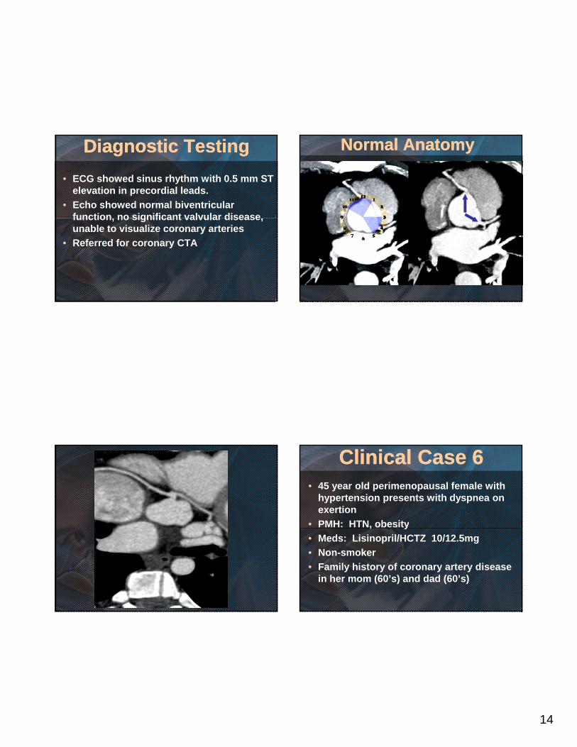
Diagnostic TestingDiagnostic Testing
• ECG showed sinus rhythm with 0.5 mm ST elevation in precordial leads.
• Echo showed normal biventricular function no significant valvular diseasefunction, no significant valvular disease, unable to visualize coronary arteries
• Referred for coronary CTA
Normal AnatomyNormal Anatomy
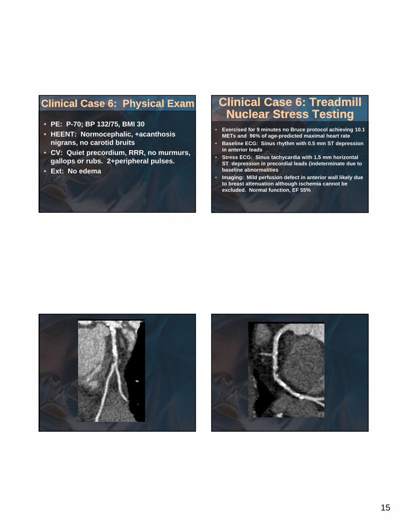
Clinical Case 6Clinical Case 6• 45 year old perimenopausal female with
hypertension presents with dyspnea on exertion
• PMH: HTN, obesity, y
• Meds: Lisinopril/HCTZ 10/12.5mg
• Non-smoker
• Family history of coronary artery disease in her mom (60’s) and dad (60’s)
15
Clinical Case 6: Physical ExamClinical Case 6: Physical Exam
• PE: P-70; BP 132/75, BMI 30
• HEENT: Normocephalic, +acanthosis nigrans, no carotid bruits
• CV: Quiet precordium RRR no murmursCV: Quiet precordium, RRR, no murmurs, gallops or rubs. 2+peripheral pulses.
• Ext: No edema
Clinical Case 6: Treadmill Nuclear Stress Testing
Clinical Case 6: Treadmill Nuclear Stress Testing
• Exercised for 9 minutes no Bruce protocol achieving 10.1 METs and 96% of age-predicted maximal heart rate
• Baseline ECG: Sinus rhythm with 0.5 mm ST depression in anterior leads
• Stress ECG: Sinus tachycardia with 1.5 mm horizontal ST depression in precordial leads (indeterminate due to baseline abnormalities
• Imaging: Mild perfusion defect in anterior wall likely due to breast attenuation although ischemia cannot be excluded. Normal function, EF 55%
16
ReferencesReferences
• ACC/AHA Cardiovascular CT Appropriateness Criteria, Journal of the American College Cardiology. 2012; 59 (9): 857-881.
• ACC/AHA Guidelines for Exercise Testing: Executive Summary, Circulation. 1991; 96: 345-354. y, ;
• OSU Department of Radiology website. https://onesource.osumc.edu/departments/radiology
Related Documents