Carbon Monoxide Recombination Dynamics in Truncated Hemoglobins Studied with Visible-Pump MidIR-Probe Spectroscopy Andrea Lapini, ¶,† Mariangela Di Donato, ¶,†,‡ Barbara Patrizi, † Agnese Marcelli, † Manuela Lima, † Roberto Righini, † Paolo Foggi, †,‡,§ Natascia Sciamanna, ⊥ and Alberto Boffi ⊥ † LENS (European Laboratory for Nonlinear Spectroscopy) via N. Carrara 1, 50019 Sesto Fiorentino (FI), Italy ‡ INO (Istituto Nazionale di Ottica), Largo Fermi 6, 50125 Firenze, Italy § Dipartimento di Chimica, Universita ̀ di Perugia, via Elce di Sotto 8, 06123 Perugia, Italy ⊥ Istituto Pasteur, Fondazione Cenci Bolognetti c/o Dipartimento di Scienze Biochimiche, Universita ̀ “La Sapienza”, piazzale Aldo Moro 5, 00185 Roma, Italy * S Supporting Information ABSTRACT: Carbon monoxide recombination dynamics upon photodissociation with visible light has been characterized by means of ultrafast visible-pump/MidIR probe spectroscopy for the truncated hemoglobins from Thermobif ida f usca and Bacillus subtilis. Photodissociation has been induced by exciting the sample at two different wavelengths: 400 nm, corresponding to the heme absorption in the B-band, and 550 nm, in the Q-bands. The bleached iron−CO coordination band located at 1850−1950 cm −1 and the free CO absorption band in the region 2050−2200 cm −1 have been observed by probe pulses tuned in the appropriate infrared region. The kinetic traces measured at 1850−1950 cm −1 reveal multiexponential subnanosecond dynamics that have been interpreted as arising from fast geminate recombination of the photolyzed CO. A compared analysis of the crystal structure of the two proteins reveals a similar structure of their distal heme pocket, which contains conserved polar and aromatic amino acid residues closely interacting with the iron ligand. Although fast geminate recombination is observed in both proteins, several kinetic differences can be evidenced, which can be interpreted in terms of a different structural flexibility of the corresponding heme distal pockets. The analysis of the free CO band-shape and of its dynamic evolution brings out novel features about the nature of the docking site inside the protein cavity. ■ INTRODUCTION Truncated hemoglobins (trHbs) are a family of small oxygen binding proteins widely distributed among bacteria, plants, and protozoa. 1,2 They are characterized by a high structural variability of the heme pocket residues, suggesting diverse functions, possibly related to the physiological response in the defense from oxygen reactive species and in the presence of other bimolecular ligands such as NO or CO. On the basis of the nature of the amino acid residues in key topological positions within the distal heme pocket, trHbs are usually divided into three groups, each presenting a certain number of conserved residues. Truncated hemoglobins from Thermobif ida f usca (Tf-trHb) and Bacillus subtilis (Bs-trHb) both belong to group II, which is characterized by the presence of a Trp residue on the bottom of the heme distal pocket (G8 position). 1 The resolution of the crystal structure of a number of group II truncated hemoglobins has revealed a common general pattern of the heme pocket, characterized by an ensemble of polar residues capable of forming hydrogen bonds with the iron-bound ligand, usually defined as “ligand inclusive hydrogen bonding network” (see Figure 1). It has been suggested that TrpG8 plays an important role in ligand binding and stabilization, though other amino acids in topological positions E7, E11, CD1, and B10 can modify drastically the Received: February 27, 2012 Revised: June 22, 2012 Published: July 3, 2012 Article pubs.acs.org/JPCB © 2012 American Chemical Society 8753 dx.doi.org/10.1021/jp3019149 | J. Phys. Chem. B 2012, 116, 8753−8761

Welcome message from author
This document is posted to help you gain knowledge. Please leave a comment to let me know what you think about it! Share it to your friends and learn new things together.
Transcript
Carbon Monoxide Recombination Dynamics in TruncatedHemoglobins Studied with Visible-Pump MidIR-Probe SpectroscopyAndrea Lapini,¶,† Mariangela Di Donato,¶,†,‡ Barbara Patrizi,† Agnese Marcelli,† Manuela Lima,†
Roberto Righini,† Paolo Foggi,†,‡,§ Natascia Sciamanna,⊥ and Alberto Boffi⊥
†LENS (European Laboratory for Nonlinear Spectroscopy) via N. Carrara 1, 50019 Sesto Fiorentino (FI), Italy‡INO (Istituto Nazionale di Ottica), Largo Fermi 6, 50125 Firenze, Italy§Dipartimento di Chimica, Universita di Perugia, via Elce di Sotto 8, 06123 Perugia, Italy⊥Istituto Pasteur, Fondazione Cenci Bolognetti c/o Dipartimento di Scienze Biochimiche, Universita “La Sapienza”, piazzale AldoMoro 5, 00185 Roma, Italy
*S Supporting Information
ABSTRACT: Carbon monoxide recombination dynamics upon photodissociation with visible light has been characterized bymeans of ultrafast visible-pump/MidIR probe spectroscopy for the truncated hemoglobins from Thermobif ida fusca and Bacillussubtilis. Photodissociation has been induced by exciting the sample at two different wavelengths: 400 nm, corresponding to theheme absorption in the B-band, and 550 nm, in the Q-bands. The bleached iron−CO coordination band located at 1850−1950cm−1 and the free CO absorption band in the region 2050−2200 cm−1 have been observed by probe pulses tuned in theappropriate infrared region. The kinetic traces measured at 1850−1950 cm−1 reveal multiexponential subnanosecond dynamicsthat have been interpreted as arising from fast geminate recombination of the photolyzed CO. A compared analysis of the crystalstructure of the two proteins reveals a similar structure of their distal heme pocket, which contains conserved polar and aromaticamino acid residues closely interacting with the iron ligand. Although fast geminate recombination is observed in both proteins,several kinetic differences can be evidenced, which can be interpreted in terms of a different structural flexibility of thecorresponding heme distal pockets. The analysis of the free CO band-shape and of its dynamic evolution brings out novelfeatures about the nature of the docking site inside the protein cavity.
■ INTRODUCTION
Truncated hemoglobins (trHbs) are a family of small oxygenbinding proteins widely distributed among bacteria, plants, andprotozoa.1,2 They are characterized by a high structuralvariability of the heme pocket residues, suggesting diversefunctions, possibly related to the physiological response in thedefense from oxygen reactive species and in the presence ofother bimolecular ligands such as NO or CO. On the basis ofthe nature of the amino acid residues in key topologicalpositions within the distal heme pocket, trHbs are usuallydivided into three groups, each presenting a certain number ofconserved residues. Truncated hemoglobins from Thermobif idafusca (Tf-trHb) and Bacillus subtilis (Bs-trHb) both belong togroup II, which is characterized by the presence of a Trp
residue on the bottom of the heme distal pocket (G8position).1 The resolution of the crystal structure of a numberof group II truncated hemoglobins has revealed a commongeneral pattern of the heme pocket, characterized by anensemble of polar residues capable of forming hydrogen bondswith the iron-bound ligand, usually defined as “ligand inclusivehydrogen bonding network” (see Figure 1). It has beensuggested that TrpG8 plays an important role in ligand bindingand stabilization, though other amino acids in topologicalpositions E7, E11, CD1, and B10 can modify drastically the
Received: February 27, 2012Revised: June 22, 2012Published: July 3, 2012
Article
pubs.acs.org/JPCB
© 2012 American Chemical Society 8753 dx.doi.org/10.1021/jp3019149 | J. Phys. Chem. B 2012, 116, 8753−8761

interaction network.3−5 The structure and sequence analysis ofgroup II trHbs shows that the nature of the residue at positionCD1 is correlated with the nature of the site E11. When theprotein has a Tyr at CD1, a nonpolar residue is found at E11; incontrast, when a non-hydrogen bond donor replaces theTyrCD1, a hydrogen bond donor is present at the E11 site.TyrB10 and TrpG8 are instead invariant residues.6,7 The crystalstructure of both Tf-trHb8 and Bs-trHb9 has been resolved,confirming a high degree of similarity among these twoproteins. In the case of Tf-trHb, the heme distal pocketinvolves, beside TrpG8, two Tyr residues, TyrB10 andTyrCD1, which are in close proximity with the iron-boundligand. In Bs-trHb, the more closely ligand interacting residuesbesides TrpG8 are TyrB10 and GlnE11.Given the high sensitivity of the ν(CO) stretching vibration
of the hemoglobin-CO adduct to the electric field generated bythe protein environment, Resonance Raman and FTIR spectrahave been used to probe the local structural characteristics ofthe heme binding pocket for the heme-bound CO state of bothTf-trHb and Bs-trHb. It has been found that each of these twoproteins can adopt two different conformations, differing by thenumber of hydrogen bonds formed with the iron-coordinatedCO.3,10,11
In the case of Tf-trHb, the comparison of the FTIR spectraof the wild type protein and of a series of combinatorialmutants in which TrpG8, TyrCD1, and TyrB10 have beenchanged into Phe, revealed that in one conformation bothTrpG8 and TyrCD1 are hydrogen bonded to the iron-coordinated CO, while in the other conformer only the H-bond with TrpG8 is maintained. The FTIR spectrum of the COadduct of the wild type protein showed two bands attributableto the ν(CO) stretching vibration, located at 1920 and 1940cm−1, respectively. These features have been ascribed to thepresence of two conformers in which the iron coordinated COis either doubly H-bonded to both TrpG8 and TyrCD1 orsingly bonded only to TrpG8, respectively.10
Very similar structural characteristics are observed also forBs-trHb. In the latter protein, the FTIR spectrum of the COadduct also shows two bands assigned to the ν(CO) stretchingvibration: one extremely downshifted, at 1888 cm−1, and theother at 1925 cm−1. The spectra have been interpreted alongthe same line as in Tf-trHb, that is, by assuming the presence ofa doubly H-bonded species in which TrpG8 and TyrB10 are
hydrogen bonded to the iron coordinated CO, and a singly H-bonded adduct in which TrpG8 is the sole H-bonding residue.The extreme downshift of the ν(CO) stretching vibration at1888 cm−1 indicated a highly polar environment around thebound CO, in which the double H-bond dominates the liganddissociation and rebinding dynamics properties of Bs-trHb.3
The sizably lower frequency of the ν(CO) stretching band inBs-trHb, as compared to Tf-trHb, can be a consequence of amore favorable orientation of the coordinating residues towardthe ligand. Such sterically favorable conformation is suggestedto generate a stronger H-bonding interaction and consequentlya higher degree of electron back-donation toward the iron. Thisfinding may account for the 1 order of magnitude higheroxygen affinity in Bs-trHb with respect to Tf-trHb despite thesimilar structural characteristics of the distal heme pockets inthe two proteins.3
The structural features of the heme distal pocket are knownto govern the dynamics of ligand escape and recombination inglobins. Recombination dynamics can vary substantially,spanning from the millisecond time scale of myoglobin andhemoglobin12,13 to the subnanosecond time scale observed forinstance in the oxygen sensory protein FixL.14,15 In Bs-trHbCO recombination dynamics has been studied by transientvisible absorption spectroscopy, revealing the existence of avery fast geminate recombination dynamics with a timeconstant of about 770 ps.3 The occurrence of fast geminaterecombination suggests that the ligand is confined within thedistal pocket with little possibility for the CO molecule toescape from the protein matrix to the solvent.To capture further details on the way such proteins operate
we analyzed the CO escape and rebinding processes afterdissociation induced by a short laser pulse by applying visible-pump MidIR-probe spectroscopy in both Bs-trHb and Tf-trHb.The combination of the high temporal resolution given byshort laser pulses with the structural information achieved bythe infrared probe, provides unique pieces of information onthe microscopic environments experienced by the ligandmolecule after photodetachment, unattainable by UV−visprobe pulses. This technique has been widely applied in thepast to study carbon monoxide recombination dynamics in anumber of similar systems, such as myoglobin, hemoglobin, andthe oxygen sensory protein FixL.13−19 In the present study, thecomparison between two proteins investigated in further details
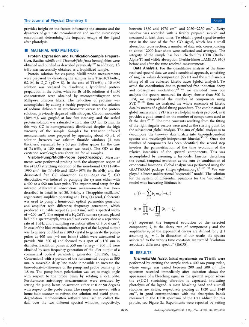
Figure 1. Close-up view of the active sites in Thermobif ida fusca and Bacillus substilis truncated hemoglobins. The key topological positions incontact with the iron bound ligand (E11, B9, B10, CD1, and G8) are shown for both proteins. Conserved residues in both proteins are coloredmagenta. Nonconserved amino acids are in red (Tf-trHb) or in cyan (Bs-trHb). Pictures generated with PyMol (DeLano Scientific LLC).
The Journal of Physical Chemistry B Article
dx.doi.org/10.1021/jp3019149 | J. Phys. Chem. B 2012, 116, 8753−87618754
provides insight on the factors influencing the amount and thedynamics of geminate recombination and on the microscopicenvironment determining the impaired escape of the ligandafter photolysis.
■ MATERIAL AND METHODSProtein Expression and Purification-Sample Prepara-
tion. Bacillus subtilis and Thermobif ida fusca hemoglobins wereobtained and purified as described previously.8,9 In addition, Tf-trHb was successfully obtained as a lyophilized sample.Protein solution for vis-pump MidIR-probe measurements
were prepared by dissolving the samples in a Tris-HCl buffer,0.2 M, in D2O (pD = 8). In the case of Tf-trHb, a 10 mMsolution was prepared by dissolving a lyophilized proteinpreparation in the buffer, while for Bs-trHb, solutions at 4 mMconcentration were obtained by microcentrifugation withMillipore ultracon filters. The reduction of proteins wasaccomplished by adding a freshly prepared anaerobic solutionof sodium dithionite in stoichiometric excess to the proteinsolution, previously degassed with nitrogen. Carbon monoxide(Rivoira), was gurgled at low flux intensity, and the sealedprotein solution was saturated with 1 atm CO for 15 min. Inthis way CO is homogeneously distributed despite the highviscosity of the sample. Samples for transient infraredmeasurements were prepared by squeezing about 40 μL ofsolution between two calcium fluoride windows (3 mmthickness) separated by a 50 μm Teflon spacer (in the caseof Bs-trHb, a 100 μm spacer was used). The OD at theexcitation wavelength was about 0.8 for all samples.Visible-Pump/MidIR-Probe Spectroscopy. Measure-
ments were performed probing both the absorption region ofthe ν(CO) stretching vibration of the iron-bound CO (1880−1990 cm−1 for Tf-trHb and 1825−1975 for Bs-trHb) and thedissociated free CO absorption (2030−2230 cm−1). COdissociation was induced by pumping the systems either witha 400 or a 550 nm laser pulse. The experimental setup for theinfrared differential absorption measurements has beendescribed in detail in ref 20. Briefly, a Ti:sapphire oscillator/regenerative amplifier, operating at 1 kHz, (Legend, Coherent)was used to pump a home-built optical parametric generatorand amplifier with difference frequency generation, whichproduced a tunable output (2.5−10 μm) with a spectral widthof ∼200 cm−1. The output of a HgCdTe camera system, placedbehind a spectrograph, was read out every shot at a repetitionrate of 1 kHz and a sampling resolution either of 3 or 6 cm−1.In case of the blue excitation, another part of the Legend outputwas frequency doubled in a BBO crystal to generate the pump-pulses at 400 nm (∼6 nm fwhm) which were attenuated toprovide 200−500 nJ and focused to a spot of ∼150 μm indiameter. Excitation pulses at 550 nm (energy = 200 nJ) wereobtained by sum frequency generation of the idler output of acommercial optical parametric generator (TOPAS, LightConversion) with a portion of the fundamental output at 800nm. A moveable delay line made it possible to increase thetime-of-arrival-difference of the pump and probe beams up to1.8 ns. The pump beam polarization was set to magic anglewith respect to the probe beam by rotating a λ/2 plate.Furthermore anisotropy measurements were executed bysetting the pump beam polarization either at 0 or 90 degreeswith respect to the probe beam. The sample was moved with ahome-built scanner to refresh the solution and avoid photo-degradation. Home-written software was used to collect thedata over the two different spectral windows, respectively,
between 1880 and 1975 cm−1 and 2030−2230 cm−1. Everywindow was recorded with a freshly prepared sample andmeasured at least three times. To obtain a good signal-to-noiseratio in the case of the free CO signal, which has a smallabsorption cross section, a number of data sets, correspondingto about 12000 laser shots were collected and averaged. Theintegrity of the sample has been checked by FTIR (BrukerAlpha-T) and visible absorption (Perkin-Elmer LAMBDA 950)before and after the time-resolved measurements.
Data Analysis. For the quantitative analysis of the time-resolved spectral data we used a combined approach, consistingof singular values decomposition (SVD) and the simultaneousfitting of all the collected kinetic traces (global analysis). Toavoid the contribution due to perturbed free induction decayand cross-phase modulation,21−25 we excluded from ouranalysis the spectra measured for delays shorter than 500 fs.First, we extrapolated the number of components usingSVD;26−28 then we analyzed the whole ensemble of kineticdata by means of a global fitting procedure. The combination ofglobal analysis and SVD is a very helpful analysis protocol, as itprovides a good control on the number of components used tofit the data.29−34 The time constants resulting from the fittingof the right singular vectors were used as the starting point forthe subsequent global analysis. The aim of global analysis is todecompose the two-way data matrix into time-independentspectra and wavelength-independent kinetics.33−36 Once thenumber of components has been identified, the second stepinvolves the parametrization of the time evolution of therelative intensities of the spectral components. This wasaccomplished by assuming a first-order kinetics, describingthe overall temporal evolution as the sum or combination ofexponential functions. Global analysis was performed using theGLOTARAN package (http://glotaran.org).34,37−40 We em-ployed a linear unidirectional “sequential” model. The solutionfor the system of differential equations for the “sequential”model with increasing lifetimes is
∑
∏ ∏
= −
= −
=
=
−
=≠
c t b k t
b k k k
( ) exp( )
/ ( )
lj
l
jl j
jlm
l
mnn j
l
n j
1
1
1
1
cl(t) represent the temporal evolution of the selectedcomponent, kj is the decay rate of component j and theamplitudes bjl of the exponential decays are defined for j ≤ lassuming b11 = 1. In discussion of our results the spectraassociated to the various time constants are termed “evolutionassociated difference spectra” (EADS).
■ RESULTSThermobif ida fusca. Initial experiments on Tf-trHb were
performed by exciting the sample with a 400 nm pump pulse,whose energy was varied between 200 and 500 nJ. Thespectrum recorded immediately after excitation shows theappearance of a bleaching signal in the spectral region wherethe ν(CO) stretching vibration is expected, indicatingphotolysis of the ligand. A main bleaching band and a smallshoulder are visible, respectively peaking at 1920 and 1940cm−1, in good correspondence with the absorption bandsmeasured in the FTIR spectrum of the CO adduct for thisprotein, see Figure 2a. Experiments were repeated by setting
The Journal of Physical Chemistry B Article
dx.doi.org/10.1021/jp3019149 | J. Phys. Chem. B 2012, 116, 8753−87618755

the pump pulse polarization either to 0 or 90 degrees withrespect to the probe pulse, and the time-dependent anisotropyof the transition was calculated, resulting in an average value of−0.18. Considering that the heme behaves like a circularabsorber when excited at 400 nm,41,42 it can be calculated thatthe coordinated CO is oriented in way that its dipole forms anangle smaller than 15 degrees with respect to the heme normal,similarly to what observed for other globin proteins.14,17,43
The analysis of the kinetic traces revealed a biphasic recovery,occurring within the subnanosecond time scale. As clearlyvisible by inspecting the kinetic traces reported in Figure 2b,the bleaching band at 1940 cm−1 recovers faster than thebleaching at 1920 cm−1. We have analyzed the data using aglobal fit with a sequential decay scheme, obtaining the EADSreported in Figure 2c. The kinetic traces can be satisfactorilyfitted with two components, whose time constants are 250 psand 1.5 ns, respectively. The fast kinetic phase accounts foralmost 30% of the recombination dynamics at 1940 cm−1. Aninspection of the kinetic traces reported in Figure 2b brings outthat the relative weight of the fast component is much higher
for the 1940 cm−1 band with respect to the low frequency band(1920 cm−1).Bleaching recovery on a subnanosecond time scale can be
directly associated to fast geminate recombination if it isassumed that the recombined CO and CO in the non-photolyzed portion of the sample have the same spectrum asthe ligated CO at equilibrium.43 To further investigate thedynamics following CO photodissociation we also analyzed thefree CO absorption band, which is expected to appear at about2100−2150 cm−1. Because of the low absorption cross sectionof the free CO vibration and the substantial water absorption inthat region, it is difficult to obtain reliable kinetic traces andtime-dependent spectra with a good signal to-noise-ratio.Tentative measurements, carried out by exciting the samplewith a 400 nm pump pulse, revealed a significant baselineproblem, due to water absorption, heating, and excess energydissipation in the system. To minimize these unwantedcontributions, CO photolysis was triggered by exciting theheme in the Q-band absorption region (550−600 nm) insteadof the B-band region. Under these experimental conditions theamount of energy dissipated by the system was significantlyreduced.Measurements were thus carried out by setting the excitation
pulse at 550 nm and probing both the ν(CO) stretching regionfor the coordinated CO and the free CO absorption region.The dynamic evolution in the bleaching region wassubstantially identical to what was previously observed byexciting the sample at 400 nm. Instead, significant improvementwas obtained in the free CO region, were time traces with goodsignal-to-noise ratio could be recorded.Spectra collected in the free CO region are reported in
Figure 3a while Figure 3b shows the kinetic trace measured atthe absorption maximum and compared with that of the 1940cm−1 bleaching band, which has been scaled to match theFigure 2. (a) Time resolved spectra recorded at different time delays,
showing the bleaching induced by the excitation at 400 nm in theν(CO) stretching region for the coordinated CO; (b) Kinetic traces(scattered points) at selected frequencies together with the fit (solidline) obtained by global analysis. The trace at 1920 cm−1 has beenscaled to overlap the trace at 1940 trace on the long time scale; (c)EADS obtained by globally analyzing all the kinetic traces recorded inthe 1850−1950 cm−1 spectral range.
Figure 3. (a) Time resolved spectra in the free CO region recorded atdifferent time delays after excitation of the sample with a 550 nm laserpulse; (b) kinetic traces at 2120 cm−1 (red line), corresponding to themaximum absorption in the free CO region. This trace has beensuperimposed with the kinetic trace at 1940 cm−1 (black line),corresponding to the maximum absorption of the coordinated CO,opportunely scaled.
The Journal of Physical Chemistry B Article
dx.doi.org/10.1021/jp3019149 | J. Phys. Chem. B 2012, 116, 8753−87618756

intensity of the free CO signal. As noticed from Figure 3b, thetwo kinetic traces are well matched, except for the short timescale, where the signal corresponding to the CO absorption(2120 cm−1) shows an initial rise component.Spectra in Figure 3a have been corrected for the presence of
a baseline, which still contributes in this region although at amore minor extent than in the previous measurements with 400nm excitation. The baseline is removed by subtracting a thirdorder polynomial fit (initial raw data and baseline subtractedare shown in Supporting Information). To avoid the influenceof the baseline correction procedure on the dynamic behaviorof the system, the region between 2080 and 2150 cm−1, wherethe signal is observed, was removed for the fit. Our data show aratio in the absorption cross section between the coordinatedand docked CO of about 50, in agreement with previousfindings.43 Baseline problems are less significant in thebleaching region, which is free from water absorption. In thisregion, the amplitude of background fluctuation has a negligibleinfluence on the kinetics (see Supporting Information fordetails).As reported in Figure 3b, the dynamic evolution in the free
CO absorption region matches that of the 1940 cm−1 bleaching,thus confirming that also the fast 250 ps component has to beascribed to geminate recombination. At the early measuredtimes a rise of the free CO absorption signal is observed, whichcan be fitted with a time constant of 30 ps. An initial risecomponent for a docked CO band has been previouslyobserved also for myoglobin17 and interpreted in terms ofprotein relaxation around the photolyzed CO molecule. In thatcase however the rise component had a much faster timeconstant of about 1.6 ps. Furthermore thermal relaxation hasalso been observed to occur for myoglobin excited at 597 nm,with a time constant of 6.2 ps.44 The longer time scale observedin this case for the rise component may indicate a combinationof slower cooling process and/or a more significant proteinrearrangement, possibly involving the rotation or reorientationof one or more amino acid side chains in the CO docking site.Such structural adjustment most likely modifies the electrostaticinteractions around the CO molecule thus increasing theoscillator strength.45 This is probably the consequence of amore flexible heme pocket structure, also brought out by thepresence of a water molecule, which could rearrange bybreaking/forming H-bonds with the tyrosine amino acids(TyrB10 and TyrCD1) located at a short distance from theheme.Besides the 30 ps component, a global fit of the kinetic traces
collected in the free CO region (2050−2200 cm−1) results intwo additional decay components, with time constants of 300ps and 1.2 ns, respectively. These time constants qualitativelyagree with those measured for the bleaching recovery, thusconfirming that the process responsible for the dynamicevolution of the system is a fast geminate recombination ofthe photolized CO. The EADS obtained by globally analyzingthe kinetic traces in the 2050−2150 cm−1 region with asequential decay scheme are reported in the SupportingInformation. To take into account of the bandwidth variationwith time, we estimated the temporal evolution of theintegrated area of the CO band, and we compared it with thekinetic trace at 2120 cm−1, corresponding to band maximum.The two traces are qualitatively in good agreement (seeSupporting Information).The measured CO absorption peak is quite asymmetrical and
has a rather large bandwidth (fwhm 30 cm−1), which suggests
the presence of an inhomogeneous distribution of twounresolved bands behind the measured line shape. The signaldynamically evolves by slightly shifting to the blue, but nosubstantial line shape variations are observed. Although it hasbeen shown that in similar systems a certain percentage(reported values span between 3.6 and 13%) of the photolizedCO is initially in a vibrationally hot state13,14,17,19,46,47 novibrational hot band could be resolved in the present case.Since the CO anharmonicity has been estimated to be 27−30cm−113,47 hot bands are probably not resolved due to baselinefluctuation problems.
Bacillus subtilis. Also in the case of Bs-trHb, time-resolvedspectra in the bleaching region were collected by exciting thesample both with a 400 nm and a 550 nm laser pulse. Again, nosignificant differences were observed in the dynamic evolutionof the system between the two excitation wavelengths.Following CO photodissociation, two bleaching bands areobserved at 1925 cm−1 and 1888 cm−1, respectively, whosepositions well correspond with the measured FTIR spectrum ofthe CO adduct for this protein, see Figure 4a. The
corresponding kinetic traces, reported in Figure 4b can befitted with two time constants of 120 ps and 2 ns, respectively.In contrast to what was observed in Tf-trHb, the two bleachingbands in Bs-trHb recover with the same kinetics. In this casethe faster kinetic component has a very low weight, onlyaccounting for less than 10% of the recombination dynamics forboth bleaching bands, implying that the fraction of picosecondgeminate recombination is lower than that observed in Tf-tbHb. Fast geminate recombination in Bs-trHb has beenpreviously observed through time-resolved measurements inthe visible spectral range.3 In the present infrared measure-ments a 30 ps component, previously identified by transientabsorption measurements in the visible region, is not observed.Since the currently probed spectral range is only sensitive to
Figure 4. (a) Time resolved spectra recorded at different time delays,showing the bleaching induced in Bs-trHb in the ν(CO) stretchingregion for the coordinated CO. Excitation wavelength was 550 nm. (b)Kinetic traces (scattered points) together with the fit (solid line)obtained by global analysis at selected frequencies, corresponding tothe two maximum absorptions in the iron coordinated CO region. Thetrace at 1888 cm−1 has been scaled to overlap the trace at 1925 cm−1.
The Journal of Physical Chemistry B Article
dx.doi.org/10.1021/jp3019149 | J. Phys. Chem. B 2012, 116, 8753−87618757
CO vibrations, we conclude that this 30 ps component is notassociated to fast ligand recombination, but probably representsa relaxation process of the heme.The free CO region was probed by exciting the sample at
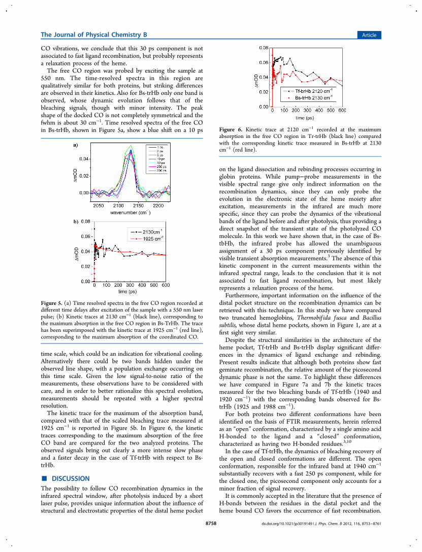
550 nm. The time-resolved spectra in this region arequalitatively similar for both proteins, but striking differencesare observed in their kinetics. Also for Bs-trHb only one band isobserved, whose dynamic evolution follows that of thebleaching signals, though with minor intensity. The peakshape of the docked CO is not completely symmetrical and thefwhm is about 30 cm−1. Time resolved spectra of the free COin Bs-trHb, shown in Figure 5a, show a blue shift on a 10 ps
time scale, which could be an indication for vibrational cooling.Alternatively there could be two bands hidden under theobserved line shape, with a population exchange occurring onthis time scale. Given the low signal-to-noise ratio of themeasurements, these observations have to be considered withcare, and in order to better rationalize this spectral evolution,measurements should be repeated with a higher spectralresolution.The kinetic trace for the maximum of the absorption band,
compared with that of the scaled bleaching trace measured at1925 cm−1 is reported in Figure 5b. In Figure 6, the kinetictraces corresponding to the maximum absorption of the freeCO band are compared for the two analyzed proteins. Theobserved signals bring out clearly a more intense slow phaseand a faster decay in the case of Tf-trHb with respect to Bs-trHb.
■ DISCUSSIONThe possibility to follow CO recombination dynamics in theinfrared spectral window, after photolysis induced by a shortlaser pulse, provides unique information about the influence ofstructural and electrostatic properties of the distal heme pocket
on the ligand dissociation and rebinding processes occurring inglobin proteins. While pump−probe measurements in thevisible spectral range give only indirect information on therecombination dynamics, since they can only probe theevolution in the electronic state of the heme moiety afterexcitation, measurements in the infrared are much morespecific, since they can probe the dynamics of the vibrationalbands of the ligand before and after photolysis, thus providing adirect snapshot of the transient state of the photolyzed COmolecule. In this work we have shown that, in the case of Bs-tbHb, the infrared probe has allowed the unambiguousassignment of a 30 ps component previously identified byvisible transient absorption measurements.3 The absence of thiskinetic component in the current measurements within theinfrared spectral range, leads to the conclusion that it is notassociated to fast ligand recombination, but most likelyrepresents a relaxation process of the heme.Furthermore, important information on the influence of the
distal pocket structure on the recombination dynamics can beretrieved with this technique. In this study we have comparedtwo truncated hemoglobins, Thermobif ida fusca and Bacillussubtilis, whose distal heme pockets, shown in Figure 1, are at afirst sight very similar.Despite the structural similarities in the architecture of the
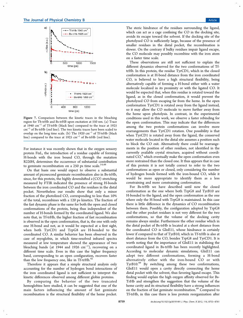
heme pocket, Tf-trHb and Bs-trHb display significant differ-ences in the dynamics of ligand exchange and rebinding.Present results indicate that although both proteins show fastgeminate recombination, the relative amount of the picoseconddynamic phase is not the same. To highlight these differenceswe have compared in Figure 7a and 7b the kinetic tracesmeasured for the two bleaching bands of Tf-trHb (1940 and1920 cm−1) with the corresponding bands observed for Bs-trHb (1925 and 1988 cm−1).For both proteins two different conformations have been
identified on the basis of FTIR measurements, herein referredas an “open” conformation, characterized by a single amino acidH-bonded to the ligand and a “closed” conformation,characterized as having two H-bonded residues.3,10
In the case of Tf-trHb, the dynamics of bleaching recovery ofthe open and closed conformations are different. The openconformation, responsible for the infrared band at 1940 cm−1
substantially recovers with a fast 250 ps component, while forthe closed one, the picosecond component only accounts for aminor fraction of signal recovery.It is commonly accepted in the literature that the presence of
H-bonds between the residues in the distal pocket and theheme bound CO favors the occurrence of fast recombination.
Figure 5. (a) Time resolved spectra in the free CO region recorded atdifferent time delays after excitation of the sample with a 550 nm laserpulse; (b) Kinetic traces at 2130 cm−1 (black line), corresponding tothe maximum absorption in the free CO region in Bs-TrHb. The tracehas been superimposed with the kinetic trace at 1925 cm−1 (red line),corresponding to the maximum absorption of the coordinated CO.
Figure 6. Kinetic trace at 2120 cm−1 recorded at the maximumabsorption in the free CO region in Tr-trHb (black line) comparedwith the corresponding kinetic trace measured in Bs-trHb at 2130cm−1 (red line).
The Journal of Physical Chemistry B Article
dx.doi.org/10.1021/jp3019149 | J. Phys. Chem. B 2012, 116, 8753−87618758
For instance it was recently shown that in the oxygen sensoryprotein FixL, the introduction of a residue capable of formingH-bonds with the iron bound CO, through the mutationR220H, determines the occurrence of substantial contributionto geminate recombination on a 250 ps time scale.14,48
On that basis one would expect to observe a substantialamount of picosecond geminate recombination also in Bs-trHb,since, for this protein, the highly downshifted ν(CO) stretchingmeasured by FTIR indicated the presence of strong H-bondsbetween the iron coordinated CO and the residues in the distalpocket. Nevertheless our results show that only a minorfraction of the photolized CO, corresponding to less than 10%of the total, recombines with a 120 ps kinetics. The fraction ofthe fast dynamic phase is the same for both the open and closedconformations of the protein, being thus independent of thenumber of H-bonds formed by the coordinated ligand. We alsonote that, in Tf-trHb, the higher fraction of fast recombinationis observed in the open conformation, that is, the one with onlyone H-bond and not, as it would be expected at a first sight,when both TyrCD1 and TrpG8 are H-bonded to thecoordinated CO. A similar behavior has been observed in thecase of myoglobin, in which time-resolved infrared spectrameasured at low temperature showed the appearance of twobleaching bands (at 1944 and 1926 cm−1), recovering on adifferent time scale. Even in this case the higher frequencyband, corresponding to an open configuration, recovers fasterthan the low frequency one, like in Tf-trHb.42
Therefore it appears that a simple structural analysis onlyaccounting for the number of hydrogen bond interactions ofthe iron coordinated ligand is not sufficient to interpret thekinetic differences observed among different globin proteins.By comparing the behavior of the two truncated
hemoglobins here studied, it can be suggested that one of themain factors influencing the amount of fast geminaterecombination is the structural flexibility of the heme pocket.
The steric hindrance of the residues surrounding the ligand,which can act as a cage confining the CO in the docking site,avoids its escape toward the solvent. If the docking site of thephotolyzed CO is sufficiently large, because of the presence ofsmaller residues in the distal pocket, the recombination isslower. On the contrary if bulky residues impair ligand escape,the CO molecule may possibly recombine with the iron atomon a faster time scale.These observations are still not sufficient to explain the
different dynamics observed for the two conformations of Tf-trHb. In this protein, the residue TyrCD1, which in the closedconformation is at H-bond distance from the iron coordinatedCO, is believed to have a high structural flexibility, beingalternatively capable of forming a H-bond either with a watermolecule localized in its proximity or with the ligated CO. Itwould be expected that, when this residue is rotated toward theligand, as in the closed conformation, it would prevent thephotolyzed CO from escaping far from the heme. In the openconformation TyrCD1 is rotated away from the ligand instead,so it may allow the CO molecule to move further away fromthe heme upon photolysis. In contrast, in the experimentalconditions used in this work, we observe a faster rebinding forthe open conformation. This may indicate that the differencesamong the two protein conformations can involve morerearrangements than TyrCD1 rotation. One possibility is thatwhen TyrCD1 is rotated away from the ligand, the conservedwater molecule located in the distal site assumes a position suchto block the CO exit. Alternatively there could be rearrange-ments in the position of other residues, not identified in thecurrently available crystal structure, acquired without coordi-nated CO,8 which eventually make the open conformation evenmore restrained than the closed one. It thus appears that in caseof this protein it is not totally correct to refer to the twoconformations as open or closed, only considering the numberof hydrogen bonds formed with the iron-bound CO, while itwould be more appropriate to identify them as a lessconstraining and more constraining conformation.For Bs-trHb we have described until now the closed
conformation as the one where both TrpG8 and TyrB10 areH-bonded to the ligand, and the open conformation as the onewhere only the H-bond with TrpG8 is maintained. In this casethere is little difference in the dynamics of CO recombinationbetween them. Possibly, the configuration adopted by TrpG8and the other pocket residues is not very different for the twoconformations, so that the volume of the docking cavityremains always similar. Furthermore the other residue which inthe distal pocket of Bs-trHb is located at a short distance fromthe coordinated CO is GlnE11, whose hindrance is certainlylower if compared to that of TyrB10, which in Tf-trHb is also atshort distance from the CO, besides TrpG8 and TyrCD1. It isworth noting that the importance of GlnE11 in stabilizing thecoordinated ligand in Bs-trHb has been recently highlighted.According to molecular dynamic simulations, GlnE11 canadopt two different conformations, forming a H-bondalternatively either with the iron-bound CO or withTyrB10.49 By switching among these two conformations,GlnE11 would open a cavity directly connecting the hemedistal pocket with the solvent, thus favoring ligand escape. Thisfinding would explain the high oxygen affinity observed for Bs-TrHb and strengthen the suggestion that the volume of theheme cavity and its structural flexibility have a strong influenceson the fraction of fast geminate recombination.49 Compared toTf-trHb, in this case there is less protein reorganization after
Figure 7. Comparison between the kinetic traces in the bleachingregion for Tb-trHb and Bs-trHB upon excitation at 550 nm. (a) Traceat 1940 cm−1 of Tf-trHb (black line) compared to the trace at 1925cm−1 of Bs-trHb (red line). The two kinetic traces have been scaled tooverlap on the long time scale. (b) The 1920 cm−1 of Tf-trHb (blackline) compared to the trace at 1925 cm−1 of Bs-trHb (red line).
The Journal of Physical Chemistry B Article
dx.doi.org/10.1021/jp3019149 | J. Phys. Chem. B 2012, 116, 8753−87618759
photolysis, possibly because the docking site is alreadysufficiently large to accommodate the dissociated ligand, dueto the topological substitution between TyrCD1 with PheCD1and LeuE11 with GlnE11. A lower degree of proteinrearrangement induced by the ligand dissociation could alsoexplain the absence of a rise component in the kinetics of thephotodissociated CO.Similar considerations regarding the caging effect50,51 played
by the amino-acid residues surrounding the photolyzed CO canalso be applied to other recently studied systems. Picosecondgeminate recombination has also been found in the COcomplex of nonglobin proteins, such as microperoxidase and inthe chemically modified form of cytochrome c, termedcarboxymethyl cytochrome c.52 In the former system a timeconstant of 110 ps has been estimated for concentratedsamples,53 while in the latter system a multiphasic recombina-tion with time constant of 16 ps, 120 ps, and 1 ns has beenmeasured.54 The authors attributed the three phases to COrebinding from different locations within the distal pocket site.The high efficiency of the ligand rebinding has been interpretedeven in this case as a consequence of a sterically hindered and“caged” nature of the distal heme pocket, from which it isdifficult for the ligand to escape. In these systems, the shorttime scale of the geminate rebinding has been correlated to aprotein configuration that assures the restraint of thereorganization energy of the active site.54
As a final comment, it should be pointed out that the kineticanalysis presented until now does not take into account that afraction of the photolyzed CO escapes to the solvent, andrecombines on a much slower time scale with a bimolecularprocess. A direct estimation of the relative amount of geminateand bimolecular recombination would require following thesystem dynamics on a time scale spanning from picoseconds tomilliseconds, which is not feasible with the currently usedexperimental set up. However, it is possible to have an estimateof the amount of geminate recombination occurring on thepicoseconds time scale by evaluating the ratio between theareas of the EADS obtained by global analysis. In the case of Tf-trHb it can be estimated that about 40% of CO undergoespicosecond geminate recombination. The evaluation of kineticcomponent in the nanosecond regime is affected by a large(∼20%) indetermination due to the lack of data regarding thebimolecular recombination.
■ CONCLUSIONSTime resolved vibrational spectroscopy, employed to studycarbon monoxide photodissociation and rebinding in twotruncated hemoglobins, has revealed key structural anddynamic properties of the ligand binding process in theseunusual globins. The results of this study highlight strikingdifferences between the dynamics of CO recombination in thetruncated hemoglobins with respect to other globins, such asvertebrate myoglobins and hemoglobins. Both the truncatedhemoglobins from Thermobif ida fusca and Bacillus subtilisexhibit subnanosecond multiexponential geminate recombina-tion, which account for at least 50% of the total yield of COrecombination. Several conserved residues in the distal hemepocket of both the analyzed proteins, capable of interactingwith the iron coordinated ligand through the formation of H-bonds, acts as a cage on the dissociated CO, preventing itsescape toward the solvent and confining it in the heme cavity,thus favoring fast rebinding. This effect is even morepronounced in the case of Tf-trHb, where at least 40% of
geminate recombination occurs in less than 300 ps. Theoccurrence of fast geminate rebinding and the differences in theCO rebinding kinetics registered for two apparently similarproteins highlight the strong influence played by the structuralorganization of the distal heme pocket and the interactionsamong the protein and the heme-ligand complex. Globinproteins are not the only example where such an effect isobserved. Other systems where protein ligand interactions canhave a strong influence on the photodynamics are found forinstance in the rhodopsin family, where strong differences inthe dynamics of retinal photoisomerization have been measuredbetween visual pigments and microbial rhodopsin.55 It isevident that a detailed understanding of such interactions isnecessary in order to completely characterize the photo-dynamics of these systems. Regarding the two proteins analyzedin this study, further analysis on site directed mutants will allowan even more accurate characterization of the specificinteractions of each of the residues present in the distal pocketwith the ligand, which will eventually help to clarify thebehavior of these proteins in their living environment.
■ ASSOCIATED CONTENT*S Supporting InformationBaseline subtraction procedure for data at 2050−2150 cm−1;EADS of time-resolved data in the free CO absorption region;influence of the baseline in the bleaching region; comparisonbetween the kinetics at 2120 cm−1; and the time dependence ofthe integrated area of the CO band. This material is availablefree of charge via the Internet at http://pubs.acs.org.
■ AUTHOR INFORMATIONAuthor Contributions¶These two authors have equally contributed to this workNotesThe authors declare no competing financial interest.
■ ACKNOWLEDGMENTSThe authors acknowledge the Italian ‘Ministero dell’Istruzionedell’Universita e della Ricerca’ (PRIN 2008, 2008BFJ34R) andIstituto Pasteur Fondazione Cenci Bolognetti. A.L. and A.M.acknowledge the financial support of the Regione Toscanathrough the found POR FSE 2007-2013 obiettivo 2 asse IV,project EPHODS. The financial support of the Cassa diRisparmio di Firenze is also gratefully acknowledged.
■ REFERENCES(1) Wittenberg, J. B.; Bolognesi, M.; Wittenberg, B. A.; Guertin, M. J.Biol. Chem. 2002, 277 (2), 871−874.(2) Wu, G.; Wainwright, L. M.; Poole, R. K., Microbial globins. InAdvances in Microbial Physiology, Academic Press: 2003; Vol. 47, pp255−310.(3) Feis, A.; Lapini, A.; Catacchio, B.; Brogioni, S.; Foggi, P.;Chiancone, E.; Boffi, A.; Smulevich, G. Biochemistry 2007, 47 (3),902−910.(4) Guallar, V.; Lu, C.; Borrelli, K.; Egawa, T.; Yeh, S.-R. J. Biol.Chem. 2009, 284 (5), 3106−3116.(5) Ouellet, H.; Milani, M.; LaBarre, M.; Bolognesi, M.; Couture, M.;Guertin, M. Biochemistry 2007, 46 (41), 11440−11450.(6) Milani, M.; Pesce, A.; Nardini, M.; Ouellet, H.; Ouellet, Y.;Dewilde, S.; Bocedi, A.; Ascenzi, P.; Guertin, M.; Moens, L.; Friedman,J. M.; Wittenberg, J. B.; Bolognesi, M. J. Inorg. Biochem. 2005, 99 (1),97−109.(7) Pesce, A.; Nardini, M.; Milani, M.; Bolognesi, M. IUBMB Life2007, 59 (8−9), 535−541.
The Journal of Physical Chemistry B Article
dx.doi.org/10.1021/jp3019149 | J. Phys. Chem. B 2012, 116, 8753−87618760

(8) Bonamore, A.; Ilari, A.; Giangiacomo, L.; Bellelli, A.; Morea, V.;Boffi, A. FEBS J. 2005, 272 (16), 4189−4201.(9) Giangiacomo, L.; Ilari, A.; Boffi, A.; Morea, V.; Chiancone, E. J.Biol. Chem. 2005, 280 (10), 9192−9202.(10) Droghetti, E.; Nicoletti, F. P.; Bonamore, A.; Boechi, L.; ArroyoManIfez, P.; Estrin, D. A.; Boffi, A.; Smulevich, G.; Feis, A. Biochemistry2010, 49 (49), 10394−10402.(11) Yoshimura, H.; Yoshioka, S.; Kobayashi, K.; Ohta, T.; Uchida,T.; Kubo, M.; Kitagawa, T.; Aono, S. Biochemistry 2006, 45 (27),8301−8307.(12) Frauenfelder, H.; McMahon, B. H.; Fenimore, P. W. Proc. Natl.Acad. Sci. 2003, 100 (15), 8615−8617.(13) Lim, M.; Jackson, T. A.; Anfinrud, P. A. J. Chem. Phys. 1995,102, 4355−4366.(14) Nuernberger, P.; Lee, K. F.; Bonvalet, A.; Bouzhir-Sima, L.;Lambry, J.-C.; Liebl, U.; Joffre, M.; Vos, M. H. J. Am. Chem. Soc. 2011,133 (43), 17110−17113.(15) van Wilderen, L. J. G. W.; Key, J. M.; Van Stokkum, I. H. M.;van Grondelle, R.; Groot, M. L. J. Phys. Chem. B 2008, 113 (11),3292−3297.(16) Kim, S.; Lim, M. J. Am. Chem. Soc. 2005, 127 (16), 5786−5787.(17) Lim, M.; Jackson, T. A.; Anfinrud, P. A. Nat. Struct. Mol. Biol.1997, 4 (3), 209−214.(18) Nienhaus, K.; Dominici, P.; Astegno, A.; Abbruzzetti, S.;Viappiani, C.; Nienhaus, G. U. Biochemistry 2010, 49 (35), 7448−7458.(19) Polack, T.; Ogilvie, J. P.; Franzen, S.; Vos, M. H.; Joffre, M.;Martin, J. L.; Alexandrou, A. Phys. Rev. Lett. 2004, 93 (1), 018102.(20) Touron Touceda, P.; Mosquera Vazquez, S.; Lima, M.; Lapini,A.; Foggi, P.; Dei, A.; Righini, R. Phys. Chem. Chem. Phys. 14 (2),1038−1047.(21) Afrawal, G. P.; Baldeck, P. L.; Alfano, R. R. Phys. Rev. A 1989,40, 5063−5072.(22) Hamm, P. Chem. Phys. 1995, 200 (3), 415−429.(23) Wynne, K.; Hochstrasser, R. M. Chem. Phys. 1995, 193 (3),211−236.(24) Lian, T.; Kholodenko, Y.; Locke, B.; Hochstrasser, R. M. J. Phys.Chem. 1995, 99 (19), 7272−7280.(25) Lapini, A.; Mosquera Vazquez, S.; Touron Touceda, P.; Lima,M. J. Mol. Struct. 2011, 993 (1−3), 470−473.(26) Hendler, R. W.; Shrager, R. I. J. Biochem. Biophys. Methods 1994,28 (1), 1−33.(27) Henry, E. R. Biophys. J. 1997, 72 (2), 652−673.(28) Henry, E. R.; Hofrichter, J.; Ludwig Brand, M. L. J. Singularvalue decomposition: Application to analysis of experimental data. InMethods Enzymolology; Academic Press: San Diego, CA, 1992; Vol.210, pp 129−192.(29) Dioumaev, A. K. Biophys. Chem. 1997, 67, 1−25.(30) Bonneau, R.; Wirz, J.; Zuberbuhler, A. D. Pure Appl. Chem.1997, 69, 979−992.(31) Henry, E. R. Biophys. J. 1997, 72, 652−673.(32) Franzen, S.; Kiger, L.; Poyart, C.; Martin, J. L. Biophys. J. 2001,80, 2372−2385.(33) van Stokkum, I. H. M.; Scherer, T.; Brouwer, A. M.; Verhoeven,J. W. J. Phys. Chem. 1994, 98 (3), 852−866.(34) van Stokkum, I. H. M.; Larsen, D. S.; van Grondelle, R. Biochim.Biophys. Acta 2004, 1657, 82.(35) Satzger, H.; Zinth, W. Chem. Phys. 2003, 295 (3), 287−295.(36) Neumann, K.; Verhoefen, M.-K.; Weber, I.; Glaubitz, C.;Wachtveitl, J. Biophys. J. 2008, 94 (12), 4796−4807.(37) Mullen, K. M.; van Stokkum, I. H. M. J. Stat. Software 2007, 18(1), 1−5.(38) Mullen, K. M.; van Stokkum, I. H. M. J. Stat. Software 2007, 18(3), 1−46.(39) Cador, O.; Chabre, F.; Dei, A.; Sangregorio, C.; Slageren, J. V.;Vaz, M. G. F. Inorg. Chem. 2003, 42 (20), 6432−6440.(40) Snellenburg, J. J.; Laptenok, S. P.; Seger, R.; Mullen, K. M.; vanStokkum, I. H. M. Glotaran: A Java-Based Graphical User Interface for
the R Package TIMP; Journal of Statistical Software, 49 (3), 1−22,http://www.jstatsoft.org/v49/i03/.(41) Lim, M.; Jackson, T. A.; Anfinrud, P. A. J. Am. Chem. Soc. 2004,126 (25), 7946−7957.(42) Moore, J. N.; Hansen, P. A.; Hochstrasser, R. M. Proc. Natl.Acad. Sci. 1988, 85 (14), 5062−5066.(43) Anfinrud, P. A.; Han, C.; Hochstrasser, R. M. Proc. Natl. Acad.Sci. 1989, 86 (21), 8387−8391.(44) Lim, M.; Jackson, T. A.; Anfinrud, P. A. J. Phys. Chem. 1996, 100(29), 12043−12051.(45) Hush, N. S.; Williams, M. L. J. Mol. Spectrosc. 1974, 50, 349−368.(46) Sagnella, D. E.; Straub, J. E.; Jackson, T. A.; Lim, M.; Anfinrud,P. A. Proc. Natl. Acad. Sci. 1999, 96 (25), 14324−14329.(47) Nuernberger, P.; Lee, K. F.; Bonvalet, A.; Vos, M. H.; Joffre, M.J. Phys. Chem. Lett. 2010, 1 (14), 2077−2081.(48) Jasaitis, A.; Hola, K.; Bouzhir-Sima, L.; Lambry, J.-C.; Balland,V.; Vos, M. H.; Liebl, U. Biochemistry 2006, 45 (19), 6018−6026.(49) Boechi, L.; Manez, P. A.; Luque, F. J.; Marti, M. A.; Estrin, D. A.Proteins: Struct., Funct., Bioinform. 2010, 78 (4), 962−970.(50) Grogan, T. G.; Bag, N.; Traylor, T. G.; Magde, D. J. Phys. Chem.1994, 98 (51), 13791−13796.(51) Jasaitis, A.; Ouellet, H.; Lambry, J.-C.; Martin, J.-L.; Friedman, J.M.; Guertin, M.; Vos, M. H. Chem. Phys. 2012, 396, 10−16.(52) Kim, J.; Park, J.; Lee, T.; Lim, M. J. Phys. Chem. B 2008, 113 (1),260−266.(53) Lim, M.; Jackson, T. A.; Anfinrud, P. A. J. Biol. Inorg. Chem.1997, 2, 531−536.(54) Silkstone, G.; Jasaitis, A.; Vos, M. H.; Wilson, M. T. DaltonTrans. 2005, 21, 3489−3494.(55) Wand, A.; Rozin, R.; Eliash, T.; Jung, K.-H.; Sheves, M.;Ruhman, S. J. Am. Chem. Soc. 2011, 133 (51), 20922−20932.
The Journal of Physical Chemistry B Article
dx.doi.org/10.1021/jp3019149 | J. Phys. Chem. B 2012, 116, 8753−87618761
Related Documents