Cannabis allergy & associated food allergies Ine Ilona Decuyper

Welcome message from author
This document is posted to help you gain knowledge. Please leave a comment to let me know what you think about it! Share it to your friends and learn new things together.
Transcript

Cannabis allergy & associated food allergies
Ine Ilona Decuyper

©2019 Ine Ilona Decuyper Cannabis allergy & associated food allergies: Exploring their true colors. Ine Decuyper Faculty of Medicine & Health Sciences. University of Antwerp. Thesis University of Antwerp-with references-with summary in English &Dutch. ISBN 9789057286124 D/2019/12.293/01 Layout: Ine Ilona Decuyper Photography: Mbridger68 Printing: Ridderprint BV, The Netherlands | www.ridderprint.nl University of Antwerp Faculty of Medicine & Health Sciences Department of Immunology-Allergology-Reumatology Campus Drie Eiken Universiteitsplein 1 B2610 Antwerp The research in this thesis was made possible by the support of the Agency for Innovation by Science and Technology (IWT) & the Research Foundation – Flanders (FWO). Printing of this thesis was financially supported by: ALK Abello, Almere, The Netherhands CAF-DCF, Brussels, Belgium HAL Allergy, Leiden, The Netherlands Mylan N.V., Pennsylvania, United States Stallergenes Greer, UK Thermofisher Scientific, Massachusetts, United States Danone Nutricia Early Life Nutrition, Schiphol, The Netherlands LA ROCHE-POSAY, L'Oréal (UK) Limited The Menarini Group, Florence, Italy

Cannabis allergy & associated food allergies: Exploring their true colors
Cannabis allergie & geassocieerde voedselallergieën onder de loep
Dissertation submitted to obtain the degree of Doctor of Medical Sciences at the University of Antwerp
To be defended by
Ine Ilona Decuyper
Promotors: Prof. dr. D.G. Ebo
Prof. dr. M.M. Hagendorens Dr. M.A. Faber
Faculty of Medicine & Health sciences Antwerp 2019


the capacity to learn is a gift, the ability to learn is a skill,
the willingness to learn is a choice
-Brian Herbert-


BOARD OF EXAMINATION
Promotors: Prof. dr. D.G. Ebo Faculty of Medicine & Health Sciences MD, PhD University of Antwerp Prof. dr. M.M. Hagendorens Faculty of Medicine & Health Sciences MD, PhD University of Antwerp dr. M.A. Faber Faculty of Medicine & Health Sciences MD, PhD University of Antwerp
Internal jury members: Prof. dr. V.F. Van Tendeloo Faculty of Medicine & Health Sciences MSc, PhD University of Antwerp Prof. dr. S.L. Verhulst Faculty of Medicine & Health Sciences MD, PhD University of Antwerp
External jury members: Prof. dr. C. Pilette Department of Pneumology-Allergology MD, PhD University Hospital of Saint-Luc (UCL) Brussels, Belgium Prof. dr. M. Morisset Department of Allergology MD, PhD University Hospital (CHU) of Angers Angers, France


9
TABLE OF CONTENT
List of abbreviations……………………………………………………………………………………………………………..11
1. INTRODUCTION………………………………………………………………………………………………………………...13 2. HYPOTHESIS & AIMS OF THE THESIS………………………………………………………………………………….35 3. OPTIMIZATION OF CANNABIS ALLERGY DIAGNOSIS…………………………………………………………..41
3.1 Cannabis allergy: a diagnostic challenge…………………….……………………………………………….43 3.2 Piecing together the IgE reactivity profile of Cannabis sativa………….…...........................59
4. OCCUPATIONAL CANNABIS EXPOSURE & ALLERGY RISKS…………………………………………………..73 5. CANNABIS ALLERGY: EXPLORING ITS TRUE COLORS…………………………………….........................91
5.1 Diagnosis, clinical & molecular profile of cannabis allergy………………………………………......93 5.2 Where there’s smoke there’s fire: Cannabis allergy by proxy …………………………………….113
6. GEOGRAPHICAL COMPARISON OF NSLTP SENSITIZATION………………………………………………..123 7. CRITICAL REFLECTION AND PERSPECTIVES………………………………………………………………………145 8. GENERAL CONCLUSION…………………………………………………………………………………………………..151
General conclusion………………………………………………………………………………………………….153 Algemene samenvatting………………………………………………………………………………………….157
9. ACKNOWLEDGEMENTS……………………………………………………………………….………………………….161 10. CURRICULUM VITAE………………………………………………………………………………………………………167 � Curriculum vitae…………………………………………………………………………………………………………169
� Publications…………………………………………………………………………………………………………………171
� Abstracts……………………………………………………………………………………………………………………..173
ADDENDUM……………………………………………………………………………………………………………….………175
� Questionnaire………………………………………………………………………………………………….…..........177
� Permit possession of sedating & psychotropic substances…………………………………………207
� Funding……………………………………………………………………………………………………………..…………209


List of abbreviations
Act d Actinidia deliciosa (kiwi fruit) AD atopic dermatitis AE angioedema AP abdominal pain Ara h Arachis hypogaea (peanut) Art v Artemisia vulgaris (mugwort) AS asymptomatic ATP adenosine triphosphate BAT basophil activation test Bet v Betula verrucosa (birch) BP blood pressure drop BE Belgium C cough CA cannabis allergic participants CA-A patients with likely-
anaphylaxis to cannabis CA-C patients with cutaneous
symptoms to cannabis CA-R patients with respiratory
symptoms to cannabis CA-RC patients with respiratory and
cutaneous symptoms to cannabis
CBA cytometric bead array CBD cannabidiol CCD cross-reactive carbohydrate
determinants CI confidence interval Cit s Citrus sinensis (tangerine) Cor a Corylus avellana (hazelnut) CRD component resolved diagnosis CS Cannabis sativa CTA cannabis tolerant but atopic
participants with pollen and LTP sensitizations
Cup a Cupressus arizonica (cypress) HC healthy controls Hev b Hevea brasiliensis (natural
rubber latex) Jug r Juglans regia (walnut) kDa kilo Dalton (unit) kUA kilounits-antibody LHR likelihood ratio Lyc e Lycopersicom esculentum
(tomato) Mal d Malus domestica (apple) MBP maltose-binding protein
MFI mean fluorescence intensity Mus a Musa acuminate (banana) n native N nausea NPV negative predictive value nsLTP non-specific lipid transfer
protein NSAIDs nonsteroidal anti-inflamma-
tory drugs OAS oral allergy syndrome OEEP2 oxygen-evolving enhancer
protein 2 ORF open reading frame P pruritus PCR polymerase-chain reaction Phl p Phleum pratense (timothy
grass) PL palpitations P+LTP- patients with a pollen but
without an nsLTP sensitization P+LTP+ patients with a pollen and
nsLTP sensitization PPV positive predictive value PR(P) pathogenesis-related
(protein) Pru av Prunus avium (cherry) Pru p Prunus persica (peach) r recombinant rCan s 3 recombinant Can s 3 protein RC rhinoconjunctivitis RuBisCO ribulose-1,5-biphosphate
carboxylase/oxygenase SE standard error (of mean) SDS-PAGE sodium dodecyl sulfate
polyacrylamide gel electrophoresis
sIgE specific immunoglobuline E SP Spain SPT skin prick test THC Δ,9-tetrahydrocannabinol TLP thaumatin-like protein Tri a Triticum aestivum (wheat) U urticaria V vomiting V* Vertigo Vit v Vitis vinifera (grape) W wheezing


1. INTRODUCTION


Introduction | 1
15
Chap
ter 1
INTRODUCTION Cannabis sativa (CS) is an annual, dioecious1 and anemophilous2 flowering plant (order: Rosales, family Cannabinaceae) native to central and southern Asia and the Caucasian region. Different preparations [dried flowering tops, hashish and hashish oil] are obtained from cannabis varieties containing elevated levels of cannabinoids, especially delta 9-tetrahydrocannabinol (THC), several of them being more or less potent psychoactive substances. Although nowadays, cannabis use is still illegal in most countries, it is widely used for its relaxing and euphoric effects. Furthermore, the illegal status of the drug has gained more and more resistance worldwide, resulting in legalization of both sale and possession of marijuana in different American states such as Colorado, Alaska, Oregon and Washington both for medicinal and recreational purposes (1). Different European countries are debating the legalization of cannabis as well. While consumption of cannabis has been legal under certain conditions for a while in the Netherlands, both the German and Belgian minister of Health debate the legalization of certain medicinal cannabis preparations (2, 3). In fact, Belgium recently legalized the use of cannabidiol (CBD) and delta-9-tetrahydrocannabinol (THC) in the form of Sativex®, for use by patients suffering from multiple sclerosis (4). Cannabis can be consumed by smoking, vaporizing or ingestion. In addition to hempseed and hempseed oil, derivatives of dried flowers or resinous extract can be incorporated in food and ingested (5-7). Cannabis in the form of industrial hemp containing lower amounts of THC is commercially used for fiber, cosmetics, clothing and animal feeding. Apart from these various uses, cannabis may elicit some undesirable effects (see below) amongst which allergy and the subsequent allergic symptoms seem to be an increasing problem with a significant health burden. This thesis focusses on identifying reliable diagnostic tools for cannabis allergy and secondly, the clinical and molecular characteristics of cannabis allergy in our region. Subsequently, because of the close relationship with cannabis allergy, this thesis will explore the reliability of different diagnostic tools in non-specific lipid transfer proteins (nsLTP) sensitized patients from different geographical regions. This introductory chapter will first focus on the physiochemical properties of cannabis before discussing the different aspects of cannabis allergy, cannabis allergens and the related nsLTP-syndrome.
1 Dioecious plants are plants with male and female reproductive organs in separate individuals. 2 Plants which reproduce by wind-pollination

1 | Introduction
16
Chapter 1
PHYSIOCHEMICAL CANNABIS PROPERTIES Phytochemicals At present, over one hundred phytochemicals have been detected in cannabis. However, there is still some discussion on the contribution of different substances to the psychotropic effects of cannabis. Phytocannabinoids are the most widely studied compounds found in natural cannabis of which CBD and THC are the two-best known (8). Extensive cultivation of the plant has resulted in significantly increased THC concentration, the molecule believed to be the main cause of the psychoactive effects of cannabis. Levels up to 23% in plant material have been reported (9). Furthermore, cannabinoids have been shown to modulate pain, spasticity, sedation, appetite, and mood (10). Different studies also highlighted the effects of cannabis as a neuroprotective antioxidant, antipruritic agent in cholestatic jaundice and its anti-inflammatory properties (11-13). Additionally, some discussion still exists on the respiratory effects of cannabis as highlighted below. Respiratory effects of cannabis inhalation The respiratory effects of cannabis inhalation have been well documented for the purified substances. Nevertheless, cannabis is inhaled and most often mixed with tobacco which makes prediction of the cannabis related respiratory effects considerably more complex. It has been shown that the composition of cannabis smoke is qualitatively similar to tobacco smoke except for the presence of cannabinoids in cannabis and nicotine in tobacco. Although a bronchodilator effect has been attributed to isolated THC, this is very short-term (14). Alternatively, several case-control studies focusing on the long-term effects of smoking cannabis show a correlation with both acute and chronic bronchitis, wheeze and exertional dyspnea (15-17). A logical consequence hereof is that lung function, more specifically the ratio of forced expiratory volume and forced vital capacity (FEV1/FVC), of habitual cannabis smokers is significantly lower than non-smokers and even lower than in tobacco-smokers (16). Although one study (18) showed a further decline of lung function with continued cannabis use, other studies failed to confirm these findings (15, 17, 18). In addition, conflicting results are available on the association of cannabis smoke and cancer risk. The International Lung Cancer Consortium found little evidence for an increased risk of lung cancer among habitual or long-term cannabis smokers (19) but a recent systematic review (20) shows that there is evidence to suggest that chronic cannabis inhalation is associated with an elevated cancer risk, mostly in the respiratory tract. This risk remained elevated even when it was adjusted for tobacco use in four different studies. Finally, it is also possible that the carcinogenic effects of marijuana in combination with tobacco in this population could be either additive or synergistic (20). Immunosuppressive effects of cannabis inhalation A risk of aspergillosis, legionnaires disease, tuberculosis, opportunistic infections and eosinophilic pneumonia were noted in several studies. One suggested explanation could be the exposure to device harboring opportunistic infectious agents that are inhaled into the respiratory system. On the other hand, accumulating evidence suggests that increased infection susceptibility could result from immunosuppressive properties of cannabis

Introduction | 1
17
Chap
ter 1
inhalation. It has been shown that alveolar macrophages of cannabis smokers have decreased function resulting in decreased phagocytic capacity with weaker responses to different Candida species and Staphylococcus aureus in comparison to non-smokers but also in comparison to tobacco-smokers. Fingers are pointing to the cannabinoids as causative mechanism as THC has been found to be a potent immunomodulator, with a predominantly immunosuppressive effect on various immune cells, including macrophages, natural killer cells and Τ-lymphocytes (14). CANNABIS ALLERGY Specific IgE mediated allergy This dissertation will focus on a specific type of allergy (type I), the so-called IgE-mediated cannabis allergy. This phenomenon occurs when an individual produces specific (s)IgE immunoglobulins targeted at the culprit allergen, in this case cannabis-constituents. In most cases, the sIgE molecule is targeted at a precise protein of the culprit. The physiochemical properties of these proteins, often designated ‘components’, can give additional information on the type of allergy such as for example the risk of possible cross-reactive sensitizations and changes in clinical responsiveness to the culprit after heat processing or gastric digestion. FIGURE 1
Specific IgE measurement to crude extract preparations (A) and single or recombinant protein components (B). Figure adapted from (21). Traditionally, allergy diagnostics rely upon crude protein extract preparations obtained from native sources, which are frequently poorly defined and contain both allergenic and non-allergenic components. As shown in figure 1, with current “component-resolved diagnostics” it becomes possible to identify the specific allergenic component(s) to which a patient’s sIgE
19
Basophil Activation Test (BAT)
During recent years, functional in vitro basophil activation assays have shown to be a sensitive
and specific instrument that can complement conventional diagnostics such as quantification
of sIgE and SPT [72]. The clinical utilities of the basophil activation test in IgE-mediated food
allergy will be extensively reviewed in chapter 6 of this thesis. Briefly, upon activation with a
specific food allergen, basophils not only secrete quantifiable mediators but also up-regulate
the expression of particular activation (CD203c) and degranulation (CD63) markers that can
be analysed flow cytometrically using specific monoclonal antibodies conjugated with a laser
excitable fluorochrome. As the technique more closely resembles the in vivo pathway leading
to symptoms than traditional IgE binding assays, basophil activation tests have been shown to
be particularly helpful in discriminating genuine allergy from merely sensitization [73-75] and
might be useful in establishing individual risk profiles, predicting persistence of allergy and
facilitating therapeutic approach [18, 72]. In this work, BAT has mainly been applied to
evaluate clinical significance of non-specific lipid transfer proteins.
Figure 4: Specific IgE measurement to crude protein extract preparations (A) and single or recombinant protein components (B)

1 | Introduction
18
Chapter 1
antibodies are reactive thus allowing to establish a personalized sensitization profile which is helpful to identify possible cross-reactivity and the risk of a severe allergic phenotype. The characteristics of the allergenic cannabis components are believed to influence the clinical symptoms related to cannabis allergy and subsequently affect diagnostic explorations. Therefore, we will first discuss the current evidence on allergenic cannabis components and their importance after which the association with clinical allergy expression and diagnostics is considered. Allergenic components The first case-report of cannabis allergy (1971) suggested THC as causative allergen (22). However, this report was based on a single case and none of the later studies could confirm this finding. Decades later, in 2007, Gamboa et al. (23) reported a case of a young male cannabis smoker with convincing evidence of a cannabis allergy. For the first time, molecular explorations suggested involvement of Can s 3 as a possible allergen playing a role in the induction of cannabis allergy (23). Can s 3 is a non-specific lipid transfer protein (nsLTP) present in Cannabis sativa and belongs to the pathogenesis-related proteins (PR)14 group (24). Non-specific lipid transfer proteins are pan-allergens ubiquitously present throughout the plant kingdom including different fruits, nuts and vegetables. NsLTPs are found to be heat stable, resistant to gastric proteases and have a strong amino-acid sequence homology. These characteristics correlate with the extensive plant-food cross-reactivities and severe clinical presentation that have been attributed to them (25). NsLTP related allergies will be further detailed (page 18). At present, only two small studies apart from Gamboa et al. have looked into the allergenic composition of CS. In 2013 Larramendi et al. (26) described six different bands with a molecular weight varying between 10 and 60-kDa in a CS leaf extract that were recognized by multiple individual sera from 21 patients with a positive skin prick tests (SPT) to CS. Firstly, the 10-kDa IgE binding band corresponded to the Can s 3 protein also suggested by Gamboa et al. and later described in other reports (23, 26-28). Sensitization to the nsLTP Can s 3 was observed in the majority of patients of a third Spanish study, assumed to be cannabis allergic according to a positive bronchial cannabis challenge (29). However critical revision of the methods discloses there is no clear information available on reported symptoms on cannabis exposure for any of the included individuals. In addition, as stated before, evidence on the physiological respiratory effects of cannabis is conflicting suggesting that a bronchial cannabis challenge is unreliable as golden standard cannabis diagnostic. Finally, only a limited number of controls were subdued to a cannabis bronchial challenge. This challenge was different from the challenge performed in cannabis users as controls were challenged with a pure unsmoked cannabis extract whereas cannabis users were asked to perform a ‘forced’ spirometry analyses after smoking a cannabis cigarette (30). These factors make it very difficult to interpret the study results. In our own case-control study sensitization to nsLTP was demonstrable in 10 out of 12 (83%) patients with symptoms to cannabis and plant-derived foods (31). Secondly, although in the study of Larramendi et al. no homology was found between the 14-kDa band and any known allergen (26).

Introduction | 1
19
Chap
ter 1
Lastly, the 38-kDa band corresponds with a thaumatin-like protein (TLP), which belongs to the PR5 family. Next to nsLTPs, TLPs constitute another important group of components that might explain extensive cross-reactivity between cannabis and plant-derived food in European patients (26). TLPs can be found in pollen and different foods such as NP24 from tomato (Lycopersicom esculatum) (32), Cup a 3 from cypress (Cupressus arizonica) (33), Act d 2 from kiwi fruit (Actinidia deliciosa) (34), Mus a 4 from banana (Musa acuminata) (35) and Mal d 2 from apple (Malus domestica) (36). The remaining bands identified by Larramendi et al. could not be correlated to any known allergenic molecules. The last study focusing on allergenic components of cannabis was performed in the North-American region by Nayak et al. (37) which also observed multiple IgE binding proteins. Nayak et al. identified a 23-kDa as an ‘oxygen-evolving enhancer protein 2’ (OEEP2), an enzyme involved in the photosynthesis. The 50-kDa band corresponds with the heavy chain subunit of ribulose-1,5-biphosphate carboxylase/oxygenase (RuBisCo). This is a highly abundant protein in nature that catalyzes a reaction that is rate-limiting for photosynthesis. Other putative allergens identified by Nayak et al. (37) are glyceraldehyde-3-phosphate and adenosine triphosphate (ATP) synthase. Finally, the authors observed that ubiquitously distributed cross-reactive carbohydrate determinants (CCDs) might also be the cause of some IgE reactivity (37). Unlike the European studies, in this North-American proteomics study almost no IgE binding sequences of the pan-allergen nsLTP were observed, even though IgE reactivity at approximately 10-kDa was observed in two patients. However, in contrast to the European series, most of the North-American patients apparently did not suffer from a plant-food cross-reactivity syndrome as is described below. Whether this indicates that cannabis allergy displays geographically different sensitization profiles with distinct clinical phenotypes remains elusive and worthwhile to investigate. In any case, it should be stressed that, apart from Can s 3, none of the other allergenic components, nor the ones reported by Larramendi et al. (26) nor the ones reported by Nayak et al.(37), were ever identified in any other reports on cannabis allergy. Table 1 and figure 2 give an overview of the most important suggested cannabis allergens and homologues. FIGURE 2
SDS-PAGE with a crude cannabis extract and an acid-based cannabis extract. The acid-based procedure is used to concentrate the non-specific lipid transfer proteins present in the substance (here Can s 3 from Cannabis sativa). kD: kilodalton.

1 | Introduction
20
Chapter 1
TABLE 1
Table adapted from Decuyper et al. (41) nsLTP: non-specific lipid protein, PR: pathogenesis-related, pbsP: photosystem subunit P. Routes of exposure and sensitization Few studies have investigated the sensitization routes of cannabis allergy. It is assumed that when cannabis is used for its psychoactive effects, drug(ab)users may become sensitized by inhaling cannabis allergens through smoking and/or vaporizing the drug (23, 31, 42). Cutaneous contact through handling of the drug is another possible route of sensitization (37, 43, 44). Apart from drug (ab)users, this last route could also be important in cannabis growers and police men seizing illegal cannabis (plants) (39, 42). A few case-reports suggest that cannabis allergy might also result from occupational exposure in laboratory personnel handling cannabis for professional purposes (43, 45). Cannabis derivatives and hemp seeds may be used in food preparations, which can cause allergic sensitization by chewing or ingestion (6) and intravenous use can also cause both sensitization and elicitation of allergic symptoms (6, 46). Finally, cannabis plants produce wind-borne pollen easily transported over long distances (47-53). For example, in Nebraska, where industrial CS is cultivated, CS pollen account for 36% of the total pollen count during mid- to late-August (48, 53). Similar observations were made in Italy (49) and Spain (51). A small number of reports were published in which allergic symptoms are presumed to result from cannabis pollen sensitization (48, 51, 53). Interestingly, the link between the nsLTP from cannabis (Can s 3) and plant-foods nsLTPs with subsequent allergy has only been described in cannabis allergic drug (ab)users. No studies have been published yet, correlating nsLTPs or TLPs to cannabis pollen allergy. However, a recent study (27) did find a 10 kDa allergenic band on SDS-PAGE in cannabis pollen allergic patients. Nevertheless, as only non-pollinating female plants are cultivated for illicit use, it is unlikely for abusers of cannabis, who grow their own plants, to become sensitized to marihuana through pollen exposure. Furthermore, a correlation between nsLTP sensitization more specifically Can s 3 and cannabis pollen has not been described yet.
WEIGHT (kDa)
ALLERGEN PROTEIN FAMILY
HOMOLOGUES (not exhaustive)
REFERENCES
9 Can s 3 nsLTP (PR14) Pru p 3, Mal d 3, Cor a 8, Hev b 12, Ara h 9, Tri a 14, Jug r 3 Art v 3
(23, 26, 28, 29, 31, 38-40)
14 (profilin?) ?
(26) 23 OEEP2
Oxygen-evolving enhancer protein2
psbP (37)
38 TLP thaumatin-like
PR5 Act d 2, Mal d 2, Mus a 4, Pru av 2, Cup a 3
(26)
50 RuBisCo Ribulose-1,5-biphosphate carboxylase/oxygenase
RuBisCo (37)

Introduction | 1
21
Chap
ter 1
Clinical manifestations The clinical presentation of an IgE-mediated cannabis allergy can vary considerably from mild symptoms to life-threatening reactions. First, respiratory symptoms like rhinitis, conjunctivitis and asthma have been described. These reactions predominantly occur when cannabis is consumed by smoking or vaporizing (7, 41) but can also arise from passive secondhand exposure to cannabis smoke (23, 31, 54), cutaneous transmission or inhalation of CS pollen (7, 47-52). In addition, (contact) urticaria and contact dermatitis (39, 43, 44) have been reported, especially following direct cutaneous cannabis contact. Finally, anaphylaxis3 as defined by Sampson et al. (55) has been described resulting from ingestion of hempseed (6), drinking marihuana tea (42) smoking (31, 37) and intravenous cannabis use (46). The available evidence on cannabis allergy suggests that IgE-mediated cannabis allergy might display geographically distinct sensitization profiles resulting in region specific clinical allergy expression. For example, sensitization to the nsLTP of cannabis, i.e. Can s 3 was mainly reported in European studies but not by the North-Americans. Can s 3 could explain the, sometimes extensive, plant-derived food allergies seen in European patients with a cannabis allergy (23, 31). However, it should also be kept in mind that in addition to plant-derived food allergies, sensitization to Can s 3 might also explain cross-reactions to nsLTPs present in various sources such as latex (Hevea brasiliensis) (56-58), alcoholic beverages such as beer and wine (59, 60) and finally also tobacco (Nicotinia tabaccum) (30, 61, 62). Figure 3 gives a non-exhaustive overview of the “cannabis-fruit/vegetable syndrome” related to Can s 3 sensitization. In short, a wide variety of clinical manifestations of cannabis allergy have been reported. However, the lion’s share of research was performed in case reports or small case series in which the diagnosis of cannabis allergy was not uniform nor standardized. Moreover, there is no reliable largescale data available on clinical manifestations of cannabis allergy and its expression in our regions.
3 Anaphylaxis is a severe, potentially fatal, systemic allergic reaction that occurs suddenly after contact with an allergy-causing substance. Throughout this dissertation, the anaphylaxis criteria as defined by Sampson et al. (55) will be employed.

1 | Introduction
22
Chapter 1
FIGURE 3 Cross-reactive substances displayed in the figure: cherry (Prunus avium), tangerine (Citrus reticulata), orange (Citrus sinensis), peach (Prunus persica), apple (Malus domestica), tomato (Lycopersicon esculatum), hazelnut (Corylus avellana), walnut (Juglans regia), banana (Musa acuminate), wheat (Triticum aestivum), latex (Hevea brasiliensis), tobacco (Nicotiana tabacum) and alcoholic beverages such as wine (grapes: Vitis vinifera) and beer (common hop: Humulus lupulus). Percentages represent amino-acid sequence homology with Can s 3. ND: no data (63).
Can s 3Cannabis
Sol l 3 (tomato)
68%
70%
75%
80%
80%
80%84%
84%
85%
85%
85%
48%
ND
Vit v 1 (grape)
Pru p 3 (peach)
Mal d 3 (apple)
Nic t LTP1 (tobacco)
Tri a 14 (wheat)
Ara h 9 (peanut)
Hev b 12 (Latex)
Cor a 8 (hazelnut)
Cit s 3 (citrus)
Humulus lupulus LTP
Act d 10 (kiwi)
Mus a 3 (banana)

Introduction | 1
23
Chap
ter 1
-------------------------------------------------------------------------------------------------------------------------- Intermezzo: the nsLTP syndrome To further clarify the role and characteristics of this “cannabis-fruit/vegetable syndrome”, it might be helpful to take a step back and take a closer look at the general “nsLTP-syndrome”. Since the late 1990s, nsLTPs have increasingly been recognized to constitute an important cause of plant-derived food allergy in the southern European region (64-66). The physicochemical properties of nsLTPs enable them to withstand gastrointestinal digestion and food processing resulting in a higher risk of more severe and generalized allergic symptoms (67, 68). Noteworthy, nsLTP sensitization sometimes requires the involvement of cofactors (e.g. exercise, intake of non-steroidal anti-inflammatory drugs and/or alcohol) to become clinically overt. Otherwise the patient remains completely asymptomatic or presents a milder clinical phenotype which can make history taking considerably more puzzling (65, 69-72). Patients with nsLTP-sensitizations often report multiple plant-food allergies now frequently referred to as “the nsLTP syndrome”. This extensive cross-sensitization is believed to be due to the high amino-acid sequence homology of the different nsLTPs. Hitherto, the nsLTP of peach (Pru p 3) has been suggested as the primary sensitizer and governor of the nsLTP sensitization, particularly in southern Europe (73). Historically, nsLTP allergy was described as an almost exclusively Mediterranean phenomenon (74-76). Nowadays evidence accumulates indicating that nsLTP sensitizations are not limited to southern Europe as nsLTP sensitization was demonstrated in up to 25% of pollen and/or food allergic patients in an earlier study of ours (77). NsLTP sensitization have since been demonstrated in other regions as well like western and central Europe but also North-America and China (56, 78-80). Diagnosis of nsLTP-related allergies is generally based upon skin prick tests (SPT) and sIgE detection which are reliable to detect nsLTP sensitization. However, in clinical practice, it remains challenging to predict clinical allergy expression in nsLTP sensitized patients as positive test results are not necessarily predictors of clinical allergy or symptom-severity. Moreover, it cannot be excluded that performance of traditional tests can exhibit geographical differences, caused by variations in pollen sensitization for example, hindering extrapolation and generalization of the data. In food allergy, it has been demonstrated that both the basophil activation test (BAT) and quantification of sIgG4 antibodies might be helpful in differentiating clinically relevant from clinically irrelevant sIgE results (81-88). Preliminary results of our earlier study (77) show that the BAT with Pru p 3 has good potential in predicting clinical reactivity to peach. Specific IgG4 and the ratio of sIgE/sIgG4 for Pru p 3 however, have mainly been evaluated in the setting of immunotherapy for nsLTP associated food allergy but their usefulness in differentiating distinct clinical allergy phenotypes has not been explored yet (83, 86). As is the case for cannabis allergy, a reliable diagnostic for other nsLTP related allergies would enable the exploration and comparison of nsLTP sensitization between geographically different regions. In addition, an nsLTP-based diagnostic capable of predicting clinical reactivity would greatly enhance diagnosis, advice and follow-up of patients with an nsLTP-syndrome. --------------------------------------------------------------------------------------------------------------------------

1 | Introduction
24
Chapter 1
Diagnosis If a patient reports a suggestive history of an IgE-mediated allergic reaction upon cannabis, diagnostic confirmation of cannabis sensitization is mandatory. Different diagnostic techniques and tests have been used in cannabis research up to now. Below is an overview of the reported techniques discussing their performance, advantages and disadvantages. Hitherto, sensitization and allergy to cannabis are almost exclusively studied or documented by prick-prick skin testing. Prick-prick skin tests use a broad variety of crude source materials such as macerated CS leaves, buds and flowers (6, 23, 29, 31, 37, 38, 42, 45, 47, 48, 52, 89, 90). These materials can be used in skin testing either raw or heated/cooked. The use of skin testing with both types of materials can give additional information to the characteristics of the allergens involved in the allergy. It is however needless to say that prick prick testing is virtually impossible to standardize, mainly because of unpredictable variations in composition and potential contaminations with other allergens of the source material. A second method that can be applied to document cannabis allergy is quantification of serum specific IgE (sIgE) antibodies towards industrial hemp, an assay that can be obtained for research purposes from Phadia Thermo Fisher Scientific (Uppsala, Sweden) but has not been thoroughly clinically validated. In an earlier preliminary case-control study by our research group, a positive industrial hemp sIgE test result was observed in all 12 cannabis allergic patients but also in 3 out of 8 pollen allergic patients without overt cannabis allergy (31). Using a whole protein extract, Larramendi et al. (26) found a positive sIgE result to a native cannabis extract in 21 out of 32 individuals who had their cannabis sensitization documented by a positive cannabis skin test. Nevertheless, no information is available on the correlation of cannabis sensitization and allergy expression. During the last two decades significant advances in biochemistry and molecular biology enabled the characterization, cloning and recombinant synthesis of relevant allergenic components and epitope-emulating peptides allowing the quantification of serum sIgE antibodies to these components or sequential epitopes; a method known as component resolved diagnosis (CRD). In contrast to traditional sIgE tests, CRD does not rely upon whole extract preparations but upon single purified native or recombinant components (e.g. proteins or peptide components) (91, 92). CRD involves unique marker components to study the sensitization of patients towards a particular allergen and the presence of sIgE antibodies to cross-reactive components (e.g. profilins and CCD) that point to cross-reactivity. In the study by Armentia et al. (29), sIgE antibodies against purified cannabis nsLTP were demonstrable in the majority of patients with a positive bronchial challenge for cannabis. Nevertheless, as previously exemplified, this study is associated with major uncertainties. Recently, Rihs et al. (28) succeeded to clone Can s 3 from Cannabis sativa L ssp. sativa Kompolti and studied its IgE binding properties showing around 30% Can s 3 sensitizations in patients mainly exhibiting respiratory symptoms on exposure to cannabis. Other in vitro tests that have been employed to document sensitization to cannabis are histamine release tests (45) and basophil activation tests (BAT) (31). In our earlier preliminary case-control study, we found BAT with a cannabis extract rich in nsLTP to have good potential in diagnosing cannabis allergy.However, larger studies are mandatory to confirm this promising finding (31).

Introduction | 1
25
Chap
ter 1
Although cannabis challenge tests might add to the diagnosis of cannabis allergy (30, 89), it is highly unlikely this technique will enter mainstream use for obvious practical, legal and ethical reasons. In summary, different non-standardized techniques have been used in different patient populations with unclear descriptions of the cannabis related symptoms. Furthermore, the large majority of studies on cannabis allergy are either case reports or small case series without the inclusion of adequate control individuals. These limitations make it impossible to compare or extrapolate studies’ findings and strongly emphasize the need for a reliable validated and standardized cannabis diagnostic. Treatment For the time being there is no available cure for IgE-mediated CS allergy nor for the cannabis-fruit/vegetable syndrome. Therefore, strict avoidance measures remain of the utmost importance. These measures comprise a complete stop of further cannabis exposure and avoidance of exposures to allergens implicated in the individual cross-reactivity syndrome. As reviewed by Ocampo et al. (7) hemp/cannabis desensitization has been described. However, these cases remain anecdotal and long-term follow-up data are lacking. Prevalence As the literature on cannabis allergy is mainly comprised of case reports and small case series, the true prevalence of cannabis allergy remains a question to be answered. Larramendi et al., a Northern Spanish allergy group (26), are the only ones who investigated prevalence and found that 0.3% of patients with respiratory and/or cutaneous symptoms consulting their Allergy Clinic display a cannabis sensitization likely to be clinically relevant. Further research should be done to see whether this finding can be extrapolated to other European and non-European regions. Natural history The natural history of a cannabis allergy is currently unknown. Longitudinal studies with larger numbers of participants are mandatory to establish whether cannabis allergy might have lit an eternal flame.

1 | Introduction
26
Chapter 1
CONCLUSION This introduction exemplifies that although there is a body of evidence suggesting that cannabis allergy exists, much is still to be discovered in the field. The physiochemical effects of cannabis on the respiratory system together with legal and practical issues make it impossible to perform a reliable cannabis challenge test. Therefore, other reliable diagnostics are needed. In addition, the availability of a reliable and standardized cannabis diagnostic would enable the exploration and comparison of cannabis allergy profiles, cannabis cross-reactivities and the differences between different geographical regions. Finally, the availability of a standardized cannabis diagnostic would also allow an allergy risk assessment of occupational cannabis exposure as current evidence thereof is very limited. Parts of this introductory text were adapted from the following published reviews: � Decuyper, II, Van Gasse AL, Cop N, Sabato V, Faber MA, Mertens C, et al. Cannabis sativa allergy: looking
through the fog. Allergy. 2017;72(2):201-6. � Decuyper II, Ryckebosch H, Van Gasse AL, Sabato V, Faber M, Bridts CH, et al. Cannabis Allergy: What do We
Know Anno 2015. Arch Immunol Ther Exp (Warsz). 2015;63(5):327-32.

Introduction | 1
27
Chap
ter 1
REFERENCES
1. Ferner M. Legal Marijuana Is The Fastest-Growing Industry In The U.S.: Report. Huffington post. 2015.
2. Berlinger J. Germany to legalize medicinal marijuana by 2017. CNN. 2016.
3. Furniere A. Medical cannabis plantation offers patients new perspectives. Flanderstoday. 2015.
4. article 6, § 1, 3°, of the Belgian law of March 25th 1964 on medical therapeutics and article 108 of the Belgian royal decree Decembre 14th 2006 on the medical therapeutics for human and animal use. .
5. Greydanus DE, Hawver EK, Greydanus MM, Merrick J. Marijuana: current concepts(dagger). Front Public Health. 2013;1:42.
6. Stadtmauer G, Beyer K, Bardina L, Sicherer SH. Anaphylaxis to ingestion of hempseed (Cannabis sativa). J Allergy Clin Immunol. 2003;112(1):216-7.
7. Ocampo TL, Rans TS. Cannabis sativa: the unconventional "weed" allergen. Annals of allergy, asthma & immunology : official publication of the American College of Allergy, Asthma, & Immunology. 2015;114(3):187-92.
8. ElSohly MA, Mehmedic Z, Foster S, Gon C, Chandra S, Church JC. Changes in Cannabis Potency Over the Last 2 Decades (1995-2014): Analysis of Current Data in the United States. Biol Psychiatry. 2016;79(7):613-9.
9. Jikomes N, Zoorob M. The Cannabinoid Content of Legal Cannabis in Washington State Varies Systematically Across Testing Facilities and Popular Consumer Products. Scientific reports. 2018;8(1):4519.
10. Russo EB. Taming THC: potential cannabis synergy and phytocannabinoid-terpenoid entourage effects. Br J Pharmacol. 2011;163(7):1344-64.
11. Evans FJ. Cannabinoids: the separation of central from peripheral effects on a structural basis. Planta Med. 1991;57(7 Suppl):S60-7.
12. Hampson AJ, Grimaldi M, Axelrod J, Wink D. Cannabidiol and (-)Delta9-tetrahydrocannabinol are neuroprotective antioxidants. Proc Natl Acad Sci U S A. 1998;95(14):8268-73.
13. Neff GW, O'Brien CB, Reddy KR, Bergasa NV, Regev A, Molina E, et al. Preliminary observation with dronabinol in patients with intractable pruritus secondary to cholestatic liver disease. Am J Gastroenterol. 2002;97(8):2117-9.
14. Tashkin DP, Baldwin GC, Sarafian T, Dubinett S, Roth MD. Respiratory and immunologic consequences of marijuana smoking. Journal of clinical pharmacology. 2002;42(11 Suppl).
15. Tashkin DP, Coulson AH, Clark VA, Simmons M, Bourque LB, Duann S, et al. Respiratory symptoms and lung function in habitual heavy smokers of marijuana alone, smokers of marijuana and tobacco, smokers of tobacco alone, and nonsmokers. Am Rev Respir Dis. 1987;135(1):209-16.

1 | Introduction
28
Chapter 1
16. Bloom JW, Kaltenborn WT, Paoletti P, Camilli A, Lebowitz MD. Respiratory effects of non-tobacco cigarettes. Br Med J (Clin Res Ed). 1987;295(6612):1516-8.
17. Taylor DR, Poulton R, Moffitt TE, Ramankutty P, Sears MR. The respiratory effects of cannabis dependence in young adults. Addiction. 2000;95(11):1669-77.
18. Sherrill DL, Krzyzanowski M, Bloom JW, Lebowitz MD. Respiratory effects of non-tobacco cigarettes: a longitudinal study in general population. Int J Epidemiol. 1991;20(1):132-7.
19. Zhang L, Morgenstern H, Greenland S, Chang SC, Lazarus P, Teare DM, et al. Cannabis smoking and lung cancer risk: Pooled analysis in the International Lung Cancer Consortium. International Journal of Cancer. 2015;136(4):894-903.
20. Martinasek MP, McGrogan JB, Maysonet A. A Systematic Review of the Respiratory Effects of Inhalational Marijuana. Respiratory Care. 2016;61(11):1543-51.
21. Faber M. Molecular diagnosis of food allergy: cracking the code. ISBN: 978 90572851 27. Faculty of Medicine and Medical Sciences, University of Antwerp. 2016.
22. Liskow B, Liss JL, Parker CW. Allergy to marihuana. Ann Intern Med. 1971;75(4):571-3.
23. Gamboa P, Sanchez-Monge R, Sanz ML, Palacin A, Salcedo G, Diaz-Perales A. Sensitization to Cannabis sativa caused by a novel allergenic lipid transfer protein, Can s 3. J Allergy Clin Immunol. 2007;120(6):1459-60.
24. Van Loon LC. Occurrence and properties of plant pathogenesis-related proteins. Pathogenesis-related proteins in plants Eds SK Datta, S Muthukrishnan, CRC Press LLC, Boca Raton. 1999:1-19.
25. Egger M, Hauser M, Mari A, Ferreira F, Gadermaier G. The role of lipid transfer proteins in allergic diseases. Curr Allergy Asthma Rep. 2010;10(5):326-35.
26. Larramendi CH, López-Matas M, Ferrer A, Huertas AJ, Pagán JA, Navarro LÁ, et al. Prevalence of sensitization to Cannabis sativa. Lipid-transfer and thaumatin-like proteins are relevant allergens. International archives of allergy and immunology. 2013;162(2):115-22.
27. Tanaka H, Degawa M, Kawata E, Hayashi J, Shoyama Y. Identification of Cannabis pollens using an allergic patient's immunoglobulin E and purification and characterization of allergens in Cannabis pollens. Forensic Sci Int. 1998;97(2-3):139-53.
28. Rihs HP, Armentia A, Sander I, Bruning T, Raulf M, Varga R. IgE-binding properties of a recombinant lipid transfer protein from Cannabis sativa. Annals of allergy, asthma & immunology : official publication of the American College of Allergy, Asthma, & Immunology. 2014;113(2):233-4.
29. Armentia A, Herrero M, Martin-Armentia B, Rihs HP, Postigo I, Martinez-Quesada J. Molecular diagnosis in cannabis allergy. J Allergy Clin Immunol Pract. 2014;2(3):351-2.
30. Armentia A, Castrodeza J, Ruiz-Munoz P, Martinez-Quesada J, Postigo I, Herrero M, et al. Allergic hypersensitivity to cannabis in patients with allergy and illicit drug users. Allergol Immunopathol (Madr ). 2011;39(5):271-9.
31. Ebo DG, Swerts S, Sabato V, Hagendorens MM, Bridts CH, Jorens PG, et al. New food allergies in a European non-Mediterranean region: is Cannabis sativa to blame? Int Arch Allergy Immunol. 2013;161(3):220-8.

Introduction | 1
29
Chap
ter 1
32. Sharma P, Singh AK, Singh BP, Gaur SN, Arora N. Allergenicity assessment of osmotin, a pathogenesis-related protein, used for transgenic crops. J Agric Food Chem. 2011;59(18):9990-5.
33. Breiteneder H. Thaumatin-like proteins -- a new family of pollen and fruit allergens. Allergy. 2004;59(5):479-81.
34. Bublin M, Mari A, Ebner C, Knulst A, Scheiner O, Hoffmann-Sommergruber K, et al. IgE sensitization profiles toward green and gold kiwifruits differ among patients allergic to kiwifruit from 3 European countries. J Allergy Clin Immunol. 2004;114(5):1169-75.
35. Palacin A, Quirce S, Sanchez-Monge R, Bobolea I, Diaz-Perales A, Martin-Munoz F, et al. Sensitization profiles to purified plant food allergens among pediatric patients with allergy to banana. Pediatric allergy and immunology : official publication of the European Society of Pediatric Allergy and Immunology. 2011;22(2):186-95.
36. Hsieh LS, Moos M, Jr., Lin Y. Characterization of apple 18 and 31 kd allergens by microsequencing and evaluation of their content during storage and ripening. J Allergy Clin Immunol. 1995;96(6 Pt 1):960-70.
37. Nayak AP, Green BJ, Sussman G, Berlin N, Lata H, Chandra S, et al. Characterization of Cannabis sativa allergens. Annals of Allergy, Asthma & Immunology. 2013;111(1):32-370000.
38. Metz-Favre C, Pauli G, Bessot JC, De BF. Molecular allergology in practice: an unusual case of LTP allergy. Eur Ann Allergy Clin Immunol. 2011;43(6):193-5.
39. Pérez-Ezquerra RP, Sánchez-Morillas L, Davila-Ferandez G, Ruiz-Hornillos FJ, García CI, Mañas HM, et al. Contact urticaria to Cannabis sativa due to a lipid transfer protein (LTP). Allergologia et Immunopathologia. 2015;43(2):231-3.
40. Omidi A, Sauvage C, Vandezande LM, Wallaert B. La LTP du cannabis : une voie de sensibilisation aux LTP alimentaires. Revue Française d'Allergologie. 2015;55(8):501-5.
41. Decuyper I, Ryckebosch H, Van Gasse A, Sabato V, Faber M, Bridts C, et al. Cannabis Allergy: What do We Know Anno 2015. Archivum immunologiae et therapiae experimentalis. 2015;63(5):327-32.
42. Tessmer A, Berlin N, Sussman G, Leader N, Chung E, Beezhold D. Hypersensitivity reactions to marijuana. Annals of Allergy, Asthma & Immunology. 2012;108(4):282-4.
43. Majmudar V, Azam NA, Finch T. Contact urticaria to Cannabis sativa. Contact Dermatitis. 2006;54(2):127.
44. Williams C, Thompstone J, Wilkinson M. Work-related contact urticaria to Cannabis sativa. Contact Dermatitis. 2008;58(1):62-3.
45. Herzinger T, Schopf P, Przybilla B, Rueff F. IgE-mediated hypersensitivity reactions to cannabis in laboratory personnel. Int Arch Allergy Immunol. 2011;156(4):423-6.
46. Perez JA, Jr. Allergic reaction associated with intravenous marijuana use. J Emerg Med. 2000;18(2):260-1.
47. Freeman GL. Allergic skin test reactivity to marijuana in the Southwest. West J Med. 1983;138(6):829-31.
48. Stokes JR, Hartel R, Ford LB, Casale TB. Cannabis (hemp) positive skin tests and respiratory symptoms. Ann Allergy Asthma Immunol. 2000;85(3):238-40.

1 | Introduction
30
Chapter 1
49. Torre FD, Limonta A, Molinari A, Masala E, Vercelloni S, Torre ED. Cannabaceae pollen in the atmosphere of Brianza, Northen Italy. Eur Ann Allergy Clin Immunol. 2007;39(1):9-11.
50. Singh AB, Shahi S. Aeroallergens in clinical practice of allergy in India- ARIA Asia Pacific Workshop report. Asian Pac J Allergy Immunol. 2008;26(4):245-56.
51. Mayoral M, Calderon H, Cano R, Lombardero M. Allergic rhinoconjunctivitis caused by Cannabis sativa pollen. J Investig Allergol Clin Immunol. 2008;18(1):73-4.
52. Prasad R, Verma SK, Dua R, Kant S, Kushwaha RA, Agarwal SP. A study of skin sensitivity to various allergens by skin prick test in patients of nasobronchial allergy. Lung India. 2009;26(3):70-3.
53. Maloney ES, Brodkey, M.H. Hemp pollen sensitivity in Omaha. Nebr Med J. 1940;Volume 25:Pages 190-1.
54. Drouet M, Moreau AS, Hoppe A. Allergie alimentaire multiple chez un enfant de 8 ans par sensibilisation passive au cannabis. Revue de Pneumologie Clinique. 2017;73(6):323-5.
55. Sampson HA, Munoz-Furlong A, Campbell RL, Adkinson NF, Jr., Bock SA, Branum A, et al. Second symposium on the definition and management of anaphylaxis: summary report--second National Institute of Allergy and Infectious Disease/Food Allergy and Anaphylaxis Network symposium. Ann Emerg Med. 2006;47(4):373-80.
56. Beezhold DH, Hickey VL, Kostyal DA, Puhl H, Zuidmeer L, van Ree R, et al. Lipid transfer protein from Hevea brasiliensis (Hev b 12), a cross-reactive latex protein. Annals of allergy, asthma & immunology : official publication of the American College of Allergy, Asthma, & Immunology. 2003;90(4):439-45.
57. Rihs HP, Rueff F, Lundberg M, Rozynek P, Barber D, Scheurer S, et al. Relevance of the recombinant lipid transfer protein of Hevea brasiliensis: IgE-binding reactivity in fruit-allergic adults. Ann Allergy Asthma Immunol. 2006;97(5):643-9.
58. Faber MA, Sabato V, Bridts CH, Nayak A, Beezhold DH, Ebo DG. Clinical relevance of the Hevea brasiliensis lipid transfer protein Hev b 12. J Allergy Clin Immunol. 2015;135(6):1645-8.
59. Jegou S, Douliez JP, Molle D, Boivin P, Marion D. Purification and structural characterization of LTP1 polypeptides from beer. J Agric Food Chem. 2000;48(10):5023-9.
60. Asero R, Mistrello G, Roncarolo D, Casarini M, Falagiani P. Allergy to nonspecific lipid transfer proteins in Rosaceae: a comparative study of different in vivo diagnostic methods. Ann Allergy Asthma Immunol. 2001;87(1):68-71.
61. Stockli SS, Bircher AJ. Generalized pruritus in a patient sensitized to tobacco and cannabis. J Dtsch Dermatol Ges. 2007;5(4):303-4.
62. Faber M, Van Gasse A, Sabato V, Hagendorens MM, Bridts CH, De Clerck LS, et al. Marihuana allergy: beyond the joint. J Investig Allergol Clin Immunol. 2015;25(1):70-2.
63. Boratyn GM, Schaffer AA, Agarwala R, Altschul SF, Lipman DJ, Madden TL. Domain enhanced lookup time accelerated BLAST. Biol Direct. 2012;7:12.
64. Pastorello EA, Pravettoni V, Farioli L, Ispano M, Fortunato D, Monza M, et al. Clinical role of a lipid transfer protein that acts as a new apple-specific allergen. J Allergy Clin Immunol. 1999;104(5):1099-106.

Introduction | 1
31
Chap
ter 1
65. Pascal M, Munoz-Cano R, Reina Z, Palacin A, Vilella R, Picado C, et al. Lipid transfer protein syndrome: clinical pattern, cofactor effect and profile of molecular sensitization to plant-foods and pollens. Clin Exp Allergy. 2012;42(10):1529-39.
66. Scala E, Till SJ, Asero R, Abeni D, Guerra EC, Pirrotta L, et al. Lipid transfer protein sensitization: reactivity profiles and clinical risk assessment in an Italian cohort. Allergy. 2015;70(8):933-43.
67. Zuidmeer L, van Ree R. Lipid transfer protein allergy: primary food allergy or pollen/food syndrome in some cases. Curr Opin Allergy Clin Immunol. 2007;7(3):269-73.
68. Salcedo G, Sanchez-Monge R, Diaz-Perales A, Garcia-Casado G, Barber D. Plant non-specific lipid transfer proteins as food and pollen allergens. Clin Exp Allergy. 2004;34(9):1336-41.
69. Romano A, Scala E, Rumi G, Gaeta F, Caruso C, Alonzi C, et al. Lipid transfer proteins: the most frequent sensitizer in Italian subjects with food-dependent exercise-induced anaphylaxis. Clin Exp Allergy. 2012;42(11):1643-53.
70. Munoz-Cano R, Pascal M, Bartra J, Picado C, Valero A, Kim DK, et al. Distinct transcriptome profiles differentiate nonsteroidal anti-inflammatory drug-dependent from nonsteroidal anti-inflammatory drug-independent food-induced anaphylaxis. J Allergy Clin Immunol. 2015.
71. Gonzalez-Mancebo E, Gonzalez-de-Olano D, Trujillo MJ, Santos S, Gandolfo-Cano M, Melendez A, et al. Prevalence of sensitization to lipid transfer proteins and profilins in a population of 430 patients in the south of Madrid. J Investig Allergol Clin Immunol. 2011;21(4):278-82.
72. Cardona V, Luengo O, Garriga T, Labrador-Horrillo M, Sala-Cunill A, Izquierdo A, et al. Co-factor-enhanced food allergy. Allergy. 2012;67(10):1316-8.
73. Schulten V, Nagl B, Scala E, Bernardi ML, Mari A, Ciardiello MA, et al. Pru p 3, the nonspecific lipid transfer protein from peach, dominates the immune response to its homolog in hazelnut. Allergy. 2011;66(8):1005-13.
74. Fernandez-Rivas M, Bolhaar S, Gonzalez-Mancebo E, Asero R, van Leeuwen A, Bohle B, et al. Apple allergy across Europe: how allergen sensitization profiles determine the clinical expression of allergies to plant foods. J Allergy Clin Immunol. 2006;118(2):481-8.
75. Gomez F, Aranda A, Campo P, Diaz-Perales A, Blanca-Lopez N, Perkins J, et al. High prevalence of lipid transfer protein sensitization in apple allergic patients with systemic symptoms. PLoS One. 2014;9(9):e107304.
76. Schocker F, Luttkopf D, Scheurer S, Petersen A, Cistero-Bahima A, Enrique E, et al. Recombinant lipid transfer protein Cor a 8 from hazelnut: a new tool for in vitro diagnosis of potentially severe hazelnut allergy. J Allergy Clin Immunol. 2004;113(1):141-7.
77. Faber MA, Van Gasse AL, Decuyper, II, Uyttebroek A, Sabato V, Hagendorens MM, et al. IgE-reactivity profiles to nonspecific lipid transfer proteins in a northwestern European country. J Allergy Clin Immunol. 2017;139(2):679-82.e5.
78. Mothes-Luksch N, Raith M, Stingl G, Focke-Tejkl M, Razzazi-Fazeli E, Zieglmayer R, et al. Pru p 3, a marker allergen for lipid transfer protein sensitization also in Central Europe. Allergy. 2017;72(9):1415-8.

1 | Introduction
32
Chapter 1
79. Gao ZS, Yang ZW, Wu SD, Wang HY, Liu ML, Mao WL, et al. Peach allergy in China: a dominant role for mugwort pollen lipid transfer protein as a primary sensitizer. J Allergy Clin Immunol. 2013;131(1):224-6 e1-3.
80. Gaier S, Oberhuber C, Hemmer W, Radauer C, Rigby NM, Marsh JT, et al. Pru p 3 as a marker for symptom severity for patients with peach allergy in a birch pollen environment. J Allergy Clin Immunol. 2009;124(1):166-7.
81. Santos AF, Douiri A, Becares N, Wu SY, Stephens A, Radulovic S, et al. Basophil activation test discriminates between allergy and tolerance in peanut-sensitized children. J Allergy Clin Immunol. 2014;134(3):645-52.
82. Santos AF, James LK, Bahnson HT, Shamji MH, Couto-Francisco NC, Islam S, et al. IgG4 inhibits peanut-induced basophil and mast cell activation in peanut-tolerant children sensitized to peanut major allergens. J Allergy Clin Immunol. 2015;135(5):1249-56.
83. Fernandez-Rivas M, Garrido Fernandez S, Nadal JA, Diaz de Durana MD, Garcia BE, Gonzalez-Mancebo E, et al. Randomized double-blind, placebo-controlled trial of sublingual immunotherapy with a Pru p 3 quantified peach extract. Allergy. 2009;64(6):876-83.
84. Gomez-Casado C, Garrido-Arandia M, Gamboa P, Blanca-Lopez N, Canto G, Varela J, et al. Allergenic characterization of new mutant forms of Pru p 3 as new immunotherapy vaccines. Clin Dev Immunol. 2013;2013:385615.
85. Palomares F, Gomez F, Bogas G, Campo P, Perkins JR, Diaz-Perales A, et al. Immunological Changes Induced in Peach Allergy Patients with Systemic Reactions by Pru p 3 Sublingual Immunotherapy. Mol Nutr Food Res. 2018;62(3).
86. Garrido-Fernandez S, Garcia BE, Sanz ML, Echechipia S, Lizaso MT, Tabar AI. Are basophil activation and sulphidoleukotriene determination useful tests for monitoring patients with peach allergy receiving sublingual immunotherapy with a Pru p 3-enriched peach extract? J Investig Allergol Clin Immunol. 2014;24(2):106-13.
87. Ebo DG, Bridts CH, Hagendorens MM, Aerts NE, De Clerck LS, Stevens WJ. Basophil activation test by flow cytometry: present and future applications in allergology. Cytometry B Clin Cytom. 2008;74(4):201-10.
88. Mayorga C, Gomez F, Aranda A, Koppelman SJ, Diaz-Perales A, Blanca-Lopez N, et al. Basophil response to peanut allergens in Mediterranean peanut-allergic patients. Allergy. 2014;69(7):964-8.
89. Vidal C, Fuente R, Iglesias A, Saez A. Bronchial asthma due to Cannabis sativa seed. Allergy. 1991;46(8):647-9.
90. de Larramendi CH, Carnes J, Garcia-Abujeta JL, Garcia-Endrino A, Munoz-Palomino E, Huertas AJ, et al. Sensitization and allergy to Cannabis sativa leaves in a population of tomato (Lycopersicon esculentum)-sensitized patients. Int Arch Allergy Immunol. 2008;146(3):195-202.
91. Valenta R, Lidholm J, Niederberger V, Hayek B, Kraft D, Gronlund H. The recombinant allergen-based concept of component-resolved diagnostics and immunotherapy (CRD and CRIT). Clin Exp Allergy. 1999;29(7):896-904.
92. Van Gasse AL, Mangodt EA, Faber M, Sabato V, Bridts CH, Ebo DG. Molecular allergy diagnosis: status anno 2015. Clin Chim Acta. 2015;444:54-61.


2. HYPOTHESIS & AIMS OF THE THESIS


Hypothesis & Aims | 2
37
Chap
ter 2
HYPOTHESIS OF THE THESIS
The hypothesis of this thesis states that cannabis allergy is a novel allergy entity with the potential of eliciting mild to severe symptoms in multiple organ systems of sensitized individuals. The current evidence is however limited to case reports and small case series and lacks standardized, validated diagnostic techniques. We believe cannabis allergy to be closely linked with nsLTP sensitizations with Can s 3 being one of the principal cannabis allergens. Therefore, it is hypothesized that our newly developed, standardized and validated tests (skin prick tests, specific IgE and basophil activation tests) based both on a recombinant Can s 3 protein and crude cannabis extracts should prove to be reliable diagnostic techniques with good performance. Finally, sensitization to Can s 3 can result in cross-reactivity to other nsLTPs and give rise to significant plant-food allergies. The availability of reliable cannabis diagnostic tests will benefit correct diagnosis of cannabis allergy and give insight into the clinical relevance of related cross-reactivities. Secondly, the expertise gathered from the validation of reliable Can s 3 based diagnostics could contribute to the development of more reliable diagnostic tests for other nsLTP-related plant-food allergies.


Hypothesis & Aims | 2
39
Chap
ter 2
AIMS OF THE THESIS
The principal aims of this dissertation are five-fold:
1. Optimization of cannabis allergy diagnosis. • Standardization and validation of sIgE rCan s 3 and basophil activation tests
(BAT) with rCan s 3 and a crude cannabis extract. (CHAPTER 3)
• Performance & comparison of five different cannabis diagnostics: sIgE hemp, sIgE rCan s 3, BAT rCan s 3, BAT crude CS extract and SPT with an nCan s 3 rich extract. (CHAPTER 5)
2. Piecing together the IgE reactivity profile of cannabis. (CHAPTER 3)
3. Assessment of allergy associated health risks of occupational cannabis exposure. • Risk of cannabis allergy by occupational cannabis exposure. (CHAPTER 4)
• The role of traditional inhalant allergy in reported symptoms during
occupational activities in illegal cannabis plantations. (CHAPTER 4)
4. Evaluation of the clinical and in vitro characteristics of cannabis allergy and the reciprocity with the nsLTP-syndrome.
• Cannabis allergy symptomatology, risk factors & molecular characteristics. (CHAPTER 5)
• Cannabis cross-reactivities and their relation to nsLTP sensitization. (CHAPTER 5)
5. Exploration of geographical differences in nsLTP sensitization.
• Performance of BAT and sIgG4/sIgE ratios for rPru p 3 and rMal d 3 to identify clinical reactivity to peach and apple respectively. (CHAPTER 6)
• Exploration of clinical and molecular differences in Pru p 3 and Mal d 3
sensitized individuals from Barcelona and Antwerp. (CHAPTER 6)


3. OPTIMIZATION OF CANNABIS ALLERGY DIAGNOSIS


Diagnostic Optimization | 3
43
Chap
ter3
Cannabis allergy: a diagnostic challenge
Decuyper I.I., Faber M.A, Lapeere H., Mertens C., Rihs H.P., Van Gasse A.G., Hagendorens
M.M., Sabato V., Bridts C.H., De Clerck L.S., Ebo D.G.
Adapted from the published letter to the editor in ‘Allergy’ 2018;
Doi: 10.1111/all.13491


Diagnostic Optimization | 3
45
Chap
ter3
ABSTRACT
BACKGROUND Although rare, Cannabis sativa allergy, can have a significant impact on quality of life as it can induce anaphylaxis and various severe cross-reactive allergies. However, correct diagnosis is challenging as there are no commercially available diagnostic tests. Therefore, this study explores the potential of a CD63-based basophil activation test (BAT) with a crude cannabis extract and a recombinant (r) Can s 3 (the nsLTP component of Cannabis sativa) and a specific IgE to rCan s 3. METHODS Specific (s)IgE to rCan s 3, BAT with rCan s 3 and a crude cannabis extract were performed. Twelve cannabis allergic patients with both cutaneous and respiratory symptoms on exposure to cannabis were included (CA). Furthermore, 14 cannabis tolerant atopic (CTA) individuals (with pollen and nsLTP sensitization) and 15 healthy controls (HC), all tolerant to cannabis were also considered. RESULTS Dose-response comparisons between HC and CA disclosed an optimal stimulation concentration of 0.1 µg/mL crude cannabis extract and 1 µg/mL rCan s 3. Cut-off values were set at 5% CD63+ basophils for both BATs and at 0.10 kUA/L for the sIgE rCan s 3. All three tests reached absolute specificity, sensitivity ranged between 60%-75%. CTA patients showed a risk of clinically irrelevant results in all three diagnostic tests with positive and negative predictive values between 67%-82%. CONCLUSION The three cannabis diagnostics show good specificity however sensitivity is not absolute. Furthermore, even when considering the CTA population, the diagnostics remain reliable with only limited numbers of clinical irrelevant positive results. Larger samples are needed to confirm these findings.


Diagnostic Optimization | 3
47
Chap
ter3
INTRODUCTION In the absence of a validated confirmatory test to document cannabis allergy, physicians use skin prick-prick testing with cannabis buds or leaves to confirm their clinical suspicion of a cannabis allergy (1-4). Obviously, this approach is difficult to standardize as results may vary according to the composition of the source material. Our earlier study (5) revealed that a basophil activation test (BAT) with an nsLTP rich cannabis extract could reliably diagnose cannabis allergy but leaves room for improvement. Here, we take advantage of the newly expressed Can s 3 protein (6) to develop three flow cytometric diagnostics for cannabis allergy; a BAT with rCan s 3, a BAT with a crude CS extract and a bead assay (CBA) quantifying sIgE to rCan s 3. METHODS Patient inclusion Patients reporting immediate respiratory and cutaneous symptoms on exposure to cannabis were selected (CA). Exposure to cannabis implied smoking or ingestion of cannabis, except for one patient who experienced repetitive generalized respiratory and cutaneous symptoms upon passive cannabis smoke exposure and was previously published in a case-report elsewhere (7). Additionally, cannabis tolerant healthy controls (HC) and cannabis tolerant atopic (CTA) patients, demonstrating pollen and nsLTP sensitizations were also enrolled. Pollen sensitization was defined by at least one positive (≥0.10 kUA/L) sIgE result to the following recombinant (r) or native (n) allergens: rBet v 1 the major allergen from birch (Betula verrucosa), sIgE against rPhl p 1 and rPhl p 5b from timothy grass (Phleum pratense)) and/or nArt v 1 of mugwort (Artemisia vulgaris). NsLTP sensitization was quantified by at least one positive sIgE result to the following nsLTP components: rAra h 9 from peanut (Arachis hypogaea), rCor a 8 from hazelnut (Corylus avellana), rMal d 3 from apple (Malus domestica), rJug r 3 from walnut (Juglans regia), rPru p 3 from peach (Prunus persica), nArt v 3 from mugwort (Artemisia vulgaris) and rPar j 2 from wall pellitory (Parietaria judaica). Furthermore, total IgE and sIgE to bromelain, as a marker for sensitization to cross-reactive carbohydrate determinants (CCD), was quantified. IgE quantifications were performed using the FEIA ImmunoCAP technique (Thermo Fisher Scientific, Uppsala Sweden) according to the manufacturer’s instructions. Participants with chronic urticaria, mastocytosis or uncontrolled asthma were excluded. Furthermore, patients with less than 15% basophil activation on stimulation with anti-IgE (positive control) were also excluded for the validation experiments. All control subjects reported repeated smoking of cannabis in the last year without symptoms apart from its psychoactive effects. These study groups were included because the evaluation of diagnostics cannot be considered complete when it fails to identify conditions that might affect the outcome e.g. multiple allergic sensitizations. Patients and controls were included through the outpatients’ clinic of the Allergology department at the University Hospital of

3| Diagnostic Optimization
48
Chapter3
Antwerp and the Dermatology department of the University Hospital of Ghent, Belgium. The local ethics committee of both hospitals approved this study (B300201524055) and patients or their representatives approved an informed consent in accordance with the Declaration of Helsinki. Preparation of the crude cannabis extract The crude cannabis extract was produced using an extraction method described by Rudeschko et al. (8) for apple extracts and is characterized by the use of enzyme inhibitors. This extraction method was chosen because it showed to have good allergen activity in vitro, is suitable for in vivo testing and is a relatively simple extraction procedure. An amount of 50 g cannabis leaves and flower(buds) was mechanically homogenized at -20 °C in 100 ml precooled diacetone alcohol (4-hydroxy-4-methyl-2-pentanon, Aldrich Chemie GmbH) for 10 min. The homogenate was mixed with 150 ml precooled acetone and equilibrated at -20 °C overnight. After removal of the supernatant by waterjet vacuum. The resulting pulverized remnant was resolubilized in 100 ml phosphate-buffered saline (PBS) by stirring for 1h at 4°C and centrifuged for 45 min at 4°C. After dialyses, the supernatants in the presence of 2.0 mmol/L EDTA, 5.0 mmol/L DIECA, 0.5 mmol/L BAHC, and 0.2 mmol/L PMSF the extract was lyophilized and stored at -20 °C. The added enzyme inhibitors prevent reactions between proteins and plant phenols or decrease protease activity. Ascorbic acid is known to be an antioxidative agent. The protein content of the extracts was determined by the method of Bradford (9). Preparation of the recombinant (r)Can s 3 protein To identify the mature peptide sequence of the lipid transfer protein of cannabis, complementary DNA (cDNA) was synthetized from total RNA of leaves from Cannabis sativa L ssp sativa cv Kompolti. The amplification of the nsLTP gene was performed with a primer mix deducted from published amino acid sequences (3, 4). Sequence information of the primer mix and amplification details are given in reference (6). The expression of the maltose-binding protein (MBP)-rCan s 3 fusion protein was performed in Escherichia coli and purification was performed exactly as previously described for Hev b 12, the nsLTP for Hevea brasiliensis (10). Basophil activation tests Basophil activation tests (BATs) were performed as described in detail elsewhere (11). Briefly, pre-warmed heparinized blood samples were stimulated with four concentrations (0.01, 0.1, 1, and 10 µg/mL) of rCan s 3 and a crude extract of Cannabis sativa. Anti-human IgE served as a positive control (10 µg/mL, BD Biosciences, Erembodegem, Belgium) and stimulation buffer was used to measure spontaneous CD63 expression by quiescent cells. Analysis of basophil activation was performed using side scatter, anti-IgE and CD203c (BD Biosciences, Erembodegem, Belgium) to characterize the basophils. Subsequently, within this gate, the percentage of activated basophils, i.e. those expressing CD203c++CD63+, was measured. Results were expressed as net percentages of CD63+ basophils, calculated by subtraction of the spontaneous expression from the allergen-induced CD63 expression. All participants were selected to exhibited 15% or more anti-IgE mediated basophil activation.

Diagnostic Optimization | 3
49
Chap
ter3
Cytometric bead array technique to quantify sIgE rCan s 3 A cytometric bead array (CBA) was developed (12) to detect sIgE to the nsLTP allergen of Cannabis sativa (rCan s 3). The method was validated and standardized as previously described (13) by using rBet v 1 (Stallergenes) from birch pollen (Betula verrucosa) expressed in E. coli. Conjugation procedure Functional beads (BD CBA functional beads; BD Biosciences, Franklin Lakes, NJ) were conjugated according to the manufacturer's procedures with rCan s 3, as described by Pomponi et al. (12). Briefly, 75 μL of microbeads were incubated with 1.9 μL of 1 mol/L dithiothreitol for 1 hour in the dark at room temperature, vortexing the tubes every 15 minutes. Afterwards, the microbeads were washed with coupling buffer (BD Biosciences) and centrifuged at 900g for 3 minutes, and the supernatant was resuspended in 20 μL of coupling buffer. Ninety microliters of the selected allergen at a concentration of 1 mg/mL were incubated with 2 μL of fresh prepared sulfosuccinimidyl-4-(N-maleimidomethyl) cyclohexane-1-carboxylate (sulfo-SMCC) solution (2 mg/mL; Thermo Scientific, Pierce, Ill) for 1 hour at room temperature in the dark vortexing every 15 minutes. The Bio Spin columns (Bio-Rad, Milan, Italy) were prewashed with coupling buffer, and the protein/sulfo-SMCC mix was transferred to the column, followed by spinning at 1000g for 2 minutes. The modified proteins in the eluate were transferred to functional microbeads and incubated for 1 hour at room temperature and protected from light. The tubes were vortexed every 15 minutes. Afterward, 2 μL of N-ethylmaleimide (2 mg/mL in dimethyl sulfoxide, Thermo Scientific) was added for 15 minutes, with vortexing every 5 minutes. After incubation, 1 mL of storage buffer was added, and the tubes were spun for 3 minutes at 900g. Washing with storage buffer was performed 3 times before the microbead pellet was resuspended in 500 μL of storage buffer at a concentration of 6 × 10⁶ microbeads/mL and stored at 2°C to 8°C and protected from light before use. Testing procedure One microliter of each allergen-conjugated microbead was used for each test. The microbeads were washed with 1 mL of PBS with 0.05% Tween 20 and 0.05% NaN3(PBS-T) and centrifuged for 5 minutes at 500g. After removing the supernatant, the microbeads were resuspended in 50 μL of PBS-T with 1% BSA (PBS-T–BSA). Fifty microliters of diluted serum samples (1:5 in PBS-T–BSA) was added to 50 μL of the mixed capture beads and incubated for 1 hour at room temperature in the dark. Tubes were then washed with PBS-T and resuspended in 100 μL of PBS-T–BSA after removing the supernatant. Then 50 μL of diluted phycoerythrin-conjugated anti-human IgE (1:100 in PBS 0.05% NaN3; BioLegend, San Diego, Calif) was added and incubated for 1 hour at room temperature in the dark. The samples were washed with 1 mL of PBS-T, centrifuged for 5 minutes at 200g, and resuspended in 200 μL of PBS-T. Afterward, the samples were measured on a FACSCanto II Flow Cytometer (BD Biosciences), and results were expressed as mean fluorescence intensity (MFI).

3| Diagnostic Optimization
50
Chapter3
Validation Standardization of the flow cytometric immunobead array was performed with rBet v 1–positive samples compared with the ImmunoCAP technique. At least 5 samples with known rBet v 1 IgE concentrations (ImmunoCAP; in kilounits of allergen per liter (kUA/L)) were pooled and tested. A 5-parameter logistic nonlinear standard curve was constructed to normalize the MFI. Reproducibility was tested, and a variation coefficient of 10% was found. Statistical analysis IBM SPSS version 24.0 (IBM, Chicago, Ill., US) software was used for data analysis. Dose finding experiments were performed using a dose-response curve, receiver operating curves (ROC) and comparison of the areas under the curve (AUC) performed using the method described by Hanley et al (14). RESULTS Demographics Twelve patients reporting immediate respiratory and cutaneous symptoms (as detailed in table 1) on exposure to cannabis were selected (CA). Fifteen cannabis tolerant healthy controls (HC) and 14 cannabis tolerant atopic (CTA) patients, all demonstrating pollen and nsLTP sensitization were also enrolled. Demographic data, sensitization profiles and history are summarized in table 2 (next page). In short, 10/12 CA patients demonstrate a pollen and nsLTP sensitization, one patient displays a pollen but no nsLTP sensitization and one patient is nsLTP-sensitized without a pollen sensitization.
U=urticaria, D=dyspnea, W=wheezing, C=cough, RC=rhinoconjunctivitis, AE=angioedema, V*=vertigo, BP=blood pressure drop, N/V=nausea and vomitus.
TABLE 1
PATIENT NUMBER SYMPTOMS ON EXPOSURE TO CANNABIS PT1 U, D, RC PT2 U, C, AE, W PT3 U, D, AE PT4 U, AE, D, RC, V* PT5 U, D, RC PT6 U, RC, BP PT7 U, D, AE, W PT8 AE, D, N/V PT9 U, RC, N, V PT10 AE, D, RC PT11 AE, D, RC PT12 U, D, RC

Diagnostic Optimization | 3
51
Chap
ter3
TABLE 2
Patient group Cannabis allergic
(CA=12) Cannabis Tolerant
Atopic (CTA=14)
Healthy Control (HC=15)
Clinical characteristics Age (years) Median (Q25-Q75)
29.0 (26.6-34.4)
30.4 (23.2-43.0)
33.9 (28.3-44.3)
Sex ratio (male: female) 4:8 9:5 11:4 Atopic dermatitis (AD) with use of topical corticosteroids
4/12 6/14 0/15
Severity of plant-food allergies*
No food allergy 2/12 2/14 15/15 Oral allergy syndrome 3/12 8/14 - Systemic reactions** 7/12 4/14 -
IgE measurements Total IgE (kU/L) Median (Q25-Q75)
213 (108-368)
458 (123-1220)
23 (8-51)
Bromelain (>0.10 kUA/L) positive results mean (range)
6/12 0.68 (0-4.69)
4/14 0.1 (0-0.47)
0/15
Pollen sensitization (≥0.10 kUA/L) Bet v 1 (birch) positive results mean (range)
8/12 17.97 (0-100)
10/14 23.24 (0-82.4)
0/15 -
Betv 2 (birch) positive results mean (range)
9/12 0.29 (0-2.94)
3/14 1.73 (0-24)
0/15 -
Phl p 1 (timothy grass) positive results mean (range)
8/12 7.07 (0-30.4)
10/14 9.91 (0-39.1)
0/15 -
Phl p 5b (timothy grass) positive results mean (range)
7/12 4.25 (0-18.1)
5/14 7.52 (0-78.8)
0/15 -
Art v 1 (mugwort) positive results mean (range)
2/12 0.05 (0-0.46)
2/14 0.26 (0-3.45)
0/15 -
nsLTP sensitization (≥0.10 kUA/L) Pru p 3 (peach) positive results mean (range)
11/12 11.21 (0-83.0)
13/14 2.69 (0-15)
0/15 -
Mal d 3 (apple) positive results mean (range)
11/12 10.71 (0-80.9)
12/14 2.02 (0-8.21)
0/15 -
Jug r 3 (walnut) positive results mean (range)
11/12 9.09 (0-68.8)
12/14 2.37 (0-12.6)
0/15 -
Tri a 14 (wheat) positive results mean (range)
8/12 2.14 (0-16.3)
5/14 0.31 (0-3.12)
0/15 -
Ara h 9 (peanut) positive results mean (range)
9/12 5.1 (0-39.9)
8/14 1.09(0-11.9)
0/15 -
Cor a 8 (hazelnut) positive results mean (range)
10/12 3.4 (0-22.5)
8/14 1.1 (0-16.8)
0/15 -
Par j 2 (wall pellitory) positive results mean (range)
3/12 0.87 (0-9.79)
6/14 0.83 (0-5)
0/15 -
Art v 3 (mugwort) positive results mean (range)
9/12 0.82 (0-3.10)
3/14 0.46(0-6.18)
0/15 -
* Plant-food allergy includes peach, apple, citrus, kiwi, banana, hazelnut, peanut, walnut and wheat. ** As defined by the WAO criteria.

3| Diagnostic Optimization
52
Chapter3
Diagnostics Figure 1A displays a dose-response curve for the BAT with rCan s 3 and the crude cannabis extract and corresponds with the mathematical comparison of the areas under de curve (ROC-analyses) showing that the three highest concentrations in both BATs are equally performant to discriminate HC from CA. The lowest allergen concentration showing optimal activation potential in the CA group and fewest clinically irrelevant results in the CTA group was 0.1 µg/mL crude cannabis extract and 1 µg/mL rCan s 3 with a set cut-off of >5% CD63+ basophils, corresponding to the highest activation percentage seen in HC and the test variability (4.5% for 500 gated basophils). Both BATs show an absolute specificity and a similar sensitivity (table 3). The individual results for both BATs with the chosen concentrations are shown in figure 1B. This figure also shows the individual results for the sIgE measurement of rCan s 3. ROC-analysis shows a specificity of 100% and a sensitivity of 75% for sIgE rCan s 3 at a cut-off value of 0.10 kUA/L (technical limit of detection for this technique). Notably, the inter-test differences for the sensitivity and specificity are not significant. The individual test results using the above chosen cut-off values for all tests are displayed in figure 2. FIGURE 1 A. B. 1A Dose-response curve showing basophil activation after 0.01-0.1-1-10 µg/mL rCan s 3 and a crude cannabis extract. 1B Basophil activation after 1 µg/mL rCan s 3 and 0.1 µg/mL crude cannabis extract and sIgE measurement for rCan s 3 expressed in kUA/L. Basophil activation is expressed as net percentage of CD63 expressing basophils. HC=healthy controls, CA=cannabis allergic patients, CTA= cannabis tolerant but atopic patients (with pollen and nsLTP sensitisations). Error bars= one standard error of the mean.

Diagnostic Optimization | 3
53
Chap
ter3
TABLE 3
Healthy Controls (HC) vs. Cannabis Allergics (CA)
Cannabis Tolerant Atopics (CTA) vs. Cannabis Allergics (CA)
BAT
rCan
s 3 SENSITIVITY (95% CI) 66.7% (34.9-90.1) -
SPECIFICITY (95% CI) 100.0% (69.2-100.0) - PPV (95% CI) 100.0% (69.2-100.0) 72.7% (48.4-88.3) NPV (95% CI) 71.4% (52.9-84.8) 66.7% (45.4-82.8)
BAT
crud
e CS
ext
ract
SENSITIVITY (95% CI) 60.0% (26.2-87.8) -
SPECIFICITY (95% CI) 100.0% (69.2-100.0) -
PPV (95% CI) 100.0% (69.2-100.0) 66.7% (40.2-85.6)
NPV (95% CI) 71.4% (53.9-84.2) 66.7% (38.5-86.5)
sIgE
rCan
s 3 SENSITIVITY (95% CI) 75.0% (42.8-94.5) -
SPECIFICITY (95% CI) 100.0% (75.3-100.0) -
PPV (95% CI) 100.0% (75.3-100.0) 81.8% (54.5-94.4)
NPV (95% CI) 81.3% (61.9-92.0) 80.0% (59.5-91.6)
PPV= positive predictive value, NPV= negative predictive value, CI= confidence interval. CS=Cannabis sativa FIGURE 2
red= positive test results, light grey= negative test results, white=missing value. CA= cannabis allergic individuals, CTA= cannabis tolerant individuals with a pollen and nsLTP sensitization. CS=Cannabis sativa
sIgEhemp
sIgErCans3
BATrCans3
BATcrudeCS
CA1
CA2
CA3
CA4
CA5
CA6
CA7
CA8
CA9
CA10
CA11
CA12
CTA1
CTA2
CTA3
CTA4
CTA5
CTA6
CTA7
CTA8
CTA9
CTA1
0CT
A11
CTA1
2CT
A13
CTA1
4

3| Diagnostic Optimization
54
Chapter3
DISCUSSION This study shows an absolute specificity and a sensitivity between 60-75% for all three tests, comparing HC and CA. When these results are compared to the initial exploration of the rCan s 3 allergen (6) which observed 30% rCan 3 sensitization in patients mainly exhibiting respiratory symptoms, applying a streptavidin-ImmunoCAP method, it appears that test sensitivity and sensitization patterns depend on the clinical phenotype, as our participants with CA had both respiratory and cutaneous reactions. Obviously, differences in test performance and sensitization profiles might also result from the use of different techniques. The validation of novel diagnostics cannot be considered complete when it fails to identify confounders that could affect the outcome of the evaluation. Therefore, the diagnostic test performance was additionally investigated in a group of individuals tolerant to cannabis but showing a pollen and nsLTP sensitization profile similar to the majority of our CA patients, the CTA group. From this “worst case scenario” it appears that both BATs and the sIgE assay are reliable with only limited clinically irrelevant results in CTA. It is likely, but currently unproven that these low numbers of clinically irrelevant Can s 3 results in the nsLTP sensitized CTA group reflect that genuine cannabis allergy results from Can s 3 specific epitopes that go undetected in more traditional nsLTP tests, mainly the rPru p 3 assay. As displayed in figure 2, three CA patients show triple negative results. One possible explanation is the potential shortcoming of our crude cannabis extract that could lack sufficient amounts of other relevant allergens to trigger basophil responsiveness and sIgE binding. Therefore, further studies are warranted to identify and produce other allergens, as these could be applied to spike natural extracts or to compose mixtures of allergens. A second possibility is that some patients might experience symptoms to contaminants such as molds that have been found in illicit cannabis plantations (15). A third hypothesis is that the reported symptoms could be caused by non-IgE mediated reactions on exposure to cannabis which cannot be demonstrated by our tests. This theory is fostered by the finding that both this study and Rihs et al. (6) show patients with clear symptoms on cannabis exposure but no response in BAT or sIgE to rCan s 3 nor a crude extract. In summary, BAT with a crude cannabis extract, rCan s 3 and sIgE (CBA) with rCan s 3 might be reliable diagnostics identifying two-thirds of our patients. However, different questions remain to be resolved including the identification and expression of novel allergens, as these could benefit extract preparation and thus test sensitivity. Furthermore, larger studies are mandatory to confirm our observations including the fact that sensitization profile and test performance might depend on the clinical phenotype.

Diagnostic Optimization | 3
55
Chap
ter3
REFERENCES 1. Tessmer A, Berlin N, Sussman G, Leader N, Chung EC, Beezhold D. Hypersensitivity reactions to marijuana. Annals of allergy, asthma & immunology : official publication of the American College of Allergy, Asthma, & Immunology. 2012;108(4):282-4.
2. Armentia A, Castrodeza J, Ruiz-Munoz P, Martinez-Quesada J, Postigo I, Herrero M, et al. Allergic hypersensitivity to cannabis in patients with allergy and illicit drug users. Allergol Immunopathol (Madr). 2011;39(5):271-9.
3. Larramendi CH, López-Matas M, Ferrer A, Huertas AJ, Pagán JA, Navarro LÁ, et al. Prevalence of sensitization to Cannabis sativa. Lipid-transfer and thaumatin-like proteins are relevant allergens. International archives of allergy and immunology. 2013;162(2):115-22.
4. Gamboa P, Sanchez-Monge R, Sanz ML, Palacin A, Salcedo G, Diaz-Perales A. Sensitization to Cannabis sativa caused by a novel allergenic lipid transfer protein, Can s 3. J Allergy Clin Immunol. 2007;120(6):1459-60.
5. Ebo DG, Swerts S, Sabato V, Hagendorens MM, Bridts CH, Jorens PG, et al. New food allergies in a European non-Mediterranean region: is Cannabis sativa to blame? Int Arch Allergy Immunol. 2013;161(3):220-8.
6. Rihs HP, Armentia A, Sander I, Bruning T, Raulf M, Varga R. IgE-binding properties of a recombinant lipid transfer protein from Cannabis sativa. Annals of allergy, asthma & immunology : official publication of the American College of Allergy, Asthma, & Immunology. 2014;113(2):233-4.
7. Decuyper, II, Faber MA, Sabato V, Bridts CH, Hagendorens MM, Rihs HP, et al. Where there's smoke, there's fire: cannabis allergy through passive exposure. J Allergy Clin Immunol Pract. 2017;5(3):864-5.
8. Rudeschko O, Fahlbusch B, Henzgen M, Schlenvoigt G, Herrmann D, Jäger L. Optimization of apple allergen preparation for in vivo and in vitro diagnostics. Allergy. 1995;50(3):262-8.
9. Bradford MM. A rapid and sensitive method for the quantitation of microgram quantities of protein utilizing the principle of protein-dye binding. Anal Biochem. 1976;72:248-54.
10. Rihs HP, Rueff F, Lundberg M, Rozynek P, Barber D, Scheurer S, et al. Relevance of the recombinant lipid transfer protein of Hevea brasiliensis: IgE-binding reactivity in fruit-allergic adults. Annals of allergy, asthma & immunology : official publication of the American College of Allergy, Asthma, & Immunology. 2006;97(5):643-9.
11. Ebo DG, Bridts CH, Hagendorens MM, Aerts NE, De Clerck LS, Stevens WJ. Basophil activation test by flow cytometry: present and future applications in allergology. Cytometry B Clin Cytom. 2008;74(4):201-10.
12. Pomponi D, Bernardi ML, Liso M, Palazzo P, Tuppo L, Rafaiani C, et al. Allergen micro-bead array for IgE detection: a feasibility study using allergenic molecules tested on a flexible multiplex flow cytometric immunoassay. PLoS One. 2012;7(4):e35697.
13. Faber MA, Sabato V, Bridts CH, Nayak A, Beezhold DH, Ebo DG. Clinical relevance of the Hevea brasiliensis lipid transfer protein Hev b 12. J Allergy Clin Immunol. 2015;135(6):1645-8.

3| Diagnostic Optimization
56
Chapter3
14. Hanley JA, McNeil BJ. A method of comparing the areas under receiver operating characteristic curves derived from the same cases. Radiology. 1983;148(3):839-43.
15. Cuypers E, Vanhove W, Gotink J, Bonneure A, Van Damme P, Tytgat J. The use of pesticides in Belgian illicit indoor cannabis plantations. Forensic science international. 2017;277:59-65.


Diagnostic Optimization | 3
59
Chap
ter 3
Piecing together the IgE reactivity profile of Cannabis sativa
Decuyper I.I., Rihs H.P., Mertens C., Van Gasse A.G., Elst J., Faber M.A., Lapeere H.,
Hagendorens M.M., Sabato V., Bridts C.H., De Clerck L.S., Ebo D.G.
presented as an abstract at the AAAAI annual meeting, United States & published in JACI 2019
Doi: 10.1016/j.jaci.2018.12.965


Diagnostic Optimization | 3
61
Chap
ter3
ABSTRACT
BACKGROUND Most recent studies point to Can s 3, the non-specific lipid transfer protein (nsLTP) of cannabis as an important and possible even major allergen. However, as up to 30% of our patients do not show sIgE reactivity to Can s 3, it is evident that this nsLTP does not cover the entire cannabis IgE reactivity profile. Therefore, this study aims at re-evaluating the role of OEEP2 and RuBisCO proteins as candidate allergens in patients with cannabis allergy in a north-western European region. METHODS 109 patients were included; 42 with a cannabis allergy (CA), 30 healthy controls (HC) and 37 atopic controls with a pollen and/or nsLTP sensitization (P+LTP+/-). A basophil activation test (BAT) with rCan s 3, a specific (s)IgE rCan s 3 and sIgE hemp were performed as well as an nCan s 3 based skin prick test (SPT) to confirm cannabis allergy. In addition, recombinant (r)RuBisCO and rOEEP2 were produced for sIgE quantification using a cytometric bead array (CBA). RESULTS After isolation of the proteins (rOEEP2 and rRuBisCO) some degradation was observed on an SDS-PAGE. Nevertheless, 2/42 CA patients displayed an OEEP2 sensitization, no RuBisco sensitization was identified (14/42 CA tested). The sIgE rRuBisCO could not be performed in all patients because of lack of patients’ sera. None of the healthy or atopic controls displayed an OEEP2 or a RuBisco sensitization. CONCLUSIONS these preliminary experiments could not identify OEEP2 or RuBisCO as important cannabis allergens in our region. However, the fact that the partial instability of these proteins after isolation could have influenced the IgE binding capacity, should also be considered. Larger collaborative studies are required to confirm and further explore these findings. .


Diagnostic Optimization | 3
63
Chap
ter3
INTRODUCTION A limited number of case-reports and small case-series have described Cannabis sativa allergy with a spectrum of symptoms varying from mild rhinoconjunctivitis to anaphylaxis (1-5). Most recent studies point to Can s 3, the non-specific lipid transfer protein (nsLTP) of cannabis as an important and possible even major allergen (1, 2, 6, 7). However, as up to 30% of our patients do not show sIgE reactivity to Can s 3 (8), it is evident that this nsLTP does not cover the entire cannabis IgE reactivity profile. Moreover, evidence has accumulated that IgE reactivity patterns might display geographic differences but remain incompletely understood (9). In their study, Nayak et al. (10) showed that 8/23 North-American patients with cannabis allergy displayed an IgE response to the oxygen-evolving enhancer protein 2 (OEEP2), an enzyme critical in plant photosynthesis. However, although its ubiquitous presence in plant kingdom, the data on the allergenic potential of OEEP2 is currently limited to a single case of Yerba Mate tea (Mate) allergy (11). Regarding the large number of available OEEP2 accession entries in PubMed, the deduced Cannabis sativa amino acid sequence identified here displayed highest amino-acid sequence homology with the OEEP2 from charcoal-tree Trema orientalis (89%; PON89684.1), the non-legume Parasponia andersonii (85%; PON35525.1), and the mulberry tree Morus notabilis (84%; XP010093189.1). Many other OEEP2 identities were in the range of 74-80%. Examples are the OEEP2 of latex (Hevea brasiliensis; XP021678457.1), potato (Solanum tuberosum; NP001274949.1), peach (Prunus persica; XP007215827.1), and tobacco (Nicotiana tabacum; CAA44292.1). However, these OEEP2 from other sources have not (yet) been found to have allergenic properties. On the other hand, multiple peptides of the large (55 kDa) of the RuBisCO protein (ribulose-1,5-bisphosphate carboxylase/oxygenase) were also put forward as possible allergenic components of cannabis (10). RuBisCO is one of the most abundant proteins in nature and catalyzes the conversion of D-ribulose,1,5-bisphosphate to 2,3-phospho-D-glycerate in the presence of carbon dioxide, a rate-limiting step for photosynthesis (12-14). It is a protein susceptible to gastric fluid digestion (15) which makes sensitization most likely through respiratory or cutaneous routes as gastro-intestinal exposure will be limited. Nevertheless, apart from Nayak et al. (10) no other reports have associated IgE mediated responses to OEEP2 or RuBisCO with symptoms on cannabis exposure. Therefore, this study aims at re-evaluating the role of these proteins as candidate allergens in patients with cannabis allergy in a north-western European region.

3 | Diagnostic Optimization
64
Chapter3
METHODS Recombinant protein production The rCan s 3 molecule was produced as described in detail elsewhere (5). The recombinant OEEP2 was produced in a 50-µl volume containing about 5 ng cDNA from Cannabis sativa leaves, 50 mmol/L potassium, 10 mmol/L Tris hydrochloride pH 9.0, 1.5 mmol/L magnesium chloride, 500 µM deoxynucleotide triphosphate, 20 pmol of the forward primer: Cs_OEEP2_F_ZraI (5’-GACGTCATGGCCTCCACCWCITGC-3’) and of the reverse primer: Cs_OEEP2_R_HindIII (5’-CCGCCAAGCTTAAGCAACACTGAAAGA-3’), and 1.5 unit of Taq polymerase. Reaction products were obtained in a thermal cycler (Life Technologies, Darmstadt, Germany) with an initial denaturation step (95°C for 5 min), followed by 40 cycles of denaturation (95°C; 1min), annealing (56°C; 1min) and extension (72°C; 1 min). The latter was finished with a 72°C step for 10 min. This resulted in a polymerase-chain-reaction (PCR) product carrying an open reading frame (ORF) of 771 base pairs which was isolated and purified by gel-electrophoresis before sub-cloning into a pDrive vector (Qiagen, Hilden, Germany) to identify its nucleotide sequence by driving BLAST. Sequence analysis revealed (EMBL accession number: PRJEB28458) the almost complete Cannabis OEEP2, an open reading frame (ORF) of 765 base pairs coding for 255 amino acid residues. This ORF was amplified with the primers OEEP2_F_Afe I (5’-AGCGCTC-TGACCACAGCAGCAG-3’) and OEEP2_R_BglII (5’-AAGATCTTAAGCAACACTGAAAGAGCCAGC-3’) and the resulting PCR product was sub-cloned after Afe I and Bgl II digest and a final gel purification into the XmnI-BamHI digested expression vector pMAL-c2 (New England Biolabs, Frankfurt, Germany) yielding an in frame maltose-binding protein (MBP)-OEEP2 hybrid, called pMAL_Cs_OEEP2. This fusion protein was expressed in E.coli host NEB5αF-Iq and purified by affinity chromatography exactly as described previously for Hev b 12 from Hevea brasiliensis (natural rubber latex tree) (16).The isolated fusion protein encoded a stable MBP-OEEP2 hybrid with an estimated molecular mass of about 70 kDa corresponding to 42.7 kDa for the carrier protein MBP and 27.3 kDa for the target protein rOEEP2. The recombinant RuBisCO nucleotide sequence of accession no. KT458035.1. from Cannabis sativa was also amplified in 50-µl volume with 20 pmol of primer Cs_RuBisCO_LC-F: 5’-CCCGGGATGTCACCACAAAC-3’ and 20 pmol of primer Cs_RuBisCO_LC-R: 5’-AAGCTTTAC-AACGTATCCATTGCTTCAAATTC-3’ exactly as described above. After sub-cloning in the pDrive vector sequence analysis revealed that this resulted in a 1431 base pairs DNA target which was cut out by Sma I-Hind III digest. After gel purification and sub-cloning into Xmn I-Hind III digested pMAL-c2 the resulting fusion protein MBP-RuBisCO was expressed in E. coli hosts C2523 and C3037 (both New England Biolabs, Frankfurt, Germany). MBP was used as Rihs et al. proved that this protein does not cause nonspecific IgE binding (5, 17). Although the expression of rOEEP2 and rRuBisCO was stable during affinity chromatography, we observed some degradation of both proteins after elution when tested on an SDS-PAGE (figure 1).

Diagnostic Optimization | 3
65
Chap
ter3
FIGURE 1 SDS-PAGE of recombinant RuBisCo and OEEP2 proteins sIgE quantification Levels of total IgE and sIgE to hemp were quantified by using a single-plexed technique (FEIA ImmunoCAP; Thermo Fisher Scientific, Uppsala, Sweden). Specific IgE to recombinant (r)Can s 3, rOEEP2 and rRuBisCO were measured using a cytometric bead array (CBA) technique (BD Biosciences, Franklin Lakes, NJ), a technique previously standardized and validated elsewhere (18). Specific IgE results measured by ImmunoCAP were considered positive if ≥0.10 kUA/L, sIgE results measured by CBA technique were considered positive if mean fluorescence intensity (MFI) was ≥ 0.10. Basophil activation tests (BAT) & skin prick tests (SPT) BAT with rCan s 3 and SPT with an nCan s 3 rich extract were performed as described before and previously validated as detailed elsewhere (2, 8, 18). BAT results were expressed as net percentages of CD63+ basophils, calculated by subtraction of the spontaneous expression from the allergen-induced CD63 expression. A result >5% CD63+ basophils was considered positive as defined by previous validation. SPT responses were read after 15 minutes and considered positive when the wheal exceeded 3 mm (largest diameter). A positive control with histamine (10 mg/mL) and a negative saline control without allergen (ALK-Abello Ltd, Berkshire, United Kingdom) were performed to rule out non-responsiveness or dermographism of the skin, respectively. Patient inclusion A total of 109 patients was included; 42 patients with a history of immediate symptoms on exposure to cannabis (CA) and 67 controls, that is, 30 healthy subjects (HC) and 37 atopic individuals with a pollen and/or nsLTP sensitization (P+LTP-/+), previously described in (8). As illustrated in figure 2, controls were further stratified according to their exposure to cannabis. 25/42 CA patients reported likely-anaphylaxis (as defined by Sampson et al. (19)) to cannabis. Of the remaining 17 patients, 6 reported localized respiratory and cutaneous symptoms, 10 reported isolated respiratory symptoms and one patient reported isolated cutaneous symptoms. All participants signed an informed consent in accordance with the Declaration of Helsinki.

3 | Diagnostic Optimization
66
Chapter3
FIGURE 2 *Patients reported respiratory and/or cutaneous symptoms not corresponding to the definition of anaphylaxis as defined by Sampson et al. (19). CA= cannabis allergic participants, HC= healthy controls, P+LTP+/-= atopic controls with a pollen and/or nsLTP sensitization, CS= Cannabis sativa.
RESULTS Demographics and cannabis diagnosis Demographics for the entire study population are shown in table 1. In summary, atopic controls and CA patients appear similar for the shown characteristics with the exception of total IgE which is higher in CA and P+LTP+ groups compared to P+LTP- (p<0.01). In addition, molecular sensitization profile and cannabis diagnostic results for the CA patients are shown in table 2. In summary, 77% of patients with CA had a positive sIgE level to hemp extract and 62% of CA patients showed a Can s 3 sensitization. TABLE 1
CA=42 HC=30 P+LTP-=19 P+LTP+=18
Demographics
AGE (years) median (Q25-Q75) 29 (25-35) 28 (23-34) 27 (22-41) 31 (23-38)
SEX % (male: female) 43% (18:24) 37% (11:19) 37% (7:12) 67% (12:6))
AD* 43% (16/37) 0% (0/30) 42% (8/19) 56% (10/18)
ASTHMA** 44% (16/36) 0% (0/30) 58% (11/19) 39% (7/18)
total IgE (kU/L) median (Q25-Q75)
244 (74-458) 14 (5-42) 87 (39-130) 213 (91-661)
*AD= atopic dermatitis with use of topical corticosteroids, ** Asthma as self-reported by the patient
n=109
CAn=42
anaphylaxisn=25
no-anaphylaxis* n=17
HCn=30
CS exposedn=16
not CS exposedn=14
P+LTP-n=19
CS exposedn=6
not CS exposedn=13
P+LTP+n=18
CS exposedn=14
not CS exposedn=4

Diagnostic Optimization | 3
67
Chap
ter3
TABLE 2
Cannabis diagnostics sIgE hemp (≥0.10 kUA/L) 77% (28/37) Can s 3 sensitization* 62% (26/42) sIgE rOEEP21 5% (2/42) sIgE RuBisCO1 0% (0/14) Molecular allergy profile (results ≥0.10 kUA/L) Bet v 1 74% (31/42) Bet v 2 21% (9/42) Phl p 1 64% (27/42) Phl p 5b 52% (22/42) Art v 1 10% (4/42) bromelain 33% (14/42) * Either by BAT rCan s 3, sIgE rCan s 3 or SPT nCan s 3 rich extract as (16) showed all have similar capacity to diagnose cannabis allergy. 1 Results shown as percentage ≥0.10 MFI
Specific IgE quantifications IgE-reactivity to rOEEP2 was demonstrable in 2/42 CA patients (confirmed by repeated measurements) as displayed in figure 3. None of the HC or atopics (P+LTP- and P+LTP+) displayed an OEEP2 sensitization. Of the two OEEP2 sensitized patients, one reported likely-anaphylaxis, the other isolated cutaneous symptoms upon cannabis exposure. Both patients also displayed a Can s 3 sensitization and a positive sIgE for hemp. Specific IgE quantification for rRuBisCO was performed in 13 CA who presented likely-anaphylaxis to cannabis, one patient with localized respiratory and cutaneous symptoms to cannabis, three HC and two P+LTP+ patients. These preliminary quantifications yielded entirely negative results in all tested individuals (figure 3). Because of sequential analyses and lack of stock sera it was not possible to perform the sIgE rRuBisCO analyses in all other included patients.

3 | Diagnostic Optimization
68
Chapter3
FIGURE 3

Diagnostic Optimization | 3
69
Chap
ter3
DISCUSSION Fueled by the observation that the sIgE reactivity profile of cannabis allergy seems incomplete, this study aimed at confirming the potential contribution of OEEP2 and RuBisCo proteins in cannabis allergy in our region. Although preliminary, our data suggest that neither OEEP2 nor RuBisCO plays an important role as cannabis allergenic components in our regions. Actually, IgE reactivity to OEEP2 was demonstrable in only 5% of our patients and no IgE reactivity was observed to RuBisCo. It cannot be excluded that the partial instability of these proteins after isolation could have influenced the IgE binding capacity as degradation might alter conformational epitopes, however, this was not analyzed. The divergences between our findings and the observations by Nayak et al. concerning the importance of OEEP2 raises some interesting hypotheses. One possibility is that cannabis allergy might display geographically distinct sensitization profiles differing between North-America and Europe. Another, closely related suggestion is that different clinical profiles might be induced by sensitization to different cannabis allergens; in this study, the majority of CA patients (25/42) reported likely-anaphylaxis to cannabis. Then again, Nayak et al. reported respiratory and cutaneous symptoms of variable severity (20). Thirdly, whereas Nayak et al. have suggested that IgE reactivity towards OEEP2 might relate to the carbohydrate determinant moieties of the molecule (10), the recombinant OEEP2 used in our study was produced in E. coli, therefore, lacking the typical plant glycans. Alternatively, it is well-known that plant cross-reactive carbohydrate determinants (CCDs) are rarely clinically relevant but frequently affect the outcome of IgE binding studies in pollen sensitized/allergic patients. Independent of this study’s results, it remains interesting that RuBisCo was previously suggested as a genuine allergenic molecule in cannabis. Multiple studies have suggested that allergenicity of proteins is correlated to heat stability and resistance to gastric digestion as these factors elongate the time of contact with mucosal tissue and so, as has been hypothesized, give the allergen more time to induce an immune response (21). RuBisCO lacks both of these characteristics (22, 23). What’s more, the thermal lability of the protein raises the question whether cannabis retains sufficient RuBisCO content to induce an allergenic response after smoking, a process which induces extremely high temperatures. In conclusion, these preliminary experiments concerning a limited number of patients could not identify OEEP2 and RuBisCO as important cannabis allergens in our region. Whether these differences with Nayak et al.’s findings are caused by geographical divergence or the result of different clinical phenotypes, remains elusive. In addition, the fact that the partial instability of these proteins after isolation could have influenced the IgE binding capacity, should also be considered. Larger collaborative studies are required to confirm and further explore these findings.

3 | Diagnostic Optimization
70
Chapter3
REFERENCES 1. Gamboa P, Sanchez-Monge R, Sanz ML, Palacin A, Salcedo G, Diaz-Perales A. Sensitization to Cannabis sativa caused by a novel allergenic lipid transfer protein, Can s 3. J Allergy Clin Immunol. 2007;120(6):1459-60.
2. Ebo DG, Swerts S, Sabato V, Hagendorens MM, Bridts CH, Jorens PG, et al. New food allergies in a European non-Mediterranean region: is Cannabis sativa to blame? Int Arch Allergy Immunol. 2013;161(3):220-8.
3. Stadtmauer G, Beyer K, Bardina L, Sicherer SH. Anaphylaxis to ingestion of hempseed (Cannabis sativa). J Allergy Clin Immunol. 2003;112(1):216-7.
4. Williams C, Thompstone J, Wilkinson M. Work-related contact urticaria to Cannabis sativa. Contact Dermatitis. 2008;58(1):62-3.
5. Rihs HP, Armentia A, Sander I, Bruning T, Raulf M, Varga R. IgE-binding properties of a recombinant lipid transfer protein from Cannabis sativa. Annals of allergy, asthma & immunology : official publication of the American College of Allergy, Asthma, & Immunology. 2014;113(2):233-4.
6. Armentia A, Herrero M, Martin-Armentia B, Rihs HP, Postigo I, Martinez-Quesada J. Molecular diagnosis in cannabis allergy. J Allergy Clin Immunol Pract. 2014;2(3):351-2.
7. Larramendi CH, López-Matas M, Ferrer A, Huertas AJ, Pagán JA, Navarro LÁ, et al. Prevalence of sensitization to Cannabis sativa. Lipid-transfer and thaumatin-like proteins are relevant allergens. International archives of allergy and immunology. 2013;162(2):115-22.
8. Decuyper II VGA, Faber MA, Elst J, Mertens C, Rihs HP, Hagendorens MM, Sabato V, Lapeere H, Bridts CH, De Clerck LS, Ebo DG. Exploring the diagnosis and profile of cannabis allergy. Journal of Allergology and Clinical Immunologie: In Practice. 2018;-in press-.
9. Decuyper, II, Van Gasse AL, Cop N, Sabato V, Faber MA, Mertens C, et al. Cannabis sativa allergy: looking through the fog. Allergy. 2016.
10. Nayak AP, Green BJ, Sussman G, Berlin N, Lata H, Chandra S, et al. Characterization of Cannabis sativa allergens. Annals of Allergy, Asthma & Immunology. 2013;111(1):32-370000.
11. Rodríguez-Rodríguez M. A-ADS-GMJB-EJO-BMÁM-DTJ-NLA-MM. Allergy to Yerba mate: new allergens detected. EAACI congress 2018 poster. 2018.
12. Vrtala S, Ball T, Spitzauer S, Pandjaitan B, Suphioglu C, Knox B, et al. Immunization with purified natural and recombinant allergens induces mouse IgG1 antibodies that recognize similar epitopes as human IgE and inhibit the human IgE-allergen interaction and allergen-induced basophil degranulation. J Immunol. 1998;160(12):6137-44.
13. Taylor SL. Protein allergenicity assessment of foods produced through agricultural biotechnology. Annu Rev Pharmacol Toxicol. 2002;42:99-112.
14. Raven JA. Rubisco: still the most abundant protein of Earth? New Phytologist. 2013;198(1):1-3.
15. Astwood JD, Leach JN, Fuchs RL. Stability of food allergens to digestion in vitro. Nat Biotechnol. 1996;14(10):1269-73.

Diagnostic Optimization | 3
71
Chap
ter3
16. Rihs HP, Rueff F, Lundberg M, Rozynek P, Barber D, Scheurer S, et al. Relevance of the recombinant lipid transfer protein of Hevea brasiliensis: IgE-binding reactivity in fruit-allergic adults. Annals of allergy, asthma & immunology : official publication of the American College of Allergy, Asthma, & Immunology. 2006;97(5):643-9.
17. Rihs HP, Chen Z, Schumacher S, Rozynek P, Cremer R, Lundberg M, et al. Recombinant Hev b 1: large-scale production and immunological characterization. Clinical and experimental allergy : journal of the British Society for Allergy and Clinical Immunology. 2000;30(9):1285-92.
18. Decuyper, II, Faber MA, Lapeere H, Mertens C, Rihs HP, Van Gasse AL, et al. Cannabis allergy: a diagnostic challenge. Allergy. 2018.
19. Sampson HA, Munoz-Furlong A, Campbell RL, Adkinson NF, Jr., Bock SA, Branum A, et al. Second symposium on the definition and management of anaphylaxis: summary report--second National Institute of Allergy and Infectious Disease/Food Allergy and Anaphylaxis Network symposium. Ann Emerg Med. 2006;47(4):373-80.
20. Tessmer A, Berlin N, Sussman G, Leader N, Chung E, Beezhold D. Hypersensitivity reactions to marijuana. Annals of Allergy, Asthma & Immunology. 2012;108(4):282-4.
21. Huby RDJ, Dearman RJ, Kimber I. Why Are Some Proteins Allergens? Toxicological Sciences. 2000;55(2):235-46.
22. Pickles J, Rafiq S, Cochrane SA, Lalljie A. In vitro pepsin resistance of proteins: Effect of non-reduced SDS-PAGE analysis on fragment observation. Toxicology reports. 2014;1:858-70.
23. Eckardt NA, Portis AR. Heat Denaturation Profiles of Ribulose-1,5-Bisphosphate Carboxylase/Oxygenase (Rubisco) and Rubisco Activase and the Inability of Rubisco Activase to Restore Activity of Heat-Denatured Rubisco. Plant physiology. 1997;113(1):243-8.


4. OCCUPATIONAL CANNABIS EXPOSURE & ALLERGY RISKS


Occupational Cannabis Exposure | 4
75
Chap
ter 4
Occupational cannabis exposure & allergy risks
Decuyper I. I., Van Gasse A.L., Faber M. A., Mertens C., Elst J., Rihs H.P., Sabato V., Lapeere H.,
Hagendorens M.M., Bridts C.H., De Clerck L.S., Ebo D.G.
Adapted from the original article published in the ‘Journal of Occupational & Environmental Medicine’ 2018;


Occupational Cannabis Exposure | 4
77
Chap
ter 4
ABSTRACT
OBJECTIVES Cannabis allergy has mainly been described following recreational use with only some anecdotal cases also pointing to cannabis sensitization as a result of work-related or occupational exposure. By consequence, little is known on the prevalence and clinical phenotype of occupational cannabis allergy. Therefore, this study aims at exploring the allergy associated health risks of occupational cannabis exposure in Belgian police force personnel. METHODS 81 participants reporting regular occupational cannabis exposure during the past 12 months were included. History was complemented by information of a standardized questionnaire on allergies and cannabis exposure. Four validated cannabis allergy diagnostics (BAT with a crude cannabis extract, BAT rCan s 3, a skin prick test (SPT) with an nCan s 3 rich extract and specific (s)IgE rCan s 3) as well as sIgE to house dust mite, pollen and mold allergens were performed. RESULTS Although 42% of the participants reported respiratory and/or cutaneous symptoms on occupational cannabis exposure, all cannabis diagnostics were entirely negative, except in one symptomatic case demonstrating a borderline result. Furthermore, no significant difference was found between the groups with and without symptoms on cannabis exposure in terms of aeroallergenic sensitizations. CONCLUSIONS The origins of the reported respiratory and cutaneous symptoms during cannabis exposure remain elusive but are probably due to non-immune reactions independent from IgE-mediated sensitization. Because only one participant reported to use fully protective gear, much improvement is to be made by focusing on protective clothing possibly reducing the number of symptoms reported on duty, independent of their origin.


Occupational Cannabis Exposure | 4
79
Chap
ter 4
INTRODUCTION Since the first report dating back to 1971 (1), IgE-mediated cannabis sativa allergy has mainly been described in a setting of recreational (ab)use (2-9). However, some anecdotal case reports and small series also point to cannabis sensitization and allergy in a context of work-related or occupational exposure (10-16). To date, cannabis allergy has been described in cannabis growers, bird breeders, factory workers and laboratory personnel reporting both cutaneous and/or respiratory symptoms upon exposure. These reports show allergic reactivity to cannabis pollen, leaves, hemp seed and/or flower tops (9, 11-16). Studies on recreational cannabis allergy put forward different potential allergenic components such as a thaumatin-like protein (TLP), Ribulose-1,5-bisphosphate carboxylase/oxygenase (RuBisCo) and Can s 3 (the non-specific lipid transfer protein (nsLTP)). It is important to note that nsLTPs are also involved in cannabis allergy resulting from mere passive exposure to cannabis smoke and/or indirect cutaneous transmission (17).Moreover, it has been suggested that recreational cannabis allergy also displays distinct geographically dependent reactivity profiles with sensitizations to RuBisCo mostly found in the United States whereas TLP and Cans s 3 sensitizations seem to predominate in Europe (4, 18-20)(3, 21) A previous report on the safety of Belgian illicit indoor cannabis plantations shows that both growers and intervention staff are faced with serious health risks caused by pesticide use (22). In contrast, little is known about cannabis-associated allergies as a potential occupational health hazard, particularly in people who are involved in the dismantling of plantations on a regular basis. Actually, to the best of our knowledge, no data are available on the prevalence, clinical phenotype or the allergenic reactivity profile of these occupationally exposed individuals. Therefore, this study aims at exploring the potential allergic health risks of occupational cannabis exposure in people responsible for the localization and dismantling of illicit cannabis plantations. METHODS Participants Participants were included in collaboration with the Belgian Federal Police and different local police departments. A research call was sent out by email as well as a poster in the predesignated offices. Inclusion criteria were defined as occupational cannabis exposure during the past 12 months with cutaneous contact and/or respiratory (environmental) exposure on entering plantations or during an arrest or seizure of drugs. Individuals using oral antihistamines and/or corticosteroids, pregnant and lactating women were excluded. Demographics and history were obtained by trained physicians and complemented by a standardized questionnaire (see addendum of the thesis). The local ethics committee of the

4 | Occupational Cannabis Exposure
80
Chapter 4
Antwerp University Hospital approved this study (B300201524055) and patients signed an informed consent in accordance with the Declaration of Helsinki. Skin Prick Tests (SPT) SPT included inhalant allergens: birch (Betula verrucosa), timothy grass (Phleum pratense), mugwort, (Artemisia vulgaris) (HAL, Haarlem, The Netherlands) and an nCan s 3 rich extract from Cannabis (Cannabis sativa) prepared as described elsewhere (3). Skin test responses were read after 15 minutes and a wheal exceeding 3 mm (longest diameter) was considered positive. A positive control with histamine (10 mg/mL) and a negative saline control without allergen (ALK-Abello Ltd, Berkshire, United Kingdom) were performed to rule out non-responsiveness or dermographism of the skin, respectively. Total and specific IgE (sIgE) measurement To identify potential alternative elicitors of symptoms on occupational cannabis exposure, sIgE was quantified to house dust mite (Dermatophagoides Pteronyssinus), recombinant (r)Bet v 1 from birch (Betula verrucosa), sIgE to rPhl p 1 and rPhl p 5b from timothy grass (Phleum pratense) and sIgE to mugwort (Artemisia vulgaris). Specific IgE to rPru p 3 from peach (Prunus persica) was quantified as a marker for nsLTP sensitization and sIgE to three different molds: Cladosporium herbarum, Penicillium chrysogenum and Aspergillus fumigatus was measured because these species were found most prevalent in illicit cannabis plantations (23, 24). Finally, total IgE was also quantified. Total and sIgE quantifications relied upon the FEIA ImmunoCAP technique (Phadia Thermo Fisher Scientific) and were carried out according to the manufacturer’s instructions. A sIgE result > 0.10 kUA/L was considered positive. Basophil activation test (BAT) BAT was performed as described in detail elsewhere (25). Briefly, pre-warmed heparinized blood samples were stimulated with 1µg/mL of recombinant Can s 3 and 0.1µg/mL of a crude Cannabis extract. Preparation of extracts and dose-finding experiments are described elsewhere (21, 26, 27). Anti-human IgE served as a positive control (10 µg/mL, BD Biosciences, Erembodegem, Belgium) to measure cell responsiveness and stimulation buffer was used to measure spontaneous CD63 expression in quiescent cells. Analysis of basophil activation was performed using side scatter, anti-IgE and anti-CD203c to characterize the basophils. Subsequently, within the gate of IgE+/CD203c+ cells, the percentage of activated basophils, i.e. those expressing CD63, was measured. Results were expressed as net percentages of CD63+ basophils, calculated by subtraction of the spontaneous expression from the allergen-induced CD63 expression. ‘Responders’ were defined as 15% or more CD63+ basophils on stimulation with the positive control. Based on our prior validation experiments, a CD63+ percentage >5% upon allergen stimulation was defined as a positive result (21).

Occupational Cannabis Exposure | 4
81
Chap
ter 4
RESULTS Study population characteristics In total, 119 individuals responded to our research call, subsequently 87 individuals were eligible for participation in the study between February and June 2017. However, six did not receive complete diagnostic testing or were not seen by a trained physician and were therefore excluded. Of the 81 remaining participants, one participant was active in the Dutch police force; all others were part of a local or federal unit of the Belgian police force. The median age was 45 years (26-60 years) with a sex ratio of 56:25 males to females. The majority (89%; 72/81) of participants reported entering cannabis plantations five times a year or more, 43% (35/81) even reported monthly exposure to cannabis. 53% (43/81) are actively involved in the dismantling of plantations with manual removal of the cannabis plants, the remainder enter cannabis plantations to perform forensic research, to make an inventory/supervise dismantling or are exposed to cannabis during drug arrests and/or at the police academy. Only 3/81 reported asymptomatic recreational use of cannabis dating back to more than 12 months ago. Notwithstanding recommendations only one participant reported the use of fully protective clothing when entering the plantations. In 17 participants (21%) a pollen allergy was confirmed by a history of seasonal rhinoconjunctivitis combined with a positive SPT for birch, timothy grass or mugwort pollen. Three participants (4%) showed a sensitization for (at least one of the tested) molds species and 32 participants (40%) exhibited a sensitization to house dust mite. Nine participants (10%) reported atopic dermatitis (with need of topical corticosteroids in the last 12 months) and 10 participants reported asthma. 34 participants (42%) reported respiratory and/or cutaneous symptoms upon occupational exposure to cannabis. Thirty-three of them (97%; 33/34) reported these symptoms in relation to entering cannabis plantations, the one remaindin participant experienced these symptoms when handling the drug outside of these environments. Eight other individuals (10%; 8/81) reported other symptoms such as headache, tiredness or facial flushing which were not specific for occupational cannabis contact. Figure 1 displays the frequency of detailed respiratory and cutaneous symptoms showing that rhinoconjunctivitis (44%) was most frequently reported and over 40% reported mild to moderate dyspnea. Six individuals (7%; 6/81) reported both respiratory and cutaneous symptoms (up)on exposure. When comparing the symptomatic and tolerant participants, the number of participants with asthma or atopic dermatitis did not significantly differ.

4 | Occupational Cannabis Exposure
82
Chapter 4
FIGURE 1
Total n=34/42; respiratory (n=20), cutaneous (n=8), respiratory and cutaneous (n=6). Severity of dyspnea as reported by participant.
Diagnostics Cannabis sensitization As summarized in figure 2, 71 out of 81 participants (88%) were categorized as BAT responders. Thirty out of these seventy-one reported respiratory and/or cutaneous symptoms. In these 71 cases, all BATs for crude cannabis extract and rCan s 3 were negative, except in one symptomatic case who demonstrated an isolated and borderline result of 7% degranulating basophils (CD63 positivity) for rCan s 3. All SPT with the nCan s 3 rich extract yielded negative results. FIGURE 2A

Occupational Cannabis Exposure | 4
83
Chap
ter 4
FIGURE 2B
FIGURE 2A: BAT with rCan s 3 (1 μg/mL), crude cannabis extract (0.1 μg/mL). FIGURE 2B: skin prick test performed with an nCan s 3 rich extract (wheal>3 mm defined as a positive result). For both BATs >5% CD63+ basophils were defined as a positive result.
Other allergic sensitizations To identify potential alternative elicitors for the respiratory and cutaneous symptoms on occupational cannabis exposure, sIgE was quantified to house dust mite, components of different endemic pollen and three different molds. The results of these quantifications can be found in table 1 (next page) and show that there is no significant difference in the number of sensitized patients to any of these allergens between the groups with and without respiratory and/or cutaneous symptoms on entering a cannabis plantation. Even when patients without respiratory or cutaneous symptoms are compared to each symptomatic subgroup e.g. patients with respiratory complaints, cutaneous symptoms or both, no significant differences were found.

4 | Occupational Cannabis Exposure
84
Chapter 4
TABLE 1 Respiratory and/or cutaneous symptoms? ABSENT PRESENT
Total n=47
Total n=34
Resp
irato
ry
sym
ptom
s n=
20
Cuta
neou
s sy
mpt
oms
n=8
Resp
irato
ry &
cu
tane
ous
sym
ptom
s n=
6
Atopic dermatitis 11% (5/47) 12% (4/34) 15% 0% 17% Asthma 11% (5/47) 12% (4/34) 15% 13% 50% Pollen allergy1 15% (7/47) 30% (10/33) 30% 38% 17% Total IgE2 91.4 (19.7) 93.6 (35.8) 100 (60.4) 68.3 (19.3) 107 (50.7) sIgE house dust mite
34% (16/47) 48% (16/33) 42% (8/19) 63% (5/8) 50% (3/6)
sIgE rBet v 1 15% (7/47) 18% (6/33) 16% (3/19) 25% (2/8) 17% (1/6) sIgE rPhl p 1 13% (6/47) 27% (9/33) 32% (6/19) 38% (3/8) 0/6 sIgE rPhl p 5b 6% (3/47) 18% (6/33) 16% (3/19) 38% (3/8) 0/6 sIgE Artemisia vulgaris
4% (2/47) 9% (3/33) 6% (1/18) 25% (2/8) 0/6
sIgE Penicillium chrysogenum
2% (1/47) 0/33 0/18 0/8 0/6
sIgE Cladosporium herbarum
2% (1/47) 0/33 0/18 0/8 0/6
sIgE Aspergillus fumigatus
2% (1/47) 0/33 0/18 0/8 0/6
sIgE rPru p 3 0/47 3% (1/33) 0/18 13% (1/8) 0/6 1 Defined as seasonal rhinoconjunctivitis and a positive (>3mm wheal) SPT for birch, timothy or mugwort pollen. 2Expressed as mean (standard error). P>0.05 for the comparison of the symptomatic and asymptomatic groups for all of the above-mentioned variables (comparison by Chi-square or Mann-Whitney U analyses)

Occupational Cannabis Exposure | 4
85
Chap
ter 4
DISCUSSION To our knowledge this is the first survey to explore the potential allergy associated health risks of occupational cannabis exposure in police forces involved in the dismantling of illegal cannabis plantations and drug arrests. Our study population consisted of participants with frequent and strong involvement in the assessment and dismantling of illegal cannabis plantations. The results demonstrate that reported respiratory and cutaneous symptoms on exposure to cannabis are common and occur mostly during or immediately after entering illegal plantations but none of the participants demonstrated an unequivocal genuine cannabis sensitization or allergy, notwithstanding the use of multiple well-standardized and validated cannabis diagnostics (3, 21). Actually, all in vitro and in vivo confirmatory tests with crude cannabis extracts and recombinant Can s 3 yield negative results except in one patient with cutaneous symptoms upon entering cannabis plantations who demonstrates an isolated and borderline basophil response to the recombinant nsLTP from cannabis. As these tests are not commercially available they were specifically manufactured and previously validated to detect a cannabis allergy. Preliminary dose-response analyses yielded optimal allergen concentrations for the BATs (see chapter 3) which confirmed to have good performance in a larger more extensive survey (see chapter 5).A small number of symptomatic patients were non-responsive in the BAT and subsequently no firm conclusions can be made about their negative BAT results for both crude cannabis extract and rCan s 3. However, false negative results are unlikely because of the negative SPT results, a test known to have a good sensitivity (3). Moreover, this study looks beyond cannabis as the cause of the occupational respiratory and/or cutaneous symptoms. As a matter of fact, the prevalence of asthma, atopic dermatitis and other environmental factors that might play a role in cannabis plantations such as other traditional inhalant allergens (house dust mite, molds and pollen) were also investigated. From these analyses it appears that the reported symptoms are unlikely to be attributable to a higher prevalence in asthma or atopic dermatitis, nor other aeroallergenic causes as no differences in sensitizations were found between the symptomatic and asymptomatic individuals. Essentially, our data indicate that the explanation for the work-related symptoms in our cases probably lies in alternative, non-immune mediated mechanisms. Previous studies (28, 29) have found that exposure to microbial contaminants or organic dust in hemp factory workers can attribute to byssinosis, a form of occupational asthma. Therefore, byssinosis could, to some extend, account for the reported respiratory symptoms but not the cutaneous symptoms. In addition, byssinosis is mainly described in outdoor plantations whereas a report of the Belgian Science Police Office (30) mainly speak of busts of indoor plantations in this region. On the other hand, Cuypers et al. (22) recently speculated that various pesticides present in indoor plantations or sprayed on the leaves might lead to muco-cutaneous exposure and represent a health risk for intervention staff. In addition, the indoor spaces in which cannabis plantations are found, are commonly very humid and poorly ventilated. Although these explanations might explain the respiratory symptoms of dyspnea, cough and even rhino-conjunctivitis, it should be questioned whether both generalized cutaneous and respiratory symptoms, as reported in this study, are likely to be caused solely by irritation. Nevertheless, this study is the first to link these toxicities to actual health problems which makes it impossible to compare these findings to previous research.

4 | Occupational Cannabis Exposure
86
Chapter 4
LIMITATIONS Collectively, our data indicate that respiratory and cutaneous symptoms are common following occupational cannabis exposure but do not originate from any of the IgE-mediated allergies tested for. However, a possible limitation of the study is that it was volunteer-based possibly causing a selection bias; people with symptoms on exposure would have been more motivated to participate in this study. A second potential criticism on our study could be that the used cannabis allergy diagnostics failed to correctly document occupational cannabis sensitization. Because unlike recreational cannabis use and because of different exposition route(s), other allergens might predominate in occupational cannabis sensitization. However, as shown by our data, results obtained with BAT with a crude cannabis extract are entirely comparable with BAT rCan s 3 and SPT with an nsLTP rich extract. Actually, virtually all symptomatic participants had entirely negative explorations. Thirdly, a recent American study (31) reported that the mold Botrytis cinerea was found most often in outdoor cannabis plantations. This differs from the findings of Van Hove et al. (22, 24) concerning molds in indoor Belgian cannabis plantations. Although this discrepancy might result from geographical climate differences or the difference between indoor and outdoor environments, it would be interesting to include sIgE to Botrytis cinerea in future research. Finally, in future prospective research on occupational cannabis exposure, it might be beneficial to quantify urine THC levels at the time of exposure. This would enable to explore whether this occupational exposure can induce any THC uptake and enables the evaluation of subsequent physiological cannabis effects. As this study was designed to retrospectively query occupational cannabis exposure, information on urine THC levels at the time of exposure was not available. CONCLUSION In conclusion, our survey confirms that respiratory and/or cutaneous symptoms are common in people with occupational cannabis exposure. However, IgE-mediated allergy for cannabis, house dust mite, molds or pollen allergy do not seem to be the causative elicitors. As a matter of fact, the exact reason(s) for these clinical manifestations remain(s) elusive but are likely due to non-immune reactions. As this study is the first study to explore the allergy associated health risks of occupational cannabis exposure, its findings should be confirmed in larger studies, especially since the overall prevalence of cannabis allergy still remains elusive. A last but important fact to highlight is that only one participant reported to use fully protective gear on entering the plantations. This observation suggests that focusing on better availability and use of protective clothing might possibly reduce the number of symptoms reported on duty, independent of their origin.

Occupational Cannabis Exposure | 4
87
Chap
ter 4
REFERENCES 1. Liskow B, Liss JL, Parker CW. Allergy to marihuana. Ann Intern Med. 1971;75(4):571-3.
2. Gamboa P, Sanchez-Monge R, Sanz ML, Palacin A, Salcedo G, Diaz-Perales A. Sensitization to Cannabis sativa caused by a novel allergenic lipid transfer protein, Can s 3. J Allergy Clin Immunol. 2007;120(6):1459-60.
3. Ebo DG, Swerts S, Sabato V, Hagendorens MM, Bridts CH, Jorens PG, et al. New food allergies in a European non-Mediterranean region: is Cannabis sativa to blame? Int Arch Allergy Immunol. 2013;161(3):220-8.
4. Armentia A, Castrodeza J, Ruiz-Munoz P, Martinez-Quesada J, Postigo I, Herrero M, et al. Allergic hypersensitivity to cannabis in patients with allergy and illicit drug users. Allergol Immunopathol (Madr). 2011;39(5):271-9.
5. Stadtmauer G, Beyer K, Bardina L, Sicherer SH. Anaphylaxis to ingestion of hempseed (Cannabis sativa). J Allergy Clin Immunol. 2003;112(1):216-7.
6. de Larramendi CH, Carnes J, Garcia-Abujeta JL, Garcia-Endrino A, Munoz-Palomino E, Huertas AJ, et al. Sensitization and allergy to Cannabis sativa leaves in a population of tomato (Lycopersicon esculentum)-sensitized patients. Int Arch Allergy Immunol. 2008;146(3):195-202.
7. Decuyper I, Ryckebosch H, Van Gasse AL, Sabato V, Faber M, Bridts CH, et al. Cannabis Allergy: What do We Know Anno 2015. Archivum immunologiae et therapiae experimentalis. 2015.
8. Stockli SS, Bircher AJ. Generalized pruritus in a patient sensitized to tobacco and cannabis. J Dtsch Dermatol Ges. 2007;5(4):303-4.
9. Rojas Perez-Ezquerra P, Sanchez-Morillas L, Davila-Ferandez G, Ruiz-Hornillos FJ, Carrasco Garcia I, Herranz Manas M, et al. Contact urticaria to Cannabis sativa due to a lipid transfer protein (LTP). Allergol Immunopathol (Madr). 2015;43(2):231-3.
10. Spiewak R, Gora A, Dutkiewicz J. Work-related skin symptoms and type I allergy among eastern-Polish farmers growing hops and other crops. Ann Agric Environ Med. 2001;8(1):51-6.
11. Williams C, Thompstone J, Wilkinson M. Work-related contact urticaria to Cannabis sativa. Contact Dermatitis. 2008;58(1):62-3.
12. Herzinger T, Schopf P, Przybilla B, Rueff F. IgE-mediated hypersensitivity reactions to cannabis in laboratory personnel. Int Arch Allergy Immunol. 2011;156(4):423-6.
13. Vidal C, Fuente R, Iglesias A, Saez A. Bronchial asthma due to Cannabis sativa seed. Allergy. 1991;46(8):647-9.
14. Mayoral M, Calderon H, Cano R, Lombardero M. Allergic rhinoconjunctivitis caused by Cannabis sativa pollen. J Investig Allergol Clin Immunol. 2008;18(1):73-4.
15. Tessmer A, Berlin N, Sussman G, Leader N, Chung EC, Beezhold D. Hypersensitivity reactions to marijuana. Annals of allergy, asthma & immunology : official publication of the American College of Allergy, Asthma, & Immunology. 2012;108(4):282-4.
16. Majmudar V, Azam NA, Finch T. Contact urticaria to Cannabis sativa. Contact Dermatitis. 2006;54(2):127.

4 | Occupational Cannabis Exposure
88
Chapter 4
17. Decuyper, II, Faber MA, Sabato V, Bridts CH, Hagendorens MM, Rihs HP, et al. Where there's smoke, there's fire: cannabis allergy through passive exposure. J Allergy Clin Immunol Pract. 2017;5(3):864-5.
18. Larramendi CH, Lopez-Matas MA, Ferrer A, Huertas AJ, Pagan JA, Navarro LA, et al. Prevalence of sensitization to Cannabis sativa. Lipid-transfer and thaumatin-like proteins are relevant allergens. Int Arch Allergy Immunol. 2013;162(2):115-22.
19. Rihs HP, Armentia A, Sander I, Bruning T, Raulf M, Varga R. IgE-binding properties of a recombinant lipid transfer protein from Cannabis sativa. Ann Allergy Asthma Immunol. 2014;113(2):233-4.
20. Nayak AP, Green BJ, Sussman G, Berlin N, Lata H, Chandra S, et al. Characterization of Cannabis sativa allergens. Annals of Allergy, Asthma & Immunology. 2013;111(1):32-370000.
21. Decuyper, II, Faber MA, Lapeere H, Mertens C, Rihs HP, Van Gasse AL, et al. Cannabis allergy: a diagnostic challenge. Allergy. 2018.
22. Cuypers E, Vanhove W, Gotink J, Bonneure A, Van Damme P, Tytgat J. The use of pesticides in Belgian illicit indoor cannabis plantations. Forensic science international. 2017;277:59-65.
23. Martyny JW, Serrano KA, Schaeffer JW, Van Dyke MV. Potential exposures associated with indoor marijuana growing operations. J Occup Environ Hyg. 2013;10(11):622-39.
24. Vanhove W, Cuypers E, Bonneure A-JJ, Gotink J, Stassen M, Tytgat J, et al. The Health Risks of Belgian Illicit Indoor Cannabis Plantations. Journal of forensic sciences. 2018.
25. Ebo DG, Bridts CH, Hagendorens MM, Aerts NE, De Clerck LS, Stevens WJ. Basophil activation test by flow cytometry: present and future applications in allergology. Cytometry B Clin Cytom. 2008;74(4):201-10.
26. Rudeschko O, Fahlbusch B, Henzgen M, Schlenvoigt G, Herrmann D, Jäger L. Optimization of apple allergen preparation for in vivo and in vitro diagnostics. Allergy. 1995;50(3):262-8.
27. Tomassen MMM, Barrett DM, van der Valk HC, Woltering EJ. Isolation and characterization of a tomato non-specific lipid transfer protein involved in polygalacturonase-mediated pectin degradation. Journal of experimental botany. 2007;58(5):1151-60.
28. Zuskin E, Mustajbegovic J, Schachter EN. Follow-up study of respiratory function in hemp workers. Am J Ind Med. 1994;26(1):103-15.
29. Er M, Emri SA, Demir AU, Thorne PS, Karakoca Y, Bilir N, et al. Byssinosis and COPD rates among factory workers manufacturing hemp and jute. Int J Occup Med Environ Health. 2016;29(1):55-68.
30. De Middeleer F. VNS, Ceulen R., Gerbrands S., Roevens E., Spapens T., Paoli L., Fijnaut C., Van Camp B., De Ruyter B., Colman C. Illegal drug scene in Belgium and the Netherlands: communicating vessels? Belgian Science Police Office (belspo). 2018.
31. Green BJ, Couch JR, Lemons AR, Burton NC, Victory KR, Nayak AP, et al. Microbial hazards during harvesting and processing at an outdoor United States cannabis farm. Journal of occupational and environmental hygiene. 2018;15(5):430-40.


5. CANNABIS ALLERGY: EXPLORING ITS TRUE COLORS


Cannabis Allergy Profile | 5
93
Chap
ter 5
Diagnosis, clinical & molecular profile of cannabis allergy
Decuyper I.I., Van Gasse A.L., Faber M.A., Elst J., Mertens C., Rihs H.P., Hagendorens M.M.,
Sabato V., Lapeere H., Bridts C.H., De Clerck L.S., Ebo D.G.
Adapted from the published original article in JACI: In Practice 2018;
Doi: 10.1016/j.jaip.2018.09.017


Cannabis Allergy Profile | 5
95
Chap
ter 5
ABSTRACT
BACKGROUND & OBJECTIVE Cannabis allergy has mainly been attributed to Can s 3, the nsLTP (non-specific lipid transfer protein) of Cannabis sativa. Nevertheless, standardized diagnostics are lacking and research is scarce. Therefore, this study aims to explore the performance of five cannabis diagnostics and the biological and phenotypic profile of cannabis allergy. METHODS 120 cannabis allergic (CA) patients were included and stratified according to the nature of their cannabis-related symptoms, 62 healthy and 189 atopic controls were included. Specific (s)IgE hemp, sIgE and BAT rCan s 3, BAT with a crude cannabis extract and a skin prick test (SPT) with an nsLTP rich cannabis extract were performed. Clinical information was based on patient-history and a standardized questionnaire. RESULTS Firstly, up to 72% of CA reporting likely-anaphylaxis (CA-A) are Can s 3 sensitized. Actually, the Can s 3-based diagnostics show the best combination of positive and negative predictive values; 80% and 60%, respectively. sIgE hemp displays 82% sensitivity but only 32% specificity. Secondly, Can s 3+CA reported significantly more cofactor mediated reactions and displayed significantly more sensitizations to other nsLTPs than Can s 3-CA. Finally, 45% of CA report systemic reactions to different plant-derived foods with the highest prevalence seen in CA-A, namely 72%. DISCUSSION The most performant and practical tests to confirm CA are the SPT with an nsLTP rich extract and the sIgE rCan s 3. Can s 3 entails a risk of systemic reactions to plant-derived foods and cofactor-mediated reactions. However, as Can s 3 sensitization is not absolute, other cannabis allergens probably play a role.


Cannabis Allergy Profile | 5
97
Chap
ter 5
INTRODUCTION Cannabis is one of the most consumed drugs worldwide (1). Despite its widespread use, reports on cannabis allergy remain rare and generally deal with relatively small numbers of cases (2-6). Nevertheless, from these reports evidence is accumulating that cannabis allergy can manifest with severe and generalized symptoms and a variety of cross-reactive plant-derived food allergies, mainly attributed to a Can s 3 sensitization, the nsLTP (non-specific lipid transfer protein) from Cannabis sativa (CS). As a matter of fact, in some European surveys, Can s 3 has been demonstrated to be a major allergen (7-9). NsLTPs are heat stable allergens widely distributed throughout the plant kingdom and showing extensive in vitro and in vivo cross-reactivity (10). Both the severe phenotype and the extensive cross-reactivity associated with cannabis allergy can be attributed to the physiochemical properties of Can s 3. Other putative cannabis allergens are ribulose-1,5-bisphosphate carboxylase/oxygenase (RuBisCo), oxygen-evolving enhancer protein 2 (OEEP2) and a thaumatin-like proteins (TLP) (2, 4). However, unlike Can s 3 (3), these allergens had not yet been successfully isolated nor expressed as a recombinant protein at the time of the study and thus, were unavailable for diagnosis. So far, in the majority of studies on cannabis allergy, diagnosis is documented by prick-prick tests with buds or leaves (4-6, 9) and therefore are difficult to standardize, because of the heterogeneous composition of the different source materials. The clinical severity and cross-reactivity of cannabis allergy together with the unpredictability of the source materials used for skin testing constitute strong incentives for more reliable cannabis diagnostic tests, in vitro or in vivo. In two preliminary studies we have standardized and presented initial performance results of four different cannabis diagnostic tests namely a basophil activation test (BAT) with rCan s 3, a BAT with a crude CS extract, a skin prick test (SPT) with an nCan s 3-rich extract and finally, a sIgE rCan s 3 assay using a cytometric bead array (CBA) technique. These diagnostic tests were compared to sIgE industrial hemp by FEIA ImmunoCAP. All four of our diagnostic tests have been found reliable in diagnosing cannabis allergy in these preliminary experiments (7, 8) and revealed Can s 3 sensitization in up to 75% of CA patients with an anaphylaxis-like phenotype. Alternatively, the sIgE hemp assay showed, albeit an excellent sensitivity, to be poorly reliable because of an important proportion of clinically irrelevant positive results in cannabis tolerant individuals sensitized and/or allergic to pollen.
Importantly, for robust validation purposes and because of the lack of a golden standard diagnostic, our recent study (8) was restricted to patients with an anaphylaxis-like phenotype on cannabis exposure. However, in general practice, physicians might frequently encounter patients with less compelling histories such as isolated respiratory symptoms and in whom sensitization to Can s 3 sensitization seems less predominant such as was observed in a recent German case series (3). Therefore, this study investigates the diagnostic test performances and inter-test differences between these five diagnostic tests in a larger study population expressing distinct clinical phenotypes on cannabis exposure. Secondly, this study explores the

5 | Cannabis Allergy Profile
98
Chapter 5
clinical phenotype and biological profile of cannabis allergy; the sensitization profiles, the severity of cross-reactivities with other plant-derived foods and the significance of cofactors, as patients presenting with nsLTP-related allergies have frequently been reported to necessitate a cofactor to become symptomatic (11, 12).
METHODOLOGY Patient inclusion Patients and controls were included through the outpatients’ clinic of the Allergology department at the Antwerp University Hospital and the Dermatology department of the Ghent University Hospital, Belgium. The local ethics committees of both hospitals approved this study (B300201524055) and patients or their representatives signed an informed consent in accordance with the Declaration of Helsinki. Patients with respiratory, gastro-intestinal, cardiovascular and/or cutaneous symptoms upon exposure to cannabis were included. Exposure to cannabis was defined as active smoking, ingestion and/or direct cutaneous contact with cannabis. Patients with generalized symptoms in two or more organ systems were categorized as likely-anaphylactic according to the criteria defined by Sampson et al. (13). Furthermore, two distinct control groups were included; firstly, healthy controls (HC) without pollen or nsLTP-sensitization, secondly, a so-called atopic control group comprising patients with a documented pollen sensitization with (P+LTP+) or without nsLTP (P+LTP-) sensitization. Controls were further stratified according to exposure and tolerance to cannabis, i.e. uneventful exposure. Pollen sensitization was defined by at least one positive sIgE result to the following recombinant (r) or native (n) allergens: rBet v 1 and rBet v 2 from birch (Betula verrucosa), rPhl p 1 and rPhl p 5b from timothy grass (Phleum pratense) and/or nArt v 1 of mugwort (Artemisia vulgaris). NsLTP sensitization was documented by at least one positive sIgE result to the following nsLTP allergens: rAra h 9 from peanut (Arachis hypogeae), rCor a 8 from hazelnut (Corylus avellana), rMal d 3 from apple (Malus domestica), rJug r 3 from walnut (Juglans regia), rPru p 3 from peach (Prunus persica), nArt v 3 from mugwort (Artemisia vulgaris) and rPar j 2 from wall pellitory (Parietaria judaica). Information on cannabis allergy, cofactor associated reactions4 and severity of plant-derived food associated reactions was gathered by history taking and a standardized questionnaire. Three cofactors were defined in this study: the use of alcoholic beverages, non-steroidal anti-inflammatory drugs (NSAIDs) and/or the performance of exercise within three hours preceding occurrence of an allergic reaction. A systemic reaction was classified as a grade 1 reaction or higher as defined by the WAO criteria of systemic allergic reactions (14). Patients with chronic spontaneous urticaria, uncontrolled asthma, eosinophilic esophagitis/colitis or systemic mastocytosis were excluded.
4 Reported plant-derived food allergies with a history of overt or more severe/generalized reactions in the presence of NSAIDs, alcohol or physical exercise than when the reaction occurred in the absence thereof.

Cannabis Allergy Profile | 5
99
Chap
ter 5
Diagnostic tests Basophil activation test Basophil activation tests (BAT) with rCan s 3 and a crude Cannabis sativa extract were performed and previously validated as described in detail elsewhere (8). Results were expressed as net percentages of CD63+ basophils, calculated by subtraction of the spontaneous expression from the allergen-induced CD63 expression. A result >5% CD63+ basophils was considered positive as defined by previous validation (8). Total and specific IgE Total and sIgE to industrial hemp, rBet v 1 and rBet v 2 from birch (Betula verrucosa), rPhl p 1 and rPhl p 5b from timothy grass (Phleum pratense), nArt v 1 and nArt v 3 of mugwort (Artemisia vulgaris), rAra h 9 from peanut (Arachis hypogeae), rCor a 8 from hazelnut (Corylus avellana), rMal d 3 from apple (Malus domesticus), rJug r 3 from walnut (Juglans regia), rPru p 3 from peach (Prunus persica), rPar j 2 from wall pellitory (Parietaria judaica) and nAna 2 c (bromelain from Ananas comosus), as a marker for sensitization to cross-reactive carbohydrate determinants (CCD), were quantified by FEIA ImmunoCAP technique (ThermoFisher Scientific, Uppsala Sweden) according to the manufacturer’s instructions. All sIgE assays are readily available, except for industrial hemp, which is available for research purposes only and was kindly provided by ThermoFisher Scientific. Specific IgE to rCan s 3 was quantified using a flow cytometric bead array (CBA) technique (BD Biosciences, Franklin Lakes, NJ, USA). The method was validated as previously described (8). Results ≥ 0.10 kUA/L were considered positive. Skin prick tests (SPT) SPT with an nCan s 3-rich CS extract, prepared as described before (7) was performed. SPT responses were read after 15 minutes and considered positive when the wheal exceeded 3 mm (largest diameter). A positive control with histamine (10 mg/mL) and a negative saline control without allergen (ALK-Abello Ltd, Berkshire, United Kingdom) were performed to rule out non-responsiveness or dermographism of the skin, respectively. Statistical analysis IBM SPSS version 24.0 (IBM, Chicago, Ill., US) software was used for data analysis. Data are expressed as medians and interquartile ranges. Non-parametric tests and χ2 analysis were used where appropriate. Test performances were compared by using McNemar’s test. Where needed, missing values were imputed by using a multiple-imputation model with five imputations based on all available information which were subsequently pooled in SPSS. Significance levels for the pooled imputed data were calculated according to the method described by Schafer et al. (15). A p-value <0.05 was regarded as statistically significant.

5 | Cannabis Allergy Profile
100
Chapter 5
RESULTS Demographics As shown in figure 1, a total of 371 individuals were included; 120 patients with symptoms on cannabis exposure (CA) of which 21% (n=25) were classified as likely-anaphylactic (CA-A), 19% (n=23) presented with mild and localized respiratory and cutaneous symptoms (CA-RC), 51% reported isolated respiratory symptoms (CA-R) and 9% report isolated cutaneous symptoms (CA-C). The remaining 251 participants were control individuals, either healthy controls (HC) or atopics with a pollen sensitization, with (P+LTP+) or without (P+LTP-) nsLTP sensitizations. As displayed by figure 1, 50-60% of each control group reported regular use of cannabis in the past 12 months without any symptoms apart from the known psychoactive effects, the other half reported no previous contact with cannabis. All CA patients displayed symptoms during active smoking, except for three patients denying any previous direct contact with cannabis (no active smoking, ingestion or cutaneous contact) but who had repeatedly experienced unequivocal symptoms on passive exposure to cannabis smoke. (Two of them will be discussed in detail in chapter 5.2). Furthermore, in total 34 CA patients reported respiratory and/or cutaneous symptoms on isolated passive exposure to cannabis smoke apart from symptoms on active smoking. Finally, four patients also reported symptoms on ingestion of cannabis processed as space cake, cannabis seeds or oil, resulting in anaphylaxis in two of the cases. FIGURE 1 CA=cannabis allergic patients, HC=healthy controls, P+LTP- pollen sensitized controls without an nsLTP sensitization, P+LTP+=pollen and nsLTP sensitized controls, CS=cannabis sativa.
n=371
CAn=120
likely-anaphylaxisn=25
respiratory symptomsn=61
cutaneous symptomsn=11
localised respiratory & cutaneous
symptomsn=23
HCn=62
CS exposedn=37
not CS exposedn=25
P+LTP-n=90
CS exposedn=47
not CS exposedn=43
P+LTP+n=99
CS exposedn=49
not CS exposedn=50

Cannabis Allergy Profile | 5
101
Chap
ter 5
The individual symptoms reported by CA-A are detailed in table 1. In summary, 23/25 reported both respiratory symptoms and cutaneous symptoms, four patients also mentioned cardiovascular symptoms comprising palpitations and/or hypotension and finally, five patients additionally reported gastro-intestinal symptoms comprising abdominal pain, nausea and vomiting TABLE 1
Patient Symptoms on cannabis exposure PT1 AE, D, RC PT2 AE, AP, C, U, W PT3 AE, D, P PT4 D, P, RC, U PT5 AE, D, RC, U PT6 AD, D, P, RC, U PT7 AE, D, P, U, W PT8 AE, C, D, RC PT9 BP, OAS, RC, U PT10 D, RC, U PT11 AE, D, N, V PT12 D, RC, U PT13 D, RC, U PT14 D, OAS, RC, U PT15 D, RC, U PT16 AE, D, P, PL, U PT17 AE, D, OAS PT18 AE, D, U PT19 D, RC, U PT20 OAS, V, U PT21 D, N, PL, W PT22 D, P, RC, U PT23 N, U, V PT24 AE, D, P, PL, RC, U PT25 D, RC, P, U
AD=atopic dermatitis flair, AE= angioedema, AP= abdominal pain, BP= blood pressure drop, C=cough, D=dyspnea, N=nausea, OAS=oral allergy syndrome, P=pruritus, PL=palpitations, RC= rhinoconjunctivitis, U=urticaria, V=vomiting, W=wheezing Table 2 displays demographic data of the different study groups revealing similar age, sex ratios and asthma prevalence in all groups. In contrast, atopic dermatitis and elevated total IgE values were significantly more prevalent in the P+LTP+ group than in the CA group and the P+LTP- group. Total IgE was also significantly higher in the P+LTP- group compared to the CA group. Finally, 84% of CA patients demonstrated a pollen sensitization and 72% an nsLTP sensitization. It is important to note that pollen sensitization was predominated by Bet v 1; 72% of CA sensitized) and 79% of P+LTP+ exhibited a Bet v 1 sensitization.

5 | Cannabis Allergy Profile
102
Chapter 5
TABLE 2 HC=62 CA=120 P+LTP-=90 P+LTP+=99
Age (years) Median Q25-Q75
28.3 (24.8-36.1)
29.2 (25.1-35.2)
28.8 (22.9-37.7)
29.9 (20.1-37.1)
Sex (% male) 42% 48% 37% 49% Atopic dermatitis (AD)1 0% 37% 37% 54% Asthma2 5% 30% 28% 39% Total IgE (kU/L) Median Q25-Q75
16.7 (6.0-46.5)
247.4 (83.0-495.0)
126.0 (65.0-314.0)
424.5 (147.0-1054.0)
Pollen sensitization3 0% 84% 100% 100% NsLTP sensitization4 0% 72% 100% 100%
1According to patient recollection and recent use of topical corticosteroids. 2according to patient recollection. 3At least one of the following sIgE’s ≥ 0.1 kUA/L: rBet v 1, rBet v 2, nArt v 1, rPhl p 1, rPhl 5b. 4At least one of the following sIgE’s ≥0.1 kUA/L: rPru p 3, rMal d 3, rJug r 3, rAra h 9, rCor a 8, nArt v 3, rPar j 2, rTri a 14.
Performance of cannabis diagnostic tests. Figure 2 shows the individual results of five different cannabis diagnostic tests: the sIgE industrial hemp, sIgE rCan s 3 (CBA), SPT with an nCan s 3-rich extract and the BAT with both rCan s 3 and a crude cannabis extract. Table 2 compares the test performances. Test performances displayed important variances between the different clinical CA groups. The three Can s 3-based diagnostic methods (BAT, sIgE and SPT) displayed a similar sensitivity; 63-72% in CA-A (45-58% in the total CA group) and a similar specificity (81-87% in the total CA group). However, 24-37% (n=34) of P+LTP+ showed clinically irrelevant Can s 3 sensitizations (measured by BAT, sIgE or SPT): 20/34 reported tolerance to active cannabis use, 14/34 reported no previous cannabis contact. In comparison, the sIgE rCan s 3 and BAT rCan s 3 showed no clinically irrelevant positive results in pollen sensitized individuals without nsLTP sensitizations (P+LTP-). Secondly, the sIgE industrial hemp displayed a significantly higher sensitivity, up to 82% (p<0.01) in the total CA group compared to the Can s 3-based diagnostics (45-58%). However, sIgE hemp also demonstrated a significantly higher number of clinically irrelevant positive results in P+LTP- and P+LTP+ i.e. 51-82% respectively compared to 0-25% for the rCan s 3 diagnostic tests (all p<0.01). Interestingly, an increase in sensitivity as seen in the sIgE hemp was not found in the BAT with a crude cannabis extract. The latter reached an overall sensitivity of 49% in the total CA group which was not superior to the Can s 3-based assays. Additionally, the BAT with the crude extract was not superior to the Can s 3 diagnostic tests in terms of specificity either, showing 19-38% of clinically irrelevant positive results in P+LTP- and P+LTP+. Collectively, for all diagnostic techniques, the majority of clinically irrelevant results was seen in the P+LTP+ group.

Cannabis Allergy Profile | 5
103
Chap
ter 5
FIG
URE
2
A. D
otpl
ots s
how
ing
HC,
P+L
TP+,
P+L
TP- a
nd C
A.
B. D
otpl
ots f
or th
e di
ffere
nt C
A gr
oups
: CA-
A, C
A-RC
, CA-
R, C
A-C.
Per
cent
ages
refle
ct th
e pr
opor
tion
of p
ositi
ve re
sults
. Hor
izon
tal l
ines
repr
esen
t gro
up m
eans
. +
=pat
ient
s with
<15
% re
spon
se to
ant
i -IgE
stim
ulat
ion
(=no
n -re
spon
ders
). 55
/371
(15%
) wer
e cl
assif
ied
as n
on- r
espo
nder
s; 1
5 H
C, 1
2 P+
LTP-
, 14
P+LT
P+ a
nd 1
4 CA

5 | Cannabis Allergy Profile
104
Chapter 5
TABLE 3
A sIgE hemp sIgE rCan s 3 BAT rCan s 3 BAT crude CS
extract SPT nCan s 3 rich
extract
SENSITIVITY 86% (66-97)
63% (41-81)
71% (48-89)
63% (38-84)
72% (51-89)
SPECIFICITY 32% (20-45)
87% (78-93)
85% (76-92)
67% (55-78)
81% (71-88)
PPV 33% (28-38)
56% (40-70)
54% (39-67)
35% (25-47)
51% (39-63)
NPV 86%
(66-95) 90%
(84-94) 93%
(86-96) 87%
(78-92) 91%
(84-95)
LHR+ 1.3
(1.0-1.6) 4.7
(2.6-8.7) 4.8
(2.7-8.6) 1.9
(1.2-3.1) 3.7
(2.3-6.0)
LHR- 0.4 (0.1-1.3)
0.40 (0.3-0.7)
0.3 (0.2-0.7)
0.6 (0.3-1.0)
0.4 (0.2-0.7)
B sIgE hemp sIgE rCan s 3 BAT rCan s 3 BAT crude CS
extract SPT nCan s 3 rich
extract
SENSITIVITY 82% (74-89)
47% (38-56)
45% (35-55)
49% (37-60)
58% (49-67)
SPECIFICITY 32% (20-45)
87% (78-93)
85% (76-92)
67% (55-78)
81% (71-88)
PPV 70%
(66-74) 82%
(72-89) 78%
(67-86) 64%
(54-73) 80%
(72-86)
NPV 47%
(34-61) 56%
(51-60) 57%
(52-62) 52%
(46-59) 58%
(53-64)
LHR+ 1.2 (1.0-1.5)
3.5 (2.0-6.2)
3.0 (1.8-5.2)
1.5 (1.0-2.2)
3.0 (1.9-4.7)
LHR- 0.6 (0.3-1.0)
0.60 (0.5-0.7)
0.7 (0.5-0.8)
0.8 (0.6-1.0)
0.5 (0.4-0.7)
A: calculations based upon CA-A group versus cannabis tolerant P+LTP- and P+LTP+. B: calculations based upon the whole CA group (respiratory and/or cutaneous symptoms) versus cannabis tolerant P+LTP- and P+LTP+. Test performance for both BATs were calculated considering both responders and non-responders to anti-IgE. In summary, when all different clinical CA groups are considered (analyses B in table 2), it appears that the three Can s 3-based diagnostic tests did not significantly differ in performance and had the best combined positive and negative predictive values around 80% and 60%, respectively. The sIgE industrial hemp lacked specificity whereas the BAT crude CS extract showed no advantage over the Can s 3-based diagnostic tests.

Cannabis Allergy Profile | 5
105
Chap
ter 5
The clinical phenotype and biological profile of cannabis allergy Figure 3 compares different clinical and in vitro characteristics for the different CA profiles and the control groups. The most prominent differences were found between CA-A and CA-R with significantly higher numbers of Pru p 3, Mal d 3, Cor a 8, Jug r 3, Tri a 14, Art v 3 sensitizations (all p<0.01) in CA-A than in CA-R. Furthermore, CA-A showed a higher prevalence of systemic reactions to plant-derived foods (72% compared to 40%, p=0.02) and cofactor mediated allergic reactions (50% compared to 18%, p=0.01) compared to CA-R. Additionally, CA-C and CA-RC showed a single difference from CA-A, namely a considerably lower prevalence of systemic reactions to plant-derived foods (71% in CA-A compared to 43% in CA-RC (p<0.01) and 18% in CA-C (p=0.08)). It appears that none of the clinical nor in vitro parameters displayed significant differences between CA-R, CA-C and CA-RC.
FIGURE 3
Red color variations represent increasing frequencies (=black=100%) of positive results for the shown variable. (sIgE measurements are shown as percentage “sensitized/not sensitized). TOL=tolerant, SR=systemic reaction defined by generalized and severe symptoms in at least one organ system (14), OAS= oral allergy syndrome defined as localized and mild oropharyngeal symptoms without generalization. Regarding, the comparison of Can s 3 sensitized and non-sensitized CA (as demonstrated in table 4), it becomes clear that Can s 3+CA had a significantly higher prevalence of other nsLTP sensitizations (92%) than Can s 3-CA (39%) with higher frequencies of all measured nsLTPs (all p<0.01), except for Par j 2. Also, Can s 3+CA displayed higher frequencies of pollen sensitizations than Can s 3-CA (92% compared to 74%) with significant more Bet v 1 sensitizations in Can s 3+CA. Additionally, Can s 3+CA showed a considerably higher prevalence of cofactor mediated allergic reactions when compared to Can s 3-CA (41% vs. 12%; p<0.01).
Ecze
ma
Asth
ma
Cofa
ctor
SR to
pla
nt-fo
ods
Polle
n se
nsiti
zatio
n
Bet v
1
Bet v
2
Phl p
1
Phl p
5b
Art v
1
LTP
sens
itiza
tion
Pru
p 3
Mal
d 3
Cor a
8
Ara
h 9
Jug
r 3
Tri a
14
Art v
3
Par j
2
Can
s 3
Brom
elai
n
P+LTP-
P+LTP+
CA total
CA-A
CA-R
CA-RC
CA-C
SCALE
0% 0%
10% 10%
20% 20%
30% 30%
40% 40%
50% 50%
60% 60%
70% 70%
80% 80%
90% 90%
100% 100%
Ecze
ma
Ast
hma
Cofa
ctor
SR to
pla
nt-fo
ods
Polle
n se
nsiti
zatio
n
Bet v
1
Bet v
2
Phl p
1
Phl p
5b
Art
v 1
LTP
sens
itiza
tion
Pru
p 3
Mal
d 3
Cor a
8
Ara
h 9
Jug
r 3
Tri a
14
Art
v 3
Par j
2
Can
s 3
Brom
elai
n
P+LTP-
P+LTP+
CA total
CA-A
CA-R
CA-RC
CA-C
SCALE
0% 0%
10% 10%
20% 20%
30% 30%
40% 40%
50% 50%
60% 60%
70% 70%
80% 80%
90% 90%
100% 100%

5 | Cannabis Allergy Profile
106
Chapter 5
TABLE 4
*Significant differences as measured by p<0.05. 1classified as a grade 1 reaction or higher as described by the WAO criteria of systemic allergic reactions (14) In a further analysis, the complete CA group was compared to the P+LTP+ group (as demonstrated in table 5). This exploration revealed a significant (p<0.01) higher prevalence of Can s 3 sensitizations in CA (63%) compared to P+LTP+ (35%). Furthermore, a significantly lower prevalence of Pru p 3, Mal d 3, Jug r 3, Par j 2 (all p<0.01) but also bromelain (p=0.02) and Phl p 1 (p<0.01) sensitizations were seen in the CA group compared to P+LTP+. Finally, as already mentioned in the demographic paragraph, significantly (p<0.01) more AD was reported in the P+LTP+ group than the CA group and subsequently total IgE values were also significantly higher in P+LTP+ than in the CA group (p<0.01). Although there was no significant difference between CA and P+LTP+ concerning the frequency of systemic reactions to plant-derived foods (p=0.11), CA-A did show double the frequency of systemic reactions to plant-derived foods than P+LTP+ (71% vs. 35%, p<0.01).
Can s 3 + CA Can s 3 - CA p-value Atopic dermatitis (AD) 31% 47% 0.08
Asthma 30% 35% 0.42 Cofactor* 42% 15% <0.01
Systemic reaction to plant-foods1 52% 35% 0,14 Pollen sensitized individuals* 92% 74% 0,03
Bet v 1* 82% 56% <0.01 Bet v 2 14% 20% 0.46 Phl p 1 54% 62% 0,45
Phl p 5b 43% 43% 1 Art v 1 17% 8% 0,15
nsLTP sensitized individuals* 92% 39% <0.01 Pru p 3* 86% 35% <0.01 Mal d 3* 87% 30% <0.01 Cor a 8* 80% 16% <0.01 Ara h 9* 76% 18% <0.01 Jug r 3* 80% 28% <0.01
Tri a 14* 55% 19% <0.01 Art v 3* 70% 10% <0.01
Par j 2 9% 20% 0.11 Bromelain 37% 20% 0.07

Cannabis Allergy Profile | 5
107
Chap
ter 5
TABLE 5
Clinical characteristics CA P+LTP+ p-value Atopic dermatitis (AD)* 37% 54% 0.01
Asthma 30% 39% 0.14 Cofactor 31% 19% 0.05
Systemic reaction to plant-foods1 45% 33% 0.11 Total IgE (mean)* 247.4 424.5 <0.01
Pollen sensitized individuals 84% 100% - Bet v 1 72% 79% 0.27 Bet v 2 16% 23% 0.17
Phl p 1* 57% 79% <0.01 Phl p 5b 43% 54% 0.09
Art v 1 14% 19% 0.32 nsLTP sensitized individuals* 72% 100% -
Pru p 3* 66% 88% <0.01 Mal d 3* 65% 82% <0.01
Cor a 8 56% 61% 0.50 Ara h 9 54% 62% 0.23
Jug r 3* 60% 79% <0.01 Tri a 14 41% 45% 0.54 Art v 3 47% 42% 0.45
Par j 2* 14% 38% <0.01 Can s 3*, ** 66% 35% <0.01 Bromelain* 30% 45% 0.02
*Significant differences as measured by p<0.05 ** measured by sIgE, BAT or SPT. 1Classified as a grade 1 reaction or higher as described by the WAO criteria of systemic allergic reactions (14)

5 | Cannabis Allergy Profile
108
Chapter 5
DISCUSSION To our best knowledge, this is the largest survey exploring diagnostic performances in different clinical phenotypes of Cannabis sativa allergy. Along with the observation that the diagnostic utilities of our tests depend on the clinical presentation, it appears that cannabis allergy in this study population displays the following peculiarities: Primarily, in terms of practicality, efficiency and standardization, the SPT with an nCans 3-rich extract and the sIgE rCan s 3 are the easiest and fastest tests to confirm a clinical suspicion of CA, both equally reliable. However, due to unavailability, in clinical practice, physicians will need to rely upon other tests to screen patients with a convincing history. As a matter of fact, according to our data it seems that the sIgE hemp assay (available upon request by Thermo Fisher) could serve as a suitable diagnostic to exclude cannabis allergy, because a negative test result reduces the risk of cannabis allergy considerably (only 18% of CA have negative sIgE hemp results). Alternatively, patients with a convincing history together with a positive sIgE hemp should undergo additional testing in order to elucidate the clinical significance of the hemp solid phase assay However, none of our diagnostic tests appear absolutely predictive for the clinical outcome. Nevertheless, for the time being, based upon our findings, we propose to perform the SPT with an nCan s 3-rich extract or quantify sIgE rCan s 3 keeping in mind that Can s 3 does not cover the entire IgE sensitization profile, particularly in patients with a less severe/pronounced phenotype. Additionally, it could be questioned whether Can s 3-negative patients, especially if reporting only milder symptoms to cannabis, should effectively be categorized as CA, since their symptoms could result from non-specific skin or airway irritation. Furthermore, due to ethical and legal limitations and most importantly, due to reliability issues, it is impossible to confirm a genuine cannabis allergy by an oral or respiratory challenge. Considering this hypothesis, it follows that the actual test performances are possibly underestimated in this study and that Can s 3 might even play a more prominent role than already suspected. Furthermore, it is likely that performance of a Can s 3 assay displays regional differences due to geographic differences in IgE reactivity profiles. The reasons why Can s 3 negative CA patients go undetected in the BAT with the full CS extract remain elusive but could relate to a sensitization to allergens that are poorly present in our crude extract or do not resist our current extraction procedure. Moreover, the low presence and the physicochemical properties of the constituent allergens might also explain the different sensitization profiles in the distinct phenotypes, namely the lower prevalence of nsLTP sensitizations in CA-R compared to CA-A. Secondly, although historically sensitization to nsLTP has mainly been recognized to occur in the Mediterranean region, characterized by severe reactions and governed by peach (10, 16), more recent data has accumulated showing that sensitization to nsLTP might also occur in other European regions and frequently go asymptomatic with uncertainties about the route(s) of sensitization (17-19). In this survey we confirm that nsLTP sensitization occurs frequently in CA and Can s 3 is a major allergen but cannabis allergy also seems to imply a risk of systemic reactions to plant-derived foods and cofactor mediated reactions. Furthermore, Can s 3 sensitization can occur as a result of in vitro cross-reactivity to nsLTPs from taxonomically related or more distant sources as suggested by the Can s 3 positive P+LTP+ patients without any

Cannabis Allergy Profile | 5
109
Chap
ter 5
previous cannabis contact. On the other hand, it seems that a Can s 3 sensitization in CA patients might also mirror a primary sensitization instead of only in vitro cross-reactivity as indicated by the significant higher prevalence of Can 3 and lower prevalence of Pru p 3, Mal d 3, Jug r 3 and Par j 2 sensitizations in CA compared to P+LTP+. Another important fact to highlight is that, because of the lack of data on the true prevalence of cannabis allergy, it is likely that the number of patients per study group in this survey do not necessarily reflect the true prevalence of CA. Therefore, the test performances would differ dependent on characteristics of the tested population and the geographic prevalence of CA itself. Finally, this study was not designed to explore the different individual characteristics of plant-derived food allergies, as symptoms to different plant-derived foods were only assessed by a standardized questionnaire complemented with history taking without systematic confirmatory testing. However, it would be interesting to further explore the actual differences in individual plant-derived food allergies within CA such as the differences in symptom-severity with and without peel ingestion, the types of plant-derived foods eliciting allergic symptoms but also the comparison of these factors between CA and other nsLTP-sensitized individuals. In conclusion, this study is the largest study exploring diagnostic test performance, clinical phenotypes and biological profiles of CA. It shows that the most effective and practical tests to confirm a clinical suspicion of cannabis allergy are the the SPT with an nCan s 3-rich extract and sIgE rCan s 3. Both tests display a positive and negative predictive value of about 80% and 60% respectively. However, due to current unavailability, screening with sIgE hemp could be a suitable tool in symptomatic cannabis users, because a negative result considerably reduces the likelihood of cannabis allergy. Alternatively, we dissuade the general use of sIgE hemp to diagnose cannabis allergy, mainly because of its limited PPV. Furthermore, we show that Can s 3 is a major allergen in patients with a history of likely-anaphylaxis upon cannabis exposure and cannabis allergy, like other nsLTP associated allergies, might indicate a risk of systemic reactions to plant-derived foods and cofactor mediated reactions. Because around 30% of CA-A and even higher proportions in other, milder CA groups are not sensitized to Can s 3, it is likely that other cannabis allergens might play a role. Further studies are thus warranted to identify and express other candidate allergens which could then be applied to spike natural extracts or to compose mixtures of allergens. Lastly, because a substantial amount of CA reported systemic plant-derived food reactions, additional research should further explore the nature of plant-derived food allergies in cannabis allergy as this study was not designed to evaluate these specific plant-derived food allergies in CA.

5 | Cannabis Allergy Profile
110
Chapter 5
REFERENCES
1. United Nations Office on Drugs and Crime, World Drug Report 2017 (ISBN: 978-92-1-148291-1, eISBN: 978-92-1-060623-3, United Nations publication, Sales No. E.17.XI.6).
2. Nayak AP, Green BJ, Sussman G, Berlin N, Lata H, Chandra S, et al. Characterization of Cannabis sativa allergens. Annals of Allergy, Asthma & Immunology. 2013;111(1):32-370000.
3. Rihs HP, Armentia A, Sander I, Bruning T, Raulf M, Varga R. IgE-binding properties of a recombinant lipid transfer protein from Cannabis sativa. Annals of allergy, asthma & immunology : official publication of the American College of Allergy, Asthma, & Immunology. 2014;113(2):233-4.
4. Larramendi CH, López-Matas M, Ferrer A, Huertas AJ, Pagán JA, Navarro LÁ, et al. Prevalence of sensitization to Cannabis sativa. Lipid-transfer and thaumatin-like proteins are relevant allergens. International archives of allergy and immunology. 2013;162(2):115-22.
5. Tessmer A, Berlin N, Sussman G, Leader N, Chung EC, Beezhold D. Hypersensitivity reactions to marijuana. Annals of allergy, asthma & immunology : official publication of the American College of Allergy, Asthma, & Immunology. 2012;108(4):282-4.
6. Gamboa P, Sanchez-Monge R, Sanz ML, Palacin A, Salcedo G, Diaz-Perales A. Sensitization to Cannabis sativa caused by a novel allergenic lipid transfer protein, Can s 3. J Allergy Clin Immunol. 2007;120(6):1459-60.
7. Ebo DG, Swerts S, Sabato V, Hagendorens MM, Bridts CH, Jorens PG, et al. New food allergies in a European non-Mediterranean region: is Cannabis sativa to blame? Int Arch Allergy Immunol. 2013;161(3):220-8.
8. Decuyper, II, Faber MA, Lapeere H, Mertens C, Rihs HP, Van Gasse AL, et al. Cannabis allergy: a diagnostic challenge. Allergy. 2018.
9. Armentia A, Herrero M, Martin-Armentia B, Rihs HP, Postigo I, Martinez-Quesada J. Molecular diagnosis in cannabis allergy. J Allergy Clin Immunol Pract. 2014;2(3):351-2.
10. Pastorello EA, Robino AM. Clinical role of lipid transfer proteins in food allergy. Mol Nutr Food Res. 2004;48(5):356-62.
11. Pascal M, Munoz-Cano R, Reina Z, Palacin A, Vilella R, Picado C, et al. Lipid transfer protein syndrome: clinical pattern, cofactor effect and profile of molecular sensitization to plant-foods and pollens. Clin Exp Allergy. 2012;42(10):1529-39.
12. González-Mancebo E, González-de-Olano D, Trujillo MJ, Santos S, Gandolfo-Cano M, Meléndez A, et al. Prevalence of sensitization to lipid transfer proteins and profilins in a population of 430 patients in the south of Madrid. Journal of investigational allergology & clinical immunology. 2011;21(4):278-82.
13. Sampson HA, Munoz-Furlong A, Campbell RL, Adkinson NF, Jr., Bock SA, Branum A, et al. Second symposium on the definition and management of anaphylaxis: summary report--second National Institute of Allergy and Infectious Disease/Food Allergy and Anaphylaxis Network symposium. Ann Emerg Med. 2006;47(4):373-80.

Cannabis Allergy Profile | 5
111
Chap
ter 5
14. Cox LS, Sanchez-Borges M, Lockey RF. World Allergy Organization Systemic Allergic Reaction Grading System: Is a Modification Needed? J Allergy Clin Immunol Pract. 2017;5(1):58-62 e5.
15. J. L. Schafer AOIMDCH, London, 1997. No.of pages: xiv#430. ISBN 0-412-04061-1.
16. Fernandez-Rivas M. Fruit and vegetable allergy. Chemical immunology and allergy. 2015;101:162-70.
17. Pascal M, Vazquez-Ortiz M, Folque MM, Jimenez-Feijoo R, Lozano J, Dominguez O, et al. Asymptomatic LTP sensitisation is common in plant-food allergic children from the Northeast of Spain. Allergol Immunopathol (Madr). 2016;44(4):351-8.
18. Faber MA, Van Gasse AL, Decuyper, II, Uyttebroek A, Sabato V, Hagendorens MM, et al. IgE-reactivity profiles to nonspecific lipid transfer proteins in a northwestern European country. J Allergy Clin Immunol. 2017;139(2):679-82.e5.
19. Azofra J, Berroa F, Gastaminza G, Saiz N, Gamboa PM, Vela C, et al. Lipid Transfer Protein Syndrome in a Non-Mediterranean Area. Int Arch Allergy Immunol. 2016;169(3):181-8.


Cannabis Allergy Profile | 5
113
Chap
ter 5
Where there's smoke, there's fire:
Cannabis allergy through passive exposure
Decuyper I.I., Faber M.A., Sabato V., Bridts C.H., Hagendorens M.M., Rihs H.P., De Clerck L.S.,
Ebo D.G.
Adapted from the published letter to the editor in JACI: In Practice 2017;
Doi: 10.1016/j.jaip.2016.10.019


Cannabis Allergy Profile | 5
115
Chap
ter 5
ABSTRACT
OBJECTIVES Marijuana allergy seems on the rise and has been attributed to a Can s 3 sensitization, the non-specific lipid protein (nsLTP) from Cannabis. Sensitization and elicitation seem to occur mainly through direct exposure. We report two patients in whom cannabis allergy resulted from secondhand exposure to cannabis. METHODS History was taken and skin prick tests (SPT) with an nCan s 3 rich extract were performed. Total and specific IgE´s were quantified for industrial hemp and recombinant (r), or native (n) components: rCan s 3 (Cannabis sativa), rPru p 3 (Prunus persica), bromelain, rBet v 1 and rBet v 2 (Betula verrucosa), rPhl p 1 and rPhl p 5b (Phleum pratensis), nArt v 1 and nArt v 3 (Artemisia vulgaris). Finally, a basophil activation test (BAT) was performed with rCan s 3. RESULTS These two patients, denying any previous active exposure to cannabis, presented with respiratory and/or cutaneous allergic symptoms upon cannabis exposure by proxy. They also reported severe allergic symptoms to multiple plant-foods. Both patients showed positive SPT results for cannabis, sIgE reactivity to rCan s 3 and basophil activation upon stimulation with rCan s 3. CONCLUSION These cases demonstrate that cannabis allergy and nsLTP related food allergies can result from secondhand exposure to cannabis without any previous direct contact with the drug. Although we cannot completely exclude Can s 3 sensitisation in our patients to result from cross-reactivity to other nsLTPs, this seems highly unlikely as none of the patients reported their plant-food allergies to have preceeded their cannabis allergy.


Cannabis Allergy Profile | 5
117
Chap
ter 5
INTRODUCTION Sensitization to cannabis has mainly been reported through active consumption with direct exposure by inhalation, cutaneous contact and/or ingestion. Most patients present with rhinoconjunctivitis and asthma and/or pruritus, contact urticaria and/or angioedema. Hitherto, it has repeatedly been demonstrated that the non-specific lipid protein (nsLTP) Can s 3 might be an important allergen (1). Other allergenic components have been described (2, 3). However, they were not available at the time of this study and thus, not explored further. Due to the high cross-reactivity between nsLTPs, patients sensitized to nsLTPs frequently display multiple allergies designated as the “nsLTP-syndrome” (4). We do not have a clear understanding of the effects of passive exposure to cannabis smoke as a potential route of allergic sensitization. Recently, we observed a 5-year-old boy who suffered from cannabis-related allergies in whom sensitization probably occurred via passive exposure to airborne cannabis allergen (4). Here, we report two additional cases in whom cannabis sensitization and allergy seems to result from passive exposure to cannabis smoke and/or indirect cutaneous transmission. METHODS Patients Patients were included from the outpatients’ clinic of the Allergology department at the University Hospital of Antwerp. A thorough history was taken and complemented by a standardized questionnaire on allergic symptoms (see addendum of this thesis). The local ethics committee of the hospital approved this study (B300201524055) and informed consents were obtained for all patients in accordance with the Declaration of Helsinki. Skin prick tests Skin prick tests (SPT) were performed with a nsLTP rich extract from Cannabis sativa prepared as described before (4). Cutaneous reactions were read after 15 minutes and a wheal exceeding 3 mm was considered positive. A positive control with histamine (10 mg/mL) and a negative saline control without allergen (ALK-Abello Ltd, Berkshire, United Kingdom) were performed to rule out non-responsiveness or dermographism of the skin, respectively. Total and sIgE quantification Total IgE and specific IgE (sIgE) to industrial hemp, rCan s 3 (Cannabis sativa), rPru p 3 (Prunus persica), and bromelain (biomarker for sensitization to cross-reactive carbohydrate determinants (CCD)) were measured. Specific IgE was also quantified to the CCD-free recombinant (r) and native (n) allergenic components of endemic pollen: rBet v 1 and rBet v 2

5 | Cannabis Allergy Profile
118
Chapter 5
(birch; Betula verrucosa), rPhl p 1 and rPhl p 5b (timothy grass; Phleum pratense), nArt v 1 and nArt v 3 (mugwort; (Artemisia vulgaris). All sIgE quantifications were performed using the ImmunoCAP technique (ThermoFisher Scientific, Uppsala Sweden) according to the manufacturer’s instructions, except for sIgE rCan s 3 that was performed using a cytometric bead array-technique (CBA) (5). Titers ³ 0.10 kUA/L were considered positive. Recombinant Can s 3 was cloned as described elsewhere (6). Basophil activation test Basophil activation tests (BATs) were performed as described in detail before (7). Briefly, aliquots of pre-warmed heparinized whole blood were stimulated with 0.01, 0.1, 1 and 10 µg/mL of rCan s 3. Monoclonal anti-human IgE served as a positive control (10 µg/mL, BD Biosciences, Erembodegem, Belgium) and stimulation buffer was used to measure spontaneous CD63 expression in quiescent cells. Flow cytometric analysis was performed using side scatter, anti-IgE and CD203c to characterize the basophils. Subsequently, within this gate, the percentage of activated basophils, i.e. those expressing CD63, was measured. Results were expressed as net percentages of CD63+ basophils after subtraction of the spontaneous expression. A BAT was considered positive if the net up-regulation of CD63 was > 5% (4). RESULTS Demographic data, clinical characteristics and laboratory results are displayed in tables 1 & 2. Both patients firmly denied any previous active contact but suffered from distinct respiratory and/or muco-cutaneous manifestations on passive exposure to cannabis smoke. Furthermore, both patients suffered from an OAS and more generalized allergic reactions upon ingestion/consumption of various plant-derived foods. Importantly, patient two reported that cannabis related symptoms clearly preceded the onset of allergic symptoms to plant-foods. As displayed in table 2, diagnosis of cannabis-related allergies and sensitization to Can s 3 was documented by skin testing, quantification of sIgE antibodies, and BAT. Tables one and two show that patient two displayed seasonal rhinoconjunctivitis due to a pollen allergy. However, she did not display sIgE reactivity to CCDs or profilins, two components well known to elicit clinically irelevant sIgE results and mimic allergy.
TABLE 1
Gender Male* Female Age (Years) 15 45
Cannabis (passive exposure) RC, cough, wheezing RC, D, diffuse U Pollen AS RC
Plant-foods + reaction(s)
Kiwi, banana, peach Raspberry, strawberry OAS, U, D AE, U, D
Sequence of onset 1. Cannabis & fruit
1. Cannabis 2. Pollen 3. Fruit
AS: asymptomatic, RC: rhinoconjunctivitis, OAS: oral allergy syndrome, AE: angioedema, U: urticaria, D: dyspnea. * Patient one even reported cannabis related symptoms since before the age of 8 years.

Cannabis Allergy Profile | 5
119
Chap
ter 5
TABLE 2 Gender, age in years Male, 15 Female, 45
Skin prick tests (wheals in mm) Cannabis (LTP rich extract) 6 9
sIgE quantification (kUA/L) total IgE ( kU/L) 475 338
sIgE hemp 11.70 4.15
sIgE rCan s 3 (CBA) 0.72 19.60
sIgE rPru p 3 36.6 <0.10
sIgE rMal d 3 39.50 1.67
sIgE rAra h 9 18.60 0.45
sIgE rCor a 8 9.80 1.79
sIgE rJug r 3 10.90 1.42
sIgE nArt v 3 9.79 0.20
sIgE rPar j 2 <0.10 <0.10
sIgE rTri a 14 3.74 <0.10
sIgE rBet v 1 <0.10 0.97
sIgE rBet v 2 <0.10 <0.10
sIgE rPhl p 1 <0.10 0.46
sIgE rPhl p 5b <0.10 0.42
sIgE nArt v 1 0.11 <0.10
sIgE bromelain <0.10 <0.10
Basophil activation tests*
rCan s 3 (1μg/ml) 33 % 77 %
*Results expressed as net % CD63 positive basophils. Normal value in 12 healthy controls below <5%

5 | Cannabis Allergy Profile
120
Chapter 5
DISCUSSION To summarize, here we report two cases in whom a cannabis allergy could have resulted from mere passive cannabis exposure, as they denied any previous active exposure to the drug. Clinical suspicion of this cannabis allergy was documented by specific IgE quantification, skin prick tests and BAT which, together with patients’ histories, disclosed an nsLTP-syndrome. Although it cannot be excluded that at least some of these food allergies result from a pre-existent pollen allergy observed in patient two, this seems unlikely to be the sole trigger. Particularly as the food-related reactions in this patient frequently were more severe than traditionally observed in a pollen related oral allergy syndrome that is common in our region. Alternatively, we cannot completely exclude sensitization to Can s 3 in our patients to result from cross-reactivity to other nsLTPs. However, none of the patients reported here, reported their plant-food allergies to have preceded their cannabis allergy. In addition, patient two confirms that Pru p 3 cannot be considered an absolute biomarker for nsLTP sensitization in our regions (8) and that screening for this component might overlook a Can s 3 sensitization. In any way, these two clinical cases confirm some particularities of cannabis-related allergies that merit some attention. It is clear that Can s 3-related allergies extend beyond muco-cutaneous and respiratory symptoms upon active use of cannabis. In essence, our case descriptions support the presumption that indirect cannabis exposure without any previous active exposure could have caused sensitization and elicitation of allergic symptoms to cannabis together with the development of various nsLTP related allergies that extend beyond the joint. Larger studies are needed to confirm these preliminary findings.

Cannabis Allergy Profile | 5
121
Chap
ter 5
REFERENCES 1. Ocampo TL, Rans TS. Cannabis sativa: the unconventional "weed" allergen. Annals of allergy, asthma & immunology : official publication of the American College of Allergy, Asthma, & Immunology. 2015;114(3):187-92.
2. Larramendi CH, López-Matas M, Ferrer A, Huertas AJ, Pagán JA, Navarro LÁ, et al. Prevalence of sensitization to Cannabis sativa. Lipid-transfer and thaumatin-like proteins are relevant allergens. International archives of allergy and immunology. 2013;162(2):115-22.
3. Nayak AP, Green BJ, Sussman G, Berlin N, Lata H, Chandra S, et al. Characterization of Cannabis sativa allergens. Annals of Allergy, Asthma & Immunology. 2013;111(1):32-370000.
4. Ebo DG, Swerts S, Sabato V, Hagendorens MM, Bridts CH, Jorens PG, et al. New food allergies in a European non-Mediterranean region: is Cannabis sativa to blame? Int Arch Allergy Immunol. 2013;161(3):220-8.
5. Faber MA, Sabato V, Bridts CH, Nayak A, Beezhold DH, Ebo DG. Clinical relevance of the Hevea brasiliensis lipid transfer protein Hev b 12. The Journal of allergy and clinical immunology. 2015;135(6):1645-8.
6. Rihs HP, Armentia A, Sander I, Bruning T, Raulf M, Varga R. IgE-binding properties of a recombinant lipid transfer protein from Cannabis sativa. Ann Allergy Asthma Immunol. 2014;113(2):233-4.
7. Ebo DG, Bridts CH, Hagendorens MM, Aerts NE, De Clerck LS, Stevens WJ. Basophil activation test by flow cytometry: present and future applications in allergology. Cytometry B Clin Cytom. 2008;74(4):201-10.
8. Faber MA, Decuyper II, Uyttebroek A, Sabato V, Hagendorens MM, Bridts CH, et al., IgE-reactivity profiles to non-specific Lipid Transfer Proteins (ns-LTP) in a north-western European country. Journal of Allergy and Clinical Immunology. 2016.


6. GEOGRAPHICAL COMPARISON OF NSLTP SENSITIZATION


Antwerp-Barcelona Comparison | 6
125
Chap
ter 6
Sensitization to non-specific Lipid Transfer Proteins: Antwerp-Barcelona-Comparison
Decuyper I.I., Pascal M., Van Gasse A.L., Mertens C., Diaz-Perales A., Bartra J., Muñoz-Cano R.M., Araujo G., Torradeflot M., Balsells S., Sabato V., Hagendorens M.M., Bridts C.H., De Clerck L. S. , Ebo D.G., Faber M.A.
Manuscript in review with the journal ‘Allergy’; 2019


6 | Antwerp-Barcelona Comparison
127
Chap
ter 6
ABSTRACT OBJECTIVES In contrast to traditional belief, recent studies show that nsLTP sensitization can occur outside of southern Europe and is not limited to severe reactions. Several diagnostics are available to detect nsLTP sensitization, however, in clinical practice it remains challenging to predict clinical allergy expression. This study evaluates the capacity of the basophil activation test (BAT) and sIgG4 to predict clinical outcome and compares sensitization profiles of nsLTP-sensitized individuals in Antwerp (Belgium) and Barcelona (Spain). METHODS Adult subjects with a sIgE to Pru p 3 or Mal d 3 ≥0.10 kUA/L (n=182) and healthy controls (n=37) were included. NsLTP sensitized individuals were stratified according to their clinical symptoms, i.e. tolerant (TOL), oral allergic symptoms (OAS), anaphylaxis (ANA) and generalized urticaria/angioedema (GU/AE) upon peach/apple. Clinical characteristics were collected by trained allergists and sIgE to various pollen and nsLTP components were measured. BAT with rPru p 3 and rMal d 3 was performed and sIgG4 antibodies to rMal d 3 and rPru p 3 were detected. RESULTS The BAT rMal d 3 was predictive of GU/AE and ANA (=systemic reactions, SR) to apple at 0.001 µg/mL in Spanish patients whereas BAT Pru p 3 (0.01-0.1-1 µg/mL) was predictive of SR to peach in the Belgian sensitized population. Furthermore, sIgG4/sIgE rMal d 3 could predict SR to apple in Barcelona but sIgG4/sIgE Pru p 3 was not helpful in neither region. The large majority of Belgian patients displayed a pollen sensitization compared to only half of Spanish patients. CONCLUSION BAT with Pru p 3 and Mal d 3 can be of additional value to predict clinical outcome, however, performance differs significantly between regions and seems component specific. In addition, Barcelona patients appear more sensitive to lower concentrations of nsLTP exposure than Antwerp patients. Finally, sensitization profiles differ between both regions with Antwerp patients displaying significantly more frequent and different pollen sensitizations compared to Barcelona.


Antwerp-Barcelona Comparison | 6
129
Chap
ter 6
INTRODUCTION Non-specific lipid transfer proteins (nsLTPs) are highly ubiquitous in the plant kingdom and have been identified as pan-allergens in different plant-foods such as fruits, vegetables, nuts, cereals (1), but also in various pollen species (2), Hevea latex (3) and Cannabis sativa (4, 5). Patients with nsLTP sensitization often report multiple plant-food allergies, also referred to as “the nsLTP-syndrome”, because of the high cross-reactivity between the nsLTPs in different plant sources. Historically, nsLTP allergy has been described as an almost exclusively Mediterranean phenomenon and was associated with severe generalized symptoms (6-8). Nowadays, evidence shows that nsLTP sensitization is not limited to southern Europe and also occurs in other European regions, as well as in Asia and North-America (3, 9-12). Moreover, from these studies, like more recent Mediterranean surveys (13, 14), it emerges that the nsLTP-syndrome can manifest very heterogeneously with a symptomatology ranging from asymptomatic sensitizations to local oropharyngeal complaints and systemic reactions (SR) which might evolve to life-threatening anaphylaxis (15-17). As a matter of fact, a recent survey involving over 800 patients, found 35-59% of all nsLTP-sensitizations to be clinically irrelevant, in other words not accompanied by clinical manifestations (12). The exact explanation for these distinct clinical phenotypes remains elusive but could relate to differences in sensitization routes, the causative plant source (ripeness, cultivation conditions), inhibitory mechanisms, specific IgG4 response and/or the affinity of IgE antibodies as well as the presence of co-sensitization to other (plant) allergens (18). In southern Europe, nsLTP sensitization is believed to be predominantly governed by peach and co-sensitization to pollen has been hypothesized to modulate the clinical allergy severity (16, 19, 20). Whether this is also the case in other European regions remains elusive. Diagnosis of nsLTP-related allergies is based upon skin prick tests (SPT) and sIgE detection which are reliable to detect nsLTP sensitization. However, in clinical practice, it remains challenging to predict clinical allergy expression in nsLTP sensitized patients as positive test results are not necessarily predictors of clinical allergy or symptom-severity. Moreover, it cannot be excluded that performance of traditional tests exhibit geographical differences, caused by variations in pollen sensitization for example, hindering extrapolation and generalization of the data. In food allergy, it has been demonstrated that both the basophil activation test (BAT) and quantification of sIgG4 antibodies might be helpful in differentiating clinically relevant from clinically irrelevant sIgE results (21-28). Preliminary results of our earlier study show that the BAT with Pru p 3 has good potential in predicting clinical reactivity to peach (12). However, concerning sIgG4 and the ratio of sIgE/sIgG4 for Pru p 3, these have mainly been evaluated in the setting of immunotherapy for nsLTP associated food allergy but their usefulness in differentiating distinct clinical allergy phenotypes has not been explored yet (22, 25).

6 | Antwerp-Barcelona Comparison
130
Chapter 6
This study aims at evaluating and comparing the performance of BAT and sIgG4 techniques and their reliability to correctly predict clinical symptoms to peach and apple in Pru p 3 and Mal d 3 sensitized patients from Antwerp and Barcelona. Secondly, clinical and molecular characteristics of Pru p 3 and Mal d 3 sensitized patients are explored in these two geographically different regions. METHODS Patient inclusion Adult subjects with a positive serum sIgE to Pru p 3 or Mal d 3 result (≥ 0.10 kUA/L by FEIA-ImmunoCAP, ThermoFisher Scientific (TFS), Uppsala, Sweden) were included via the outpatients’ clinics of the Allergology department at the Antwerp University Hospital (Belgium) and the Allergy Section of the Pneumology department of the Hospital Clínic de Barcelona (Spain) between September 2015 and December 2017. A total of 219 individuals were included; 182/219 sensitized to Pru p 3 and/or Mal d 3. The remaining 37 individuals were included as healthy control individuals (HC), and matched for age and sex between both centers. 89/219 (41%) of the total population originated from Barcelona and 130/219 (59%) from Antwerp. As shown in figure 1, patients were stratified according to their clinical response to peach and/or apple ingestion: tolerance (TOL), oral allergy syndrome (OAS), generalized urticaria and/or angioedema (GU/AE) and anaphylaxis (ANA) as defined by the WAO anaphylaxis guidelines (29). If patients had no clear recollection of peach/apple ingestion or these foods were avoided for more than a year, patients were excluded for the respective analyses. Other exclusion criteria were chronic spontaneous urticaria, eosinophilic esophagitis and systemic mastocytosis. Demographic data and allergy history were obtained by trained physicians in both medical centers. The local ethics committees of the University Hospital of Antwerp (B300201524055) and Hospital Clinic de Barcelona (HCB/2016/0361) approved this study.

Antwerp-Barcelona Comparison | 6
131
Chap
ter 6
Striped=Spanish patients, grey=Belgian patients. HC= healthy controls, TOL=tolerant, OAS=oral allergy syndrome, ANA= anaphylaxis as defined by WAO (29) GU/AE= generalized urticaria and/or angioedema. * Mal d 3 and Pru p 3 sensitizations were based on sIgE rPru p 3 and rMal d 3 >0.1 kU A /L (ImmunoCAP). Patients with a positive sIgE result for both Mal d 3 and Pru p 3 were considered in
both analyses.
nsLTP sensitizedn=182
142 Pru p 3* sensitized
TOL=13 TOL=50
OAS=22 OAS=21
ANA=14 ANA=11
GU/AE=11
130 Mal d 3* sensitized
TOL=33 TOL=33
OAS=6 OAS=28
ANA=11 ANA=8
GU/AE=6 GU/AE=5
37 HC
n=16 n=21
n=219Included
FIGURE 1 Total and specific (s)IgE Serum total IgE, sIgE to rPru p 3 from peach (Prunus persica) and rMal d 3 from apple (Malus domestica) were quantified by FEIA-ImmunoCAP (TFS, Uppsala Sweden). Sensitization to other relevant recombinant (r) or native (n) allergens were also explored by ImmunoCAP or ImmunoCAP ISAC technique (TFS) according to the manufacturer’s instructions: PR10 (i.e., quantification of sIgE to rBet v 1 from birch (Betula verrucosa), rPru p 1 from peach (Prunus persicae) or rMal d 1 from apple (Malus domestica)); profilin (i.e., rBet v 2 from birch (Betula verrucosa), Phl p 12 from Timothy grass (Phleum pratense) or Pru p 4 from peach); plane tree pollen (i.e., whole extract and/or rPla a 1, rPla a 2 from plane tree (Platanus acerifolia); timothy grass (i.e., rPhl p 1 and rPhl p 2); mugwort nArt v 1 (Artemisa vulgaris) and other nsLTPs (i.e., nArt v 3 from mugwort (Artemisa vulgaris), rTri a 14 from wheat (Triticum aestivum), rCor a 8 from hazelnut (Corylus avellana) and rAra h 9 from peanut (Arachis hypogaea)). ImmunoCAP results ≥ 0.10 kUA/L and ImmunoCAP ISAC microarray results ≥ 0.3 ISU were considered positive as advised by the manufacturer’s instructions. sIgG4 and sIgG4/sIgE ratios sIgG4 rPru p 3 and rMal d 3 were measured with a FEIA ImmunoCAP (TFS). Specific IgG4/IgE ratios were determined after conversion of milligrams per millilitre to kilounits per litre (factor 2.4 x10⁻³).

6 | Antwerp-Barcelona Comparison
132
Chapter 6
Basophil Activation Tests Basophil activation tests (BATs) were performed in both centres strictly employing the same protocol as previously detailed (26). Briefly, pre-warmed fresh heparinized blood samples were stimulated with four concentrations (0.001-0.01-0.1-1 µg/mL) of rPru p 3 or rMal d 3, that had been obtained, cloned and characterized as described elsewhere (30). Anti-human IgE served as a positive control (10 µg/mL, BD Biosciences, Erembodegem, Belgium) and stimulation buffer was used to measure spontaneous CD63 expression in quiescent cells. Analysis of basophil activation was performed using side scatter, anti-IgE and CD203c expression to characterize the basophil population. Within this gate (of at least 500 basophils to consider the test valid), the percentage of activated basophils, i.e. those expressing CD63, was measured. Results were expressed as net percentages of CD63+ basophils, calculated by subtraction of the spontaneous expression from the allergen-induced CD63 expression as recommended elsewhere (31). The technical limit of detection for this technique at 500 gated basophils (the minimum basophil count adhered to in these BATs) was set at 5% CD63+ basophils. All HC individuals (n=37) were selected as anti-IgE responders in the BAT (≥15% CD63-basophils after stimulation with anti-IgE). In the nsLTP sensitized patients, 17% (31/182) were found to be non-responders. However, 10/31 of these non-responders did show a detectable basophil response on allergen specific stimulation with rPru p 3/rMal d 3. Consequently, non-responders (for anti-IgE) were included for further analysis. Statistical analysis IBM SPSS version 24.0 (IBM, Chicago, Ill., US) software and JMP 13.0 (SAS Institute Inc., US) were used for data analysis. Dose-finding experiments were performed using a dose-response curve. (Non-)parametric tests and 𝜒2-analyses were used where appropriate. A p-value ≤0.05 was regarded as statistically significant. RESULTS Demographics Table 1 displays an overview of the demographics, clinical and molecular characteristics of our population according to geographic origin. In summary, Antwerp patients are significantly younger (p<0.01), have higher total IgE values (p<0.01) and report atopic dermatitis (AD) in 29% whereas none of the Spanish patients suffer from AD. Furthermore, Belgian patients are more frequently pollen sensitized (87 vs. 56%, p<0.01), mostly to birch (PR10; 73% of Belgian pollen sensitized patients) and timothy grass (Phl p 1; 77% of Belgian pollen sensitized patients) whereas in Barcelona, less than 5% is sensitized to birch or timothy grass components. On the other hand, 32/37 (86%) of the Spanish patients tested, were sensitized to plane tree. Finally, sex ratio, sIgE rPru p 3 and rMal d 3 do not show any differences.

Antwerp-Barcelona Comparison | 6
133
Chap
ter 6
Clinical details and sIgE results to various pollen and nsLTP allergens are compared between the different clinical groups (TOL, OAS, GU/AE, ANA) for peach/apple and are presented in the heatmap of figure 2. Additionally, numerical and statistical comparisons are demonstrated in detail in table 2A (peach) and 2B (apple). TABLE 1
* Comparison of nsLTP-sensitized patients (Antwerp vs. Barcelona), p-values calculated by𝜒2 or independent T-test where appropriate. CI=95% confidence interval. ** measured by at least one positive sIgE for the following components: Bet v 1 (PR10), Bet v 2 (PROFILIN), Art v 1, Phl p 1, Phl p 5b, Pla a 1, Pla a 1 or the crude plane tree extract. FIGURE 2
HC nsLTP sensitized HC nsLTP sensitizedage (mean-CI) matched 30 (18-42) matched 39 (27-50) <0.01sex (male:female) 43% 44% 44% 30% 0.08eczema 0% 29% 0% 0% <0.01pollen sensitization** 0% 87% 0% 56% <0.01total IgE (mean-CI) 54 (0-143) 1076 (0-3321) 161 (0-373) 224 (0-575) <0.01Pru p 3 (mean-CI) 0 6.49 (0.1-18.59) 0 8.37 (0.1-19.29) 0.29Mal d 3 (mean-CI) 0 7.49 (0.1-20.78) 0 7.56 (0.1-19.04) 0.97
BARCELONAANTWERPp-value*
* Comparison of nsLTP-sensitized patients (Antwerp vs. Barcelona), p-values calculated by Chi-square or independent T-test where appropriate. CI=95% confidence interval. ** measured by at least one positive sIgE for the following components: Bet v 1 (PR10), Bet v 2 (PROFILIN), Art v 1, Phl p 1, Phl p 5b, Pla a 1, Pla a 1 or the crude plane tree extract.
0 % 0 % - 1 8 %
0 % 0 % 0 % 2 1 % cofactor associated peach/apple allergy 0 % 4 % 0 % 2 5 % 0 % 0 % 1 7 % 2 7 % 0%3 8 % 3 8 % - 0 %
0 % 0 % 0 % 0 %atopic dermatitis
3 0 % 4 1 % 0 % 0 % 0 % 0 % 0 % 0 % 10%9 0 % 8 6 % - 9 1 %
4 6 % 6 4 % 5 5 % 5 7 % Pollen sensitized** 7 9 % 9 3 % 1 0 0 % 7 5 % 4 4 % 5 0 % 5 0 % 6 4 % 20%5 8 % 7 6 % - 8 2 %
8 % 0 % 0 % 7 % PR10 5 2 % 7 5 % 1 0 0 % 6 3 % 3 % 0 % 0 % 9 % 30%2 3 % 1 4 % - 9 %
0 % 0 % 0 % 7 % PROFILIN 1 6 % 2 6 % 2 0 % 2 9 % 3 % 0 % 0 % 9 % 40%1 9 % 1 5 % - 2 7 %
0 % 0 % 0 % 0 % nArt v 1 2 6 % 2 0 % 2 0 % 4 3 % 0 % 0 % 0 % 0 % 50%7 3 % 7 1 % - 5 5 %
1 5 % 2 7 % 0 % 2 3 %rPhl p 1
6 6 % 6 7 % 6 0 % 7 1 % 1 9 % 2 0 % 3 3 % 2 7 % 60%5 3 % 4 8 % - 2 7 %
8 % 1 4 % 0 % 8 % rPhl p 5b 5 0 % 6 3 % 2 0 % 4 3 % 6 % 0 % 0 % 9 % 70%
- - - - 3 1 % 5 5 % 5 5 % 5 0 % plane tree*** - - - - 4 1 % 5 0 % 3 3 % 6 0 % 80%1 0 0 % 9 3 % - 8 8 % 1 0 0 % 1 0 0 % 1 0 0 % 1 0 0 % rMal d 3 | rPru p 3 1 0 0 % 1 0 0 % 1 0 0 % 1 0 0 % 1 0 0 % 1 0 0 % 1 0 0 % 1 0 0 % 90%5 7 %
4 8 % - 7 3 %
5 4 % 5 9 % 3 6 % 6 4 % sensitized to ≥5 nsLTPs 6 5 % 6 1 % 8 0 % 7 5 % 4 8 % 8 3 % 1 7 % 8 2 % 100%
TOL
OAS
GU
/AE
ANA
TOL
OAS
GU/AE
ANA
TOL
OAS
GU
/AE
ANA
TOL
OAS
GU/AE
ANA
*coded as 'known ingestion of peel/no known ingestion of peel'. ** measured by at least one positive sIgE for the following components: Bet v 1 (PR10), Bet v 2 (PROFILIN), Art v 1, Phl p 1, Phl p 5b, Pla a 1, Pla a 2 or the crude plane tree extract. *** Measured by a positive sIgE to the crude plane tree extract, Pla a 1 or Pla a 2. - = not performed
PEACH APPLE
ANT BCN ANT BCN

6 | Antwerp-Barcelona Comparison
134
Chapter 6
Pru p 3 sensitized individuals Although Belgian patients display a higher prevalence of pollen sensitization, it is shown that ANA and TOL groups within each geographical region have similar proportions of pollen sensitized patients (p=0.71-1.00). Actually, sensitization patterns for the different clinical groups within each region, are similar for all of the tested pollen components (p=0.18-1.00). In addition, similar proportions of all clinical groups are sensitized to five or more different nsLTPs in each region (p=0.50-0.70). When interregional differences are examined, it appears that AD was exclusively reported in the Belgian TOL and OAS groups. The Belgian OAS group also demonstrates the highest total IgE values, higher than the Spanish OAS group (p=0.01). Finally, Spanish patients, independent of the clinical group, report ingestion of unpeeled peach more often in comparison to their Belgian equals (p<0.01). Interestingly, in contrast to the previous finding, a notable number of both Belgian (82-86%) and Spanish (21-23%) symptomatic patients report these symptoms on ingestion of peeled peach. Mal d 3 sensitized individuals The pollen and nsLTP sensitization distributions and distribution of AD are in line with the observations for the Pru p 3 sensitized patients. In addition, the highest total IgE values are seen in the OAS and TOL group, with a marked difference between the Spanish and Belgian individuals in both groups (p=0.001-0.07). Finally, as seen for peach, Spanish patients of all clinical groups report ingestion of unpeeled apple more often than Belgians. Nevertheless, an important part of both Belgian (50-86%) and Spanish (17-27%) symptomatic patients report symptoms on ingestion of peeled apple.

Antwerp-Barcelona Comparison | 6
135
Chap
ter 6
BE
SPB
ESP
BE
SPB
ESP
cofa
ctor
ass
ocia
ted
peac
h al
lerg
y-
-0.
20%
0%1.
0018
%21
%1.
00-
0%-
atop
ic d
erm
atit
is38
%0%
<0.01
38%
0%<0.01
0%0%
1.00
-0%
-po
llen
sens
itis
atio
n**
90%
46%
<0.01
86%
64%
0.16
91%
57%
0.09
-55
%-
PR10
58%
8%<0.01
76%
0%<0.01
82%
7%<0.01
-0%
-PR
OFI
LIN
23%
0%0.
0614
%0%
0.11
9%7%
1.00
-0%
-nA
rt v
119
%0%
0.10
15%
0%0.
127
%0%
0.07
-0%
-rP
hl p
173
%15
%<0.01
71%
27%
<0.01
55%
23%
0.21
-0%
-rP
hl p
5b
53%
8%<0.01
48%
14%
0.02
27%
8%0.
3-
0%-
plan
e tr
ee**
*-
31%
--
55%
--
50%
--
55%
-
Mal
d 3
100%
100%
1.00
93%
100%
0.47
88%
100%
0.4
-10
0%-
sens
itiz
ed t
o ≥5
nsL
TPs
57%
54%
1.00
48%
59%
0.55
73%
64%
1.00
-36
%-
tota
l IgE
kU
/L (
mea
n-Q
25-7
5)34
5 (9
9-89
2)18
7 (4
3-50
6)0.
1152
9 (1
47-1
216)
130
(63-
320)
0.01
173
(83-
362)
96 (
44-3
11)
0.37
3-
143
(88-
229)
-
PEA
CH
PP
PP
AN
AG
U/A
ETO
LO
AS
*cod
ed a
s 'k
now
n in
gest
ion
of p
eel/
no k
now
n in
gest
ion
of p
eel'.
**
mea
sure
d by
at
leas
t on
e po
siti
ve s
IgE
for
the
follo
win
g co
mpo
nent
s: B
et v
1 (
PR10
), B
et v
2 (
PRO
FILI
N),
Art
v 1
, Phl
p 1
, Phl
p 5
b, P
la a
1 o
r th
e cr
ude
plan
e tr
ee e
xtra
ct. *
** M
easu
red
by a
pos
itiv
e sI
gE t
o th
e cr
ude
plan
e tr
ee e
xtra
ct, r
Pla
a 1
or n
Pla
a 2.
P=
p-va
lue
for
com
pari
son
of B
elgi
an a
nd S
pani
sh p
atie
nts
wit
hin
the
sam
e cl
inic
al g
roup
. all
com
pari
son
mad
e by
Chi
-squ
are
exce
pt t
otal
IgE
valu
es (
Man
n-W
hitn
ey U
)
BE
SPB
ESP
BE
SPB
ESP
cofa
ctor
ass
ocia
ted
appl
e al
lerg
y-
-1.
004%
0%1.
0025
%27
%1.
000%
17%
1.00
atop
ic d
erm
atit
is30
%0%
<0.01
41%
0%0.
080%
0%1.
000%
0%1.
00po
llen
sens
itis
atio
n**
79%
44%
0.08
93%
50%
0.03
75%
64%
0.29
100%
50%
0.18
PR10
52%
3%<0.01
75%
0%<0.01
63%
9%0.04
100%
0%<0.01
PRO
FILI
N16
%3%
0.20
26%
0%0.
3029
%9%
0.53
20%
0%0.
46nA
rt v
126
%0%
<0.01
20%
0%0.
5543
%0%
0.04
20%
0%0.
46rP
hl p
166
%19
%<0.01
67%
20%
0.13
71%
27%
0.15
60%
33%
0.57
rPhl
p 5
b50
%6%
<0.01
63%
0%0.02
43%
9%0.
2520
%0%
0.46
plan
e tr
ee**
*-
42%
--
50%
--
55%
--
33%
-
Pru
p 3
100%
100%
1.00
100%
100%
1.00
100%
100%
1.00
100%
100%
1.00
sens
itiz
ed t
o ≥5
nsL
TPs
65%
48%
0.22
61%
83%
0.39
75%
82%
1.00
80%
17%
0.08
tota
l IgE
kU
/L (
mea
n-Q
25-7
5)31
9 (1
56-1
767)
75 (
36-2
79)
<0.01
356
(122
-139
5)11
7 (6
5-23
9)0.
0710
8 (8
4-20
0)11
7 (9
5-22
8)0.
7811
6 (8
2-31
7)10
1 (3
2-20
5)0.
54
APP
LE
PP
PP
GU
/AE
TOL
OA
SA
NA
*cod
ed a
s 'k
now
n in
gest
ion
of p
eel/
no k
now
n in
gest
ion
of p
eel'.
**
mea
sure
d by
at
leas
t on
e po
siti
ve s
IgE
for
the
follo
win
g co
mpo
nent
s: B
et v
1 (
PR10
), B
et v
2 (
PRO
FILI
N),
Art
v 1
, Phl
p 1
, Phl
p 5
b, P
la a
1 o
r th
e cr
ude
plan
e tr
ee e
xtra
ct. *
** M
easu
red
by a
pos
itiv
e sI
gE t
o th
e cr
ude
plan
e tr
ee e
xtra
ct, r
Pla
a 1
or n
Pla
a 2.
P=
p-va
lue
for
com
pari
son
of B
elgi
an a
nd S
pani
sh p
atie
nts
wit
hin
the
sam
e cl
inic
al g
roup
. all
com
pari
son
mad
e by
Chi
-squ
are
exce
pt t
otal
IgE
valu
es (
Man
n-W
hitn
ey U
)
TABLE 2A TABLE 2B

6 | Antwerp-Barcelona Comparison
136
Chapter 6
BAT Pru p 3 and Mal d 3 Results of the BAT with rPru p 3 and rMal d 3 in HC, TOL and systemic reactions (SR= ANA or GU/AE) are displayed in figure 3. First of all, none of the HC display any detectable basophil activation to rMal d 3 or rPru p 3. In the nsLTP sensitized patients, it is demonstrated that BAT with 0.01-0.1-1 µg/mL rPru p 3 can significantly differentiate Belgian patients tolerating peach (TOL) from SR upon peach (p=0.01-0.04), whereas in Barcelona only 0.01 µg/mL rPru p 3 approaches a difference in basophil activation between TOL and SR (p=0.07, 1-0.1-0.001 µg/mL; p=0.48-0.72). On the other hand, the results show that none of the tested rMal d 3 concentrations discriminate Belgian TOL from SR to apple (p=0.13-0.83) but the lowest rMal d 3 concentration (0.001 µg/mL) elicits a difference in basophil responses between Spanish apple TOL and SR (p=0.001). In addition, 72% of Spanish SR display detectable basophil activation in all tested rMal d 3 concentrations whereas the Belgian SR show a trend towards gradually decreasing basophil activation with declining allergen concentrations (rPru p 3 and rMal d 3) and no detectable activation in the lowest rMal d 3 concentration in 80% of the SR. Finally, it is interesting that in three patients with SR to peach (one Belgian, two Spanish) BAT did not show any detectable (≥5% CD63 basophils) basophil activation to any of the Pru p 3 concentrations despite a good response to anti-IgE. Similarly, two additional Belgian SR to apple showed no basophil response to any of the rMal d 3 concentrations despite good response to anti-IgE.

Antwerp-Barcelona Comparison | 6
137
Chap
ter 6
FIGURE 3

6 | Antwerp-Barcelona Comparison
138
Chapter 6
Discriminating capacity of sIgG4/sIgE Pru p 3 and Mal d 3 ratios Figure 4 displays results of HC, TOL and SR to evaluate the discriminative power of sIgG4 and sIgG4/sIgE rMal d 3 and rPru p 3. It appears that sIgG4/sIgE rMal d 3 could only discriminate Spanish TOL from SR (p=0.04), it was not predictive in Belgian patients (p=0.59). Nevertheless, sIgG4/sIgE Pru p 3 ratio cannot discriminate SR from TOL patients in neither region (p=0.61-0.66) as is the case for sIgG4 Mal d 3 and Pru p 3 (p=0.13-0.73). FIGURE 4

Antwerp-Barcelona Comparison | 6
139
Chap
ter 6
DISCUSSION Current diagnostics such as SPT and sIgE quantification can easily identify nsLTP sensitization. However, in clinical practice, the management and guidance of nsLTP sensitized patients remains oftentimes complex because it can be challenging to differentiate clinically irrelevant sensitizations from those inciting severe systemic symptoms. Preliminary data demonstrated that BAT Pru p 3 could be of additional help in predicting clinical reactivity to peach (12) in our region. Therefore, this study further explored the performance of the BAT with Pru p 3 and Mal d 3 to predict clinical reactivity to peach and apple respectively. In addition, sIgG4/sIgE ratio’s for rPru p 3 and rMal d 3 were quantified in an nsLTP sensitized population to try and meet these needs for better clinically predictive diagnostics. Test performances were compared between two regions to explore geographical differences. Our results show that nsLTP sensitization can demonstrate important geographical differences resulting in different diagnostic performances of both BAT and sIgG4. In this study BAT rPru 3 was able to discriminate Belgian TOL from SR to peach in higher stimulation concentrations whereas, in Spain, BAT rMal d 3 enabled discrimination between apple TOL and SR at the lowest tested concentration. These results indicate that not only does test performance differ for both regions, performance seems to be component specific as well. In addition, our results suggest that Spanish patients might be more sensitive to lower doses of nsLTP. This hypothesis is strengthened by the higher basophil response after stimulation with the lower allergen concentrations for Spanish compared to Belgian SR. Furthermore, sIgG4 and sIgG4/sIgE Pru p 3 ratio was not predictive, nor in Antwerp nor in Barcelona. Nevertheless, in correspondence with the results of the BAT, sIgG4/sIgE Mal d 3 was able to differentiate Spanish TOL from SR to apple. The results of these diagnostic explorations further underscore the importance of test validation for different regions and populations. One possible explanation for these distinct test performances could be that nsLTP sensitized patients show geographical different sensitization profiles. We found that the large majority of Antwerp nsLTP sensitized patients are also pollen sensitized (mostly to birch (PR10) or timothy grass) whereas only half of patients from Barcelona are pollen sensitized, predominantly plane tree pollen. Nevertheless, it is interesting to note that pollen sensitization did not differ between TOL and ANA groups within each region. Therefore, these results failed to confirm the earlier southern European findings (16, 19, 20, 32) suggesting that a PR10/pollen co-sensitization correlates to a milder or tolerant clinical phenotype. In addition, as both TOL and SR groups within each region had similar proportions of patients sensitized to five nsLTPs or more, this study was unable to confirm that sensitization to ≥5 nsLTPs correlates to a higher risk of severe and generalized symptoms either, a hypothesis proposed by an earlier Italian study (16). Apart from the sensitization profile, another factor possibly influencing test performance is the presence of AD and/or elevated titers of total IgE values. A significant number of Belgian nsLTP sensitized patients, especially those reporting TOL or OAS to peach/apple, report AD, a characteristic which is completely absent in the studied Spanish nsLTP sensitized population. These Belgian OAS and TOL patients, also display high total IgE values, possibly inducing a higher risk of nonspecific IgE binding. In other words, these patients could display clinically

6 | Antwerp-Barcelona Comparison
140
Chapter 6
irrelevant Pru p 3/Mal d 3 sensitizations unrelated to their clinical reaction to peach or apple respectively. However, the differences in AD frequency between both regions warrants careful interpretation as the Spanish patients were included via an Allergology/Pneumology department (not Dermatology), whereas the Belgian patients presented themselves through an Immunology/Allergology department in which a significant number of consultations include dermatological problems. Another factor that may contribute to this difference is the younger age of Belgian patients as it is known that AD is seen more frequently in younger individuals (33, 34). Finally, the fact that Belgian patient history frequently disclosed apple ingestion without peel is a possible reason why both BAT and sIgG4 did not always display the anticipated results in the Belgian population. As the peel is the part of the fruit which contains the bulk of nsLTP concentration (up to 30x more than the pulp in the case of apple (35, 36)) it is plausible that the reported apple related symptoms do not solely correlate to Mal d 3 exposure but can be elicited by other allergen components present in apple. Moreover, the four Belgian SR patients without basophil activation in any of the tested rMal d 3 concentrations contribute to this theory. On the other hand, TOL patients only reporting ingestion of unpeeled apple might remain asymptomatic because of the low nsLTP concentration in apple pulp. These patients might become symptomatic and shift to the OAS/SR group when apple is ingested with peel. In addition, it should be noted that in the case of peach, the concentration of nsLTP in peach pulp is 6-7 times higher than in apple pulp (35, 36) which might be the reason why our patient history of peach ingestion (with or without peel) correlates better to the BAT results. By consequence, the applied patient inclusion based upon clinical history of ingestion ‘with or without peel’ might have influenced the results. Nevertheless, it should be noted that ingestion ‘with’ and ‘without peel’ cannot be used as a substitute for the absence or presence of nsLTP contact as the peeling method, ripeness, variety and washing of the fruit might also alter nsLTP concentration and exposure. In conclusion, this study is the first to compare nsLTP sensitization and diagnostic performances of BAT and sIgG4/sIgE between different geographical regions. Looking at these results, it becomes very clear that nsLTP related allergy profiles and diagnostic performances show important geographical and component-specific differences. Consequently, findings concerning nsLTP sensitization and allergy in a certain area cannot be extrapolated to another population without thorough comparative research. Collectively, it remains difficult to predict clinical reactivity to a certain allergen component as the golden standards (both patient history and oral food challenges) are based on ingestion of the whole fruit, often in different matrices, implying exposure to a variety of allergen components and different concentrations. Nevertheless, this study indicates that a basophil activation test (BAT), can be a useful additional tool to help predict clinical outcome in nsLTP sensitized patients both in the Belgian and Spanish population.

Antwerp-Barcelona Comparison | 6
141
Chap
ter 6
REFERENCES
1. Egger M, Hauser M, Mari A, Ferreira F, Gadermaier G. The role of lipid transfer proteins in allergic diseases. Curr Allergy Asthma Rep. 2010;10(5):326-35.
2. Salcedo G, Sanchez-Monge R, Diaz-Perales A, Garcia-Casado G, Barber D. Plant non-specific lipid transfer proteins as food and pollen allergens. Clin Exp Allergy. 2004;34(9):1336-41.
3. Beezhold DH, Hickey VL, Kostyal DA, Puhl H, Zuidmeer L, van Ree R, et al. Lipid transfer protein from Hevea brasiliensis (Hev b 12), a cross-reactive latex protein. Annals of allergy, asthma & immunology : official publication of the American College of Allergy, Asthma, & Immunology. 2003;90(4):439-45.
4. Ebo DG, Swerts S, Sabato V, Hagendorens MM, Bridts CH, Jorens PG, et al. New food allergies in a European non-Mediterranean region: is Cannabis sativa to blame? Int Arch Allergy Immunol. 2013;161(3):220-8.
5. Gamboa P, Sanchez-Monge R, Sanz ML, Palacin A, Salcedo G, Diaz-Perales A. Sensitization to Cannabis sativa caused by a novel allergenic lipid transfer protein, Can s 3. J Allergy Clin Immunol. 2007;120(6):1459-60.
6. Fernandez-Rivas M, Bolhaar S, Gonzalez-Mancebo E, Asero R, van Leeuwen A, Bohle B, et al. Apple allergy across Europe: how allergen sensitization profiles determine the clinical expression of allergies to plant foods. J Allergy Clin Immunol. 2006;118(2):481-8.
7. Gomez F, Aranda A, Campo P, Diaz-Perales A, Blanca-Lopez N, Perkins J, et al. High prevalence of lipid transfer protein sensitization in apple allergic patients with systemic symptoms. PLoS One. 2014;9(9):e107304.
8. Schocker F, Luttkopf D, Scheurer S, Petersen A, Cistero-Bahima A, Enrique E, et al. Recombinant lipid transfer protein Cor a 8 from hazelnut: a new tool for in vitro diagnosis of potentially severe hazelnut allergy. J Allergy Clin Immunol. 2004;113(1):141-7.
9. Mothes-Luksch N, Raith M, Stingl G, Focke-Tejkl M, Razzazi-Fazeli E, Zieglmayer R, et al. Pru p 3, a marker allergen for lipid transfer protein sensitization also in Central Europe. Allergy. 2017;72(9):1415-8.
10. Gaier S, Oberhuber C, Hemmer W, Radauer C, Rigby NM, Marsh JT, et al. Pru p 3 as a marker for symptom severity for patients with peach allergy in a birch pollen environment. J Allergy Clin Immunol. 2009;124(1):166-7.
11. Gao ZS, Yang ZW, Wu SD, Wang HY, Liu ML, Mao WL, et al. Peach allergy in China: a dominant role for mugwort pollen lipid transfer protein as a primary sensitizer. J Allergy Clin Immunol. 2013;131(1):224-6 e1-3.
12. Faber MA, Van Gasse AL, Decuyper, II, Uyttebroek A, Sabato V, Hagendorens MM, et al. IgE-reactivity profiles to nonspecific lipid transfer proteins in a northwestern European country. J Allergy Clin Immunol. 2017;139(2):679-82.e5.
13. Gonzalez-Mancebo E, Gonzalez-de-Olano D, Trujillo MJ, Santos S, Gandolfo-Cano M, Melendez A, et al. Prevalence of sensitization to lipid transfer proteins and profilins in a population of 430 patients in the south of Madrid. J Investig Allergol Clin Immunol. 2011;21(4):278-82.

6 | Antwerp-Barcelona Comparison
142
Chapter 6
14. Pascal M, Vazquez-Ortiz M, Folque MM, Jimenez-Feijoo R, Lozano J, Dominguez O, et al. Asymptomatic LTP sensitisation is common in plant-food allergic children from the Northeast of Spain. Allergol Immunopathol (Madr). 2016;44(4):351-8.
15. Pascal M, Munoz-Cano R, Reina Z, Palacin A, Vilella R, Picado C, et al. Lipid transfer protein syndrome: clinical pattern, cofactor effect and profile of molecular sensitization to plant-foods and pollens. Clin Exp Allergy. 2012;42(10):1529-39.
16. Scala E, Till SJ, Asero R, Abeni D, Guerra EC, Pirrotta L, et al. Lipid transfer protein sensitization: reactivity profiles and clinical risk assessment in an Italian cohort. Allergy. 2015;70(8):933-43.
17. Sanchez-Lopez J, Tordesillas L, Pascal M, Munoz-Cano R, Garrido M, Rueda M, et al. Role of Art v 3 in pollinosis of patients allergic to Pru p 3. J Allergy Clin Immunol. 2014;133(4):1018-25.
18. Faber MA, Gasse AL, Decuyper II, Sabato V, Hagendorens MM, Mertens C, et al. Cross-reactive aeroallergens: which need to cross our mind in food allergy diagnosis? The journal of allergy and clinical immunology In practice. 2018.
19. Asero R, Pravettoni V. Anaphylaxis to plant-foods and pollen allergens in patients with lipid transfer protein syndrome. Curr Opin Allergy Clin Immunol. 2013;13(4):379-85.
20. Scala E, Abeni D, Guerra EC, Locanto M, Pirrotta L, Meneguzzi G, et al. Co-sensitization to Profilin is associated with less severe reactions to Foods in nsLTPs- and Storage Proteins-Reactors and with less severe respiratory allergy. Allergy. 2018.
21. Santos AF, James LK, Bahnson HT, Shamji MH, Couto-Francisco NC, Islam S, et al. IgG4 inhibits peanut-induced basophil and mast cell activation in peanut-tolerant children sensitized to peanut major allergens. J Allergy Clin Immunol. 2015;135(5):1249-56.
22. Fernandez-Rivas M, Garrido Fernandez S, Nadal JA, Diaz de Durana MD, Garcia BE, Gonzalez-Mancebo E, et al. Randomized double-blind, placebo-controlled trial of sublingual immunotherapy with a Pru p 3 quantified peach extract. Allergy. 2009;64(6):876-83.
23. Gomez-Casado C, Garrido-Arandia M, Gamboa P, Blanca-Lopez N, Canto G, Varela J, et al. Allergenic characterization of new mutant forms of Pru p 3 as new immunotherapy vaccines. Clin Dev Immunol. 2013;2013:385615.
24. Palomares F, Gomez F, Bogas G, Campo P, Perkins JR, Diaz-Perales A, et al. Immunological Changes Induced in Peach Allergy Patients with Systemic Reactions by Pru p 3 Sublingual Immunotherapy. Mol Nutr Food Res. 2018;62(3).
25. Garrido-Fernandez S, Garcia BE, Sanz ML, Echechipia S, Lizaso MT, Tabar AI. Are basophil activation and sulphidoleukotriene determination useful tests for monitoring patients with peach allergy receiving sublingual immunotherapy with a Pru p 3-enriched peach extract? J Investig Allergol Clin Immunol. 2014;24(2):106-13.
26. Ebo DG, Bridts CH, Hagendorens MM, Aerts NE, De Clerck LS, Stevens WJ. Basophil activation test by flow cytometry: present and future applications in allergology. Cytometry B Clin Cytom. 2008;74(4):201-10.
27. Santos AF, Douiri A, Becares N, Wu SY, Stephens A, Radulovic S, et al. Basophil activation test discriminates between allergy and tolerance in peanut-sensitized children. J Allergy Clin Immunol. 2014;134(3):645-52.

Antwerp-Barcelona Comparison | 6
143
Chap
ter 6
28. Mayorga C, Gomez F, Aranda A, Koppelman SJ, Diaz-Perales A, Blanca-Lopez N, et al. Basophil response to peanut allergens in Mediterranean peanut-allergic patients. Allergy. 2014;69(7):964-8.
29. Simons FE, Ardusso LR, Dimov V, Ebisawa M, El-Gamal YM, Lockey RF, et al. World Allergy Organization Anaphylaxis Guidelines: 2013 update of the evidence base. Int Arch Allergy Immunol. 2013;162(3):193-204.
30. Diaz-Perales A, Garcia-Casado G, Sanchez-Monge R, Garcia-Selles FJ, Barber D, Salcedo G. cDNA cloning and heterologous expression of the major allergens from peach and apple belonging to the lipid-transfer protein family. Clinical and experimental allergy : journal of the British Society for Allergy and Clinical Immunology. 2002;32(1):87-92.
31. Hoffmann HJ, Santos AF, Mayorga C, Nopp A, Eberlein B, Ferrer M, et al. The clinical utility of basophil activation testing in diagnosis and monitoring of allergic disease. Allergy. 2015;70(11):1393-405.
32. Fernández-Rivas M, Bolhaar S, González-Mancebo E, Asero R, van Leeuwen A, Bohle B, et al. Apple allergy across Europe: How allergen sensitization profiles determine the clinical expression of allergies to plant foods. Journal of Allergy and Clinical Immunology. 2006;118(2):481-8.
33. Furue M, Chiba T, Takeuchi S. Current status of atopic dermatitis in Japan. Asia Pacific Allergy. 2011;1(2):64-72.
34. Nutten S. Atopic Dermatitis: Global Epidemiology and Risk Factors. Annals of Nutrition and Metabolism. 2015;66(Suppl. 1):8-16.
35. Barre A, Brulé C, Borges JP, Culerrier R, Jauneau A, Didier A, et al. Concentration des LTP dans la peau et la pulpe des fruits. Revue Française d'Allergologie. 2009;49(3):166-9.
36. Borges JP, Jauneau A, Brulé C, Culerrier R, Barre A, Didier A, et al. The lipid transfer proteins (LTP) essentially concentrate in the skin of Rosaceae fruits as cell surface exposed allergens. Plant Physiology and Biochemistry. 2006;44(10):535-42.


7. CRITICAL REFLECTIONS & PERSPECTIVES


Critical Reflection & Perspectives | 7
147
Chap
ter 7
CRITICAL REFLECTION & PERSPECTIVES Because cannabis allergy is a relatively newly recognized clinical entity and no reference diagnostic was available at the start of this dissertation, some difficulties were encountered. However, we believe we addressed these difficulties in our research with the needed care so that the presented results are reliable and not flawed by major methodological errors or limitations. First of all, cannabis as a drug still has an illegal status in Belgium. We believe that this illegal status causes patients to be very cautious when it comes to talking about drug exposure to their physician. By consequence, inclusion of patients with active cannabis use, with or without allergic symptoms was challenging. It is likely that this reticence will have influenced patient inclusion. Therefore, it would be interesting for future research to re-evaluate our findings in a cannabis allergic population, originating from regions in which cannabis use is not illegal or frowned upon. Another consequence of cannabis’ illegal status and its physiochemical properties, is the impossibility to perform any reliable challenge tests in our patient population, a test regarded as the reference test in general allergy diagnosis. In our research we have tried to overcome this factor as much as possible by complementing results of history taking with results of a standardized questionnaire. Additionally, the validation of cannabis allergy diagnostics was performed using extremely strict clinical entry criteria pointing to likely-anaphylaxis upon cannabis exposure. Thirdly, because of the illegal status, we needed approval for the handling of cannabis and cannabis products by the Belgian Federal Agency for medicine and Health products (see addendum) to be able to standardize the production and ameliorate the composition of the cannabis extracts used in our diagnostic tests. Lastly, although Cannabis sativa and Cannabis indica are the most popular varieties in recreational cannabis use, due to the illegality it was not possible to reliably determine which variety of cannabis was used or obtain insight into the exact manufacturing process of the cannabis-derived products. As both factors strongly influence the protein content, symptomatology of patients after cannabis exposure could vary significantly dependent on the type and composition of the used cannabis products. Furthermore, because little is known on the prevalence of cannabis allergy and the differences thereof in different geographical regions, it is important to emphasize that our results on diagnostic performance and cannabis allergy profile should, in future, be compared to other geographic regions to assess whether they can be extrapolated or whether important geographical differences are found. It seems that Can s 3 plays a significant role in cannabis allergy in our regions. Consequently, the Can s 3 based diagnostics show good potential. Nevertheless, in a previous preliminary study in North America (1), Can s 3 sensitization was not identified in patients with symptoms to cannabis exposure and another small-scale Spanish study (2) highlighted the possible role of a cannabis thaumatin-like protein (TLP). With this in
7| Critical Reflection & Perspectives
148
Chapter 7
mind, we would suggest to reevaluate the applicability of diagnostic performances and to explore the variation of the cannabis allergy profiles in geographically different, well-characterized populations. Anyhow, as Can s 3 does not cover the entire sIgE reactivity profile in our patients, further research should explore the significance of other cannabis components such as a cannabis thaumatin-like proteins (TLP), ribulose-1,5-bifosfaat carboxylase oxygenase (RuBisCo), the oxygen-evolving enhancer protein 2 (OEEP2) and others. Our pilot study on occupational cannabis exposure and allergy risks comprised over 80 individuals of which more than a third reported respiratory and/or cutaneous symptoms on cannabis exposure. Nonetheless, we were unable to identify any IgE-mediated causes for the reported symptoms. Yet, it is likely that a cannabis allergy in this population has a low prevalence, but in those affected, it can still cause an important health impact with a significant burden of disease. Therefore, we believe that further research in this field is warranted. Hence, we recommend further research to focus on larger study populations with occupational cannabis exposure to reevaluate the risk of subsequent allergenic disease. Although our study only focused on police personnel, regions in which cannabis use and production is not legally prohibited, could also explore populations of cannabis and hemp factory workers. Looking to the future, our research showed that it would be interesting to further explore the extend and characteristics of cannabis related cross-reactivities as our studies were not set up to determine the specific fruit, vegetable and nut allergies reported by cannabis allergic individuals. It would be our recommendation that further research focuses on these cross-reactivities within a cannabis allergic group using both in vitro and in vivo diagnostic techniques confirmed by challenge tests where possible. Finally, the main limitation encountered in our geographical comparison of nsLTP sensitized patients, was considered the fact that nor history nor a challenge test could reliably differentiate apple/peach allergy caused by a pollen sensitization from a causal nsLTP-sensitization. This is important especially in the Antwerp population as pollen-related food allergies, predominantly Bet v 1 related, are the most common in our region and virtually all Antwerp patients in our study displayed pollen-sensitizations next to their nsLTP sensitization. The lack of this causal differentiation might have been responsible for the inclusion of “mixed” clinical groups. In other words, both patients with a Pru p 3 elicited peach allergy as well as peach allergic individuals with asymptomatic Pru p 3 but clinically relevant pollen cross-sensitizations were included. These ‘mixed’ clinical groups might have affected the performance results of the BAT and sIgG4/sIgE ratios in the Antwerp population. However, in reality there is no golden standard to make this differentiation as challenge tests with recombinant proteins, for ethical reasons, are not allowed. In future, larger studies should look into north-western nsLTP sensitized patients without a pollen sensitization, as our study could not identify this type of patient.

Critical Reflection & Perspectives | 7
149
Chap
ter 7
In summary, the illegal status of cannabis and the impossibility to use recombinant components in in vivo diagnostic tools has caused several hurdles in the research on cannabis allergy and other nsLTP related allergies. Nevertheless, we believe that our results are reliable and not flawed by major methodological errors or limitations. However, our findings should be interpreted with care as they are based on an allergic population limited to a certain geographical area. Future research should unravel the true prevalence of cannabis allergy and can lift the veil on the relevance of cannabis allergens, other than Can s 3, the characteristics of cannabis related plant-food allergies and the variation of these features in geographically different populations. In addition, geographical comparison will show how the performance of the diagnostic tools described and studied in this thesis might vary and should be used differently according to the populations at hand. REFERENCES 1. Nayak AP, Green BJ, Sussman G, Berlin N, Lata H, Chandra S, et al. Characterization of Cannabis sativa allergens. Annals of Allergy, Asthma & Immunology. 2013;111(1):32-370000. 2. Larramendi CH, López-Matas M, Ferrer A, Huertas AJ, Pagán JA, Navarro LÁ, et al. Prevalence of sensitization to Cannabis sativa. Lipid-transfer and thaumatin-like proteins are relevant allergens. International archives of allergy and immunology. 2013;162(2):115-


8. GENERAL CONCLUSION


General Conclusion | 8
153
Chap
ter 8
GENERAL CONCLUSION
This dissertation makes it clear that cannabis allergy merits a place as a genuine allergy entity. It has been shown that cannabis allergy can manifest itself with a wide variety of clinical symptoms ranging from mild respiratory or cutaneous complaints to severe anaphylaxis with involvement of respiratory, cutaneous, cardiovascular and/or gastro-intestinal tract. Furthermore, it appears that symptoms can be induced by both cannabis smoking and the ingestion of cannabis products such as oils and seeds. Moreover, a considerable number of patients report to experience symptoms on mere passive smoke exposure. To start with, this dissertation focused on the search for a reliable cannabis diagnostic. Up to now, diagnosis of cannabis allergy was mainly performed by poorly standardized skin prick tests with buds, leaves or flowers. However, the clinical severity and the extent of cross-reactivities related to cannabis allergy together with the unpredictability of the extracts used for skin testing strongly indicated the need for more reliable cannabis diagnostics. This dissertation is the first to validate multiple cannabis allergy diagnostics and to compare their diagnostic performances. It was shown that in our cannabis allergic population, the Can s 3 based diagnostics (BAT rCan s 3, sIgE rCan s 3 and SPT with an nCan s 3 rich extract) have the best performances. Pragmatically, we would recommend the use of sIgE rCan s 3 or the SPT as these are easy, quick and relatively inexpensive to perform and do not require fresh blood. However, because of commercial unavailability, we propose to perform the sIgE hemp assay where there’s an equivocal history of cannabis related symptoms, as a negative result entails a very low risk of cannabis allergy. Nevertheless, when sIgE hemp is positive, further diagnostic explorations remain necessary to identify or exclude a cannabis allergy. In addition to the Can s 3 based diagnostic, we also looked at OEEP2 and RuBisCO proteins as candidate allergens in cannabis allergy. Our preliminary results could not identify OEEP2 and RuBisCO as important allergens in our cannabis allergic population. Nevertheless, the relevance of these other candidate allergens as well as the cannabis TLP should be further researched in larger, preferably, multicenter studies. The results of our diagnostic exploration have thus strongly confirmed the findings of previous research linking cannabis allergy to a Can s 3 sensitization, the non-specific lipid transfer protein (nsLTP) found in Cannabis sativa. NsLTPs are known to be omnipresent throughout the plant kingdom, are heat stable proteins resistant to gastric proteolysis and show a high amino-acid sequence homology. These features correlate with the characteristics found in cannabis allergic individuals; namely multiple plant-derived food allergies, often showing more severe symptomatology than is suspected from the traditional PR10-related cross-reactivities which are the most prevalent cause of plant-derived food allergy in our geographical region (1).

8 | General Conclusion
154
Chapter 8
What’s more, our research has indicated that, in our region, Can s 3 is a major cannabis allergen with up to 72% Can s 3 sensitization in cannabis allergic individuals reporting likely anaphylaxis to cannabis. In addition, almost half of the cannabis allergic individuals report systemic reactions to plant-derived foods and they display a higher prevalence of cofactor mediated reactions than their cannabis tolerant, pollen and nsLTP-sensitized peers (P+LTP+). Moreover, patients with likely-anaphylaxis to cannabis even report a higher prevalence of systemic reactions to plant-derived foods then P+LTP+. These findings suggest that cannabis allergy entails a risk of severe reactions to cannabis but also seems to be correlated with a more severe allergy profile for plant-derived foods. In addition, it was shown that Can s 3 sensitization can occur as a result of in vitro cross-reactivity to nsLTPs from taxonomically related or more distant sources such as pollen and/or plant-derived foods as is suggested by the Can s 3 positive controls without any previous cannabis contact. However, it could be argued that a Can s 3 sensitization in cannabis allergic patients might also mirror a primary sensitization as is indicated by the significant higher prevalence of Can 3 and lower prevalence of Pru p 3, Mal d 3, Jug r 3 and Par j 2 in cannabis allergic individuals compared to pollen and nsLTP sensitized controls. As it has been suggested that cannabis allergy might differ according to geographical location with more focus on RubisCO and OEEP2 overseas and more Can s 3 and clinically severe cross-reactivities to plant-foods in western and southern Europe, our results should be treated carefully and cannot be extrapolated to any other cannabis allergic population. The prevalence of cannabis allergy also remains a question to be resolved. In addition, future research should focus on the specific plant-derived food allergies found in cannabis allergy to be able to further elucidate the extent of cannabis related food-allergic disease. Concerning the risk of allergy and more specifically cannabis allergy in professionals with regular occupational cannabis exposure, our pilot study was not able to identify any allergenic factors as causative mechanisms for the symptoms reported by this population. However, as the magnitude of reported symptoms is significant, we would recommend extensive protective clothing while in contact with cannabis or derivatives thereof. Finally, our comparison of Pru p 3 and Mal d 3 sensitized individuals from Antwerp and Barcelona has taught us that nsLTP related allergy shows interesting geographical differences. It was demonstrated that the BAT with rPru p 3 and rMal d 3 could be of additional value in predicting clinical outcome. However, the BATs performance seems to display geographical and component specific differences. In addition, Spanish patients seem to be more sensitive to lower concentrations of nsLTP in food. This hypothesis of increased sensitivity is strengthened by the results of the BAT in which Spanish patients’ basophils react to very low concentrations of nsLTP in vitro whereas Antwerp patients are more responsive to the higher concentrations. In addition, the majority of Antwerp patients demonstrated a pollen co-sensitization (mostly Bet v 1 and Phl p 1 related) which is markedly different from the allergy profile of patients from Barcelona. The frequency of the pollen sensitization together with the fact that the majority of Antwerp symptomatic patients ingested peach and apple without peel (the part of the fruit containing the bulk of nsLTP) suggest that the nsLTP sensitizations in our region might be less frequently responsible for clinical symptoms to apple and peach than expected. In summary, findings concerning nsLTP sensitization and allergy in a certain area cannot be extrapolated to

General Conclusion | 8
155
Chap
ter 8
another population without thorough comparative research. Collectively, it remains difficult to predict clinical reactivity to a certain allergen component as the golden standards (both patient history and oral food challenges) are based on ingestion of the whole fruit, often in different matrices, implying exposure to a variety of allergen components and different concentrations. In conclusion, this dissertation has identified reliable standardized diagnostics for cannabis allergy with Can s 3 playing an important role as cannabis allergen in our region, causing a variety of symptoms and cross-reactive plant-derived food allergies. In addition, the comparison of nsLTP sensitized patients between Antwerp and Barcelona has shown that there are important geographical differences both in allergy profile and in the ability of diagnostics to discriminate clinically relevant from irrelevant nsLTP-sensitizations.

8 | General Conclusion
156
Chapter 8
General Conclusion | 8
157
Chap
ter 8
ALGEMENE SAMENVATTING
Deze thesis toont aan dat cannabis allergie erkenning verdient als een aparte allergie entiteit die zich kan manifesteren aan de hand van een breed scala aan symptomen gaande van milde respiratoire of cutane klachten tot ernstige anafylaxie met betrekking van het respiratoir, cutaan, cardiovasculair en/of gastro-intestinaal systeem. Daarenboven blijkt dat allergische klachten kunnen geïnduceerd worden door blootstelling via actief roken van cannabis maar ook passieve blootstelling aan cannabisrook en ingestie van cannabis (bevattende) producten zoals olie en zaden kunnen klachten uitlokken. Ten eerste werd er in deze thesis gefocust op de zoektocht naar een betrouwbare en gestandardiseerde diagnostische test voor het aantonen van cannabis allergie. Voor de start van dit onderzoek werd de diagnose cannabis allergie vooral gesteld op basis van niet gestandaardiseerde huidpriktesten met knop, blad of bloem van de cannabis plant. Desalniettemin toont de ernst van de aandoening, de uitgebreidheid van de kruisallergieën en de onvoorspelbaarheid van de gebruikte technieken aan dat een betrouwbare gestandaardiseerde diagnostische test geen overbodige luxe is. Deze thesis omvat het eerste uitgebreide onderzoek naar validatie, standaardisatie en performantie vergelijking van verschillende diagnostische technieken voor het aantonen van cannabis allergie. De resultaten geven aan dat de technieken gebaseerd op het Can s 3 eiwit (BAT rCan s 3, sIgE rCan s 3 en de huidpriktest met het nCan s 3 rijk extract) de beste performantie vertonen. Praktisch gezien, adviseren we het gebruik van de sIgE rCan s 3 meting of de huidpriktest aangezien dit gemakkelijke, snelle en relatief goedkope tests zijn die geen vers bloed vereisen. Echter, aangezien deze testen niet commercieel beschikbaar zijn, stellen we voor sIgE voor hemp te kwantificeren in die gevallen waar er een sterke klinische verdenking van cannabis allergie rijst. Een negatief resultaat voor sIgE hemp reduceert het risico op cannabis allergie immers aanzienlijk. Bij een positief resultaat dient alsnog aanvullende diagnostiek uitgevoerd te worden om een cannabis allergie te kunnen uitsluiten dan wel aantonen. Naast de diagnostiek gebaseerd op Can s 3, werden ook OEEP2 en RuBisCO onder de loep genomen als kandidaat cannabis allergenen. Onze preliminaire experimenten konden het belang van OEEP2 en RuBisCO als cannabis allergenen in onze populatie echter niet bevestigen. Niettegenstaande dient de relevantie van deze kandidaat allergenen als ook het cannabis TLP verder nagegaan te worden in grotere, bij voorkeur, multicentrische studies. De resultaten van deze thesis hebben zo de bevindingen van eerdere studies kunnen bevestigen en tonen een verband aan tussen cannabis allergie en een Can s 3 sensitisatie, het niet specifiek lipide transfer proteïne (nsLTP) aanwezig in Cannabis sativa. NsLTPs zijn eiwitten die wijdverspreid voorkomen in het plantenrijk en belangrijke hitte- en proteolyse resistente eigenschappen vertonen. Daarnaast is geweten dat de homologie van de aminozuursequentie

8 | General Conclusion
158
Chapter 8
van de verschillende nsLTPs zeer hoog is. Deze karakteristieken kunnen een verklaring vormen voor de gerapporteerde observaties in cannabis allergische patiënten met een Can s 3 sensitisatie; multipele kruisallergieën met andere, niet steeds verwante, plantaardige voedingsmiddelen met vaak een ernstiger, meer uitgebreid verloop dan traditioneel gerapporteerd bij de klassieke PR10-gerelateerde kruisallergieën, de meest prevalente voedselallergie in onze contreien (1). Bovendien heeft deze thesis aangetoond Can s 3 een majeur cannabis allergeen is met 72% Can s 3 sensitiaties bij patiënten met een vermoedelijke anafylaxie op cannabis. Daarenboven rapporteerde bijna de helft van alle cannabis allergische patiënten systeemreacties op plantaardige voedingsmiddelen en zij vermelden vaker cofactorgemedieerde reacties dan de cannabis tolerante, pollen en nsLTP gesensitiseerde controles. Deze bevindingen suggereren dat een Can s 3 sensitisatie een risico met zich mee brengt op veralgemeende reacties bij cannabis blootstelling maar ook een ernstiger en meer veralgemeend profiel voor plantaardige voedselallergieën. Verder werd aangetoond dat een Can s 3 sensitisatie voor kan komen als gevolg van in vitro kruisreactiviteit met nsLTPs van andere taxonomisch verwante of minder verwante soorten zoals pollen en/of plantaardige voedingsmiddelen. Dit wordt gesuggereerd door het bestaan van Can s 3 positieve controles zonder enig voorgaand contact met cannabis. Anderzijds kan men argumenteren dat een Can s 3 sensitisatie in cannabis allergische patiënten ook kan ontstaan als een primaire sensitisatie aangezien cannabis allergische patiënten significant vaker Can s 3 en minder Pru p 3, Mal d 3, Jug r 3 en Par j 2 sensitisaties vertonen in vergelijking met nsLTP gesensitiseerde controles. Omdat zowel deze resultaten als eerder onderzoek lijken aan te tonen dat het profiel van cannabis allergie geografische verschillen kan vertonen met meer focus op RuBisCO en OEEP2 overzees en meer Can s 3 sensitisaties met ernstige kruisallergieën voor plantaardige voedingsmiddelen in West- en Zuid-Europa, moeten de resultaten van deze thesis met enige voorzichtigheid en binnen hun context geïnterpreteerd worden. Daarnaast blijft het tasten in het duister omtrent de prevalentie van cannabis allergie en zou toekomstig onderzoek binnen dit onderzoeksveld ook meer licht kunnen werpen op de specifieke soorten kruisallergieën die geassocieerd worden met een cannabis allergie zodat de uitgebreidheid en het karakter hiervan nog beter getypeerd kunnen worden. Omtrent allergie geassocieerde gezondheidsrisico’s bij werkgerelateerde cannabis blootstelling kon geen oorzakelijke IgE-gemedieerde trigger geïdentificeerd worden voor de gerapporteerde symptomen tijdens werkgerelateerde cannabis blootstelling. Niettegenstaande is het percentage van mensen die klachten rapporteren tijdens deze professionele activiteiten aanzienlijk. Om die reden raden we aan om uitgebreide beschermende kledij te gebruiken bij contact met cannabis, cannabis afgeleide producten of tijdens aanwezigheid in cannabis plantages. Tenslotte, toonde de geografische vergelijking van Pru p 3 en Mal d 3 gesensitizeerde patiënten afkomstig uit Antwerpen en Barcelona aan dat nsLTP gerelateerde allergie interessante geografische verschillen kan vertonen. Ten eerste werd aangetoond dat de BAT met rPru p 3 en rMal d 3 nuttige aanvullende informatie kan verschaffen bij het voorspellen van klinische reactiviteit. Niettegenstaande, vertoonde de performantie van de BAT belangrijke geografische en componentspecifieke verschillen. Bovendien lijken de Spaanse patiënten gevoeliger voor lagere nsLTP-concentraties

General Conclusion | 8
159
Chap
ter 8
in voeding dan de Belgische nsLTP gesensitiseerde patiënten. Deze hypothese van verhoogde gevoeligheid wordt bevestigd door de resultaten van de BAT welke bij bij Spaanse patiënten reeds basofielen activatie vertoont bij lagere concentraties in vergelijking met Belgische patiënten. Ten tweede vertoonde de meerderheid van de Antwerpse patiënten een co-sensitisatie met pollen (voornamelijk Bet v 1 en Phl p 1 gerelateerd), sensitizaties die in minder dan 5% van de Spaanse patiënten aanwezig zijn. Het frequentieverschil van specifieke pollen sensitisaties samen met het feit dat de meerderheid van de Antwerpse symptomatische patiënten deze symptomen ervaarde bij perzik of appel ingestie zonder schil (het deel van de vrucht waarin de overgrote meerderheid van het nsLTP eiwit aanwezig is) lijkt te suggereren dat de nsLTP sensitisatie in onze regio waarschijnlijk minder frequent de oorzakelijke trigger is voor de gerapporteerde symptomen dan initieel gedacht. Samengevat tonen deze resultaten dat bevindingen omtrent nsLTP sensitisatie in een bepaalde regio niet geëxtrapoleerd kunnen worden naar een andere populatie zonder uitgebreid vergelijkend onderzoek. In het algemeen blijft het uitdagend om klinische reactiviteit ten opzichte van een bepaalde component te voorspellen aangezien de gouden standaard (zowel anamnese als provocatietesten) gebaseerd is op de ingestie van de hele vrucht, vaak in verschillende matrices met bijgevolg blootstelling aan een waaier van allergene componenten en verschillende allergenen concentraties. Samengevat heeft deze thesis betrouwbare en gestandaardiseerde diagnostische testen geïdentificeerd voor het aantonen van cannabis allergie waarbij de belangrijke rol van Can s 3 bij het uitlokken van een cannabis allergie en de verschillende kruisallergieën in onze regio bevestigd wordt. Verder toonde de vergelijking van nsLTP gesensitiseerde patiënten uit Antwerpen en Barcelona aan dat er belangrijke geografische verschillen zijn zowel in het allergie profiel als in de performantie van diagnostiek om klinische reactiviteit aan te tonen.
REFERENCES
1. Andersen MB, Hall S, Dragsted LO. Identification of european allergy patterns to the allergen families PR-10, LTP, and profilin from Rosaceae fruits. Clin Rev Allergy Immunol. 2011;41(1):4-19.


9. ACKNOWLEDGEMENTS


Acknowledgements | 9
163
Chap
ter 9
ACKNOWLEDGEMENTS
Although this PhD might be only a tiny step for mankind, it was a giant leap for me. I was and am lucky to be surrounded by a group of wonderful people who helped, supported and encouraged me to keep on going. Without them, this dissertation would never have become what it is today. I would like to start by sincerely thanking my promotors prof. Didier Ebo, Prof. Margo Hagendorens & dr. Margriet Faber. I am so grateful for the opportunity you gave me to work on and learn from this doctoral project and most of all, for the life lessons it taught me. Didier, thank you for being available at all times in all stages of the project. You taught me that a day cannot start too early as supported by the numerous email-replies before 6 am in the morning even during weekends. Thank you also for the plentiful but patient revisions of texts, presentations, abstracts etc. Thanks to you I was able to develop my academic writing and presentation skills which will be a very valuable asset in the future. Margo, you introduced me to the world of clinical allergy and showed me the difference and difficulty between theory and practice. Thank you for the encouragement to keep going and to keep a good work-life balance. Margriet, I met you when you were a PhD student yourself, you showed me the way when I just started and have always been positive and supportive, thank you. In addition, Mrs. Christel Mertens, one of the most talented laboratory technicians I know. I am so grateful that I have had the opportunity to learn from you. Thank you for lending your expertise to this dissertation and patiently explaining and teaching me every step of the BAT, sIgE measurements and CBA technique. You have turned laboratory magic into logic for me. I would also like to thank Lisa Caboor and Michel Van Houdt. Thank you for all the work and effort you put into the measurements and experiments. Christel, Michel and Lisa, without you, this project would never have been possible. Then, I would like to thank prof. Vito Sabato for his valuable critical appraisals of both my scientific work and his guidance and supervision in clinical practice. I keep being amazed at the extent of your clinical and research knowledge demonstrated by all the times you could pinpoint the exact title, journal and year of publication in which a certain fact was published. These events have amazed us all more than once and I am sure, you will keep on inspiring both students and experienced researchers.

9 | Acknowledgements
164
Chapter 9
I would also like to give thanks to Mr. Chris Bridts, Mrs. Mariona Pascal and Prof. Hans-Peter Rihs for their cooperation on the different studies and help with the construction of the manuscripts. I feel very grateful to have had the opportunity to learn from you and have a peek at a different, more technical perspective of allergy research. I would like to express my gratitude for the cooperation during this project with Prof. dr. Hilde Lapeere and Nele Maes at the University Hospital of Ghent but also to the Belgian Federal and National Police forces, especially Mr. Benny Van Camp, Mr. Michel Bruneau, dr. Wim Castelein. In addition, I am grateful for the time and effort put in by Prof. dr. Luc De Clerck, prof. dr. Stijn Verhulst, Prof. Viggo Van Tendeloo, Prof. dr. Charles Pilette and prof. dr. Martine Morisset to read and critically review this work. I would also very much like to thank Mrs. Kristien Wouters, a wonderful statistician and a kind and ever helpful person. She was always happy to answer my questions, explain new methods and help me disentangle the chaos of first-time analyses and results. During my time at the Immunology department, luck was on my side as I had the opportunity to learn from and work with the talented Prof. Filomeen Haerynck during her consultations on Pediatric Immunodeficiencies. Filomeen, you have shown me true passion and what it looks like to be both an excellent physician and an ambitious researcher. You have taught me a lot, maybe most of all, how much I still have to learn. Another role model for me has been prof. dr. Wojciechowski. As the mentor of my Masters thesis he was the first to spark my interest in research. Both during clinical rotations and in research I’ve learned to know him as a brilliant clinician and one of the warmest and most friendly doctors and people I know. Thank you for your support and encouragements. I am very thankful for my fellow PhD students: Athina, Jessy, Astrid, Nathalie and Leander. Athina, when you arrived at our department, a fellow pediatrician in training, I found my buddy. In dutch we would say “twee handen op een buik’! Thank you for all the pep talks, discussions, support but also your critical reviews of my ideas. Jessy, Athina, Astrid, Nathalie and Leander, I look back smiling to the memories of our (too few and too short) lunchbreaks in the sun, the ‘violette bollekes’ and ice-cream escapes. Many thanks go out to the nurses and secretary personnel of the Immunology-Allergology-rheumatology department in the Antwerp University Hospital and Kathleen Van Bendegem, the secretary of our department at the UA. Thank you for making my time here a pleasant one. I am so grateful for their help in the organization and performance of skin prick tests and blood samples. I would like to give special thanks to Mrs. Katrien Vandebos, our brilliantly skilled and efficient study nurse who accompanied me in the journeys all over Belgium at all hours of the day for the inclusion of our study on professional cannabis exposure. Doing research with you Katrien, makes everything feel easier and lighter.

Acknowledgements | 9
165
Chap
ter 9
Of course, I’d like to sincerely thank my family and friends: my brother and parents who have encouraged me, been there for me and always supported me throughout. Thank you for all the critical dinner discussions about science and about life. Jan, my little big brother, with your outstanding PhD project and defense last year, you raised the bar very high. I hope, after today, you can be proud of me too. Mom and Dad, thank you for passing on the love for research and your never-ending loving support. Alja, Wiebe, Mariken and Vincent, my extended family, thank you to you too for the support, always believing in me and for putting things into perspective. To all my friends, thank you for helping me keep the balance between work and life in the past few years. Thank you for all the wonderful brunchbunch brunches, thank you for the lovely dinner evenings, thank you for being there for me whenever I needed you. Last but not least, Niels, the love of my life. I don’t know where to start and I am sure that in such a short text I cannot do you justice. I understand that this PhD must have been a bumpy ride for you, almost as much as it has been for me. I am so grateful for your love, caring, patience and believe in me. You make me a better person. I love you. Thank you to all the people mentioned above and all others who have supported this project and my work in the past four years. I would like to end with a quote which might not be applicable to scientific research but surely, is how I feel looking back on this interesting adventure that is my PhD thesis:
“On ne voit bien qu’avec le coeur. L’essentiel est invisible pour les yeux”
-Antoine de Saint Exupéry-


10. CURRICULUM VITAE


Curriculum vitae | 10
169
C
Chap
ter 1
0
CURRICULUM VITAE
Ine Decuyper was born on the 3rd of April 1990, in Jette (Brussels).
After obtaining her high school diploma in the Sciences-Mathematics department of the Royal Atheneum of Grimbergen (2008), she studied Medicine at the University of Antwerp. She obtained her Medical diploma in June 2015 with great distinction. Subsequently she started a PhD training at the laboratory for Immunology-Allergology-Rheumatology of the University of Antwerp. As part of her specialist training as a Pediatrician, she combined this PhD project with her clinical responsibilities at the Pediatrics department and Immunology-Allergology department of the University Hospital of Antwerp.
In the context of this PhD project she was awarded the following honors:
• 2nd price oral abstract presentation, the Belgian society of Allergology and Clinical Immunology 2015, Belgium.
• Travel Grant of the European Association of Allergy and Clinical Immunology (EAACI) for the Food Allergy School of 2017, Manchester, UK.
• Best poster presentation awarded by the EAACI at the Food Allergy School 2017, Manchester, UK.
• Best oral poster presentation, the Belgian Society of Allergology and Clinical Immunology 2017, Belgium.
• 2nd price oral abstract presentation, the Belgian society of Allergology and Clinical Immunology 2018, Belgium.
• International Travel Grant, Research Foundation – Flanders for abstract presentation & attendance of cannabis allergy focus group at the annual meeting of the AAAAI, San Francisco, United States.
After the defense of her dissertation, Ine will resume her full-time clinical training in pediatrics.


Curriculum vitae | 10
171
Chap
ter 1
0
C
PUBLICATIONS 1. Decuyper I, Ryckebosch H, Van Gasse AL, Sabato V, Faber M, Bridts CH, et al. Cannabis Allergy: What do We Know Anno 2015. Archivum immunologiae et therapiae experimentalis. 2015;63(5):327-32. 2. Decuyper I.I., Van Gasse A.L., Faber M.A., Hagendorens M.M., Sabato V, Bridts C.H., Ebo D.G., Le syndrome cannabis – fruits et légumes: une mise au point., Revue francaise d'allergologie et d'immunologie Clinique, Novembre 2015;55(7):480-82. 3. Decuyper, I.I., Faber MA, Sabato V, Bridts CH, Hagendorens MM, Rihs HP, et al. Where there's smoke, there's fire: cannabis allergy through passive exposure. J Allergy Clin Immunol Pract. 2017;5(3):864-5. 4. Decuyper I.I., Van Gasse AL, Cop N, Sabato V, Faber MA, Mertens C, et al. Cannabis sativa allergy: looking through the fog. Allergy. 2017;72(2):201-6. 5. Decuyper I.I., Ebo D.G., Le syndrome cannabis–fruits/légumes: quoi de neuf en 2017, Revue francaise d'allergologie et d'immunologie Clinique, 2017;57(3):150-51. 6. Decuyper I.I., Faber MA, Lapeere H, Mertens C, Rihs HP, Van Gasse AL, et al. Cannabis allergy: A diagnostic challenge. Allergy. 2018;73(9):1911-4. 7. Decuyper I.I., Van Gasse A.L., Faber M.A., Elst J., Mertens C., Rihs H.P., Hagendorens M.M., Sabato V., Lapeere H., Bridts C.H., De Clerck L.S., Ebo D.G., Exploring the diagnosis and profile of cannabis allergy, J Allergy Clin Immunol Pract. 2018 – In Press- 8. Decuyper I.I., Van Gasse A, Faber MA, Mertens C, Elst J, Rihs HP, et al. Occupational cannabis exposure and allergy risks. Occup Environ Med. 2019;76(2):78-82. 9. Decuyper I.I., Rihs H-P, Mertens C, Van Gasse AL, Faber MA, Hagendorens MM, et al. Piecing Together the IgE-Reactivity Profile of Cannabis sativa in a North-Western European Region. Journal of Allergy and Clinical Immunology. 2019;143(2):AB428. -------------------------------------------------------------------------------------------------------------------------- 10. Decuyper I.I., Ebo DG, Uyttebroek AP, Hagendorens MM, Faber MA, Bridts CH, et al. Quantification of specific IgE antibodies in immediate drug hypersensitivity: More shortcomings than potentials? Clin Chim Acta. 2016; 460:184-9. 11. Faber MA, Sabato V, Decuyper II, Van Gasse AL, Hagendorens MM, Bridts CH, et al. Basophil Activation Test in IgE-Mediated Food Allergy: Should We Follow the Flow? Current Treatment Options in Allergy. 2016;3(2):158-68.

10 | Curriculum vitae
172
Chapter 10
12. Decuyper I.I., Mangodt EA, Van Gasse AL, Claesen K, Uyttebroek A, Faber M, et al. In Vitro Diagnosis of Immediate Drug Hypersensitivity Anno 2017: Potentials and Limitations. Drugs in R&D. 2017;17(2):265-78. 13. Mangodt EA, Van Gasse AL, Decuyper I, Uyttebroek A, Faber MA, Sabato V, et al. In vitro Diagnosis of Immediate Drug Hypersensitivity: Should We Go with the Flow. Int Arch Allergy Immunol. 2015;168(1):3-12. 14. Cop N, Decuyper I.I, Faber MA, Sabato V, Bridts CH, Hagendorens MM, et al. Phenotypic and functional characterization of in vitro cultured human mast cells. Cytometry B Clin Cytom. 2017;92(5):348-54. 15. Faber MA, Pascal M, El Kharbouchi O, Sabato V, Hagendorens MM, Decuyper I.I., et al. Shellfish allergens: tropomyosin and beyond. Allergy. 2017;72(6):842-8. 16. Mangodt EA, Van Gasse AL, Bastiaensen A, Decuyper, I.I., Uyttebroek A, Faber M, et al. Flow-assisted basophil activation tests in immediate drug hypersensitivity: two decades of Antwerp experience. Acta Clin Belg. 2016;71(1):19-25. 17. Uyttebroek AP, Decuyper I.I., Bridts CH, Romano A, Hagendorens MM, Ebo DG, et al. Cefazolin Hypersensitivity: Toward Optimized Diagnosis. J Allergy Clin Immunol Pract. 2016;4(6):1232-6. 18. Uyttebroek AP, Sabato V, Cop N, Decuyper I.I., Faber MA, Bridts CH, et al. Diagnosing cefazolin hypersensitivity: Lessons from dual-labeling flow cytometry. J Allergy Clin Immunol Pract. 2016;4(6):1243-5. 19. Faber MA, Van Gasse AL, Decuyper I.I., Uyttebroek A, Sabato V, Hagendorens MM, et al. IgE-reactivity profiles to nonspecific lipid transfer proteins in a northwestern European country. J Allergy Clin Immunol. 2017;139(2):679-82.e5. 20. Faber MA, Van Gasse AL, Decuyper I.I., Sabato V, Hagendorens MM, Mertens C, et al. Cross-reactive aeroallergens: which need to cross our mind in food allergy diagnosis? The Journal of Allergy and Clinical Immunology: In Practice. 2018-accepted- 21. Faber MA, Decuyper, I.I., Van Gasse AL, Sabato V, Hagendorens MM, Ebo DG. Letter to the Authors Concerning the Published Manuscript by Rial and Sastre: Food Allergies Caused by Allergenic Lipid Transfer Proteins: What Is Behind the Geographic Restriction? Current allergy and asthma reports. 2018;18(12):70. -------------------------------------------------------------------------------------------------------------------------- • Cannabis allergie: een addertje onder het “gras”, Medi-sfeer, nº 505, 21 Januari 2016 • Cannabis allergie: een addertje onder het “gras”, Tijdschrift van de Vereniging voor Alcohol-
en andere Drugproblemen vzw, nº 1, Maart 2016. • Decuyper I. I, Ns-LTP associated food and cannabis allergy: the odd one out, Nederlands
Tijdschrift voor Allergie en Astma, 2016.

Curriculum vitae | 10
173
Chap
ter 1
0
C
ABSTRACTS 1. Decuyper I.I., FaberM. A., Uyttebroek A. P., Sabato V., Hagendorens M.M., Bridts C.H., Rihs
H.P., Lapeere H., Ebo D.G. Basophil activation test: ‘getting high’ with the recombinant non-specific lipid transfer protein of cannabis in marihuana allergic patients. Oral presentation annual BelSACI meeting 2015, Bruges-Belgium; 2nd prize oral presentations.
2. Decuyper I.I., Kerchofs A., Verheyen K., Bael A., Wojciechowski M., Down syndrome &
Continence Development., Annual congress BVK 2016, Brussel-Belgium. Poster presentation.
3. Decuyper I.I., Faber M. A., Sabato V., Bridts C.H., Hagendorens M.M., Rihs H.P., Nayak A.P.,
Beezhold D., Ebo D.G. Where there’s smoke there’s fire: cannabis allergy through passive exposure. Annual EAACI congress 2016, Vienna-Austria. Poster presentation.
4. Decuyper I.I., Pascal M., Van Gasse A.G., diaz-Perales A., Mertens C., Bridts C. H., Ebo D. G.,
Faber M.A. Potential of the basophil activation test in Pru p 3 sensitized population. Food Allergy School EAACI 2017, Manchester-UK; Prize best poster presentation-Travel Grant.
5. Decuyper I.I., Pascal M., Van Gasse A.G., Mertens C., Bridts C.H., Ebo D.G., Faber M.A.
Potential of the basophil activation test in Pru p 3 sensitized population. Annual BelSACI meeting 2017, Brussels-Belgium; Prize best poster presentation.
6. Decuyper I.I., Van Loocke Y., Goossens E., Mertens C., Hagendorens M.M., Ebo D.E.,
Legume allergy: cross-reactivity in a pea allergic population. Annual congress BVK 2017, Antwerp-Belgium. Poster presentation
7. Decuyper I.I., Wojciechowski M., Compliance of Belgian Expatriate children with
international vaccine recommendations. Annual congress BVK 2017, Antwerp-Belgium. Poster presentation
8. Decuyper I.I., Faber M.A., Sabato V., Bridts C.H., Hagendorens M.M., Rihs H.P., Nayak A.P.,
Beezhold D., Ebo D.G. Where there’s smoke there’s fire: cannabis allergy through passive exposure. Annual congress BVK 2017, Antwerp-Belgium. Poster presentation
9. Decuyper I.I., Cannabis allergy: Exploring its true colors. EAACI-FAAM meeting October
2018, Copenhagen-Denmark. -oral presentation- 10. Decuyper I.I., Cannabis allergy: Exploring its true colors. Annual BelSACI meeting December
2018, Leuven, Belgium. -abstract accepted and selected for first prize oral presentations. 11. Decuyper I.I, Rihs H.P., Mertens C., Van Gasse A.G., Faber M.A., Hagendorens M.M., Sabato V., Bridts C.H., Ebo D.G. Piecing together the IgE-reactivity profile of Cannabis sativa in a North-Western European region. AAAAI annual meeting San Francisco, US 2019. -FWO travel grant-


ADDENDUM


177
1. QUESTIONNAIRE CANNABIS ALLERGY & FOOD ALLERGY
Available in NL-FR-ENG 1 Hello, Thank you for taking part in this study. This questionnaire is part of the research on cannabis allergy performed by the University of Antwerp in collaboration with the University of Ghent. In this questionnaire you will find questions on your (known) allergies, allergic symptoms on contact with different foods and contact with cannabis. We would like to ensure you once more that all information is handled strictly confidential and only visible to the researchers. Processing of data and potential publication of results will be anonymous. 2 Who is filling in this questionnaire?
o I, the patient itself
o I, parent/guardian of the patient 3 Date of birth (dd/mm/jjjj) _____________________________________________________________________ 4 Surname and given name ______________________________________________________________________ 5 To keep you informed on your blood results and/or inquire additional information we would like to ask you to write down your contact information (e-mail address/telephone number) below. _______________________________________________________________________ _______________________________________________________________________ _______________________________________________________________________ _______________________________________________________________________

178
6 Sex
o man
o woman 7 Who is your treating physician?
▢ Prof. dr. Ebo
▢ Prof. dr. Hagendorens
▢ dr. Sabato
▢ dr. Uyttebroek
▢ dr. Faber
▢ dr. Decuyper
▢ dr. Van Gasse
▢ I don't have a treating physician
▢ I don't know 8 You work as
▢ a police officer/employee
▢ laboratory personnel
▢ employee responsible for the evacuation of drugs/cannabis plantations
▢ other ____________________________________________________

179
9 Can you describe if and how you are exposed to cannabis through your professional activity? _____________________________________________________________________ _____________________________________________________________________ _____________________________________________________________________ _____________________________________________________________________ _________________________________________________________________ 10 Your country of origin is:
o Belgium
o The Netherlands
o I come from another country: ... ______________________________________________________________
11 Did you suffer from eczema during your childhood?
o Yes
o No 12 Do you suffer from asthma (now or did you in the past)? Asthma is characterized by shortness of breath, acute couching episodes and/or wheezing)
o Yes
o No 13 Do you use any of the medication mentioned below?
▢ I do not use any medication
▢ Antihistamines (Cetirizine, Desloratadine, Rupatall, Ebastine, …)
▢ Nose spray (for example Nasonex, Avamys, mometasone…)
▢ Inhaler/aerosols (for example Symbicort, Avamys etc.)

180
▢ I use other medication (then described above) 14 Do you use any of the medication/products mentioned below?
▢ Diet pills
▢ Nutritional supplements
▢ Muscle reinforcing medication
▢ I do not use any of the above medication 15. You have indicated using diet pills/muscle reinforcing medication and/or nutritional supplements. Could you write down the exact name of the products your using below? ______________________________________________________________________ 16 Have you ever suffered from allergic signs on contact/eating fruits? (for example, peach, apple, citrus, kiwi, cherries, banana etc.?)
o Yes
o No
o I don't know 17 What allergic sings do you experience on contact with/eating peach?
▢ I eat peach without any allergic signs
▢ Within minutes, I develop signs limited to mouth and throat: itchiness lips/mouth/throat, limited redness around the mouth, swelling lips/tongue
▢ Within minutes, I develop at least two of the following: redness of the skin/ generalized itchiness/ swelling of face/lips/tongue/throat, shortness of breath, vomiting, diarrhea, loss of consciousness

181
▢ I experience worsening of my atopic eczema
▢ Allergic signs are unknown, because I don't eat peaches and never had an allergic reaction on peaches in the past
▢ Other allergic signs 18 What allergic signs did you experience on contact with apple?
▢ I eat apples without any allergic signs
▢ Within minutes, I develop signs limited to mouth and throat: itchiness lips/mouth/throat, limited redness around the mouth, swelling lips/tongue
▢ Within minutes, I develop at least two of the following: redness of the skin/ generalised itchiness/ swelling of face/lips/tongue/throat, shortness of breath, vomiting, diarrhea, loss of consciousness
▢ I experience worsening of my atopic eczema
▢ Allergic signs are unknown, because I don't eat apples and never had an allergic reaction on apples in the past
▢ Other allergic signs 19 What allergic signs did you experience on contact with citrus fruit (orange, lemon, grape fruit, …)?
▢ I eat citrus fruit without any allergic signs
▢ Within minutes, I develop signs limited to mouth and throat: itchiness lips/mouth/throat, limited redness around the mouth, swelling lips/tongue
▢ Within minutes, I develop at least two of the following: redness of the skin/ generalized itchiness/ swelling of face/lips/tongue/throat, shortness of breath, vomiting, diarrhea, loss of consciousness

182
▢ I experience worsening of my atopic eczema
▢ Allergic signs are unknown, because I don't eat citrus fruit and never had an allergic reaction on citrus fruit in the past
▢ Other allergic signs 20 What allergic signs did you experience on contact with kiwi?
▢ I eat kiwi fruit without any allergic signs
▢ Within minutes, I develop signs limited to mouth and throat: itchiness lips/mouth/throat, limited redness around the mouth, swelling lips/tongue
▢ Within minutes, I develop at least two of the following: redness of the skin/ generalised itchiness/ swelling of face/lips/tongue/throat, shortness of breath, vomiting, diarrhea, loss of consciousness
▢ I experience worsening of my atopic eczema
▢ Allergic signs are unknown, because I don't eat kiwi fruit and never had an allergic reaction on kiwi fruit in the past
▢ Other allergic signs 21 What allergic signs did you experience on contact with cherries?
▢ I eat cherries without any allergic signs
▢ Within minutes, I develop signs limited to mouth and throat: itchiness lips/mouth/throat, limited redness around the mouth, swelling lips/tongue
▢ Within minutes, I develop at least two of the following: redness of the skin/ generalized itchiness/ swelling of face/lips/tongue/throat, shortness of breath, vomiting, diarrhea, loss of consciousness

183
▢ I experience worsening of my atopic eczema
▢ Allergic signs are unknown, because I don't eat cherries and never had an allergic reaction on cherries in the past
▢ Other allergic signs 22. What allergic signs did you experience on contact with banana?
▢ I eat bananas without any allergic signs
▢ Within minutes, I develop signs limited to mouth and throat: itchiness lips/mouth/throat, limited redness around the mouth, swelling lips/tongue
▢ Within minutes, I develop at least two of the following: redness of the skin/ generalized itchiness/ swelling of face/lips/tongue/throat, shortness of breath, vomiting, diarrhea, loss of consciousness
▢ I experience worsening of my atopic eczema
▢ Allergic signs are unknown, because I don't eat bananas and never had an allergic reaction on bananas in the past
▢ Other allergic signs 23 Have you experienced allergic signs on contact with other fruit then already mentioned before?
o Yes; Which fruits, other than the ones mentioned before also induce allergic signs? _____________________________________________________________________
o No 24 You experience allergic signs on fruit only when they are:
o Raw fruit
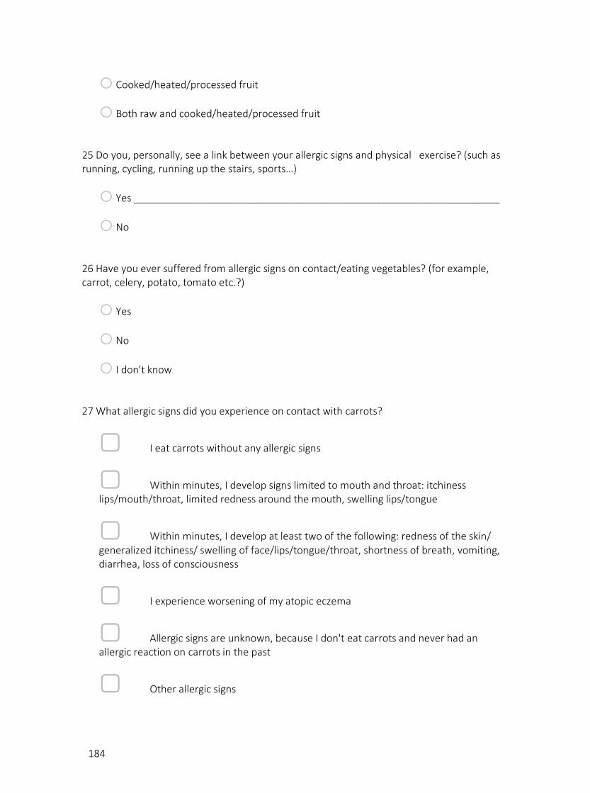
184
o Cooked/heated/processed fruit
o Both raw and cooked/heated/processed fruit 25 Do you, personally, see a link between your allergic signs and physical exercise? (such as running, cycling, running up the stairs, sports…)
o Yes _________________________________________________________________
o No 26 Have you ever suffered from allergic signs on contact/eating vegetables? (for example, carrot, celery, potato, tomato etc.?)
o Yes
o No
o I don't know 27 What allergic signs did you experience on contact with carrots?
▢ I eat carrots without any allergic signs
▢ Within minutes, I develop signs limited to mouth and throat: itchiness lips/mouth/throat, limited redness around the mouth, swelling lips/tongue
▢ Within minutes, I develop at least two of the following: redness of the skin/ generalized itchiness/ swelling of face/lips/tongue/throat, shortness of breath, vomiting, diarrhea, loss of consciousness
▢ I experience worsening of my atopic eczema
▢ Allergic signs are unknown, because I don't eat carrots and never had an allergic reaction on carrots in the past
▢ Other allergic signs

185
28 What allergic signs did you experience on contact with potatoes?
▢ I eat potatoes without any allergic signs
▢ Within minutes, I develop signs limited to mouth and throat: itchiness lips/mouth/throat, limited redness around the mouth, swelling lips/tongue
▢ Within minutes, I develop at least two of the following: redness of the skin/ generalized itchiness/ swelling of face/lips/tongue/throat, shortness of breath, vomiting, diarrhea, loss of consciousness
▢ I experience worsening of my atopic eczema
▢ Allergic signs are unknown, because I don't eat potatoes and never had an allergic reaction on potatoes in the past
▢ Other allergic signs 29 What allergic signs did you experience on contact with celery?
▢ I eat celery without any allergic signs
▢ Within minutes, I develop signs limited to mouth and throat: itchiness lips/mouth/throat, limited redness around the mouth, swelling lips/tongue
▢ Within minutes, I develop at least two of the following: redness of the skin/ generalized itchiness/ swelling of face/lips/tongue/throat, shortness of breath, vomiting, diarrhea, loss of consciousness
▢ I experience worsening of my atopic eczema
▢ Allergic signs are unknown, because I don't eat celery and never had an allergic reaction on celery in the past
▢ Other allergic signs 30 What allergic signs did you experience on contact with tomatoes?

186
▢ I eat tomatoes without any allergic signs
▢ Within minutes, I develop signs limited to mouth and throat: itchiness lips/mouth/throat, limited redness around the mouth, swelling lips/tongue
▢ Within minutes, I develop at least two of the following: redness of the skin/ generalized itchiness/ swelling of face/lips/tongue/throat, shortness of breath, vomiting, diarrhea, loss of consciousness
▢ I experience worsening of my atopic eczema
▢ Allergic signs are unknown, because I don't eat tomatoes and never had an allergic reaction on tomatoes in the past
▢ Other allergic signs 31 Have you experienced allergic signs on contact with other vegetables then already mentioned before?
o Yes. Which vegetables, other than the ones mentioned before, also induce allergic signs? ________________________________________________________________
o No 32 You experience allergic signs on vegetables only when they are:
o Only raw vegetables
o Only cooked/heated/processed vegetables
o Both raw and cooked/heated/processed vegetables 33 Have you ever suffered from allergic signs on contact/eating legumes? (for example peas, soy, chick peas, lentils, beans, etc.?)
o Yes
o No

187
o I don't know whether I experience allergic symptoms on contact with/eating legumes 34 What allergic signs did you experience on contact with peas?
▢ I eat peas without any allergic signs
▢ Within minutes, I develop signs limited to mouth and throat: itchiness lips/mouth/throat, limited redness around the mouth, swelling lips/tongue
▢ Within minutes, I develop at least two of the following: redness of the skin/ generalized itchiness/ swelling of face/lips/tongue/throat, shortness of breath, vomiting, diarrhea, loss of consciousness
▢ I experience worsening of my atopic eczema
▢ Allergic signs are unknown, because I don't eat peas and never had an allergic reaction on peas in the past
▢ Other allergic signs 35 What allergic signs did you experience on contact with soy?
▢ I eat soy (products) without any allergic signs
▢ Within minutes, I develop signs limited to mouth and throat: itchiness lips/mouth/throat, limited redness around the mouth, swelling lips/tongue
▢ Within minutes, I develop at least two of the following: redness of the skin/ generalized itchiness/ swelling of face/lips/tongue/throat, shortness of breath, vomiting, diarrhea, loss of consciousness
▢ I experience worsening of my atopic eczema
▢ Allergic signs are unknown, because I don't eat soy and never had an allergic reaction on soy (products) in the past

188
▢ Other allergic signs 36 What allergic signs did you experience on contact with beans?
▢ I eat beans without any allergic signs
▢ Within minutes, I develop signs limited to mouth and throat: itchiness lips/mouth/throat, limited redness around the mouth, swelling lips/tongue
▢ Within minutes, I develop at least two of the following: redness of the skin/ generalized itchiness/ swelling of face/lips/tongue/throat, shortness of breath, vomiting, diarrhea, loss of consciousness
▢ I experience worsening of my atopic eczema
▢ Allergic signs are unknown, because I don't eat beans and never had an allergic reaction on beans in the past
▢ Other allergic signs 37 What allergic signs did you experience on contact with lentils?
▢ I eat lentils without any allergic signs
▢ Within minutes, I develop signs limited to mouth and throat: itchiness lips/mouth/throat, limited redness around the mouth, swelling lips/tongue
▢ Within minutes, I develop at least two of the following: redness of the skin/ generalized itchiness/ swelling of face/lips/tongue/throat, shortness of breath, vomiting, diarrhea, loss of consciousness
▢ I experience worsening of my atopic eczema
▢ Allergic signs are unknown, because I don't eat lentils and never had an allergic reaction on lentils in the past

189
▢ Other allergic signs 38 What allergic signs did you experience on contact with chick peas?
▢ I eat chick peas without any allergic signs
▢ Within minutes, I develop signs limited to mouth and throat: itchiness lips/mouth/throat, limited redness around the mouth, swelling lips/tongue
▢ Within minutes, I develop at least two of the following: redness of the skin/ generalized itchiness/ swelling of face/lips/tongue/throat, shortness of breath, vomiting, diarrhea, loss of consciousness
▢ I experience worsening of my atopic eczema
▢ Allergic signs are unknown, because I don't eat chick peas and never had an allergic reaction on chick peas in the past
▢ Other allergic signs 39 Have you experienced allergic signs on contact with other legumes then already mentioned before?
o Yes. Which legumes, other than the ones mentioned before, also induce allergic signs? ______________________________________________________________
o No 40 You experience allergic signs on legumes only when they are:
o Only raw legumes
o Only cooked/heated/processed legumes
o Both raw and cooked/heated/processed legumes

190
41 Have you ever suffered from allergic signs on contact/eating nuts/seeds? (for example, peanut, hazelnut, almond, sesame, cashew nut, pine nuts etc.?)
o yes
o No
o I don't know 42 What allergic signs did you experience on contact with peanuts?
▢ I eat peanuts without any allergic signs
▢ Within minutes, I develop signs limited to mouth and throat: itchiness lips/mouth/throat, limited redness around the mouth, swelling lips/tongue
▢ Within minutes, I develop at least two of the following: redness of the skin/ generalized itchiness/ swelling of face/lips/tongue/throat, shortness of breath, vomiting, diarrhea, loss of consciousness
▢ I experience worsening of my atopic eczema
▢ Allergic signs are unknown, because I don't eat peanuts and never had an allergic reaction on peanuts in the past
▢ Other allergic signs 43 What allergic signs did you experience on contact with hazelnuts? I eat hazelnuts without any allergic signs
▢ Within minutes, I develop signs limited to mouth and throat: itchiness lips/mouth/throat, limited redness around the mouth, swelling lips/tongue
▢ Within minutes, I develop at least two of the following: redness of the skin/ generalised itchiness/ swelling of face/lips/tongue/throat, shortness of breath, vomiting, diarrhea, loss of consciousness
▢ I experience worsening of my atopic eczema

191
▢ Allergic signs are unknown, because I don't eat hazelnuts and never had an allergic reaction on hazelnuts in the past
▢ Other allergic signs 44 What allergic signs did you experience on contact with pine nuts?
▢ I eat pine nuts without any allergic signs
▢ Within minutes, I develop signs limited to mouth and throat: itchiness lips/mouth/throat, limited redness around the mouth, swelling lips/tongue
▢ Within minutes, I develop at least two of the following: redness of the skin/ generalized itchiness/ swelling of face/lips/tongue/throat, shortness of breath, vomiting, diarrhea, loss of consciousness
▢ I experience worsening of my atopic eczema
▢ Allergic signs are unknown, because I don't eat pine nuts and never had an allergic reaction on pine nuts in the past
▢ Other allergic signs 45 What allergic signs did you experience on contact with cashew nuts?
▢ I eat cashew nuts without any allergic signs
▢ Within minutes, I develop signs limited to mouth and throat: itchiness lips/mouth/throat, limited redness around the mouth, swelling lips/tongue
▢ Within minutes, I develop at least two of the following: redness of the skin/ generalized itchiness/ swelling of face/lips/tongue/throat, shortness of breath, vomiting, diarrhea, loss of consciousness
▢ I experience worsening of my atopic eczema

192
▢ Allergic signs are unknown, because I don't eat cashew nuts and never had an allergic reaction on cashew nuts in the past
▢ Other allergic signs 46 What allergic signs did you experience on contact with walnut?
▢ I eat walnuts without any allergic signs
▢ Within minutes, I develop signs limited to mouth and throat: itchiness lips/mouth/throat, limited redness around the mouth, swelling lips/tongue
▢ Within minutes, I develop at least two of the following: redness of the skin/ generalized itchiness/ swelling of face/lips/tongue/throat, shortness of breath, vomiting, diarrhea, loss of consciousness
▢ I experience worsening of my atopic eczema
▢ Allergic signs are unknown, because I don't eat walnuts and never had an allergic reaction on walnuts in the past
▢ Other allergic signs 47. What allergic signs did you experience on contact with almonds?
▢ I eat almonds without any allergic signs
▢ Within minutes, I develop signs limited to mouth and throat: itchiness lips/mouth/throat, limited redness around the mouth, swelling lips/tongue
▢ Within minutes, I develop at least two of the following: redness of the skin/ generalized itchiness/ swelling of face/lips/tongue/throat, shortness of breath, vomiting, diarrhea, loss of consciousness
▢ I experience worsening of my atopic eczema

193
▢ Allergic signs are unknown, because I don't eat almonds and never had an allergic reaction on almonds in the past
▢ Other allergic signs 48 What allergic signs did you experience on contact with sesame?
▢ I eat sesame seeds without any allergic signs
▢ Within minutes, I develop signs limited to mouth and throat: itchiness lips/mouth/throat, limited redness around the mouth, swelling lips/tongue
▢ Within minutes, I develop at least two of the following: redness of the skin/ generalized itchiness/ swelling of face/lips/tongue/throat, shortness of breath, vomiting, diarrhea, loss of consciousness
▢ I experience worsening of my atopic eczema
▢ Allergic signs are unknown, because I don't eat sesame seeds and never had an allergic reaction on sesame in the past
▢ Other allergic signs 49 What allergic signs did you experience on contact with poppy seeds?
▢ I eat poppy seeds without any allergic signs
▢ Within minutes, I develop signs limited to mouth and throat: itchiness lips/mouth/throat, limited redness around the mouth, swelling lips/tongue
▢ Within minutes, I develop at least two of the following: redness of the skin/ generalized itchiness/ swelling of face/lips/tongue/throat, shortness of breath, vomiting, diarrhea, loss of consciousness
▢ I experience worsening of my atopic eczema

194
▢ Allergic signs are unknown, because I don't eat poppy seeds and never had an allergic reaction on poppy seeds in the past
▢ other allergic signs 50 Have you experienced allergic signs on contact with other seeds/nuts then already mentioned before?
o Yes. Which nuts/seeds, other than the ones mentioned before, also induce allergic signs? ________________________________________________________________
o no 51 You experience allergic signs on seeds/nuts only when they are:
o Only raw uncooked nuts/seeds
o Only cooked/heated/processed nuts/seeds
o Both raw and cooked/heated/processed nuts/seeds 52 Have you ever suffered from allergic signs on contact/eating wheat/wheat containing products?
o Yes
o No
o I don't know 53 You have indicated to have experienced allergic signs on contact with wheat (containing products). Can you describe these signs?
▢ Within minutes, I develop signs limited to mouth and throat: itchiness lips/mouth/throat, limited redness around the mouth, swelling lips/tongue
▢ Within minutes, I develop at least two of the following: redness of the skin/ generalized itchiness/ swelling of face/lips/tongue/throat, shortness of breath, vomiting, diarrhea, loss of consciousness

195
▢ other allergic signs _______________________________________________________________________
54 Do you, personally, see a link between your allergic signs on wheat and physical exercise? (such as running, cycling, running up the stairs, sports, …)
o yes
o No
o I don't know 55 Do you suffer from hay fever? (hay fever can induce itchy nose/throat/ears, runny nose/ nose congestion, frequent sneezing, teary eyes, …)
o Yes
o No 56 In which period do you suffer from hay fever? (multiple responses allowed)
▢ The whole year round, these signs are not linked with pollen exposure
▢ The whole year round, also during winter
▢ January/February/March
▢ April/May/June
▢ July/August/September 57 Have you ever suffered from allergic signs on contact with/eating animal-based foods? (for example, cow's milk, eggs, meat, fish, shellfish/molluscs)
o Yes
o No 58 On exposure to which animal-based foods have you experienced allergic signs?

196
▢ Cow's milk
▢ Hen's egg
▢ Meat
▢ Fish
▢ Crustaceans/shellfish
▢ None of the above 59 What kind of allergic signs did you experience on contact with/drinking milk? (multiple responses allowed)
▢ I drink milk/eat dairy products without any allergic signs
▢ I experience worsening of my atopic eczema
▢ Skin signs (redness/rash, swelling (eyes/lips/extremities), hives, …)
▢ Abdominal complaints (cramps, bloating, flatulence, diarrhoea, vomiting, nausea, …)
▢ Breathing problems (cough, shortness of breath, wheezing, asthma attack, …)
▢ Generalized malaise
▢ Other
▢ Signs unknown because I don't drink milk 60 What kind of allergic signs did you experience on contact with/eating eggs?

197
▢ I eat eggs without any allergic signs
▢ I experience worsening of my atopic eczema
▢ Skin signs (redness/rash, swelling (eyes/lips/extremities), hives, …)
▢ Abdominal complaints (cramps, bloating, flatulence, diarrhoea, vomiting, nausea, …)
▢ Breathing problems (cough, shortness of breath, wheezing, asthma attack,…)
▢ Generalized malaise
▢ Other allergic signs
▢ Signs unkown, I don't eat eggs 61 Contact/ingestion of which types of meat made you experience allergic signs?
▢ I eat meat without any allergic signs
▢ Poultry (chicken, turkey, quail, …)
▢ Beef or veal
▢ Pork
▢ other types of meat 62 What kind of allergic signs did you experience on contact with/eating meat?
▢ I eat meat without any allergic signs
▢ I experience worsening of my atopic eczema

198
▢ Skin signs (redness/rash, swelling (eyes/lips/extremities), hives, …)
▢ Abdominal complaints (cramps, bloating, flatulence, diarrhoea, vomiting, nausea, …)
▢ Breathing problems (cough, shortness of breath, wheezing, asthma attack, …)
▢ Generalized malaise
▢ Other allergic signs
▢ Signs unknown because I don’t eat these products. 63 Contact/ingestion of which types of fish made you experience allergic signs?
▢ Salmon
▢ Tuna
▢ Cod fish
▢ Other _________________________________________________________
▢ None 64 What kind of allergic signs did you experience on contact with/eating these types of fish?
▢ I eat fish without any allergic signs
▢ I experience worsening of my atopic eczema
▢ Skin signs (redness/rash, swelling (eyes/lips/extremities), hives, …)

199
▢ Abdominal complaints (cramps, bloating, flatulence, diarrhoea, vomiting, nausea, …)
▢ Breathing problems (cough, shortness of breath, wheezing, asthma attack…)
▢ Generalized malaise
▢ Other allergic signs
▢ Signs unknown because I don’t eat these products. 65 What kind of allergic signs did you experience on contact with/eating shellfish/molluscs?
▢ I eat crustaceans/shellfish without any allergic signs
▢ I experience worsening of my atopic eczema
▢ Skin signs (redness/rash, swelling (eyes/lips/extremities), hives, …)
▢ Abdominal complaints (cramps, bloating, flatulence, diarrhoea, vomiting, nausea, …)
▢ Breathing problems (cough, shortness of breath, wheezing, asthma attack…)
▢ Generalized malaise
▢ Other allergic signs
▢ Signs unknown because I don’t eat these products. 66 Have you ever suffered from allergic signs on contact with/latex? (for example, balloons, latex gloves, condoms, etc.)
o Yes

200
o No
o I don't know 67 What kind of allergic signs did you experience on contact with/ latex?
▢ Allergic signs of the skin, limited to the area of latex contact.
▢ Allergic signs limited to the upper respiratory system: sneezing, itchy nose/eyes, cough, runny nose/nasal congestion, mild shortness of breath
▢ Generalized allergic signs: redness/itchiness/swelling of the skin beyond the area of contact AND/OR shortness of breath AND/OR diarrhoea/vomiting AND/OR loss of consciousness
▢ Other allergic signs
▢ I do not develop allergic signs upon contact with latex (products) 68 Have you ever smoked (tobacco) before?
o Yes
o No 69 Have you experienced allergic signs on contact with tobacco/smoke or when someone is smoking in your vicinity?
o Yes
o No
o I don't know

201
70 What kind of allergic signs did you experience on contact with tobacco/passive smoking?
▢ I do not develop allergic signs upon contact with tobacco (products)
▢ Allergic signs limited to the respiratory system: sneezing, itchy nose/eyes, cough, runny nose/nasal congestion, mild shortness of breath
▢ I develop generalized allergic signs; at least two of the following: redness of the skin/ generalized itchiness/ swelling of face/lips/tongue/throat, shortness of breath, vomiting, diarhoea, loss of consciousness
▢ Other allergic signs ______________________________________________________________________
71 Have you experienced allergic signs on contact with/drinking alcoholic beverages?
o Yes
o No
o I don't know 72 What kind of allergic signs did you experience on contact with/drinking beer?
▢ I drink beer without any allergic signs
▢ Within minutes, I develop signs limited to mouth and throat: itchiness lips/mouth/throat, limited redness around the mouth, swelling lips/tongue
▢ Within minutes, I develop at least two of the following: redness of the skin/ generalized itchiness/ swelling of face/lips/tongue/throat, shortness of breath, vomiting, diarhoea, loss of consciousness
▢ I experience worsening of my atopic eczema
▢ Allergic signs are unknown, because I don't drink beer and never had an allergic reaction on beer in the past

202
▢ Other allergic signs 73 You have indicated to have experienced allergic signs on drinking beer. Can you describe the specific beer brand or type? __________________________________________________________________________ __________________________________________________________________________ __________________________________________________________________________ __________________________________________________________________________ __________________________________________________________________________ 74 What kind of allergic signs did you experience on contact with/drinking red wine?
▢ I drink red wine without any allergic signs
▢ Within minutes, I develop signs limited to mouth and throat: itchiness lips/mouth/throat, limited redness around the mouth, swelling lips/tongue
▢ Within minutes, I develop at least two of the following: redness of the skin/ generalized itchiness/ swelling of face/lips/tongue/throat, shortness of breath, vomiting, diarhoea, loss of consciousness
▢ I experience worsening of my atopic eczema
▢ Allergic signs are unknown, because I don't drink red wine and never had an allergic reaction on red wine in the past
▢ Other allergic signs 75 What kind of allergic signs did you experience on contact with/drinking white wine?
▢ I drink white wine without any allergic signs
▢ Within minutes, I develop signs limited to mouth and throat: itchiness lips/mouth/throat, limited redness around the mouth, swelling lips/tongue

203
▢ Within minutes, I develop at least two of the following: redness of the skin/ generalized itchiness/ swelling of face/lips/tongue/throat, shortness of breath, vomiting, diarhoea, loss of consciousness
▢ I experience worsening of my atopic eczema
▢ Allergic signs are unknown, because I don't drink white wine and never had an allergic reaction on white wine in the past
▢ Other allergic signs 76 Have you ever smoked/eaten/touched cannabis or cannabis products? (we would like to reassure you that all information gathered through this questionnaire will be confidential and processing of data is anonymous)
o Yes
o No 77 How frequent do you use/come in contact with cannabis?
o At least once or multiple times a day
o Once a week
o Once a month
o Seldom, a couple of times a year 78 What was the last time you were in contact with cannabis?
o Last week
o Last month
o More than one month ago, within the past year
o More than one year ago
o More than 10 years ago

204
79 Do you know someone in your environment who uses/comes in contact with cannabis OR do you have contact with cannabis on a professional level due to your job (policeman, laboratory personnel, …)
o Yes
o No
o I don't know 80 How frequent do you have contact with others using cannabis/do you have professional exposure to cannabis?
o I don't know
o At least once or multiple times a day
o Once a week
o Once a month
o Seldom, a couple of times a year 81 The most recent environmental/professional cannabis exposure dates to:
o Last week
o Last month
o More than one month ago, within the past year
o More than one year ago
o More than 10 years ago 82 What kind of allergic signs did you experience on (environmental) contact with cannabis?
▢ I do not develop allergic signs on contact with cannabis (products)
▢ Allergic signs limited to the upper respiratory system: sneezing, itchy nose/eyes, cough, runny nose/nasal congestion, mild shortness of breath

205
▢ I develop generalized signs; at least two of the following: redness of the skin/ generalized itchiness/ swelling of face/lips/tongue/throat, shortness of breath, vomiting, diarhoea, loss of consciousness
▢ Other allergic signs ________________________________________________________________________
▢ I have no exposure to cannabis 83 At last, could you define the order in which your allergic symptoms developed? I first developed allergic symptoms to:
o Fruit
o Vegetables
o Legumes
o Nuts/seeds
o Hay fever
o Latex
o Cannabis
o Tobacco
o Beer/wine
o Wheat (products)
o I don't suffer from allergies 84 Secondly, I developed allergic symptoms to:
o I don't suffer from any of these allergies
o Fruit
o Vegetables

206
o Legumes
o Nuts/seeds
o Hay fever
o Latex
o Cannabis
o Tobacco
o Beer/wine
o Wheat (products) 85 We would like to thank you for filling in this questionnaire.

207
2. PERMIT POSSESSION OF SEDATING & PSYCHOTROPIC SUBSTANCES

208
209
3. FUNDING
Related Documents


![Allergy Clinician Manual-06172016lrgdesigns.com/wp-content/uploads/2019/01/Allergy...the allergies, and ultimately obtain success by building up the patient’s tolerance. “[Immunotherapy]](https://static.cupdf.com/doc/110x72/600b74df86744a016f5e32ff/allergy-clinician-manual-the-allergies-and-ultimately-obtain-success-by-building.jpg)