Canine Sinonasal Mycosis A retrospective and prospective study of different treatment options, their successes and influence of different variables Research project Veterinary Medicine Utrecht University N.S. Ruijgrok Students number: 3259889 6 September 2013 Project supervisor: Drs. A.N. Haagsman Second assessor: Dr. M.E. Peeters Department coordinator: Prof. B.P. Meij Cooperation with Microbiology Department of Biology, Utrecht University: Dr. J.J.P.A. de Cock and N. Escobar Salazar MSc Department of Clinical Sciences of Companion Animals Faculty of Veterinary Medicine, Utrecht University

Welcome message from author
This document is posted to help you gain knowledge. Please leave a comment to let me know what you think about it! Share it to your friends and learn new things together.
Transcript
Canine Sinonasal Mycosis
A retrospective and prospective study of different treatment options, their successes and
influence of different variables
Research project Veterinary Medicine Utrecht University
N.S. Ruijgrok
Students number: 3259889
6 September 2013
Project supervisor: Drs. A.N. Haagsman
Second assessor: Dr. M.E. Peeters
Department coordinator: Prof. B.P. Meij
Cooperation with Microbiology Department of Biology, Utrecht University: Dr. J.J.P.A. de
Cock and N. Escobar Salazar MSc
Department of Clinical Sciences of Companion Animals
Faculty of Veterinary Medicine, Utrecht University

A retrospective and prospective study of different treatment options, their successes and influence of different variables
N.S. Ruijgrok Bsc.
2
Abstract
This study reviews treatment outcome of commonly used treatment techniques of dogs with
Sinonasal Mycosis (SNM). Treatment options considered are:
Treatment 1: Removal of local abnormalities (e.g. corpus alienum); removal mycotic plaques
by rhinoscopy, followed by the local application of clotrimazole cream (1%).
Treatment 2: Removal of mycotic plaques by rhinoscopy and one hour flushing with
clotrimazole solution (1%) without trepanation.
Treatment 3: Removal of mycotic plaques by rhinoscopy and trepanation of the frontal sinus
and one hour flushing with clotrimazole solution (1%), followed by
administration of local clotrimazole cream (1%).
Treatment 4: Removal of mycotic plaques by rhinoscopy and trepanation of the frontal sinus
with placement of tubes to administer enilconazole (1%) twice daily for 10-14
days.
Treatment 5: Removal of mycotic plaques by rhinoscopy and one hour flushing with
clotrimazole solution (1%) without trepanation, removal of mycotic plaques
followed by local application of clotrimazole cream (1%).
Influence of different variables on treatment failure will be examined. Variables considered
are: age, gender, ipsilateral versus bilateral affection, affection of the frontal sinus, severity of
the infection and enlargement of the mandibular lymph node.
Furthermore, a prospective case control study was done on 4 new patients with SNM in order
to determine specific changes in blood values, signs of underlying disease and specific
properties of the fungus itself.
Study design:
Retrospective (n = 51) and prospective study (n=4).
Methods:
Retrospective study: Medical reports of 51 dogs treated for sinonasal mycosis were obtained
via research of the clinical database of the Faculty of Veterinary Science, Medicine of
Companion Animals at Utrecht University for the time period March 2005 to March 2013.
First treatment outcome and influence of variables described above were evaluated.
Prospective case control study: Clinical examination, blood analyses, CT, rhinoscopy and biopsies
were performed at 4 new patients with SNM at Department of Clinical Sience of Companion
Animals, Faculty of Veterinary Medicine, Utrecht University. Mycotic plaques were
collected, pH was measured, samples were cultured and DNA was isolated and typed.
Determination of a resistance profile was started.
Results:
Retrospective study: When all topical treatment options were combined together, treatment
success was 52.9% after first treatment and 78.4% after two treatments.
No statistical difference was found in treatment outcome (recurrence of infection) over the
entire follow-up between the different treatment groups 1-5.
A retrospective and prospective study of different treatment options, their successes and influence of different variables
N.S. Ruijgrok Bsc.
3
Treatment failure was not associated with age, gender, SNM in the frontal sinus, ipsilateral
versus bilateral SNM infection, severity of the infection or enlargement of the mandibular
lymph node.
Significant proof that recurrence of SNM will not occur within 6 weeks after being treated
with treatment option 4 (p= 0.049) was found. Recurrence rates of a combined group
consisting of treatment 2 and 5 (to enlarge the sample size) showed a significant (p= 0.039)
reduction in recurrence rate within 6 weeks when compared with other treatments.
Relative risks and odds ratios calculated in this study to compare treatment outcomes and
influence of different variables are not reliable because calculated results are not significant,
95% confidence intervals are wide and they cross the value 1. This indicates that the results
are unreliable.
Prospective case control study: Mycotic plaques collected in 4 patients were all typed as
Aspergillus fumigatus. The pH of fresh collected fungus was between 7.9 and 8.2. Histological
evaluation of biopsies shows that the fungus grows only superficially on the mucosa. The lamina
propria was not infiltrated. Determination of a resistance profile was not completed while
writing this report.
Three patients were treated with treatment option 3 and one patient was treated with treatment
option 5. One patient was euthanized because of clinical deterioration. Recurrence of clinical
signs of SNM was seen in two dogs (patient 3 and 4, 50% of total). However, recurrence of
SNM in patient 4 was not proven after he was euthanized. Slight hypo-hypoalbuminemia was
seen in three out of four patients.
A retrospective and prospective study of different treatment options, their successes and influence of different variables
N.S. Ruijgrok Bsc.
4
Contents
Abstract 2
Introduction 5
Research goals: Retrospective study 10
Materials and methods: Retrospective study 11
Results: Retrospective study 19
Research goals: Prospective case control study 27
Material and methods: Prospective case control study 28
Results: Prospective case control study 30
Discussion 35
Conclusion 39
Acknowledgements 40
Appendix 1 41
Appendix 2 42
References 43
A retrospective and prospective study of different treatment options, their successes and influence of different variables
N.S. Ruijgrok Bsc.
5
Introduction
Chronic nasal discharge is common in dogs. Chronic sneezing, depigmentation of the nasal
planum and (unilateral) nasal discharge are symptoms that may indicate an abnormality in the
nasal cavities, sinuses and/or nasopharynx. A fungus infection in the nasal cavity and frontal
sinus may be one of the causes of these clinical signs. (Benitah, 2006; Peeters & Clercx, 2007; M.
J. Sharman & Mansfield, 2012). Research has shown that 7 to 34% of dogs with chronic nasal
discharge is caused by fungal rhinosinusitis (M. J. Sharman & Mansfield, 2012). Fungi that
can cause such a clinical picture are Aspergillus fumigatus, Aspergillus flavus, Aspergillus
niger or Penicillium spp. Of these fungi A. fumigatus is by far the most demonstrated in
patients suspected of a sinonasal fungal infection (Benitah, 2006; Peeters & Clercx, 2007;
Quinn, Markey, Carter, Donnelly, & Leonard, 2002; M. J. Sharman & Mansfield, 2012; N.
Sharp, Sullivan, & Harvey, 1992). Sinonasal mycosis (SNM) is diagnosed by a thorough
history, clinical signs, physical examination and diagnostic imaging, such as computed
tomography (CT) and rhinoscopy. Sinonasal Aspergillosis (SNA) should not be confused with
the rare systemic form of Aspergillosis. The latter disease is mainly seen in German
Shepherds, the nasal region is usually not involved and it is caused by A. terreurs instead of
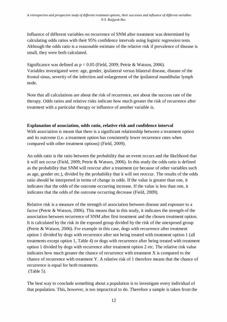
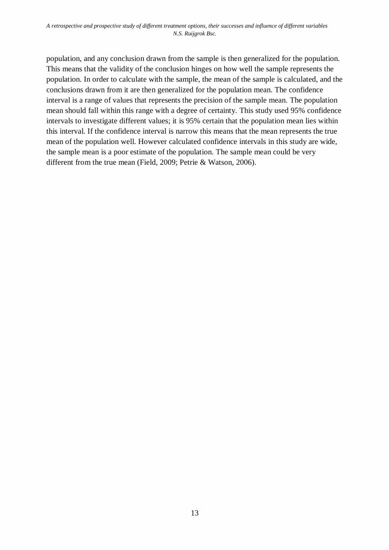
A. fumigatus (M. J. Sharman & Mansfield, 2012). SNM is characterized by destruction of the
nasal conchae (see figure 3 and 4). In an advanced stage there may also be damage to the
frontal bone, the periorbital tissue and cribriforme plate (ethmoid). Bone destruction is caused
by toxins produced by A. fumigatus and by the inflammatory process of the patient. (Peeters
& Clercx, 2007). Clinical signs seen in dogs with SNM is: chronic nasal discharge (mostly
mucopurulent). This usually starts unilaterally, but may eventually occur bilaterally.
Moreover there are signs of pain at the nose and sinuses, depigmentation and ulceration of the
nasal planum Changes at the nasal planum are most likely (caused by toxins of the fungus)
(Nelson et al., 2009; Peeters & Clercx, 2007; M. J. Sharman & Mansfield, 2012; N. Sharp et
al., 1992). Often there is increased air flow through the nasal sinus because of increased
destruction of conchal structures. Furthermore epistaxis, sneezing, reversed sneezing, loss of
appetite and depression is reported (Peeters & Clercx, 2007; M. J. Sharman & Mansfield,
2012; N. Sharp et al., 1992). In severe cases deformities of the skull can be seen caused by
hyperostosis of the frontal bone. Sometimes epiphora secondary to SNM in the orbit and brain
damage is seen if the ethmoid bone is no longer intact (Peeters & Clercx, 2007). SNM can
occur secondary to an underlying primary cause. Primary causes are as a foreign body in the
nasal sinus, previous trauma to the nose, neoplasia or a oronasal fistula. Removal of this
primary cause will in most cases resolve the secondary fungal infection. Although, an
opportunistic fungal infection can be the primary cause which is more challenging to treat. In
this study, we focus on the latter group.
In order to determine the extent of the infection and subsequently to determine which
treatment is most appropriate for the individual patient, diagnostic imaging such as computed
tomography (CT) and rhinoscopy are necessary. CT evaluation of the skull is necessary to
determine if the frontal sinus is affected and if the cribriform plate and the orbita is intact.
These factors will influence the best treatment technique. For instance, one hour flushing with
clotrimazole under pressure is not possible if the cribriform plate is not intact; this will lead to
A retrospective and prospective study of different treatment options, their successes and influence of different variables
N.S. Ruijgrok Bsc.
6
severe complications or even death. Trepanation should be performed if the frontal sinus is
affected (Benitah, 2006; Peeters & Clercx, 2007).
A. fumigatus is a ubiquitous prevalent fungus, an infection can be acquired by inhalation of
spores (Quinn et al., 2002). A. fumigatus can be found in the nose, throat and pharynx of
healthy animals and humans (Nelson et al., 2009; Peeters & Clercx, 2007; Quinn et al., 2002;
M. J. Sharman & Mansfield, 2012; N. Sharp et al., 1992). It is still unclear why only a subset
of the dogs exposed to A. fumigatus will develop clinically relevant disease (Peeters & Clercx,
2007). The fungus infection is relatively more prevalent among dogs of mesocephal and
dolichocephal races and is seen more among young or middle aged dogs (Peeters & Clercx,
2007; Quinn et al., 2002; M. J. Sharman & Mansfield, 2012; N. Sharp et al., 1992). Rarely it
is also seen in cats. It is thought that a compromised immune system plays a role in the
development of the fungus infection, but this is not evidence-based.
Several treatment protocols are described with variable success rates (Burbidge et al., 1997;
Claeys, Lefebvre, Schuller, Hamaide, & Clercx, 2006; Clercx, 2006; Mathews et al., 1998;
Schuller & Clercx, 2007; M. Sharman et al., 2010; M. J. Sharman & Mansfield, 2012; N.
Sharp et al., 1992; Sissener, Bacon, Friend, Anderson, & White, 2006; Zonderland et al.,
2002). In many cases treatment should be repeated to achieve a higher success rate and
recurrence is not uncommon. To treat SNM, topical antimycotics are preferred to systemic
antifungals because of poor efficacy of the latter (Benitah, 2006; Nelson et al., 2009; Peeters
& Clercx, 2007; Quinn et al., 2002; M. J. Sharman & Mansfield, 2012; N. Sharp et al., 1992).
This study investigates five different topical treatment options to cure SNM in dogs (see
research goals). Three of them include one hour contact time with clotrimazole solution, one
of them administered through surgically placed catheters in the frontal sinus. Two of the
treatment options were followed by local application of clotrimazole cream. One treatment
option consists only of removal of mycotic plaques followed by local application of
clotrimazole cream (1%). Another treatment option is trepanation of the frontal sinus with
placement of tubes to administer enilconazole twice daily for 10-14 days.
Literature results
Few studies described topical clotrimazole treatments in dogs with sinonasal Aspergillosis
(Burbidge et al., 1997; Hayes & Demetriou, 2012; Mathews et al., 1998; M. Sharman et al.,
2010; M. Sharman, Lenard, Hosgood, & Mansfield, 2012; Sissener et al., 2006). Burbridge et
al. (1997) showed that noninvasive intranasal infusion with one hour contact time appears to
be an effective treatment for sinonasal Aspergillosis in dogs, their sample size was small
however (n=5) (Burbidge et al., 1997). Mathews et al. (1998) compared the effect on outcome
of topical administration of clotrimazole (1%) of surgically placed versus non-surgically
placed catheters in 60 dogs. In 65% of the cases there was resolution of clinical disease after
one treatment with clotrimazole through non-surgical placement of catheters. A higher
percentage of success (87%) was seen after multiple treatments (Mathews et al., 1998). In a

A retrospective and prospective study of different treatment options, their successes and influence of different variables
N.S. Ruijgrok Bsc.
7
retrospective study of Sharman et al. (2010), first treatment success after commonly used
treatments was examined. Thirty nine of the 85 dogs (45.8%) treated with a topical treatment
had resolution of disease. Treatment success was associated with younger age. After multiple
topical treatments 69.4% of patients were cured. When considering only non-invasive
treatment of a one hour clotrimazole (1%) flush inserted via catheters placed in the nares,
40% (18 of 45) of the dogs were cured after first treatment. Seventy percent (7 of 10) of the
dogs that were treated with a short 5-minute clotrimazole (1%) soak followed by
administration of clotrimazole cream (1%) were cured after first treatment. Trepanation of the
frontal sinus for temporary placement of the catheter and a one-hour flush with clotrimazole
solution was performed in 24 dogs. Two dogs were soaked with enilconazole instead of
clotrimazole. Fifty percent of these dogs were considered cured after first treatment. No
statistical difference between different treatment groups was found in this study (M. Sharman
et al., 2010).
Pomrantz et al. (2010) repeated rhinoscopic evaluation to assess the effectiveness of
intranasally administered clotrimazole in 23 dogs. When the frontal sinus was affected,
trepanation was performed. All dogs had rhinoscopic follow-up examination 1 to 4 months
after treatment. In 48% (11 of 23) of the dogs no fungal plaques were seen during rhinoscopic
follow-up evaluation and were classified as treatment success. Persistent fungal disease was
found in 52% (12 of 23). Three of seven dogs (42.8%) were cured after second treatment and
one of three dogs (33.3%) were free of disease after 3 treatments. Overall the efficiency of
intranasal administration of clotrimazole solution could be confirmed in 15 of 17 dogs (1-3
treatments). Delayed recurrence was seen in three dogs, all of them had involvement of the
frontal sinus. Treatment success was 67% (Pomrantz & Johnson, 2010).
Combined clotrimazole flush and depot therapy was examined by Sissener et al (2006). In this
study fourteen dogs were treated by frontal sinus trepanation with a short 5-minute flushing of
1% clotrimazole solution followed by administration of 1% clotrimazole cream. Twelve of
the fourteen dogs (85.7%) had no clinical signs after treatment or had only signs of mild
rhinitis during follow-up period of six months (Sissener et al., 2006).
For several years the standard treatment for sinonasal aspergillosis was an enilconazole
emulsion administered by surgically placed catheters into the frontal sinus and nasal chambers
(Benitah, 2006; N. Sharp et al., 1992). Enilconazole is less toxic and irritating to mucous
membranes than clotrimazole, especially in low concentrations (Peeters & Clercx, 2007).
Enilconazole was administered twice daily for 7-14 days (N. Sharp et al., 1992; N. J. Sharp,
Sullivan, Harvey, & Webb, 1993). Sharp et al. (1992, 1993) described that enilconazole cured
90% of the dogs with nasal aspergillosis (N. Sharp et al., 1992; N. J. Sharp et al., 1993).
However in some dogs treatment was followed by a six week course of ketoconazole orally
(N. J. Sharp et al., 1993).
Long term outcomes of dogs with sinonasal aspergillosis treated with non-invasive treatments
of topical enilconazole were examined in a study of Shuller et al. (2007). Fifteen dogs were
treated with an infusion of 1% enilconazole emulsion through a blindly placed catheter and
twelve dogs were treated with a 2% emulsion of enilconazole infused through endoscopically
A retrospective and prospective study of different treatment options, their successes and influence of different variables
N.S. Ruijgrok Bsc.
8
placed catheters in the frontal sinus. Fifty percent of the patients were asymptomatic
throughout the follow-up period of 38±17 months. The other 50% had only mild clinical signs
which were interpreted as chronic lymphoplasmacytic rhinitis/sinusitis and episodes of
bacterial infection (Schuller & Clercx, 2007).
Distribution of clotrimazole and enilconazole has been examined in multiple studies (Burrow,
Baker, White, & McConnell, 2013; Hayes & Demetriou, 2012; M. Sharman et al., 2012).
Hayes et al. (2012) reported that clotrimazole cream persists in the frontal sinus and is
distributed effectively in normal canine cadavers. Retention time is probably shorter in living
dogs because of head movements and sneezing. Drainage of cream would probably be faster
in animals with conchal atrophy (Hayes & Demetriou, 2012). Based on Hayes & Demetriou
(2012), this study assumes that administration of a clotrimazole cream prolongs drug contact
time in comparison with a clotrimazole solution. Because of this, treatment option 5
fundamentally differs from option 2 (both consist of a one hour flush with clotrimazol
solution, however option 5 ends with administration of clotrimazole cream), which is why
they are considered as separate treatments. However, because of the small sample size and the
same treatment principle, their data will be pooled together as well.
A Computed Tomography (CT) study of the distribution of clotrimazole cream installed in the
frontal sinus by trepanation in canine cadavers was performed by Burrow et al. (2011). Sinus
filling was excellent in 10 of 12 dogs (83.3%) (22 sinuses) and filling of caudal nasal cavities
was excellent in all dogs (Burrow et al., 2013).
Distribution of clotrimazole and enilconazole solutions were assessed with CT scan
performed 5 minutes after treatment by Sharman et al. (2012). He reported that distribution of
clotrimazole (1%) and enilconazole (10%) solutions in all regions of the nasal cavity and
frontal sinuses was achievable using temporary trepanation of the frontal sinus. However
distribution results varied considerably and retention was poor in 10 of the 18 regions
assessed.
A retrospective and prospective study of different treatment options, their successes and influence of different variables
N.S. Ruijgrok Bsc.
9
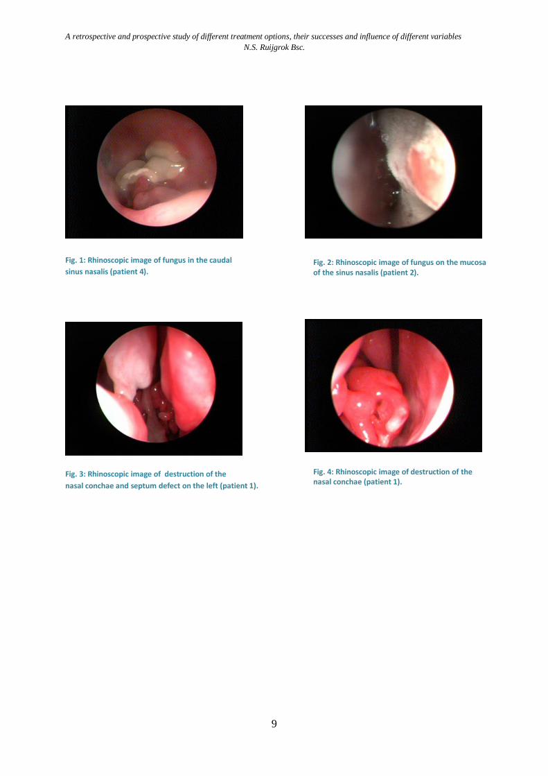
Fig. 1: Rhinoscopic image of fungus in the caudal
sinus nasalis (patient 4).
Fig. 3: Rhinoscopic image of destruction of the
nasal conchae and septum defect on the left (patient 1).
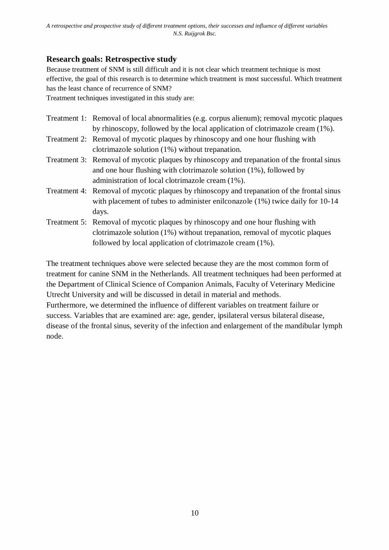
Fig. 2: Rhinoscopic image of fungus on the mucosa of the sinus nasalis (patient 2).
Fig. 4: Rhinoscopic image of destruction of the nasal conchae (patient 1).
A retrospective and prospective study of different treatment options, their successes and influence of different variables
N.S. Ruijgrok Bsc.
10
Research goals: Retrospective study
Because treatment of SNM is still difficult and it is not clear which treatment technique is most
effective, the goal of this research is to determine which treatment is most successful. Which treatment
has the least chance of recurrence of SNM?
Treatment techniques investigated in this study are:
Treatment 1: Removal of local abnormalities (e.g. corpus alienum); removal mycotic plaques
by rhinoscopy, followed by the local application of clotrimazole cream (1%).
Treatment 2: Removal of mycotic plaques by rhinoscopy and one hour flushing with
clotrimazole solution (1%) without trepanation.
Treatment 3: Removal of mycotic plaques by rhinoscopy and trepanation of the frontal sinus
and one hour flushing with clotrimazole solution (1%), followed by
administration of local clotrimazole cream (1%).
Treatment 4: Removal of mycotic plaques by rhinoscopy and trepanation of the frontal sinus
with placement of tubes to administer enilconazole (1%) twice daily for 10-14
days.
Treatment 5: Removal of mycotic plaques by rhinoscopy and one hour flushing with
clotrimazole solution (1%) without trepanation, removal of mycotic plaques
followed by local application of clotrimazole cream (1%).
The treatment techniques above were selected because they are the most common form of
treatment for canine SNM in the Netherlands. All treatment techniques had been performed at
the Department of Clinical Science of Companion Animals, Faculty of Veterinary Medicine
Utrecht University and will be discussed in detail in material and methods.
Furthermore, we determined the influence of different variables on treatment failure or
success. Variables that are examined are: age, gender, ipsilateral versus bilateral disease,
disease of the frontal sinus, severity of the infection and enlargement of the mandibular lymph
node.

A retrospective and prospective study of different treatment options, their successes and influence of different variables
N.S. Ruijgrok Bsc.
11
Material and methods: Retrospective study
Medical reports of dogs treated for sinonasal mycosis were obtained via research of the
clinical database of the Department of Clinical Science of Companion Animals, Faculty of
Veterinary Medicine Utrecht University for the time period March 2005 through March 2013.
Requirements that the patients had to meet in order to be included in the study are: confirmed
diagnosis of sinonasal mycosis by either computed tomography and rhinoscopy, pathology or
fungal culture. They have to have been treated with one of the treatment options described
above and their medical records need to be complete. Patients treated with one of the
treatment options described above in combination with systemic antifungals were excluded
from this study.
Treatment success was defined as resolution of nasal mucopurulent/purulent discharge,
epistaxis, sneezing and other clinical signs such as pain of the head or nose and lethargy.
Treatment failure was defined as recurrence of clinical symptoms described above and
confirmed by rhinoscopic visualization of mycotic plaques or fungal culture.
Patients with only mild serous nasal discharge without evidence of fungal recurrence were
classified as treatment success.
A subdivision was made for recurrence within 6 weeks, within 12 weeks, after 12 weeks and
over the entire follow-up. With the entire follow-up is meant the whole period between first
visit to the Departement of Clinical Sciences of Companion Animals with symptoms of SNM
until April 2013. This subdivision was made because recurrence within 6 or 12 weeks is likely
due to unsuccessful treatment whereas recurrence after e.g. one year is more likely due to
reinfection.
Statistics
Data was calculated by hand and by using the Statistical Package for Social Siences (IBM
SPSS version 21.0.0.0). To calculate the significance of first treatment failure and determine
if there is an association (relationship) between treatment option and recurrence of SNM, Chi-
squared tests or Fishers exact tests were used. The same tests were used to determine if there
is an association between different variables and recurrence over the entire follow-up (Fishers
exact test was used when expected frequencies were less than 5 (Field, 2009; Petrie &
Watson, 2006)).
Relative risks and odds ratios with their 95% confidence intervals were calculated for each
treatment option to determine which treatment option is best by determining treatment failure
(i.e. recurrence during follow-up) after first treatment. (Table 4).
Because lack of a control group (i.e. patients that did not receive treatment after diagnosis of
SNM), each treatment option was compared with the total of all other treatment options (e.g.
option 1 is compared with option 2+3+4+5+(2+5)). Each treatment option was also compared
to each other treatment option individually (e.g. the relative risk of option 1 to option 2 equals
option 1 divided by option 2: 20.00/53.125 = 0.376 which means that option 1 has a 0.376
greater chance of recurrence than option 2). (Table 5).
A retrospective and prospective study of different treatment options, their successes and influence of different variables
N.S. Ruijgrok Bsc.
12
Influence of different variables on recurrence of SNM after treatment was determined by
calculating odds ratios with their 95% confidence intervals using logistic regression tests.
Although the odds ratio is a reasonable estimate of the relative risk if prevalence of disease is
small, they were both calculated.
Significance was defined as p < 0.05 (Field, 2009; Petrie & Watson, 2006).
Variables investigated were: age, gender, ipsilateral versus bilateral disease, disease of the
frontal sinus, severity of the infection and enlargement of the ipsilateral mandibular lymph
node.
Note that all calculations are about the risk of recurrence, not about the success rate of the
therapy. Odds ratios and relative risks indicate how much greater the risk of recurrence after
treatment with a particular therapy or influence of another variable is.
Explanation of association, odds ratio, relative risk and confidence interval
With association is meant that there is a significant relationship between a treatment option
and its outcome (i.e. a treatment option has consistently lower recurrence rates when
compared with other treatment options) (Field, 2009).
An odds ratio is the ratio between the probability that an event occurs and the likelihood that
it will not occur (Field, 2009; Petrie & Watson, 2006). In this study the odds ratio is defined
as the probability that SNM will reoccur after a treatment (or because of other variables such
as age, gender etc.), divided by the probability that it will not reoccur. The results of the odds
ratio should be interpreted in terms of change in odds. If the value is greater than one, it
indicates that the odds of the outcome occurring increase. If the value is less than one, it
indicates that the odds of the outcome occurring decrease (Field, 2009).
Relative risk is a measure of the strength of association between disease and exposure to a
factor (Petrie & Watson, 2006). This means that in this study, it indicates the strength of the
association between recurrence of SNM after first treatment and the chosen treatment option.
It is calculated by the risk in the exposed group divided by the risk of the unexposed group
(Petrie & Watson, 2006). For example in this case, dogs with recurrence after treatment
option 1 divided by dogs with recurrence after not being treated with treatment option 1 (all
treatments except option 1, Table 4) or dogs with recurrence after being treated with treatment
option 1 divided by dogs with recurrence after treatment option 2 etc. The relative risk value
indicates how much greater the chance of recurrence with treatment X is compared to the
chance of recurrence with treatment Y. A relative risk of 1 therefore means that the chance of
recurrence is equal for both treatments.
(Table 5).
The best way to conclude something about a population is to investigate every individual of
that population. This, however, is too impractical to do. Therefore a sample is taken from the
A retrospective and prospective study of different treatment options, their successes and influence of different variables
N.S. Ruijgrok Bsc.
13
population, and any conclusion drawn from the sample is then generalized for the population.
This means that the validity of the conclusion hinges on how well the sample represents the
population. In order to calculate with the sample, the mean of the sample is calculated, and the
conclusions drawn from it are then generalized for the population mean. The confidence
interval is a range of values that represents the precision of the sample mean. The population
mean should fall within this range with a degree of certainty. This study used 95% confidence
intervals to investigate different values; it is 95% certain that the population mean lies within
this interval. If the confidence interval is narrow this means that the mean represents the true
mean of the population well. However calculated confidence intervals in this study are wide,
the sample mean is a poor estimate of the population. The sample mean could be very
different from the true mean (Field, 2009; Petrie & Watson, 2006).
A retrospective and prospective study of different treatment options, their successes and influence of different variables
N.S. Ruijgrok Bsc.
14
Description of the implementation of the different treatment options
Treatment 1: Removal of local abnormalities (e.g. corpus alienum); removal of mycotic
plaques by rhinoscopy, followed by local application of clotrimazole cream.
Under general anesthesia rhinoscopy is performed. Local foreign bodies that might be present
and mycotic plaques are removed by surgical hook and suction. Further local clotrimazole-
cream (1%) will be applied at the place where the fungus was located using a urinecatheter
attached to a syringe to do so.
This treatment technique is only used in mild cases of SNM, when there are only a few
mycotic plaques, most of them primarily caused by a corpus alienum.
In this research 5 dogs were treated with this treatment technique.
Treatment 2: Removal of mycotic plaques by rhinoscopy and one hour flushing with
clotrimazole solution (1%) without trepanation.
Under general anesthesia rhinoscopy is performed and mycotic plaques are removed by
surgical hook and suction. The dog is positioned in sternal recumbency and has to be
intubated with a cuffed endotracheal tube. Then the tip of a 20- French Foley catheter is
inserted through the mouth and placed at the junction between the hard and the soft palate in
the caudal nasopharynx. The nasopharynx will be occluded by inflating the balloon. After, the
Foley catheter is clamped. Moist surgical gauzes are counted and placed in the pharynx to
avoid leakage of the clotrimazole-solution into the trachea. Next, the side holes of two 18-
French Foley catheters (used to flush) are clipped off and one is placed in each nostril into the
dorsal sinus nasalis. Within these Foley catheters we yellow urine catheters are placed, also
with the side holes cut off, though which the clotrimazole will be administered. Each catheter
is connected to a 50 ml infusion syringe with clotrimazole-solution. After that the balloons are
inflated which close off the nostrils and so prevent leaking out of the nose. Clotrimazole
solution (1%) is flushed into the nasal cavities through the catheter until the nasal cavity and
frontal sinus are sufficiently filled and leakage along the Foley catheters or mouth is noticed
(approximately 25 ml in each nostril in medium to large sized dogs). Then the catheters are
clamped. The dog is positioned 15 minutes in sternal recumbency and then rotated every 15
minutes to left lateral recumbency, right lateral recumbency and dorsal recumbency to ensure
drug contact with all the sinonasal surfaces. Every 15 minutes more clotrimazole solution is
added if possible (with a maximum of 75 ml per nasal cavity) to ensure maximal drug contact.
At the end of the infusion (after 1 hour) the dog is positioned in sternal recumbency with the
head pointed downwards. The catheters in the nostrils are removed to drain the nose for 15
minutes. At the end of the procedure the 20-French Foley catheter of the nasopharynx and the
gauzes in the pharynx are removed. The oral cavity is checked and fluid is removed by
suction before recovery from anesthesia. For this treatment procedure CT evaluation is
necessary to determine if the frontal sinus is not affected and if the cribriform plate is intact. If
the sinus frontalis is affected, preferred therapy are treatment 3 and 4 because these provide
direct access to mycotic plaques by trepanation.
A retrospective and prospective study of different treatment options, their successes and influence of different variables
N.S. Ruijgrok Bsc.
15
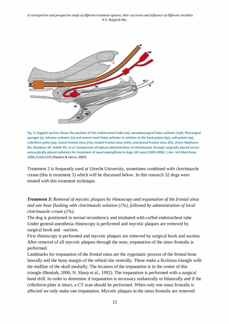
Fig. 5: Saggital section shows the position of the endotracheal tube (et), nasopharyngeal Foley catheter (npf), Pharyngeal
sponges (s), infusion catheter (ic) and rostral nasal Foley catheter in relation to the hard palate (hp), soft palate (sp),
cribriform plate (cp), rostral frontal sinus (rfs), medial frontal sinus (mfs), and lateral frontal sinus (lfs). (From Mathews
KG, Davidson AP, Koblik PD, et al. Comparison of topical administration of clotrimazole through surgically placed versus
nonsurgically placed catheters for treatment of nasal aspergillosis in dogs: 60 cases (1990-1996). J.Am. Vet Med Assoc
1998,213(4):503) (Peeters & Clercx, 2007)
Treatment 2 is frequently used at Utrecht University, sometimes combined with clotrimazole
cream (this is treatment 5) which will be discussed below. In this research 32 dogs were
treated with this treatment technique.
Treatment 3: Removal of mycotic plaques by rhinoscopy and trepanation of the frontal sinus
and one hour flushing with clotrimazole solution (1%), followed by administration of local
clotrimazole cream (1%).
The dog is positioned in sternal recumbency and intubated with cuffed endotracheal tube.
Under general anesthesia rhinoscopy is performed and mycotic plaques are removed by
surgical hook and -suction.
First rhinoscopy is performed and mycotic plaques are removed by surgical hook and suction.
After removal of all mycotic plaques through the nose, trepanation of the sinus frontalis is
performed.
Landmarks for trepanation of the frontal sinus are the zygomatic process of the frontal bone
laterally and the bony margin of the orbital rim ventrally. These make a fictitious triangle with
the midline of the skull medially. The location of the trepanation is in the center of this
triangle (Benitah, 2006; N. Sharp et al., 1992). The trepanation is performed with a surgical
hand drill. In order to determine if trepanation is necessary unilaterally or bilaterally and if the
cribriform plate is intact, a CT scan should be performed. When only one sinus frontalis is
affected we only make one trepanation. Mycotic plaques in the sinus frontalis are removed
A retrospective and prospective study of different treatment options, their successes and influence of different variables
N.S. Ruijgrok Bsc.
16
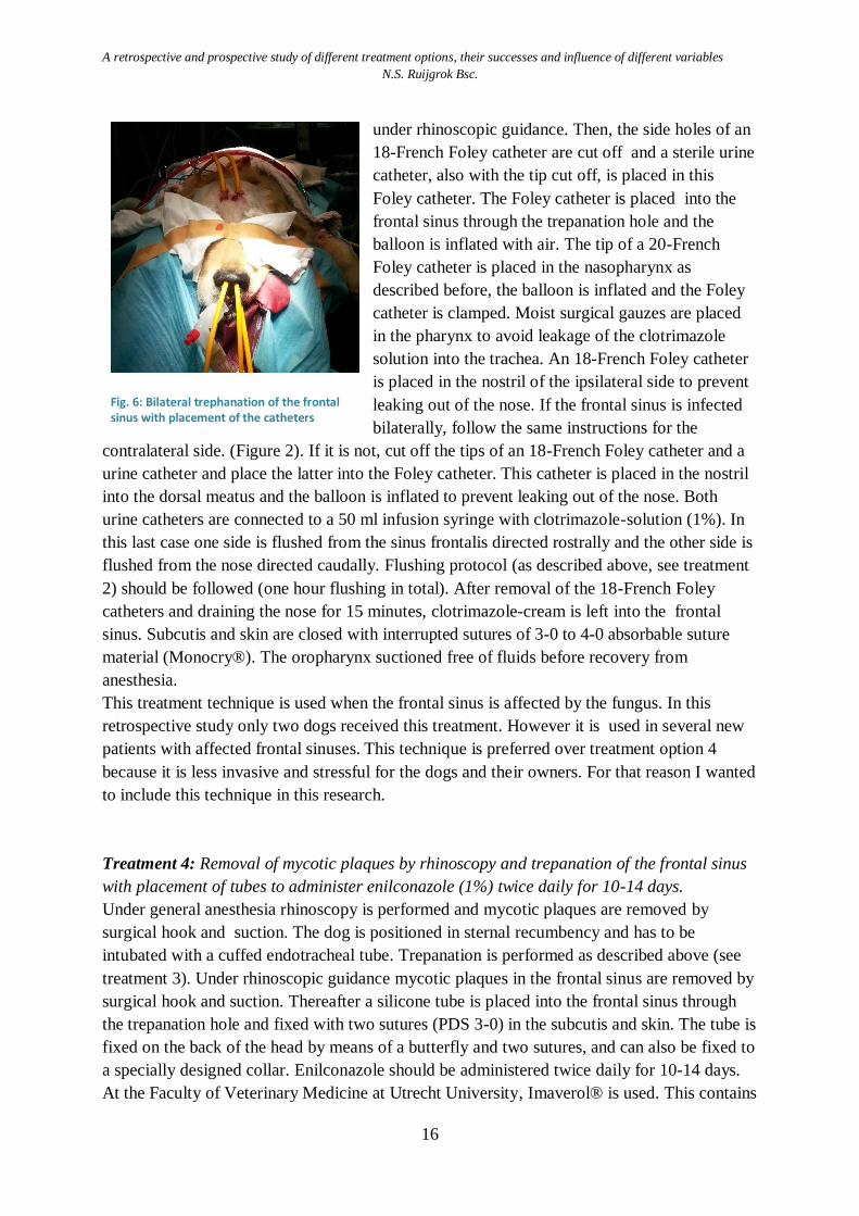
under rhinoscopic guidance. Then, the side holes of an
18-French Foley catheter are cut off and a sterile urine
catheter, also with the tip cut off, is placed in this
Foley catheter. The Foley catheter is placed into the
frontal sinus through the trepanation hole and the
balloon is inflated with air. The tip of a 20-French
Foley catheter is placed in the nasopharynx as
described before, the balloon is inflated and the Foley
catheter is clamped. Moist surgical gauzes are placed
in the pharynx to avoid leakage of the clotrimazole
solution into the trachea. An 18-French Foley catheter
is placed in the nostril of the ipsilateral side to prevent
leaking out of the nose. If the frontal sinus is infected
bilaterally, follow the same instructions for the
contralateral side. (Figure 2). If it is not, cut off the tips of an 18-French Foley catheter and a
urine catheter and place the latter into the Foley catheter. This catheter is placed in the nostril
into the dorsal meatus and the balloon is inflated to prevent leaking out of the nose. Both
urine catheters are connected to a 50 ml infusion syringe with clotrimazole-solution (1%). In
this last case one side is flushed from the sinus frontalis directed rostrally and the other side is
flushed from the nose directed caudally. Flushing protocol (as described above, see treatment
2) should be followed (one hour flushing in total). After removal of the 18-French Foley
catheters and draining the nose for 15 minutes, clotrimazole-cream is left into the frontal
sinus. Subcutis and skin are closed with interrupted sutures of 3-0 to 4-0 absorbable suture
material (Monocry®). The oropharynx suctioned free of fluids before recovery from
anesthesia.
This treatment technique is used when the frontal sinus is affected by the fungus. In this
retrospective study only two dogs received this treatment. However it is used in several new
patients with affected frontal sinuses. This technique is preferred over treatment option 4
because it is less invasive and stressful for the dogs and their owners. For that reason I wanted
to include this technique in this research.
Treatment 4: Removal of mycotic plaques by rhinoscopy and trepanation of the frontal sinus
with placement of tubes to administer enilconazole (1%) twice daily for 10-14 days.
Under general anesthesia rhinoscopy is performed and mycotic plaques are removed by
surgical hook and suction. The dog is positioned in sternal recumbency and has to be
intubated with a cuffed endotracheal tube. Trepanation is performed as described above (see
treatment 3). Under rhinoscopic guidance mycotic plaques in the frontal sinus are removed by
surgical hook and suction. Thereafter a silicone tube is placed into the frontal sinus through
the trepanation hole and fixed with two sutures (PDS 3-0) in the subcutis and skin. The tube is
fixed on the back of the head by means of a butterfly and two sutures, and can also be fixed to
a specially designed collar. Enilconazole should be administered twice daily for 10-14 days.
At the Faculty of Veterinary Medicine at Utrecht University, Imaverol® is used. This contains
Fig. 6: Bilateral trephanation of the frontal sinus with placement of the catheters
A retrospective and prospective study of different treatment options, their successes and influence of different variables
N.S. Ruijgrok Bsc.
17
100mg/ml enilconazole. Dilute 1 ml Imaverol® into 9 ml of warm sterile NaCl before
administering through the tubes. After treatment, the tubes are removed and subcutis and skin
are closed with interrupted sutures of 3-0 to 4-0 absorbable suture material (Monocry®).
This treatment technique is administered to patients with affected frontal sinus(es), severe
bone destruction or if the cribriform plate is not intact.
Whether tubes are placed unilaterally or bilaterally should be determined by CT evaluation.
In the past this treatment was frequently used. Nowadays, treatment option 3 is preferred over
this treatment technique because it is less invasive and stressful for the dogs and their owners.
However, this is only possible if the cribriform plate is intact and if there is no severe bone
destruction. If the cribriform plate is damaged or there is severe bone destruction whereby a
connection has formed between the frontal sinus and the brains, flushing under pressure
should not be performed. Trepanation of the frontal sinus and placement of tubes to
administer enilconazole twice daily for 10-14 days is the only treatment option.
Since the patients are conscious during daily treatment, some dogs do not cooperate when
administering enilconazole. Most dogs are admitted to the veterinary hospital during the
treatment phase. Nine dogs were treated with this treatment protocol.
Treatment 5: Removal of mycotic plaques by rhinoscopy and one hour flushing with
clotrimazole solution (1%) without trepanation, removal of mycotic plaques followed by local
application of clotrimazole cream (1%).
Treatment technique 5 is almost identical to treatment technique 2, the only difference is that
in technique 5 the one hour flush with clotrimazole solution (1%) is followed by local
administration of clotrimazole-cream (1%). See treatment 2 for clotrimazole flushing
protocol.
Three patients were treated with treatment option 5.
A retrospective and prospective study of different treatment options, their successes and influence of different variables
N.S. Ruijgrok Bsc.
18
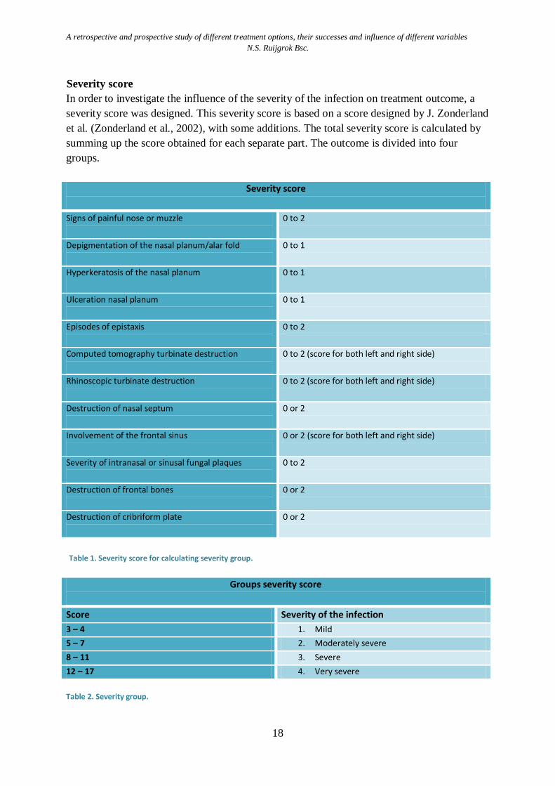
Severity score
In order to investigate the influence of the severity of the infection on treatment outcome, a
severity score was designed. This severity score is based on a score designed by J. Zonderland
et al. (Zonderland et al., 2002), with some additions. The total severity score is calculated by
summing up the score obtained for each separate part. The outcome is divided into four
groups.
Severity score
Signs of painful nose or muzzle
0 to 2
Depigmentation of the nasal planum/alar fold
0 to 1
Hyperkeratosis of the nasal planum
0 to 1
Ulceration nasal planum
0 to 1
Episodes of epistaxis
0 to 2
Computed tomography turbinate destruction
0 to 2 (score for both left and right side)
Rhinoscopic turbinate destruction
0 to 2 (score for both left and right side)
Destruction of nasal septum
0 or 2
Involvement of the frontal sinus
0 or 2 (score for both left and right side)
Severity of intranasal or sinusal fungal plaques
0 to 2
Destruction of frontal bones
0 or 2
Destruction of cribriform plate
0 or 2
Table 1. Severity score for calculating severity group.
Groups severity score
Score Severity of the infection
3 – 4 1. Mild
5 – 7 2. Moderately severe
8 – 11 3. Severe
12 – 17 4. Very severe
Table 2. Severity group.

A retrospective and prospective study of different treatment options, their successes and influence of different variables
N.S. Ruijgrok Bsc.
19
Results: Retrospective study
In the time period March 2005 to March 2013, 78 dogs were diagnosed with SNM at the
Faculty of Veterinary Science, Medicine of Companion Animals. Twenty-seven dogs were
excluded from the study because they did not match the inclusion criteria. The remaining 51
patients were included in the study and statistical analyses were performed. Affected breeds
are: the Golden retriever (n=5), German Shepherd Dogs (n=4), Belgian Shepherd Tervuren
(n=3), Flatcoated retrievers (n=3), Labrador retrievers (n=3), Jack Russel Terriers, , Border
Collies (n=2), Rottweilers (n=3), Bull Mastiffs (n=3), Bull terriers (n=2), Greater Swiss
Mountain dogs (n=2) and mixed breeds (n=7). Furthermore, one dog of each of the following
breeds: Dutch Shepherd Dog, Scottish Shepherd Dog, Small Münsterlander, German hunt
terrier, Airedale terrier, Rhodesian Ridgebacks, Dobermans, Spinone Italiano, Portuguese
water dog, Staffordshire Bullterrier, Afghan hound and a Dachshund. All dogs were
mesocephalic or doliocephalic breeds. Twenty-four dogs were intact males, 12 castrated
males, 7 intact females and 7 neutered females. Dogs referred at the University clinic with
clinical signs of SNM were aged 6 months to 13 years. The mean age was 6 years.
Treatments
In total 51 dogs were treated with one of the described treatment techniques. Twenty four
(47.1%) of these dogs had recurrence of SNM over the entire follow-up period after one
treatment. Treatment success was 52.9% after one treatment. After two treatments 11 of these
24 dogs had again recurrence of SNM. This is 21.6% (11 of 51) of all the treated patients in
total and 45.8% (11 of 24) of the patients which had recurrence of SNM after one treatment.
In conclusion, 78.4% of patients receiving two treatments were successful. Patients requiring
multiple treatments to cure often received treatment techniques different from the initial
protocol.
Treatment option 1
Five dogs were treated with treatment option 1, local abnormalities e.g. corpus alienum and
mycotic plaques were removed after which clotrimazole cream (10 mg/ml) was installed.
Recurrence after one treatment was seen in one (20%) of these dogs. Success after one
treatment was 80%.
Treatment option 2
A one hour long noninvasive flushing with clotrimazole (1%) without trepanation or
administration of local clotrimazole cream was performed in 32 dogs (treatment 2). Seventeen
(53.1%) of these dogs had recurrence; 40.6% within 12 weeks and 34.4% within 6 weeks.
Overall success rate of treatment 2 was 46.9%.
Of the 32 dogs initially treated with treatment 2, nine dogs with recurrence were retreated
with treatment 2. Three 33.3% (3 of 9) of these dogs again had recurrence after the second
treatment. After two treatments of treatment 2, 9.4% (3 of 32) of patients still had recurrence.
Treatment success after treated twice with treatment 2 is 90.6%.
A retrospective and prospective study of different treatment options, their successes and influence of different variables
N.S. Ruijgrok Bsc.
20
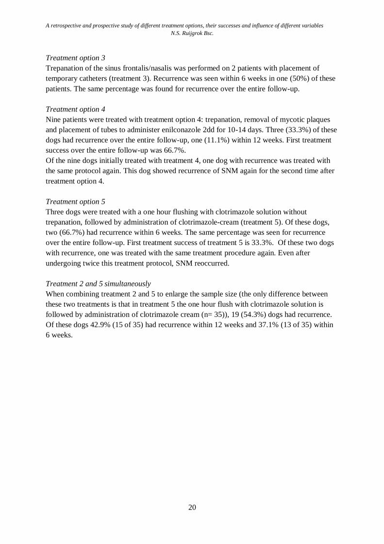
Treatment option 3
Trepanation of the sinus frontalis/nasalis was performed on 2 patients with placement of
temporary catheters (treatment 3). Recurrence was seen within 6 weeks in one (50%) of these
patients. The same percentage was found for recurrence over the entire follow-up.
Treatment option 4
Nine patients were treated with treatment option 4: trepanation, removal of mycotic plaques
and placement of tubes to administer enilconazole 2dd for 10-14 days. Three (33.3%) of these
dogs had recurrence over the entire follow-up, one (11.1%) within 12 weeks. First treatment
success over the entire follow-up was 66.7%.
Of the nine dogs initially treated with treatment 4, one dog with recurrence was treated with
the same protocol again. This dog showed recurrence of SNM again for the second time after
treatment option 4.
Treatment option 5
Three dogs were treated with a one hour flushing with clotrimazole solution without
trepanation, followed by administration of clotrimazole-cream (treatment 5). Of these dogs,
two (66.7%) had recurrence within 6 weeks. The same percentage was seen for recurrence
over the entire follow-up. First treatment success of treatment 5 is 33.3%. Of these two dogs
with recurrence, one was treated with the same treatment procedure again. Even after
undergoing twice this treatment protocol, SNM reoccurred.
Treatment 2 and 5 simultaneously
When combining treatment 2 and 5 to enlarge the sample size (the only difference between
these two treatments is that in treatment 5 the one hour flush with clotrimazole solution is
followed by administration of clotrimazole cream (n= 35)), 19 (54.3%) dogs had recurrence.
Of these dogs 42.9% (15 of 35) had recurrence within 12 weeks and 37.1% (13 of 35) within
6 weeks.
A retrospective and prospective study of different treatment options, their successes and influence of different variables
N.S. Ruijgrok Bsc.
21
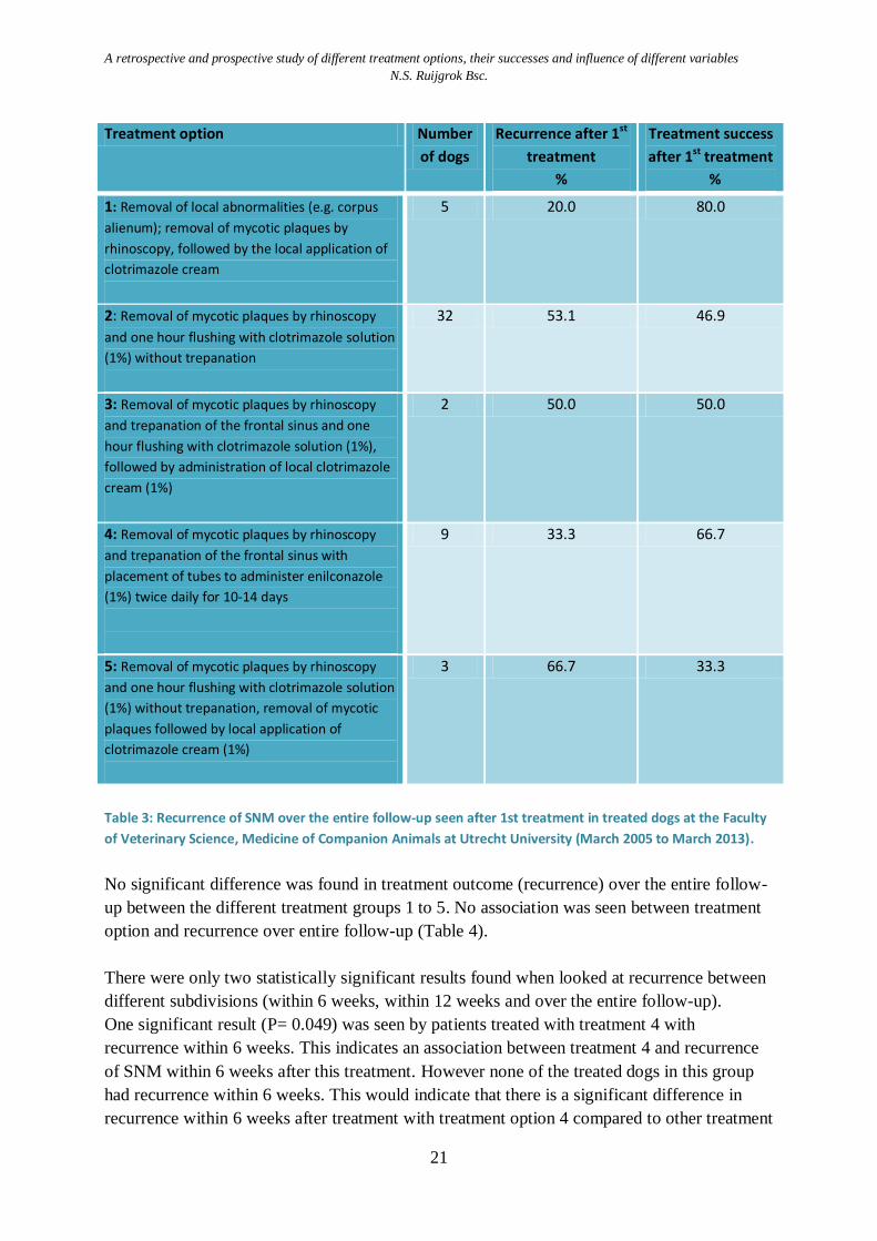
Treatment option Number
of dogs
Recurrence after 1st
treatment
%
Treatment success
after 1st treatment
%
1: Removal of local abnormalities (e.g. corpus
alienum); removal of mycotic plaques by
rhinoscopy, followed by the local application of
clotrimazole cream
5 20.0 80.0
2: Removal of mycotic plaques by rhinoscopy
and one hour flushing with clotrimazole solution
(1%) without trepanation
32 53.1 46.9
3: Removal of mycotic plaques by rhinoscopy
and trepanation of the frontal sinus and one
hour flushing with clotrimazole solution (1%),
followed by administration of local clotrimazole
cream (1%)
2 50.0 50.0
4: Removal of mycotic plaques by rhinoscopy
and trepanation of the frontal sinus with
placement of tubes to administer enilconazole
(1%) twice daily for 10-14 days
9 33.3 66.7
5: Removal of mycotic plaques by rhinoscopy
and one hour flushing with clotrimazole solution
(1%) without trepanation, removal of mycotic
plaques followed by local application of
clotrimazole cream (1%)
3 66.7 33.3
Table 3: Recurrence of SNM over the entire follow-up seen after 1st treatment in treated dogs at the Faculty
of Veterinary Science, Medicine of Companion Animals at Utrecht University (March 2005 to March 2013).
No significant difference was found in treatment outcome (recurrence) over the entire follow-
up between the different treatment groups 1 to 5. No association was seen between treatment
option and recurrence over entire follow-up (Table 4).
There were only two statistically significant results found when looked at recurrence between
different subdivisions (within 6 weeks, within 12 weeks and over the entire follow-up).
One significant result (P= 0.049) was seen by patients treated with treatment 4 with
recurrence within 6 weeks. This indicates an association between treatment 4 and recurrence
of SNM within 6 weeks after this treatment. However none of the treated dogs in this group
had recurrence within 6 weeks. This would indicate that there is a significant difference in
recurrence within 6 weeks after treatment with treatment option 4 compared to other treatment
A retrospective and prospective study of different treatment options, their successes and influence of different variables
N.S. Ruijgrok Bsc.
22
options. This indicates that there is 0% chance of recurrence within 6 weeks after treatment
option 4.
The other significant result was seen by recurrence within 6 weeks, when treatment groups 2
and 5 were joined together (the only difference between these two treatments is that in
treatment 5 the one hour flush with clotrimazole solution is followed by administration of
clotrimazole cream) (p= 0.039). This indicates that there is an association between recurrence
of SNM within 6 weeks after treatment with treatment option 2 or 5.
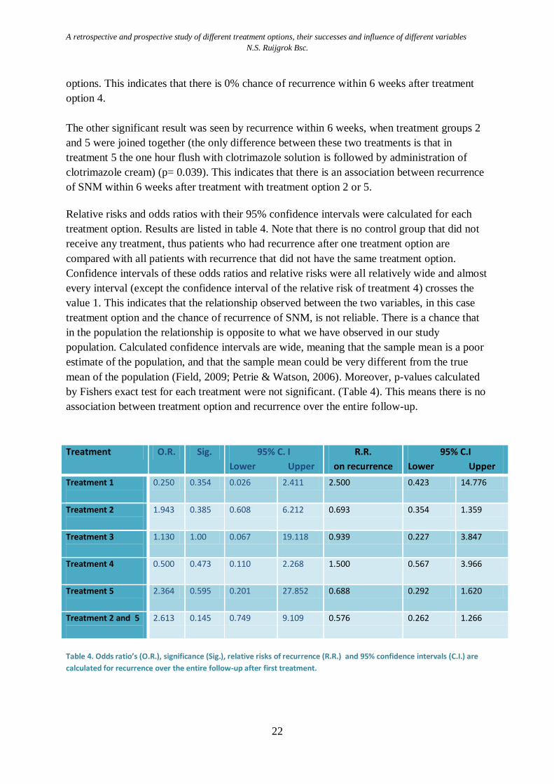
Relative risks and odds ratios with their 95% confidence intervals were calculated for each
treatment option. Results are listed in table 4. Note that there is no control group that did not
receive any treatment, thus patients who had recurrence after one treatment option are
compared with all patients with recurrence that did not have the same treatment option.
Confidence intervals of these odds ratios and relative risks were all relatively wide and almost
every interval (except the confidence interval of the relative risk of treatment 4) crosses the
value 1. This indicates that the relationship observed between the two variables, in this case
treatment option and the chance of recurrence of SNM, is not reliable. There is a chance that
in the population the relationship is opposite to what we have observed in our study
population. Calculated confidence intervals are wide, meaning that the sample mean is a poor
estimate of the population, and that the sample mean could be very different from the true
mean of the population (Field, 2009; Petrie & Watson, 2006). Moreover, p-values calculated
by Fishers exact test for each treatment were not significant. (Table 4). This means there is no
association between treatment option and recurrence over the entire follow-up.
Treatment O.R. Sig. 95% C. I
Lower Upper
R.R.
on recurrence
95% C.I
Lower Upper
Treatment 1
0.250 0.354 0.026 2.411 2.500 0.423 14.776
Treatment 2
1.943 0.385 0.608 6.212 0.693 0.354 1.359
Treatment 3
1.130 1.00 0.067 19.118 0.939 0.227 3.847
Treatment 4
0.500 0.473 0.110 2.268 1.500 0.567 3.966
Treatment 5
2.364 0.595 0.201 27.852 0.688 0.292 1.620
Treatment 2 and 5
2.613 0.145 0.749 9.109 0.576 0.262 1.266
Table 4. Odds ratio’s (O.R.), significance (Sig.), relative risks of recurrence (R.R.) and 95% confidence intervals (C.I.) are
calculated for recurrence over the entire follow-up after first treatment.
A retrospective and prospective study of different treatment options, their successes and influence of different variables
N.S. Ruijgrok Bsc.
23
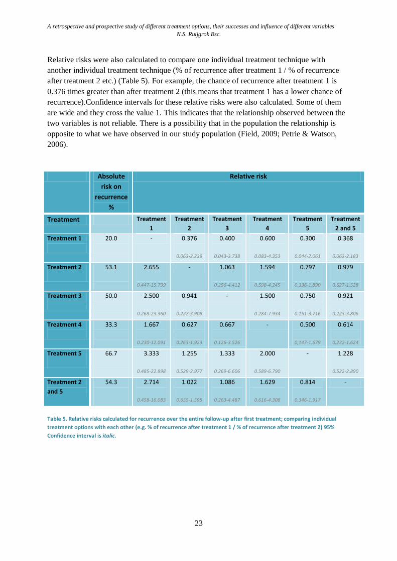
Relative risks were also calculated to compare one individual treatment technique with
another individual treatment technique (% of recurrence after treatment 1 / % of recurrence
after treatment 2 etc.) (Table 5). For example, the chance of recurrence after treatment 1 is
0.376 times greater than after treatment 2 (this means that treatment 1 has a lower chance of
recurrence).Confidence intervals for these relative risks were also calculated. Some of them
are wide and they cross the value 1. This indicates that the relationship observed between the
two variables is not reliable. There is a possibility that in the population the relationship is
opposite to what we have observed in our study population (Field, 2009; Petrie & Watson,
2006).
Absolute
risk on
recurrence
%
Relative risk
Treatment Treatment
1
Treatment
2
Treatment
3
Treatment
4
Treatment
5
Treatment
2 and 5
Treatment 1
20.0
- 0.376
0.063-2.239
0.400
0.043-3.738
0.600
0.083-4.353
0.300
0.044-2.061
0.368
0.062-2.183
Treatment 2
53.1
2.655
0.447-15.799
- 1.063
0.256-4.412
1.594
0.598-4.245
0.797
0.336-1.890
0.979
0.627-1.528
Treatment 3
50.0
2.500
0.268-23.360
0.941
0.227-3.908
- 1.500
0.284-7.934
0.750
0.151-3.716
0.921
0.223-3.806
Treatment 4
33.3
1.667
0.230-12.091
0.627
0.263-1.923
0.667
0.126-3.526
- 0.500
0,147-1.679
0.614
0.232-1.624
Treatment 5
66.7
3.333
0.485-22.898
1.255
0.529-2.977
1.333
0.269-6.606
2.000
0.589-6.790
- 1.228
0.522-2.890
Treatment 2
and 5
54.3
2.714
0.458-16.083
1.022
0.655-1.595
1.086
0.263-4.487
1.629
0.616-4.308
0.814
0.346-1.917
-
Table 5. Relative risks calculated for recurrence over the entire follow-up after first treatment; comparing individual
treatment options with each other (e.g. % of recurrence after treatment 1 / % of recurrence after treatment 2) 95%
Confidence interval is italic.
A retrospective and prospective study of different treatment options, their successes and influence of different variables
N.S. Ruijgrok Bsc.
24
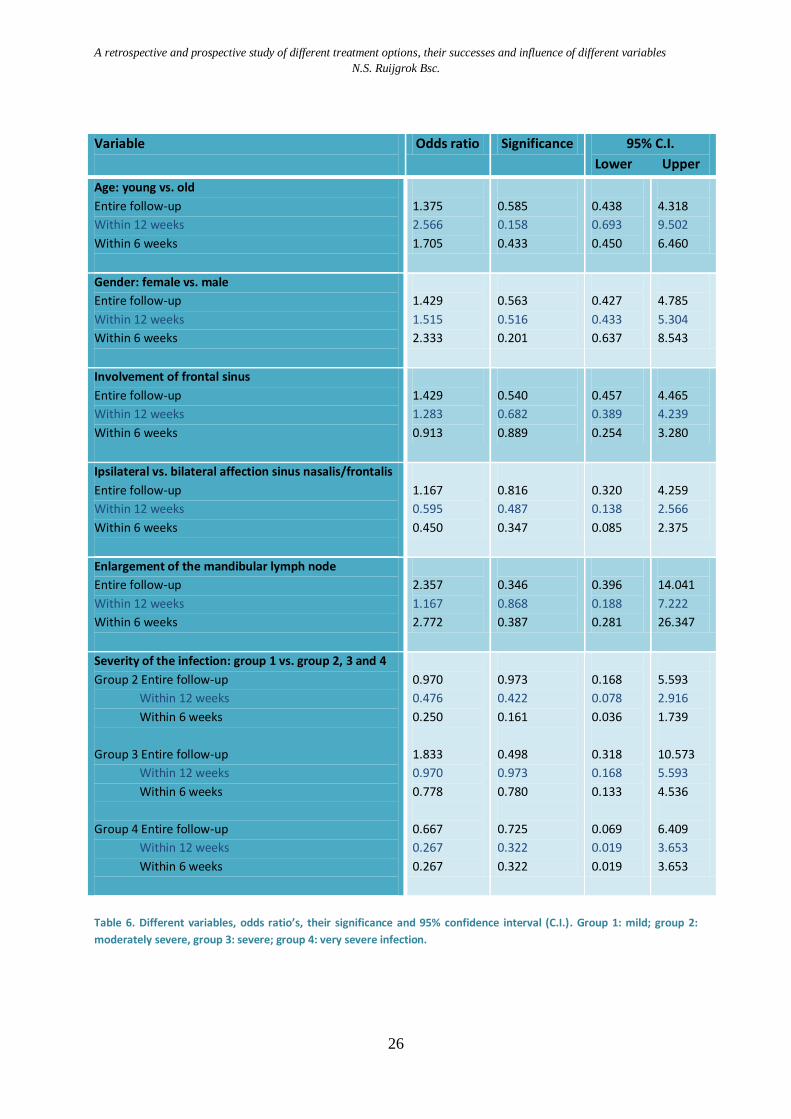
Influence of different variables on recurrence of sinonasal mycosis
The Chi-squared test or the Fischer exact test calculate an association between different
variables and recurrence after treatment. None of the calculated p values were smaller than
0.05. This means that treatment failure was not associated with age (0-5 years versus > 5
years; p=0.585), gender (p=0.759, affection of the frontal sinus (p=0.575), ipsilateral versus
bilateral affection (p=1.00), severity of the infection (p=0.707) or enlargement of the
ipsilateral mandibular lymph node (p=0.431). Odds ratios were calculated for each variable to
determine an association between the variable and recurrence of SNM. None of these were
significant, meaning that none of the variables were associated with recurrence over the entire
follow-up. All 95% confidence intervals calculated cross the value 1, meaning that they are
not accurate estimates. See table 6. Results of each variable will be discussed below.
Age
Fifty percent (16 of 32) of the dogs older than > 5 years of age had recurrence over the entire
follow-up compared to 42.1% (8 of 19) of the dogs 0-5 years of age. Within 12 weeks after
treatment recurrence was seen in 40.6% (13 of 32) of the animals > 5 years of age and in
21.1% (4 of 19) of the animals < 5 years of age. Within 6 weeks this was respectively 21.1%
(4 of 19) in young and 31.3% (10 of 32) in older dogs. Odds ratios were all greater than one,
but none of them were significant. See table 6.
Gender
The recurrence rate was higher in female dogs than in male dogs (53.3% versus 44.4% after
treatment over the entire follow-up). This was also the case when calculating the percentages
within the different subdivisions (male 12 weeks: 30.6% (11 of 36); male 6 weeks: 22.2% (8
of 36); female 12 weeks: 40.0% (6 of 15); females within 6 weeks: 40% (6 of 15)). The
calculated odds ratios indicate that female dogs are 1.4 times more likely to have recurrence
after first treatment than male dogs. However this difference is not significant (p= 0.563).
Involvement of frontal sinus
In total the frontal sinus was affected in 37.3% (19 of 51) of cases. Ten of these dogs (52.6%)
had recurrence over the entire follow-up. Forty-three point eight percent43.8% (14 of 32) of
the dogs without diseased frontal sinuses had recurrence. Recurrence percentages seen within
6 weeks are 26.3% (5 of 19) with an affected frontal sinus versus 28.1% (9 of 32) with only
an affected sinus nasalis. Odds ratios calculated for recurrence over the entire follow-up and
within 12 weeks were greater than one, but not significant. (Table 6).
Ipsilateral versus bilateral affection of the sinus nasalis/frontalis
Over the entire follow-up period, 50% (6 of 12) of the dogs that had bilaterally affected
nasal/frontal sinusses had recurrence while 46.2% (18 of 39) of the dogs that only had an
ipsilaterally affected sinus nasal/frontal sinusses had recurrence.
However, only 16.7% (2 of 12) of dogs with a bilaterally affected nasal/frontal sinusses
showed recurrence within 6 weeks compared to 30.8% (12 of 39) of the dogs with an
A retrospective and prospective study of different treatment options, their successes and influence of different variables
N.S. Ruijgrok Bsc.
25
ipsilaterally affected sinus nasalis/frontalis. No significant odds ratios for recurrence of SNM
was found for ipsilateral versus unilateral affection of the sinus nasalis/frontalis. (Table 6).
There was no statistical difference between the odds ratios for the sinus nasalis and the sinus
frontalis.
Enlargement of the mandibular lymph node
Lymph node size was not always included in patient files. Mandibular lymph node
enlargement ipsilateral to the diseased nasal/frontal sinus was seen in 75.8% (25 of 33) of the
dogs with SNM. In 44% of these cases with enlarged mandibular lymph nodes recurrence was
seen over the entire follow-up. When lymph node was not enlarged 25% (2 of 8) had
recurrence over the entire follow-up. Twenty-eight percent (6 of 25) of the patients with
enlarged mandibular lymph nodes had recurrence within 6 weeks versus 12.5% in animals
with no enlarged mandibular lymph nodes. Calculated odds ratios were all greater than one
but none of them were significant. (Table 6).
It was hypothesized that the size of the mandibular lymph nodes could possibly predict and
affect the course of the disease. However, this assumption is not supported as results were not
significant (p= 0.431) and the exact size of each mandibular lymph node was not measured.
Severity of the infection
Using the severity score (see table 1 and 2), 11.8% (6 of 51) of the total number of dogs
treated had a very severe infection, 37.3% (19 of 51) had a severe infection; 37.3 % (19 of 51)
had a moderate infection and 13.7% (7 of 51) had an mild infection.
Of the group with very severe infection 33.3% had recurrence over the entire follow-up and
16.7% (1 of 6) had recurrence within 12 and 6 weeks.
In the group with severe infection 57.9% (11 of 19) had recurrence over the entire follow-up
and 42.1 (8 of 19) and 36.8% (7 of 19) had recurrence within 12 and 6 weeks respectively.
The group with moderate infection had recurrence over the entire follow-up in 42.1% (8 of
19) of cases. 26.3% (5 of 19) had recurrence within 12 weeks and 15.8% (3 of 19) within 6
weeks.
Because lack of a control group, the severity group 1 (mild infection), was used as a control
group to calculate the odds ratios with. None of the odds ratios were significant. See table 6.
Depigmentation of the nasal planum was seen in 18 dogs (35.3%), hyperkeratosis was seen in
6 dogs (11.8%) and ulceration of the nasal planum was observed in 8 of the total 51 dogs
(15.7%). Eleven dogs (21.7%) had local destruction of the nasal septum and on CT 13 dogs
(25.4%) showed bone degeneration.
A retrospective and prospective study of different treatment options, their successes and influence of different variables
N.S. Ruijgrok Bsc.
26
Variable
Odds ratio Significance 95% C.I.
Lower Upper
Age: young vs. old
Entire follow-up
Within 12 weeks
Within 6 weeks
1.375
2.566
1.705
0.585
0.158
0.433
0.438
0.693
0.450
4.318
9.502
6.460
Gender: female vs. male
Entire follow-up
Within 12 weeks
Within 6 weeks
1.429
1.515
2.333
0.563
0.516
0.201
0.427
0.433
0.637
4.785
5.304
8.543
Involvement of frontal sinus
Entire follow-up
Within 12 weeks
Within 6 weeks
1.429
1.283
0.913
0.540
0.682
0.889
0.457
0.389
0.254
4.465
4.239
3.280
Ipsilateral vs. bilateral affection sinus nasalis/frontalis
Entire follow-up
Within 12 weeks
Within 6 weeks
1.167
0.595
0.450
0.816
0.487
0.347
0.320
0.138
0.085
4.259
2.566
2.375
Enlargement of the mandibular lymph node
Entire follow-up
Within 12 weeks
Within 6 weeks
2.357
1.167
2.772
0.346
0.868
0.387
0.396
0.188
0.281
14.041
7.222
26.347
Severity of the infection: group 1 vs. group 2, 3 and 4
Group 2 Entire follow-up
Within 12 weeks
Within 6 weeks
Group 3 Entire follow-up
Within 12 weeks
Within 6 weeks
Group 4 Entire follow-up
Within 12 weeks
Within 6 weeks
0.970
0.476
0.250
1.833
0.970
0.778
0.667
0.267
0.267
0.973
0.422
0.161
0.498
0.973
0.780
0.725
0.322
0.322
0.168
0.078
0.036
0.318
0.168
0.133
0.069
0.019
0.019
5.593
2.916
1.739
10.573
5.593
4.536
6.409
3.653
3.653
Table 6. Different variables, odds ratio’s, their significance and 95% confidence interval (C.I.). Group 1: mild; group 2:
moderately severe, group 3: severe; group 4: very severe infection.
A retrospective and prospective study of different treatment options, their successes and influence of different variables
N.S. Ruijgrok Bsc.
27
Research goals: Prospective case control study
Another part of this study is a prospective case control study of new patients with SNM. The
goal is to define risk factors for the development of SNM in dogs and to define if there are
specific abnormalities in these dogs that can be observed by performing blood analyses.
Blood analysis on each patient sample was performed: hematocrit, leucocyte differentiation,
albumin, total protein and protein spectrum (only measured if total protein was not within
reference values). If the patient has been abroad (particularly in the south of Europe) and is
suspected of Leishmania or other foreign diseases, blood analysis was performed to rule this
out.
New patients participating in the study were used to investigate possible abnormalities of
blood values described above, which can indicate an underlying disease and determine if there
are specific blood values that are abnormal in patients with SNM. Extra blood for DNA
analysis was taken from the patients (with informed consent) and stored for later research to
investigate similarities between them. Specific similar DNA sequences in these patients may
explain their sensitivity to Aspergillus spp.
In cooperation with Microbiology, Department of Biology, Faculty of Science, Utrecht
University we did not only focus on the characteristics of the patients, but also on the
characteristics of the fungus itself. Of the new patients with SNM that are examined and
treated at the Faculty of Veterinary Science in Utrecht, the type of fungus that causes the
infection is determined, its DNA is isolated and typed and its resistance profile and acidity is
determined. Histologic evaluation of biopsies of the affected tissue is required to determine
how the fungus is growing on the tissue and how deep it is capable of penetrating the tissue.
These results will allow us to get a better idea of the pathogen and how to treat it.
Inclusion criteria of the study are the same as described in the retrospective part. It was
decided that there should be a minimum of six patients cooperating in this part of the research.
However, within the timeframe available for my research project, less than six patients with
proven diagnosis of SNM contributed to the study and not all data were available yet. Not all
fungal DNA was typed and the complete resistance profile was not determined. Therefore,
this study will mainly focus on the retrospective aspect of it.
A retrospective and prospective study of different treatment options, their successes and influence of different variables
N.S. Ruijgrok Bsc.
28
Material and methods: Prospective case control study
In the time period of April 25, 2013 to September 1, 2013 new patients suspected of SNM
were followed. Clinical examination, CT and rhinoscopy was performed of all new patients
suspected of SNM at the Faculty of Veterinary Science, Medicine of Companion Animals at
Utrecht University. Dogs were under anesthesia during CT, rhinoscopy and treatment.
Anesthetics used depended on the anesthetist and American Society of Anesthesiologists
(ASA) scale of physical status of the dog.
All patients were scheduled for control rhinoscopy six weeks after treatment. Every two
weeks the owners were contacted for follow-up.
Computed tomography:
CT images were evaluated by the Radiology Department of Clinical Science of Companion
Animals, Faculty of Veterinary Medicine Utrecht University.
Rhinoscopy:
During the rhinoscopy, mycotic plaques and biopsies from affected tissue were collected.
- Pieces of fungus for culturing and species typing. (The samples were sent to
Microbiology, Department of Biology, Faculty of Science, Utrecht University. This
department cultures the fungus and sends samples to Centraalbureau voor
Schimmelculturen (CBS) at Utrecht for typing of DNA of the fungus.).
- Pieces of fungus were transferred to a sterile tube and frozen in liquid nitrogen, these
tubes were stored at -70⁰C and saved for later DNA isolation. (The samples will be
sent to Microbiology, Department of Biology, Faculty of Science, Utrecht University
for RNA isolation).
- 1-2 biopsies from affected tissue were sent to the Veterinary Pathology Diagnostic
Centre in Utrecht for histologic evaluation to determine fungal invasion.
After removal of the fungal tissue, an anti-mycotic treatment was performed.
pH measurement:
The nasal pH at the place where the fungus grows was determined. The goal is to examine if
the fungus adapts to the hosts environment and how it migrates to the frontal sinus and
whether or not the pH is more favorable for fungal growth. pH- measurement was performed
directly (within 30 minutes) after the collection of the fungal plaques by Microbiology,
Department of Biology by the means of pH paper.
Blood analyses:
Blood samples were taken while the patient was under general anesthesia and samples were
analyzed by the University Veterinary Diagnostic Laboratory (UVDL). Blood parameters that
were analyzed are the: hematocrit, leucocyte differentiation, albumin, total protein. Protein
spectrum was only analyzed if values of total protein were high. With informed consent of the
client (dog owner) extra blood (4cc EDTA) was taken for storage and later DNA research.
A retrospective and prospective study of different treatment options, their successes and influence of different variables
N.S. Ruijgrok Bsc.
29
Culturing:
- Pieces of fungus were collected and transferred directly to Microbiology.
- Small pieces of the plaque were removed by cutting and then placed onto a PDA plate
and cultured at 37⁰C for two days. By this time sufficiently large colonies had formed
that started to sporulate. From those plates sub culturing was performed until a single
type of the fungus was defined.
- Isolates were evaluated by light microscopy, especially spore head structure to see if it
resembles with Aspergillus fumigatus.
- Of each isolate a culture was prepared to isolate spores that were stored at -80⁰C (at
Microbiology, Utrecht University).
Resistance profile:
- Of all isolates, a resistance profile will be determined with a relative set of antifungals
(Clotrimazol, Itraconazole, Fluconazole, Voriconazole, Amphotericin and
Flucytosine).
The resistance profile will be estimated by two tests: the broth dilution method and the
epsilometer test (E-test method). (The E-test utilizes a rectangular strip that has been
impregnated with the drug to be studied. A fungus is spread and grown on an plate,
and the E-test strip is laid on top; the drug diffuses, producing an exponential gradient
of the drug to be tested.) However, at the time of writing only an E-test with
Itroconazol was performed.
DNA isolation and typing:
DNA of the collected fungal strains were isolated and typed at CBS (Centraalbureau voor
Schimmelculturen) by sequencing relevant genes (Balajee et al., 2007).
DNA was isolated of each isolate, tagged with a barcode to identify the isolate (see appendix
1) and sequenced via the next generation sequencing methods.
- DNA isolation is performed by the TRIzol method.
Ultra Clean® Microbial DNA Isolation Kit of Mo Bio Laboratories, Inc. was used to
isolate DNA.
The genome of A. fumigatus 293 (clinical isolate from humans) is used to identify
expressed genes. Control for expression profiling is AF293 grown in vitro according to
protocol used by Eric Bathoorn (who also performed a gene expression profiling,
Manuscript in preparation).
- Different primer pairs were used to sequence DNA fragments, see appendix 2.

A retrospective and prospective study of different treatment options, their successes and influence of different variables
N.S. Ruijgrok Bsc.
30
Results: Prospective case control study
In the time period of April 25, 2013 to September 1, 2013, nine patients suspected of
sinonasal mycosis were presented at the Faculty of Veterinary Science, Medicine of
Companion Animals at Utrecht University. These patients presented with clinical signs that
are normally seen in cases of SNM (see introduction). However, other differential diagnoses
or primary causes (e.g. nasal tumor, lymphoplasmacytic rhinitis, tooth root inflammation,
corpus alienum and nasal fistula) were not yet excluded.
Clinical examination, CT and rhinoscopy were performed on all new patients suspected of
SNM.
Ultimately four dogs were diagnosed with SNM. Breed, gender, age and severity of the
infection are listed in Table 7 and 8.
With informed consent blood was collected for DNA research.
Every two weeks owners were contacted for follow-up.
Patient 4, had a previous history of sinonasal Aspergillosis. Its first diagnosis of sinonasal
mycosis was established in 2011 and the right sinus nasalis was affected. In July 2013 the left
side of the frontal sinus was affected. No clinical symptoms matching SNM were seen in the
interim period.
Patient 2 showed recurrence of nasal discharge within two weeks after treatment, because of
severe clinical deterioration probably caused by recurrence of SNM or a systemic form of
Aspergillosis. The owners decided to euthanize the dog.
Treatment and recurrence of SNM
As is listened in table 9, one patient was treated with treatment option 5 (patient 1) and the
other three patients were treated with treatment option 3.
Recurrence of clinical signs of SNM were seen in two of total dogs (50%) (patient 3 and 4).
However, recurrence of SNM in patient 2 was not confirmed after he was euthanized.
Patient Breed Gender
Age
(years)
Severity group
Patient 1
Labrador Retriever Male 2 3: severe (9 points)
Patient 2
Mixed breed Male 6 4: very severe (12 points)
Patient 3
Alaska Malamute Male 4 4: very severe (12 points)
Patient 4
Golden Retriever Neutered female 4 3: very severe (11 points)
Table 7. Data of new patients diagnosed with SNM at the Faculty of Veterinary Medicine, Utrecht University (time period
April 25, 2013 to September 1, 2013).
A retrospective and prospective study of different treatment options, their successes and influence of different variables
N.S. Ruijgrok Bsc.
31
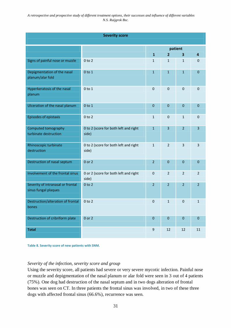
Severity score
patient
1 2 3 4
Signs of painful nose or muzzle
0 to 2 1 1 1 0
Depigmentation of the nasal
planum/alar fold
0 to 1 1 1 1 0
Hyperkeratosis of the nasal
planum
0 to 1 0 0 0 0
Ulceration of the nasal planum
0 to 1 0 0 0 0
Episodes of epistaxis
0 to 2 1 0 1 0
Computed tomography
turbinate destruction
0 to 2 (score for both left and right
side)
1 3 2 3
Rhinoscopic turbinate
destruction
0 to 2 (score for both left and right
side)
1 2 3 3
Destruction of nasal septum
0 or 2 2 0 0 0
Involvement of the frontal sinus
0 or 2 (score for both left and right
side)
0 2 2 2
Severity of intranasal or frontal
sinus fungal plaques
0 to 2 2 2 2 2
Destruction/alteration of frontal
bones
0 to 2 0 1 0 1
Destruction of cribriform plate
0 or 2 0 0 0 0
Total
9 12 12 11
Table 8. Severity score of new patients with SNM.
Severity of the infection, severity score and group
Using the severity score, all patients had severe or very severe mycotic infection. Painful nose
or muzzle and depigmentation of the nasal planum or alar fold were seen in 3 out of 4 patients
(75%). One dog had destruction of the nasal septum and in two dogs alteration of frontal
bones was seen on CT. In three patients the frontal sinus was involved, in two of these three
dogs with affected frontal sinus (66.6%), recurrence was seen.
A retrospective and prospective study of different treatment options, their successes and influence of different variables
N.S. Ruijgrok Bsc.
32
Patient
1st Treatment 2nd Treatment 3rd Treatment
1
Treatment 5: Removal of
mycotic plaques by rhinoscopy
and one hour flushing with
clotrimazole solution (1%)
without trepanation, removal of
mycotic plaques followed by
local application of clotrimazole
cream (1%)
No 2nd
Treatment No 3rd
Treatment
2
Treatment 3: Removal of
mycotic plaques by rhinoscopy
and trepanation of the frontal
sinus and one hour flushing with
clotrimazole solution (1%),
followed by administration of
local clotrimazole cream (1%)
No 2nd
Treatment
†
No 3rd
Treatment
†
3
Treatment 3: Removal of
mycotic plaques by rhinoscopy
and trepanation of the frontal
sinus and one hour flushing with
clotrimazole solution (1%),
followed by administration of
local clotrimazole cream (1%)
Treatment 1: Removal of
mycotic plaques by rhinoscopy,
followed by the local
application of clotrimazole
cream (1%)
Treatment 3: Removal of
mycotic plaques by rhinoscopy
and trepanation of the frontal
sinus and one hour flushing
with clotrimazole solution (1%),
followed by administration of
local clotrimazole cream (1%)
4
Treatment 3: Removal of
mycotic plaques by rhinoscopy
and trepanation of the frontal
sinus and one hour flushing with
clotrimazole solution (1%),
followed by administration of
local clotrimazole cream (1%)
No 2nd Treatment No 3rd Treatment
Table 9. Treatment option of every new dog with SNM.
† dog was euthanized
Mandibular lymph node
In three patients (patients 1, 3 and 4) the mandibular lymph node was enlarged on the affected
side. Patient 3 had enlarged mandibular lymph nodes on the right and left side. In this dog
both the right and left nasal sinus and the left frontal sinus were affected.
A retrospective and prospective study of different treatment options, their successes and influence of different variables
N.S. Ruijgrok Bsc.
33
Foreign diseases
None of the four patients had been to the south of Europe. Two had been in Germany and the
others had never been abroad. None of these patients were suspected of Leishmania or other
foreign diseases.
Histology
Biopsies were sent to the Veterinary Pathology Diagnostic Centre (VPDC) in Utrecht for
histologic evaluation. In the biopsies of three patients, the fungus grows only superficially on
the mucosa, the lamina propria was not infiltrated. No vital tissue was found in biopsies of
patient 2 and for that reason infiltration of the fungus could not be examined. In all patients
biopsies were classified as lymphoplasmacellulair rhinitis or sinusitis, some with neutrophil
infiltration.
Blood analyses
Blood parameters analyzed were: hematocrit, leucocyte differentiation, albumin, total protein
and protein spectrum. The values of each patient are displayed in table 10. There was no clear
similarity between blood values of the patients. The only similarity seen was that albumin was
low in three of four patients. Values found were slightly lower than references values. Patients
did not show any signs of hepatic disease, renal losses or immune mediated diseases that can
explain hypoalbuminemia. Patient 2 showed high rod like neutrophils and low segmented
neutrophils. This is seen as a left shift of the blood cells, which can possibly be explained by
severe acute infection by the fungus. Patient 1 had high segmented neutrophils, that are
normally seen during infection. Patient 4 had a slightly higher value of basophils which may
indicate an infection and possibly an allergic component. There was no evidence for
immunologic disease in any of these dogs.
pH of the mycotic plaques
After removal of fungus from the nose or frontal sinus, the pH of the fungal plaque was
determined by pH-paper. All pH measurements were between 7.9 and 8.2 (patient 1: 7.9 – 8.1,
patient 2: 8.0 – 8.4 and patient 3: 8.0 – 8.2, the pH of patient 4 is unknown).
DNA isolation and typing of the fungus
Isolation and typing of fungal DNA was performed on fungus collected from patients 1, 2 and
3. Although there were some small differences in nucleotide sequence, all isolates were
identified as Aspergillus fumigatus. Isolation and typing of fungal DNA collected from patient
4 will be examined at CBS when more samples are collected.

A retrospective and prospective study of different treatment options, their successes and influence of different variables
N.S. Ruijgrok Bsc.
34
Blood analyses
Patient Ht L/L
Leucocytes Alb. g/L
T. P. g/L
Lymph. 10^9/L
Mono.
10^9/L Blasts 10^9/L
Rodl. 10^9/L
Seg. 10^9/L
Eo. 10^9/L
Baso. 10^9/L
Norm. 10^9/L
1
0.42 2.2 0.8 0 0 12.1 0.9 0 0 21 57
2
0.47 0.3 0.1 0 1.0 1.4 0.2 0 0 25 59
3
0.46 1.4 0.6 0 0 7.8 1.4 0 0 22 68
4
0.48 1.2 0.3 0 0 6.3 0.8 0.1 0 29 61
Table 10. Blood values analyzed and their references: Hematocrit(Ht): 0.42-0.61 L/L, Lymphocytes (Lymph.): 0.8-4.7
x10^9/L, Monocytes (Mono.): 0.0-0.9 x10^9/L, Blasts: < 0.0 x10^9/L, Rod like (Rodl.): 0.0-0.3 x10^9/L, Segmented (Seg.):
2.9-11.0 x10^9/L, Eosinophils (Eo.): 0.0-1.6 x10^9/L, Basophils (Baso.): 0.0-0.1 x10^9/L, Albumine (Alb): 26-37 g/L, Total
Protein (T.P.): 55-72 g/L, Protein Spectrum (P.S.): red = value above reference value, blue = value below reference value.
Resistance profile
Investigation of the resistance profile of the fungus was not yet completed. Resistance to the
antifungal Itraconazol was determined by the E-test method on samples collected from
patients 1, 2 and 3. However, MIC values are unknown at the time of writing. More tests with
different concentrations should be performed in order to determine the resistance profile.
A retrospective and prospective study of different treatment options, their successes and influence of different variables
N.S. Ruijgrok Bsc.
35
Discussion
All patients with SNM reviewed in this study showed clinical signs as described in the
literature and had mesocephalic or doliocephalic skulls (Nelson et al., 2009; Peeters & Clercx,
2007; Quinn et al., 2002; M. J. Sharman & Mansfield, 2012; N. Sharp et al., 1992).
Lack of treatment success is likely to be multifactorial. It is determined by severity of the
infection, the treatment option, failure to completely debride of fungal plaques, distribution of
antifungal agent and the experience of the treating veterinarian (Friend, Williams, & White,
2002; Mathews et al., 1998; Schuller & Clercx, 2007; M. Sharman et al., 2010; Zonderland et
al., 2002).
The goal of this retrospective study is to determine which topical treatment option is most
successful. In other words, which treatment has the least chance of recurrence of sinonasal mycosis
in dogs? Treatment techniques investigated were:
Treatment 1: Removal of local abnormalities (e.g. corpus alienum); removal mycotic plaques
by rhinoscopy, followed by the local application of clotrimazole cream (1%).
Treatment 2: Removal of mycotic plaques by rhinoscopy and one hour flushing with
clotrimazole solution (1%) without trepanation.
Treatment 3: Removal of mycotic plaques by rhinoscopy and trepanation of the frontal sinus
and one hour flushing with clotrimazole solution (1%), followed by
administration of local clotrimazole cream (1%).
Treatment 4: Removal of mycotic plaques by rhinoscopy and trepanation of the frontal sinus
with placement of tubes to administer enilconazole (1%) twice daily for 10-14
days.
Treatment 5: Removal of mycotic plaques by rhinoscopy and one hour flushing with
clotrimazole solution (1%) without trepanation, removal of mycotic plaques
followed by local application of clotrimazole cream (1%).
Furthermore, the influence of different variables on treatment failure or success were
determined. Variables taken into account were: age, gender, ipsilateral versus bilateral
affection, affection of the frontal sinus, severity of the infection and enlargement of the
mandibular lymph node.
Of the total 51 dogs treated with one of the therapies described above, 24 (47.1%) had a
recurrent infection of SNM after their first treatment over the entire follow-up period. In other
words, first treatment success was 52.9%. Some dogs still had some clear nasal discharge
after treatment, however this is probably due to conchal destruction and is not seen as
recurrence. After two treatments, the chance of success was 78.4%. This is higher than
described by Sharman et al. (2010) (45.8% first treatment success) in his retrospective study
(M. Sharman et al., 2010). Sharman et al. (2010) reviewed medical records of patients of six
veterinary referral centers which might give a more realistic indication of clinical outcome
(M. Sharman et al., 2010). In this study only the medical records of the Faculty of Veterinary
Medicine at Utrecht University were reviewed. A difference in treatment protocol between the
A retrospective and prospective study of different treatment options, their successes and influence of different variables
N.S. Ruijgrok Bsc.
36
different hospitals Sharp et al. (2010) examined may explain the difference in percentages
between Sharp et al. (2010) and this study.
When only taking treatment 2 (one hour clotrimazole infusion through non-surgically placed
catheters) into consideration 46.9% of the dogs were cured after one treatment. This
percentage is higher than described by Sharman et al. (2010) (40%), but is less than described
by Mathews et al. (1998) (65%) (Mathews et al., 1998; M. Sharman et al., 2010). First
treatment success of surgically placed tubes into the frontal sinus to administer enilconazole
(1%) twice daily for 10-14 days (treatment 4) was 66.7%. This is less than described by Sharp
et al. (1992, 1993) (90%) (N. Sharp et al., 1992; N. J. Sharp et al., 1993). It is not clear if all
patients treated by Sharp et al. (1993) had frontal sinus involvement. However, at Utrecht
University, patients were only treated with treatment option 4 if the frontal sinus was
severely affected. If infection was more extensive in treated animals at the Faculty of
Veterinary Science compared with treated animals by Sharp et al. (1993), this might
contribute to smaller treatment success.
The results from patients treated with treatment options 1, 3 and 5 had a small sample size,
which makes calculated results less reliable. Treatment successes seen in this group were
80%, 50% and 33.3%.
This study did not find any significant difference in first treatment outcome (recurrence) over
the entire follow-up between the different treatment groups 1 to 5. Nor was treatment failure
associated with age (0-5 years versus > 5 years; p=0.585), gender (p=0.759, affection of the
frontal sinus (p=0.575), ipsilateral versus bilateral affection (p=1.00), severity of the infection
(p=0.707) or enlargement of the mandibular lymph nodes (p=0,431).
However, an association was seen between patients treated with treatment option 4:
trepanation of the frontal sinus with placement of tubes to administer enilconazole (1%) twice
daily for 10-14 days, and recurrence within 6 weeks (p=0.049). None of the patients treated
with treatment 4 had recurrence within six weeks after treatment, indicating that there is
significant proof that no recurrence of SNM will occur within 6 weeks after being treated with
treatment option 4. Although this knowledge can be useful, sample size of treatment 4 was
small (n=9) and we prefer to know what the chance of recurrence is seen over a longer period
of time after treatment.
Recurrence rates of a combined group consisting of treatment 2 and 5 (in order to enlarge the
sample size) showed significant (p=0.039) reduction in recurrence rates within 6 weeks when
compared with other treatments. Groups were combined in order to achieve a higher sample
size to improve the validity for statistic calculation. Nevertheless, administration of
clotrimazole cream after one hour flush with clotrimazole solution can prolong drug contact
time which may be favorable for treatment success (Burrow et al., 2013; Hayes & Demetriou,
2012; M. J. Sharman & Mansfield, 2012; Sissener et al., 2006). In all new patients with SNM,
treatment was always followed by administration of local clotrimazole cream (1%).
There was no control group of dogs with SNM who did not receive any treatment as this
would be ethically unsound. Patients who had recurrence after one treatment option are
compared with all patients with recurrence that did not receive the same treatment option.
A retrospective and prospective study of different treatment options, their successes and influence of different variables
N.S. Ruijgrok Bsc.
37
Treatment options are also compared individually (Table 4 and 5). As a result, however, it is
not clear how much greater the risk of recurrence after treatment with a therapy is compared
to animals that are not treated.
In this study different treatment options are compared with each other. Outcomes of relative
risks can be used to compare each treatment technique with each other treatment technique to
determine which treatment is best. However it is known that therapy choice is determined by
the severity of infection. This calculation does not consider the severity of the infection. Thus,
it is not quite valid to compare treatment outcomes of different treatment options. For
instance, a dog with SNM with involvement of the frontal sinus probably has a higher chance
at successful treatment when antifungals are inserted into the frontal sinus through surgically
placed catheters compared to only administration of clotrimazole cream in the nasal sinus.
Influence of the severity of the infection was determined by using a severity score. If infection
is more severe it would be more likely that higher percentages of recurrence will occur.
However, no relation was found between recurrence in total and severity score (p=0.707).
Most recurrences occur within 6 or 12 weeks after treatment, indicating that a control
rhinoscopy after 6 weeks would be beneficial to detect recurrence early. Because in most
cases, recurrence was seen within 6 or 12 weeks after treatment, it is more likely that in these
cases first treatment was not 100% successful. It is likely that some fungus survived the
treatment and that it continues to grow.
It was expected that the chance of recurrence was higher when the frontal sinus was affected.
Yet only small differences between treatment outcomes were seen (recurrence over the entire
follow-up with frontal sinus involvement: 52.6% vs. 43.8% where the frontal sinus was not
affected). Odds ratios calculated for the effect of frontal sinus involvement over the entire
follow-up and within 12 weeks were greater than one but were not significant, and their
confidence intervals cross 1.
Also, no large differences in treatment outcome calculated over the entire follow-up period
were seen between ipsilateral and bilateral affected nasal and frontal sinuses (50.0% vs.
46.2%). It was remarkable that the percentage of recurrence within 6 weeks was less in
bilaterally affected dogs compared to unilateral affected dogs (16.7% vs. 30.8%). This was
also seen in the odds ratios but these were not significant and confidence intervals cross the
value 1, which makes those results unreliable.
Age and gender seemed to play a role in treatment outcome after first treatment. Females
were more likely to show recurrence than males (53.3% vs. 44.4%, seen over the entire
follow-up period; OR: 1.4). However, this difference was not significant (p=0.563).
It is thought that an immunomodulated disease plays a part in the pathogenesis of SNM.
Blood analyses can be done to detect underlying disease. Unfortunately, no blood analyses
were performed in the past of patients in the retrospective study. In order to determine any
underlying disease, blood analyses were performed on the 4 new patients with SNM (see
prospective study). Nevertheless, no clear similarities between blood values of the patients
were found. The only similarity seen was that albumin was low in three out of four patients.

A retrospective and prospective study of different treatment options, their successes and influence of different variables
N.S. Ruijgrok Bsc.
38
Values found were slightly lower than references values. In the future, more blood analyses
should be performed on more patients with SNM to find similarities and determine if specific
values can indicate SNM.
Often, it was unknown whether or not the patients had visited abroad, in particular countries
in the south of Europe. Therefore, association with SNM could not be evaluated.
The same applies for the notation of mandibular lymph node size. In 18 of the 51 dogs
(35.3%) it is unknown if the lymph node was enlarged at the time of referral. Lymph node
size evaluation is a clinical investigation that is not very specific or sensitive; individual
variation exists between researchers and the mandibular lymph node is easy to confuse with
the salivary gland. CT evaluation of mandibular lymph nodes has a much higher sensitivity
and specificity. For this reason, statistic outcomes could be slightly different in the real
population. In this study no association between treatment outcome and enlarged mandibular
lymph node was seen.
Relative risks and odds ratios were calculated to compare treatment outcomes. Confidence
intervals of these odds ratios and relative risks were all relatively wide and almost every one,
crosses the value 1. This indicates that the sample mean is a poor estimate of the population
and that the relationship observed between the two variables is not reliable. The width of the
confidence interval depends on the degree of confidence required, the sample size (a larger
sample provides a more precise estimate and therefore a narrower confidence interval) and the
variability of the characteristic under investigation (a more variable set of observations
provides a less precise estimate and a wider confidence interval) (Field, 2009; Petrie &
Watson, 2006). The number of patients treated with treatment options 1, 3 and 5 are too small
for reliable statistic evaluation. These groups are not a good estimate of the population. The
sample size should be enlarged by reviewing more patient files, or follow up on more new
patients.
The prospective study of new patients with SNM was not completed within the time available
for this research project. Not enough data was obtained to draw conclusions.
A retrospective and prospective study of different treatment options, their successes and influence of different variables
N.S. Ruijgrok Bsc.
39
Conclusion
Sinonasal mycosis is an uncommon disease which causes chronic rhinosinusitis in dogs. SNM
is mostly caused by the fungus Aspergillus fumigatus.
The overall success rate of topical treatments performed at the Faculty of Veterinary Science,
Medicine of Companion Animals at Utrecht University is 52.9% after one treatment and
78.4% after two treatments.
No statistical difference is found in treatment outcome (recurrence) over the entire follow-up
between the different treatment groups 1 to 5. Nor is treatment failure associated with age,
gender, disease of the frontal sinus, ipsilateral versus bilateral disease, severity of the
infection or enlargement of the ipsilateral mandibular lymph node.
There is significant proof that recurrence of SNM will not occur within 6 weeks after
treatment with treatment option 4 (p=0.049).
Recurrence rates of a combined group consisting of treatment 2 and 5 (to enlarge the sample
size) shows a significant (p=0.039) reduction in recurrence rates within 6 weeks when
compared with other treatments.
Relative risks and odds ratios calculated in this study to compare treatment outcomes and the
influence of different variables (age, gender, involvement of frontal sinus, ipsilateral vs.
bilateral affection, enlargement of mandibular lymph nodes and severity score) are not
reliable, nothing can be concluded from these values (these results are not significant, the
95% confidence interval is wide and crosses the value 1).
The sample size per treatment option in this study is low, the highest sample size is 32 and the
lowest is 2. This is most probably the reason that only a few results are significant and odds
ratios and relative risks are unreliable. The sample size should be enlarged by reviewing more
patient files and following up more patients diagnosed with SNM.
More research is needed into the treatment of SNM to determine which treatment is most
successful.
The prospective study of new patients with SNM was not finished within the time available
for this research project. Not enough results are obtained to draw conclusions.
However, mycotic samples collected from the 4 patients are all typed as Aspergillus
fumigatus. The pH of collected mycotic plaques lies between 7.9 and 8.2. Histological
evaluation of biopsies of 3 different patients shows that the fungus grows only superficially
on the mucosa, the lamina propria is not infiltrated Slight hypoalbuminemia is seen in three
out of four patients (75%). Values found are slightly lower than references values. More
blood analyses of new patients with SNM should be done in the future to determine specific
differences in parameters and/or values and to screen for parameters and/or values that can
indicate an Aspergillus fumigatus infection, or a predisposing condition. No risk factors for
SNM are found as of yet.

A retrospective and prospective study of different treatment options, their successes and influence of different variables
N.S. Ruijgrok Bsc.
40
Acknowledgements
I would like to thank drs. A.N. Haagsman, dr. D. Klinkenberg, dr. J.J.P.A. de Cock and N.
Escobar Salazar MSc for their assistance in writing this research paper.
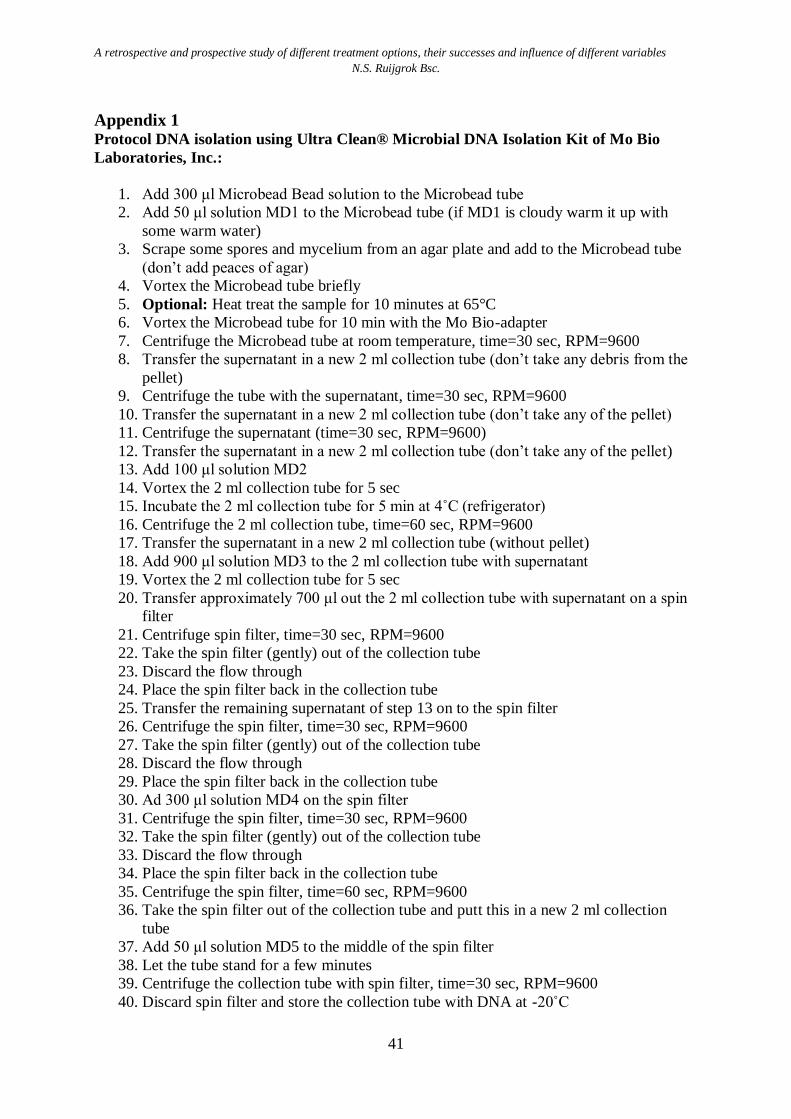
A retrospective and prospective study of different treatment options, their successes and influence of different variables
N.S. Ruijgrok Bsc.
41
Appendix 1
Protocol DNA isolation using Ultra Clean® Microbial DNA Isolation Kit of Mo Bio
Laboratories, Inc.:
1. Add 300 μl Microbead Bead solution to the Microbead tube
2. Add 50 μl solution MD1 to the Microbead tube (if MD1 is cloudy warm it up with
some warm water)
3. Scrape some spores and mycelium from an agar plate and add to the Microbead tube
(don’t add peaces of agar)
4. Vortex the Microbead tube briefly
5. Optional: Heat treat the sample for 10 minutes at 65°C
6. Vortex the Microbead tube for 10 min with the Mo Bio-adapter
7. Centrifuge the Microbead tube at room temperature, time=30 sec, RPM=9600
8. Transfer the supernatant in a new 2 ml collection tube (don’t take any debris from the
pellet)
9. Centrifuge the tube with the supernatant, time=30 sec, RPM=9600
10. Transfer the supernatant in a new 2 ml collection tube (don’t take any of the pellet)
11. Centrifuge the supernatant (time=30 sec, RPM=9600)
12. Transfer the supernatant in a new 2 ml collection tube (don’t take any of the pellet)
13. Add 100 μl solution MD2
14. Vortex the 2 ml collection tube for 5 sec
15. Incubate the 2 ml collection tube for 5 min at 4˚C (refrigerator)
16. Centrifuge the 2 ml collection tube, time=60 sec, RPM=9600
17. Transfer the supernatant in a new 2 ml collection tube (without pellet)
18. Add 900 μl solution MD3 to the 2 ml collection tube with supernatant
19. Vortex the 2 ml collection tube for 5 sec
20. Transfer approximately 700 μl out the 2 ml collection tube with supernatant on a spin
filter
21. Centrifuge spin filter, time=30 sec, RPM=9600
22. Take the spin filter (gently) out of the collection tube
23. Discard the flow through
24. Place the spin filter back in the collection tube
25. Transfer the remaining supernatant of step 13 on to the spin filter
26. Centrifuge the spin filter, time=30 sec, RPM=9600
27. Take the spin filter (gently) out of the collection tube
28. Discard the flow through
29. Place the spin filter back in the collection tube
30. Ad 300 μl solution MD4 on the spin filter
31. Centrifuge the spin filter, time=30 sec, RPM=9600
32. Take the spin filter (gently) out of the collection tube
33. Discard the flow through
34. Place the spin filter back in the collection tube
35. Centrifuge the spin filter, time=60 sec, RPM=9600
36. Take the spin filter out of the collection tube and putt this in a new 2 ml collection
tube
37. Add 50 μl solution MD5 to the middle of the spin filter
38. Let the tube stand for a few minutes
39. Centrifuge the collection tube with spin filter, time=30 sec, RPM=9600
40. Discard spin filter and store the collection tube with DNA at -20˚C
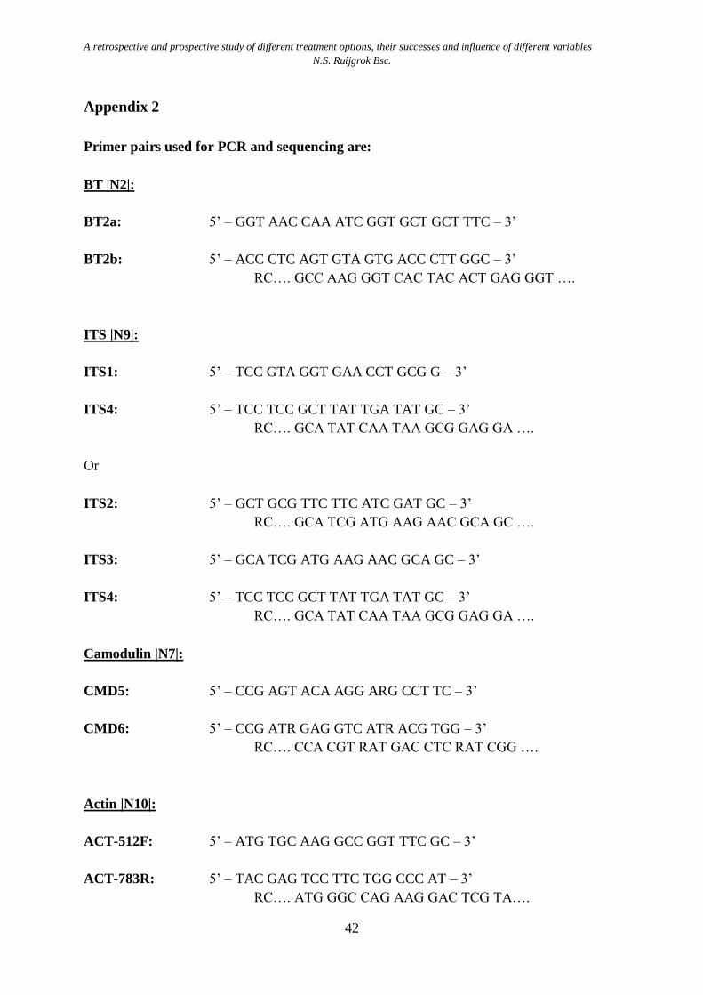
A retrospective and prospective study of different treatment options, their successes and influence of different variables
N.S. Ruijgrok Bsc.
42
Appendix 2
Primer pairs used for PCR and sequencing are:
BT |N2|:
BT2a: 5’ – GGT AAC CAA ATC GGT GCT GCT TTC – 3’
BT2b: 5’ – ACC CTC AGT GTA GTG ACC CTT GGC – 3’
RC…. GCC AAG GGT CAC TAC ACT GAG GGT ….
ITS |N9|:
ITS1: 5’ – TCC GTA GGT GAA CCT GCG G – 3’
ITS4: 5’ – TCC TCC GCT TAT TGA TAT GC – 3’
RC…. GCA TAT CAA TAA GCG GAG GA ….
Or
ITS2: 5’ – GCT GCG TTC TTC ATC GAT GC – 3’
RC…. GCA TCG ATG AAG AAC GCA GC ….
ITS3: 5’ – GCA TCG ATG AAG AAC GCA GC – 3’
ITS4: 5’ – TCC TCC GCT TAT TGA TAT GC – 3’
RC…. GCA TAT CAA TAA GCG GAG GA ….
Camodulin |N7|:
CMD5: 5’ – CCG AGT ACA AGG ARG CCT TC – 3’
CMD6: 5’ – CCG ATR GAG GTC ATR ACG TGG – 3’
RC…. CCA CGT RAT GAC CTC RAT CGG ….
Actin |N10|:
ACT-512F: 5’ – ATG TGC AAG GCC GGT TTC GC – 3’
ACT-783R: 5’ – TAC GAG TCC TTC TGG CCC AT – 3’
RC…. ATG GGC CAG AAG GAC TCG TA….
A retrospective and prospective study of different treatment options, their successes and influence of different variables
N.S. Ruijgrok Bsc.
43
References
Benitah, N. (2006). Canine nasal aspergillosis. Clinical Techniques in Small Animal Practice,
21(2), 82-88.
Burbidge, H. M., Clark, W. T., Read, R., Labuc, R., Davey, T., & Broome, C. (1997). Canine
nasal aspergillosis: Results of treatment using clotrimazole as a topical agent. Australian
Veterinary Practitioner, 27(2), 79-83.
Burrow, R., Baker, M., White, L., & McConnell, J. F. (2013). Trephination of the frontal
sinuses and instillation of clotrimazole cream: A computed tomographic study in canine
cadavers. Veterinary Surgery, 42(3), 322-328.
Claeys, S., Lefebvre, J. B., Schuller, S., Hamaide, A., & Clercx, C. (2006). Surgical treatment
of canine nasal aspergillosis by rhinotomy combined with enilconazole infusion and oral
itraconazole. The Journal of Small Animal Practice, 47(6), 320-324. doi:10.1111/j.1748-
5827.2006.00154.x
Clercx, C. (2006). Surgical treatment of canine nasal aspergillosis. The Journal of Small
Animal Practice, 47(7), 416. doi:10.1111/j.1748-5827.2006.00201.x
Field, A. (2009). Discovering statistics using spss [Discovering statistics using spss] (third
ed.). Oriental Press, Dubai: SAGE Publications.
Friend, E. J., Williams, J. M., & White, R. A. (2002). Invasive treatment of canine nasal
aspergillosis with topical clotrimazole. The Veterinary Record, 151(10), 298-299.
Hayes, G. M., & Demetriou, J. L. (2012). Distribution and persistence of topical clotrimazole
after sinus infusion in normal canine cadavers. The Journal of Small Animal Practice,
53(2), 95-100. doi:10.1111/j.1748-5827.2011.01164.x; 10.1111/j.1748-
5827.2011.01164.x
Mathews, K. G., Davidson, A. P., Koblik, P. D., Richardson, E. F., Komtebedde, J.,
Pappagianis, D., . . . Kass, P. H. (1998). Comparison of topical administration of
clotrimazole through surgically placed versus nonsurgically placed catheters for
treatment of nasal aspergillosis in dogs: 60 cases (1990-1996). Journal of the American
Veterinary Medical Association, 213(4), 501-506.
Nelson, R. W., Couto, C. G., Grauwer, G. F., Hawkins, E. C., Jhonson, C. A., Lappin, M. R., .
. . Willard, M. D. (2009). Nasal mycoses. In R. W. Nelson, & C. G. Couto (Eds.), Small
animal internal medicine [Small Animal Internal Medicine] (pp. 226-229)
A retrospective and prospective study of different treatment options, their successes and influence of different variables
N.S. Ruijgrok Bsc.
44
Peeters, D., & Clercx, C. (2007). Update on canine sinonasal aspergillosis. The Veterinary
Clinics of North America.Small Animal Practice, 37(5), 901-16, vi.
doi:10.1016/j.cvsm.2007.05.005
Petrie, A., & Watson, P. (2006). Statistics for veterinary and animal sience [Statistics for
Veterinary and Animal Sience] (second ed.). UK: Blackwell Publishing.
Pomrantz, J. S., & Johnson, L. R. (2010). Repeated rhinoscopic and serologic assessment of
the effectiveness of intranasally administered clotrimazole for the treatment of nasal
aspergillosis in dogs. Journal of the American Veterinary Medical Association, 236(7),
757-762. doi:10.2460/javma.236.7.757; 10.2460/javma.236.7.757
Quinn, P. J., Markey, B. K., Carter, M. E., Donnelly, W. J., & Leonard, F. C. (2002).
Aspergillus species. Veterinary microbiology and microbial disease [Veterinary
Microbiology and Microbial Disease] (pp. 229-232)
Schuller, S., & Clercx, C. (2007). Long-term outcomes in dogs with sinonasal aspergillosis
treated with intranasal infusions of enilconazole. Journal of the American Animal
Hospital Association, 43(1), 33-38.
Sharman, M., Lenard, Z., Hosgood, G., & Mansfield, C. (2012). Clotrimazole and
enilconazole distribution within the frontal sinuses and nasal cavity of nine dogs with
sinonasal aspergillosis. The Journal of Small Animal Practice, 53(3), 161-167.
doi:10.1111/j.1748-5827.2011.01181.x; 10.1111/j.1748-5827.2011.01181.x
Sharman, M., Paul, A., Davies, D., MacKay, B., Swinney, G., Barrs, V., . . . Mansfield, C.
(2010). Multi-centre assessment of mycotic rhinosinusitis in dogs: A retrospective study
of initial treatment success (1998 to 2008). Journal of Small Animal Practice, 51(8), 423-
427.
Sharman, M. J., & Mansfield, C. S. (2012). Sinonasal aspergillosis in dogs: A review. The
Journal of Small Animal Practice, 53(8), 434-444. doi:10.1111/j.1748-
5827.2012.01245.x; 10.1111/j.1748-5827.2012.01245.x
Sharp, N., Sullivan, M., & Harvey, C. (1992). Treatment of canine nasal aspergillosis. In
Practice, 14(1), 27-28.
Sharp, N. J., Sullivan, M., Harvey, C. E., & Webb, T. (1993). Treatment of canine nasal
aspergillosis with enilconazole. Journal of Veterinary Internal Medicine / American
College of Veterinary Internal Medicine, 7(1), 40-43.
Sissener, T. R., Bacon, N. J., Friend, E., Anderson, D. M., & White, R. A. S. (2006).
Combined clotrimazole irrigation and depot therapy for canine nasal aspergillosis.
Journal of Small Animal Practice, 47(6), 312-315.

A retrospective and prospective study of different treatment options, their successes and influence of different variables
N.S. Ruijgrok Bsc.
45
Zonderland, J. L., Stork, C. K., Saunders, J. H., Hamaide, A. J., Balligand, M. H., & Clercx,
C. M. (2002). Intranasal infusion of enilconazole for treatment of sinonasal aspergillosis
in dogs. Journal of the American Veterinary Medical Association, 221(10), 1421-1425.
Related Documents