ISSN 2234-3806 • eISSN 2234-3814 http://dx.doi.org/10.3343/alm.2014.34.6.413 www.annlabmed.org 413 Ann Lab Med 2014;34:413-425 http://dx.doi.org/10.3343/alm.2014.34.6.413 Review Article Diagnostic Genetics Cancer Cytogenetics: Methodology Revisited Thomas S. K. Wan, Ph.D. Haematology Division, Department of Pathology, The University of Hong Kong, Hong Kong The Philadelphia chromosome was the first genetic abnormality discovered in cancer (in 1960), and it was found to be consistently associated with CML. The description of the Philadelphia chromosome ushered in a new era in the field of cancer cytogenetics. Accu- mulating genetic data have been shown to be intimately associated with the diagnosis and prognosis of neoplasms; thus, karyotyping is now considered a mandatory investigation for all newly diagnosed leukemias. The development of FISH in the 1980s overcame many of the drawbacks of assessing the genetic alterations in cancer cells by karyotyping. Karyo- typing of cancer cells remains the gold standard since it provides a global analysis of the abnormalities in the entire genome of a single cell. However, subsequent methodological advances in molecular cytogenetics based on the principle of FISH that were initiated in the early 1990s have greatly enhanced the efficiency and accuracy of karyotype analysis by marrying conventional cytogenetics with molecular technologies. In this review, the de- velopment, current utilization, and technical pitfalls of both the conventional and molecu- lar cytogenetics approaches used for cancer diagnosis over the past five decades will be discussed. Key Words: Cancer cytogenetics, FISH, Karyotyping, Molecular cytogenetics Received: June 17, 2014 Revision received: July 31, 2014 Accepted: October 6, 2014 Corresponding author: Thomas S. K. Wan Hematology Division, Department of Pathology, The University of Hong Kong, Queen Mary Hospital, 102 Pokfulam Road, Hong Kong Tel: +852-22553172 Fax: +852-28177565 E-mail: [email protected] © The Korean Society for Laboratory Medicine This is an Open Access article distributed under the terms of the Creative Commons Attribution Non-Commercial License (http://creativecom- mons.org/licenses/by-nc/3.0) which permits unrestricted non-commercial use, distribution, and reproduction in any medium, provided the original work is properly cited. INTRODUCTION The discovery of a tiny abnormal chromosome, the Philadelphia chromosome, as a hallmark of CML in 1960 by Peter Nowell and David Hungerford marked the first time cancer was shown to result from a specific genetic abnormality [1]. As cytogenetic techniques improved, in 1973, Rowley [2] discovered the exis- tence of a translocation between the long arms of chromosome 9 and 22 in CML. Subsequent work revealed that this transloca- tion resulted in a new fusion protein that was expressed in the cancer cells [3]. Strikingly, the description of the Philadelphia chromosome ushered in a new era of genetic diagnosis. Over the past several decades, accumulating genetic data has been shown to be intimately associated with the diagnosis and prog- nosis of neoplasms, thereby moving cancer cytogenetics out of research laboratories and into clinical practice. Recognizing the association between specific cytogenetic abnormalities and cer- tain morphologic and clinical features, the World Health Organi- zation has categorized four unique AML subtypes according to cytogenetics [4]. Therefore, conventional cytogenetic analysis is considered mandatory for all newly diagnosed leukemias, owing to its usefulness in diagnosis, classification, and prognostication. FISH can be used to map loci on specific chromosomes [5], de- tect both numerical and structural chromosomal abnormalities, and reveal cryptic abnormalities. It has overcome many of the drawbacks of karyotyping, such as low specimen cell yield, low mitotic index, poor quality metaphases, and other technical dif- ficulties. Currently, FISH is used as an indispensable tool for the detection of structural rearrangements such as translocations, inversions, insertions, and microdeletions, and for the identifica- tion of marker chromosomes and the delineation of chromo- some breakpoints [6, 7]. Therefore, FISH has greatly enhanced the efficiency and accuracy of karyotype analysis by bringing to- gether conventional cytogenetics and molecular technologies. This review summarizes the development, current utilization, and technical pitfalls of the conventional and molecular cytoge- netic approaches used in clinical laboratories for cancer diagnosis.

Welcome message from author
This document is posted to help you gain knowledge. Please leave a comment to let me know what you think about it! Share it to your friends and learn new things together.
Transcript

ISSN 2234-3806 • eISSN 2234-3814
http://dx.doi.org/10.3343/alm.2014.34.6.413 www.annlabmed.org 413
Ann Lab Med 2014;34:413-425http://dx.doi.org/10.3343/alm.2014.34.6.413
Review ArticleDiagnostic Genetics
Cancer Cytogenetics: Methodology RevisitedThomas S. K. Wan, Ph.D.Haematology Division, Department of Pathology, The University of Hong Kong, Hong Kong
The Philadelphia chromosome was the first genetic abnormality discovered in cancer (in 1960), and it was found to be consistently associated with CML. The description of the Philadelphia chromosome ushered in a new era in the field of cancer cytogenetics. Accu-mulating genetic data have been shown to be intimately associated with the diagnosis and prognosis of neoplasms; thus, karyotyping is now considered a mandatory investigation for all newly diagnosed leukemias. The development of FISH in the 1980s overcame many of the drawbacks of assessing the genetic alterations in cancer cells by karyotyping. Karyo-typing of cancer cells remains the gold standard since it provides a global analysis of the abnormalities in the entire genome of a single cell. However, subsequent methodological advances in molecular cytogenetics based on the principle of FISH that were initiated in the early 1990s have greatly enhanced the efficiency and accuracy of karyotype analysis by marrying conventional cytogenetics with molecular technologies. In this review, the de-velopment, current utilization, and technical pitfalls of both the conventional and molecu-lar cytogenetics approaches used for cancer diagnosis over the past five decades will be discussed.
Key Words: Cancer cytogenetics, FISH, Karyotyping, Molecular cytogenetics
Received: June 17, 2014Revision received: July 31, 2014Accepted: October 6, 2014
Corresponding author: Thomas S. K. WanHematology Division, Department of Pathology, The University of Hong Kong, Queen Mary Hospital, 102 Pokfulam Road, Hong KongTel: +852-22553172Fax: +852-28177565E-mail: [email protected]
© The Korean Society for Laboratory MedicineThis is an Open Access article distributed under the terms of the Creative Commons Attribution Non-Commercial License (http://creativecom-mons.org/licenses/by-nc/3.0) which permits unrestricted non-commercial use, distribution, and reproduction in any medium, provided the original work is properly cited.
INTRODUCTION
The discovery of a tiny abnormal chromosome, the Philadelphia
chromosome, as a hallmark of CML in 1960 by Peter Nowell
and David Hungerford marked the first time cancer was shown
to result from a specific genetic abnormality [1]. As cytogenetic
techniques improved, in 1973, Rowley [2] discovered the exis-
tence of a translocation between the long arms of chromosome
9 and 22 in CML. Subsequent work revealed that this transloca-
tion resulted in a new fusion protein that was expressed in the
cancer cells [3]. Strikingly, the description of the Philadelphia
chromosome ushered in a new era of genetic diagnosis. Over
the past several decades, accumulating genetic data has been
shown to be intimately associated with the diagnosis and prog-
nosis of neoplasms, thereby moving cancer cytogenetics out of
research laboratories and into clinical practice. Recognizing the
association between specific cytogenetic abnormalities and cer-
tain morphologic and clinical features, the World Health Organi-
zation has categorized four unique AML subtypes according to
cytogenetics [4]. Therefore, conventional cytogenetic analysis is
considered mandatory for all newly diagnosed leukemias, owing
to its usefulness in diagnosis, classification, and prognostication.
FISH can be used to map loci on specific chromosomes [5], de-
tect both numerical and structural chromosomal abnormalities,
and reveal cryptic abnormalities. It has overcome many of the
drawbacks of karyotyping, such as low specimen cell yield, low
mitotic index, poor quality metaphases, and other technical dif-
ficulties. Currently, FISH is used as an indispensable tool for the
detection of structural rearrangements such as translocations,
inversions, insertions, and microdeletions, and for the identifica-
tion of marker chromosomes and the delineation of chromo-
some breakpoints [6, 7]. Therefore, FISH has greatly enhanced
the efficiency and accuracy of karyotype analysis by bringing to-
gether conventional cytogenetics and molecular technologies.
This review summarizes the development, current utilization,
and technical pitfalls of the conventional and molecular cytoge-
netic approaches used in clinical laboratories for cancer diagnosis.

Thomas S.K.WanCancer cytogenetics methodology revisited
414 www.annlabmed.org http://dx.doi.org/10.3343/alm.2014.34.6.413
CONVENTIONAL CYTOGENETICS
Chromosome analysis is a simple technique. Under optimal
conditions, in most cases of acute leukemia, clonal cytogenetic
abnormalities are detected by using this method. Chromosome
analysis requires five principal steps: 1) cell culture, 2) harvest
of metaphase chromosomes, 3) chromosome preparation, 4)
banding and staining using a special protocol, and 5) analysis
by light microscopy or karyotype assisted computer analysis (Fig.
1). The discovery that colchicine (or colcemid) pretreatment re-
sulted in mitotic arrest and that treatment of arrested cells with a
hypotonic solution improved the yield and quality of metaphases
spreads. Therefore, counted and analyzed individual chromo-
somes in human cells are then possible. Chromosome analysis
provides an overview of all chromosomal aberrations in a single
tumor cell.
1. Technology considerationsChromosomal studies of malignancies pose a particular techni-
cal challenge. As the results are so unpredictable, there is no a
single technique that can be guaranteed to work consistently
and reliably. Therefore, every laboratory should adopt a slight
variation of the basic protocol. In addition to setting up a bone
marrow suspension cell culture of hematologic neoplasms, pe-
ripheral blood can also be investigated, if it contains more than
10% circulating blasts or immature cells. In addition, a lymph
node biopsy is required, as lymph nodes are the preferred tissue
for studies of most lymphomas [8]. The duration of the cell cycle
in malignant cells varies greatly among patients; a range of 16 hr
to 292 hr was obtained in a series of 37 patients with AML [9].
Therefore, one of the most significant factors in obtaining a suc-
cessful result is setting up multiple cultures to maximize the
chances of obtaining optimal malignant cell divisions: 1) direct
harvest of bone marrow cells, 2) overnight culture, and 3) over-
night culture with synchronization (by blocking at S-phase of the
cell cycle) [10].
High-resolution banding of long chromosomes with good mor-
phology can be achieved by applying synchronization techniques
[10]. It enables the identification of subtle structural chromo-
some aberrations that are commonly found in malignant cells.
However, it has been reported that fluorodeoxyuridine synchroni-
zation cultures are inferior to short-term cultures for chromo-
some analysis in ALL [11]. ALL is a frustrating disease for most
cytogeneticists, as it has several technical challenges, including
frequent poor chromosome morphology, low mitotic index, and
samples that have a marked tendency to clot during harvest.
Fig. 1. Protocol for the preparation of a karyotype from a leukemic patient.
Thomas S. K. WanCancer cytogenetics methodology revisited
http://dx.doi.org/10.3343/alm.2014.34.6.413 www.annlabmed.org 415
A B C D E F
J
NML
IH
K
G
2. Acquired abnormal clones versus constitutional chromosomal anomalies or cytogenetic heteromorphisms
In cases where the cytogenetic analysis reveals only abnormal
metaphases, especially in those with a balanced translocation, it
may be necessary to rule out a constitutional chromosomal
anomaly. In addition, the constitutive heterochromatic regions
just below the centromeres of chromosomes 1, 9, and 16 and
the telomeric end of the long arm of the Y-chromosome can
vary widely in size among individuals and can sometimes ap-
pear abnormal (Fig. 2A). Constitutional chromosomal anomalies
are best differentiated from differentiate acquired abnormal
clones through cytogenetic examination of phytohemagglutinin
(PHA)-stimulated lymphocytes from peripheral blood in disease
remission. The Cytogenetics Resource Committee (CyRC) of the
College of American Pathologists (CAP)/American College of
Medical Genetics & Genomics (ACMG) has asked participants
not to include heteromorphisms when responding to a chal-
lenge of the cytogenetics survey. However, the guidelines for re-
porting heteromorphisms have not been standardized. Of 223
cytogenetic laboratories, 136 (61%) stated that they would in-
clude selected heteromorphism information identified by routine
G-banding in a clinical cytogenetic report in a CyRC survey on
Fig. 2. Partial karyotype showing normal variants and chromosome abnormalities of leukemia. (A) Normal variant with 1qh+ (arrow), (B) Normal variant with inv(9)(p11q13) (arrow), (C) dup(1)(q21q32) chromosome with duplication of a 1q21-q32 chromosomal fragment (ar-row), (D) trp(1)(q21q32) chromosome with triplication of a 1q21-q32 chromosomal fragment (arrow), (E) del(7)(q) chromosome with ter-minal deletion ofa7q22-qter chromosomal fragment (arrow), (F) del(5)(q13q33) chromosome with an interstitial deletion of a 5q13-q33 chromosomal fragment (arrow), (G) der(1)t(1;1)(p35;q25) chromosome with loss of a 1p35-pter chromosomal fragment and duplication of a 1q25-qter chromosomal fragment (arrow), (H) i(17)(q10) chromosome with a loss of the whole short arm and duplication of the whole long of chromosome 17 (arrow), (I) t(9;22)(q34;q11.2), a balanced translocation between 9q34 and 22q11.2 (arrows), (J) t(2;9;22)(q37;q34;q11.2), a balanced three-way translocation between 2q37, 9q34, and 22q11.2 (arrows), (K) der(1;7)(q10;p10), a centric fusion of the whole arms of 1q and 7p with a gain of 1q and a loss of 7q (arrow), (L) der(5)ins(5;?)(q13;?) chromosome with an unknown chro-mosomal fragment inserted into 5q13 (arrow), (M) inv(16)(p13q22) chromosome with G-banding (arrow), (N) inv(16)(p13q22) chromo-some with R-banding (arrow).

Thomas S.K.WanCancer cytogenetics methodology revisited
416 www.annlabmed.org http://dx.doi.org/10.3343/alm.2014.34.6.413
cytogenetic heteromorphisms [12]. The majority of the clinical
cytogenetics surveyed would not include the more common
chromosomal variants (such as prominent short arms, large or
double satellites, and increased stalk length or double stalks on
acrocentric chromosomes, and long arm heterochromatin varia-
tions of chromosomes 1, 9, 16, and Y), but would report most
pericentric inversions [such as inv(9)(p11q13); Fig. 2B] and
other rare heteromorphisms [12].
Constitutional pericentric inversion of chromosome 9 [inv(9)
(p11q13)] occurs in 0.8% to 2% of the normal population and
has long been considered a normal variant. Whether constitu-
tional inv(9) is a predisposing factor for cancer remains contro-
versial. We were the first group to document a case of an ac-
quired pericentric inv(9) in essential thrombocythemia [13].
Since then, several other hematological malignancies with ac-
quired inv(9) have been reported. However, inv(9) is not over-
represented in patients with hematological malignancy; there-
fore, there is no evidence to suggest that the presence of consti-
tutional inv(9) increases the risk of hematological malignancy
[13, 14].
Furthermore, some abnormalities are both acquired and in-
herited. For example, trisomy 21 is common both as an inher-
ited abnormality in Down syndrome and as an acquired abnor-
mality in AML, MDS, and childhood ALL [15-17]. Trisomy 21 is
the second most common trisomy in AML and MDS after tri-
somy 8 [18]. Therefore, if trisomy 21 is found in a karyotype, it
is necessary to consider both inherited and acquired abnormali-
ties. However, most individuals with Down syndrome have char-
acteristic physical features.
3. Mechanisms that generate chromosome abnormalities1) Net gain and loss of chromosomal materialIn general, gain or loss of chromosomal material results in gene
amplification or loss of heterozygosity, respectively. Two main
classes of cancer-relevant genes, oncogenes and tumor sup-
pressor genes have been recognized as the major pathogenic
targets for cancer-associated karyotypic abnormalities. Numeri-
cal chromosome aberrations are detected as the sole clonal
change in approximately 15% of all cytogenetically abnormal he-
matological malignancies, and they show substantial variations
in frequency among the various disease subgroups [19]. Despite
their relatively frequent occurrence, numerical changes, includ-
ing single autosomal trisomies, have received less attention than
structural changes. There are several reasons for this discrep-
ancy. First, the high degree of correlation between cytogenetic
changes and morphology that is observed with structural
changes like translocation is lacking for numerical aberrations.
Second, the role that numerical changes play in leukemogenesis
tends to be obscured. Third, the molecular consequences of
whole chromosome gains and losses are not yet clear.
Net gain of chromosomal material may be caused by duplica-
tion (Fig. 2C) and triplication (Fig. 2D) of particular chromo-
somal segments or regions, which may also lead to an unbal-
anced gene product. Mis-segregation of entire chromosomes in
cell division may also result in trisomies or more extensive poly-
somies [20]. A single autosomal trisomy is a common numerical
cytogenetic abnormality in hematological malignancies, and it
shows a predilection for myeloid disorders [15, 16, 18, 21].
However, gain of a sex chromosome as the sole acquired abnor-
mality is very rare in hematological malignancies [22]. Interest-
ingly, hand-mirror cell morphology has been described in AML
with trisomy 13 [23], particularly in AML-M0 and -M1, and it
may not be detected in the more differentiated subtypes of AML
[24]. In addition, chromosome gain is nonrandom in childhood
ALL, and eight chromosomes (+4, +6, +10, +14, +17, +18,
+21, and +X) account for almost 80% of all gains. However, tri-
somy 4 has also been reported as the sole karyotypic abnormal-
ity in ALL [25].
Net loss of chromosomal material may be caused by loss of
an entire chromosome (monosomy) [26-28], deletions of partic-
ular chromosomal segments [29] (Fig. 2E, F), unbalanced
translocations (Fig. 2G), or isochromosomes (Fig. 2H). Terminal
deletions results from a single break in the chromosome arm,
with loss of the distal segment (Fig. 2E). Interstitial deletions
emerge when two breaks occur within the same chromosome
arm and the intervening segment is lost (Fig. 2F).
2) Chromosomal rearrangementsA chromosome rearrangement occurs when a piece of one
chromosome breaks off and attaches to another chromosome.
A gene fusion can be created when the translocation joins two
other separate genes, the occurrence of which is common in
cancer. It can be a balanced translocation, unbalanced translo-
cation, insertion, or inversion.
In a balanced translocation, pieces of chromosomes are rear-
ranged, but no genetic material is gained or lost in the cell (Fig.
2I). A balanced translocation can involve more than two chro-
mosomes and form a complex variant translocation [30, 31]
(Fig. 2J). In CML, a complex, 3-way variant translocation, involv-
ing 3 chromosomes, often occurs as a single-step process with
3 breakpoints and no reciprocal ABL1-BCR fusion [32] (Fig.
2J). Interestingly, a complex 3-way translocation resulting from

Thomas S. K. WanCancer cytogenetics methodology revisited
http://dx.doi.org/10.3343/alm.2014.34.6.413 www.annlabmed.org 417
4 breakpoints, 2-step process and a reciprocal gene fusion on
the third chromosome has been detected in acute promyelo-
cytic leukemia and CML, using dual color dual fusion transloca-
tion FISH probes [32, 33].
In an unbalanced translocation, the exchange of chromo-
some material is unequal, resulting in extra or missing chromo-
somal fragments [34, 35]. Derivative (1;7)(q10;p10) with an un-
balanced whole-arm translocation is a recurrent cytogenetic ab-
normality in myeloid disorders [36, 37] (Fig. 2K). It had long
been regarded as a poor prognostic indicator in MDS and AML
[38], until the publication of a large study on the clinicopatho-
logical features of myeloid neoplasms with this karyotypic abnor-
mality [39]. It has been proposed that myeloid neoplasms with
der(1;7)(q10;p10) may not have a homogeneously favorable
clinical behavior compared to MDS, which has known poor-risk
cytogenetics [37].
An insertion is a structural rearrangement, in which part of a
chromosome is typically interstitially repositioned into a different
area of the karyotype (Fig. 2L). The insertion can be cryptic,
and at the gene level, we have previously reported a case of
childhood CML with a cryptic insertion of BCR at 9q34 and
morphologically normal chromosomes 9 and 22 on G-banding
[40]. FISH confirmed the presence of the BCR/ABL1 gene fu-
sion on chromosome 9 in metaphase chromosomes. Therefore,
in clinical practice, atypical genetic test results should not be in-
terpreted in isolation and should be integrated with information
gathered through different genetic studies.
A chromosomal aberration, in which a segment of a chromo-
some is reversed in orientation but not relocated, is called an in-
version. Inversion of chromosome 16 [inv(16)(p13q22)] is the
most common chromosomal inversion observed in leukemia
(Fig. 2M). It has been detected in approximately 5% of de novo
AML cases, which are mostly classified as the M4Eo subtype,
and is associated with a relatively favorable outcome. In the
AML M4Eo subgroup, inv(16) is much more prevalent (88%)
[41]. However, ethnic differences have been reported, including
a very low prevalence of inv(16)(p13q22) abnormalities in two
Chinese AML cohorts [41, 42]. Interestingly, R-banding is un-
suitable for detecting this inv(16)(p13q22) aberration (Fig. 2N)
[42], and it is far easier to recognize by G-banding. Therefore,
FISH, reverse-transcription (RT)-PCR, and Southern blot analy-
ses are reliable tools for detecting masked inv(16).
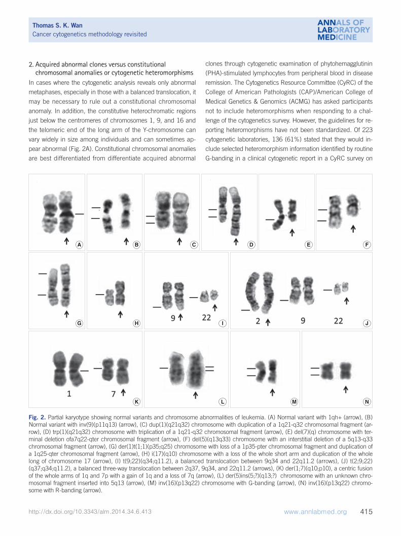
MOLECULAR CYTOGENETICS
Molecular cytogenetics involves the use of a series of techniques
referred to as FISH, in which DNA probes are labeled with differ-
ent colored fluorescent tags to visualize one or more specific re-
gions of the genome (Fig. 3). It is used as a rapid, sensitive test
for the detection of cryptic or subtle chromosomal changes. Fur-
Fig. 3. FISH protocol. It includes sample pretreatment, denaturation of probe and sample, hybridization, post-hybridization washing, and fluorescent signal detection.
Thomas S.K.WanCancer cytogenetics methodology revisited
418 www.annlabmed.org http://dx.doi.org/10.3343/alm.2014.34.6.413
thermore, it can be used to detect genetic alterations in non-di-
viding cell populations, and it is a convenient method to support
the practice of personalized medicine. However, FISH assays are
still hampered by reagent costs, which prevent its adoption by
large-scale oncological screening.
1. MethodologyThe standard FISH protocol is illustrated in Fig. 3. Briefly, it in-
cludes five steps: 1) sample pretreatment, 2) denaturation of
probe and sample, 3) hybridization of probe to target cells or
metaphase spreads (annealing), 4) post-hybridization washing,
and 5) detection using a simple epifluorescence microscope
with appropriate filter sets. When a FISH test is initially imple-
mented, the assay performance characteristics assessed should
include sensitivity, accuracy, precision, and specificity [43]. The
upper cutoff for normal results in a FISH assay can be deter-
mined by calculating the 95% confidence interval for probe sig-
nal patterns detected in normal control samples that are repre-
sentative of the sample type to be analyzed.
ACMG sets internationally accepted standards for FISH analy-
sis to ensure that FISH results are clear and interpretable [44].
Furthermore, ongoing monitoring of interobserver reproducibil-
ity, accomplished in part by having two laboratory personnel
read every case, can help detect changes in assay performance
or loss of consistency in applying scoring criteria. The standard
FISH nomenclature has been simplified and expanded in the
latest edition of the International System for Human Cytogenetic
Nomenclature, ISCN 2013 [45]. However, since the use of the
full FISH ISCN is likely to make it more difficult for physicians to
understand, it is not recommended by the European Myeloma
Network [46].
2. Selection of FISH probes for use in oncologyThere are three broad types of FISH probes used in clinical ge-
netics laboratories, each with a different application: 1) whole-
chromosome painting (WCP) probes for deciphering cytogenetic
aberrations [8, 34, 47, 48]; 2) repetitive sequence probes for
chromosome enumeration [21, 25, 26, 49-51]; and 3) locus-
specific identifier (LSI) probes for gene fusions [17, 33, 52],
gene deletions [35, 53, 54] or duplications [55]. These probes
can also be used in various combinations when investigating
complex chromosome abnormalities [56, 57].
WCP probes are designed to mark the entire chromosome of
interest and are useful for deciphering cytogenetic aberrations
that are difficult to resolve on morphological grounds, such as
marker chromosomes of uncertain nature or complex changes.
However, the use of WCP probes in interphase cells is very lim-
ited, as the chromosomes are dispersed in the cell nucleus dur-
ing interphase and they do not form discrete units.
Centromeric enumeration probes (CEPs) hybridize to alpha
(or beta) satellite repeat sequences within the specific centro-
meric regions of each chromosome and are used for chromo-
somal enumeration. Interphase FISH analysis using dual color
CEPs for chromosomes X and Y is an efficient method to predict
relapse or failure of engraftment in patients after sex-mis-
matched bone marrow transplantation (Fig. 4A) [58].
There are two main LSI FISH probe systems for the detection
of gene rearrangements in oncology, dual-color translocation
probes and dual-color break-apart probe. The dual-color trans-
location probes are designed to detect chromosomal transloca-
tions involving know partner genes, such as BCR-ABL1 that re-
sult from t(9;22)(q34;q11.2) in CML (Fig. 4B). Furthermore,
dual-color break-apart probes are useful for detecting chromo-
somal translocations that involve genes with unknown or multi-
ple translocation partners, such as MLL, ALK, and RARα (Fig.
4C). Break-apart translocation FISH probes conveniently pro-
vide important information on the presence of gene rearrange-
ments, although they are unable to identify the specific partner
gene. Recently, with the use of long distance inverse-poly-
merase chain reaction (LDI)-PCR, it has become possible to
identify unknown translocation partners and to map breakpoints
at the base-pair level. LDI-PCR requires only approximate se-
quence information for one partner, rendering it ideal for use in
combination with FISH to extend and refine cytogenetic break-
point data [59]. LSI FISH probes have also been used to identify
the origin of the amplified DNA that constitutes homogeneously
staining regions (HSRs) and double minutes (DMs), which oc-
cur in a variety of tumor cells (Fig. 4D) [60].
3. Advances in FISH technologyNumerous methodological advances in molecular cytogenetic
technology were initiated in the early 1990s, including compara-
tive genomic hybridization (CGH) [54], spectral karyotyping
(SKY) [62], multicolor FISH (MFISH) [63], multicolor banding
(mBAND) [64], and array CGH (aCGH) [65]. All of these cyto-
genetic techniques add colors to the monotonous world of con-
ventional banding. Two multicolor fluorescence technologies
have been introduced, MFISH [63] and SKY [62]. These tech-
nologies are based on simultaneous hybridization of 24 chromo-
some-specific composite probes. This technique is suitable for
the identification of subtle chromosomal aberrations that include
an unidentified chromosome (marker chromosome) and an un-

Thomas S. K. WanCancer cytogenetics methodology revisited
http://dx.doi.org/10.3343/alm.2014.34.6.413 www.annlabmed.org 419
balanced chromosomal translocation. Regarding probe design,
these chromosome-painting probes are generated from flow-
sorted human chromosomes. Unique chromosome-specific col-
ors are produced by labeling each chromosome library with ei-
ther a single fluorochrome or specific combinations of multiple
fluorochromes.
mBAND has been developed to facilitate the identification of
intrachromosomal rearrangements and to map the exact break-
point by using human overlapping microdissection libraries that
are differentially labeled [64]. The color bands have great value
for delineating intrachromosomal exchanges, such as inver-
sions, deletions, duplications, and insertions [6].
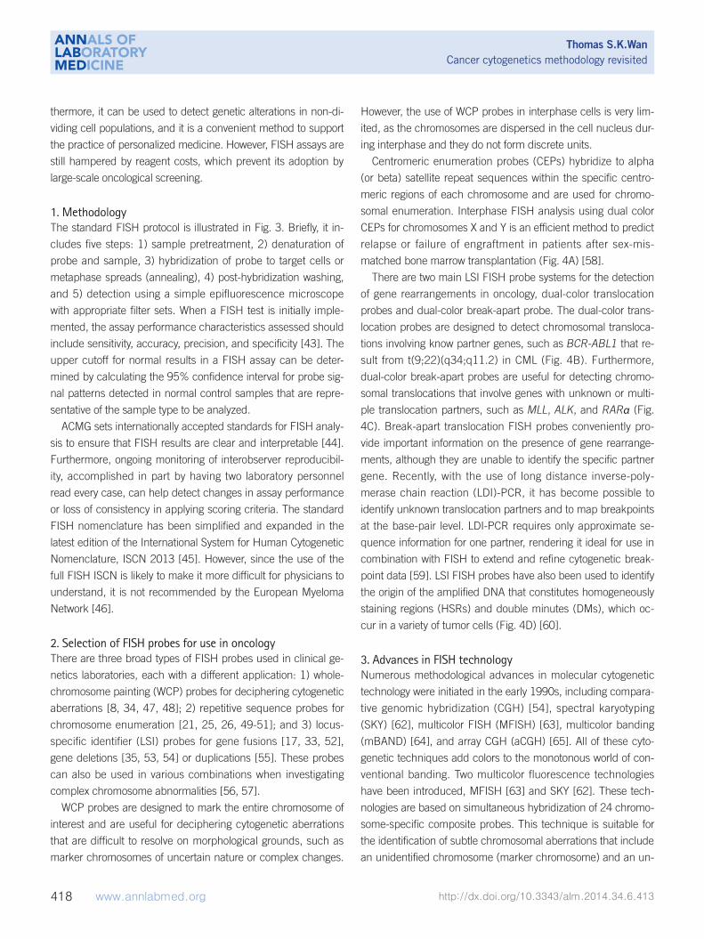
Fig. 4. Interphase FISH images. (A) Interphase FISH using dual color centromeric-specific probes for chromosomes X (red) and Y (green) to determine the proportion of donor cells in the peripheral blood of the recipient (XY, arrow; XX, block arrow). (B) Interphase FISH using a dual color dual fusion BCR-ABL1 translocation probe, showing a 2G2R pattern in a normal cell (block arrow) and 1G1R2F in a Philadel-phia-positive cell (arrow). (C) Interphase FISH using a dual color breakapart MLL translocation probe, showing an MLL split signal (distal MLL region, arrow; proximal MLL region, block arrow), and indicating MLL gene rearrangement. (D) MYC amplification in a neuroblastoma (arrow) and a normal cell with a 2G2R pattern (block arrow). The MYCN gene is labeled with a green fluorochrome, whereas the centro-meric probe for chromosome 2 is labeled with a red fluorochrome.
A B
C D

Thomas S.K.WanCancer cytogenetics methodology revisited
420 www.annlabmed.org http://dx.doi.org/10.3343/alm.2014.34.6.413
CGH is based on quantitative dual-color FISH along each
chromosome [61]. CGH can be used to detect genetic imbal-
ances in test genomes, and to determine the chromosomal map
positions of gains and losses of entire chromosomes or chromo-
somal subregions present in normal reference metaphase prep-
arations. A distinct advantage of CGH is that tumor DNA is the
only requirement for this analysis. Thus, archived, formalin-fixed
and paraffin-embedded tissues can be used as well. CGH is
useful for cancer research, especially for determining the low
mitotic index of malignant cells with poor chromosome morphol-
ogy and resolution [66-68].
Tremendous technical advances in cytogenetics have changed
the approaches used for clinical diagnostics and research, in
particular, the development of array CGH (aCGH) technology for
“molecular karyotyping” with a resolution of 100 kb to 1 Mb [65].
aCGH greatly improves the resolution of this technique by substi-
tuting the hybridization targets, the metaphase chromosome
spread, with genomic segments spotted in an array format. The
complexity of the genomic aberrations in most human tumors
hampers delineation of the genes that drive the tumorigenic pro-
cess. Interestingly, cognate mouse models have been shown to
recapitulate these genetic alterations with unexpected fidelity
[69]. These results indicate that cross-species aCGH analysis is
a powerful strategy to identify the responsible genes and assess
their oncogenic capacity in the appropriate genetic context [69].
Recently developed genomic microarray methodologies, includ-
ing aCGH and single-nucleotide polymorphism (SNP)-based ar-
rays, are innovative methods that provide genomic data for mul-
tiple neoplastic disorders [70-72]. In addition, these methods
can reveal additional important information about the genetics of
specific disorders, such as leukemia with normal cytogenetic
and FISH analyses [73, 74]. In 2013, ACMG developed profes-
sional standards and guidelines to assist clinical laboratories in
the validation, consistent use, and the interpretation and report-
ing of results from these microarray methodologies [75].
CANCER CYTOGENETICS RESOURCES
1. The Mitelman Database of Chromosome Aberrations and Gene Fusions in Cancer
The Mitelman Database of Chromosome Aberrations and Gene
Fusions in Cancer (http://cgap.nci.nih.gov/Chromosomes/Mitel-
man) includes a comprehensive database of all published neo-
plasia-associated karyotypes and their corresponding gene fu-
sions. The available information on chromosome abnormalities in
human neoplasias has steadily increased over the past three de-
cades. The data in this database have been manually culled from
the literature by Felix Mitelman, Bertil Johansson, and Fredrik
Mertens. The total number of tumor cases, in which clonal cyto-
genetic aberrations have been reported has reached 64,319,
and the database was updated with the addition of 2,072 chime-
ric fusion genes in May 2014 [76].
2. The Atlas of Genetics and Cytogenetics in Oncology and Haematology
The Atlas of Genetics and Cytogenetics in Oncology and Hae-
matology (http://atlasgeneticsoncology.org/) [77], which was es-
tablished in 1997, is a peer-reviewed, open access, online jour-
nal, encyclopedia, and database that is devoted to genes, cyto-
genetics, and clinical entities in cancer and cancer-prone dis-
eases. Approximately 2,200 authors have contributed to the At-
las so far, making 2,167 review articles available. The Atlas con-
tains peer-reviewed articles on 1,135 genes, 503 leukemia enti-
ties, 177 solid tumors, and 104 cancer-prone inherited diseases.
It also contains “automated cards” on 8,190 other genes that
are potentially implicated in cancer [78].
3. Cytogenetic nomenclatureIn the medical genetics community, interpretation and scientific
communication is often facilitated by universally accepted no-
menclature with precisely defined terms and syntax conventions
that minimize complexity and add precision to the process. Cy-
togenetic nomenclature is based on the reports of an interna-
tional committee that was established in 1960, known as the In-
ternational System for Human Cytogenetic Nomenclature (ISCN)
[79]. The nomenclature is updated periodically, most recently in
2013 with expanded guidelines for cancer cytogenetics, FISH,
and microarray [45]. In cancer cytogenetic, an abnormal cyto-
genetic clone is defined as a population of cells with the same
chromosome complement that is derived from a single progeni-
tor. A clone must have at least two cells with the same aberra-
tion if the aberration is a chromosome gain or a structural rear-
rangement. If the abnormality is loss of a chromosome, the
same loss must be present in at least three cells to be accepted
as clonal. However, in the current version of the ISCN (2013),
two cells with identical losses of one or more chromosomes and
the same structural aberration(s) may be considered clonal and
newly included [45]. However, whether two cells with the same
loss of a single chromosome or one cell with a gain of a single
chromosome in a composite karyotype should be counted and
included in the size of the clone has not been mentioned in the
current nomenclature system.

Thomas S. K. WanCancer cytogenetics methodology revisited
http://dx.doi.org/10.3343/alm.2014.34.6.413 www.annlabmed.org 421
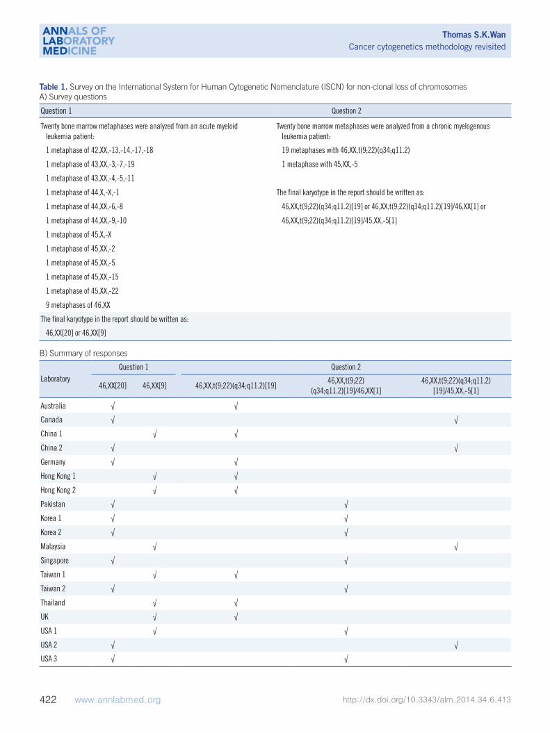
Recently, we conducted a pilot survey of the reporting prac-
tices for the random loss or gain of a single chromosome in clin-
ical cytogenetics reports. The questionnaire, which consisted of
two questions, was sent to 19 cancer cytogenetic laboratories in
13 counties and cities, and we invited them to report to us using
their usual reporting system (Table 1). In question 1, partici-
pants were presented with a bone marrow sample from an AML
patient with non-clonal (random) loss of chromosome(s), and
asked whether these metaphases should be considered as nor-
mal and included in the clone size of the normal karyotype. In
question 2, participants were presented with a marrow blood
sample from a CML patient with 19 metaphases of t(9;22)(q34;
q11.2) and one metaphase with only -5. We asked whether the
normal metaphase with a loss of a chromosome 5 should be re-
ported in final karyotype in addition to the 19 metaphases of
t(9;22)(q34;q11.2). To our surprise, the responses were not
unanimous. For question 1, 11 of the 19 laboratories (57.9%)
reported 46,XX[20], and the remaining 8 (42.1%) reported 46,
XX[9] (Table 1). Therefore, 57.9% of the cytogenetic laboratories
assumed that metaphases with non-clonal loss of chromosomes
were normal, and they included all these metaphases in the
clone size of normal clone. Most laboratory accreditation bodies
require the “total number of metaphases analyzed” in the final
report. Thus, for the 42.1% of cytogenetic laboratories that re-
ported 46,XX[9], there is a general understanding by the clini-
cians that the patient has 11 metaphases with non-clonal loss
or gain of chromosomes among the 20 metaphases analyzed.
For question 2, 8 of the 19 laboratories (42.1%) reported 46,
XX,t(9;22)(q34;q11.2) [19] and omitted the only metaphase with
-5; 7 laboratories (36.8%) reported 46,XX,t(9;22)(q34;q11.2)
[19]/46,XX[1] and counted the only metaphase with -5 as a nor-
mal clone; and 4 laboratories (21.1%) reported 46,XX,t(9;22)
(q34;q11.2)[19]/45,XX,-5 [1] and simply wrote down the only
metaphase with -5 in the final karyotype (Table 1). Taken to-
gether, the ISCN standing committee should continue to discuss
such discrepancies and make efforts to align the reporting sys-
tem used by cytogenetic laboratories [80].
Interestingly, a review report of the CAP FISH proficiency sur-
veys between 1997 and 2001 showed that syntax errors were
far more common than diagnostic errors [81]. In addition, the
syntax errors that are common in the proficiency surveys are
also common in FISH surveys. Recently, since the full FISH
ISCN is likely more difficult for physicians to understand, the
use of FISH ISCN is not recommended by the European My-
eloma Network [46]. Therefore, continuous improvement of the
nomenclature guidelines to address some of these issues and
identify the cause of these variations is essential [78, 82].
FUTURE PROSPECTS AND CONCLUDING REMARKS
Over the past five decades, innovative technical advances in the
field of cancer cytogenetics have greatly enhanced the detection
of chromosomal alterations and have facilitated the research and
diagnostic potential of chromosomal studies in malignancies.
Karyotyping of a single cell is still the easiest way to understand
the relationship between clonal evolution and disease progres-
sion. The use of advanced FISH techniques allows for the identi-
fication of chromosomal alterations that are unresolved by karyo-
typing. Recently, DNA microarray technologies have been de-
veloped that provide a high-resolution view of the whole genome,
which may yield massive amounts of new information on cancer
genomics. Strikingly, cancer cytogenetics not only provides key
information to improve the care of patients with leukemia and
various cancers but also acts as a guide to identify the genes re-
sponsible for the development of these neoplastic states.
Ongoing research and development on automated cytogenet-
ics procedures includes: an automated metaphase harvester,
automated metaphase spreader, high-throughput slide stainer,
high-throughput metaphase finder, automated karyotyper, auto-
mated FISH pretreatment processor, automated FISH spot
counting, and computerized sample tracking system. However,
microscopy is still an essential technique for a final check of the
chromosomes. Obviously, cytogenetic laboratories need to obtain
more capital investment to keep up with the fast advancing and
robotic technologies, since equipment costs are currently high.
The main goal of the FISH laboratory is to identify the tech-
niques that are most useful and informative for a particular
study and perform thorough analyses to arrive at an interpreta-
tion that is useful for research and diagnostic purposes. The
unique characteristics of peptide nucleic acids (PNAs) allow for
the use of shorter probes, which combined with the hydropho-
bic nature of the peptide backbone, enables PNA probes to
more easily traverse the hydrophobic core of cell membranes. A
staining efficiency of nearly 100% can be achieved by using
telomere probes based on PNA [83]. Telomere length can be
measured by quantitative FISH in human [84] and other verte-
brate cells [85]. Recently, the inherent flexibility of de novo syn-
thesized oligonucleotide libraries was shown to have a powerful
advantage that could aid in the visualization of high-resolution
fine-scale genomic structure [86].
In the future, targeted therapies will find broader applications
Thomas S.K.WanCancer cytogenetics methodology revisited
422 www.annlabmed.org http://dx.doi.org/10.3343/alm.2014.34.6.413
Table 1. Survey on the International System for Human Cytogenetic Nomenclature (ISCN) for non-clonal loss of chromosomesA) Survey questions
Question 1 Question 2
Twenty bone marrow metaphases were analyzed from an acute myeloid leukemia patient:
Twenty bone marrow metaphases were analyzed from a chronic myelogenous leukemia patient:
1 metaphase of 42,XX,-13,-14,-17,-18 19 metaphases with 46,XX,t(9;22)(q34;q11.2)
1 metaphase of 43,XX,-3,-7,-19 1 metaphase with 45,XX,-5
1 metaphase of 43,XX,-4,-5,-11
1 metaphase of 44,X,-X,-1 The final karyotype in the report should be written as:
1 metaphase of 44,XX,-6,-8 46,XX,t(9;22)(q34;q11.2)[19] or 46,XX,t(9;22)(q34;q11.2)[19]/46,XX[1] or
1 metaphase of 44,XX,-9,-10 46,XX,t(9;22)(q34;q11.2)[19]/45,XX,-5[1]
1 metaphase of 45,X,-X
1 metaphase of 45,XX,-2
1 metaphase of 45,XX,-5
1 metaphase of 45,XX,-15
1 metaphase of 45,XX,-22
9 metaphases of 46,XX
The final karyotype in the report should be written as:
46,XX[20] or 46,XX[9]
B) Summary of responses
LaboratoryQuestion 1 Question 2
46,XX[20] 46,XX[9] 46,XX,t(9;22)(q34;q11.2)[19]46,XX,t(9;22)
(q34;q11.2)[19]/46,XX[1]46,XX,t(9;22)(q34;q11.2)
[19]/45,XX,-5[1]
Australia √ √
Canada √ √
China 1 √ √
China 2 √ √
Germany √ √
Hong Kong 1 √ √
Hong Kong 2 √ √
Pakistan √ √
Korea 1 √ √
Korea 2 √ √
Malaysia √ √
Singapore √ √
Taiwan 1 √ √
Taiwan 2 √ √
Thailand √ √
UK √ √
USA 1 √ √
USA 2 √ √
USA 3 √ √

Thomas S. K. WanCancer cytogenetics methodology revisited
http://dx.doi.org/10.3343/alm.2014.34.6.413 www.annlabmed.org 423
in leukemia and cancer. Personalized oncology, in addition to
FISH and drug target information for treatment decision making,
other aspects include the application of genetic markers for pa-
tient risk stratification. This is particularly relevant to CLL [87] and
multiple myeloma. Since the neoplastic population in both CLL
and multiple myeloma is mitotically inactive, the use of inter-
phase FISH for the detection of genetic abnormalities plays a sig-
nificant role in the prognostication and risk stratification of these
disorders [88]. The identification or selection of malignant cells
by morphology, immunophenotyping, or through sorting of
plasma cells is required before FISH probes can yield reliable re-
sults [89]. In addition, although personalized genomic medicine
in the clinic may be very attractive, there is a need for succinct
clinicopathological correlation and the rational use of faster, more
cost-effective methods among the large array of genomic tests
available for drug-target selection. Recently, the power and appli-
cability of whole-genome sequencing for the diagnosis of leuke-
mia patients with cryptic gene fusion has demonstrated, and it
hugely affected the clinical management and prognosis of these
patients. However, leukemic patients with cryptic gene rearrange-
ments can often be diagnosed without whole-genome analysis –
a costly, time consuming, and highly specialized procedure.
Conventional cytogenetics is now complemented by FISH and
molecular biology. A FISH-negative cryptic PML/RARα rear-
rangement detected by long-distance PCR and sequencing
analyses has also been reported [90]. Therefore, FISH analysis
should be conducted, if the morphologic, cytogenetic, and mo-
lecular findings are inconsistent. It is envisaged that efforts
made towards the characterization of molecular defects in neo-
plasms will ultimately be translated into better clinical outcomes
for patients. Taken together, the morphologic, karyotyping, FISH,
and molecular features should all be considered to obtain accu-
rate diagnoses of malignancies. This highlights the clinical im-
portance of a combined modality approach for the accurate di-
agnosis and classification of cancers.
Author’s Disclosure of Potential Conflicts of Interest
No potential conflicts of interest relevant to this article were re-
ported.
Acknowledgements
The author thanks Eden Wan for drawing Figs. 1 and 3, Sophia
Ho for expert clerical assistance, Dr. Huifang Huang of Fujian
Medical University (China) for providing the R-banded image of
inv(16) shown in Fig. 2N, and all of the laboratories who partici-
pated in the ICSN nomenclature survey presented in Table 1.
REFERENCES
1. Nowell PC and Hungerford DA. A minute chromosome in human chron-ic granulocytic leukemia. Science 1960;132:1497.
2. Rowley JD. A new consistent chromosomal abnormality in chronic my-elogeneous leukaemia identified by quinacrine fluorescence and Giem-sa staining. Nature 1973;243:290-3.
3. de Klein A, van Kessel AG, Grosveld G, Bartram CR, Hagemeijer A, Bootsma D, et al. A cellular oncogene is translocated to Philadelphia chromosome in chronic myelocytic leukemia. Nature 1982;300:765-7.
4. Vardiman JW, Thiele J, Arber DA, Brunning RD, Borowitz MJ, Porwit A, et al. The 2008 revision of the World Health Organization (WHO) classi-fication of myeloid neoplasm and acute leukemia: rationale and impor-tant changes. Blood 2009;114:937-51.
5. Manuelidis L, Langer-Safer PR, Ward DC. High-resolution mapping of satellite DNA using biotin-labeled DNA probes. J Cell Biol 1982;95:619-25.
6. Wan TS and Ma ES. Molecular cytogenetics: an indispensable tool for cancer diagnosis. Chang Gung Med J 2012;35:96-110.
7. Wan TS and Ma ES. The role of FISH in hematologic cancer. Int J He-matol Oncol 2012;1:71-86.
8. Wan TS, Ma SK, Chan GC, Ching LM, Ha SY, Chan LC. Complex cyto-genetic abnormalities in T-lymphoblastic lymphoma: resolution by spec-tral karyotyping. Cancer Genet Cytogenet 2000;118:24-7.
9. Raza A, Maheshwari Y, Preisler HD. Differences in cell cycle character-istics among patients with acute nonlymphocytic leukemia. Blood 1987; 69:1647-53.
10. Yunis JJ. Comparative analysis of high-resolution chromosome tech-niques for leukemic bone marrows. Cancer Genet Cytogenet 1982;7: 43-50.
11. Garipidou V and Secker-Walker LM. The use of fluorodeoxyuridine syn-chronization for cytogenetic investigation of acute lymphoblastic leuke-mia. Cancer Genet Cytogenet 1991;52:107-11.
12. Brothman AR, Schneider NR, Saikevych I, Cooley LD, Butler MG, Patil S, et al. Cytogenetic Heteromorphisms. Survey results and reporting prac-tices of Giemsa-band regions that we have pondered for years. Arch Pathol Lab Med 2006;130:947-9.
13. Wan TS, Ma SK, Chan LC. Acquired pericentric inversion of chromo-some 9 in essential thrombocythemia. Hum Genet 2000;106:669-70.
14. Lee SG, Park TS, Lim G, Lee KA, Song J, Choi JR. Constitutional peri-centric inversion 9 and hematological disorders: a Korean tertiary insti-tution’s experience over eight years. Ann Clin Lab Sci 2010;40:273-7.
15. Wan TS, Au WY, Chan JC, Chan LC, Ma SK. Trisomy 21 as the sole ac-quired karyotypic abnormality in acute myeloid leukemia and myelodys-plastic syndrome. Leuk Res 1999;23:1079-83.
16. Wan TS, Ma SK, Au WY, Liu HS, Chan JC, Chan LC. Trisomy 21 and other chromosomal abnormalities in acute promyelocytic leukemia. Cancer Genet Cytogenet 2003;140:170-3.
17. Ma SK, Wan TS, Cheuk AT, Fung LF, Chan GC, Chan SY, et al. Charac-terization of additional genetic events in childhood acute lymphoblastic leukemia with TEL/AML1 gene fusion: a molecular cytogenetics study. Leukemia 2001;15:1442-7.
18. Ma SK and Wan TS. Single autosomal trisomy in acute myeloid leuke-mia and myelodysplastic syndrome. Curr Genomics 2000;1:153-73.

Thomas S.K.WanCancer cytogenetics methodology revisited
424 www.annlabmed.org http://dx.doi.org/10.3343/alm.2014.34.6.413
19. Heim S and Mitelman F. Numerical chromosome aberrations in human neoplasia. Cancer Genet Cytogenet 1986;22:99-108.
20. Rajagopalan H and Lengauer C. Aneuploidy and cancer. Nature 2004; 432:338-41.
21. Ma SK, Wan TS, Au EY, Chan LC. Trisomy 5 in two cases of acute mono-cytic leukemia with hyperdiploid clones. Leuk Res 1998;22:961-4.
22. Wan TS, Yip SF, Yeung YM, Chan LC, Ma SK. Fatal diffuse alveolar dam-age complicating acute myeloid leukemia with abnormal eosinophils and trisomy X. Ann Hematol 2002;81:167-9.
23. Mehta AB, Bain BJ, Fitchett M, Shah S, Secker-Walker LM. Trisomy 13 and myeloid malignancy-characteristic blast cell morphology: a United Kingdom Cancer Cytogenetics Group survey. Br J Haematol 1998;101: 749-52.
24. Ma SK and Wan TS. Blast cell morphology in acute myeloid leukemia with trisomy 13. Leuk Res 1999;23:767-9.
25. Yip SF, Wan TS, Chan LC, Chan GC. Trisomy 4 as sole karyotypic ab-normality in acute lymphoblastic leukemia: different clinical features and treatment response between B and T phenotypes? Cancer Genet Cytogenet 2006;164:94-5.
26. Au WY, Ma SK, Wan TS, Jim MH, Kwong YL. Subvalvular pulmonary stenosis, demyelination and myelodysplasia with monosomy 7. Leuk Lymphoma 2002;43:1505-7.
27. Au WY, Wan TS, Leung RY, Lie AK. Sequential chronic myelogenous leukemia, B-lineage lymphoma and erythroleukemia with monosomy 7 over 10 years. Leuk Lymphoma 2012;53:733-5.
28. Surapolchai P, Ha SY, Chan GC, Lukito J, Wan TS, So CC, et al. Central diabetes insipidus: an unusual complication in a child with juvenile my-elomonocytic leukemia and monosomy 7. J Pediatr Hematol Oncol 2013;35:e84-7.
29. Wan TS, Ma ES, Lam CC, Chan LC, Lee KK, Au WY. Deletion 9q as the sole karyotypic abnormality in myelocytic disorders: a new case of my-elodysplastic syndrome and its prognostic implications in acute myelo-cytic leukemia. Cancer Genet Cytogenet 2003;145:184-6.
30. Wan TS, Chim CS, So CK, Chan LC, Ma SK. Complex variant 15;17 translocations in acute promyelocytic leukemia. A case report and review of three-way translocations. Cancer Genet Cytogenet 1999;111:139-43.
31. So CW, Ma SK, Wan TS, Chan GC, Ha SY, Chan LC. Analysis of MLL-derived transcripts in infant acute monocytic leukemia with a complex translocation (1;11;4)(q21;q23;p16). Cancer Genet Cytogenet 2000; 117:24-7.
32. So CC, Wan TS, Yip SF, Chan LC. A dual colour dual fusion fluorescence in situ hybridization study on the genesis of complex variant transloca-tions in chronic myelogenous leukemia. Oncol Rep 2008;19:1181-4.
33. Wan TS, So CC, Hui KC, Yip SF, Ma ES, Chan LC. Diagnostic utility of dual fusion PML/RARα translocation DNA probe (D-FISH) in acute pro-myelocytic leukemia. Oncol Rep 2007;17:799-805.
34. Wan TS, Ma SK, Yip SF, Yeung YM, Chan LC. Molecular characteriza-tion of der(15)t(11;15) as a secondary cytogenetic abnormality in acute promyelocytic leukemia with cryptic PML-RARα fusion on chromosome 17q. Cancer Genet Cytogenet 2000;121:90-3.
35. So CC, Wan TS, Ma ES, Chan LC. An unbalanced translocation, der(17)t(1;17)(p13;p11.2), leads to heterozygous loss of TP53 and is associat-ed with clinical evolution in myelodysplastic syndrome. Br J Biomed Sci 2008;65:36-8.
36. Willem P, Pinto M, Bernstein R. Translocation t(1;7) revisited. Report of three further cases and review. Cancer Genet Cytogenet 1988;36:45-54.
37. So CC, Ma ES, Wan TS, Yip SF, Chan LC. Clinicopathological features of unbalanced translocation Der(1;7)(q10;p10) in myeloid neoplasms. Leuk Res 2008;32:1000-1.
38. Pedersen B, Nørgaard JM, Pedersen BB, Clausen N, Rasmussen IH,
Thorling K. Many unbalanced translocation show duplication of a trans-location participant. Clinical and cytogenetic implications in myeloid he-matologic malignancies. Am J Hematol 2000;64:161-9.
39. Sanada M, Uike N, Ohyashiki K, Ozawa K, Lili W, Hangaishi A, et al. Un-balanced translocation der(1;7)(q10;p10) defines a unique clinicopatho-logical subgroup of myeloid neoplasms. Leukemia 2007;21:992-7.
40. Wan TS, Ma SK, Li CK, Chan LC. Atypical fluorescence in situ hybrid-ization pattern in chronic myeloid leukemia due to cryptic insertion of BCR at 9q34. Leukemia 2004;18:161-2.
41. So CC, Wan TS, Chow JL, Hui KC, Choi WW, Lam CC, et al. A single-center cytogenetic study of 629 Chinese patients with de novo acute myeloid leukemia – evidence of major ethnic differences and a high prevalence of acute promyelocytic leukemia in Chinese patients. Can-cer Genet 2011;204:430-8.
42. Cheng Y, Wang Y, Wang H, Chen Z, Lou J, Xu H, et al. Cytogenetic pro-file of de novo acute myeloid leukemia: a study based on 1432 patients in a single institution of China. Leukemia 2009;23:1801-6.
43. Saxe DF, Persons DL, Wolff DJ, Theil KS; Cytogenetics Resource Com-mittee of the College of American Pathologists. Validation of fluores-cence in situ hybridization using an analyste-specific reagent for detec-tion of abnormalities involving the mixed lineage leukemia gene. Arch Pathol Lab Med 2012;136:47-52.
44. American College of Medical Genetics, Standards and Guidelines for Clinical Genetics Laboratories. https://www.acmg.net/StaticContent/SGs/Section_E_2011.pdf (2009 edition, Revised on Jan 2010).
45. Shaffer LG, McGowan-Jordan J, Schmid M, eds. ISCN (2013): An In-ternational System for Human Cytogenetic Nomenclature. Basel: S. Karger, 2013.
46. Ross FM, Avet-Loiseau H, Ameye G, Gutiérrez NC, Liebisch P, O’Connor S,et al. Report from the European Myeloma Network on interphase FISH in multiple myeloma and related disorders. Heamatologica 2012;97: 1272-7.
47. Ma SK, Wan TS, Au WY, Kwong YL, Chan LC. Atypical chronic myeloid leukemia with der(20)t(17;20)(q21;q13). Cancer Genet Cytogenet 1999; 112:130-3.
48. Wan TS, Ma, SK, Yip SF, Yeung YM, Chan LC. Two balanced and novel chromosomal translocations in myeloid malignancies.characterization by multiplex fluorescence in situ hybridization. Cancer Genet Cytogenet 2002;139:52-6.
49. Ma SK, Lee AC, Wan TS, Lam CK, Chan LC. Trisomy 8 as a secondary genetic change in acute megakaryoblastic leukemia associated with Down’s syndrome. Leukemia 1999;13:491-2.
50. Ma SK, Kwong YL, Shek TW, Wan TS, Chow EY, Chan JC, et al. The role of trisomy 8 in the pathogenesis of chronic eosinophilic leukemia. Hum Pathol 1999;30:864-8.
51. Cheung AM, Wan TS, Leung JC, Chan LY, Huang H, Kwong YL, et al. Aldehyde dehydrogenase activity in leukemia blasts defines a subgroup of acute myeloid leukemia with adverse prognosis and superior NOD/SCID engrafting potential. Leukemia 2007;21:1423-30.
52. Meyer C, Kowarz E, Yip SF, Wan TS, Chan TK, Dingermann T, et al. A complex MLL rearrangement identified five years after initial MDS diag-nosis results in out-of-frame fusions without progression to acute leuke-mia. Cancer Genet 2011;204:557-62.
53. Ma SK, Wan TS, Au WY, Fung LF, So CK, Chan LC. Chromosome 11q deletion in myeloid malignacies. Leukemia 2002;16:953-5.
54. Cheung AM, Fung TK, Fan AK, Wan TS, Chow HC, Leung JC, et al. Successful engraftment by leukemia initiating cells in adult acute lym-phoblastic leukemia after direct intrahepatic injection into uncondi-tioned newborn NOD/SCID mice. Exp Hematol 2010;38:3-10.
55. Wan TS, Ma ES, Chen YT. Near-tetraploid acute myeloid leukemia. Br J

Thomas S. K. WanCancer cytogenetics methodology revisited
http://dx.doi.org/10.3343/alm.2014.34.6.413 www.annlabmed.org 425
Haematol 2011;155:285.56. So CC, Yung KH, Chu ML, Wan TS. Diagnostic challenges in a case of
B cell lymphoma unclassifiable with features intermediate between dif-fuse large B-cell lymphoma and Burkitt lymphoma. Int J Hematol 2013; 98:478-82.
57. Lee JH, Wan TS, Ha JS. Acute myeloid leukemia with a novel t(8;21) variant: paracentric inversion-associatedins(21;8). Leuk Lymphoma 2014;55:441-3.
58. Li YH, Ma SK, Wan TS, Au WY, Fung LF, Leung AY. Lineage-specific dif-ferences in telomere length after bone marrow transplantation. Bone Marrow Transplant 2002;30:475-7.
59. Yang JJ, Marschalek R, Meyer C, Park TS. Diagnostic usefulness of ge-nomic breakpoint analysis of various gene rearrangements in acute leu-kemias: a perspective of long distance- or long distance inverse-PCR-based approaches. Ann Lab Med 2012;32:316-8.
60. Wan TS, Ma ES, Chan GC, Chan LC. Investigation of MYCN status in neuroblastoma by fluorescence insitu hybridization. Int J Mol Med 2004;14:981-7.
61. Kallioniemi A, Kallioniemi OP, Sudar D,Rutovitz D, Gray JW, Waldman F, et al. Compararive genomic hybridization for molecular cytogenetic analysis of solid tumors. Science 1992:258:818-21.
62. Schröck E, du Manoir S, Veldman T, Schoell B, Wienberg J, Ferguson-Smith MA, et al. Multicolor spectral karyotyping of human chromo-somes. Science 1996;273:494-7.
63. Speicher MR, Gwyn Ballard S, Ward DC. Karyotyping human chromo-somes by combinatorial multi-fluor FISH. Nat Genet 1996;12:368-75.
64. Chudoba I, Plesch A, Lörch T, Lemke J, Claussen U, Senger G. High resolution multicolor-banding: a new technique for refine FISH analysis of human chromosomes. Cytogenet Cell Genet 1999;84:156-60.
65. Pinkel D, Segraves R, Sudar D,Clark S, Poole I, Kowbel D, et al. High resolution analysis of DNA copy number variation using comparative ge-nomic hybridization to microarrays. Nature Genetics 1998;20:207-11.
66. Tsao SW, Wong N, Wang X, Liu Y, Wan TS, Fung LF, et al. Nonrandom chromosomal imbalances in human ovarian surface epithelial cells im-mortalized by HPV16-E6E7 viral oncogenes. Cancer Genet Cytogenet 2001;130:141-9.
67. Hu YC, Lam KY, Law SY, Wan TS, Ma ES, Kwong YL, et al. Establish-ment, characterization, karyotyping, and comparative genomic hybrid-ization analysis of HKESC-2 and HKESC-3, two newly established hu-man esophageal squamous cell carcinoma cell lines. Cancer Genet Cy-togenet 2002;135:120-7.
68. Wong MP, Fung LF, Wang E, Chow WS, Chiu SW, Lam WK, et al. Chro-mosomal aberrations of primary lung adenocarcinomas in nonsmokers. Cancer 2003;97:1263-70.
69. Peeper D and Berns A. Cross-species oncogenomics in cancer gene identification. Cell 2006;125:1230-3.
70. Slovak ML, Bedell V, Hsu YH,Estrine DB, Nowak NJ, Delioukina ML, et al. Molecular karyotypes of Hodgkin and Reed/Sternberg cells at dis-ease onset reveal distinct copy number alterations in chemosensitive versus refractory Hodgkin lymphoma. Clin Cancer Res 2011;17:3443-54.
71. Walter MJ, Payton JE, Ries RE, Shannon WD, Deshmukh H, Zhao Y, et al. Acquired copy number alterations in adult acute myeloid leukemia genomes. Proc Natl Acad Sci U S A 2009;106:12950-5.
72. Yu L, Slovak ML, Mannoor K, Chen C, Hunger SP, Carroll AJ, et al. Mi-croarray detection of multiple recurring submicroscopic chromosomal aberrations in pediatric T-cell acute lymphoblastic leukemia. Leukemia 2011;25:1042-6.
73. Kawamata N, Ogawa S, Zimmermann M, Kato M, Sanada M, Hemmin-ki K, et al. Molecular allelokaryotyping of pediatric acute lymphoblastic leukemias by high-resolution single nucleotide polymorphism oligonu-cleotide genomic microarray. Blood 2008;111:776-84.
74. O’Keefe C, McDevitt MA, Maciejewski JP. Copy neutral loss of heterozy-gosity: a novel chromosomal lesion in myeloid malignancies. Blood 2010;115:2731-9.
75. Cooley LD, Lebo M, Li MM, Slovak ML, Wolff DJ for AWorking Group of the American College of Medical Genetics and Genomics (ACMG) Lab-oratory Quality Assurance Committee. American College of Medical Ge-netics and Genomics technical standards and guidelines: microarray analysis for chromosome abnormalities in neoplastic disorders. Genet Med 2013;15:484-94.
76. Mitelman F, Johansson B, Mertens F (Eds.), Mitelman database of chromosome aberrations in cancer. http://cgap.nci.nih.gov/Chromo-somes/Mitelman (Updated on May 2014).
77. Atlas of Genetics and Cytogenetics in Oncology and Haematology. http://AtlasGeneticsOncology.org (Updated on May 2012).
78. Huret JL, Ahmad M, Arsaban M, Bernheim A, Cigna J, Desangles F, et al. Atlas of genetics and cytogenetics in oncology and haematology in 2013. Nucleic Acids Res 2013;41(Database issue):D920-4.
79. Shaffer LG, Tommerup N, eds. ISCN (2005): an International System for Human Cytogenetic Nomenclature. Basel: S. Karger, 2005.
80. Mitelman F and Rowley JD. ISCN (2005) is not acceptable for describ-ing clonal evolution in cancer. Genes Chromosomes Cancer 2007;46: 213-4.
81. Mascarello JT, Cooley LD, Davison K, Dewald GW, Brothman AR, Her-rman M, et al. Problems with ISCN FISH Nomenclature make it not practical for use in clinical test reports or cytogenetic databases [cor-rected]. Genet Med 2003;5:370-7.
82. Shaffer LG, Slovak ML, Campbell LJ, eds. ISCN (2009): an International System for Human Cytogenetic Nomenclature. Basel: S. Karger, 2009.
83. Lansdorp PM, Verwoerd NP, van de Rijke FM, Dragowska V, Little MT, Dirks RW, et al. Heterogeneity in telomere length of human chromo-somes. Hum Mol Genet 1996;5:685-91.
84. Wan TS, Martens UM, Poon SS, Tsao SW, Chan LC, Lansdorp PM. Ab-sence or low number of telomere repeats at junctions of dicentric chro-mosomes. Genes Chromosomes Cancer 1999;24:83-6.
85. Zijlmans JM, Martens UM, Poon SS, Raap AK, Tanke HJ, Ward RK, et al. Telomeres in the mouse have large inter-chromosomal variations in the number of T2AG3 repeats. Proc Natl Acad Sci USA1997;94:7423-8.
86. Yamada NA, Rector LS, Tsang P, Carr E, Scheffer A, Sederberg MC, et al. Visualization of fine-scale genomic structure by oligonucleotide-based high-resolution FISH. Cytogenet Genome Res 2011;132:248-54.
87. Stevens-Kroef MJ, van den Berg E, Olde Weghuis D, Geurts van Kessel A, Pfundt R, Linssen-Wiersma M, et al. Identification of prognostic rele-vant chromosomal abnormalities in chronic lymphocytic leukemia using microarray-based genomic profiling. Mol Cytogenet 2014;7:3.
88. Hallek M, Bergsagel PL, Anderson KC. Multiple myeloma: increasing evidence for a multistep transformation process. Blood 1998;91:3-21.
89. Leung EW, Sin PL, Wan TS. The impact of FICTION on the detection of genetic aberrations in multiple myeloma. J H K Inst Med Lab Sci 2012: 13:1-8.
90. Kim MJ, Cho SY, Kim MH, Lee JJ, Kang SY, Cho EH, et al. FISH-nega-tive cryptic PML-RARA rearrangement detected by long-distance poly-merase chain reaction and sequencing analyses: a case study and re-view of the literature. Cancer Genet Cytogenet 2010;203:278-83.
Related Documents