Molecules 2012, 17, 3202-3242; doi:10.3390/molecules17033202 molecules ISSN 1420-3049 www.mdpi.com/journal/molecules Review Cancer Chemoprevention by Carotenoids Takuji Tanaka 1,2, *, Masahito Shnimizu 3 and Hisataka Moriwaki 3 1 Tohkai Cytopathology Institute, Cancer Research and Prevention (TCI-CaRP), 5-1-2 Minami-Uzura, Gifu 500-8285, Japan 2 Department of Tumor Pathology, Gifu University Graduate School of Medicine, 1-1 Yanagido, Gifu 501-1194, Japan 3 Department of Medicine, Gifu University Graduate School of Medicine, 1-1 Yanagido, Gifu 501-1194, Japan * Author to whom correspondence should be addressed; E-Mail: [email protected]; Tel.: +81-58-273-4399; Fax: +81-58-273-4392. Received: 22 December 2011; in revised form: 15 February 2012 / Accepted: 6 March 2012 / Published: 14 March 2012 Abstract: Carotenoids are natural fat-soluble pigments that provide bright coloration to plants and animals. Dietary intake of carotenoids is inversely associated with the risk of a variety of cancers in different tissues. Preclinical studies have shown that some carotenoids have potent antitumor effects both in vitro and in vivo, suggesting potential preventive and/or therapeutic roles for the compounds. Since chemoprevention is one of the most important strategies in the control of cancer development, molecular mechanism-based cancer chemoprevention using carotenoids seems to be an attractive approach. Various carotenoids, such as β-carotene, -carotene, lycopene, lutein, zeaxanthin, β-cryptoxanthin, fucoxanthin, canthaxanthin and astaxanthin, have been proven to have anti-carcinogenic activity in several tissues, although high doses of β-carotene failed to exhibit chemopreventive activity in clinical trials. In this review, cancer prevention using carotenoids are reviewed and the possible mechanisms of action are described. Keywords: carotenoids; xanthophylls; cancer chemoprevention; mechanisms OPEN ACCESS

Welcome message from author
This document is posted to help you gain knowledge. Please leave a comment to let me know what you think about it! Share it to your friends and learn new things together.
Transcript

Molecules 2012, 17, 3202-3242; doi:10.3390/molecules17033202
molecules ISSN 1420-3049
www.mdpi.com/journal/molecules
Review
Cancer Chemoprevention by Carotenoids
Takuji Tanaka 1,2,*, Masahito Shnimizu 3 and Hisataka Moriwaki 3
1 Tohkai Cytopathology Institute, Cancer Research and Prevention (TCI-CaRP),
5-1-2 Minami-Uzura, Gifu 500-8285, Japan 2 Department of Tumor Pathology, Gifu University Graduate School of Medicine, 1-1 Yanagido,
Gifu 501-1194, Japan 3 Department of Medicine, Gifu University Graduate School of Medicine, 1-1 Yanagido,
Gifu 501-1194, Japan
* Author to whom correspondence should be addressed; E-Mail: [email protected];
Tel.: +81-58-273-4399; Fax: +81-58-273-4392.
Received: 22 December 2011; in revised form: 15 February 2012 / Accepted: 6 March 2012 /
Published: 14 March 2012
Abstract: Carotenoids are natural fat-soluble pigments that provide bright coloration to
plants and animals. Dietary intake of carotenoids is inversely associated with the risk of a
variety of cancers in different tissues. Preclinical studies have shown that some carotenoids
have potent antitumor effects both in vitro and in vivo, suggesting potential preventive
and/or therapeutic roles for the compounds. Since chemoprevention is one of the most
important strategies in the control of cancer development, molecular mechanism-based
cancer chemoprevention using carotenoids seems to be an attractive approach. Various
carotenoids, such as β-carotene, -carotene, lycopene, lutein, zeaxanthin, β-cryptoxanthin,
fucoxanthin, canthaxanthin and astaxanthin, have been proven to have anti-carcinogenic
activity in several tissues, although high doses of β-carotene failed to exhibit chemopreventive
activity in clinical trials. In this review, cancer prevention using carotenoids are reviewed
and the possible mechanisms of action are described.
Keywords: carotenoids; xanthophylls; cancer chemoprevention; mechanisms
OPEN ACCESS

Molecules 2012, 17 3203
Abbreviations
ABCA1, ATP-binding cassette transporter 1; AFB1, aflatoxin B1; Akt, protein kinase B; AMD,
age-related macular degeneration; AOM, azoxymethane; AP-1, activator 1; ARE, antioxidant response
element; CAR, constitutive androstane receptor; Cdks, cyclin-dependent kinases; CHRP,
β-cryptoxanthin- and hesperidin-rich powder; CMO-1, β-carotene 15,15'-monooxygenase; COM2,
β-carotene 9',10'-monooxygenase; COX, cyclooxygenase; CUSM, citrus unshiu segment membrane;
CVD, cardiovascular disease; CYP, cytochrome P450; DMH, 1,2-dimethylhydrazine; EGF, early
growth response gene; ERK, extracellular signal-regulated kinase; GJIC, gap junctional intercellular
communication; GSK3β, glycogen synthase kinase 3β; GSTs, glutathione S-transferases; HDL,
high-density lipoproteins; HO-1, heme oxygenase-1; IGF, insulin growth factor; IGFBPs, IGF binding
proteins; IL, interleukin; LDL, low-density lipoproteins; MJ, satsuma mandarin (Citrus unshiu Marc)
juice; MMP, matrix metalloproteinases; NF-kB, nuclear factor kappaB; 4-NQO, 4-nitroquinoline
1-oxide; NQO1, NAD(P)H:quinone oxidoreductase; Nrf2, NF-E2-related factor 2; OH-BBN,
N-butyl-N(4-hydroxybutyl)nitrosamine; PPARs, peroxisome proliferator-activated receptors; PSA,
prostate-specific antigen; RAR, retinoic acid receptor; ROS, reactive oxygen species; RXR, retinoid X
receptor; SXR/PXR, steroid and xenobiotic receptor/pregnane X receptor; TCF/LEF, transcription
factors T cell factor/lymphoid enhancer factor; TNF, tumor necrosis factor; TRE, TPA response
element; UV, ultraviolet; VDR, vitamin D3 receptor.
1. Introduction
To date, the cancer problem and the failure of conventional chemotherapy to achieve a reduction in
the mortality rates for common epithelial malignancies such as carcinomas of the lung, colon, breast,
prostate and pancreas, indicates a critical need for new approaches to control cancer development [1,2].
One of these approaches is chemoprevention, which is a pharmacological approach to intervention
with the objective of arresting or reversing the process of multi-step carcinogenesis. The carcinogenic
process may be driven by mutation(s), and followed by subsequent alterations in phenotypic, epigenetic
and genetic events. Pharmacologic modulation of these regulatory pathways, involving the effective
use of drugs, micronutrients and non-nutrients that block mutational damage of DNA, thus offers great
potential for cancer prevention.
There is a clear link between dietary intake or dietary habits and cancer development in man [3–5].
Dietary risk factors have ranked higher than smoking and much higher than pollution or occupational
hazards in their association with death due to cancer [6]. However, a number of compounds naturally
occurring in foods, particularly antioxidative compounds in plants, have shown promise as potential
chemopreventive agents [2,6–8]. These phytonutrients include the yellow, orange and red carotenoid
pigments that have recently been investigated. Epidemiologically, vegetable and fruit consumption
has constantly been associated with a reduced incidence of a variety of cancers [7–9], and dietary
carotenoid intake from these sources has similarly been correlated with a reduced cancer risk [10–12].
However, several recent large-scale intervention trials failed to find any chemopreventive effects due
to long-term supplementation with β-carotene, the most abundant dietary carotenoid [13–15]. In
contrast, several naturally occurring carotenoids other than β-carotene have exhibited chemopreventive
Molecules 2012, 17 3204
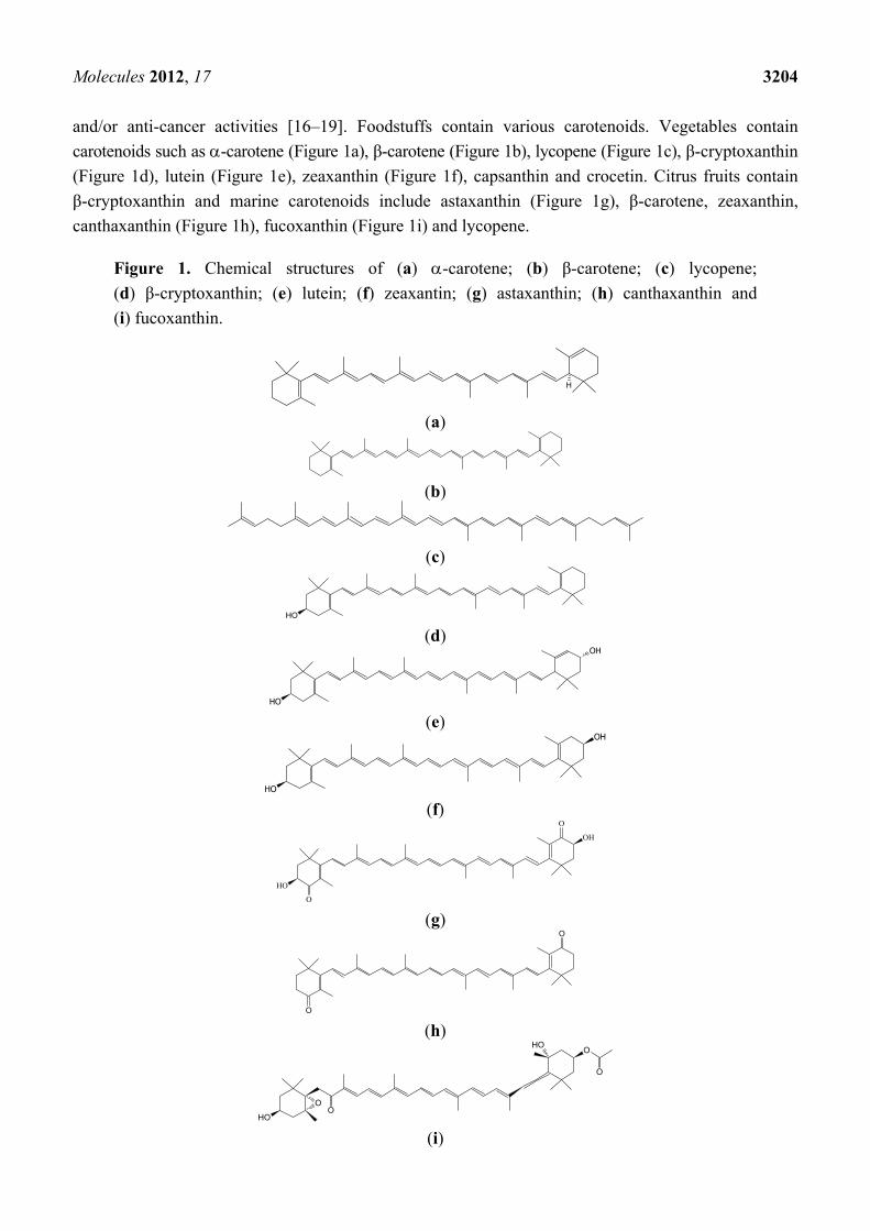
and/or anti-cancer activities [16–19]. Foodstuffs contain various carotenoids. Vegetables contain
carotenoids such as -carotene (Figure 1a), β-carotene (Figure 1b), lycopene (Figure 1c), β-cryptoxanthin
(Figure 1d), lutein (Figure 1e), zeaxanthin (Figure 1f), capsanthin and crocetin. Citrus fruits contain
β-cryptoxanthin and marine carotenoids include astaxanthin (Figure 1g), β-carotene, zeaxanthin,
canthaxanthin (Figure 1h), fucoxanthin (Figure 1i) and lycopene.
Figure 1. Chemical structures of (a) -carotene; (b) β-carotene; (c) lycopene;
(d) β-cryptoxanthin; (e) lutein; (f) zeaxantin; (g) astaxanthin; (h) canthaxanthin and
(i) fucoxanthin.
H
(a)
(b)
(c)
OH
(d) OH
OH (e)
OH
OH
(f)
O
OH
OH
O
(g)
O
O
(h)
O
OHO
OOH
O
(i)
Molecules 2012, 17 3205
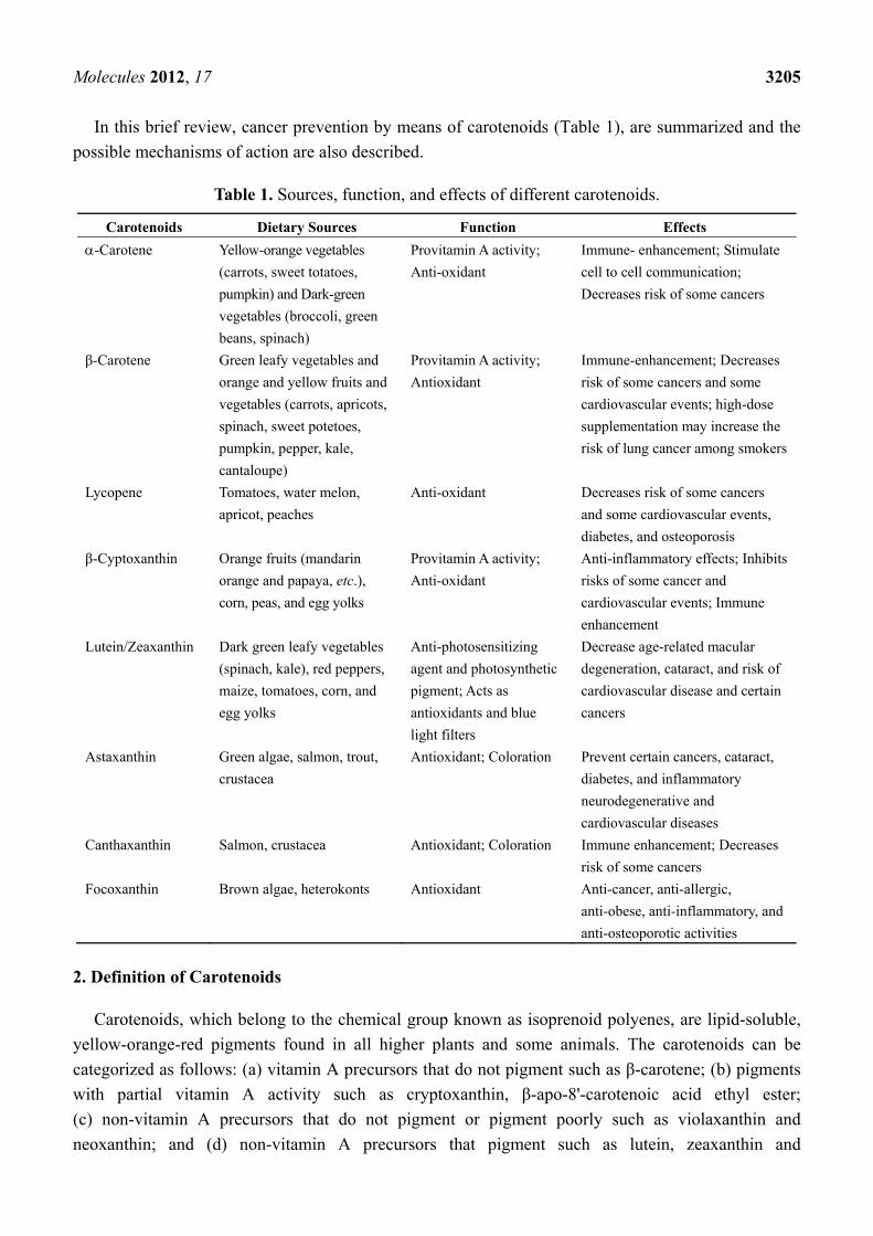
In this brief review, cancer prevention by means of carotenoids (Table 1), are summarized and the
possible mechanisms of action are also described.
Table 1. Sources, function, and effects of different carotenoids.
Carotenoids Dietary Sources Function Effects
-Carotene Yellow-orange vegetables
(carrots, sweet totatoes,
pumpkin) and Dark-green
vegetables (broccoli, green
beans, spinach)
Provitamin A activity;
Anti-oxidant
Immune- enhancement; Stimulate
cell to cell communication;
Decreases risk of some cancers
β-Carotene Green leafy vegetables and
orange and yellow fruits and
vegetables (carrots, apricots,
spinach, sweet potetoes,
pumpkin, pepper, kale,
cantaloupe)
Provitamin A activity;
Antioxidant
Immune-enhancement; Decreases
risk of some cancers and some
cardiovascular events; high-dose
supplementation may increase the
risk of lung cancer among smokers
Lycopene Tomatoes, water melon,
apricot, peaches
Anti-oxidant Decreases risk of some cancers
and some cardiovascular events,
diabetes, and osteoporosis
β-Cyptoxanthin Orange fruits (mandarin
orange and papaya, etc.),
corn, peas, and egg yolks
Provitamin A activity;
Anti-oxidant
Anti-inflammatory effects; Inhibits
risks of some cancer and
cardiovascular events; Immune
enhancement
Lutein/Zeaxanthin Dark green leafy vegetables
(spinach, kale), red peppers,
maize, tomatoes, corn, and
egg yolks
Anti-photosensitizing
agent and photosynthetic
pigment; Acts as
antioxidants and blue
light filters
Decrease age-related macular
degeneration, cataract, and risk of
cardiovascular disease and certain
cancers
Astaxanthin Green algae, salmon, trout,
crustacea
Antioxidant; Coloration Prevent certain cancers, cataract,
diabetes, and inflammatory
neurodegenerative and
cardiovascular diseases
Canthaxanthin Salmon, crustacea Antioxidant; Coloration Immune enhancement; Decreases
risk of some cancers
Focoxanthin Brown algae, heterokonts Antioxidant Anti-cancer, anti-allergic,
anti-obese, anti-inflammatory, and
anti-osteoporotic activities
2. Definition of Carotenoids
Carotenoids, which belong to the chemical group known as isoprenoid polyenes, are lipid-soluble,
yellow-orange-red pigments found in all higher plants and some animals. The carotenoids can be
categorized as follows: (a) vitamin A precursors that do not pigment such as β-carotene; (b) pigments
with partial vitamin A activity such as cryptoxanthin, β-apo-8'-carotenoic acid ethyl ester;
(c) non-vitamin A precursors that do not pigment or pigment poorly such as violaxanthin and
neoxanthin; and (d) non-vitamin A precursors that pigment such as lutein, zeaxanthin and

Molecules 2012, 17 3206
canthaxanthin. Due to the numerous conjugated double bonds and cyclic end groups, carotenoids
present a variety of stereoisomers with different chemical and physical properties. The most important
forms commonly found among carotenoids are geometric (E-/Z-). A double bond links the two residual
parts of the molecule either in an E-configuration with both parts on opposite sites of the plane, or a
Z-configuration with both parts on the same side of the plane. Geometrical isomers of this type are
interconvertible in solution. This stereoisomerism exerts a marked influence on the physical properties.
Isomers differ not only in their melting points, solubility and stability, but also in respect to absorption
affinity, color and color intensity. Animals cannot synthesize carotenoids, so their presence in the body
is due to dietary intake of foods such as pink salmon flesh. The plumage of many birds owes its color
to carotenoids. Plant, algae, fungal and synthetic (nature-identical) carotenoids are permitted as
colorants in food products, but not animal carotenoids.
Carotenoids owe their name to carrots (Daucus carota), and xanthophylls (originally phylloxanthins)
are derived from the Greek words for yellow (xanthos) and leaf (phyllon). Together with anthocyanins,
carotenoids are the most complex class of natural food colorants with over 750 different
structures identified.
3. Absorption, Metabolism, and Bioavailability of Carotenes and Xanthophylls
Carotenoids, being mostly fat soluble, follow the same intestinal absorption path as dietary fat.
Carotenoids are released from food matrices and solubilized in the gut. This is carried out in the
presence of fat and conjugated bile acids. For carotenoid absorption, as little as 3~5 g of fat in a meal
is sufficient [20,21]. Absorption is affected by the same factors that influence fat absorption. Thus, the
absence of bile or any generalized malfunction of the lipid absorption system, such as diseases of the
small intestine and pancreas, will interfere with the absorption of carotenoids. Chylomicrons are
responsible for the transport of carotenoids from the intestinal mucosa to the bloodstream via the
lymphatics for delivery to tissues. Carotenoids are transported in the plasma exclusively by lipoproteins.
Oxygen functionalized carotenoids are more polar than carotenes. Thus, -carotene, β-carotene and
lycopene tend to predominate in low-density lipoproteins (LDL) in the circulation, whereas high-density
lipoproteins (HDL) are major transporters of xanthophylls such as cryptoxanthins, lutein and
zeaxanthin [22,23]. The delivery of carotenoids to extrahepatic tissues is accomplished through the
interaction of lipoprotein particles with receptors and the degradation by lipoprotein lipase.
Although no less than forty carotenoids are usually ingested in the diet, only six carotenoids and
their metabolites have been found in human tissues, suggesting selectivity in the intestinal absorption
of carotenoids [24,25]. In contrast, thirty-four carotenoids and eight metabolites are detected in breast
milk and serum of lactating mothers [26]. Recently, facilitated diffusion in addition to simple diffusion
has been reported to mediate the intestinal absorption of carotenoids in mammals. The selective
absorption of carotenoids may be due to uptake to the intestinal epithelia by means of facilitated
diffusion and an unknown mechanism of excretion into the intestinal lumen. It is well known that
β-carotene can be metabolized to vitamin A after intestinal absorption of carotenoids, but little is
known about the metabolic transformation of non-provitamin A xanthophylls. The enzymatic
oxidation of the secondary hydroxyl group leading to keto-carotenoids would occur as a common
pathway of xanthophyll metabolism in mammals [24].

Molecules 2012, 17 3207
4. Distribution and Nature of Certain Carotenoids
Numerous studies have reported that carotenoids have the potential to prevent cancers, diabetes, and
inflammatory and cardiovascular disease (CVD). Some of these carotenoids are listed below.
4.1. Hydrocarbone Carotenoids
Under EU legislation, plant carotenoids may be derived from edible plants, carrots, vegetable oils,
grass, alfalfa and nettle. However, according to U.S. legislation carotenes may only be derived from
carrots. A good source of plant carotenoids is the mesocarp of oil palm (Elaeis guineensis) fruits,
which contains an oil rich in carotenes. After separation of the carotenes from the palm fruit oil, which
is used for making detergents, the carotenes are suspended in vegetable oil at a concentration of 30%.
The predominant carotenes are - and β-carotene in the ratio 2:3. Other carotenes, including phytoene,
phytofluene, -carotene, -carotene and lycopene, which are all precursors in the biosynthesis of
- and β-carotene, are present in smaller amounts. Due to heat treatment of the oil palm fruit used in
obtaining the oil, a complex mixture of geometric isomers is formed, with only 60% of - and
β-carotene as the trans-forms. Synthetic β-carotene is predominantly trans-β-carotene. The presence of
β-carotene and cis-isomers of - and β-carotene in palm fruit carotenes means that synthetic
β-carotene is more orange than palm fruit carotenes, which is more yellow. Carotene from B. trispora
is also mainly trans-β-carotene, with approximately 3% of other carotenoids. Carotene from D. salina
also primarily consists of β-carotene with 5–6% of other carotenoids (-carotene, lutein, zeaxanthin
and β-cryptoxanthin); according to legislation, the content of transisomers coming from this source
should be in the range 50–71%. This means that its color shade would be between that of oil palm
carotenes and synthetic β-carotene. Besides being used as colorants, carotenes are also used for
nutritional purposes, such as provitamin A agents or as dietary supplements.
β-Carotene is the major source of vitamin A as a provitamin A carotenoid. Two metabolic pathways
exist for its conversion to vitamin A, and they are known as the central cleavage pathway and the
excentric cleavage pathway. For provitamin A carotenoids, central cleavage is the main pathway
leading to the formation of vitamin A [27,28]. β-Carotene, -carotene, and β-cryptoxanthin are cleaved
symmetrically at their central double bond by β-carotene 15,15'-monooxygenase (CMO1), formerly
called β-carotene 15,15'-dioxygenase. An alternative excentric cleavage pathway was also reported [29,30]
and confirmed by molecular identification of an excentric cleavage enzyme, β-carotene
9',10'-monooxygenase (CMO2) in mice, humans, and zebrafish [31]. CMO2 has the ability to catalyze
the asymmetric cleavage of β-carotene to produce β-apo-10'-carotenal and β-ionone [31].
Apo-β-carotenals can be precursors of vitamin A in vitro and in vivo, by further cleavage enzyme,
CMO1 [32]. They can also be oxidized to their corresponding apo-β-carotenoic acids, which undergo a
process similar to β-oxidation of fatty acids, to produce retinoic acid [33]. The coexistence of these
two cleavage pathways reveals a greater complexity of β-carotene metabolism in organisms and raises
a potential link between effects from β-carotene and/or its metabolites and anti-carcinogenesis.
Common non-synonymous single-nucleotide polymorphisms (SNPs) exist in the human CMO1 gene
and alter β-carotene metabolism [34,35].

Molecules 2012, 17 3208
4.2. Lycopene
Being a precursor in the biosynthesis of β-carotene, lycopene can be expected to be found in plants
containing β-carotene, albeit usually at very low and sometimes undetectable concentrations. The
best-known sources of lycopene are tomatoes, watermelon, guava and pink grapefruit. Lycopene may
also be produced synthetically and by B. trispora. Lycopene is permitted as a food colorant in the EU
and was also approved for use as a food supplement in the USA in July 2005. The only permitted
source is tomatoes (Lycopersicon esculentum, Lycopersicon, meaning wolf peach). Besides lycopene,
tomato oleoresin also contains appreciable amounts of β-carotene, phytoene and phytofluene. In
solution, lycopene appears orange and not bright red as in the tomato. Lycopene is very prone to
oxidative degradation, much more so than β-carotene.
Carotenoids absorb light, transfer energy to chlorophyll in the process of photosynthesis and protect
against photo-oxidative damage [36,37]. In man, carotenoids function primarily as dietary sources of
provitamin A. However, lycopene lacks the β-ionone ring structure required to form vitamin A and has
no provitamin A activity. Therefore, lycopene has no known physiological function in man. However,
some potential molecular targets in cells have been identified for lycopene. They include molecules
that are involved in antioxidant activity, the antioxidant response element (ARE), apoptosis induction,
cell cycle arrest, growth factors and signaling pathways, and invasion and metastasis [38–42].
4.3. Lutein and Zeaxanthin
Lutein and zeaxanthin are the two major components of the macular pigments of the retina. The
macula lutea “yellow spot” in the retina is responsible for central vision and visual activity. Lutein and
zeaxanthin are the only carotenoids found in both the macula and lens of the human eye, and have dual
functions in both tissues to act as powerful antioxidants and to filter high-energy blue light [43]. Lutein
is found in high amounts in human serum [26]. In the diet it occurs in highest concentrations in dark
green leafy vegetables (spinach, kale, collard greens and others), corn and egg yolks [44]. Zeaxanthin
is the major carotenoid found in corn, orange peppers, oranges and tangerines.
Lutein is also a very common carotenoid and one of the major xanthophylls present in green leafy
vegetables. Lutein and zeaxanthin are known to selectively accumulate in the macula of the human
retina. They are thought to function as antioxidants [45,46] and as blue light filters [47] to protect the
eyes from oxidative stresses such as cigarette smoke and sunlight, which can lead to age-related
macular degeneration (AMD) and cataracts. The name lutein is derived from the Latin word for yellow
(compare xanthophyll, vide supra). The most interesting source is Aztec marigold (Tagetes erecta) in
which lutein is primarily found esterified with saturated fatty acids (lauric, myristic, palmitic and
stearic acid). Lutein made from Aztec marigold also contains some zeaxanthin (typically less than 10%).
Containing only 10 conjugated double bonds, lutein is more yellowish-green than oil palm carotenes.
Zeaxanthin, the principal pigment of yellow corn, Zeaxanthin mays L. (from which its name is
derived) is the compound that consists of 40 carbon atoms. It also occurs in egg yolks and some of the
orange and yellow vegetables and fruits, such as alfalfa and marigold flowers [48]. Zeaxanthin exhibits
no vitamin A activity. Zeaxanthin and its close relative lutein play a critical role in the prevention of
AMD, the leading cause of blindness [49]. Zeaxanthin is isomeric with lutein; the two carotenols only

Molecules 2012, 17 3209
differ from each other in terms of the shift of a single double bond, so that in zeaxanthin all double
bonds are conjugated. Zeaxanthin is used as a feed additive and colorant in the food industry for birds,
swine and fish [50]. The pigment imparts a yellow coloration to the skin of birds and their egg yolk,
whereas in pigs and fish it is used for skin pigmentation [51].
4.4. β-Cryptoxanthin
β-Cryptoxanthin is found in human blood together with -carotene, β-carotene, lycopene, lutein
and zeaxanthin. Unlike other abundant carotenoids, β-cryptoxanthin is not found in most fruits or
vegetables but only in specific ones, namely hot pepper, persimmon and Satsuma mandarin
(Citrus unshiu Marc.) [52]. Satsuma mandarin, also known as table orange or Satsuma in Western
countries, is one of the most popular citrus fruits in Japan. It is sweet, tasty and rich in vitamin C. It is
notable that Satsuma mandarin is one of the most common β-cryptoxanthin rich fruits in the world.
The edible part of the Satsuma mandarin contains about 1.8 mg/100 g of β-cryptoxanthin, while the
β-cryptoxanthin content is 0.2 mg/100 g in Valencia orange and almost nothing in grapefruits. As
β-cryptoxanthin is rarely found in most fruits or vegetables, the serum β-cryptoxanthin concentration
in the Japanese population is almost parallel to their consumption of the Satsuma mandarin, and is
higher than in western populations [53]. Although the nutritional functions and metabolism of
abundant carotenoids, for example β-carotene and lycopene, have been well studied [54,55], those of
β-cryptoxanthin have not been examined in detail. Recent reports strongly suggest a significant
negative correlation between serum β-cryptoxanthin concentrations and disease morbidity such as liver
disorders [56,57], cancer [58,59] and mutagenesis [60], and post-menopausal osteoporosis [61–63].
β-Cryptoxanthin intake is beneficial for human health. The anti-obesity effects of β-cryptoxanthin have
recently been reported [64,65]. The major xanthophyll, β-cryptoxanthin, was also reported to decrease
the gene expression of interleukin (IL)-1 in mouse macrophage RAW264 cells [66], to promote
osteoblastic differentiation of mouse MC3T3 cells [67] and to prevent a decrease of calcium content in
the bone of ovariectomized rats [63].
4.5. Astaxanthin
Astaxanthin contains two keto groups on each ring structure as compared with other carotenoids,
resulting in enhanced antioxidant properties. This compound occurs naturally in a wide variety of
living organisms including microalgae (Haematococcus pluvialis, Chlorella zofingiensis and
Chlorococcum sp.), fungi (Phaffia rhodozyma, red yeast), complex plants, seafood and some birds such
as flamingos and quail; it has a reddish color and gives salmon, shrimp and lobster their distinctive
coloration [68]. The microalga Haematococcus pluvialis has the highest capacity to accumulate
astaxanthin at up to 4–5% of cell dry weight. Astaxanthin has been attributed with the extraordinary
potential of protecting the organism against a wide range of diseases. It also has considerable potential
and promising applications in the prevention and treatment of various diseases such as cancers, chronic
inflammatory diseases, metabolic syndrome, diabetes, diabetic nephropathy, CVD, gastrointestinal and
liver diseases, and neurodegenerative diseases [69]. Astaxanthin cannot be manufactured in animals or
converted to vitamin A, and therefore must be consumed in the diet. Astaxanthin and canthaxanthin
have antioxidant activity, are free radical scavengers, potent quenchers of reactive oxygen species

Molecules 2012, 17 3210
(ROS) and nitrogen oxygen species, and chain-breaking antioxidants. They are superior antioxidants
and scavengers of free radicals as compared with the carotenoids such as β-carotene [70]. Astaxanthin
is even called superantioxidant.
4.6. Canthaxanthin
Canthaxanthin was first isolated from the edible mushroom, Cantharellus cinnabarinus. In addition,
canthaxanthin is said to be produced at the end of the growth phase in several green algae, and also in
blue-green algae, as secondary carotenoids instead of, or in addition to, primary carotenoids. It has also
been found in bacteria, crustacea and various species of fish including carp (Cyprinus carpio), golden
mullet (Mugil auratus), annular seabream (Diplodus annularis) and trush wrasse (Crenilabrus tinca).
Canthaxanthin is not encountered in wild Atlantic salmon, but represents a minor carotenoid in wild
Pacific salmon. It has also been reported in wild trout (Salmo trutta). Canthaxanthin is used widely as
a drug or as a food and cosmetic colorant (skin tanning), but it may have some undesirable effects on
human health. These are mainly caused by the formation of crystals in the macula lutea membranes of
the retina. This condition is called canthaxanthin retinopathy [71]. It has been shown that this type of
dysfunction of the eye is strongly connected with damage to the blood vessels around the locations of
crystal deposition.
Canthaxanthin is one of the carotenoids without provitamin A activity, but may have
anti-carcinogenic, immune-enhancing, antioxidative activities. The mechanisms by which canthaxanthin
may exert anti-tumor activity are associated with its antioxidant properties through radical trapping or
chain-breaking processes [72,73], or its enhancement of gap-junction cell to cell communication
through upregulation of the gap-junction protein, connexin [74].
4.7. Fucoxanthin
The allenic carotenoid fucoxanthin is one of the most abundant carotenoids, and contributes to
nature more than 10% of the estimated total production of carotenoids in nature, especially in the
marine environment [75]. Fucoxanthin is a naturally occurring brown- or orange-colored pigment that
belongs to the class of non-provitamin A carotenoids. Fucoxanthin acts as an antioxidant under anoxic
conditions. The typical antioxidants are usually proton donors (ascorbic acid, -tocopherol and
glutathione). Fucoxanthin, on the other hand, donates an electron as a part of its free-radical quenching
function. A combination of these distinct properties is very rarely found among naturally occurring
compounds [76,77]. During normal metabolism the body produces heat. Fucoxanthin increases the
amount of energy released as heat in fat tissue, a process known as thermogenesis. In a published study
it has been reported that fucoxanthin affects multiple enzymes involved in fat metabolism causing an
increase in the production of energy from fat [78].
Fucoxanthin is present in Chromophyta (Heterokontophyta or Ochrophyta), including brown
seaweeds (Phaeophyceae) and diatoms (Bacillariophyta) [79]. Based on its unique molecular structure,
fucoxanthin has remarkable biological properties similar to neoxanthin, dinoxanthin and peridinin,
which make it different to other carotenoids. Fucoxanthin does not exhibit toxicity and mutagenicity
under experimental conditions [79–81]. Fucoxanthin may have the ability to increase circulating
cholesterol levels in rodents as a common feature [79].

Molecules 2012, 17 3211
5. Clinical Trials with Long-Term β-Carotene Supplementation
Epidemiologic studies have shown an inverse relationship between the presence of various
cancers and dietary or blood carotenoid levels [82]. However, three [13–15] out of four intervention
trials [13–15,83] using high-doses of β-carotene supplements did not show protective effects against
cancer or CVD. Rather, the high-dose intervention trials showed an increase in cancer and angina
pectoris [13–15,83]. Therefore, carotenoids may promote health when taken at dietary levels, but may
have adverse effects when taken high doses by subjects who smoke or who have been exposed to asbestos.
The epidemiologic observations of the possible protective effects of high dietary (not supplemental)
β-carotene intakes against cancer, along with what is known about carotenoid biochemical functions,
has led to further study of the effect of β-carotene on cancer risk. Long-term large randomized
intervention trials were designed to test the efficacy of high doses of β-carotene (20–30 mg/day) in
the prevention of cancer (Table 2). As stated above, the results from two trials provided possible
evidence of harm from β-carotene supplements in relation to cancer among high-risk individuals such
as smokers and asbestos workers [15], but no effect (either beneficial or detrimental) in a generally
well-nourished population [84]. Moreover, the Linxian (Chinese) Cancer Prevention Study [83] found
that supplementation with β-carotene, vitamin E and selenium led to a significant reduction in total
mortality (9%), especially from cancer (13%) and stomach cancer in particular (21%) (Table 2). The
positive results of the Chinese study probably reflect the correction of a vitamin A deficiency in the
study population. A number of mechanisms have been proposed to account for the association between
β-carotene supplementation and lung cancer in smokers and asbestos workers, including an imbalance
of other carotenoids or antioxidants, a pro-oxidant activity of β-carotene at the high oxygen tensions
found in the lungs, induction of P450 enzymes and the production of damaging β-carotene oxidation
products by components of cigarette smoke [85]. The Women’s Health Study [86] indicated no
statistically significant differences in incidence of cancer, CVD, or total mortality, although the
treatment duration is short (a median treatment duration of 2.1 years and a median total follow-up of
4.1 years).
Table 2. β-Carotene supplementation trials.
Studies Study Designs Ref.
No. Population Intervention Duration Cancer outcome
ATBC 29,133 Finish male
smokers (50–69 years
of age)
β-carotene, 20 mg/day;
vitamin E, 50 mg/day
5–8 years 18% increase in lung
cancer; 8% increase in
mortality
13
CARET 18,314 men and women
and asbestoss workers
(45–74 years of age)
β-carotene, 30 mg/day;
vitamin A, 25,000 IU
<4 years 28% increase in lung
cancer; 17% increase in
deaths
15
PHS 22,071 male physicians
(40–84 years of age)
β-carotene, 50 mg on
alternate days
12 years No effect of
supplementation in
incidence of cancer
14

Molecules 2012, 17 3212
Table 2. Cont.
Studies Study designs Ref.
No. Population Intervention Duration Cancer outcome
Linxian 29,584 men and
women, vitamin and
mineral deficient
(40–69 years of age)
β-carotene, 15 mg/day;
selenium, 50 mg/day;
-tocopherol,
30 mg/day
5 years 13% decrease in total
cancers; 9% decrease in
overall deaths
84
Women’s
Health
Study
39,876 female health
professionals (over
45 years of age)
β-carotene, 50 mg on
alternate days
4.1 years (2.1
years’ treatment
and 2.0 years’
follow-up)
No effect of
supplementation in
incidence of cancer
87
The epidemiologic studies reported an inverse relationship between diet and/or blood β-carotene
levels and cancer prevention. It is probable that β-carotene serves as a marker of increased fruit and
vegetable intake and, therefore, of all components that have cancer prevention potential, for example
vitamin C, folic acid, other carotenoids and polyphenols. Alternatively, low-dose dietary levels could
have a protective effect against cancer, whereas high-dose β-carotene supplementation could have a
cancer stimulating effect.
6. Cancer Chemoprevention by Carotenoids in Preclinical Studies
Cancer chemoprevention is a rapidly expanding discipline that focuses on the discovery and
identification of dietary agents and drugs that prevent or inhibit malignant tumor development [4,5].
Since approximately one-third of the overall risk of cancer is attributable to diet, a large number
of dietary compounds have been tested to determine their chemopreventive ability using animal
carcinogenesis models [87–90]. The higher eukaryotic aerobic organisms, including man, cannot exist
without oxygen, yet oxygen represents a danger to their very existence due to its high reactivity. This
fact has been termed the paradox of aerobic life [91]. A number of ROS are generated during normal
aerobic metabolism such as the superoxide, hydrogen peroxide and the hydroxyl radical. In addition,
singlet oxygen can be generated through photochemical events (in skin and eyes), and lipid
peroxidation can lead to peroxyl radical formation [92]. These oxidants collectively contribute to aging
and degenerative diseases such as cancer and atherosclerosis through oxidation of DNA, proteins and
lipids [91–93]. Antioxidant compounds can decrease mutagenesis, and thus carcinogenesis, both by
decreasing oxidative damage of DNA and by decreasing oxidant-stimulated cell division [92]. The
human body maintains an array of endogenous antioxidants such as catalase and superoxide dismutase;
however, exogenous dietary antioxidants such as ascorbic acid (vitamin C), -tocopherol (vitamin E)
and carotenoids play important roles in reducing oxidative damage as well [91–93], and their serum
levels have the potential to be manipulated [93]. Major carotenoids with antioxidant activity that have
been extensively evaluated with regard to their cancer chemopreventive ability include - and
β-carotenes, β-cryptoxanthin, lycopene, lutein and zeaxanthin.

Molecules 2012, 17 3213
6.1. - and β-Carotene
Carotenoids have been studied vigorously to see if these colorful compounds can decrease the risk
of cancer. In ecological studies and early case-control studies it appeared that β-carotene was a
cancer-protective agent. Randomized controlled trials of β-carotene found that the isolated nutrient
was either without effect [14] or actually increased the risk of lung cancer in smokers [13,15].
β-Carotene may be a marker for the intake of fruits and vegetables, but it does not have a powerful
protective effect in isolated pharmacological doses. However, there is a large body of literature
indicating that dietary carotenoids are cancer preventative. -Carotene has been found to be a stronger
protective agent than its well-known isomer β-carotene [94]. Studies tend to agree that overall intake
of carotenoids is more protective than a high intake of a single carotenoid [94]. Hence, a variety of
fruits and vegetables is still a better anti-cancer strategy than just using a single vegetable high in a
specific carotenoid [94]. The richest source of -carotene is carrots and carrot juice, with pumpkins
and winter squash as a second densest source [94]. There is approximately 1 μg of -carotene for
every 2 μg of β-carotene in carrots. Previous studies in our laboratory have demonstrated the
chemopreventive ability of β-carotene against oral carcinogenesis in rats [95].
Several experimental animal studies have shown that -carotene possesses higher activity than
β-carotene in suppressing tumorigenesis in the skin, lung, liver and colorectum [18,96]. In a skin
tumorigenesis experiment conducted by Murakoshi et al. [18], the incidence of tumor-bearing mice in
the positive control group was 69%, whereas those in the groups treated with β- and -carotene were
13% and 25%, respectively. The average multiplicity (number of tumors/mouse) of tumors in the
positive control group was 3.73/mouse, whereas the -carotene-treated group had 0.13/mouse (p < 0.01).
β-Carotene treatment also decreased tumor multiplicity (1.31/mouse), but the difference from the
positive control group was insignificant (p < 0.05). The higher potency of -carotene relative to
β-carotene in the suppression of tumor promotion was further confirmed in their studies [18]. In a
mouse lung carcinogenesis model initiated by 4-nitroquinoline 1-oxide (4-NQO) and promoted by
glycerol, the average multiplicity of lung tumors per mouse in the positive control group was
4.06/mouse, whereas the -carotene-treated group had 1.33/mouse (p < 0.001). β-Carotene treatment
did not show any suppressive effect on tumor multiplicity, which was significantly increased
(4.93/mouse, p < 0.02). In their liver carcinogenesis experiment [18], male C3H/He mice, which have
a high incidence of spontaneous liver tumor development, were treated with drinking water containing
0.05% - and β-carotene for 40 weeks. The mean number of hepatomas (3.00/mouse; p < 0.001) in the
mice that received -carotene was significantly decreased as compared with the untreated control
group (6.31/mouse). On the other hand, the β-carotene-treated group only showed a tendency toward a
decrease in tumors (4.71/mouse), as compared with the control group [18]. Narisawa et al. [96] also
demonstrated the protective effects of -carotene, lycopene and lutein, but not β-carotene, on
preneoplastic colorectal adenocarcinoma lesions.
6.2. β-Cryptoxanthin
It is known that certain carotenoids and flavonoids can inhibit cancer development in animal
carcinogenesis models [87–90]. β-Cryptoxanthin and hesperidin are such compounds. β-Cryptoxanthin

Molecules 2012, 17 3214
with non-substituted β-ionone cycles and provitamin A properties exhibits several biological activities,
including the scavenging of free radicals, enhancement of gap junctions, immunomodulation and
regulation of the enzyme activity involved in carcinogenesis [97,98]. The most common sources of
β-cryptoxanthin are citrus fruits and red sweet peppers. β-Cryptoxanthin is reported to inhibit
mouse skin tumorigenesis [58] and rat colon carcinogenesis [99]. Narisawa et al. also reported that
25 ppm of β-cryptoxanthin administered for 30 weeks in the diet significantly suppressed
N-methylnitrosourea-induced colon carcinogenesis in rats [99]. This suggested that dietary
β-cryptoxanthin may affect colon carcinogenesis after accumulation in the colonic mucosa, perhaps
due to absorption from the colon as well as the small intestine. β-Cryptoxanthin-rich juice (Satsuma
mandarin juice [MJ]) has also been found to inhibit colon [59] and lung [100] carcinogenesis.
Hesperidin, present in several vegetables and fruits, has antioxidant properties, and anti-inflammatory
and inhibitory effects on prostaglandin biosynthesis. This flavonoid has been shown to inhibit
chemically induced carcinogenesis in several organs [87–90,101–105]. β-Cryptoxanthin and hesperidin
are thus considered to be potential cancer chemopreventive compounds. However, edible plants contain
only small amounts of these chemicals. Therefore, to obtain higher contents of these compounds
in foods we prepared a pulp (CHRP) containing high amounts of β-cryptoxanthin and hesperidin
during the process of making MJ. CHRP (100 g) contained 0.67 g of β-cryptoxanthin and 3.58 g of
hesperidin; the contents of β-cryptoxanthin and hesperidin were 583 times and 38 times greater than
those in the edible parts of Satsuma mandarin, respectively. In addition, we prepared Satsuma
mandarin juices, which we called MJ2 (1.7 mg of β-cryptoxanthin and 84 mg of hesperidin/100 g) and
MJ5 (84 mg of β-cryptoxanthin and 100 mg of hesperidin/100 g), by adding CHRP to the standard
Satsuma mandarin juice (MJ: 0.8 mg of β-cryptoxanthin and 79 mg of hesperidin/100 g). We have
demonstrated the chemopreventive effects of CHRP and MJs on chemically induced oncogenesis in rat
colon and tongue and mouse lung [59,100,106,107].
Citrus compounds act on multiple key elements in signal transduction pathways related to cellular
proliferation, differentiation, apoptosis, inflammation and obesity. We have found that Citrus unshiu
segment membrane (CUSM) containing β-cryptoxanthin and fiber suppresses colitis- and obesity-related
colon tumorigenesis in animal models [108,109]. Feeding involving a diet with CUSM treatment also
decreased the serum level of triglycerides.
6.3. Lycopene
There are relatively few reports on the cancer chemopreventive effects of lycopene or other tomato
carotenoids in animal models. The majority, but not all, of these studies have indicated a protective
effect. Inhibitory effects were seen in two studies using aberrant crypt foci (putative precursors of
colon cancer) [96] and colon cancer [110] as biomarkers, and in two mammary tumor studies, one
using the dimethylbenz(a)anthracene model [111] and the other the spontaneous mouse model [112].
Inhibitory effects were also reported in mouse lung [113] and rat hepatocarcinoma [114] and
bladder cancer [115] models. However, a study by Cohen et al. [116] found no effect in the
N-nitrosomethylurea-induced mammary tumor model when crystalline lycopene or a lycopene-rich
tomato carotenoid oleoresin was administered in the diet. Unfortunately, differences in routes of
administration (gavage, intraperitoneal injection, intra-rectal instillation, drinking water and diet

Molecules 2012, 17 3215
supplementation), species and strain differences, form of lycopene (pure crystalline, beadlet and mixed
carotenoid suspension), varying diets (grain-based and casein based) and dose ranges (0.5–500 ppm)
resulted in no prevention effect on development of chemically induced mammary cancer. It is clear
that the majority of ingested lycopene is excreted in the feces and that 1,000-fold more lycopene is
absorbed and stored in the liver than in other target organs. Nonetheless, physiologically significant
(nanogram) levels of lycopene are assimilated by key organs such as breast, prostate, lung and colon,
and there is a rough dose-response relationship between lycopene intake and blood levels. Pure
lycopene was absorbed less efficiently than the lycopene-rich tomato carotenoid oleoresin, and blood
levels of lycopene in rats fed a grain based diet were consistently lower than those in rats fed lycopene
in a casein-based diet. The latter suggests that the matrix in which lycopene is incorporated is an
important determinant of lycopene uptake.
High intake of lycopene has been associated with a lower risk of a variety of cancers including
lung cancer. Lycopene can be converted to apo-10'-lycopenoids [117] in mammalian tissues and can
be cleaved by carotene 9',10'-oxygenase at its 9',10' double bond to form apo-10'-lycopenoids, including
apo-10'-lycopenal, apo-10'-lycopenol, and apo-10'-lycopenoic acid. Among apo-10'-lycopenoids,
apo-10'-lycopenoic acid has been recently shown to inhibit lung carcinogenesis both in vivo and
in vitro [118]. Since enzymatic metabolites of lycopene induce NF-E2-related factor 2 (Nrf2)-mediated
expression of phase II detoxifying/antioxidant enzymes including heme oxygenase-1 (HO-1), NQO1,
GSTs, and glutamate-cysteine ligases in human bronchial epithelial cells, BEAS-2B [119] and human
liver cell cancer cells, HepG2 [120], the anti-carcinogenic and antioxidant functions of lycopene are
mediated by apo-10'-lycopenoids, especially apo-10'-lycopenoic acid, via activating Nrf2 and inducing
phase II detoxifying/antioxidant enzymes [119].
Of the various carotenoids lycopene has been found to be very protective, particularly for prostate
cancer. The major dietary source of lycopene is tomatoes, with the lycopene in cooked tomatoes being
more bioavailable than that in raw tomatoes. Several prospective cohort studies have found associations
between high intake of lycopene and reduced incidence of prostate cancer, although not all studies
have produced consistent results [121,122]. Some studies suffer from a lack of good correlation
between lycopene intake assessed by questionnaire and actual serum levels, and other studies measured
intakes among a population that consumed very few tomato products. In the Health Professionals
Follow-up Study there was a 21% decrease in prostate cancer risk, when comparing the highest
quintile of lycopene intake with the lowest quintile. Combined intake of tomatoes, tomato sauce,
tomato juice and pizza (which accounted for 82% of the lycopene intake) was associated with a 35%
lower risk of prostate cancer. Furthermore, lycopene was even more protective for advanced stages of
prostate cancer, with a 53% decrease in risk [123]. A more recent follow-up report on this same cohort
of men confirmed these original findings that lycopene or frequent tomato intake is associated with
about a 30–40% decrease in the risk of developing prostate cancer, especially advanced prostate
cancer [124]. In addition to the two reports detailed above, a nested case control study from the Health
Professional Follow-up Study involving 450 cases and controls found an inverse relationship between
plasma lycopene and prostate cancer risk (OR 0.48) among older subjects (>65 years of age) without a
family history of prostate cancer [125].
In addition to these observational studies, two clinical trials have been conducted to supplement
lycopene for a short period before radical prostatectomy. In one study 30 mg/day of lycopene were

Molecules 2012, 17 3216
given to 15 men in the intervention group, while 11 men in the control group were instructed to follow
the National Cancer Institute’s recommendations to consume at least five servings of fruits and
vegetables daily. Results showed that lycopene slowed the growth of prostate cancer. Prostate tissue
lycopene concentration was 47% higher in the intervention group. Subjects that took lycopene for
3 weeks had smaller tumors, less involvement of the surgical margins and less diffuse involvement of
the prostate by pre-cancerous high-grade prostatic intraepithelial neoplasia [126]. In another study
carried out before radical prostatectomy surgery, 32 men were given a tomato sauce-based pasta dish
every day, which supplied 30 mg of lycopene per day. After 3 weeks serum and prostate lycopene
levels had increased 2-fold and 2.9-fold, respectively. Prostate-specific antigen (PSA) levels had
decreased by 17%, as also reported by Kucuk et al. [126]. Oxidative DNA damage was 21% lower in
the patients’ leukocytes and 28% lower in prostate tissue, as compared with the non-study controls.
The apoptotic index was 3-fold higher in the resected prostate tissue, relative to biopsy tissue [127].
A number of issues remain to be resolved before any definitive conclusions can be drawn
concerning the anticancer effects of lycopene. These include the following: the optimal dose and form of
lycopene; interactions among lycopene and other carotenoids and fat soluble vitamins such as vitamin
E and D; the role of dietary fat in regulating lycopene uptake and disposition; organ and tissue
specificity; and the problem of extrapolation from rodent models to human populations [128].
6.4. Lutein and Zeaxanthin
In addition to playing pivotal roles in ocular health, lutein and zeaxanthin are important nutrients
for the prevention of CVD, stroke and lung cancer. They may also be protective in skin conditions
attributed to excessive ultraviolet (UV) light exposure. In a 10-year study following 120,000 U.S. men
and women, a significant reduction in lung cancer was observed in patients with the highest intake of
total carotenoids including lutein and zeaxanthin [129]. A second 14-year study assessed the same
relationship in 27,000 Finnish male smokers via a food-item questionnaire. Consumption of carotenoid
containing fruits and vegetables was associated with a decreased risk of lung cancer. A decreased risk
of lung cancer was also observed in individuals in the highest quintiles of lutein/zeaxanthin intake
versus the lowest quintiles. A population-based survey of 20 South Pacific Island populations examined
the association between lutein consumption and lung cancer rates. Researchers found an inverse
association between lutein and lung cancer and a markedly lower incidence rate for lung cancer among
Fijians, as compared with other South Pacific populations. Fijians consume an average of 200 g of
dark green vegetables (25 mg lutein) daily; whereas inhabitants of other South Pacific countries
consume diets in which colorful fruits and vegetables are less plentiful [130].
Carotenoids singly or in combination could lower cancer risk due to their antimutagenic properties
and ability to scavenge free radicals, to protect against tumor development and to improve immune
response [131,132]. Lutein and β-carotene quench peroxy radicals and demonstrate antioxidant
properties against oxidative damage in vitro [133,134]. Plasma lutein analyzed from 37 women
correlated inversely with measured oxidative indices [135]. It has been shown in vitro using multilamellar
liposomes, that carotenoids in combination elicit a greater antioxidant defense than singly. The
strongest synergistic effect was obtained in the presence of lutein or lycopene [136]. Lutein may be
anticarcinogenic as well. This is suggested by its ability to interact with the mutagens 1-nitropyrene
Molecules 2012, 17 3217
and aflatoxin B1 (AFB1) [137,138]. Lutein may also exert an anticarcinogenic effect by stimulating
certain genes involved in T-cell transformations activated by mitogens, cytokines and antigens [139].
Investigation of lutein’s protective effects in relation to site-specific cancers is beginning to evolve
in epidemiologic studies and animal models. No associations have been detected between plasma
lutein and zeaxanthin concentrations and gastric cancer [140]. Slattery et al. [141] detected an inverse
association between dietary lutein intake and colon cancer in men and women. The reduction in risk
was significant only in patients who were diagnosed with colon cancer at a younger age [141].
Carotenoid esters are found in human skin [142]. A combination of carotenoids may protect against the
development of erythema in human skin [143] and are correlated with the presence or absence of skin
cancer and precancerous lesions [141]. The specific effects of lutein on skin cancer are yet to be
determined. Previous research has shown modest relationships between the consumption of nutrients
found in carotenoid rich foods such as β-carotene and vitamin A, and a reduced risk of breast
cancer [144–146]. Focus on the potential protective effects of lutein in relation to developing breast
cancer has evolved only recently. Recent research in mice showed that low levels of dietary lutein at
0.002 and 0.02% of the diet inhibited mammary tumor incidence, growth and latency [19]. Lutein has
been shown to induce apoptosis in transformed but not in normal human mammary cells, and to
protect normal cells from apoptosis induced in cell culture [147]. Freudenheim et al. [148] have shown
that the intake of carotenoid-rich foods, specifically vegetables, as well as lutein and zeaxanthin, is
significantly associated with a lower risk of developing premenopausal breast cancer. In a case-control
study, increasing serum levels of lutein and zeaxanthin were associated with a reduced breast cancer
risk, but the trend was only marginally significant [149]. A decreased risk of cancer was associated
with increasing levels of breast adipose tissue lutein and zeaxanthin concentrations in women with
breast cancer as compared with women with benign breast biopsies, but the association was not
significant [150]. The Nurse’s Health Study [12] showed a weak, but significant, inverse association
between lutein and zeaxanthin intake and the risk of developing breast cancer among premenopausal
women. The protective effect of lutein and zeaxanthin in relation to breast cancer was strongest among
women with a family history of breast cancer. A nested case-control study from the prospective New
York University Women’s Health Study [151] indicated an inverse association between plasma lutein,
but not zeaxanthin, and risk of breast cancer. However, plasma - and β-carotene levels were also
significantly related to a decrease in risk. Other case-control studies have shown no differences in
breast adipose tissue concentrations of lutein and zeaxanthin between women with benign breast
tumors and those with breast cancer [152].
6.5. Astaxanthin
Because astaxanthin has not typically been identified as a major carotenoid in human serum,
information on its epidemiology in human health is lacking. Salmon, the principal dietary source of
astaxanthin, is an important component of the traditional diets of Eskimos and certain coastal tribes in
North America; these groups have shown an unusually low prevalence of cancer [153,154]. This low
cancer incidence has been attributed to the high levels of certain fatty acids in salmon, notably
eicosapentaenoic acid [154], yet it is possible that astaxanthin has played a role in cancer
chemoprevention among these peoples as well. Regardless, the existing data on the potential for

Molecules 2012, 17 3218
astaxanthin to directly prevent cancer is limited to in vitro cell culture studies and in vivo studies with
rodent models.
We previously investigated the possible preventive effects of astaxanthin and canthaxanthin on
N-butyl-N(4-hydroxybutyl)nitrosamine (OH-BBN)-induced mouse urinary bladder carcinogenesis [155],
4-NQO-induced rat oral carcinogenesis [156] and azoxymethane (AOM)-induced rat colon
carcinogenesis [157]. Both of these xanthophylls exhibited inhibitory activity in relation to cancer
development in urinary bladder [155], tongue [156] and colorectum [157] through the suppression of
cell proliferation. In urinary bladder carcinogenesis, the inhibitory effect of astaxanthin was greater
than that of canthaxanthin through the suppression of cell proliferation [155]. A recent study of ours
demonstrated the anti-inflammatory ability and anti-carcinogenesis effects of astaxanthin in inflamed
colon due to modulation of the expression of several inflammatory cytokines that are involved
in inflammation-associated carcinogenesis [158]. Indeed, astaxanthin may aid cyclooxygenase
(COX)-2 down-regulation [159]. A recent study using a 1,2-dimethylhydrazine (DMH)-induced colon
carcinogenesis model also showed that daily administration of astaxanthin (15 mg/kg body weight)
significantly inhibited colon carcinogenesis by modulating nuclear factor kappaB (NF-kB), COX-2,
matrix etalloproteinases (MMP) 2/9, extracellular signal-regulated kinase (ERK)-2 and protein kinase
B (Akt) [160]. Astaxanthin, canthaxanthin and β-carotene, but not lycopene, are reported to be able to
suppressed the development of preneoplastic liver cell lesions induced by AFB1 in rats through the
deviation of AFB1 metabolism towards detoxification pathways [161]. In addition, tetrasodium
diphosphate astaxanthin has been reported to completely inhibit methylcholanthrene-induced neoplastic
transformation of C3H/10T1/2 cells by upregulation of connexin 43 and gap junctional intercellular
communication (GJIC) [162].
6.6. Canthaxanthin
Epidemiological data on canthaxanthin in disease prevention is lacking. However, this carotenoid
has exhibited potential anticancer properties in vitro and in animal models. In earlier studies, canthaxanthin
exerted cancer chemopreventive activities in UV-B-induced mouse skin tumorigenesis [163] and
chemically-induced gastric [164] and breast carcinogenesis [164,165]. Canthaxanthin can also suppress
the proliferation of human colon cancer cells [166], and protect mouse embryo fibroblasts from
transformation [167] and mice from mammary and skin tumor development [16,168]. Canthaxanthin
has also proved effective in inhibiting both oral and colon carcinogenesis in rats [156,157].
Canthaxanthin and astaxanthin have been found to lower the incidence of urinary bladder cancers
induced by OH-BBN, but the inhibitory effects of canthaxanthin were weak when compared to
astaxanthin [155]. As was the case with astaxanthin and β-carotene, canthaxanthin suppressed
AFB1-induced preneoplastic hepatocellular lesions in rats [161]. Although it is a potent antioxidant,
the chemopreventive effects of canthaxanthin may also be related to its ability to up-regulate gene
expression, resulting in enhanced gap junctional cell-cell communication [74,169]. The chemopreventive
effects of canthaxanthin may also be related to its ability to induce xenobiotic metabolizing enzymes,
as has been demonstrated in the liver, lung and kidney of rats [170,171]. The apoptosis-inducing
effects of canthaxanthin may also contribute to its cancer chemopreventive effects [172]. Unfortunately,
canthaxanthin overuse as a sunless tanning product has led to the appearance of crystalline deposits in
Molecules 2012, 17 3219
the human retina [173]. Although these retinal inclusions are reversible [174] and appear to have no
adverse effects [173], their existence has prompted caution regarding the intake of this xanthophyll.
6.7. Fucoxanthin
There are several in vitro studies that have demonstrated the inhibitory effects of fucoxanthin on
human cancer cell lines developed in liver (HepG2) [175], colon (Caco-2, HT-29 and DLD-1) [176]
and urinary bladder [177]. The induction of apoptosis [176,177] and the suppression of cyclin D
levels [175] have been considered to be the biochemical mechanisms by which fucoxanthin exerts
its inhibitory effects on the growth of cancer cells. Since mice actively convert fucoxanthin into
keto-carotenoids by oxidizing the secondary hydroxyl groups and accumulating them in tissues [178],
it may be possible that keto-carotenoids are active chemicals responsible for the effects of fucoxanthin.
In a preclinical study, fucoxanthin was found to significantly inhibit DMH-induced mouse colon
carcinogenesis [179]. Fucoxanthin has been proven to suppress spontaneous liver tumorigenesis
in C3H/He male mice and showed antitumor-promoting activity in a two-stage carcinogenesis
experiment involving the skin of ICR mice, initiated with 7,12-dimethylbenz[a]anthracene and
promoted with 12-O-teradecanoylphorbol-13-acetate and mezerein [180]. In addition, fucoxanthin
has been reported to inhibit duodenal carcinogenesis induced by N-ethyl-N′-nitro-N-nitrosoguanidine
in mice [181].
Although the antitumor effects of fucoxanthin are known, the precise mechanism of action has yet
to be elucidated [182]. The anticancer activity of fucoxanthin has been shown to be partly based on its
regulative effect on biomolecules related to the cell cycle and apoptosis [183,184] and those associated
with antioxidant activity through its pro-oxidant action [185]. In addition, fucoxanthin has been found
to be able to selectively inhibit mammalian DNA polymerase activities, especially replicative DNA
polymerases (i.e., pol , δ and ε), and thus has anti-neoplastic activity [186]. Further investigations
using animal models are needed to assess the details of the molecular mechanisms involved in
fucoxanthin’s activity against different types of cancer cells.
7. Mechanisms of Cancer Chemoprevention by Carotenoids
The mechanisms underlying the anticancer and/or cancer chemopreventive activities of carotenoids
may involve changes in pathways leading to cell growth or cell death. These include immune
modulation, hormone and growth factor signaling, regulatory mechanisms of cell cycle progression,
cell differentiation and apoptosis. Examples of carotenoid effects on some of these pathways are listed
below, with the emphasis being placed on the changes in protein expression associated with these
effects. The main question is, by what mechanism do carotenoids affect so many and diverse cellular
pathways as described above? The changes in the levels of many proteins suggest that the initial effect
involves modulation of transcription. As described below, such modulation can occur at the level of
ligand-activated nuclear receptors or other transcription factors. As illustrated in Figure 2, carotenoids
have multiple targets that contribute to their efficacy as chemoprevention agents.
Molecules 2012, 17 3220
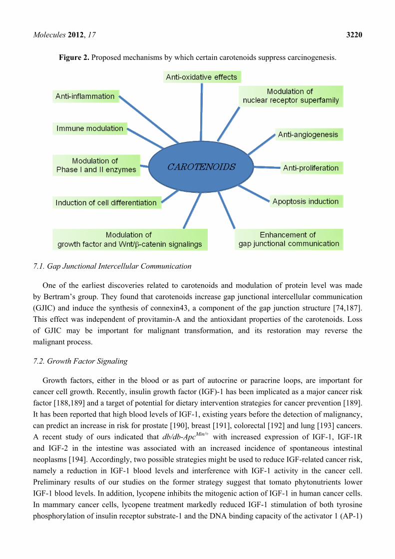
Figure 2. Proposed mechanisms by which certain carotenoids suppress carcinogenesis.
7.1. Gap Junctional Intercellular Communication
One of the earliest discoveries related to carotenoids and modulation of protein level was made
by Bertram’s group. They found that carotenoids increase gap junctional intercellular communication
(GJIC) and induce the synthesis of connexin43, a component of the gap junction structure [74,187].
This effect was independent of provitamin-A and the antioxidant properties of the carotenoids. Loss
of GJIC may be important for malignant transformation, and its restoration may reverse the
malignant process.
7.2. Growth Factor Signaling
Growth factors, either in the blood or as part of autocrine or paracrine loops, are important for
cancer cell growth. Recently, insulin growth factor (IGF)-1 has been implicated as a major cancer risk
factor [188,189] and a target of potential for dietary intervention strategies for cancer prevention [189].
It has been reported that high blood levels of IGF-1, existing years before the detection of malignancy,
can predict an increase in risk for prostate [190], breast [191], colorectal [192] and lung [193] cancers.
A recent study of ours indicated that db/db-ApcMin/+ with increased expression of IGF-1, IGF-1R
and IGF-2 in the intestine was associated with an increased incidence of spontaneous intestinal
neoplasms [194]. Accordingly, two possible strategies might be used to reduce IGF-related cancer risk,
namely a reduction in IGF-1 blood levels and interference with IGF-1 activity in the cancer cell.
Preliminary results of our studies on the former strategy suggest that tomato phytonutrients lower
IGF-1 blood levels. In addition, lycopene inhibits the mitogenic action of IGF-1 in human cancer cells.
In mammary cancer cells, lycopene treatment markedly reduced IGF-1 stimulation of both tyrosine
phosphorylation of insulin receptor substrate-1 and the DNA binding capacity of the activator 1 (AP-1)

Molecules 2012, 17 3221
transcription factor [195]. These effects were not associated with changes in the number or affinity of
IGF-1 receptors, but rather with an increase in membrane-associated IGF binding proteins (IGFBPs).
This finding can explain the suppression of IGF-1-signaling by lycopene based on the finding that
membrane-associated IGFBP-3 inhibits IGF-1 receptor signaling in an IGF-dependent manner [196].
7.3. Cell Cycle Progression
Growth factors have a major effect in promoting cell cycle progression, primarily during the
G1 phase. Lycopene treatment of MCF-7 mammary cancer cells has been shown to slow down
IGF-1-stimulated cell cycle progression [195], which was not accompanied by either apoptotic or
necrotic cell death. Lycopene-induced delay in progression through the G1 and S phases has also been
observed in other human cancer cell lines (leukemia and cancers of endometrium, lung and prostate) [197].
Similar effects of another carotenoid, -carotene, were reported in human neuroblastoma cells
(GOTO) [198]. Likewise, β-carotene was found to induce a cell-cycle delay in the G1 phase in normal
human fibroblasts [199]. Fucoxanthin is reported to alter cell cycle progression [182,184,200]. In
addition, metabolites of lycopene, apo-10'-lycopenoic acid [118] and apo-12'-lycopenal [201] can
induce cell cycle arrest in cancer cells. Cancer cells arrested by serum deprivation in the presence of
lycopene are incapable of returning to the cell cycle after serum re-addition [202]. This inhibition
correlated with a reduction in cyclin D1 protein levels that resulted in inhibition of both Cdk4 and
Cdk2 kinase activity and in hypophosphorylation of pRb.
7.4. Differentiation-Related Proteins
Induction of malignant clonogenic cells to differentiate into mature cells with distinct functions
similar to those of nonmalignant cells has been proposed as an alternative to cytotoxic chemotherapy,
and may be useful for chronic chemoprevention. Differentiation therapy has been quite effective in
treating acute promyelocytic leukemia and is currently being investigated for the treatment of solid
tumors. Differentiation inducers that are presently under laboratory and clinical investigation include
vitamin D and its analogs, retinoids, polyamine inhibitors and others. We have shown that lycopene
alone induces differentiation of HL-60 promyelocytic leukemia cells [197]. A similar effect has also
been described for other carotenoids such as β-carotene, lutein and the saffron carotenoids [197,203,204].
The differentiation effect of lycopene was associated with elevated expression of several
differentiation-related proteins such as cell surface antigen (CD14) and oxygen burst oxidase (as
measured by phorbol ester-stimulated reduction of nitroblue tetrazolium) [197]. The mechanism of the
differentiating activity of lycopene and its ability to synergize with 1,25(OH)2D3 in this effect [197] is
largely unclear. However, the differentiation-enhancing effect of another phytonutrient, carnosic
acid from rosemary, is associated with the induction of multiple differentiation-related proteins such as
Cdk inhibitor, p21Cip1, early growth response gene (EGF)-1 and Cdk5 and its activator protein,
p35Nck5a [205,206]. Most importantly, carnosic acid and its combinations with 1,25(OH)2D3 and
retinoic acid transcriptionally activated the expression of nuclear hormone receptors such as vitamin
D3 receptor (VDR), retinoic acid receptor (RAR), and retinoid X receptor (RXR) [205,206]. This
may represent a molecular basis for synergy between phytonutrients and differentiation inducers. The
Molecules 2012, 17 3222
possibility that lycopene, as well as other carotenoids and/or their derivatives, may affect nuclear
signaling pathways is an attractive suggestion, but requires experimental proof.
7.5. RAR
The structural similarity between lycopene and β-carotene suggests that lycopene or some of
its oxidized derivatives may activate retinoid-like receptors. Acyclo-retinoic acid, a hypothetical
oxidation product of lycopene, is the open chain analog of retinoic acid [207] and was found to be able
to transactivate RAR, but the growth-inhibitory effect of lycopene was not mediated directly via this
classical retinoid receptor [208]. In addition, acyclo-retinoic acid has been reported not to have a role
in gap junctional communication [207]. Muto et al. [209] synthesized acyclo-retinoic acid and tested
its biological activity as part of a series of acyclic retinoids, but did not observe transactivation by this
compound in the RAR or RXR reporter gene systems [210]. However, they did find that other acyclic
retinoids, lacking one or two double bonds (geranyl geranoic acid and 4,5-didehydrogeranylgeranoic
acid), caused transactivation of the reporter gene comparable to that achieved by retinoic acid. It is
interesting to note that these acyclic retinoids may be potential derivatives of phytoene and phytofluene
carotenoids present in tomatoes. These studies suggest that carotenoids, their oxidized derivatives, and
other phytonutrients interact with a network of transcription factors that are activated by different
ligands at low affinity and specificity. The activation of several transcription factor systems by
different compounds may lead to the synergistic inhibition of cell growth. In addition to the retinoid
receptors, other candidate transcription systems that may participate in this network are the peroxisome
proliferator-activated receptors (PPARs) [211–214], ARE [215,216], AP-1 [217], the xenobiotic
receptors [218] and yet unidentified orphan receptors.
Recent elucidation of the pathways that are activated by retinoids will help to exploit the beneficial
aspects of this class of compounds for cancer therapy and prevention [219,220]. Retinoids and
carotenoids are important dietary factors which regulate cellular differentiation and growth, so that
they are thought to be particularly effective at preventing the development of certain tumors. They play
this role as ligands of the nuclear retinoic acid receptors, RAR and RXR [220]. These ligand-activated
nuclear receptors induce the transcription of target genes by binding to retinoic acid-responsive
elements in the promoter regions. Among these target genes, the RARβ gene is of great interest, being
able to encode a potential tumor suppressor. It should be emphasized that most breast carcinomas and
breast cancer cell lines show loss or down-regulation of RARβ receptor expression, whereas RAR and
γ, as well as RXRs, appear to be variably expressed in both normal and tumor cells [220]. Expression
of RARβ could be modulated by chemopreventive intervention [221,222] and may therefore serve
as an intermediate biomarker in chemoprevention trials for some cancers [223]. Provitamin A
carotenoids, such as β-carotene and its excentric cleavage metabolites, can serve as direct precursors
for (all-trans)-retinoic acid and (9-cis)-retinoic acid which are ligands for RAR and RXR, respectively.
β-Carotene and its oxidative metabolite, apo-14'-carotenoic acid, are reported to reverse the
down-regulation of RARβ by smoke-borne carcinogens in normal bronchial epithelial cells [224]. In
addition, the transactivation of the RARβ promoter by β-apo-14'-carotenoic acid appears to occur via
its metabolism to all-trans-retinoic acid [224]. Therefore, the molecular mode of the action of
β-carotene might be mediated by retinoic acid through transcriptional activation of a series of genes

Molecules 2012, 17 3223
with distinct anti-proliferative or pro-apoptotic activity, which allows for the elimination of neoplastic
and preneoplastic cells with irreparable alterations.
7.6. PPAR
These nuclear receptors have a key role in the differentiation of adipocytes, but recently their role in
cancer cell growth inhibition and differentiation has also been demonstrated. PPARγ is expressed at
significant levels in a variety of human primary and metastatic carcinomas [214,225–227]. Human
colorectal cancer was found to be associated with loss-of-function mutations in PPARγ [228]. Ligand
activation of PPARγ was reported in cultured breast cancer cells [213]. Human prostate cancer cells
have been shown to express PPARγ at prominent levels, while its expression in normal prostate tissues
was very low [212,213]. Activation of this receptor with specific ligands such as troglitazone exerts an
inhibitory effect on the growth of prostate cancer cells, and favorable changes in PSA dynamics in
prostate cancer patients [213]. The presence of PPARγ receptors in various cancer cells, their
activation by fatty acids, prostaglandins and related hydrophobic agents in the M range makes this
liganded transcription factor an interesting target for carotenoid derivatives. We have previous
demonstrated that fucoxanthin can induce apoptosis and enhance the antiproliferative effects of the
PPARγ ligand, troglitazone, and inhibit the growth of human colon cancer cells [176].
Recently, Simone et al. [229] reported new molecular mechanisms by which lycopene regulates
cigarette smoke-driven inflammation in human macrophages, THP-1. They have shown that lycopene
inhibits the production of the pro-inflammatory cytokine interleukin (IL)-8 induced by cigarette
smoke. More recently, Yang et al. [230] demonstrated that the anti-proliferative effect of lycopene on
human prostate cancer cells (LNCaP) involves the activation of the PPARγ-LXR-ATP-binding
cassette transporter 1 (ABCA1) pathway.
7.7. Xenobiotic and other Orphan Nuclear Receptors
Orphan receptors include gene products that are structurally related to nuclear hormone receptors,
but lack known physiological ligands. Thus, like all the recognized nuclear receptors they should have
multiple regulatory roles, some of which may be related to diet-derived compounds. Mammals encounter
numerous xenobiotics which are metabolized and eliminated mainly by cytochrome P450 (CYP)
enzymes [218]. CYP enzymes are induced by various xenobiotic substrates, including phytonutrients,
through the response element of several orphan nuclear receptors such as the steroid and xenobiotic
receptor/pregnane X receptor (SXR/PXR), and the constitutive androstane receptor (CAR) [218,231].
St. John’s wort, the herbal remedy used widely for the treatment of depression, illustrates the possible
role of phytonutrients in this system. It has recently been found that its active compound, hyperforin, is
a potent ligand for PXR that promotes the expression of CYP 3A4 [232].
7.8. Antioxidant Response Element
Induction of phase 2 enzymes that neutralize reactive electrophiles and act as indirect antioxidants
appears to be an effective means for achieving protection against a variety of carcinogens in animals
and man. Transcriptional control of the expression of these enzymes is mediated, at least in part,
Molecules 2012, 17 3224
through the antioxidant response element (ARE) found in the regulatory regions of their genes. The
transcription factor Nrf2, which binds to the ARE, appears to be essential for the induction of prototypical
phase 2 enzymes such as glutathione S-transferases (GSTs), NAD(P)H:quinone oxidoreductase
(NQO1) [233] and thioredoxin [234]. The constitutive hepatic and gastric activities of GST and NQO1
were reduced by 50–80% in Nrf2-deficient mice as compared with wild-type mice [65]. Under basal
conditions, Nrf1 and Nrf2 are located in the cytoplasm and are bound to the inhibitory protein, Keap1.
Upon challenge with inducing agents, they are released from Keap1 and translocate to the
nucleus [235,236]. Within the nucleus, these basic region leucine zipper transcription factors are
recruited to the ARE as heterodimers with either small Maf proteins, FosB, c-Jun or JunD. Several
studies have shown that dietary antioxidants such as terpenoids [237], phenolic flavonoids including
green tea polyphenols and epigallocatechin-3-gallate [238,239] and isothiocyanates, may work as
anticancer agents by activating this transcription system. By way of illustration, an isothiocyanate
compound from Japanese horseradish extract has been demonstrated to induce both nuclear localization
of Nrf2, which binds to the ARE, and expression of phase 2 enzyme genes. These effects were
completely abrogated in Nrf2-deficient mice [240].
7.9. AP-1 Transcriptional Complex
The activation of the AP-1 transcriptional complex is a middle-term event (1–2 h) in the mitogenic
signaling pathway of IGF-1 and other growth factors [241]. The AP-1 complex consists of protein
from the Jun (c-Jun, JunB and JunD) and Fos (c-Fos, FosB, Fra-1 and Fra-2) families, which associate
as homo- (Jun/Jun) or heterodimers (Jun/Fos). These proteins are often induced by mitogenic stimuli
and tumor-promoting agents. They bind to the AP-1 site, known also as the TPA response element
(TRE), on the promoter of many genes that are related to cell proliferation such as cyclin-D [242].
Interestingly, some of these proteins participate in the ARE transcription complex as well. This
transcriptional system is modulated by carotenoids. It is possible that lycopene and retinoic acid reduce
growth factor-induced stimulation of AP-1 transcriptional activity by altering the composition of AP-1
complexes that bind to DNA [39,243]. Wang et al. [217] reported that the expression of c-Jun and
c-Fos genes in the lungs of ferrets, supplemented with high-dose β-carotene and exposed to tobacco
smoke, was elevated 3- to 4-fold. In addition, they observed a strong proliferative response in lung
tissue and squamous metaplasia, as well as an increase in the level of a cell proliferation marker,
proliferating cell nuclear antigen. In β-carotene-supplemented animals, this increase was enhanced
further by tobacco smoke. Their report offers a possible explanation for the enhancing effect of
β-carotene supplementation on lung carcinogenesis in smokers, as has been reported in large
intervention studies [13,15].
7.10. Wnt/β-Catenin Pathway
The Wnt/β-catenin pathway has been demonstrated to modulate cell proliferation, migration,
apoptosis, differentiation and stem cell self-renewal [244]. It has been shown that Wnt/β-catenin
signaling is implicated in the maintenance of stem cells in a variety of cancers, including colorectal
cancer [245]. The link between Wnt/β-catenin and the PI3K/Akt pathway has been established by
several studies. Activated Akt was shown to be able to phosphorylate Ser9 on glycogen synthase
Molecules 2012, 17 3225
kinase 3β (GSK3β), which may decrease the activity of GSK3β, thereby stabilizing β-catenin [246].
Furthermore, the PI3K/Akt pathway is important in regulating the mammary stem/progenitor cells by
promoting β-catenin downstream events through the phosphorylation of GSK3β [247]. In colon cancer
cells, lycopene suppressed Akt activation and nonphosphorylated β-catenin protein levels, and
augmented the phosphorylated form of β-catenin, which were associated with reduced protein expression
of cyclin D1 [248]. Hence, lycopene may inhibit Wnt/β-catenin signaling via the connection along the
Akt/GSK3β/β-catenin [249]. Further studies on cancer stem cells in response to lycopene would
perhaps provide promising new data.
7.11. Inflammatory Cytokines
Cancer frequently develops in inflamed tissues, suggesting that the inflammatory condition is
closely related to carcinogenesis [250,251]. Examples of this relationship are: chronic hepatitis (HBV
and HCV infection) and liver cancer; Barrett dysplasia and esophageal cancer; chronic gastritis
(H. pylori infection) and gastric cancer; and inflammatory bowel disease and colorectal cancer [250].
The common denominator of all these conditions is that chronic inflammation leads to an increased
incidence of cancer [250]. Thus, suppression of inflammatory cytokine expression leads to inhibition
of carcinogenesis. These inflammatory cytokines include IL-1β, IL-6 and tumor necrosis factor
(TNF)-. Cytokine expression is mainly regulated by NF-B. A recent study of ours demonstrated that
astaxanthin suppressed the expression of these inflammatory cytokines and NF-B, and inhibited
inflammation-associated colon carcinogenesis in mice [158]. In addition, lycopene is reported to
inhibit pancreatitis [252]. Chronic pancreatitis and hereditary pancreatitis are believed to increase the
risk of pancreatic cancer [253,254].
8. Conclusions
Beneficial effects of carotenoid-rich vegetables and fruits in relation to cancer risk have been found
in many epidemiological studies. However, the metabolism and molecular biological properties of
carotenoids remain to be determined through further research. Provitamin A carotenoids (-carotene,
β-carotene and β-cryptoxanthin) combined with other antioxidants (ascorbic acid, -tocopherol and
lycopene) limit the oxidative cleavage products of carotenoids, formed in large quantities in the highly
oxidative conditions of the smoke-exposed lung and enhance retinoid signaling, by blocking the
activation of MAPK. In considering the efficacy and complex biological functions of carotenoids in
the prevention of human lung cancer [255], it seems that these provitamin A carotenoids and antioxidants
used in combination could be employed as a chemopreventive strategy against certain human cancers.
However, there appear to be detrimental interactions between β-carotene, cigarette smoke and alcohol.
In addition, the molecular mechanisms that underlie these interactions need to be understood before
β-carotene can be further pursued for the prevention of carcinogenesis in man. As we await a better
scientific understanding of carotenoid metabolism and the mechanisms of action, a prudent strategy to
reduce the risk of cancer incidence and mortality would include increased consumption of vegetables
and fruits as a part of a healthy, balanced diet. This would include eating between five to nine servings
of fruits and vegetables every day. There is currently no evidence of any dangers associated with high
levels of dietary β-carotene from natural food sources, aside from the occasional appearance of

Molecules 2012, 17 3226
carotenodermia, an accumulation of β-carotene in the skin that gives it a yellow or orange tint. At
present, supplemental doses of β-carotene taken to meet vitamin A needs beyond the recommended
dietary intake dose are not advisable for the general population. Smokers and alcohol drinkers are
especially encouraged to avoid high doses of supplemental β-carotene.
Acknowledgments
This work was supported in part by a Grant-in-Aid for the 3rd Term Comprehensive 10-Year Strategy
for Cancer Control from the Ministry of Health, Labour and Welfare of Japan; a Grant-in-Aid for Cancer
Research from the Ministry of Health, Labour and Welfare of Japan; and Grants-in-Aid for Scientific
Research (No. 23501324) from the Ministry of Education, Culture, Sports, Science and Technology of Japan.
Conflict of Interest
The authors declare no conflict of interest.
References
1. Sporn, M.B.; Suh, N. Chemoprevention of cancer. Carcinogenesis 2000, 21, 525–530.
2. Sporn, M.B.; Suh, N. Chemoprevention: An essential approach to controlling cancer. Nat. Rev.
Cancer 2002, 2, 537–543.
3. Chesson, A.; Collins, A. Assessment of the role of diet in cancer prevention. Cancer Lett. 1997,
114, 237–245.
4. Tanaka, T. Chemoprevention of human cancer: Biology and therapy. Crit. Rev. Oncol. Hematol.
1997, 25, 139–174.
5. Tanaka, T. Effect of diet on human carcinogenesis. Crit. Rev. Oncol. Hematol. 1997, 25, 73–95.
6. Lee, B.M.; Park, K.K. Beneficial and adverse effects of chemopreventive agents. Mutat. Res.
2003, 523–524, 265–278.
7. Potter, J.D. Cancer prevention: Epidemiology and experiment. Cancer Lett. 1997, 114, 7–9.
8. Wargovich, M.J. Experimental evidence for cancer preventive elements in foods. Cancer Lett.
1997, 114, 11–17.
9. Eastwood, M.A. Interaction of dietary antioxidants in vivo: How fruit and vegetables prevent
disease? QJM 1999, 92, 527–530.
10. Holick, C.N.; Michaud, D.S.; Stolzenberg-Solomon, R.; Mayne, S.T.; Pietinen, P.; Taylor, P.R.;
Virtamo, J.; Albanes, D. Dietary carotenoids, serum β-carotene, and retinol and risk of lung
cancer in the -tocopherol,β-carotene cohort study. Am. J. Epidemiol. 2002, 156, 536–547.
11. Rock, C.L. Carotenoid update. J. Am. Diet Assoc. 2003, 103, 423–425.
12. Zhang, S.; Hunter, D.J.; Forman, M.R.; Rosner, B.A.; Speizer, F.E.; Colditz, G.A.; Manson, J.E.;
Hankinson, S.E.; Willett, W.C. Dietary carotenoids and vitamins A, C, and E and risk of breast
cancer. J. Natl. Cancer Inst. 1999, 91, 547–556.
13. Heinonen, O.P.; Albanes, D. The effect of vitamin E and β carotene on the incidence of lung
cancer and other cancers in male smokers. The -Tocopherol, β Carotene Cancer Prevention
Study Group. N. Engl. J. Med. 1994, 330, 1029–1035.
Molecules 2012, 17 3227
14. Hennekens, C.H.; Buring, J.E.; Manson, J.E.; Stampfer, M.; Rosner, B.; Cook, N.R.; Belanger, C.;
LaMotte, F.; Gaziano, J.M.; Ridker, P.M.; et al. Lack of effect of long-term supplementation with
β carotene on the incidence of malignant neoplasms and cardiovascular disease. N. Engl. J. Med.
1996, 334, 1145–1149.
15. Omenn, G.S.; Goodman, G.E.; Thornquist, M.D.; Balmes, J.; Cullen, M.R.; Glass, A.;
Keogh, J.P.; Meyskens, F.L.; Valanis, B.; Williams, J.H.; et al. Effects of a combination of β
carotene and vitamin A on lung cancer and cardiovascular disease. N. Engl. J. Med. 1996, 334,
1150–1155.
16. Chew, B.P.; Park, J.S.; Wong, M.W.; Wong, T.S. A comparison of the anticancer activities of dietary
β-carotene, canthaxanthin and astaxanthin in mice in vivo. Anticancer Res. 1999, 19, 1849–1853.
17. Levy, J.; Bosin, E.; Feldman, B.; Giat, Y.; Miinster, A.; Danilenko, M.; Sharoni, Y. Lycopene is
a more potent inhibitor of human cancer cell proliferation than either -carotene or β-carotene.
Nutr. Cancer 1995, 24, 257–266.
18. Murakoshi, M.; Nishino, H.; Satomi, Y.; Takayasu, J.; Hasegawa, T.; Tokuda, H.; Iwashima, A.;
Okuzumi, J.; Okabe, H.; Kitano, H.; et al. Potent preventive action of -carotene against
carcinogenesis: Spontaneous liver carcinogenesis and promoting stage of lung and skin
carcinogenesis in mice are suppressed more effectively by -carotene than by β-carotene. Cancer
Res. 1992, 52, 6583–6587.
19. Park, J.S.; Chew, B.P.; Wong, T.S. Dietary lutein from marigold extract inhibits mammary tumor
development in BALB/c mice. J. Nutr. 1998, 128, 1650–1656.
20. Jalal, F.; Nesheim, M.C.; Agus, Z.; Sanjur, D.; Habicht, J.P. Serum retinol concentrations in
children are affected by food sources of β-carotene, fat intake, and anthelmintic drug treatment.
Am. J. Clin. Nutr. 1998, 68, 623–629.
21. van Het Hof, K.H.; West, C.E.; Weststrate, J.A.; Hautvast, J.G. Dietary factors that affect the
bioavailability of carotenoids. J. Nutr. 2000, 130, 503–506.
22. Clevidence, B.A.; Bieri, J.G. Asociation of carotenoids with human plasma lipoproteins. In
Methods in Enzymology; Abelson, J.N., Simon, M.I., Eds.; Academic Press: San Diego, CA,
USA, 1993; pp. 33–46.
23. Wang, W.; Connor, S.L.; Johnson, E.J.; Klein, M.L.; Hughes, S.; Connor, W.E. Effect of dietary
lutein and zeaxanthin on plasma carotenoids and their transport in lipoproteins in age-related
macular degeneration. Am. J. Clin. Nutr. 2007, 85, 762–769.
24. Kotake-Nara, E.; Nagao, A. Absorption and metabolism of xanthophylls. Mar. Drugs 2011, 9,
1024–1037.
25. Nagao, A. Absorption and metabolism of dietary carotenoids. Biofactors 2011, 37, 83–87.
26. Khachik, F.; Spangler, C.J.; Smith, J.C., Jr.; Canfield, L.M.; Steck, A.; Pfander, H. Identification,
quantification, and relative concentrations of carotenoids and their metabolites in human milk
and serum. Anal. Chem. 1997, 69, 1873–1881.
27. Goodman, D.S.; Huang, H.S. Biosynthesis of vitamin a with rat intestinal enzymes. Science 1965,
149, 879–880.
28. Olson, J.A.; Hayaishi, O. The enzymatic cleavage of β-carotene into vitamin A by soluble
enzymes of rat liver and intestine. Proc. Natl. Acad. Sci. USA 1965, 54, 1364–1370.
Molecules 2012, 17 3228
29. Wang, X.D.; Krinsky, N.I. The bioconversion of β-carotene into retinoids. Subcell. Biochem.
1998, 30, 159–180.
30. Wang, X.D.; Tang, G.W.; Fox, J.G.; Krinsky, N.I.; Russell, R.M. Enzymatic conversion of
β-carotene into β-apo-carotenals and retinoids by human, monkey, ferret, and rat tissues. Arch.
Biochem. Biophys. 1991, 285, 8–16.
31. Kiefer, C.; Hessel, S.; Lampert, J.M.; Vogt, K.; Lederer, M.O.; Breithaupt, D.E.; von Lintig, J.
Identification and characterization of a mammalian enzyme catalyzing the asymmetric oxidative
cleavage of provitamin A. J. Biol. Chem. 2001, 276, 14110–14116.
32. Lakshmanan, M.R.; Pope, J.L.; Olson, J.A. The specificity of a partially purified carotenoid
cleavage enzyme of rabbit intestine. Biochem. Biophys. Res. Commun. 1968, 33, 347–352.
33. Wang, X.D.; Russell, R.M.; Liu, C.; Stickel, F.; Smith, D.E.; Krinsky, N.I. β-oxidation in rabbit
liver in vitro and in the perfused ferret liver contributes to retinoic acid biosynthesis from
β-apocarotenoic acids. J. Biol. Chem. 1996, 271, 26490–26498.
34. Ferrucci, L.; Perry, J.R.; Matteini, A.; Perola, M.; Tanaka, T.; Silander, K.; Rice, N.; Melzer, D.;
Murray, A.; Cluett, C.; et al. Common variation in the β-carotene 15,15'-monooxygenase 1 gene
affects circulating levels of carotenoids: A genome-wide association study. Am. J. Hum. Genet.
2009, 84, 123–133.
35. Leung, W.C.; Hessel, S.; Meplan, C.; Flint, J.; Oberhauser, V.; Tourniaire, F.; Hesketh, J.E.;
von Lintig, J.; Lietz, G. Two common single nucleotide polymorphisms in the gene encoding
β-carotene 15,15'-monoxygenase alter β-carotene metabolism in female volunteers. FASEB J. 2009,
23, 1041–1053.
36. Armstrong, G.A.; Hearst, J.E. Carotenoids 2: Genetics and molecular biology of carotenoid
pigment biosynthesis. FASEB J. 1996, 10, 228–237.
37. Demmig-Adams, B.; Gilmore, A.M.; Adams, W.W., 3rd. Carotenoids 3: In vivo function of
carotenoids in higher plants. FASEB J. 1996, 10, 403–412.
38. van Breemen, R.B.; Pajkovic, N. Multitargeted therapy of cancer by lycopene. Cancer Lett. 2008,
269, 339–351.
39. Huang, C.S.; Fan, Y.E.; Lin, C.Y.; Hu, M.L. Lycopene inhibits matrix metalloproteinase-9
expression and down-regulates the binding activity of nuclear factor-kappa B and stimulatory
protein-1. J. Nutr. Biochem. 2007, 18, 449–456.
40. Huang, C.S.; Liao, J.W.; Hu, M.L. Lycopene inhibits experimental metastasis of human
hepatoma SK-Hep-1 cells in athymic nude mice. J. Nutr. 2008, 138, 538–543.
41. Huang, C.S.; Shih, M.K.; Chuang, C.H.; Hu, M.L. Lycopene inhibits cell migration and invasion
and upregulates Nm23-H1 in a highly invasive hepatocarcinoma, SK-Hep-1 cells. J. Nutr. 2005,
135, 2119–2123.
42. Yang, C.M.; Yen, Y.T.; Huang, C.S.; Hu, M.L. Growth inhibitory efficacy of lycopene and
β-carotene against androgen-independent prostate tumor cells xenografted in nude mice.
Mol. Nutr. Food Res. 2011, 55, 606–612.
43. Landrum, J.T.; Bone, R.A. Lutein, zeaxanthin, and the macular pigment. Arch. Biochem. Biophys.
2001, 385, 28–40.
44. Krinsky, N.I.; Landrum, J.T.; Bone, R.A. Biologic mechanisms of the protective role of lutein
and zeaxanthin in the eye. Annu. Rev. Nutr. 2003, 23, 171–201.
Molecules 2012, 17 3229
45. Miller, N.J.; Sampson, J.; Candeias, L.P.; Bramley, P.M.; Rice-Evans, C.A. Antioxidant activities
of carotenes and xanthophylls. FEBS Lett. 1996, 384, 240–242.
46. di Mascio, P.; Kaiser, S.; Sies, H. Lycopene as the most efficient biological carotenoid singlet
oxygen quencher. Arch. Biochem. Biophys. 1989, 274, 532–538.
47. Junghans, A.; Sies, H.; Stahl, W. Macular pigments lutein and zeaxanthin as blue light filters
studied in liposomes. Arch. Biochem. Biophys. 2001, 391, 160–164.
48. Humphries, J.M.; Khachik, F. Distribution of lutein, zeaxanthin, and related geometrical isomers
in fruit, vegetables, wheat, and pasta products. J. Agric. Food. Chem. 2003, 51, 1322–1327.
49. Moeller, S.M.; Jacques, P.F.; Blumberg, J.B. The potential role of dietary xanthophylls in cataract
and age-related macular degeneration. J. Am. Coll. Nutr. 2000, 19, 522S–527S.
50. Hadden, W.L.; Watkins, R.H.; Levy, L.W.; Regalado, E.; Rivadeneira, D.M.; van Breemen, R.B.;
Schwartz, S.J. Carotenoid composition of marigold (Tagetes erecta) flower extract used as
nutritional supplement. J. Agric. Food Chem. 1999, 47, 4189–4194.
51. Nelis, H.J.; de Leenheer, A.P. Microbial sources of carotenoid pigments used in foods and feeds.
J. Appl. Bacteriol. 1991, 70, 181–191.
52. Mangels, A.R.; Holden, J.M.; Beecher, G.R.; Forman, M.R.; Lanza, E. Carotenoid content of
fruits and vegetables: An evaluation of analytic data. J. Am. Diet Assoc. 1993, 93, 284–296.
53. Sugiura, M.; Matsumoto, H.; Kato, M.; Ikoma, Y.; Yano, M.; Nagao, A. Multiple linear
regression analysis of the seasonal changes in the serum concentration of β-cryptoxanthin.
J. Nutr. Sci. Vitaminol. (Tokyo) 2004, 50, 196–202.
54. Kaplan, L.A.; Lau, J.M.; Stein, E.A. Carotenoid composition, concentrations, and relationships in
various human organs. Clin. Physiol. Biochem. 1990, 8, 1–10.
55. Nair, P.P.; Lohani, A.; Norkus, E.P.; Feagins, H.; Bhagavan, H.N. Uptake and distribution of
carotenoids, retinol, and tocopherols in human colonic epithelial cells in vivo. Cancer Epidemiol.
Biomark. Prev. 1996, 5, 913–916.
56. Sugiura, M.; Nakamura, M.; Ikoma, Y.; Yano, M.; Ogawa, K.; Matsumoto, H.; Kato, M.;
Ohshima, M.; Nagao, A. High serum carotenoids are inversely associated with serum
gamma-glutamyltransferase in alcohol drinkers within normal liver function. J. Epidemiol. 2005,
15, 180–186.
57. Sugiura, M.; Nakamura, M.; Ikoma, Y.; Yano, M.; Ogawa, K.; Matsumoto, H.; Kato, M.;
Ohshima, M.; Nagao, A. Serum carotenoid concentrations are inversely associated with serum
aminotransferases in hyperglycemic subjects. Diabetes Res. Clin. Pract. 2006, 71, 82–91.
58. Nishino, H.; Tokuda, H.; Murakoshi, M.; Satomi, Y.; Masuda, M.; Onozuka, M.; Yamaguchi, S.;
Takayasu, J.; Tsuruta, J.; Okuda, M.; et al. Cancer prevention by natural carotenoids. Biofactors
2000, 13, 89–94.
59. Tanaka, T.; Kohno, H.; Murakami, M.; Shimada, R.; Kagami, S.; Sumida, T.; Azuma, Y.;
Ogawa, H. Suppression of azoxymethane-induced colon carcinogenesis in male F344 rats by
mandarin juices rich in β-cryptoxanthin and hesperidin. Int. J. Cancer 2000, 88, 146–150.
60. Rauscher, R.; Edenharder, R.; Platt, K.L. In vitro antimutagenic and in vivo anticlastogenic
effects of carotenoids and solvent extracts from fruits and vegetables rich in carotenoids.
Mutat. Res. 1998, 413, 129–142.

Molecules 2012, 17 3230
61. Sugiura, M.; Nakamura, M.; Ogawa, K.; Ikoma, Y.; Ando, F.; Shimokata, H.; Yano, M. Dietary
patterns of antioxidant vitamin and carotenoid intake associated with bone mineral density:
Findings from post-menopausal Japanese female subjects. Osteoporos. Int. 2011, 22, 143–152.
62. Sugiura, M.; Ogawa, K.; Yano, M. Effect of chronic administration of fruit extract (Citrus unshiu
Marc.) on glucose tolerance in GK rats, a model of type 2 diabetes. Biosci. Biotechnol. Biochem.
2006, 70, 293–295.
63. Uchiyama, S.; Yamaguchi, M. Oral administration of β-cryptoxanthin prevents bone loss in
ovariectomized rats. Int. J. Mol. Med. 2006, 17, 15–20.
64. Takayanagi, K. Prevention of adiposity by oral administration of β-cryptoxanthin. Front. Neurol.
2011, 2, 67.
65. Takayanagi, K.; Morimoto, S.I.; Shirakura, Y.; Mukai, K.; Sugiyama, T.; Tokuji, Y.; Ohnishi, M.
Mechanism of visceral fat reduction in Tsumura Suzuki Obese, Diabetes (TSOD) mice orally
administered β-cryptoxanthin from Satsuma mandarin oranges (Citrus unshiu Marc).
J. Agric. Food Chem. 2011, 59, 12342–12351.
66. Katsuura, S.; Imamura, T.; Bando, N.; Yamanishi, R. β-Carotene and β-cryptoxanthin but not
lutein evoke redox and immune changes in RAW264 murine macrophages. Mol. Nutr. Food Res.
2009, 53, 1396–1405.
67. Yamaguchi, M.; Weitzmann, M.N. The bone anabolic carotenoid β-cryptoxanthin enhances
transforming growth factor-β1-induced SMAD activation in MC3T3 preosteoblasts. Int. J. Mol.
Med. 2009, 24, 671–675.
68. Paterson, E.; Gordon, M.H.; Niwat, C.; George, T.W.; Parr, L.; Waroonphan, S.; Lovegrove, J.A.
Supplementation with fruit and vegetable soups and beverages increases plasma carotenoid
concentrations but does not alter markers of oxidative stress or cardiovascular risk factors. J. Nutr.
2006, 136, 2849–2855.
69. Yuan, J.P.; Peng, J.; Yin, K.; Wang, J.H. Potential health-promoting effects of astaxanthin: A
high-value carotenoid mostly from microalgae. Mol. Nutr. Food Res. 2011, 55, 150–165.
70. Pashkow, F.J.; Watumull, D.G.; Campbell, C.L. Astaxanthin: A novel potential treatment for
oxidative stress and inflammation in cardiovascular disease. Am. J. Cardiol. 2008, 101, 58D–68D.
71. Hulisz, D.T.; Boles, G.L. Clinical review of canthaxanthin (‘tanning pills’). Am. Pharm. 1993,
NS33, 44–46.
72. Palozza, P.; Krinsky, N.I. Astaxanthin and canthaxanthin are potent antioxidants in a membrane
model. Arch. Biochem. Biophys. 1992, 297, 291–295.
73. Terao, J. Antioxidant activity of β-carotene-related carotenoids in solution. Lipids 1989, 24,
659–661.
74. Zhang, L.X.; Cooney, R.V.; Bertram, J.S. Carotenoids up-regulate connexin43 gene expression
independent of their provitamin A or antioxidant properties. Cancer Res. 1992, 52, 5707–5712.
75. Dembitsky, V.M.; Maoka, T. Allenic and cumulenic lipids. Prog. Lipid Res. 2007, 46, 328–375.
76. Nomura, T.; Kikuchi, M.; Kubodera, A.; Kawakami, Y. Proton-donative antioxidant activity
of fucoxanthin with 1,1-diphenyl-2-picrylhydrazyl (DPPH). Biochem. Mol. Biol. Int. 1997, 42,
361–370.
77. Yan, X.; Chuda, Y.; Suzuki, M.; Nagata, T. Fucoxanthin as the major antioxidant in Hijikia
fusiformis, a common edible seaweed. Biosci. Biotechnol. Biochem. 1999, 63, 605–607.
Molecules 2012, 17 3231
78. Woo, M.N.; Jeon, S.M.; Shin, Y.C.; Lee, M.K.; Kang, M.A.; Choi, M.S. Anti-obese property of
fucoxanthin is partly mediated by altering lipid-regulating enzymes and uncoupling proteins of
visceral adipose tissue in mice. Mol. Nutr. Food Res. 2009, 53, 1603–1611.
79. Beppu, F.; Niwano, Y.; Tsukui, T.; Hosokawa, M.; Miyashita, K. Single and repeated oral dose
toxicity study of fucoxanthin (FX), a marine carotenoid, in mice. J. Toxicol. Sci. 2009, 34, 501–510.
80. Iio, K.; Okada, Y.; Ishikura, M. Bacterial reverse mutation test and micronucleus test of
fucoxanthin oil from microalgae. Shokuhin Eiseigaku Zasshi 2011, 52, 190–193.
81. Beppu, F.; Niwano, Y.; Sato, E.; Kohno, M.; Tsukui, T.; Hosokawa, M.; Miyashita, K. In vitro
and in vivo evaluation of mutagenicity of fucoxanthin (FX) and its metabolite fucoxanthinol
(FXOH). J. Toxicol. Sci. 2009, 34, 693–698.
82. Block, G.; Patterson, B.; Subar, A. Fruit, vegetables, and cancer prevention: A review of the
epidemiological evidence. Nutr. Cancer 1992, 18, 1–29.
83. Blot, W.J.; Li, J.Y.; Taylor, P.R.; Guo, W.; Dawsey, S.; Wang, G.Q.; Yang, C.S.; Zheng, S.F.;
Gail, M.; Li, G.Y.; et al. Nutrition intervention trials in Linxian, China: Supplementation with
specific vitamin/mineral combinations, cancer incidence, and disease-specific mortality in the
general population. J. Natl. Cancer Inst. 1993, 85, 1483–1492.
84. Ziegler, R.G.; Mayne, S.T.; Swanson, C.A. Nutrition and lung cancer. Cancer Causes Control
1996, 7, 157–177.
85. Wang, X.D.; Russell, R.M. Procarcinogenic and anticarcinogenic effects of β-carotene.
Nutr. Rev. 1999, 57, 263–272.
86. Lee, I.M.; Cook, N.R.; Manson, J.E.; Buring, J.E.; Hennekens, C.H. β-Carotene supplementation
and incidence of cancer and cardiovascular disease: The women’s health study. J. Natl. Cancer
Inst. 1999, 91, 2102–2106.
87. Kelloff, G.J.; Boone, C.W.; Crowell, J.A.; Steele, V.E.; Lubet, R.A.; Doody, L.A.; Malone, W.F.;
Hawk, E.T.; Sigman, C.C. New agents for cancer chemoprevention. J. Cell. Biochem. Suppl.
1996, 26, 1–28.
88. Tanaka, T. Chemoprevention of oral carcinogenesis. Eur. J. Cancer B Oral Oncol. 1995, 31B, 3–15.
89. Tanaka, T.; Mori, H. Inhibition of colon carcinogenesis by non-nutritive constituents in foods.
J. Toxicol. Pathol. 1996, 9, 139–149.
90. Tanaka, T.; Sugie, S. Inhibition of colon carcinogenesis by dietary non-nutritive compounds.
J. Toxicol. Pathol. 2008, 20, 215–235.
91. Davies, K.J. Oxidative stress: The paradox of aerobic life. Biochem. Soc. Symp. 1995, 61, 1–31.
92. Ames, B.N.; Shigenaga, M.K.; Hagen, T.M. Oxidants, antioxidants, and the degenerative
diseases of aging. Proc. Natl. Acad. Sci. USA 1993, 90, 7915–7922.
93. Ames, B.N.; Shigenaga, M.K. Oxidants are a major contributor to aging. Ann. N.Y. Acad. Sci.
1992, 663, 85–96.
94. Donaldson, M.S. Nutrition and cancer: A review of the evidence for an anti-cancer diet. Nutr. J.
2004, 3, 19.
95. Tanaka, T.; Makita, H.; Ohnishi, M.; Hirose, Y.; Wang, A.; Mori, H.; Satoh, K.; Hara, A.;
Ogawa, H. Chemoprevention of 4-nitroquinoline 1-oxide-induced oral carcinogenesis by dietary
curcumin and hesperidin: comparison with the protective effect of β-carotene. Cancer Res. 1994,
54, 4653–4659.
Molecules 2012, 17 3232
96. Narisawa, T.; Fukaura, Y.; Hasebe, M.; Ito, M.; Aizawa, R.; Murakoshi, M.; Uemura, S.; Khachik, F.;
Nishino, H. Inhibitory effects of natural carotenoids, -carotene, β-carotene, lycopene and lutein,
on colonic aberrant crypt foci formation in rats. Cancer Lett. 1996, 107, 137–142.
97. Faure, H.; Fayol, V.; Galabert, C.; Grolier, P.; Le Moel, G.; Steghens, J.P.; van Kappel, A.; Nabet, F.
Carotenoids: 1. Metabolism and physiology. Ann. Biol. Clin. (Paris) 1999, 57, 169–183.
98. Tanaka, T.; Sugiura, H.; Inaba, R.; Nishikawa, A.; Murakami, A.; Koshimizu, K.; Ohigashi, H.
Immunomodulatory action of citrus auraptene on macrophage functions and cytokine production
of lymphocytes in female BALB/c mice. Carcinogenesis 1999, 20, 1471–1476.
99. Narisawa, T.; Fukaura, Y.; Oshima, S.; Inakuma, T.; Yano, M.; Nishino, H. Chemoprevention by
the oxygenated carotenoid β-cryptoxanthin of N-methylnitrosourea-induced colon carcinogenesis in
F344 rats. Jpn. J. Cancer Res. 1999, 90, 1061–1065.
100. Kohno, H.; Taima, M.; Sumida, T.; Azuma, Y.; Ogawa, H.; Tanaka, T. Inhibitory effect of
mandarin juice rich in β-cryptoxanthin and hesperidin on 4-(methylnitrosamino)-1-(3-pyridyl)-
1-butanone-induced pulmonary tumorigenesis in mice. Cancer Lett. 2001, 174, 141–150.
101. Tanaka, T.; Makita, H.; Kawabata, K.; Mori, H.; Kakumoto, M.; Satoh, K.; Hara, A.; Sumida, T.;
Fukutani, K.; Ogawa, H. Modulation of N-methyl-N-amylnitrosamine-induced rat oesophageal
tumourigenesis by dietary feeding of diosmin and hesperidin, both alone and in combination.
Carcinogenesis 1997, 18, 761–769.
102. Tanaka, T.; Makita, H.; Kawabata, K.; Mori, H.; Kakumoto, M.; Satoh, K.; Hara, A.; Sumida, T.;
Ogawa, H. Chemoprevention of azoxymethane-induced rat colon carcinogenesis by the naturally
occurring flavonoids, diosmin and hesperidin. Carcinogenesis 1997, 18, 957–965.
103. Tanaka, T.; Makita, H.; Ohnishi, M.; Hirose, Y.; Wang, A.; Mori, H.; Satoh, K.; Hara, A.; Ogawa, H.
Chemoprevention of 4-nitroquinoline 1-oxide-induced oral carcinogenesis by dietary curcumin
and hesperidin: Comparison with the protective effect of β-carotene. Cancer Res. 1994, 54,
4653–4659.
104. Tanaka, T.; Makita, H.; Ohnishi, M.; Mori, H.; Satoh, K.; Hara, A.; Sumida, T.; Fukutani, K.;
Ogawa, H. Chemoprevention of 4-nitroquinoline 1-oxide-induced oral carcinogenesis in rats by
flavonoids diosmin and hesperidin, each alone and in combination. Cancer Res. 1997, 57, 246–252.
105. Yang, M.; Tanaka, T.; Hirose, Y.; Deguchi, T.; Mori, H.; Kawada, Y. Chemopreventive effects of
diosmin and hesperidin on N-butyl-N-(4-hydroxybutyl)nitrosamine-induced urinary-bladder
carcinogenesis in male ICR mice. Int. J. Cancer 1997, 73, 719–724.
106. Kohno, H.; Maeda, M.; Honjo, S.; Murakami, M.; Shimada, R.; Masuda, S.; Sumida, T.;
Azuma, Y.; Ogawa, H.; Tanaka, T. Prevention of colonic preneoplastic lesions by the
β-cryptoxanthin and hesperidin rich powder prepared from Citrus unshiu Marc. Juice in male
F344 rats. J. Toxicol. Pathol. 1999, 12, 209–215.
107. Tanaka, T.; Tanaka, T.; Tanaka, M.; Kuno, T. Cancer chemoprevention by citrus pulp and juices
containing high amounts of β-cryptoxanthin and hesperidin. J. Biomed. Biotechnol. 2012,
doi:10.1155/2012/516981.
108. Suzuki, R.; Kohno, H.; Yasui, Y.; Hata, K.; Sugie, S.; Miyamoto, S.; Sugawara, K.; Sumida, T.;
Hirose, Y.; Tanaka, T. Diet supplemented with citrus unshiu segment membrane suppresses
chemically induced colonic preneoplastic lesions and fatty liver in male db/db mice. Int. J.
Cancer 2007, 120, 252–258.

Molecules 2012, 17 3233
109. Tanaka, T.; Yasui, Y.; Ishigamori-Suzuki, R.; Oyama, T. Citrus compounds inhibit inflammation-
and obesity-related colon carcinogenesis in mice. Nutr. Cancer 2008, 60, S70–S80.
110. Narisawa, T.; Fukaura, Y.; Hasebe, M.; Nomura, S.; Oshima, S.; Sakamoto, H.; Inakuma, T.;
Ishiguro, Y.; Takayasu, J.; Nishino, H. Prevention of N-methylnitrosourea-induced colon
carcinogenesis in F344 rats by lycopene and tomato juice rich in lycopene. Jpn. J. Cancer Res.
1998, 89, 1003–1008.
111. Sharoni, Y.; Giron, E.; Rise, M.; Levy, J. Effects of lycopene-enriched tomato oleoresin on
7,12-dimethyl-benz[a]anthracene-induced rat mammary tumors. Cancer Detect. Prev. 1997, 21,
118–123.
112. Nagasawa, H.; Mitamura, T.; Sakamoto, S.; Yamamoto, K. Effects of lycopene on spontaneous
mammary tumour development in SHN virgin mice. Anticancer Res. 1995, 15, 1173–1178.
113. Kim, D.J.; Takasuka, N.; Kim, J.M.; Sekine, K.; Ota, T.; Asamoto, M.; Murakoshi, M.;
Nishino, H.; Nir, Z.; Tsuda, H. Chemoprevention by lycopene of mouse lung neoplasia after
combined initiation treatment with DEN, MNU and DMH. Cancer Lett. 1997, 120, 15–22.
114. Astorg, P.; Gradelet, S.; Berges, R.; Suschetet, M. Dietary lycopene decreases the initiation of
liver preneoplastic foci by diethylnitrosamine in the rat. Nutr. Cancer 1997, 29, 60–68.
115. Okajima, E.; Tsutsumi, M.; Ozono, S.; Akai, H.; Denda, A.; Nishino, H.; Oshima, S.; Sakamoto, H.;
Konishi, Y. Inhibitory effect of tomato juice on rat urinary bladder carcinogenesis after
N-butyl-N-(4-hydroxybutyl)nitrosamine initiation. Jpn. J. Cancer Res. 1998, 89, 22–26.
116. Cohen, L.A.; Zhao, Z.; Pittman, B.; Khachik, F. Effect of dietary lycopene on
N-methylnitrosourea-induced mammary tumorigenesis. Nutr. Cancer 1999, 34, 153–159.
117. Hu, K.Q.; Liu, C.; Ernst, H.; Krinsky, N.I.; Russell, R.M.; Wang, X.D. The biochemical
characterization of ferret carotene-9',10'-monooxygenase catalyzing cleavage of carotenoids
in vitro and in vivo. J. Biol. Chem. 2006, 281, 19327–19338.
118. Lian, F.; Smith, D.E.; Ernst, H.; Russell, R.M.; Wang, X.D. Apo-10'-lycopenoic acid inhibits
lung cancer cell growth in vitro, and suppresses lung tumorigenesis in the A/J mouse model
in vivo. Carcinogenesis 2007, 28, 1567–1574.
119. Lian, F.; Wang, X.D. Enzymatic metabolites of lycopene induce Nrf2-mediated expression of
phase II detoxifying/antioxidant enzymes in human bronchial epithelial cells. Int. J. Cancer 2008,
123, 1262–1268.
120. Yang, C.M.; Huang, S.M.; Liu, C.L.; Hu, M.L. Apo-8'-lycopenal induces expression of HO-1 and
NQO-1 via the ERK/p38-Nrf2-ARE pathway in human HepG2 cells. J. Agric. Food Chem. 2012,
60, 1576–7585.
121. Hsing, A.W.; Comstock, G.W.; Abbey, H.; Polk, B.F. Serologic precursors of cancer. Retinol,
carotenoids, and tocopherol and risk of prostate cancer. J. Natl. Cancer Inst. 1990, 82, 941–946.
122. Schuurman, A.G.; Goldbohm, R.A.; Brants, H.A.; van den Brandt, P.A. A prospective cohort
study on intake of retinol, vitamins C and E, and carotenoids and prostate cancer risk
(Netherlands). Cancer Causes Control 2002, 13, 573–582.
123. Giovannucci, E.; Ascherio, A.; Rimm, E.B.; Stampfer, M.J.; Colditz, G.A.; Willett, W.C. Intake
of carotenoids and retinol in relation to risk of prostate cancer. J. Natl. Cancer Inst. 1995, 87,
1767–1776.

Molecules 2012, 17 3234
124. Giovannucci, E.; Rimm, E.B.; Liu, Y.; Stampfer, M.J.; Willett, W.C. A prospective study of
tomato products, lycopene, and prostate cancer risk. J. Natl. Cancer Inst. 2002, 94, 391–398.
125. Wu, K.; Erdman, J.W., Jr.; Schwartz, S.J.; Platz, E.A.; Leitzmann, M.; Clinton, S.K.; DeGroff, V.;
Willett, W.C.; Giovannucci, E. Plasma and dietary carotenoids, and the risk of prostate cancer: A
nested case-control study. Cancer Epidemiol. Biomark. Prev. 2004, 13, 260–269.
126. Kucuk, O.; Sarkar, F.H.; Sakr, W.; Djuric, Z.; Pollak, M.N.; Khachik, F.; Li, Y.W.; Banerjee, M.;
Grignon, D.; Bertram, J.S.; et al. Phase II randomized clinical trial of lycopene supplementation
before radical prostatectomy. Cancer Epidemiol. Biomark. Prev. 2001, 10, 861–868.
127. Bowen, P.; Chen, L.; Stacewicz-Sapuntzakis, M.; Duncan, C.; Sharifi, R.; Ghosh, L.; Kim, H.S.;
Christov-Tzelkov, K.; van Breemen, R. Tomato sauce supplementation and prostate cancer:
Lycopene accumulation and modulation of biomarkers of carcinogenesis. Exp. Biol. Med.
(Maywood) 2002, 227, 886–893.
128. Cohen, L.A. A review of animal model studies of tomato carotenoids, lycopene, and cancer
chemoprevention. Exp. Biol. Med. (Maywood) 2002, 227, 864–868.
129. Michaud, D.S.; Feskanich, D.; Rimm, E.B.; Colditz, G.A.; Speizer, F.E.; Willett, W.C.;
Giovannucci, E. Intake of specific carotenoids and risk of lung cancer in 2 prospective US
cohorts. Am. J. Clin. Nutr. 2000, 72, 990–997.
130. Le Marchand, L.; Hankin, J.H.; Bach, F.; Kolonel, L.N.; Wilkens, L.R.; Stacewicz-Sapuntzakis, M.;
Bowen, P.E.; Beecher, G.R.; Laudon, F.; Baque, P.; et al. An ecological study of diet and lung
cancer in the South Pacific. Int. J. Cancer 1995, 63, 18–23.
131. Chew, B.P.; Park, J.S. Carotenoid action on the immune response. J. Nutr. 2004, 134, 257S–261S.
132. Kawashima, T. A marine carotenoid, fucoxanthin, induces regulatory T cells and inhibits Th17
cell differentiation in vitro. Biosci. Biotechnol. Biochem. 2011, 75, 2066–2069.
133. Iannone, A.; Rota, C.; Bergamini, S.; Tomasi, A.; Canfield, L.M. Antioxidant activity of
carotenoids: An electron-spin resonance study on β-carotene and lutein interaction with free
radicals generated in a chemical system. J. Biochem. Mol. Toxicol. 1998, 12, 299–304.
134. Sujak, A.; Gabrielska, J.; Grudzinski, W.; Borc, R.; Mazurek, P.; Gruszecki, W.I. Lutein and
zeaxanthin as protectors of lipid membranes against oxidative damage: The structural aspects.
Arch. Biochem. Biophys. 1999, 371, 301–307.
135. Haegele, A.D.; Gillette, C.; O’Neill, C.; Wolfe, P.; Heimendinger, J.; Sedlacek, S.;
Thompson, H.J. Plasma xanthophyll carotenoids correlate inversely with indices of oxidative
DNA damage and lipid peroxidation. Cancer Epidemiol. Biomark. Prev. 2000, 9, 421–425.
136. Stahl, W.; Junghans, A.; de Boer, B.; Driomina, E.S.; Briviba, K.; Sies, H. Carotenoid mixtures
protect multilamellar liposomes against oxidative damage: Synergistic effects of lycopene and
lutein. FEBS Lett. 1998, 427, 305–308.
137. Gonzalez de Mejia, E.; Loarca-Pina, G.; Ramos-Gomez, M. Antimutagenicity of xanthophylls
present in Aztec Marigold (Tagetes erecta) against 1-nitropyrene. Mutat. Res. 1997, 389, 219–226.
138. Gonzalez de Mejia, E.; Ramos-Gomez, M.; Loarca-Pina, G. Antimutagenic activity of natural
xanthophylls against aflatoxin B1 in Salmonella typhimurium. Environ. Mol. Mutagen. 1997, 30,
346–353.

Molecules 2012, 17 3235
139. Park, J.S.; Chew, B.P.; Wong, T.S.; Zhang, J.X.; Magnuson, N.S. Dietary lutein but
not astaxanthin or β-carotene increases pim-1 gene expression in murine lymphocytes.
Nutr. Cancer 1999, 33, 206–212.
140. Tsubono, Y.; Tsugane, S.; Gey, K.F. Plasma antioxidant vitamins and carotenoids in five Japanese
populations with varied mortality from gastric cancer. Nutr. Cancer 1999, 34, 56–61.
141. Slattery, M.L.; Benson, J.; Curtin, K.; Ma, K.N.; Schaeffer, D.; Potter, J.D. Carotenoids and
colon cancer. Am. J. Clin. Nutr. 2000, 71, 575–582.
142. Wingerath, T.; Sies, H.; Stahl, W. Xanthophyll esters in human skin. Arch. Biochem. Biophys.
1998, 355, 271–274.
143. Stahl, W.; Heinrich, U.; Jungmann, H.; Sies, H.; Tronnier, H. Carotenoids and carotenoids plus
vitamin E protect against ultraviolet light-induced erythema in humans. Am. J. Clin. Nutr. 2000,
71, 795–798.
144. Hunter, D.J.; Manson, J.E.; Colditz, G.A.; Stampfer, M.J.; Rosner, B.; Hennekens, C.H.;
Speizer, F.E.; Willett, W.C. A prospective study of the intake of vitamins C, E, and A and the risk
of breast cancer. N. Engl. J. Med. 1993, 329, 234–240.
145. Potischman, N.; McCulloch, C.E.; Byers, T.; Nemoto, T.; Stubbe, N.; Milch, R.; Parker, R.;
Rasmussen, K.M.; Root, M.; Graham, S.; et al. Breast cancer and dietary and plasma
concentrations of carotenoids and vitamin A. Am. J. Clin. Nutr. 1990, 52, 909–915.
146. Rohan, T.E.; McMichael, A.J.; Baghurst, P.A. A population-based case-control study of diet and
breast cancer in Australia. Am. J. Epidemiol. 1988, 128, 478–489.
147. Sumantran, V.N.; Zhang, R.; Lee, D.S.; Wicha, M.S. Differential regulation of apoptosis in
normal versus transformed mammary epithelium by lutein and retinoic acid. Cancer Epidemiol.
Biomark. Prev. 2000, 9, 257–263.
148. Freudenheim, J.L.; Marshall, J.R.; Vena, J.E.; Laughlin, R.; Brasure, J.R.; Swanson, M.K.;
Nemoto, T.; Graham, S. Premenopausal breast cancer risk and intake of vegetables, fruits, and
related nutrients. J. Natl. Cancer Inst. 1996, 88, 340–348.
149. Dorgan, J.F.; Sowell, A.; Swanson, C.A.; Potischman, N.; Miller, R.; Schussler, N.;
Stephenson, H.E., Jr. Relationships of serum carotenoids, retinol, -tocopherol, and selenium
with breast cancer risk: Results from a prospective study in Columbia, Missouri (United States).
Cancer Causes Control 1998, 9, 89–97.
150. Zhang, S.; Tang, G.; Russell, R.M.; Mayzel, K.A.; Stampfer, M.J.; Willett, W.C.; Hunter, D.J.
Measurement of retinoids and carotenoids in breast adipose tissue and a comparison of
concentrations in breast cancer cases and control subjects. Am. J. Clin. Nutr. 1997, 66, 626–632.
151. Toniolo, P.; van Kappel, A.L.; Akhmedkhanov, A.; Ferrari, P.; Kato, I.; Shore, R.E.; Riboli, E.
Serum carotenoids and breast cancer. Am. J. Epidemiol. 2001, 153, 1142–1147.
152. Yeum, K.J.; Ahn, S.H.; Rupp de Paiva, S.A.; Lee-Kim, Y.C.; Krinsky, N.I.; Russell, R.M.
Correlation between carotenoid concentrations in serum and normal breast adipose tissue of
women with benign breast tumor or breast cancer. J. Nutr. 1998, 128, 1920–1926.
153. Anonymous. Eskimo diets and diseases. Lancet 1983, 1, 1139–1141.
154. Bates, C.; van Dam, C.; Horrobin, D.F.; Morse, N.; Huang, Y.S.; Manku, M.S. Plasma essential
fatty acids in pure and mixed race American Indians on and off a diet exceptionally rich in
salmon. Prostaglandins Leukot. Med. 1985, 17, 77–84.
Molecules 2012, 17 3236
155. Tanaka, T.; Morishita, Y.; Suzui, M.; Kojima, T.; Okumura, A.; Mori, H. Chemoprevention of
mouse urinary bladder carcinogenesis by the naturally occurring carotenoid astaxanthin.
Carcinogenesis 1994, 15, 15–19.
156. Tanaka, T.; Makita, H.; Ohnishi, M.; Mori, H.; Satoh, K.; Hara, A. Chemoprevention of rat oral
carcinogenesis by naturally occurring xanthophylls, astaxanthin and canthaxanthin. Cancer Res.
1995, 55, 4059–4064.
157. Tanaka, T.; Kawamori, T.; Ohnishi, M.; Makita, H.; Mori, H.; Satoh, K.; Hara, A. Suppression of
azoxymethane-induced rat colon carcinogenesis by dietary administration of naturally occurring
xanthophylls astaxanthin and canthaxanthin during the postinitiation phase. Carcinogenesis 1995,
16, 2957–2963.
158. Yasui, Y.; Hosokawa, M.; Mikami, N.; Miyashita, K.; Tanaka, T. Dietary astaxanthin inhibits
colitis and colitis-associated colon carcinogenesis in mice via modulation of the inflammatory
cytokines. Chem. Biol. Interact. 2011, 193, 79–87.
159. McCarty, M.F. Minimizing the cancer-promotional activity of cox-2 as a central strategy in
cancer prevention. Med. Hypotheses 2011, 78, 45–57.
160. Nagendraprabhu, P.; Sudhandiran, G. Astaxanthin inhibits tumor invasion by decreasing
extracellular matrix production and induces apoptosis in experimental rat colon carcinogenesis
by modulating the expressions of ERK-2, NFkB and COX-2. Invest. New Drugs 2011, 29,
207–224.
161. Gradelet, S.; Le Bon, A.M.; Berges, R.; Suschetet, M.; Astorg, P. Dietary carotenoids inhibit
aflatoxin B1-induced liver preneoplastic foci and DNA damage in the rat: Role of the modulation
of aflatoxin B1 metabolism. Carcinogenesis 1998, 19, 403–411.
162. Hix, L.M.; Frey, D.A.; McLaws, M.D.; Østerlie, M.; Lockwood, S.F.; Bertram, J.S. Inhibition of
chemically-induced neoplastic transformation by a novel tetrasodium diphosphate astaxanthin
derivative. Carcinogenesis 2005, 26, 1634–1641.
163. Black, H.S.; Mathews-Roth, M.M. Protective role of butylated hydroxytoluene and certain
carotenoids in photocarcinogenesis. Photochem. Photobiol. 1991, 53, 707–716.
164. Santamaria, L.; Bianchi, A.; Arnaboldi, A.; Ravetto, C.; Bianchi, L.; Pizzala, R.; Andreoni, L.;
Santagati, G.; Bermond, P. Chemoprevention of indirect and direct chemical carcinogenesis by
carotenoids as oxygen radical quenchers. Ann. N.Y. Acad. Sci. 1988, 534, 584–596.
165. Grubbs, C.J.; Eto, I.; Juliana, M.M.; Whitaker, L.M. Effect of canthaxanthin on chemically
induced mammary carcinogenesis. Oncology 1991, 48, 239–245.
166. Onogi, N.; Okuno, M.; Matsushima-Nishiwaki, R.; Fukutomi, Y.; Moriwaki, H.; Muto, Y.;
Kojima, S. Antiproliferative effect of carotenoids on human colon cancer cells without
conversion to retinoic acid. Nutr. Cancer 1998, 32, 20–24.
167. Bertram, J.S.; Pung, A.; Churley, M.; Kappock, T.J., IV; Wilkins, L.R.; Cooney, R.V. Diverse
carotenoids protect against chemically induced neoplastic transformation. Carcinogenesis 1991,
12, 671–678.
168. Mathews-Roth, M.M.; Krinsky, N.I. Carotenoid dose level and protection against UV-B induced
skin tumors. Photochem. Photobiol. 1985, 42, 35–38.

Molecules 2012, 17 3237
169. Hanusch, M.; Stahl, W.; Schulz, W.A.; Sies, H. Induction of gap junctional communication by
4-oxoretinoic acid generated from its precursor canthaxanthin. Arch. Biochem. Biophys. 1995,
317, 423–428.
170. Gradelet, S.; Astorg, P.; Leclerc, J.; Chevalier, J.; Vernevaut, M.F.; Siess, M.H. Effects of
canthaxanthin, astaxanthin, lycopene and lutein on liver xenobiotic-metabolizing enzymes in the
rat. Xenobiotica 1996, 26, 49–63.
171. Jewell, C.; O’Brien, N.M. Effect of dietary supplementation with carotenoids on xenobiotic
metabolizing enzymes in the liver, lung, kidney and small intestine of the rat. Br. J. Nutr. 1999,
81, 235–242.
172. Palozza, P.; Maggiano, N.; Calviello, G.; Lanza, P.; Piccioni, E.; Ranelletti, F.O.; Bartoli, G.M.
Canthaxanthin induces apoptosis in human cancer cell lines. Carcinogenesis 1998, 19, 373–376.
173. Goralczyk, R.; Buser, S.; Bausch, J.; Bee, W.; Zuhlke, U.; Barker, F.M. Occurrence of
birefringent retinal inclusions in cynomolgus monkeys after high doses of canthaxanthin. Invest.
Ophthalmol. Vis. Sci. 1997, 38, 741–752.
174. Leyon, H.; Ros, A.M.; Nyberg, S.; Algvere, P. Reversibility of canthaxanthin deposits within the
retina. Acta Ophthalmol. (Copenh.) 1990, 68, 607–611.
175. Das, S.K.; Hashimoto, T.; Kanazawa, K. Growth inhibition of human hepatic carcinoma HepG2
cells by fucoxanthin is associated with down-regulation of cyclin D. Biochim. Biophys. Acta
2008, 1780, 743–749.
176. Hosokawa, M.; Kudo, M.; Maeda, H.; Kohno, H.; Tanaka, T.; Miyashita, K. Fucoxanthin induces
apoptosis and enhances the antiproliferative effect of the PPARgamma ligand, troglitazone, on
colon cancer cells. Biochim. Biophys. Acta 2004, 1675, 113–119.
177. Zhang, Z.; Zhang, P.; Hamada, M.; Takahashi, S.; Xing, G.; Liu, J.; Sugiura, N. Potential
chemoprevention effect of dietary fucoxanthin on urinary bladder cancer EJ-1 cell line.
Oncol. Rep. 2008, 20, 1099–1103.
178. Yonekura, L.; Kobayashi, M.; Terasaki, M.; Nagao, A. Keto-carotenoids are the major
metabolites of dietary lutein and fucoxanthin in mouse tissues. J. Nutr. 2010, 140, 1824–1831.
179. Kim, J.M.; Araki, S.; Kim, D.J.; Park, C.B.; Takasuka, N.; Baba-Toriyama, H.; Ota, T.; Nir, Z.;
Khachik, F.; Shimidzu, N.; et al. Chemopreventive effects of carotenoids and curcumins on mouse
colon carcinogenesis after 1,2-dimethylhydrazine initiation. Carcinogenesis 1998, 19, 81–85.
180. Nishino, H.; Murakoshi, M.; Tokuda, H.; Satomi, Y. Cancer prevention by carotenoids.
Arch. Biochem. Biophys. 2009, 483, 165–168.
181. Okuzumi, J.; Takahashi, T.; Yamane, T.; Kitao, Y.; Inagake, M.; Ohya, K.; Nishino, H.; Tanaka, Y.
Inhibitory effects of fucoxanthin, a natural carotenoid, on N-ethyl-N'-nitro-N-nitrosoguanidine-
induced mouse duodenal carcinogenesis. Cancer Lett. 1993, 68, 159–168.
182. Yoshiko, S.; Hoyoku, N. Fucoxanthin, a natural carotenoid, induces G1 arrest and GADD45 gene
expression in human cancer cells. In Vivo 2007, 21, 305–309.
183. Miyashita, K.; Nishikawa, S.; Beppu, F.; Tsukui, T.; Abe, M.; Hosokawa, M. The allenic
carotenoid fucoxanthin, a novel marine nutraceutical from brown seaweeds. J. Sci. Food Agric.
2011, 91, 1166–1174.

Molecules 2012, 17 3238
184. Liu, C.L.; Huang, Y.S.; Hosokawa, M.; Miyashita, K.; Hu, M.L. Inhibition of proliferation of a
hepatoma cell line by fucoxanthin in relation to cell cycle arrest and enhanced gap junctional
intercellular communication. Chem. Biol. Interact. 2009, 182, 165–172.
185. Liu, C.L.; Chiu, Y.T.; Hu, M.L. Fucoxanthin enhances HO-1 and NQO1 expression in murine
hepatic BNL CL.2 cells through activation of the Nrf2/ARE system partially by its pro-oxidant
activity. J. Agric. Food Chem. 2011, 59, 11344–11351.
186. Murakami, C.; Takemura, M.; Sugiyama, Y.; Kamisuki, S.; Asahara, H.; Kawasaki, M.;
Ishidoh, T.; Linn, S.; Yoshida, S.; Sugawara, F.; et al. Vitamin A-related compounds, all-trans
retinal and retinoic acids, selectively inhibit activities of mammalian replicative DNA
polymerases. Biochim. Biophys. Acta 2002, 1574, 85–92.
187. Zhang, L.X.; Cooney, R.V.; Bertram, J.S. Carotenoids enhance gap junctional communication
and inhibit lipid peroxidation in C3H/10T1/2 cells: relationship to their cancer chemopreventive
action. Carcinogenesis 1991, 12, 2109–2114.
188. LeRoith, D.; Roberts, C.T., Jr. The insulin-like growth factor system and cancer. Cancer Lett.
2003, 195, 127–137.
189. Voskuil, D.W.; Vrieling, A.; van’t Veer, L.J.; Kampman, E.; Rookus, M.A. The insulin-like
growth factor system in cancer prevention: Potential of dietary intervention strategies. Cancer
Epidemiol. Biomark. Prev. 2005, 14, 195–203.
190. Mantzoros, C.S.; Tzonou, A.; Signorello, L.B.; Stampfer, M.; Trichopoulos, D.; Adami, H.O.
Insulin-like growth factor 1 in relation to prostate cancer and benign prostatic hyperplasia.
Br. J. Cancer 1997, 76, 1115–1118.
191. Hankinson, S.E.; Willett, W.C.; Colditz, G.A.; Hunter, D.J.; Michaud, D.S.; Deroo, B.;
Rosner, B.; Speizer, F.E.; Pollak, M. Circulating concentrations of insulin-like growth factor-I
and risk of breast cancer. Lancet 1998, 351, 1393–1396.
192. Ma, J.; Pollak, M.N.; Giovannucci, E.; Chan, J.M.; Tao, Y.; Hennekens, C.H.; Stampfer, M.J.
Prospective study of colorectal cancer risk in men and plasma levels of insulin-like growth factor
(IGF)-I and IGF-binding protein-3. J. Natl. Cancer Inst. 1999, 91, 620–625.
193. Yu, H.; Spitz, M.R.; Mistry, J.; Gu, J.; Hong, W.K.; Wu, X. Plasma levels of insulin-like growth
factor-I and lung cancer risk: A case-control analysis. J. Natl. Cancer Inst. 1999, 91, 151–156.
194. Hata, K.; Kubota, M.; Shimizu, M.; Moriwaki, H.; Toshiya, K.; Tanaka, T.; Hara, A.; Hirose, Y.
C57BL/KsJ-db/db-ApcMin/+ mice exhibit an increased incidence of intestinal neoplasms. Int. J.
Mol. Sci. 2011, 12, 8133–8145.
195. Karas, M.; Amir, H.; Fishman, D.; Danilenko, M.; Segal, S.; Nahum, A.; Koifmann, A.; Giat, Y.;
Levy, J.; Sharoni, Y. Lycopene interferes with cell cycle progression and insulin-like growth
factor I signaling in mammary cancer cells. Nutr. Cancer 2000, 36, 101–111.
196. Karas, M.; Danilenko, M.; Fishman, D.; LeRoith, D.; Levy, J.; Sharoni, Y. Membrane-associated
insulin-like growth factor-binding protein-3 inhibits insulin-like growth factor-I-induced
insulin-like growth factor-I receptor signaling in ishikawa endometrial cancer cells. J. Biol.
Chem. 1997, 272, 16514–16520.
197. Amir, H.; Karas, M.; Giat, J.; Danilenko, M.; Levy, R.; Yermiahu, T.; Levy, J.; Sharoni, Y.
Lycopene and 1,25-dihydroxyvitamin D3 cooperate in the inhibition of cell cycle progression
and induction of differentiation in HL-60 leukemic cells. Nutr. Cancer 1999, 33, 105–112.
Molecules 2012, 17 3239
198. Murakoshi, M.; Takayasu, J.; Kimura, O.; Kohmura, E.; Nishino, H.; Iwashima, A.; Okuzumi, J.;
Sakai, T.; Sugimoto, T.; Imanishi, J.; et al. Inhibitory effects of -carotene on proliferation of the
human neuroblastoma cell line GOTO. J. Natl. Cancer Inst. 1989, 81, 1649–1652.
199. Stivala, L.A.; Savio, M.; Quarta, S.; Scotti, C.; Cazzalini, O.; Rossi, L.; Scovassi, I.A.; Pizzala, R.;
Melli, R.; Bianchi, L.; et al. The antiproliferative effect of β-carotene requires p21waf1/cip1 in
normal human fibroblasts. Eur. J. Biochem. 2000, 267, 2290–2296.
200. Satomi, Y.; Nishino, H. Implication of mitogen-activated protein kinase in the induction of G1
cell cycle arrest and gadd45 expression by the carotenoid fucoxanthin in human cancer cells.
Biochim. Biophys. Acta 2009, 1790, 260–266.
201. Ford, N.A.; Elsen, A.C.; Zuniga, K.; Lindshield, B.L.; Erdman, J.W., Jr. Lycopene and
apo-12'-lycopenal reduce cell proliferation and alter cell cycle progression in human prostate
cancer cells. Nutr. Cancer 2011, 63, 256–263.
202. Nahum, A.; Hirsch, K.; Danilenko, M.; Watts, C.K.; Prall, O.W.; Levy, J.; Sharoni, Y. Lycopene
inhibition of cell cycle progression in breast and endometrial cancer cells is associated with
reduction in cyclin D levels and retention of p27(Kip1) in the cyclin E-cdk2 complexes.
Oncogene 2001, 20, 3428–3436.
203. Gross, M.D.; Bishop, T.D.; Belcher, J.D.; Jacobs, D.R., Jr. Induction of HL-60 cell differentiation
by carotenoids. Nutr. Cancer 1997, 27, 169–173.
204. Tarantilis, P.A.; Morjani, H.; Polissiou, M.; Manfait, M. Inhibition of growth and induction of
differentiation of promyelocytic leukemia (HL-60) by carotenoids from Crocus sativus L.
Anticancer Res. 1994, 14, 1913–1918.
205. Danilenko, M.; Wang, X.; Studzinski, G.P. Carnosic acid and promotion of monocytic differentiation
of HL60-G cells initiated by other agents. J. Natl. Cancer Inst. 2001, 93, 1224–1233.
206. Steiner, M.; Priel, I.; Giat, J.; Levy, J.; Sharoni, Y.; Danilenko, M. Carnosic acid inhibits proliferation
and augments differentiation of human leukemic cells induced by 1,25-dihydroxyvitamin D3 and
retinoic acid. Nutr. Cancer 2001, 41, 135–144.
207. Stahl, W.; von Laar, J.; Martin, H.D.; Emmerich, T.; Sies, H. Stimulation of gap junctional
communication: Comparison of acyclo-retinoic acid and lycopene. Arch. Biochem. Biophys.
2000, 373, 271–274.
208. Ben-Dor, A.; Nahum, A.; Danilenko, M.; Giat, Y.; Stahl, W.; Martin, H.D.; Emmerich, T.;
Noy, N.; Levy, J.; Sharoni, Y. Effects of acyclo-retinoic acid and lycopene on activation of the
retinoic acid receptor and proliferation of mammary cancer cells. Arch. Biochem. Biophys. 2001,
391, 295–302.
209. Muto, Y.; Moriwaki, H.; Omori, M. In vitro binding affinity of novel synthetic polyprenoids
(polyprenoic acids) to cellular retinoid-binding proteins. Gann 1981, 72, 974–977.
210. Araki, H.; Shidoji, Y.; Yamada, Y.; Moriwaki, H.; Muto, Y. Retinoid agonist activities of
synthetic geranyl geranoic acid derivatives. Biochem. Biophys. Res. Commun. 1995, 209, 66–72.
211. Butler, R.; Mitchell, S.H.; Tindall, D.J.; Young, C.Y. Nonapoptotic cell death associated with
S-phase arrest of prostate cancer cells via the peroxisome proliferator-activated receptor gamma
ligand, 15-deoxy-delta12,14-prostaglandin J2. Cell Growth Differ. 2000, 11, 49–61.

Molecules 2012, 17 3240
212. Kubota, T.; Koshizuka, K.; Williamson, E.A.; Asou, H.; Said, J.W.; Holden, S.; Miyoshi, I.;
Koeffler, H.P. Ligand for peroxisome proliferator-activated receptor gamma (troglitazone) has
potent antitumor effect against human prostate cancer both in vitro and in vivo. Cancer Res. 1998,
58, 3344–3352.
213. Mueller, E.; Smith, M.; Sarraf, P.; Kroll, T.; Aiyer, A.; Kaufman, D.S.; Oh, W.; Demetri, G.;
Figg, W.D.; Zhou, X.P.; et al. Effects of ligand activation of peroxisome proliferator-activated
receptor gamma in human prostate cancer. Proc. Natl. Acad. Sci. USA 2000, 97, 10990–10995.
214. Yasui, Y.; Kim, M.; Tanaka, T. PPAR Ligands for Cancer Chemoprevention. PPAR Res. 2008,
2008, 548919.
215. Jaiswal, A.K. Regulation of genes encoding NAD(P)H:quinone oxidoreductases. Free Radic.
Biol. Med. 2000, 29, 254–262.
216. Venugopal, R.; Jaiswal, A.K. Nrf1 and Nrf2 positively and c-Fos and Fra1 negatively regulate the
human antioxidant response element-mediated expression of NAD(P)H:quinone oxidoreductase1
gene. Proc. Natl. Acad. Sci. USA 1996, 93, 14960–14965.
217. Wang, X.D.; Liu, C.; Bronson, R.T.; Smith, D.E.; Krinsky, N.I.; Russell, M. Retinoid signaling
and activator protein-1 expression in ferrets given β-carotene supplements and exposed to
tobacco smoke. J. Natl. Cancer Inst. 1999, 91, 60–66.
218. Xie, W.; Evans, R.M. Orphan nuclear receptors: The exotics of xenobiotics. J. Biol. Chem. 2001,
276, 37739–37742.
219. Altucci, L.; Gronemeyer, H. The promise of retinoids to fight against cancer. Nat. Rev. Cancer
2001, 1, 181–193.
220. Pavan, B.; Biondi, C.; Dalpiaz, A. Nuclear retinoic acid receptor β as a tool in chemoprevention
trials. Curr. Med. Chem. 2006, 13, 3553–3563.
221. Shimizu, M.; Sakai, H.; Shirakami, Y.; Iwasa, J.; Yasuda, Y.; Kubota, M.; Takai, K.; Tsurumi, H.;
Tanaka, T.; Moriwaki, H. Acyclic retinoid inhibits diethylnitrosamine-induced liver
tumorigenesis in obese and diabetic C57BLKS/J- +Leprdb/+Leprdb mice. Cancer Prev. Res.
(Phila.) 2011, 4, 128–136.
222. Moriwaki, H.; Shimizu, M.; Okuno, M.; Nishiwaki-Matsushima, R. Chemoprevention of liver
carcinogenesis with retinoids: Basic and clinical aspects. Hepatol. Res. 2007, 37, S299–S302.
223. Fabricius, E.M.; Kruse-Boitschenko, U.; Schneeweiss, U.; Wildner, G.P.; Hoffmeister, B.;
Raguse, J.D. Model examination of chemoprevention with retinoids in squamous cell carcinomas
of the head and neck region and suitable biomarkers for chemoprevention. Int. J. Oncol. 2011,
39, 1083–1097.
224. Prakash, P.; Liu, C.; Hu, K.Q.; Krinsky, N.I.; Russell, R.M.; Wang, X.D. β-Carotene
and β-apo-14'-carotenoic acid prevent the reduction of retinoic acid receptor β in
benzo[a]pyrene-treated normal human bronchial epithelial cells. J. Nutr. 2004, 134, 667–673.
225. Reuter, S.; Gupta, S.C.; Chaturvedi, M.M.; Aggarwal, B.B. Oxidative stress, inflammation, and
cancer: How are they linked? Free Radic. Biol. Med. 2010, 49, 1603–1616.
226. Schmidt, M.V.; Brune, B.; von Knethen, A. The nuclear hormone receptor PPARgamma as a
therapeutic target in major diseases. ScientificWorldJournal 2010, 10, 2181–2197.
227. Youssef, J.; Badr, M. Peroxisome proliferator-activated receptors and cancer: Challenges and
opportunities. Br. J. Pharmacol. 2011, 164, 68–82.
Molecules 2012, 17 3241
228. Sarraf, P.; Mueller, E.; Smith, W.M.; Wright, H.M.; Kum, J.B.; Aaltonen, L.A.; de la Chapelle, A.;
Spiegelman, B.M.; Eng, C. Loss-of-function mutations in PPAR gamma associated with human
colon cancer. Mol. Cell 1999, 3, 799–804.
229. Simone, R.E.; Russo, M.; Catalano, A.; Monego, G.; Froehlich, K.; Boehm, V.; Palozza, P.
Lycopene inhibits NF-kB-mediated IL-8 expression and changes redox and PPARgamma
signalling in cigarette smoke-stimulated macrophages. PLoS One 2011, 6, e19652.
230. Yang, C.M.; Lu, I.H.; Chen, H.Y.; Hu, M.L. Lycopene inhibits the proliferation of
androgen-dependent human prostate tumor cells through activation of PPARγ-LXR-ABCA1
pathway. J. Nutr. Biochem. 2012, 23, 8–17.
231. Xie, W.; Barwick, J.L.; Simon, C.M.; Pierce, A.M.; Safe, S.; Blumberg, B.; Guzelian, P.S.;
Evans, R.M. Reciprocal activation of xenobiotic response genes by nuclear receptors SXR/PXR
and CAR. Genes Dev. 2000, 14, 3014–3023.
232. Moore, L.B.; Goodwin, B.; Jones, S.A.; Wisely, G.B.; Serabjit-Singh, C.J.; Willson, T.M.;
Collins, J.L.; Kliewer, S.A. St. John’s wort induces hepatic drug metabolism through activation
of the pregnane X receptor. Proc. Natl. Acad. Sci. USA 2000, 97, 7500–7502.
233. Ramos-Gomez, M.; Kwak, M.K.; Dolan, P.M.; Itoh, K.; Yamamoto, M.; Talalay, P.; Kensler, T.W.
Sensitivity to carcinogenesis is increased and chemoprotective efficacy of enzyme inducers is
lost in nrf2 transcription factor-deficient mice. Proc. Natl. Acad. Sci. USA 2001, 98, 3410–3415.
234. Kim, Y.C.; Masutani, H.; Yamaguchi, Y.; Itoh, K.; Yamamoto, M.; Yodoi, J. Hemin-induced
activation of the thioredoxin gene by Nrf2. A differential regulation of the antioxidant responsive
element by a switch of its binding factors. J. Biol. Chem. 2001, 276, 18399–18406.
235. Dhakshinamoorthy, S.; Jaiswal, A.K. Functional characterization and role of INrf2 in antioxidant
response element-mediated expression and antioxidant induction of NAD(P)H:quinone
oxidoreductase1 gene. Oncogene 2001, 20, 3906–3917.
236. Hayes, J.D.; McMahon, M. Molecular basis for the contribution of the antioxidant responsive
element to cancer chemoprevention. Cancer Lett. 2001, 174, 103–113.
237. Kwak, M.K.; Egner, P.A.; Dolan, P.M.; Ramos-Gomez, M.; Groopman, J.D.; Itoh, K.;
Yamamoto, M.; Kensler, T.W. Role of phase 2 enzyme induction in chemoprotection by
dithiolethiones. Mutat. Res. 2001, 480–481, 305–315.
238. Kong, A.N.; Owuor, E.; Yu, R.; Hebbar, V.; Chen, C.; Hu, R.; Mandlekar, S. Induction of
xenobiotic enzymes by the MAP kinase pathway and the antioxidant or electrophile response
element (ARE/EpRE). Drug Metab. Rev. 2001, 33, 255–271.
239. Xu, C.; Li, C.Y.; Kong, A.N. Induction of phase I, II and III drug metabolism/transport by
xenobiotics. Arch. Pharm. Res. 2005, 28, 249–268.
240. Morimitsu, Y.; Nakagawa, Y.; Hayashi, K.; Fujii, H.; Kumagai, T.; Nakamura, Y.; Osawa, T.;
Horio, F.; Itoh, K.; Iida, K.; et al. A sulforaphane analogue that potently activates the
Nrf2-dependent detoxification pathway. J. Biol. Chem. 2002, 277, 3456–3463.
241. Angel, P.; Karin, M. The role of Jun, Fos and the AP-1 complex in cell-proliferation and
transformation. Biochim. Biophys. Acta 1991, 1072, 129–157.
242. Albanese, C.; Johnson, J.; Watanabe, G.; Eklund, N.; Vu, D.; Arnold, A.; Pestell, R.G.
Transforming p21ras mutants and c-Ets-2 activate the cyclin D1 promoter through
distinguishable regions. J. Biol. Chem. 1995, 270, 23589–23597.

Molecules 2012, 17 3242
243. Palozza, P.; Parrone, N.; Catalano, A.; Simone, R. Tomato lycopene and inflammatory cascade:
Basic interactions and clinical implications. Curr. Med. Chem. 2010, 17, 2547–2563.
244. Polakis, P. Wnt signaling and cancer. Genes Dev. 2000, 14, 1837–1851.
245. Schulenburg, A.; Cech, P.; Herbacek, I.; Marian, B.; Wrba, F.; Valent, P.; Ulrich-Pur, H.
CD44-positive colorectal adenoma cells express the potential stem cell markers musashi antigen
(msi1) and ephrin B2 receptor (EphB2). J. Pathol. 2007, 213, 152–160.
246. Cohen, P.; Frame, S. The renaissance of GSK3. Nat. Rev. Mol. Cell Biol. 2001, 2, 769–776.
247. Korkaya, H.; Paulson, A.; Charafe-Jauffret, E.; Ginestier, C.; Brown, M.; Dutcher, J.;
Clouthier, S.G.; Wicha, M.S. Regulation of mammary stem/progenitor cells by PTEN/Akt/β-catenin
signaling. PLoS Biol. 2009, 7, e1000121.
248. Tang, F.Y.; Shih, C.J.; Cheng, L.H.; Ho, H.J.; Chen, H.J. Lycopene inhibits growth of human colon
cancer cells via suppression of the Akt signaling pathway. Mol. Nutr. Food Res. 2008, 52, 646–654.
249. Lin, M.C.; Wang, F.Y.; Kuo, Y.H.; Tang, F.Y. Cancer chemopreventive effects of lycopene:
Suppression of MMP-7 expression and cell invasion in human colon cancer cells. J. Agric. Food
Chem. 2011, 59, 11304–11318.
250. Tanaka, T.; Suzuki, R. Inflammation and cancer. In Cancer: Disease Progression and
Chemoprevention; Tanaka, T., Ed.; Research Signpost: Kerala, India, 2007; pp. 27–44.
251. Tanaka, T.; Kohno, H.; Suzuki, R.; Yamada, Y.; Sugie, S.; Mori, H. A novel inflammation-related
mouse colon carcinogenesis model induced by azoxymethane and dextran sodium sulfate.
Cancer Sci. 2003, 94, 965–973.
252. Kim, H. Inhibitory mechanism of lycopene on cytokine expression in experimental pancreatitis.
Ann. N.Y. Acad. Sci. 2011, 1229, 99–102.
253. Erkan, M.; Reiser-Erkan, C.; Michalski, C.W.; Kleeff, J. Tumor microenvironment and
progression of pancreatic cancer. Exp. Oncol. 2010, 32, 128–131.
254. Nitsche, C.; Simon, P.; Weiss, F.U.; Fluhr, G.; Weber, E.; Gartner, S.; Behn, C.O.; Kraft, M.;
Ringel, J.; Aghdassi, A.; et al. Environmental risk factors for chronic pancreatitis and pancreatic
cancer. Dig. Dis. 2011, 29, 235–242.
255. Gallicchio, L.; Boyd, K.; Matanoski, G.; Tao, X.G.; Chen, L.; Lam, T.K.; Shiels, M.; Hammond, E.;
Robinson, K.A.; Caulfield, L.E.; et al. Carotenoids and the risk of developing lung cancer:
A systematic review. Am. J. Clin Nutr. 2008, 88, 372–383.
© 2012 by the authors; licensee MDPI, Basel, Switzerland. This article is an open access article
distributed under the terms and conditions of the Creative Commons Attribution license
(http://creativecommons.org/licenses/by/3.0/).
Related Documents