Cancer Cell Article Androgen Receptor Gene Expression in Prostate Cancer Is Directly Suppressed by the Androgen Receptor Through Recruitment of Lysine-Specific Demethylase 1 Changmeng Cai, 1 Housheng Hansen He, 2,3 Sen Chen, 1 Ilsa Coleman, 4 Hongyun Wang, 1 Zi Fang, 1 Shaoyong Chen, 1 Peter S. Nelson, 4 X. Shirley Liu, 3 Myles Brown, 2 and Steven P. Balk 1, * 1 Hematology-Oncology Division, Department of Medicine, Beth Israel Deaconess Medical Center and Harvard Medical School, Boston, MA 02215, USA 2 Division of Molecular and Cellular Oncology, Dana-Farber Cancer Institute and Harvard Medical School, Boston, MA 02115, USA 3 Department of Biostatistics and Computational Biology, Dana-Farber Cancer Institute and Harvard School of Public Health, Boston, MA 02115, USA 4 Fred Hutchinson Cancer Research Center, University of Washington, Seattle, Washington 91809, USA *Correspondence: [email protected] DOI 10.1016/j.ccr.2011.09.001 SUMMARY Androgen receptor (AR) is reactivated in castration-resistant prostate cancer (CRPC) through mechanisms including marked increases in AR gene expression. We identify an enhancer in the AR second intron contrib- uting to increased AR expression at low androgen levels in CRPC. Moreover, at increased androgen levels, the AR binds this site and represses AR gene expression through recruitment of lysine-specific demethylase 1 (LSD1) and H3K4me1,2 demethylation. AR similarly represses expression of multiple genes mediating androgen synthesis, DNA synthesis, and proliferation while stimulating genes mediating lipid and protein biosynthesis. Androgen levels in CRPC appear adequate to stimulate AR activity on enhancer elements, but not suppressor elements, resulting in increased expression of AR and AR repressed genes that contribute to cellular proliferation. INTRODUCTION The standard treatment for metastatic prostate cancer (PCa) is surgical or medical castration to reduce circulating androgens (androgen deprivation therapy [ADT]) and suppress activity of the androgen receptor (AR), but patients invariably relapse with more aggressive castration-resistant prostate cancer (CRPC). Significantly, early studies showed that AR was highly expressed in CRPC (Ruizeveld de Winter et al., 1994), and further studies in clinical samples and xenograft models have confirmed that AR mRNA is highly expressed and consistently increased in CRPC compared to levels prior to ADT (Taplin et al., 1995; Gregory et al., 2001; Holzbeierlein et al., 2004; Chen et al., 2004; Stan- brough et al., 2006). Multiple androgen regulated-genes, including prostate-specific antigen (PSA) and the TMPRSS2:ERG fusion gene, are also highly expressed in CRPC, indicating that AR tran- scriptional activity has been reactivated despite castrate serum androgen levels (Stanbrough et al., 2006; Cai et al., 2009). Mecha- nisms that may contribute to restoring AR activity in CRPC include AR mutations or alternative splicing, increased intratumoral androgen synthesis, increased coactivator expression, and acti- vation of several kinases that may directly or indirectly sensitize AR to low levels of androgens (Yuan and Balk, 2009). Moreover, studies in xenograft models indicate that even modest increases in AR protein expression may alone render tumors resistant to castration and to available AR antagonists (Chen et al., 2004). Despite the critical role AR plays in PCa development and progression to CRPC, the mechanisms that regulate its Significance This study shows that AR can function through a suppressor element to repress its own expression and the expression of additional genes, including those that mediate androgen synthesis. This negative feedback loop suppresses AR signaling at high androgen levels but allows increased AR and androgen synthesis in CRPC. Moreover, decreased androgen levels in CRPC, although adequate to stimulate AR on enhancer elements, may relieve AR suppression of genes mediating DNA synthesis/proliferation and thereby contribute to tumor growth. Distinct mechanisms of AR action on enhancer versus suppressor elements may make it possible to selectively augment AR transcriptional repressor function and thereby prevent or delay emergence of CRPC. Cancer Cell 20, 457–471, October 18, 2011 ª2011 Elsevier Inc. 457

Welcome message from author
This document is posted to help you gain knowledge. Please leave a comment to let me know what you think about it! Share it to your friends and learn new things together.
Transcript

Cancer Cell
Article
AndrogenReceptorGeneExpressioninProstateCancerIs Directly Suppressed by the Androgen ReceptorThrough Recruitment of Lysine-Specific Demethylase 1Changmeng Cai,1 Housheng Hansen He,2,3 Sen Chen,1 Ilsa Coleman,4 Hongyun Wang,1 Zi Fang,1 Shaoyong Chen,1
Peter S. Nelson,4 X. Shirley Liu,3 Myles Brown,2 and Steven P. Balk1,*1Hematology-Oncology Division, Department of Medicine, Beth Israel Deaconess Medical Center and Harvard Medical School, Boston,
MA 02215, USA2Division of Molecular and Cellular Oncology, Dana-Farber Cancer Institute and Harvard Medical School, Boston, MA 02115, USA3Department of Biostatistics and Computational Biology, Dana-Farber Cancer Institute and Harvard School of Public Health, Boston,
MA 02115, USA4Fred Hutchinson Cancer Research Center, University of Washington, Seattle, Washington 91809, USA*Correspondence: [email protected]
DOI 10.1016/j.ccr.2011.09.001
SUMMARY
Androgen receptor (AR) is reactivated in castration-resistant prostate cancer (CRPC) through mechanismsincluding marked increases in AR gene expression. We identify an enhancer in the AR second intron contrib-uting to increased AR expression at low androgen levels in CRPC. Moreover, at increased androgen levels,theARbinds this site and repressesARgene expression through recruitment of lysine-specific demethylase 1(LSD1) and H3K4me1,2 demethylation. AR similarly represses expression of multiple genes mediatingandrogen synthesis, DNA synthesis, and proliferation while stimulating genes mediating lipid and proteinbiosynthesis. Androgen levels in CRPC appear adequate to stimulate AR activity on enhancer elements,but not suppressor elements, resulting in increased expression of AR and AR repressed genes thatcontribute to cellular proliferation.
INTRODUCTION
The standard treatment for metastatic prostate cancer (PCa) is
surgical or medical castration to reduce circulating androgens
(androgen deprivation therapy [ADT]) and suppress activity of the
androgen receptor (AR), but patients invariably relapse with
more aggressive castration-resistant prostate cancer (CRPC).
Significantly, early studies showed that AR was highly expressed
in CRPC (Ruizeveld de Winter et al., 1994), and further studies in
clinical samples and xenograft models have confirmed that AR
mRNA is highly expressed and consistently increased in CRPC
compared to levels prior to ADT (Taplin et al., 1995; Gregory
et al., 2001; Holzbeierlein et al., 2004; Chen et al., 2004; Stan-
brough et al., 2006).Multiple androgen regulated-genes, including
Significance
This study shows that AR can function through a suppressor eadditional genes, including those that mediate androgen synthehigh androgen levels but allows increased AR and androgen sCRPC, although adequate to stimulate AR on enhancer elemsynthesis/proliferation and thereby contribute to tumor growsuppressor elements may make it possible to selectively auprevent or delay emergence of CRPC.
C
prostate-specific antigen (PSA) and the TMPRSS2:ERG fusion
gene, are also highly expressed in CRPC, indicating that AR tran-
scriptional activity has been reactivated despite castrate serum
androgen levels (Stanbrough et al., 2006; Cai et al., 2009).Mecha-
nisms thatmay contribute to restoring AR activity in CRPC include
AR mutations or alternative splicing, increased intratumoral
androgen synthesis, increased coactivator expression, and acti-
vation of several kinases that may directly or indirectly sensitize
AR to low levels of androgens (Yuan and Balk, 2009). Moreover,
studies in xenograft models indicate that even modest increases
in AR protein expression may alone render tumors resistant to
castration and to available AR antagonists (Chen et al., 2004).
Despite the critical role AR plays in PCa development
and progression to CRPC, the mechanisms that regulate its
lement to repress its own expression and the expression ofsis. This negative feedback loop suppresses AR signaling atynthesis in CRPC. Moreover, decreased androgen levels inents, may relieve AR suppression of genes mediating DNAth. Distinct mechanisms of AR action on enhancer versusgment AR transcriptional repressor function and thereby
ancer Cell 20, 457–471, October 18, 2011 ª2011 Elsevier Inc. 457

Figure 1. Androgen Decreases AR Protein Expression in VCaP Cells
(A) LNCaP, CWR22Rv1, LAPC4, or VCaP cells were treated with 0, 1, or 10 nM DHT for 24 hr and AR or b-actin were immunoblotted.
(B) VCaP cells were treated with and without DHT for 4, 8, or 24 hr, and AR, PSA, or b-actin were immunoblotted.
(C) VCaP cells were treated with 0, 0.1, 1, or 10 nM DHT and with 0, 10, or 40 mM bicalutamide for 24 hr and immunoblotted for AR, Ser 81 phosphorylated AR,
PSA, or b-actin.
(D) VCaP or LNCaP cells were pretreated with and without 10 nM DHT for 24 hr and then treated with MG115/MG132 for 4 hr.
(E) VCaP or LNCaP cells were pretreated with and without DHT for 2 hr and then treated with cycloheximide (10 ng/mL) for 0, 2, 4, or 6 hr.
(F) VCaP or LNCaP cells were transiently transfected with empty vector or 3xFlag-AR. After 24 hr, cells were treated with and without 10 nM DHT for 24 hr (note:
the prostate cancer cells were steroid-depleted by culturing in medium with charcoal/dextran stripped serum, CSS, for 3 days before treatments in all experi-
ments). See also Figure S1.
Cancer Cell
AR Suppresses Its Gene Transcription
expression and contribute to its increased expression in CRPC
are not well understood. AR mRNA levels may be controlled
physiologically by a suppressor element in the 50 UTR of the
AR gene that regulates transcription (Kumar et al., 1994; Wang
et al., 2004, 2008) and by an element in the 30 UTR that regulates
mRNA stability (Yeap et al., 2002). Mechanisms contributing to
the increased AR mRNA in CRPC include AR gene amplification
in about one-third of patients with CRPC (Visakorpi et al., 1995)
and increased E2F activity in RB-deficient tumors (Sharma et al.,
2010). Previous studies in androgen-sensitive rodent tissues and
in LNCaP PCa cells have shown that androgens can negatively
regulate AR gene transcription, suggesting that AR mRNA may
also increase after ADT as a result of relief from this negative
regulation (Quarmby et al., 1990; Shan et al., 1990; Krongrad
et al., 1991; Blok et al., 1992). However, the androgen-mediated
changes in AR mRNA levels in LNCaP cells are modest, and
the molecular basis for this negative regulation has not been
determined. In contrast to these findings in LNCaP cells, we
reported recently that AR mRNA levels in VCaP PCa cells and
xenografts were rapidly and substantially increased in response
to androgen deprivation, suggesting that relief from AR-medi-
ated negative regulation of AR gene expression may make a
significant contribution to increasing AR mRNA in CRPC (Cai
et al., 2009). This study addresses the molecular basis for this
458 Cancer Cell 20, 457–471, October 18, 2011 ª2011 Elsevier Inc.
negative regulation of AR gene expression by the androgen
liganded AR.
RESULTS
Androgen Decreases AR Protein in VCaP CellsThe VCaP PCa cell line was derived from a vertebral metastasis
in a patient with CRPC, and it expresses wild-type (WT) AR and
AR-regulated genes, such as PSA and the TMPRSS2:ERG
fusion gene (Korenchuk et al., 2001; Loberg et al., 2006; Cai
et al., 2009). In the absence of exogenous androgen, AR protein
expression in VCaP cells was higher than in other PCa cell lines,
including LNCaP, LAPC4, and CWR22Rv1 cells (the latter
express a mutant AR with a duplicated exon 3) (Figure 1A). AR
protein was increased by 24 hr of DHT treatment in LNCaP,
LAPC4, and CWR22Rv1 cells, consistent with previous data
showing that androgen binding increases AR protein stability
(Kemppainen et al., 1992). In contrast, although AR protein in
VCaP was modestly increased after 4 hr of DHT (Figure 1B), it
was markedly decreased at 24 hr (Figure 1A) and after 3 days
of DHT (see Figure S1, which is available with this article online).
This decrease could be blocked by bicalutamide, an AR antago-
nist, indicating it was dependent on the agonist liganded AR (Fig-
ure 1C). Although AR protein was decreased by DHT, serine 81
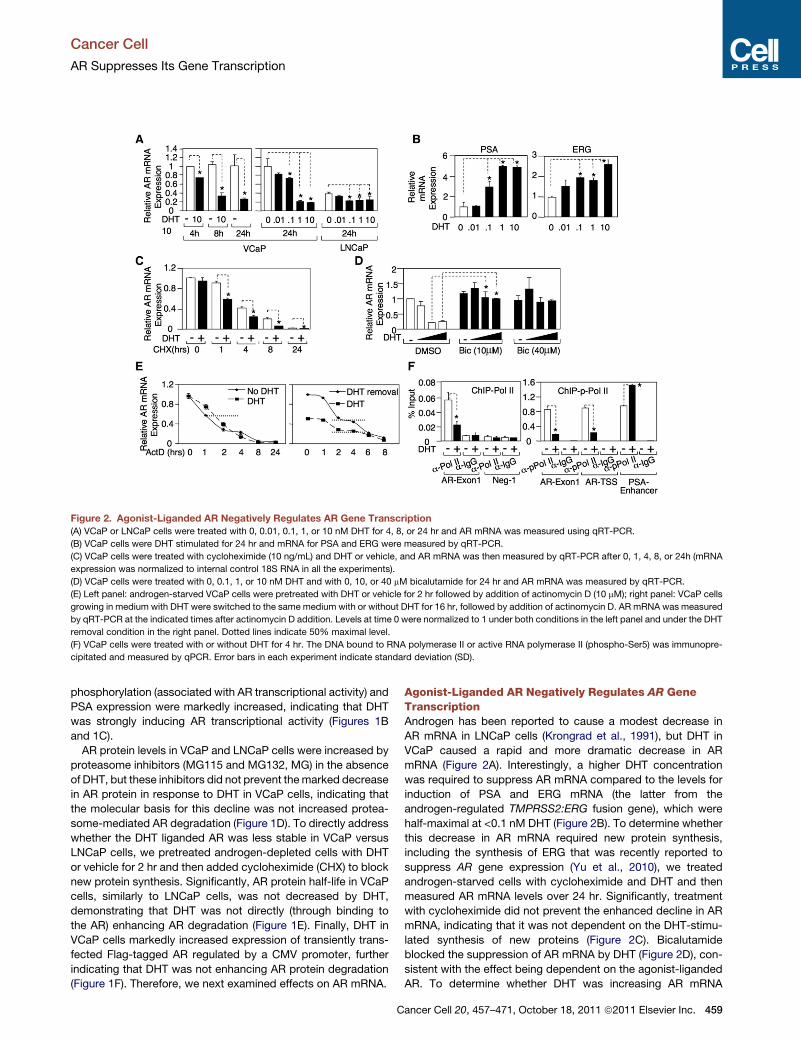
Figure 2. Agonist-Liganded AR Negatively Regulates AR Gene Transcription
(A) VCaP or LNCaP cells were treated with 0, 0.01, 0.1, 1, or 10 nM DHT for 4, 8, or 24 hr and AR mRNA was measured using qRT-PCR.
(B) VCaP cells were DHT stimulated for 24 hr and mRNA for PSA and ERG were measured by qRT-PCR.
(C) VCaP cells were treated with cycloheximide (10 ng/mL) and DHT or vehicle, and AR mRNA was then measured by qRT-PCR after 0, 1, 4, 8, or 24h (mRNA
expression was normalized to internal control 18S RNA in all the experiments).
(D) VCaP cells were treated with 0, 0.1, 1, or 10 nM DHT and with 0, 10, or 40 mM bicalutamide for 24 hr and AR mRNA was measured by qRT-PCR.
(E) Left panel: androgen-starved VCaP cells were pretreated with DHT or vehicle for 2 hr followed by addition of actinomycin D (10 mM); right panel: VCaP cells
growing in medium with DHT were switched to the same medium with or without DHT for 16 hr, followed by addition of actinomycin D. AR mRNA was measured
by qRT-PCR at the indicated times after actinomycin D addition. Levels at time 0 were normalized to 1 under both conditions in the left panel and under the DHT
removal condition in the right panel. Dotted lines indicate 50% maximal level.
(F) VCaP cells were treated with or without DHT for 4 hr. The DNA bound to RNA polymerase II or active RNA polymerase II (phospho-Ser5) was immunopre-
cipitated and measured by qPCR. Error bars in each experiment indicate standard deviation (SD).
Cancer Cell
AR Suppresses Its Gene Transcription
phosphorylation (associated with AR transcriptional activity) and
PSA expression were markedly increased, indicating that DHT
was strongly inducing AR transcriptional activity (Figures 1B
and 1C).
AR protein levels in VCaP and LNCaP cells were increased by
proteasome inhibitors (MG115 and MG132, MG) in the absence
of DHT, but these inhibitors did not prevent themarked decrease
in AR protein in response to DHT in VCaP cells, indicating that
the molecular basis for this decline was not increased protea-
some-mediated AR degradation (Figure 1D). To directly address
whether the DHT liganded AR was less stable in VCaP versus
LNCaP cells, we pretreated androgen-depleted cells with DHT
or vehicle for 2 hr and then added cycloheximide (CHX) to block
new protein synthesis. Significantly, AR protein half-life in VCaP
cells, similarly to LNCaP cells, was not decreased by DHT,
demonstrating that DHT was not directly (through binding to
the AR) enhancing AR degradation (Figure 1E). Finally, DHT in
VCaP cells markedly increased expression of transiently trans-
fected Flag-tagged AR regulated by a CMV promoter, further
indicating that DHT was not enhancing AR protein degradation
(Figure 1F). Therefore, we next examined effects on AR mRNA.
C
Agonist-Liganded AR Negatively Regulates AR GeneTranscriptionAndrogen has been reported to cause a modest decrease in
AR mRNA in LNCaP cells (Krongrad et al., 1991), but DHT in
VCaP caused a rapid and more dramatic decrease in AR
mRNA (Figure 2A). Interestingly, a higher DHT concentration
was required to suppress AR mRNA compared to the levels for
induction of PSA and ERG mRNA (the latter from the
androgen-regulated TMPRSS2:ERG fusion gene), which were
half-maximal at <0.1 nM DHT (Figure 2B). To determine whether
this decrease in AR mRNA required new protein synthesis,
including the synthesis of ERG that was recently reported to
suppress AR gene expression (Yu et al., 2010), we treated
androgen-starved cells with cycloheximide and DHT and then
measured AR mRNA levels over 24 hr. Significantly, treatment
with cycloheximide did not prevent the enhanced decline in AR
mRNA, indicating that it was not dependent on the DHT-stimu-
lated synthesis of new proteins (Figure 2C). Bicalutamide
blocked the suppression of AR mRNA by DHT (Figure 2D), con-
sistent with the effect being dependent on the agonist-liganded
AR. To determine whether DHT was increasing AR mRNA
ancer Cell 20, 457–471, October 18, 2011 ª2011 Elsevier Inc. 459

Cancer Cell
AR Suppresses Its Gene Transcription
degradation, we pretreated androgen-starved VCaP cells with
DHT for 2 hr and then added actinomycin D to block new
mRNA synthesis. Significantly, AR mRNA half-life was not
decreased by DHT (Figure 2E, left panel), suggesting that DHT
was decreasing AR gene transcription. We also assessed AR
mRNA half-life in VCaP cells growing in mediumwith DHT versus
cells where DHT was removed for 16 hr before the addition of
actinomycin D. Although AR mRNA was decreased in the pres-
ence of DHT, there was no evident decrease in AR half-life (Fig-
ure 2E, right panel). Finally, we found by chromatin immunopre-
cipitation (ChIP) that DHT decreased the binding of RNA
polymerase II to exon 1 in the AR gene (Figure 2F, left panel)
and also decreased binding of active RNA polymerase II as
shown by anti-phospho-RNA polymerase II ChiP (Figure 2F, right
panel). Together these results indicated that the DHT liganded
AR in VCaP cells was directly repressing AR gene transcription.
Androgen Stimulates AR Recruitment to a ConservedSite in Intron 2 of the AR GeneData from a recent ChIP-chip analysis of AR binding sites
(ARBSs) in LNCaP cells identified three sites linked to the AR
gene: ARBS1 in the promoter region (10% FDR), ARBS2 in intron
2 (5% FDR), and ARBS3 in the 30 downstream region (5% FDR)
(Wang et al., 2009) (Figure S2A). To assess these binding sites in
VCaP cells, we designed two pairs of primers for each ARBS and
utilized ChIP coupledwith quantitative real-time PCR tomeasure
AR binding. Only the ARBS2 site (ARBS2-1) showed clear DHT
induced AR binding, although basal and androgen-induced AR
binding to the well-characterized major ARE upstream of the
PSA gene (ARE III) were higher (Figure 3A). Because important
regulatory elements may be conserved between species, we
compared the human ARBS2 region to the corresponding
regions in other species. Interestingly, a fragment of ARBS2
(�400 bp) that overlapped ARBS2-1 was highly conserved
among species (100% identical between mouse and rat and
88% identical between mouse or rat and human) and contained
multiple binding sites for FOXA1, a pioneer transcription factor
that interacts with AR and is generally found at steroid-respon-
sive enhancer elements (Figure 3B and Figure S2B). Therefore,
we synthesized an additional set of primer pairs spanning this
conserved region (ARBS2a, 2b, and 2c) and repeated the AR
ChIP assays. AR binding to all three sites was substantially
increased by DHT, and this binding was blocked by the AR
antagonist bicalutamide (Figure 3C). The DHT-stimulated
increase was comparable to the �5-fold increase on the AREs
in the control PSA and TMPRSS2 enhancers, but basal binding
to ARBS2 was again lower (Figure 3C). As observed on the
PSA enhancer, DHT-stimulated AR recruitment to ARBS2 was
maximal at early times (2 hr) but still persisted after 24 hr (Fig-
ure S2C). As noted for suppression of ARmRNA versus induction
of PSA and ERGmRNA (Figure 2), AR binding to ARBS2 required
higher DHT concentrations (Figure 3D). Finally, anti-FOXA1 ChIP
showed that FOXA1 was associated constitutively with ARBS2
(Figure 3E).
Androgen Stimulates Demethylationof H3K4 Associated with ARBS2Consistent with ARBS2 functioning as an enhancer, ChIP with an
anti-TATA binding protein (TBP) antibody indicated that there
460 Cancer Cell 20, 457–471, October 18, 2011 ª2011 Elsevier Inc.
was an interaction between this site and AR gene promoter (Fig-
ure S3A). Significantly, we also detected a basal association
between activated RNA polymerase II and ARBS2 that was
decreased by DHT, suggesting that the agonist liganded AR
may be mediating repression through this site (Figure 4A).
Further evidence for an interaction between the AR recruited to
ARBS2 and the AR gene promoter was obtained by anti-AR
ChIP followed by a chromatin conformation capture (3C) assay,
which identified a DHT-dependent association between AR,
ARBS2, and the AR gene promoter (Figure S3B).
The agonist liganded AR generally stimulates transcription
through recruitment of coactivator proteins and histone acetyl-
transferases, but can more weakly mediate recruitment of
transcriptional corepressors, such as NCoR or SMRT, and their
associated histone deacetylases (HDACs) (Cheng et al., 2002).
Therefore, we next used ChIP to determine whether DHT
was directly or indirectly stimulating recruitment of an HDAC to
AR-binding sites in the AR gene. Interestingly, control experi-
ments indicated that HDAC3 (which forms a complex with
NCoR and SMRT) was associated with ARE III in the PSA
enhancer and that this association was decreased by DHT (Fig-
ure S3C). There also appeared to be a very weak association of
HDAC3 with each of the ChIP-chip identified AR-binding sites
(ARBS1, 2, and 3) in the AR gene, but these were not increased
by DHT (Figure S3C). Moreover, ChIP with antibodies against
acetylated H3K9/14 did not detect decreases in histone acetyla-
tion at any of the sites in response toDHT (Figure S3D). As a posi-
tive control, in the absence of DHT, we detected high levels of
histone acetylation in AR exon 1 and this decreased in response
to DHT, consistent with down-regulation of AR gene expression.
Because interaction with the promoter and FOXA1 binding
suggested that ARBS2 may function as an enhancer, we next
assessed changes in histone marks that are associated with
active enhancers (H3K4 mono- and dimethylation) at ARBS1,
2, and 3. Substantial H3K4 methylation was detected at each
site, but there were no changes in response to DHT at ARBS1
or ARBS3, or at the ARE III site in the PSA enhancer (Figure 4B).
The TMPRSS2 enhancer ARE was similarly unaffected (Fig-
ure 4C). In contrast, DHT caused a decrease in both H3K4me1
and H3K4me2 levels at ARBS2-1 (Figure 4B), and this was con-
firmed using the set of ARBS2 primers (ARBS2a, b, and c) span-
ning the conserved region (Figure 4C). Taken together, these
results suggested that ARBS2 contains an enhancer that is
rapidly inactivated by androgen.
VCaP xenografts that relapse after castration have higher
levels of AR mRNA and renewed expression of AR-regulated
genes, similarly to what is observed in patients who progress
to CRPC (Cai et al., 2009). To determine whether the ARBS2
site contributes to the increased AR gene expression in these
relapsed tumors, we generated a cell line (VCS2) from a relapsed
VCaP xenograft tumor. VCS2 cells in steroid-depleted medium
had higher levels of AR, PSA, and ERG (from the androgen-regu-
lated TMPRSS2:ERG fusion gene) relative to the parental VCaP
cells (Figure 4D) and were less dependent on androgens for
cell survival (Figure S3E), but AR protein was still markedly
decreased by DHT. An analysis of basal (in steroid depleted
medium without exogenous DHT) mRNA levels confirmed that
AR, PSA, and ERG mRNA were increased in VCS2 cells com-
pared to VCaP and showed that AR mRNA was markedly

Figure 3. Androgen Stimulates AR Recruitment to a Site in Intron 2 of the AR Gene
(A) VCaP cells in steroid-depleted medium (CSSmedium) were treated with 0, 1, or 10 nMDHT for 4 hr and the DNA bound to ARwasmeasured by ChIP followed
by qPCR.
(B) The conserved region of ARBS2 (intron2) among 17 vertebrate species was plotted using UCSC Genome Browser.
(C) VCaP cells were pretreated with or without 10 mMbicalutamide for 4 hr followed by treatment with 10 nMDHT for 4 hr. The DNA bound to ARwasmeasured by
ChIP followed by qPCR.
(D) VCaP cells were treated for 4 hr with 0, 0.1, 1, or 10 nM DHT. AR binding to ARBS2 or the PSA enhancer ARE were measured by ChIP followed by qPCR.
(E) VCaP cells were treated with or without 10 nM DHT for 4 hr and the DNA bound to FOXA1 was measured by ChIP and qPCR. Error bars in each experiment
indicate SD. See also Figure S2 and see Table S1 for raw qPCR data for experiments shown.
Cancer Cell
AR Suppresses Its Gene Transcription
decreased in response to DHT (Figure 4E). AR ChIP showed that
DHT stimulated recruitment of AR to ARBS2 in the VCS2 cells,
with the increased binding compared to VCaP being consistent
with higher AR levels in the VCS2 cells (Figure 4F, left panel).
Significantly, basal ARBS2 H3K4 methylation was increased in
C
the VCS2 cells compared to VCaP, but was still decreased by
DHT (Figure 4F, right panel). Finally, transcription factors shown
previously to interact with AR on enhancers, Oct1 and GATA-2
(Wang et al., 2007), were associated with ARBS2 and were
increased in VCS2 (Figure 4G). Overall, these findings further
ancer Cell 20, 457–471, October 18, 2011 ª2011 Elsevier Inc. 461

Figure 4. Androgen Stimulates Rapid Demethylation of H3K4 in VCaP and VCaP-Derived VCS2 Cells
(A–C) VCaP cells were treated with or without DHT for 4 hr and the DNA bound to active RNA polymerase II, mono- or di-methylated H3K4 were measured ChIP
and qPCR.
(D and E) VCaP or VCS2 cells were treated with 0, 1, or 10 nM DHT for 24 hr and AR, PSA, ERG, and b-tubulin proteins were immunoblotted or mRNA were
measured by ChIP followed by qRT-PCR (18S as internal control).
(F and G) VCaP or VCS2 cells were treated with or without DHT for 4 hr and the DNA bound to AR, mono-methylated H3K4, Oct1, or GATA2 were measured by
ChIP followed by qPCR. Error bars in each experiment indicate SD. See also Figure S3 and see Table S2 for raw qPCR data for experiments shown.
Cancer Cell
AR Suppresses Its Gene Transcription
462 Cancer Cell 20, 457–471, October 18, 2011 ª2011 Elsevier Inc.

Figure 5. Androgen Deprivation Activates the ARBS2 Site in LNCaP Cells
(A) LNCaP cells were treated with or without 10 nM DHT for 4 hr and the DNA bound to AR was immunoprecipitated and measured by qPCR.
(B) LNCaP cells were treated with or without 10 nMDHT for 4 hr and the DNA bound to AR, mono- or di-methylated H3K4 was immunoprecipitated andmeasured
by qPCR.
(C) LNCaP or LNCaP-CSS3 (adapted to steroid-depleted medium for > 3 w) were treated with 0, 1, or 10 nM DHT for 24 hr and AR mRNA was measured by
qRT-PCR (18S as internal control).
(D) LNCaP or LNCaP-CSS3 cells were treatedwith or without 10 nMDHT for 4 hr and the DNA bound to AR ormono-methylated H3K4wasmeasured by ChIP and
qPCR. Error bars in each experiment indicate SD. See Table S3 for raw qPCR data for experiments shown.
Cancer Cell
AR Suppresses Its Gene Transcription
supported the conclusion that ARBS2 contains an enhancer that
contributes to increased AR gene expression at low androgen
levels in CRPC and indicated that this enhancer is repressed
by the agonist liganded AR.
Androgen Deprivation Activates the ARBS2 Sitein LNCaP CellsWe next examined the LNCaP PCa cell line, which shows only
a small decrease in ARmRNA in response toDHT (see Figure 2A).
Anti-AR ChIP showed DHT stimulated recruitment of AR to
ARBS2-1 (Figure 5A), which was confirmed using the ARBS2a,
b, and c primers (Figure 5B, left panel). However, in contrast to
VCaP cells, there was less AR binding to ARBS2 and no marked
DHT stimulated decreases in H3K4me1 or me2 (Figure 5B, right
panel). On the basis of the results above in VCaP versus VCS2
cells, we next examined LNCaP cells that were passaged
in vitro in steroid-depletedmedium (basal mediumwith 5%char-
coal/dextran stripped serum, CSS). As shown in Figure 5C, after
3 weeks in steroid-depleted medium, the cells expressed higher
levels of AR mRNA, which markedly declined in response to
DHT. AR ChIP in these LNCaP-CSS3 cells showed increased
DHT-stimulated AR recruitment to ARBS2 relative to the parental
LNCaP cells (Figure 5D, upper panel). Most significantly, basal
H3K4 methylation of ARBS2 was increased in the LNCaP-
C
CSS3 cells, and it declined in response to DHT (Figure 5D, lower
panel). These results in LNCaP cells further support the conclu-
sion that ARBS2 contains an androgen-repressed enhancer that
contributes to increased AR gene expression in response to
androgen deprivation.
Lysine-Specific Demethylase 1 (LSD1) Is Recruitedto ARBS2 In Vitro and In Vivo by the DHT LigandedAR and Mediates RepressionThe decrease in H3K4 mono- and dimethylation over the ARBS2
site indicated that AR was either suppressing the activity of a
histone methyltransferase or increasing a histone demethylase.
Significantly, lysine-specific demethylase 1 (LSD1) has been
shown to interact with AR (Metzger et al., 2005; Wissmann
et al., 2007), and we confirmed this interaction by coimmunopre-
cipitation of endogenous AR and LSD1 (Figure 6A). LSD1 is re-
ported to function as an AR coactivator on the PSA gene ARE
III enhancer through demethylation of repressive mono- and di-
methylated H3K9 (Metzger et al., 2005; Wissmann et al., 2007).
However, mono- and dimethylated H3K4 are also substrates
for LSD1, and in most contexts LSD1 appears to function as a
repressor through H3K4me1 and H3K4me2 demethylation (Shi
et al., 2004). Therefore, we next tested the hypothesis that
DHT stimulates LSD1 recruitment to ARBS2. An association
ancer Cell 20, 457–471, October 18, 2011 ª2011 Elsevier Inc. 463

Figure 6. LSD1 Is Recruited to ARBS2 by the DHT Liganded AR In Vitro and In Vivo
(A) VCaP cells were treated with or without 10 nM DHT for 24 hr and protein was then immunoprecipitated using anti-AR antibody or IgG control, followed by
immunoblotting for LSD1 and AR.
(B) VCS2 cells were treated with 0 or 10 nM DHT for 4 hr and the DNA bound to LSD1 was measured by ChIP and qPCR.
(C) VCaP cells were grown in steroid-depleted medium supplemented with 10 nMDHT for 3 days and then DHTwas removed for 3 days. The DNA bound to AR or
LSD1 was measured by ChIP and qPCR.
(D) The tissue of VCaP xenograft tumor (precastrated [�] or 4-day postcastration [+] mice) was formalin fixed, lysed, and sonicated. The DNA bound to ARor LSD1
was immunoprecipitated and measured by qPCR.
(E) VCaP cells were transfected with 20 nM LSD1 siRNA (Dharmacon) for 2 days and then treated with or without DHT for 24 hr. AR, LSD1, and b-actin were
immunoblotted.
(F)VCaPcells transfectedwithLSD1orcontrol siRNAwerestimulatedwith10nMDHTandLSD1,AR,AKR1C3,orHSD17B6mRNAweremeasuredusingqRT-PCR.
(G) VCaP cells were pretreated with pargyline (2 mM) for 8 hr and then treated with or without DHT for 16 hr. LSD1, AR, AKR1C3, or HSD17B6 mRNA were
measured using qRT-PCR (normalized to GAPDH as internal control).
(H) VCaP cells were transfectedwith 20 nM LSD1 siRNA for 2 days and then treatedwith or without 10 nMDHT for 4 hr. The DNA bound tomono- or di-methylated
H3K4 was immunoprecipitated and measured by qPCR. Error bars in each experiment indicate SD. See also Figure S4 and see Table S4 for raw qPCR data for
experiments shown.
Cancer Cell
AR Suppresses Its Gene Transcription
464 Cancer Cell 20, 457–471, October 18, 2011 ª2011 Elsevier Inc.

Cancer Cell
AR Suppresses Its Gene Transcription
between LSD1 and ARBS2 was detected by ChIP in VCaP cells
(Figure S4A) and in VCS2 cells (Figure 6B), and this interaction
was increased by DHT. Consistent with previous reports in
LNCaP cells (Metzger et al., 2005; Wissmann et al., 2007),
LSD1 was constitutively associated with the ARE III in the PSA
enhancer and was not clearly increased by DHT (Figure 6B).
LSD1 was similarly constitutively associated with the ARE in
the TMPRSS2 enhancer (Figure 6B). Finally, we confirmed that
DHT stimulated the recruitment of LSD1 to ARBS2 in LNCaP
cells and found that LSD1 recruitment to ARBS2 was increased
in the LNCaP-CSS3 cells (Figure S4B).
In the converse experiment, we examined VCaP cells cultured
in medium with androgen that were then shifted to steroid-
depleted medium for 3 days. As shown in Figure 6C, both AR
and LSD1 binding to ARBS2 were decreased in the steroid-
depleted cells. We showed previously that AR mRNA levels in
VCaP xenografts were markedly increased at 4 days after
castration (Cai et al., 2009). To determine whether this increase
in AR mRNA in vivo correlated with decreased binding of AR
and LSD1 to ARBS2, we used ChIP to examine VCaP xenografts
prior to castration and at 4 days after castration. As shown in Fig-
ure 6D, both AR and LSD1 were associated with ARBS2 prior to
castration, and these associations were markedly decreased
4 days after castration.
LSD1 can potentially function as a coactivator or corepressor
by demethylating H3K9 or H3K4, respectively, and we found
that DHT also stimulated a decline in H3K9 methylation as well
as H3K4 methylation across the ARBS2 site (Figure S4C, left
panel). In contrast, DHT did not cause a decrease in H3K4me3,
which is associated with both promoters and enhancers but is
not a substrate for LSD1 (Figure S4C, right panel). Therefore,
as these changes in methylation would be consistent with
LSD1 functioning as a coactivator or corepressor, we next
utilized siRNA to address directly whether LSD1 was mediating
the down-regulation of AR gene expression in response to
DHT. Expression of LSD1 protein (Figure 6E) and mRNA (Fig-
ure 6F) were substantially decreased by the LSD1 siRNA, and
the DHT-stimulated decrease in AR protein was diminished (Fig-
ure 6E). An analysis of AR mRNA confirmed that the DHT-stimu-
lated decrease in AR expression was blunted by LSD1 siRNA
(Figure 6F).
To determine whether this LSD1-dependent suppression was
unique to the AR gene, we also examined expression of AKR1C3
and HSD17B6, which are androgen repressed and increased
in CRPC. AKR1C3 catalyzes synthesis of testosterone from
androstenedione and HSD17B6 oxidizes 5a-androstene-3a,
17b-diol back to DHT (Bauman et al., 2006). Similarly to AR,
we reported previously that mRNA expression of AKR1C3 was
consistently increased in CRPC (Stanbrough et al., 2006), and
both AKR1C3 and HSD17B6 were negatively regulated by
androgens in VCaP cells (Cai et al., 2009). As shown in Figure 6F,
the DHT-stimulated declines in AKR1C3 and HSD17B6 mRNA
were abrogated by the LSD1 siRNA. Similar results were ob-
tained using a chemical inhibitor of LSD1, pargyline (Figure 6G),
which also prevented the DHT-stimulated decline in AR protein
(Figure S4D). Consistent with previous data showing that LSD1
functions as a coactivator on the PSA gene (Metzger et al.,
2005; Wissmann et al., 2007), pargyline also blocked the DHT
stimulated increase in PSA protein (Figure S4D).
C
The LSD1 siRNA did not decrease the DHT-stimulated recruit-
ment of AR to ARBS2 (Figure S4E, left panel). However, the DHT-
stimulated declines in H3K9methylation (Figure S4E, right panel)
and H3K4 methylation (Figure 6H) across ARBS2 were impaired
or abrogated by the LSD1 siRNA. Pargyline similarly impaired
DHT-stimulated H3K4me1 demethylation across ARBS2 (Fig-
ure S4F). Together, these data indicated that AR was mediating
repression through recruitment of LSD1 and H3K4 demethyla-
tion. Finally, we used pargyline to assess whether LSD1 was
mediating the DHT-stimulated repression of AR gene expression
in other PCa cell lines. C4-2 cells were derived from a castration-
resistant LNCaP xenograft and CWR22Rv1 cells were from
a castration-resistant CWR22 xenograft. In both cells, pargyline
abrogated the DHT-stimulated decrease in AR mRNA (Fig-
ure S4G). Moreover, consistent with LSD1 functioning as an
AR coactivator on androgen-stimulated genes, pargyline sup-
pressed the DHT-stimulated increase in FKBP5.
Previous studies have shown that LSD1 functions as a coacti-
vator for AR on the PSA (KLK3) and KLK2 genes because of
phosphorylation of H3T6 and H3T11, which suppress LSD1-
mediated H3K4 demethylation and enhance H3K9 demethyla-
tion, respectively (Metzger et al., 2008; Metzger et al., 2010).
Therefore, we next used ChIP to determine whether differences
in H3T6 or H3T11 phosphorylation were a basis for the distinct
effects of AR and LSD1 on the AR gene versus AR-stimulated
genes. Significantly, DHT-stimulated H3T6 and H3T11 phos-
phorylation were lower across ARBS2 and were also lower in
the androgen-suppressed OPRK1 (see Figure 7) and AKR1C3
genes, compared to AREs in the androgen-stimulated PSA,
KLK2, and FKBP5 genes (Figure S4G). However, H3T6 and
H3T11 phosphoryation were also low in the strongly androgen-
stimulated TMPRSS2 gene. These findings are consistent with
the conclusion that phosphorylation of H3T6 and H3T11
contribute to the regulation of LSD1 substrate specificity, but
additional mechanisms may also contribute to this regulation.
Expression of Androgen Repressed Genes Is Increasedin CRPC XenograftsExpression microarrays were used to identify genes that were
androgen repressed in both VCaP and VCS2 cells in vitro, and
to then assess the expression of these genes in vivo in an-
drogen-dependent versus relapsed castration-resistant VCaP
xenografts. AR, AKR1C3, and HSD17B6 were again found to
be androgen repressed in VCaP (4.2-, 2.8-, and 3.7-fold higher
in the absence of androgen, respectively) and were even
more highly androgen repressed in VCS2 cells (6.4-, 8.5-, and
4.7-fold, respectively) (Table S5). In contrast, expression of these
genes was highly up-regulated in the relapsed VCaP xenografts
(5.4-, 2.3-, and 3.5-fold for AR, AKR1C3, and HSD17B6, respec-
tively). These findings, in conjunction with the low intratumoral
androgen levels in these castration-resistant tumors (Fig-
ure S5A), support a feedback mechanism that negatively regu-
lates AR signaling at high androgen levels and enhances
signaling at the lower androgen levels.
To more systematically assess the significance of additional
in vitro identified androgen-repressed genes, we next focused
on the 411 genes that were repressed by >2-fold in VCS2
and >1.5-fold in VCaP (the lower threshold in VCaP being based
on the more robust repression of AR, AKR1C3, and HSD17B6 in
ancer Cell 20, 457–471, October 18, 2011 ª2011 Elsevier Inc. 465

Figure 7. Identification of Androgen-Repressed Genes in VCaP Cells and Xenografts
(A) VCaP or VCS2 cells were treated with or without 10 nM DHT for 24 hr and were analyzed on Affymetrix U133A microarrays. The numbers of DHT-repressed
genes or DHT-induced genes in VCaP and VCS2 cells and their overlaps are shown.
(B) VCaP xenografts were established and biopsied at three stages: androgen-dependent tumor (AD), 4 days after castration (CS), and castration-resistant
relapsed tumor (CRPC). mRNA were extracted from the biopsies of tumors of AD or CRPC stages and analyzed on Agilient microarrays. The data was analyzed
using SAM software (Significance Analysis of Microarrays). The top 30 genes with lowest q-value are shown, with black arrows indicating DHT-repressed genes.
(C) GO term analysis of DHT-repressed genes (left panel) versus androgen-induced genes (right panel). See also Figure S5 and Table S5.
Cancer Cell
AR Suppresses Its Gene Transcription
VCS2 cells) (Figure 7A and Table S5). Remarkably, among the
top 30 genes with most significantly elevated expression in the
castration-resistant VCaP xenografts, 12 were in this group of
411 androgen-repressed genes (Figure 7B). In addition, further
genes among this group of 30 that appeared to be androgen-
repressed were ANKRD22 (1.64-fold in VCaP and 1.82-fold in
VCS2), MMP10 (1.32-fold in VCaP and 4.2-fold in VCS2), and
STXBP6 (1.60-fold in VCaP and 1.93-fold in VCS2).
We next took advantage of recent AR ChIP-seq data in VCaP
cells (Yu et al., 2010) to assess the frequency of AR-binding sites
in androgen-repressed versus androgen-activated genes in
VCaP cells. AR-binding sites were found in 20% of AR-activated
genes and in 14% of AR-repressed genes, with the background
being 11% (fraction of total 31,810 genes that contain AR-
466 Cancer Cell 20, 457–471, October 18, 2011 ª2011 Elsevier Inc.
binding sites), indicating that there is enrichment for AR-binding
sites within the AR-repressed genes (Figure S5B). The lower
enrichment versus the AR-activated genes could mean that
more genes in the AR-activated group are directly regulated by
AR, but could also be in part technical and reflect somewhat
weaker binding of AR to AR-repressed genes. To further assess
whether suppression of these genes was mediated directly by
AR through an LSD1-dependent mechanism, we focused on
another androgen-repressed gene (OPRK1) that was strongly
up-regulated in the VCaP CRPC xenografts. Using real-time
RT-PCR, we first confirmed that DHT markedly decreases
OPRK1 mRNA in VCaP cells, similarly to the decreases in AR,
AKR1C3, and HSD17B6 (Figure S5C). Using AR siRNA we also
showed that AR down-regulation could blunt the DHT-mediated

Cancer Cell
AR Suppresses Its Gene Transcription
repression of these genes, providing further evidence that the
repression was AR mediated (Figure S5C). The AR siRNA also
decreased basal, but not DHT stimulated PSA or TMPRSS2
expression, consistent with AR functioning more efficiently on
AR-stimulated genes. OPRK1 has a single AR-binding site in
its 30 UTR based on ChIP-chip and ChIP-seq data in both LNCaP
and VCaP cells (Wang et al., 2009; Yu et al., 2010) (Figure S5D).
Therefore, we usedChIPwith primers covering this site to assess
AR and LSD1 binding. Significantly, DHT stimulated AR and
LSD1 recruitment to this site and also decreased H3K4 methyl-
ation (Figure S5E). Together, these data indicate that AR is
directly negatively regulating a set of genes that are up-regulated
in the VCaP CRPC xenografts.
To assess the potential functional consequences of failing to
suppress androgen-repressed genes after castration, we deter-
mined the pathways that were associated with the 411
androgen-repressed genes identified in VCaP and VCS2 cells.
Importantly, expression of these genes was most significantly
associated with increased DNA replication and cell cycle
progression (Figure 7C, left panel), whereas genes that were
increased in response to DHT in VCaP and VCS2 cells were
associated with synthesis of lipids, proteins, and other metabolic
processes distinct from DNA replication (Figure 7C, right panel).
Finally, we treated VCaP CRPC xenografts with testosterone to
assess effects on AR repressed genes in vivo, and found by
RT-PCR that AR, AKR1C3, HSD17B6, and OPRK1 were
repressed (Figure S5F). Testosterone also suppressed expres-
sion of BCL11A, another strongly AR repressed gene that was
increased in castration-resistant VCaP xenografts, but did not
clearly suppress PSA or TMPRSS2.Moreover, therewasmarked
regression in the xenografts (Figure S5G). These findings indi-
cated that a partial restoration of androgen levels and AR
transcriptional activity in CRPC cells may drive tumor growth
by activating cellular metabolism while failing to suppress DNA
replication and proliferation.
Increased Expression of Androgen Repressed Genesin Patients with CRPCTo determine whether increased expression of androgen-
repressed genes may contribute to CRPC in patients, we used
expression data from a set of CRPC bone marrow metastases
versus primary prostate cancers that had not received hormonal
therapy (Stanbrough et al., 2006; Mendiratta et al., 2009). Con-
sistent with lower androgen levels and reduced AR transcrip-
tional activity in CRPC, only a small fraction of the genes that
were androgen induced in VCaP/VCS2 were overexpressed in
CRPC (18/556), whereas a much larger fraction were underex-
pressed (71/556) (Figure 8A). Similarly, very few of the AR re-
pressed genes were underexpressed in CRPC (9/411), whereas
many more were overexpressed (53/411) (Table S6). As noted
previously, genes that are overexpressed in CRPC are highly
associated with proliferation (Stanbrough et al., 2006; Wang
et al., 2009) (Figure 8B), whereas genes that are underexpressed
are more associated with developmental pathways (Figure S6A).
Significantly, the set of 53 androgen-repressed genes that were
overexpressed in the CRPCbiopsy samples were similarly highly
associated with DNA replication and proliferation (Figure 8C).
To further assess the biological importance of these 53
androgen-repressed genes in CRPC, we removed them from
C
the set of 1490 genes that were overexpressed in the CRPC
biopsy samples and repeated the Gene Ontology analysis on
the remaining 1437 genes. Although these 1437 genes were still
associated with cell cycle progression and DNAmetabolism, the
significance of all these associations was markedly decreased,
and DNA replication was no longer among the most highly asso-
ciated pathways in the absence of these 53 androgen-repressed
genes (Figure S6B). Finally, we selected for further analysis a set
of eight genes that were androgen repressed in VCaP/VCS2 cells
andwere also overexpressed in the relapsed VCaP xenografts or
the clinical CRPC biopsies. Quantitative real-time RT-PCR
confirmed that they were all DHT repressed in VCaP and VCS2
cells, and that this could be prevented with bicalutamide (Fig-
ure S6C). Moreover, in all cases the androgen-stimulated
down-regulation was decreased or abrogated by treatment
with pargyline, indicating that it was mediated by LSD1 (Fig-
ure 8D). Together, these findings elucidate a mechanism by
which loss of negative regulation by the agonist liganded AR,
in association with LSD1, increases the expression of AR and
of multiple genes that contribute to increased androgen
synthesis, DNA replication, and proliferation in CRPC.
DISCUSSION
Studies in clinical samples and xenograft models indicate that
increased AR gene expression plays a major role in the progres-
sion to CRPC. We observed previously in VCaP cells in vitro and
in VCaP xenografts in vivo that ARmRNA levels decline rapidly in
response to androgen stimulation and increase rapidly in
response to androgen withdrawal (Cai et al., 2009). In this report
we have identified a highly conserved site in the second intron of
the AR gene that regulates its expression in response to
androgen stimulation and withdrawal. RNA polymerase II and
FOXA1 are associated with this ARBS2 site, as are OCT1,
GATA2, and substantial levels of H3K4 mono- and dimethylation
that are further increased in cells adapted to androgen depriva-
tion, consistent with this element functioning as an enhancer
that contributes to increased AR gene expression in CRPC.
Moreover, we show that the agonist liganded AR decreases
AR gene expression by functioning as a transcriptional repressor
at this site through recruitment of LSD1 and demethylation of
H3K4me1,2. The rapid androgen-mediated down-regulation of
AKR1C3 and HSD17B6 is similarly LSD1 dependent, indicating
that the agonist liganded AR directly mediates a physiological
intracellular negative feedback loop to regulate AR activity.
Taken together, these findings elucidate a mechanism that
contributes to increased AR gene expression and restored AR
activity in CRPC, and identify a suppressor element and tran-
scriptional repressor function for the agonist liganded AR.
Further analysis of gene expression in androgen-starved
versus androgen-stimulated VCaP and VCS2 cells showed that
the agonist liganded AR also suppressed the expression of
multiple genes mediating DNA synthesis and cell cycle progres-
sion, while it increased the expression of genes mediating
synthesis of lipids, amino acids, and other metabolic processes.
This profile is consistent with AR function in normal prostate
epithelium to drive terminal differentiation and synthesis of
seminal fluid and provides a molecular basis for the biphasic
response to androgen stimulation whereby PCa cells proliferate
ancer Cell 20, 457–471, October 18, 2011 ª2011 Elsevier Inc. 467

Figure 8. Expression of Androgen-Repressed Genes Is Increased in Human CRPC Samples
(A) Affymetrix microarray expression data showing overlaps between androgen repressed/induced genes and the expression of 1490 genes that were increased
and 626 genes that were decreased (p < 0.001 and fold-change > 1.5) in 34 CRPC bone marrow metastases compared with 27 primary tumors prior to any
hormonal therapy.
(B and C) GO term analysis of the group of 1490 CRPC-overexpressed genes (B) and 53 AR-repressed genes that were overexpressed in CRPC (C).
(D) VCaP cells were pretreated with pargyline (2 mM) for 8 hr and then were treated with or without DHT for 16 hr. OPKR1, THBS1, BCL11A, STXBP6, MCM2,
MCM4, MCM6, or MCM7 mRNA were measured using qRT-PCR (normalized to GAPDH as internal control). Error bars in each experiment indicate SD.
(E) Graphical summary showing divergent effects of androgen deprivation on expression of AR-stimulated genes, which are decreased, versus AR-repressed
genes (including the AR gene), which are increased. In castration-resistant PCa, mechanisms including further increases in intratumoral androgen synthesis
result in partial restoration of AR transcriptional activation function on genes mediating lipid and protein biosynthesis, but do not restore AR repressor function on
the AR gene, or on genes mediating androgen synthesis, DNA synthesis, and cell cycle progression. See also Figure S6 and Table S6.
Cancer Cell
AR Suppresses Its Gene Transcription
468 Cancer Cell 20, 457–471, October 18, 2011 ª2011 Elsevier Inc.

Cancer Cell
AR Suppresses Its Gene Transcription
in response to low levels of androgen but are growth arrested at
high concentrations (Xu et al., 2006). Significantly, a set of these
androgen-repressed genes associated with increased DNA
synthesis and proliferation were overexpressed in vivo in castra-
tion-resistant VCaP xenografts and in CRPC patient samples.
We suggest that androgen levels in CRPC cells are adequate
to stimulate AR activity on enhancer elements of genes medi-
ating certain critical metabolic functions such as lipid synthesis,
which are sensitive to lower levels of androgens, but are not
adequate to effectively recruit AR and LSD1 to suppressor
elements in multiple genes that negatively regulate AR signaling
and cellular proliferation. A graphical summary showing diver-
gent effects of AR on expression of AR-stimulated versus AR-
repressed genes after androgen deprivation and in CRPC is
shown in Figure 8E.
LSD1 was initially identified in corepressor complexes and
shown to function by demethylating mono- and dimethylated
H3K4 (Shi et al., 2004). However, it was subsequently shown
to function as a coactivator through demethylation of repressive
mono- and dimethylated H3K9 when associated with AR and
possibly other nuclear receptors including estrogen receptor
a (Metzger et al., 2005, Garcia-Bassets et al., 2007, Perillo
et al., 2008). The results of this study indicate that the association
with AR does not determine the coactivator versus corepressor
function of LSD1, and that it is instead determined by properties
of the element to which it is being recruited. For example, hypo-
acetylated nucleosomes are more susceptible substrates for
LSD1 mediated demethylation (Shi et al., 2005). Moreover,
recent data indicate that phosphorylation of H3T11 by an AR-
associated kinase (PRK1/PKN1) enhances the demethylation
of H3K9me3 by JMJD2C and subsequent demethylation of
H3K9me1,2 by LSD1 (Metzger et al., 2008), whereas phosphor-
ylation of H3T6 by a distinct kinase (PKCb1) can suppress the
LSD1-mediated demethylation of H3K4me1,2 (Metzger et al.,
2010). Our data indicate that lower H3T6 and H3T11 phosphor-
ylation may contribute to the substrate specificity and core-
pressor function of LSD1 at AR repressed genes, although
LSD1 may be regulated by a distinct mechanism on the
TMPRSS2 gene. It will clearly be important to further charac-
terize these and additional AR suppressor elements and deter-
mine the extent to which histone modifications or other factors
regulate the function of AR and LSD1 on these suppressor
versus AR enhancer elements.
It has been well appreciated for many years that AR has both
growth-promoting and growth-suppressing activities and that
androgen deprivation therapies may directly or indirectly stimu-
late some pathways that contribute to growth and eventual
relapse. Indeed, androgens can suppress the growth of some
CRPC-derived cell lines, and high-dose androgens have been
explored as a therapy for CRPC (Umekita et al., 1996, Morris
et al., 2009). However, the molecular basis for androgen-stimu-
lated growth suppression has not been clear, and there have
been no previous studies suggesting that distinct AR transcrip-
tional mechanisms may underlie these functions. Therefore,
the results of this study provide a paradigm with implications
for both basic molecular mechanisms of steroid action and for
AR targeted therapy of prostate cancer. In particular, the distinct
mechanisms of AR action on enhancer versus suppressor
elements may make it possible to selectively augment AR tran-
C
scriptional repressor function and thereby prevent or delay the
emergence of CRPC.
EXPERIMENTAL PROCEDURES
Cell Culture and Xenografts
LNCaP or C4-2 cells were cultured in RPMI1640mediumwith 10% FBS. VCaP
cells were cultured in DMEM medium with 10% FBS, and VCS2 cells were
cultured in DMEM medium with 8% charcoal/dextran-stripped FBS (CSS)
plus 2% FBS. For most immunoblotting, RT-PCR, or ChIP assays, cells
were grown to 50%–60% confluence in 5% (CSS) medium for 3 days and
then treated with androgens or drugs. VCaP xenografts were established in
the flanks of male scid mice by injecting �2 million cells in 50% Matrigel.
When the tumors reached �1 cm, biopsies were obtained and then the mice
were castrated. Additional biopsies were obtained 4 days after castration,
and the tumors were harvested at relapse. Frozen sections were examined
to confirm that the samples used for RNA and protein extraction contained
predominantly nonnecrotic tumor. All animal experiments were approved by
the Beth Israel Deaconess Institutional Animal Care and Use Committee and
were performed in accordance with institutional and national guidelines.
RT-PCR and Immunoblotting
Quantitative real-time RT-PCR amplification was performed on RNA extracted
from tissue samples or cell lines using TRIZOL reagent. RNA (50 ng) was used
for each reaction and the result was normalized by coamplification of 18S
RNA. Reactions were performed on an ABI Prism 7700 Sequence Detection
System using Taqman one-step RT-PCR reagents. Primers and probes are
listed in Supplemental Information. PCR data are represented as mean ±
STD for repeats. Protein extracts were prepared by boiling for 15 min in 2%
SDS. Blots were incubated with anti-PSA (1:3000, polyclonal, BioDesign),
anti-AR (1:2000, polyclonal, Upstate), anti-LSD1 (1:1000, Abcam), anti-b-actin
(1:5000, monoclonal, Abcom), or anti-b-tubulin (1:2000, Upstate), and then
with 1:5000 anti-rabbit or anti-mouse secondary antibodies (Promega).
Coimmunoprecipitation
VCaP cells were harvested in Triton lysis buffer (0.5% Triton X-100, 20 mM
Tris-HCl, 150 mM NaCl, 5 mM EDTA, and 2 mM dithiothreitol) with protease
inhibitors. The protein was immunoprecipitated using monoclonal anti-AR
(AR441 from NeoMarkers) or mouse IgG control and then subjected to
immunoblotting.
Chromatin-Immunoprecipitation (ChIP) Assay
Cellswere formalin fixed, lysed, and sonicated to break the chromatin into 500–
800 bp fragments. Anti-AR (Santa Cruz), anti-FOXA1 (Abcam), anti-OCT1
(Santa Cruz), anti-GATA2 (Santa Cruz), anti-RNA Polymerase II (Santa Cruz),
anti-RNA Polymerase II CTD repeat (phospho Ser5), anti-TBP (Santa Cruz),
anti-LSD1 (Abcam), anti-HDAC1 (Santa Cruz), anti-HDAC2 (Santa Cruz), anti-
HDAC3 (Santa Cruz), anti-H3K4me1 (Abcam), anti-H3K4me2 (Upstate), anti-
H3K4me3 (Abcam), anti-H3K9me1 (Abcam), anti-H3K9/14ace (Upstate),
anti-H3T6pho (Abcam), anti-H3T11pho (Abcam), or rabbit IgG (Santa Cruz)
were used to precipitate chromatin fragments from cell extracts. Quantitative
real-time PCR was used to analyze binding to the ARBS-1, �2, and �3; PSA
enhancer (ARE3); TMPRSS2 enhancer (�14 k upstream); OPRK1 enhancer
(30 UTR); or negative-1 (30 irrelevant region of PSA) or -2 (irrelevant region of
chromosome 18). The primers are listed in the Supplemental Information. We
used real-time quantitative PCR (SYBR green) to amplify the DNA fragment
in the antibody precipitated DNA and the unprecipitated input DNA to calculate
DCT values. The RQ values (RQ = 2-DCT) are presented and reflect the precipi-
tatedDNAasapercentageof the inputDNA.Results are representedasmean±
STD for replicate samples. Data are representative of at least three ex-
periments. Significant differences are indicated (*) in the experiments. Raw
data for the real-time quantitative PCR are provided in Tables S1–S4.
Gene Expression Microarray Assay
VCaP or VCS2 cells treated with ethanol or 10 nM DHT were subjected to mi-
croarray assay (Affymetrix) to identify genes whose expression was repressed
by DHT in both VCaP and VCS2 cells. TissuemRNAwas extracted and purified
ancer Cell 20, 457–471, October 18, 2011 ª2011 Elsevier Inc. 469
Cancer Cell
AR Suppresses Its Gene Transcription
from three sets (precastrated, 4 days after castration, and relapsed) of xeno-
graft tumors (3 mice) and then subjected to microarray assay (Agilent). SAM
software was used to perform t test on these three biological repeats (three
mice) to determine the score and q-value. The genes whose expression was
significantly elevated in relapsed tumors (q < 0.05) were picked for the next
screening to determine whether they were DHT-repressed in VCaP and VCS2.
ACCESSION NUMBERS
The expression microarray data has been deposited in the Gene Expression
Omnibus (GEO) database (www.ncbi.nlm.nih.gov/geo) under accession
number GSE31410.
SUPPLEMENTAL INFORMATION
Supplemental Information includes six figures and six tables andmay be found
with this article online at doi:10.1016/j.ccr.2011.09.001.
ACKNOWLEDGMENTS
We thank J. He for analyzing three AR binding sites among species; and E. A.
Mostaghel, B. Marck, and A. M. Matsumoto for measuring intratumoral level of
steroids. This work was supported by the National Institutes of Health (grant
R01 CA111803 to S.P.B., Prostate SPORE P50 CA090381 to S.P.B. and
M.B., a Career Development Award from the Prostate SPORE to C.C., grant
R00 CA135592 to S.C., and grants Prostate SPORE P50 CA097186 and
RC1 CA146849 to P.S.N.), Department of Defense Prostate Cancer Research
Program (grant PC060807 to S.P.B., postdoctoral fellowships to C.C. and
H.W., and grant PC093509 to P.S.N.), and a Challenge Grant from the Prostate
Cancer Foundation.
Received: August 26, 2010
Revised: January 14, 2011
Accepted: September 1, 2011
Published: October 17, 2011
REFERENCES
Bauman, D.R., Steckelbroeck, S., Williams, M.V., Peehl, D.M., and Penning,
T.M. (2006). Identification of the major oxidative 3alpha-hydroxysteroid
dehydrogenase in human prostate that converts 5alpha-androstane-
3alpha,17beta-diol to 5alpha-dihydrotestosterone: a potential therapeutic
target for androgen-dependent disease. Mol. Endocrinol. 20, 444–458.
Blok, L.J., Themmen, A.P., Peters, A.H., Trapman, J., Baarends, W.M.,
Hoogerbrugge, J.W., and Grootegoed, J.A. (1992). Transcriptional regulation
of androgen receptor gene expression in Sertoli cells and other cell types.
Mol. Cell. Endocrinol. 88, 153–164.
Cai, C., Wang, H., Xu, Y., Chen, S., and Balk, S.P. (2009). Reactivation of
androgen receptor-regulated TMPRSS2:ERG gene expression in castration-
resistant prostate cancer. Cancer Res. 69, 6027–6032.
Chen, C.D., Welsbie, D.S., Tran, C., Baek, S.H., Chen, R., Vessella, R.,
Rosenfeld, M.G., and Sawyers, C.L. (2004). Molecular determinants of resis-
tance to antiandrogen therapy. Nat. Med. 10, 33–39.
Cheng, S., Brzostek, S., Lee, S.R., Hollenberg, A.N., and Balk, S.P. (2002).
Inhibition of the dihydrotestosterone-activated androgen receptor by nuclear
receptor corepressor. Mol. Endocrinol. 16, 1492–1501.
Garcia-Bassets, I., Kwon, Y.S., Telese, F., Prefontaine, G.G., Hutt, K.R.,
Cheng, C.S., Ju, B.G., Ohgi, K.A., Wang, J., Escoubet-Lozach, L., et al.
(2007). Histone methylation-dependent mechanisms impose ligand depen-
dency for gene activation by nuclear receptors. Cell 128, 505–518.
Gregory, C.W., Johnson, R.T., Jr., Mohler, J.L., French, F.S., and Wilson, E.M.
(2001). Androgen receptor stabilization in recurrent prostate cancer is associ-
ated with hypersensitivity to low androgen. Cancer Res. 61, 2892–2898.
Holzbeierlein, J., Lal, P., LaTulippe, E., Smith, A., Satagopan, J., Zhang, L.,
Ryan, C., Smith, S., Scher, H., Scardino, P., et al. (2004). Gene expression
analysis of human prostate carcinoma during hormonal therapy identifies
470 Cancer Cell 20, 457–471, October 18, 2011 ª2011 Elsevier Inc.
androgen-responsive genes and mechanisms of therapy resistance. Am. J.
Pathol. 164, 217–227.
Kemppainen, J.A., Lane, M.V., Sar, M., and Wilson, E.M. (1992). Androgen
receptor phosphorylation, turnover, nuclear transport, and transcriptional
activation: specificity for steroids and antihormones. J. Biol. Chem. 267,
968–974.
Korenchuk, S., Lehr, J.E., McLean, L., Lee, Y.G., Whitney, S., Vessella, R., Lin,
D.L., and Pienta, K.J. (2001). VCaP, a cell-based model system of human
prostate cancer. In Vivo 15, 163–168.
Krongrad, A., Wilson, C.M., Wilson, J.D., Allman, D.R., and McPhaul, M.J.
(1991). Androgen increases androgen receptor protein while decreasing
receptor mRNA in LNCaP cells. Mol. Cell. Endocrinol. 76, 79–88.
Kumar, M.V., Jones, E.A., Grossmann, M.E., Blexrud, M.D., and Tindall, D.J.
(1994). Identification and characterization of a suppressor element in the
50-flanking region of the mouse androgen receptor gene. Nucleic Acids Res.
22, 3693–3698.
Loberg, R.D., St John, L.N., Day, L.L., Neeley, C.K., and Pienta, K.J. (2006).
Development of the VCaP androgen-independent model of prostate cancer.
Urol. Oncol. 24, 161–168.
Mendiratta, P., Mostaghel, E., Guinney, J., Tewari, A.K., Porrello, A., Barry,
W.T., Nelson, P.S., and Febbo, P.G. (2009). Genomic strategy for targeting
therapy in castration-resistant prostate cancer. J. Clin. Oncol. 27, 2022–2029.
Metzger, E., Wissmann, M., Yin, N., Muller, J.M., Schneider, R., Peters, A.H.,
Gunther, T., Buettner, R., and Schule, R. (2005). LSD1 demethylates repres-
sive histone marks to promote androgen-receptor-dependent transcription.
Nature 437, 436–439.
Metzger, E., Yin, N., Wissmann, M., Kunowska, N., Fischer, K., Friedrichs, N.,
Patnaik, D., Higgins, J.M., Potier, N., Scheidtmann, K.H., et al. (2008).
Phosphorylation of histone H3 at threonine 11 establishes a novel chromatin
mark for transcriptional regulation. Nat. Cell Biol. 10, 53–60.
Metzger, E., Imhof, A., Patel, D., Kahl, P., Hoffmeyer, K., Friedrichs, N., Muller,
J.M., Greschik, H., Kirfel, J., Ji, S., et al. (2010). Phosphorylation of histone
H3T6 by PKCbeta(I) controls demethylation at histone H3K4. Nature 464,
792–796.
Morris, M.J., Huang, D., Kelly, W.K., Slovin, S.F., Stephenson, R.D., Eicher, C.,
Delacruz, A., Curley, T., Schwartz, L.H., and Scher, H.I. (2009). Phase 1 trial of
high-dose exogenous testosterone in patients with castration-resistant meta-
static prostate cancer. Eur. Urol. 56, 237–244.
Perillo, B., Ombra, M.N., Bertoni, A., Cuozzo, C., Sacchetti, S., Sasso, A.,
Chiariotti, L., Malorni, A., Abbondanza, C., and Avvedimento, E.V. (2008).
DNA oxidation as triggered by H3K9me2 demethylation drives estrogen-
induced gene expression. Science 319, 202–206.
Quarmby, V.E., Yarbrough, W.G., Lubahn, D.B., French, F.S., andWilson, E.M.
(1990). Autologous down-regulation of androgen receptor messenger ribonu-
cleic acid. Mol. Endocrinol. 4, 22–28.
Ruizeveld de Winter, J.A., Janssen, P.J., Sleddens, H.M., Verleun-Mooijman,
M.C., Trapman, J., Brinkmann, A.O., Santerse, A.B., Schroder, F.H., and van
der Kwast, T.H. (1994). Androgen receptor status in localized and locally
progressive hormone refractory human prostate cancer. Am. J. Pathol. 144,
735–746.
Shan, L.X., Rodriguez, M.C., and Janne, O.A. (1990). Regulation of androgen
receptor protein and mRNA concentrations by androgens in rat ventral pros-
tate and seminal vesicles and in human hepatoma cells. Mol. Endocrinol. 4,
1636–1646.
Sharma, A., Yeow, W.S., Ertel, A., Coleman, I., Clegg, N., Thangavel, C.,
Morrissey, C., Zhang, X., Comstock, C.E., Witkiewicz, A.K., et al. (2010). The
retinoblastoma tumor suppressor controls androgen signaling and human
prostate cancer progression. J. Clin. Invest. 120, 4478–4492.
Shi, Y., Lan, F., Matson, C., Mulligan, P., Whetstine, J.R., Cole, P.A., Casero,
R.A., and Shi, Y. (2004). Histone demethylation mediated by the nuclear amine
oxidase homolog LSD1. Cell 119, 941–953.
Shi, Y.J., Matson, C., Lan, F., Iwase, S., Baba, T., and Shi, Y. (2005). Regulation
of LSD1 histone demethylase activity by its associated factors. Mol. Cell. 19,
857–864.

Cancer Cell
AR Suppresses Its Gene Transcription
Stanbrough, M., Bubley, G.J., Ross, K., Golub, T.R., Rubin, M.A., Penning,
T.M., Febbo, P.G., and Balk, S.P. (2006). Increased expression of genes con-
verting adrenal androgens to testosterone in androgen-independent prostate
cancer. Cancer Res. 66, 2815–2825.
Taplin, M.E., Bubley, G.J., Shuster, T.D., Frantz, M.E., Spooner, A.E., Ogata,
G.K., Keer, H.N., and Balk, S.P. (1995). Mutation of the androgen-receptor
gene in metastatic androgen-independent prostate cancer. N. Engl. J. Med.
332, 1393–1398.
Umekita, Y., Hiipakka, R.A., Kokontis, J.M., and Liao, S. (1996). Human
prostate tumor growth in athymic mice: inhibition by androgens and stimula-
tion by finasteride. Proc. Natl. Acad. Sci. USA 93, 11802–11807.
Visakorpi, T., Hyytinen, E., Koivisto, P., Tanner, M., Keinanen, R., Palmberg,
C., Palotie, A., Tammela, T., Isola, J., and Kallioniemi, O.P. (1995). In vivo
amplification of the androgen receptor gene and progression of human
prostate cancer. Nat. Genet. 9, 401–406.
Wang, L.G., Johnson, E.M., Kinoshita, Y., Babb, J.S., Buckley, M.T., Liebes,
L.F., Melamed, J., Liu, X.M., Kurek, R., Ossowski, L., and Ferrari, A.C.
(2008). Androgen receptor overexpression in prostate cancer linked to Pur
alpha loss from a novel repressor complex. Cancer Res. 68, 2678–2688.
Wang, L.G., Ossowski, L., and Ferrari, A.C. (2004). Androgen receptor level
controlled by a suppressor complex lost in an androgen-independent prostate
cancer cell line. Oncogene 23, 5175–5184.
Wang, Q., Li, W., Liu, X.S., Carroll, J.S., Janne, O.A., Keeton, E.K., Chinnaiyan,
A.M., Pienta, K.J., and Brown, M. (2007). A hierarchical network of transcrip-
C
tion factors governs androgen receptor-dependent prostate cancer growth.
Mol. Cell 27, 380–392.
Wang, Q., Li, W., Zhang, Y., Yuan, X., Xu, K., Yu, J., Chen, Z., Beroukhim, R.,
Wang, H., Lupien, M., et al. (2009). Androgen receptor regulates a distinct
transcription program in androgen-independent prostate cancer. Cell 138,
245–256.
Wissmann, M., Yin, N., Muller, J.M., Greschik, H., Fodor, B.D., Jenuwein, T.,
Vogler, C., Schneider, R., Gunther, T., Buettner, R., et al. (2007).
Cooperative demethylation by JMJD2C and LSD1 promotes androgen
receptor-dependent gene expression. Nat. Cell Biol. 9, 347–353.
Xu, Y., Chen, S.Y., Ross, K.N., and Balk, S.P. (2006). Androgens induce
prostate cancer cell proliferation through mammalian target of rapamycin
activation and post-transcriptional increases in cyclin D proteins. Cancer
Res. 66, 7783–7792.
Yeap, B.B., Voon, D.C., Vivian, J.P., McCulloch, R.K., Thomson, A.M., Giles,
K.M., Czyzyk-Krzeska, M.F., Furneaux, H., Wilce, M.C., Wilce, J.A., and
Leedman, P.J. (2002). Novel binding of HuR and poly(C)-binding protein to
a conserved UC-rich motif within the 30-untranslated region of the androgen
receptor messenger RNA. J. Biol. Chem. 277, 27183–27192.
Yu, J., Yu, J., Mani, R.S., Cao, Q., Brenner, C.J., Cao, X., Wang, X., Wu, L., Li,
J., Hu, M., et al. (2010). An integrated network of androgen receptor, poly-
comb, and TMPRSS2-ERG gene fusions in prostate cancer progression.
Cancer Cell 17, 443–454.
Yuan, X., and Balk, S.P. (2009). Mechanisms mediating androgen receptor
reactivation after castration. Urol. Oncol. 27, 36–41.
ancer Cell 20, 457–471, October 18, 2011 ª2011 Elsevier Inc. 471
Related Documents