Cancer Cell Article EGF Receptor Signaling Is Essential for K- Ras Oncogene-Driven Pancreatic Ductal Adenocarcinoma Carolina Navas, 1 Isabel Herna ´ ndez-Porras, 1 Alberto J. Schuhmacher, 1,3 Maria Sibilia, 2 Carmen Guerra, 1, * and Mariano Barbacid 1, * 1 Molecular Oncology Programme, Centro Nacional de Investigaciones Oncolo ´ gicas (CNIO), E-28029 Madrid, Spain 2 Institute of Cancer Research, Department of Medicine I, Comprehensive Cancer Center, Medical University of Vienna, Borschkegasse 8a, A1090 Vienna, Austria 3 Present address: Cancer Biology and Genetics Program, Memorial Sloan-Kettering Cancer Center, New York, NY 10021, USA *Correspondence: [email protected] (C.G.), [email protected] (M.B.) http://dx.doi.org/10.1016/j.ccr.2012.08.001 SUMMARY Clinical evidence indicates that mutation/activation of EGF receptors (EGFRs) is mutually exclusive with the presence of K-RAS oncogenes in lung and colon tumors. We have validated these observations using genetically engineered mouse models. However, development of pancreatic ductal adenocarcinomas driven by K-Ras oncogenes are totally dependent on EGFR signaling. Similar results were obtained using human pancreatic tumor cell lines. EGFRs were also essential even in the context of pancreatic injury and absence of p16Ink4a/p19Arf. Only loss of p53 made pancreatic tumors independent of EGFR signaling. Additional inhibition of PI3K and STAT3 effectively prevented proliferation of explants derived from these p53-defective pancreatic tumors. These findings may provide the bases for more rational approaches to treat pancreatic tumors in the clinic. INTRODUCTION Patients with pancreatic ductal adenocarcinoma (PDAC) have an average survival of less than a year with fewer than 5% surviving more than 5 years (Vincent et al., 2011). Current standard of care for PDAC patients is Gemcitabine, a nucleoside analog that only prolongs survival for few weeks (Burris et al., 1997; Li et al., 2004). Hence, there is an urgent medical need to find more effec- tive therapeutic approaches to treat this deadly disease (Hidalgo, 2010). PDAC is likely to stem from a process known as acinar to ductal metaplasia that involves either transdifferentiation of adult acinar cells or misdifferentiation of their progenitors into ductal-like cells. These cells can subsequently progress into malignant adenocarcinoma through a series of histopatholog- ical lesions known as pancreatic intraepithelial neoplasias (PanINs) (Maitra and Hruban, 2008). Early pancreatic lesions including low-grade PanINs already carry mutations in K-RAS oncogenes, along with loss or inactivation of the P16INK4a tumor suppressor (Kanda et al., 2012). High-grade lesions develop upon accumulation of further mutational events, mainly involving inactivation of other tumor suppressors such as TP53, SMAD4, or BRCA2 (Hong et al., 2011). Exome sequencing anal- ysis of PDAC genomes has revealed an incredibly complex pattern of mutations affecting as many as 12 different signaling pathways (Jones et al., 2008). In a recent study describing the exomic sequence of different areas of a single PDAC tumor, Campbell et al. (2010) have illustrated the perverse molecular evolution of these tumors even before they spread to other organs. In 2007, a clinical trial combining Gemcitabine with the EGFR inhibitor, Erlotinib, reported some responses in a limited number of PDAC patients (Moore et al., 2007). Yet, the overall results were not sufficiently significant for the FDA to recommend the Significance Previous clinical studies have suggested a therapeutic benefit of Erlotinib, an EGFR inhibitor, in pancreatic ductal adeno- carcinoma patients. Here, we show that these observations may have a mechanistic base. EGFRs are expressed during pancreatic injury and in preneoplastic PanIN lesions. Loss of p53, but not of p16INK4a/p19ARF tumor suppressors, relieved the need of tumor cells to maintain EGFR signaling. Yet, loss of EGFRs increased tumor latency and survival. Tumor explants lacking p53 and EGFRs were sensitive to the combined inhibition of PI3K and STAT3. Thus, successful treatment of advanced human pancreatic tumors may require inhibition of at least four distinct signaling cascades including those driven by K-RAS, EGFRs, PI3K, and STAT3. 318 Cancer Cell 22, 318–330, September 11, 2012 ª2012 Elsevier Inc.

Welcome message from author
This document is posted to help you gain knowledge. Please leave a comment to let me know what you think about it! Share it to your friends and learn new things together.
Transcript
Cancer Cell
Article
EGF Receptor Signaling Is Essentialfor K-Ras Oncogene-DrivenPancreatic Ductal AdenocarcinomaCarolina Navas,1 Isabel Hernandez-Porras,1 Alberto J. Schuhmacher,1,3 Maria Sibilia,2 Carmen Guerra,1,*and Mariano Barbacid1,*1Molecular Oncology Programme, Centro Nacional de Investigaciones Oncologicas (CNIO), E-28029 Madrid, Spain2Institute of Cancer Research, Department of Medicine I, Comprehensive Cancer Center, Medical University of Vienna, Borschkegasse 8a,A1090 Vienna, Austria3Present address: Cancer Biology and Genetics Program, Memorial Sloan-Kettering Cancer Center, New York, NY 10021, USA
*Correspondence: [email protected] (C.G.), [email protected] (M.B.)
http://dx.doi.org/10.1016/j.ccr.2012.08.001
SUMMARY
Clinical evidence indicates that mutation/activation of EGF receptors (EGFRs) is mutually exclusive withthe presence of K-RAS oncogenes in lung and colon tumors. We have validated these observations usinggenetically engineered mouse models. However, development of pancreatic ductal adenocarcinomas drivenby K-Ras oncogenes are totally dependent on EGFR signaling. Similar results were obtained using humanpancreatic tumor cell lines. EGFRs were also essential even in the context of pancreatic injury and absenceof p16Ink4a/p19Arf. Only loss of p53 made pancreatic tumors independent of EGFR signaling. Additionalinhibition of PI3K and STAT3 effectively prevented proliferation of explants derived from these p53-defectivepancreatic tumors. These findings may provide the bases for more rational approaches to treat pancreatictumors in the clinic.
INTRODUCTION
Patients with pancreatic ductal adenocarcinoma (PDAC) have an
average survival of less than a year with fewer than 5% surviving
more than 5 years (Vincent et al., 2011). Current standard of care
for PDAC patients is Gemcitabine, a nucleoside analog that only
prolongs survival for few weeks (Burris et al., 1997; Li et al.,
2004). Hence, there is an urgent medical need to findmore effec-
tive therapeutic approaches to treat this deadly disease
(Hidalgo, 2010).
PDAC is likely to stem from a process known as acinar to
ductal metaplasia that involves either transdifferentiation of
adult acinar cells or misdifferentiation of their progenitors into
ductal-like cells. These cells can subsequently progress into
malignant adenocarcinoma through a series of histopatholog-
ical lesions known as pancreatic intraepithelial neoplasias
(PanINs) (Maitra and Hruban, 2008). Early pancreatic lesions
Significance
Previous clinical studies have suggested a therapeutic benefitcarcinoma patients. Here, we show that these observations mpancreatic injury and in preneoplastic PanIN lesions. Loss of p5the need of tumor cells tomaintain EGFR signaling. Yet, loss of Elacking p53 and EGFRs were sensitive to the combined inhadvanced human pancreatic tumorsmay require inhibition of atby K-RAS, EGFRs, PI3K, and STAT3.
318 Cancer Cell 22, 318–330, September 11, 2012 ª2012 Elsevier In
including low-grade PanINs already carry mutations in K-RAS
oncogenes, along with loss or inactivation of the P16INK4a
tumor suppressor (Kanda et al., 2012). High-grade lesions
develop upon accumulation of further mutational events, mainly
involving inactivation of other tumor suppressors such as TP53,
SMAD4, or BRCA2 (Hong et al., 2011). Exome sequencing anal-
ysis of PDAC genomes has revealed an incredibly complex
pattern of mutations affecting as many as 12 different signaling
pathways (Jones et al., 2008). In a recent study describing the
exomic sequence of different areas of a single PDAC tumor,
Campbell et al. (2010) have illustrated the perverse molecular
evolution of these tumors even before they spread to other
organs.
In 2007, a clinical trial combining Gemcitabine with the EGFR
inhibitor, Erlotinib, reported some responses in a limited number
of PDAC patients (Moore et al., 2007). Yet, the overall results
were not sufficiently significant for the FDA to recommend the
of Erlotinib, an EGFR inhibitor, in pancreatic ductal adeno-ay have a mechanistic base. EGFRs are expressed during3, but not of p16INK4a/p19ARF tumor suppressors, relievedGFRs increased tumor latency and survival. Tumor explantsibition of PI3K and STAT3. Thus, successful treatment ofleast four distinct signaling cascades including those driven
c.
Cancer Cell
EGFR Signaling Is Essential for Pancreatic Cancer
combination of these two drugs as standard of care. These
observations are intriguing because the EGFR is known to signal
upstream of K-RAS and hence, its inhibition should have little or
no effect on downstream K-RAS-driven oncogenic signals
(Yarden and Sliwkowski, 2001). Indeed, in nonsmall lung adeno-
carcinoma (NSCLC) mutations in EGFR and in K-RAS are mutu-
ally exclusive (Shigematsu et al., 2005). Likewise, a large clinical
trial carried out in patients with advanced colorectal carcinomas
(CRC) has determined that patients carrying tumors with K-RAS
mutations do not benefit from treatment with Cetuximab,
a monoclonal antibody that blocks EGFR signaling (Karapetis
et al., 2008). In spite of these odds, we decided to interrogate
by genetic means whether EGFRs might play a role in K-Ras
oncogene-driven PDAC using a well-characterized genetically
engineered mouse (GEM) model for this disease (Guerra et al.,
2007, 2011).
RESULTS
Acinar to Ductal Metaplasia Requires EGFR SignalingEven in the Presence of K-Ras OncogenesPancreatic acinar to ductal metaplasia is a precursor of the pre-
neoplastic PanIN lesions that eventually lead to PDAC develop-
ment (Parsa et al., 1985). In normal mice, generation of acinar to
ductal metaplasia is largely dependent on activation of EGFRs
(Means et al., 2005). Because EGFRs signal through the Ras
pathway, we examined whether expression of a constitutive
active K-Ras oncoprotein could bypass the requirement for
EGFR activity during the generation of metaplasia. Pancreatic
cell explants obtained from K-Ras+/LSLG12Vgeo;Elas-tTA/tetO-
Cre mice (from now on ElasK-RasG12V) in which the K-RasG12V
oncogene is selectively expressed in acinar cells, efficiently
transdifferentiated into metaplastic ductal-like cells leading to
the generation of 5- to 10-fold more metaplastic structures
than those not expressing the oncogene (Figure S1A available
online). Yet, K-RasG12V-driven metaplasia was still largely
dependent on activation of EGFRs because addition of their
cognate ligands EGF or TGFa, effectively increased the number
of metaplastic figures (Figure S1B). Ablation of Egfr alleles signif-
icantly reduced, but did not eliminate the ability of acinar cell
explants to generate metaplastic structures (Figures S1B–
S1D). These observations suggest that EGF and TGFa may
contribute to acinar to ductal metaplasia by activating additional
receptors, at least in vitro. Pancreatic acinar cells also expressed
high levels of amphiregulin, but not of other members of the
EGFR family of ligands (Figure S1E).
Human and Mouse Pancreatic Lesions ExpressAbundant EGFRsMouse acinar cells did not express detectable levels of EGFRs
regardless of whether they expressed a K-Ras oncogene or
not (Figure 1A). In contrast, PanINs, regardless of their grade,
were decorated with high levels of the receptor (Figure 1A)
(Ueda et al., 2004; Hingorani et al., 2005). Elevated expression
of EGFRs was maintained during tumor progression including
well-differentiated glandular structures within PDAC tumors (Fig-
ure 1A). However, expression levels decreased in poorly differ-
entiated tumor cells (Figure 1B) (Ueda et al., 2004; Hingorani
et al., 2005). Human normal pancreata also displayed undetect-
Can
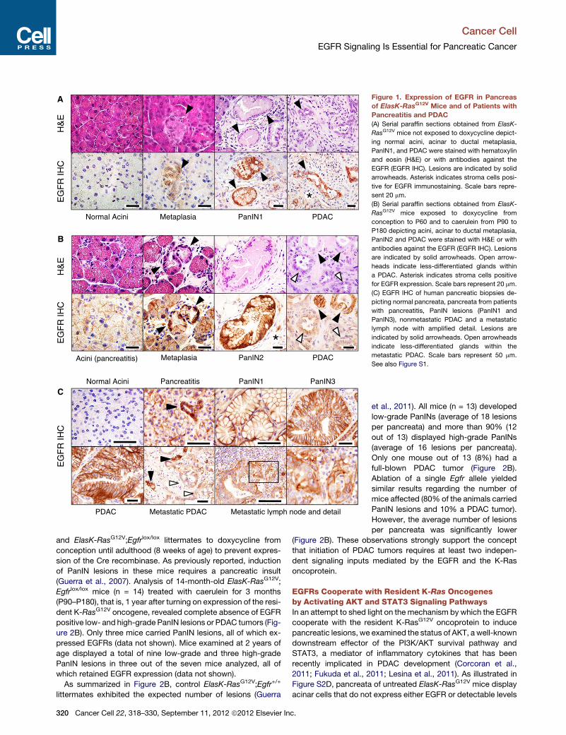
able levels of EGFRs (Figure 1C). However, morphologically
normal acinar cells of pancreatitis patients expressed significant
levels of EGFRs in a manner highly reminiscent of the result
obtained in pancreata derived from mice exposed to caerulein
(Figures 1B and 1C).
These observations are in agreement with an early study
describing overexpression of EGFRs in patients with chronic
pancreatitis (Korc et al., 1994). We also observed that metapla-
sias present in pancreatitis biopsies displayed elevated levels
of EGFRs (Figure 1C). Low-grade and high-grade PanINs
present in human PDAC tumors also expressed high levels of
EGFRs (Figure 1C). Interestingly, their pattern of expression in
tumored areas closely resembled that observed in mouse
PDACs (Figure 1B). Whereas well-differentiated tumor glands
were uniformly decorated with EGFR antibodies, less-differenti-
ated glands expressed significantly lower levels of the receptors
(Figure 1C). Finally, metastatic cells localized in a regional lymph
node retained detectable, albeit somewhat attenuated levels of
EGFRs (Figure 1C). These observations indicate that induction
of EGFRs in acinar cells of injured pancreata as well as in PanIN
and PDAC lesions is a common event in mouse and human
pancreatic tissues.
EGFRs Are Essential for the Generation of K-RasOncogene-Driven PanIN LesionsTo determine whether development of PanIN lesions and PDAC
tumors require EGFR signaling, we generated ElasK-RasG12V;
Egfr+/+ and ElasK-RasG12V; Egfr lox/lox strains and analyzed their
pancreata at 1 year of age. These mice were not exposed to
doxycycline to allow expression of the Elastase-driven Cre re-
combinase during late embryonic development (E16.5). Cre-
mediated recombination allowed concomitant expression of
the resident K-RasG12V oncogene and ablation of the floxed
Egfr alleles in acinar cells (Figure S2A). As illustrated in Figure 2A,
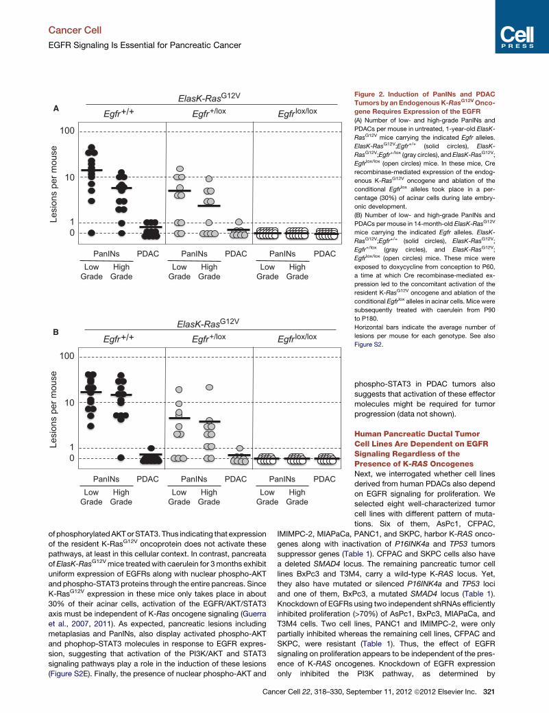
control ElasK-RasG12V;Egfr+/+ littermates (12 out of 13 animals,
92%) exhibited abundant low- and high-grade PanIN lesions
(average of 16 and 5 lesions per pancreata, respectively). More-
over, three animals (23%) displayed sizable PDAC tumors.
Animals heterozygous for the Egfr locus also harbored low-
and high-grade PanIN lesions albeit at reduced numbers
(average of 5 and 2.5 lesions per pancreata, respectively). Like-
wise, only one out of ten heterozygous mice carried a PDAC
tumor (Figure 2A).
In contrast, careful analysis of serial sections of pancreata
from 1-year-old ElasK-RasG12V; Egfr lox/lox animals (n = 24) only
revealed the presence of a limited number of PanIN lesions
(ten low-grade and two high-grade PanINs) in eight mice. More
importantly, all of these lesions expressed EGFRs due to incom-
plete recombination of the floxed Egfr alleles (Figures S2B and
S2C). Similar results were obtained in older mice sacrificed at
2 years of age (data not shown). These observations indicate
that EGFRs are essential for the induction of PanINs and PDAC
by K-Ras oncogenes.
Adult Mice Also Require EGFR Signaling for PDACDevelopmentTo exclude the possibility that these observations were due to
developmental defects in acinar cells lacking EGFRs during
embryonic development, we exposed ElasK-RasG12V;Egfr+/+
cer Cell 22, 318–330, September 11, 2012 ª2012 Elsevier Inc. 319
EG
FR
IHC
H&
E
Metaplasia PanIN1 PDACNormal Acini
*
Metaplasia PanIN2 PDACAcini (pancreatitis)
EG
FR
IHC
H&
E
*
Pancreatitis PanIN1 PanIN3Normal Acini
Metastatic PDAC Metastatic lymph node and detailPDAC
EG
FR
IHC
A
B
C
Figure 1. Expression of EGFR in Pancreas
of ElasK-RasG12V Mice and of Patients with
Pancreatitis and PDAC
(A) Serial paraffin sections obtained from ElasK-
RasG12V mice not exposed to doxycycline depict-
ing normal acini, acinar to ductal metaplasia,
PanIN1, and PDAC were stained with hematoxylin
and eosin (H&E) or with antibodies against the
EGFR (EGFR IHC). Lesions are indicated by solid
arrowheads. Asterisk indicates stroma cells posi-
tive for EGFR immunostaining. Scale bars repre-
sent 20 mm.
(B) Serial paraffin sections obtained from ElasK-
RasG12V mice exposed to doxycycline from
conception to P60 and to caerulein from P90 to
P180 depicting acini, acinar to ductal metaplasia,
PanIN2 and PDAC were stained with H&E or with
antibodies against the EGFR (EGFR IHC). Lesions
are indicated by solid arrowheads. Open arrow-
heads indicate less-differentiated glands within
a PDAC. Asterisk indicates stroma cells positive
for EGFR expression. Scale bars represent 20 mm.
(C) EGFR IHC of human pancreatic biopsies de-
picting normal pancreata, pancreata from patients
with pancreatitis, PanIN lesions (PanIN1 and
PanIN3), nonmetastatic PDAC and a metastatic
lymph node with amplified detail. Lesions are
indicated by solid arrowheads. Open arrowheads
indicate less-differentiated glands within the
metastatic PDAC. Scale bars represent 50 mm.
See also Figure S1.
Cancer Cell
EGFR Signaling Is Essential for Pancreatic Cancer
and ElasK-RasG12V;Egfrlox/lox littermates to doxycycline from
conception until adulthood (8 weeks of age) to prevent expres-
sion of the Cre recombinase. As previously reported, induction
of PanIN lesions in these mice requires a pancreatic insult
(Guerra et al., 2007). Analysis of 14-month-old ElasK-RasG12V;
Egfrlox/lox mice (n = 14) treated with caerulein for 3 months
(P90–P180), that is, 1 year after turning on expression of the resi-
dent K-RasG12V oncogene, revealed complete absence of EGFR
positive low- and high-grade PanIN lesions or PDAC tumors (Fig-
ure 2B). Only three mice carried PanIN lesions, all of which ex-
pressed EGFRs (data not shown). Mice examined at 2 years of
age displayed a total of nine low-grade and three high-grade
PanIN lesions in three out of the seven mice analyzed, all of
which retained EGFR expression (data not shown).
As summarized in Figure 2B, control ElasK-RasG12V;Egfr+/+
littermates exhibited the expected number of lesions (Guerra
320 Cancer Cell 22, 318–330, September 11, 2012 ª2012 Elsevier Inc.
et al., 2011). All mice (n = 13) developed
low-grade PanINs (average of 18 lesions
per pancreata) and more than 90% (12
out of 13) displayed high-grade PanINs
(average of 16 lesions per pancreata).
Only one mouse out of 13 (8%) had a
full-blown PDAC tumor (Figure 2B).
Ablation of a single Egfr allele yielded
similar results regarding the number of
mice affected (80% of the animals carried
PanIN lesions and 10% a PDAC tumor).
However, the average number of lesions
per pancreata was significantly lower
(Figure 2B). These observations strongly support the concept
that initiation of PDAC tumors requires at least two indepen-
dent signaling inputs mediated by the EGFR and the K-Ras
oncoprotein.
EGFRs Cooperate with Resident K-Ras Oncogenesby Activating AKT and STAT3 Signaling PathwaysIn an attempt to shed light on the mechanism by which the EGFR
cooperate with the resident K-RasG12V oncoprotein to induce
pancreatic lesions, we examined the status of AKT, a well-known
downstream effector of the PI3K/AKT survival pathway and
STAT3, a mediator of inflammatory cytokines that has been
recently implicated in PDAC development (Corcoran et al.,
2011; Fukuda et al., 2011; Lesina et al., 2011). As illustrated in
Figure S2D, pancreata of untreated ElasK-RasG12V mice display
acinar cells that do not express either EGFR or detectable levels
0
10
100
1Lesi
ons
per m
ouse
Egfr+/+ Egfr+/lox Egfr lox/loxA
PDAC PDAC PDACLow
GradeHigh
Grade
PanINsLow
GradeHigh
Grade
PanINsLow
GradeHigh
Grade
PanINs
0
10
100
1Lesi
ons
per m
ouse
Egfr+/+ Egfr+/lox Egfr lox/loxB
PDAC PDAC PDACLow
GradeHigh
Grade
PanINsLow
GradeHigh
Grade
PanINsLow
GradeHigh
Grade
PanINs
ElasK-RasG12V
ElasK-RasG12V
Figure 2. Induction of PanINs and PDAC
Tumors by an Endogenous K-RasG12VOnco-
gene Requires Expression of the EGFR
(A) Number of low- and high-grade PanINs and
PDACs per mouse in untreated, 1-year-old ElasK-
RasG12V mice carrying the indicated Egfr alleles.
ElasK-RasG12V;Egfr+/+ (solid circles), ElasK-
RasG12V;Egfr+/lox (gray circles), and ElasK-RasG12V;
Egfrlox/lox (open circles) mice. In these mice, Cre
recombinase-mediated expression of the endog-
enous K-RasG12V oncogene and ablation of the
conditional Egfrlox alleles took place in a per-
centage (30%) of acinar cells during late embry-
onic development.
(B) Number of low- and high-grade PanINs and
PDACs per mouse in 14-month-old ElasK-RasG12V
mice carrying the indicated Egfr alleles. ElasK-
RasG12V;Egfr+/+ (solid circles), ElasK-RasG12V;
Egfr+/lox (gray circles), and ElasK-RasG12V;
Egfrlox/lox (open circles) mice. These mice were
exposed to doxycycline from conception to P60,
a time at which Cre recombinase-mediated ex-
pression led to the concomitant activation of the
resident K-RasG12V oncogene and ablation of the
conditional Egfrlox alleles in acinar cells. Mice were
subsequently treated with caerulein from P90
to P180.
Horizontal bars indicate the average number of
lesions per mouse for each genotype. See also
Figure S2.
Cancer Cell
EGFR Signaling Is Essential for Pancreatic Cancer
of phosphorylatedAKTorSTAT3. Thus indicating that expression
of the resident K-RasG12V oncoprotein does not activate these
pathways, at least in this cellular context. In contrast, pancreata
ofElasK-RasG12Vmice treatedwith caerulein for 3months exhibit
uniform expression of EGFRs along with nuclear phospho-AKT
and phospho-STAT3 proteins through the entire pancreas. Since
K-RasG12V expression in these mice only takes place in about
30% of their acinar cells, activation of the EGFR/AKT/STAT3
axis must be independent of K-Ras oncogene signaling (Guerra
et al., 2007, 2011). As expected, pancreatic lesions including
metaplasias and PanINs, also display activated phospho-AKT
and phophop-STAT3 molecules in response to EGFR expres-
sion, suggesting that activation of the PI3K/AKT and STAT3
signaling pathways play a role in the induction of these lesions
(Figure S2E). Finally, the presence of nuclear phospho-AKT and
Cancer Cell 22, 318–330, Se
phospho-STAT3 in PDAC tumors also
suggests that activation of these effector
molecules might be required for tumor
progression (data not shown).
Human Pancreatic Ductal TumorCell Lines Are Dependent on EGFRSignaling Regardless of thePresence of K-RAS OncogenesNext, we interrogated whether cell lines
derived from human PDACs also depend
on EGFR signaling for proliferation. We
selected eight well-characterized tumor
cell lines with different pattern of muta-
tions. Six of them, AsPc1, CFPAC,
IMIMPC-2, MIAPaCa, PANC1, and SKPC, harbor K-RAS onco-
genes along with inactivation of P16INK4a and TP53 tumors
suppressor genes (Table 1). CFPAC and SKPC cells also have
a deleted SMAD4 locus. The remaining pancreatic tumor cell
lines BxPc3 and T3M4, carry a wild-type K-RAS locus. Yet,
they also have mutated or silenced P16INK4a and TP53 loci
and one of them, BxPc3, a mutated SMAD4 locus (Table 1).
Knockdown of EGFRs using two independent shRNAs efficiently
inhibited proliferation (>70%) of AsPc1, BxPc3, MIAPaCa, and
T3M4 cells. Two cell lines, PANC1 and IMIMPC-2, were only
partially inhibited whereas the remaining cell lines, CFPAC and
SKPC, were resistant (Table 1). Thus, the effect of EGFR
signaling on proliferation appears to be independent of the pres-
ence of K-RAS oncogenes. Knockdown of EGFR expression
only inhibited the PI3K pathway, as determined by
ptember 11, 2012 ª2012 Elsevier Inc. 321
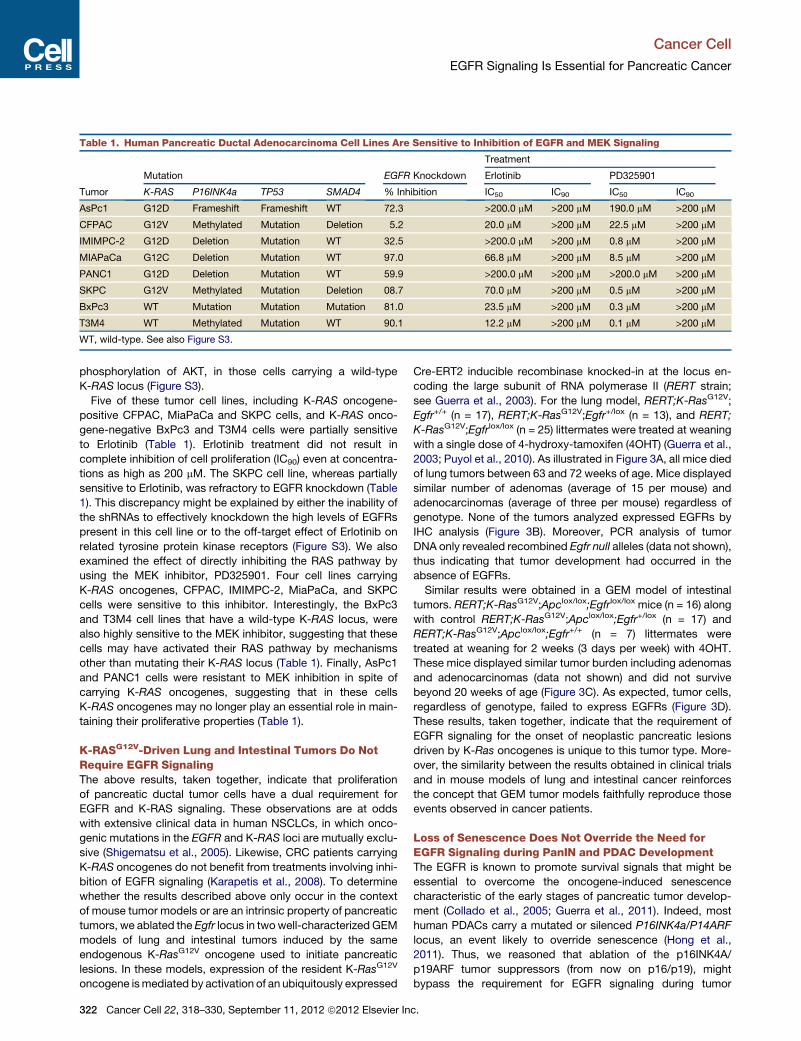
Table 1. Human Pancreatic Ductal Adenocarcinoma Cell Lines Are Sensitive to Inhibition of EGFR and MEK Signaling
Tumor
Mutation EGFR Knockdown
Treatment
Erlotinib PD325901
K-RAS P16INK4a TP53 SMAD4 % Inhibition IC50 IC90 IC50 IC90
AsPc1 G12D Frameshift Frameshift WT 72.3 >200.0 mM >200 mM 190.0 mM >200 mM
CFPAC G12V Methylated Mutation Deletion 5.2 20.0 mM >200 mM 22.5 mM >200 mM
IMIMPC-2 G12D Deletion Mutation WT 32.5 >200.0 mM >200 mM 0.8 mM >200 mM
MIAPaCa G12C Deletion Mutation WT 97.0 66.8 mM >200 mM 8.5 mM >200 mM
PANC1 G12D Deletion Mutation WT 59.9 >200.0 mM >200 mM >200.0 mM >200 mM
SKPC G12V Methylated Mutation Deletion 08.7 70.0 mM >200 mM 0.5 mM >200 mM
BxPc3 WT Mutation Mutation Mutation 81.0 23.5 mM >200 mM 0.3 mM >200 mM
T3M4 WT Methylated Mutation WT 90.1 12.2 mM >200 mM 0.1 mM >200 mM
WT, wild-type. See also Figure S3.
Cancer Cell
EGFR Signaling Is Essential for Pancreatic Cancer
phosphorylation of AKT, in those cells carrying a wild-type
K-RAS locus (Figure S3).
Five of these tumor cell lines, including K-RAS oncogene-
positive CFPAC, MiaPaCa and SKPC cells, and K-RAS onco-
gene-negative BxPc3 and T3M4 cells were partially sensitive
to Erlotinib (Table 1). Erlotinib treatment did not result in
complete inhibition of cell proliferation (IC90) even at concentra-
tions as high as 200 mM. The SKPC cell line, whereas partially
sensitive to Erlotinib, was refractory to EGFR knockdown (Table
1). This discrepancy might be explained by either the inability of
the shRNAs to effectively knockdown the high levels of EGFRs
present in this cell line or to the off-target effect of Erlotinib on
related tyrosine protein kinase receptors (Figure S3). We also
examined the effect of directly inhibiting the RAS pathway by
using the MEK inhibitor, PD325901. Four cell lines carrying
K-RAS oncogenes, CFPAC, IMIMPC-2, MiaPaCa, and SKPC
cells were sensitive to this inhibitor. Interestingly, the BxPc3
and T3M4 cell lines that have a wild-type K-RAS locus, were
also highly sensitive to the MEK inhibitor, suggesting that these
cells may have activated their RAS pathway by mechanisms
other than mutating their K-RAS locus (Table 1). Finally, AsPc1
and PANC1 cells were resistant to MEK inhibition in spite of
carrying K-RAS oncogenes, suggesting that in these cells
K-RAS oncogenes may no longer play an essential role in main-
taining their proliferative properties (Table 1).
K-RASG12V-Driven Lung and Intestinal Tumors Do NotRequire EGFR SignalingThe above results, taken together, indicate that proliferation
of pancreatic ductal tumor cells have a dual requirement for
EGFR and K-RAS signaling. These observations are at odds
with extensive clinical data in human NSCLCs, in which onco-
genic mutations in the EGFR and K-RAS loci are mutually exclu-
sive (Shigematsu et al., 2005). Likewise, CRC patients carrying
K-RAS oncogenes do not benefit from treatments involving inhi-
bition of EGFR signaling (Karapetis et al., 2008). To determine
whether the results described above only occur in the context
of mouse tumor models or are an intrinsic property of pancreatic
tumors, we ablated the Egfr locus in twowell-characterized GEM
models of lung and intestinal tumors induced by the same
endogenous K-RasG12V oncogene used to initiate pancreatic
lesions. In these models, expression of the resident K-RasG12V
oncogene ismediated by activation of an ubiquitously expressed
322 Cancer Cell 22, 318–330, September 11, 2012 ª2012 Elsevier In
Cre-ERT2 inducible recombinase knocked-in at the locus en-
coding the large subunit of RNA polymerase II (RERT strain;
see Guerra et al., 2003). For the lung model, RERT;K-RasG12V;
Egfr+/+ (n = 17), RERT;K-RasG12V;Egfr+/lox (n = 13), and RERT;
K-RasG12V;Egfrlox/lox (n = 25) littermates were treated at weaning
with a single dose of 4-hydroxy-tamoxifen (4OHT) (Guerra et al.,
2003; Puyol et al., 2010). As illustrated in Figure 3A, all mice died
of lung tumors between 63 and 72 weeks of age. Mice displayed
similar number of adenomas (average of 15 per mouse) and
adenocarcinomas (average of three per mouse) regardless of
genotype. None of the tumors analyzed expressed EGFRs by
IHC analysis (Figure 3B). Moreover, PCR analysis of tumor
DNA only revealed recombined Egfr null alleles (data not shown),
thus indicating that tumor development had occurred in the
absence of EGFRs.
Similar results were obtained in a GEM model of intestinal
tumors. RERT;K-RasG12V;Apclox/lox;Egfrlox/lox mice (n = 16) along
with control RERT;K-RasG12V;Apclox/lox;Egfr+/lox (n = 17) and
RERT;K-RasG12V;Apclox/lox;Egfr+/+ (n = 7) littermates were
treated at weaning for 2 weeks (3 days per week) with 4OHT.
These mice displayed similar tumor burden including adenomas
and adenocarcinomas (data not shown) and did not survive
beyond 20 weeks of age (Figure 3C). As expected, tumor cells,
regardless of genotype, failed to express EGFRs (Figure 3D).
These results, taken together, indicate that the requirement of
EGFR signaling for the onset of neoplastic pancreatic lesions
driven by K-Ras oncogenes is unique to this tumor type. More-
over, the similarity between the results obtained in clinical trials
and in mouse models of lung and intestinal cancer reinforces
the concept that GEM tumor models faithfully reproduce those
events observed in cancer patients.
Loss of Senescence Does Not Override the Need forEGFR Signaling during PanIN and PDAC DevelopmentThe EGFR is known to promote survival signals that might be
essential to overcome the oncogene-induced senescence
characteristic of the early stages of pancreatic tumor develop-
ment (Collado et al., 2005; Guerra et al., 2011). Indeed, most
human PDACs carry a mutated or silenced P16INK4a/P14ARF
locus, an event likely to override senescence (Hong et al.,
2011). Thus, we reasoned that ablation of the p16INK4A/
p19ARF tumor suppressors (from now on p16/p19), might
bypass the requirement for EGFR signaling during tumor
c.
H&E EGFR IHC
Egf
r+/+
Egf
rlox/
lox
-catenin IHCH&E EGFR IHC SPC IHC
Egf
r+/+
Egf
rlox/
lox
A
B
C
D
Sur
viva
l (%
)
700
20
40
60
80
100
30 40 50
Age (weeks)60
0
20
40
60
80
100
Sur
viva
l (%
)
155 10 20Age (weeks)
NSCLC GEM tumor model Intestinal GEM tumor model
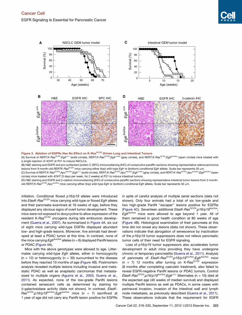
Figure 3. Ablation of EGFRs Has No Effect on K-RasG12V-Driven Lung and Intestinal Tumors
(A) Survival of RERT;K-RasG12V;Egfr+/+ (solid circles), RERT;K-RasG12V;Egfr+/lox (gray circles), and RERT;K-RasG12V;Egfrlox/lox (open circles) mice treated with
a single injection of 4OHT at P21 to induce NSCLCs.
(B) H&E staining and EGFR and pro-surfactant protein C (SPC) immunostaining (IHC) of consecutive paraffin sections showing representative adenocarcinoma
lesions from 9-month-old RERTK-RasG12V mice carrying either (top) wild-type Egfr or (bottom) conditional Egfr alleles. Scale bar represents 50 mm.
(C) Survival of RERT;K-RasG12V;Apclox/lox;Egfr+/+ (solid circles), RERT;K-RasG12V;Apclox/lox;Egfr+/lox (gray circles), and RERT;K-RasG12V;Apclox/lox;Egfrlox/lox (open
circles) mice treated with 4OHT (3 days per week, for 2 weeks) at P21 to induce intestinal tumors.
(D) H&E staining and EGFR and b-catenin immunostaining (IHC) of consecutive paraffin sections showing representative intestinal tumor lesions from 2-month-
old RERT;K-RasG12V;Apclox/lox mice carrying either (top) wild-type Egfr or (bottom) conditional Egfr alleles. Scale bar represents 50 mm.
Cancer Cell
EGFR Signaling Is Essential for Pancreatic Cancer
initiation. Conditional floxed p16/p19 alleles were introduced
into ElasK-RasG12V mice carrying wild-type or floxed Egfr alleles
and their pancreata examined at 16 weeks of age, before they
displayed any obvious signs of overt tumor development. These
mice were not exposed to doxycycline to allow expression of the
resident K-RasG12V oncogene during late embryonic develop-
ment (Guerra et al., 2007). As summarized in Figure 4A, six out
of eight mice carrying wild-type EGFRs displayed abundant
low- and high-grade lesions. Moreover, five animals had devel-
oped at least a PDAC tumor at this time. In contrast, none of
themice carrying Egfrlox/lox alleles (n = 6) displayed PanIN lesions
or PDAC (Figure 4A).
Mice with the above genotypes were allowed to age. Litter-
mates carrying wild-type Egfr alleles, either in homozygocity
(n = 12) or heterozygocity (n = 30) succumbed to the disease
before they reached 10 months of age (Figure 4B). Postmortem
analysis revealed multiple lesions including invasive and meta-
static PDAC as well as anaplastic carcinomas that metasta-
sized to multiple organs (Aguirre et al., 2003; Guerra et al.,
2011). As expected, none of the low-grade PanIN lesions
contained senescent cells as determined by staining for
b-galactosidase activity (data not shown). In contrast, ElasK-
RasG12V;p16/p19lox/lox;Egfrlox/lox mice (n = 7) sacrificed at
1 year of age did not carry any PanIN lesion positive for EGFRs
Can
in spite of careful analysis of multiple serial sections (data not
shown). Only four animals had a total of six low-grade and
two high-grade PanIN ‘‘escaper’’ lesions positive for EGFRs
(Figure 4C). Seventeen additional ElasK-RasG12V;p16/p19lox/lox;
Egfrlox/lox mice were allowed to age beyond 1 year. All of
them remained in good health condition at 80 weeks of age
(Figure 4B). Histological examination of their pancreata at this
time did not reveal any lesions (data not shown). These obser-
vations indicate that abrogation of senescence by inactivation
of the p16/p19 tumor suppressors does not relieve pancreatic
tumor cells of their need for EGFR signaling.
Loss of p16/p19 tumor suppressors also accelerates tumor
development in adult mice providing they have undergone
chronic or temporary pancreatitis (Guerra et al., 2011). Analysis
of pancreata of ElasK-RasG12V;p16/p19lox/lox;Egfrlox/lox mice
(n = 7) 12 months after turning on K-RasG12V expression
(8 months after completing caerulein treatment), also failed to
reveal EGFR-negative PanIN lesions or PDAC tumors. Control
ElasK-RasG12V;p16/p19lox/lox;Egfr+/+ littermates (n = 10) died at
the expected age (40 weeks of median survival) and displayed
multiple PanIN lesions as well as PDACs, in some cases with
perineural invasion, invasion of the intestinal wall and lymph
node metastasis, as previously described (Guerra et al., 2011).
These observations indicate that the requirement for EGFR
cer Cell 22, 318–330, September 11, 2012 ª2012 Elsevier Inc. 323
A
B
C
H&E EGFR IHC
0
10
30
1
Lesi
ons
per m
ouse
Egfr+/+ Egfr lox/lox
PDACLow
GradeHigh
Grade
PanINs
3
Age (weeks)
Sur
viva
l (%
)
0
20
40
60
80
100
20 3010 40 70
ElasK-RasG12V;p16/p19lox/lox
PDACLow
GradeHigh
Grade
PanINs
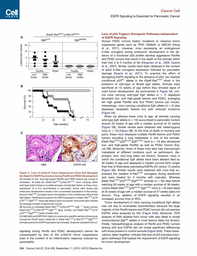
Figure 4. Loss of p16/p19 Tumor Suppressors Does Not Abrogate
the Need for EGFR Expression during PanIN and PDACDevelopment
(A) Number of low- and high-grade PanINs and PDAC lesions per mouse in
untreated, 16-week-old ElasK-RasG12V;p16/p19lox/lox mice carrying either
wild-type (solid circles) or conditional (open circles) Egfr alleles. In these mice,
expression of a Cre recombinase in pancreatic acinar cells during late
embryonic development results in the concomitant expression of the endog-
enous K-RasG12V oncogene and in the ablation of the conditional p16/p19 and
Egfr alleles. PanIN lesions positive for EGFR expression in ElasK-RasG12V;p16/
p19lox/lox;Egfrlox/lox mice (see below) were not scored. Horizontal bars indicate
the average number of lesions per mouse.
(B) Survival of untreated ElasK-RasG12V;p16/p19lox/lox; Egfr +/+ (solid circles),
ElasK-RasG12V;p16/p19lox/lox;Egfr+/lox (gray circles), and ElasK-RasG12V;p16/
p19lox/lox;Egfrlox/lox (open circles) mice.
(C) H&E (left) and EGFR IHC (right) of consecutive paraffin sections showing an
occasional PanIN lesion observed in ElasK-RasG12V;p16/p19lox/lox;Egfrlox/lox
animals positive for EGFRexpression (arrowhead). Scale bar represents 50mm.
Cancer Cell
EGFR Signaling Is Essential for Pancreatic Cancer
signaling during PanIN and PDAC development cannot be
compensated by loss of the p16/p19 tumor suppressors
even in the context of an inflammatory response induced by
pancreatitis.
324 Cancer Cell 22, 318–330, September 11, 2012 ª2012 Elsevier In
Loss of p53 Triggers Oncogenic Pathways Independentof EGFR SignalingHuman PDAC tumors harbor mutations in classical tumor
suppressor genes such as TP53, SMAD4, or BRCA2 (Hong
et al., 2011). Likewise, mice expressing an endogenous
K-Ras oncogene during embryonic development in the ab-
sence of a functional p53 protein develop aggressive PanINs
and PDAC tumors that result in the death of the animals within
their first 4 to 5 months of life (Hingorani et al., 2005; Guerra
et al., 2007). Similar results have been obtained in the context
of adult K-Ras oncogene expression followed by pancreatic
damage (Guerra et al., 2011). To examine the effect of
abrogating EGFR signaling in the absence of p53, we inserted
conditional p53lox alleles in the ElasK-RasG12V strain in the
presence of wild-type or floxed Egfr alleles. Animals were
sacrificed at 10 weeks of age before they showed signs of
overt tumor development. As summarized in Figure 5A, con-
trol mice carrying wild-type Egfr alleles (n = 7) displayed
abundant low- and high-grade lesions and PDAC, averaging
ten high grade PanINs and four PDAC tumors per mouse.
Interestingly, mice carrying conditional Egfr alleles (n = 5) also
displayed neoplastic lesions but with reduced incidence
(Figure 5A).
When we allowed these mice to age, all animals carrying
wild-type Egfr alleles (n = 10) succumbed to pancreatic tumors
around 20 weeks of age with a median survival of 12 weeks
(Figure 5B). Similar results were obtained with heterozygous
mice (n = 13) (Figure 5B). At the time of death or humane end
point, these mice displayed multiple PanIN lesions and PDAC
tumors including a lung metastasis in one of the animals.
ElasK-RasG12V;p53lox/lox;Egfrlox/lox mice (n = 13) also developed
low- and high-grade PanINs as well as PDAC tumors (Fig-
ure 5B). Moreover, three of these mice also had macroscopic
metastasis at different locations such as peritoneum, dia-
phragm, liver, and lung (data not shown). However, mice in
which the conditional Egfr alleles have been ablated died by
40 weeks of age and displayed a median survival 83% longer
than that of littermates expressing EGFRs (22 versus 12 weeks)
(Figure 5B). Similar results were obtained with mice that ex-
pressed the resident K-RasG12V oncogene during adulthood
and were treated for 3 months with caerulein. Whereas
ElasK-RasG12V;p53lox/lox;Egfrlox/lox animals (n = 26) died before
reaching 60 weeks of age with a median survival of 38 weeks,
control ElasK-RasG12V;p53lox/lox;Egfr+/lox mice (n = 8) were dead
at 35 weeks of age with a median survival of 27 weeks (data not
shown). Thus, ablation of EGFR signaling resulted in an
increased survival time of 40%.
Tumor development in mice carrying conditional Egfr alleles
was not due to incomplete recombination because the large
majority of the PanIN lesions and PDAC tumors did not express
EGFRs when analyzed by IHC (Figure S4A). Moreover, PCR
analysis of DNA isolated from tumor cells also failed to reveal
unrecombined Egfrlox alleles in most lesions (data not shown).
Finally, histopathological analysis of PanIN and PDAC tumors
lacking p53 and EGFRs did not reveal significant differences
with those present in control animals (Figure S4A). These obser-
vations, taken together, indicate that loss of p53 activates onco-
genic pathways that bypass the requirement of EGFR signaling
for tumor development.
c.
A
B
C
0
10
30
1
Lesi
ons
per m
ouse
Egfr+/+ Egfr lox/lox
PDACLow
GradeHigh
Grade
PanINs PDACLow
GradeHigh
Grade
PanINs
3
ElasK-RasG12V;p53lox/lox
Lesi
ons
per m
ouse
Sur
viva
l(%
)
Age (weeks)30
0
20
40
60
80
100
10 15 205 25 35 40
0
10
30
1
Vehicle Erlotinib
PDACLow
GradeHigh
Grade
PanINs
3
PDACLow
GradeHigh
Grade
PanINs
ElasK-RasG12V;p53lox/lox
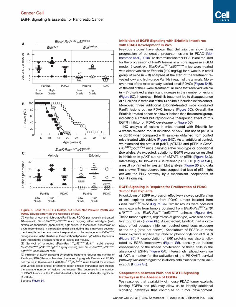
Figure 5. Loss of EGFRs Delays but Does Not Prevent PanIN and
PDAC Development in the Absence of p53
(A) Number of low- and high-grade PanINs and PDACs permouse in untreated,
10-week-old ElasK-RasG12V;p53lox/lox mice carrying either wild-type (solid
circles) or conditional (open circles) Egfr alleles. In these mice, expression of
a Cre recombinase in pancreatic acinar cells during late embryonic develop-
ment results in the concomitant expression of the endogenous K-RasG12V
oncogene and in the ablation of the conditional p53 and Egfr alleles. Horizontal
bars indicate the average number of lesions per mouse.
(B) Survival of untreated ElasK-RasG12V;p53lox/lox;Egfr+/+ (solid circles),
ElasK-RasG12V;p53lox/lox;Egfr+/lox (gray circles), and ElasK-RasG12V;p53lox/lox;
Egfrlox/lox (open circles) mice.
(C) Inhibition of EGFR signaling by Erlotinib treatment reduces the number of
PanIN and PDAC lesions. Number of low- and high-grade PanINs and PDACs
per mouse in 6-week-old ElasK-RasG12V;p53lox/lox mice treated for 4 weeks
with vehicle (solid circles) or Erlotinib (open circles). Horizontal bars indicate
the average number of lesions per mouse. The decrease in the number
of PDAC tumors in the Erlotinib-treated cohort was statistically significant
(p < 0.05).
See also Figure S4.
Cancer Cell
EGFR Signaling Is Essential for Pancreatic Cancer
Can
Inhibition of EGFR Signaling with Erlotinib Interfereswith PDAC Development In VivoPrevious studies have shown that Gefitinib can slow down
progression of pancreatic precursor lesions to PDAC (Mo-
hammed et al., 2010). To determine whether EGFRs are required
for the progression of PanIN lesions in a more aggressive GEM
model, 6-week-old ElasK-RasG12V;p53lox/lox mice were treated
with either vehicle or Erlotinib (100 mg/Kg) for 4 weeks. A small
group of mice (n = 3) analyzed at the start of the treatment re-
vealed low- and high-grade PanINs in each of the animals. More-
over, two of the mice already carried small PDACs (Figure S4B).
At the end of the 4-week treatment, all mice that received vehicle
(n = 7) displayed a significant increase in the number of lesions
(Figure 5C). In contrast, Erlotinib treatment led to disappearance
of all lesions in three out of the 14 animals included in this cohort.
Moreover, three additional Erlotinib-treated mice contained
PanIN lesions but no PDAC tumors (Figure 5C). Overall, the
Erlotinib-treated cohort had fewer lesions than the control group,
indicating a limited but reproducible therapeutic effect of this
EGFR inhibitor on PDAC development (Figure 5C).
IHC analysis of lesions in mice treated with Erlotinib for
4 weeks revealed robust inhibition of pAKT but not of pSTAT3
or pERK when compared with samples obtained from control
mice treated with vehicle (Figure S4C). As an additional control,
we examined the status of pAKT, pSTAT3 and pERK in ElasK-
RasG12V;p53lox/lox mice carrying either wild-type or conditional
Egfr alleles. As expected, ablation of EGFR expression resulted
in inhibition of pAKT but not of pSTAT3 or pERK (Figure S4D).
Interestingly, full blown PDACs retained pAKT IHC (Figure S4E),
a result confirmed by western blot analysis (Figure S5 and data
not shown). These observations suggest that loss of p53 might
activate the PI3K pathway by a mechanism independent of
EGFR signaling.
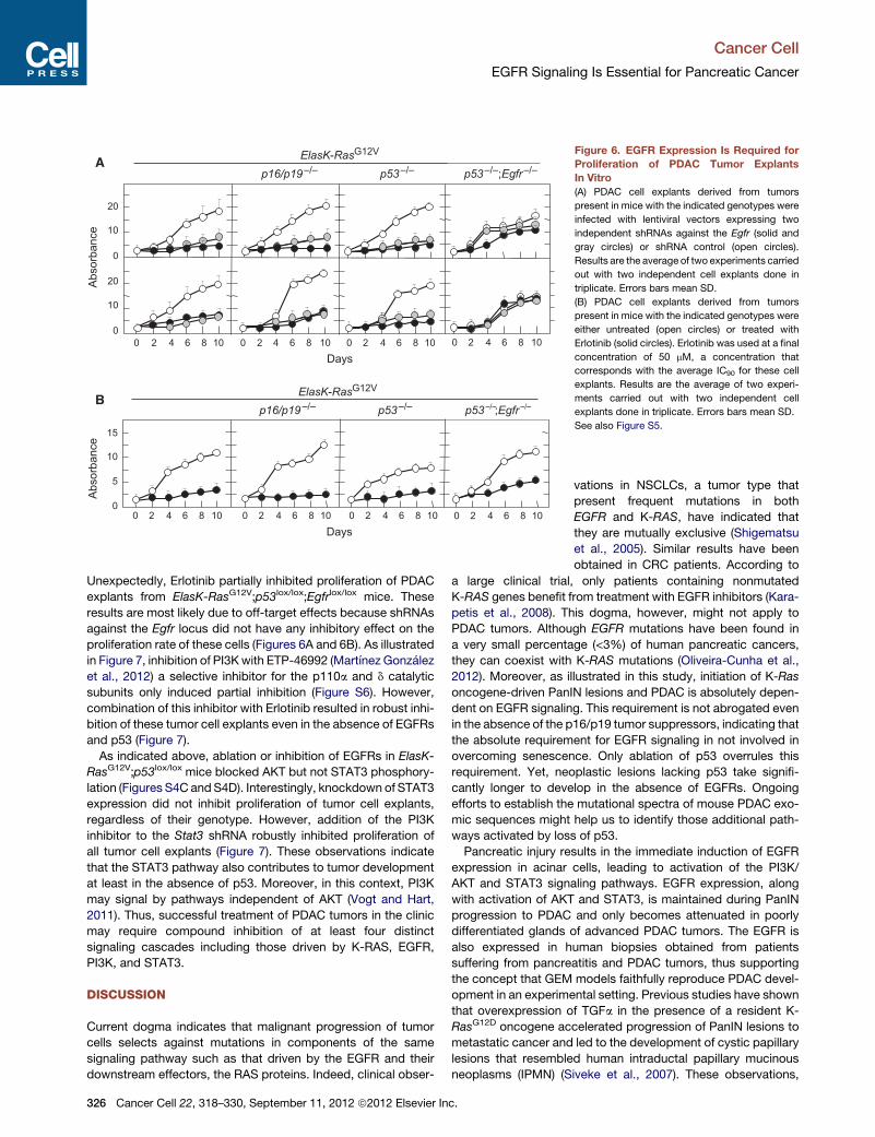
EGFR Signaling Is Required for Proliferation of PDACTumor Cell ExplantsKnockdown of EGFR expression effectively slowed proliferation
of cell explants derived from PDAC tumors isolated from
ElasK-RasG12V mice (Figure 6A). Similar results were obtained
using explants from tumors obtained from ElasK-RasG12V;p16/
p19lox/lox and ElasK-RasG12V;p53lox/lox animals (Figure 6A).
These tumor explants, regardless of genotype, were also sensi-
tive to Erlotinib (Figure 6B). As expected, Erlotinib had a cyto-
static effect because inhibition required continuous exposure
to the drug (data not shown). Knockdown of EGFRs in these
tumor explants significantly inhibited phosphorylation of STAT3
(Figure S5). Phosphorylation of ERK proteins was also amelio-
rated by EGFR knockdown (Figure S5), possibly an indirect
consequence of the limited proliferation of these cells in the
absence of EGFRs (Figure 6A). Interestingly, phosphorylation
of AKT, a marker for the activation of the PI3K/AKT survival
pathway was downregulated in all explants except in those lack-
ing p53 (Figure S5).
Cooperation between PI3K and STAT3 SignalingPathways in the Absence of EGFRsWe reasoned that availability of mouse PDAC tumor explants
lacking EGFRs and p53 may allow us to identify additional
signaling pathways that contribute to tumor development.
cer Cell 22, 318–330, September 11, 2012 ª2012 Elsevier Inc. 325
B
5
10
0
Days
Abs
orba
nce 10
20
0
10
20
0
A
0 2 4 6 8 10 0 2 4 6 8 10 0 2 4 6 8 10 0 2 4 6 8 10
0 2 4 6 8 10 0 2 4 6 8 10 0 2 4 6 8 10
ElasK-RasG12V
p53–/–p16/p19–/– p53–/–;Egfr–/–
0 2 4 6 8 10
Days
ElasK-RasG12V
p53–/–p16/p19–/– p53 –/–;Egfr –/–
Abs
orba
nce
15
Figure 6. EGFR Expression Is Required for
Proliferation of PDAC Tumor Explants
In Vitro
(A) PDAC cell explants derived from tumors
present in mice with the indicated genotypes were
infected with lentiviral vectors expressing two
independent shRNAs against the Egfr (solid and
gray circles) or shRNA control (open circles).
Results are the average of two experiments carried
out with two independent cell explants done in
triplicate. Errors bars mean SD.
(B) PDAC cell explants derived from tumors
present in mice with the indicated genotypes were
either untreated (open circles) or treated with
Erlotinib (solid circles). Erlotinib was used at a final
concentration of 50 mM, a concentration that
corresponds with the average IC90 for these cell
explants. Results are the average of two experi-
ments carried out with two independent cell
explants done in triplicate. Errors bars mean SD.
See also Figure S5.
Cancer Cell
EGFR Signaling Is Essential for Pancreatic Cancer
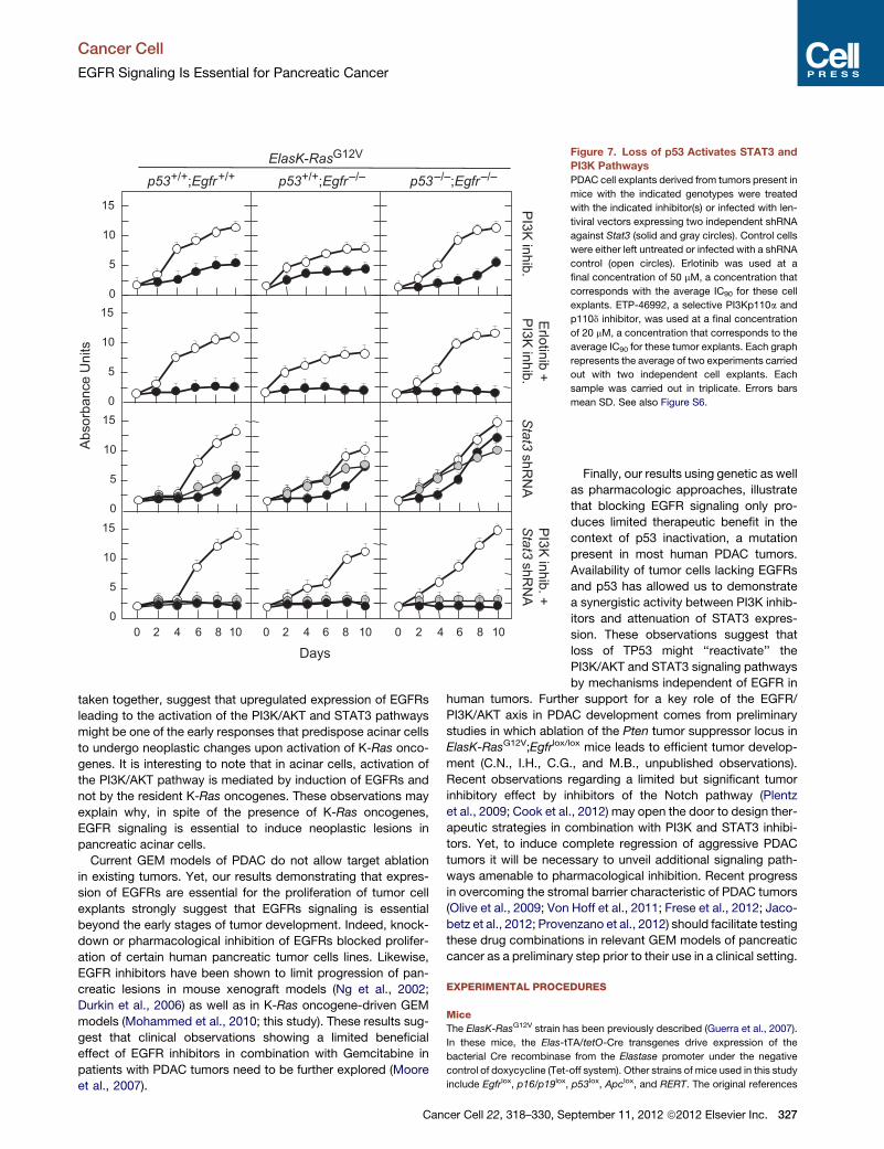
Unexpectedly, Erlotinib partially inhibited proliferation of PDAC
explants from ElasK-RasG12V;p53lox/lox;Egfrlox/lox mice. These
results are most likely due to off-target effects because shRNAs
against the Egfr locus did not have any inhibitory effect on the
proliferation rate of these cells (Figures 6A and 6B). As illustrated
in Figure 7, inhibition of PI3Kwith ETP-46992 (Martınez Gonzalez
et al., 2012) a selective inhibitor for the p110a and d catalytic
subunits only induced partial inhibition (Figure S6). However,
combination of this inhibitor with Erlotinib resulted in robust inhi-
bition of these tumor cell explants even in the absence of EGFRs
and p53 (Figure 7).
As indicated above, ablation or inhibition of EGFRs in ElasK-
RasG12V;p53lox/lox mice blocked AKT but not STAT3 phosphory-
lation (Figures S4C andS4D). Interestingly, knockdown of STAT3
expression did not inhibit proliferation of tumor cell explants,
regardless of their genotype. However, addition of the PI3K
inhibitor to the Stat3 shRNA robustly inhibited proliferation of
all tumor cell explants (Figure 7). These observations indicate
that the STAT3 pathway also contributes to tumor development
at least in the absence of p53. Moreover, in this context, PI3K
may signal by pathways independent of AKT (Vogt and Hart,
2011). Thus, successful treatment of PDAC tumors in the clinic
may require compound inhibition of at least four distinct
signaling cascades including those driven by K-RAS, EGFR,
PI3K, and STAT3.
DISCUSSION
Current dogma indicates that malignant progression of tumor
cells selects against mutations in components of the same
signaling pathway such as that driven by the EGFR and their
downstream effectors, the RAS proteins. Indeed, clinical obser-
326 Cancer Cell 22, 318–330, September 11, 2012 ª2012 Elsevier Inc.
vations in NSCLCs, a tumor type that
present frequent mutations in both
EGFR and K-RAS, have indicated that
they are mutually exclusive (Shigematsu
et al., 2005). Similar results have been
obtained in CRC patients. According to
a large clinical trial, only patients containing nonmutated
K-RAS genes benefit from treatment with EGFR inhibitors (Kara-
petis et al., 2008). This dogma, however, might not apply to
PDAC tumors. Although EGFR mutations have been found in
a very small percentage (<3%) of human pancreatic cancers,
they can coexist with K-RAS mutations (Oliveira-Cunha et al.,
2012). Moreover, as illustrated in this study, initiation of K-Ras
oncogene-driven PanIN lesions and PDAC is absolutely depen-
dent on EGFR signaling. This requirement is not abrogated even
in the absence of the p16/p19 tumor suppressors, indicating that
the absolute requirement for EGFR signaling in not involved in
overcoming senescence. Only ablation of p53 overrules this
requirement. Yet, neoplastic lesions lacking p53 take signifi-
cantly longer to develop in the absence of EGFRs. Ongoing
efforts to establish the mutational spectra of mouse PDAC exo-
mic sequences might help us to identify those additional path-
ways activated by loss of p53.
Pancreatic injury results in the immediate induction of EGFR
expression in acinar cells, leading to activation of the PI3K/
AKT and STAT3 signaling pathways. EGFR expression, along
with activation of AKT and STAT3, is maintained during PanIN
progression to PDAC and only becomes attenuated in poorly
differentiated glands of advanced PDAC tumors. The EGFR is
also expressed in human biopsies obtained from patients
suffering from pancreatitis and PDAC tumors, thus supporting
the concept that GEM models faithfully reproduce PDAC devel-
opment in an experimental setting. Previous studies have shown
that overexpression of TGFa in the presence of a resident K-
RasG12D oncogene accelerated progression of PanIN lesions to
metastatic cancer and led to the development of cystic papillary
lesions that resembled human intraductal papillary mucinous
neoplasms (IPMN) (Siveke et al., 2007). These observations,
Abs
orba
nce
Uni
ts
5
10
0
15
5
10
0
15
5
10
0
15
5
10
0
15
Days0 2 4 6 8 10 0 2 4 6 8 10 0 2 4 6 8 10
p53–/–;Egfr–/–p53+/+;Egfr+/+ p53+/+;Egfr–/–
Erlotinib
+P
I3K inhib.
PI3K
inhib. +Stat3
shRN
AP
I3K inhib.
Stat3
shRN
A
ElasK-RasG12V Figure 7. Loss of p53 Activates STAT3 and
PI3K Pathways
PDAC cell explants derived from tumors present in
mice with the indicated genotypes were treated
with the indicated inhibitor(s) or infected with len-
tiviral vectors expressing two independent shRNA
against Stat3 (solid and gray circles). Control cells
were either left untreated or infected with a shRNA
control (open circles). Erlotinib was used at a
final concentration of 50 mM, a concentration that
corresponds with the average IC90 for these cell
explants. ETP-46992, a selective PI3Kp110a and
p110d inhibitor, was used at a final concentration
of 20 mM, a concentration that corresponds to the
average IC90 for these tumor explants. Each graph
represents the average of two experiments carried
out with two independent cell explants. Each
sample was carried out in triplicate. Errors bars
mean SD. See also Figure S6.
Cancer Cell
EGFR Signaling Is Essential for Pancreatic Cancer
taken together, suggest that upregulated expression of EGFRs
leading to the activation of the PI3K/AKT and STAT3 pathways
might be one of the early responses that predispose acinar cells
to undergo neoplastic changes upon activation of K-Ras onco-
genes. It is interesting to note that in acinar cells, activation of
the PI3K/AKT pathway is mediated by induction of EGFRs and
not by the resident K-Ras oncogenes. These observations may
explain why, in spite of the presence of K-Ras oncogenes,
EGFR signaling is essential to induce neoplastic lesions in
pancreatic acinar cells.
Current GEM models of PDAC do not allow target ablation
in existing tumors. Yet, our results demonstrating that expres-
sion of EGFRs are essential for the proliferation of tumor cell
explants strongly suggest that EGFRs signaling is essential
beyond the early stages of tumor development. Indeed, knock-
down or pharmacological inhibition of EGFRs blocked prolifer-
ation of certain human pancreatic tumor cells lines. Likewise,
EGFR inhibitors have been shown to limit progression of pan-
creatic lesions in mouse xenograft models (Ng et al., 2002;
Durkin et al., 2006) as well as in K-Ras oncogene-driven GEM
models (Mohammed et al., 2010; this study). These results sug-
gest that clinical observations showing a limited beneficial
effect of EGFR inhibitors in combination with Gemcitabine in
patients with PDAC tumors need to be further explored (Moore
et al., 2007).
Cancer Cell 22, 318–330, Se
Finally, our results using genetic as well
as pharmacologic approaches, illustrate
that blocking EGFR signaling only pro-
duces limited therapeutic benefit in the
context of p53 inactivation, a mutation
present in most human PDAC tumors.
Availability of tumor cells lacking EGFRs
and p53 has allowed us to demonstrate
a synergistic activity between PI3K inhib-
itors and attenuation of STAT3 expres-
sion. These observations suggest that
loss of TP53 might ‘‘reactivate’’ the
PI3K/AKT and STAT3 signaling pathways
by mechanisms independent of EGFR in
human tumors. Further support for a key role of the EGFR/
PI3K/AKT axis in PDAC development comes from preliminary
studies in which ablation of the Pten tumor suppressor locus in
ElasK-RasG12V;Egfrlox/lox mice leads to efficient tumor develop-
ment (C.N., I.H., C.G., and M.B., unpublished observations).
Recent observations regarding a limited but significant tumor
inhibitory effect by inhibitors of the Notch pathway (Plentz
et al., 2009; Cook et al., 2012) may open the door to design ther-
apeutic strategies in combination with PI3K and STAT3 inhibi-
tors. Yet, to induce complete regression of aggressive PDAC
tumors it will be necessary to unveil additional signaling path-
ways amenable to pharmacological inhibition. Recent progress
in overcoming the stromal barrier characteristic of PDAC tumors
(Olive et al., 2009; Von Hoff et al., 2011; Frese et al., 2012; Jaco-
betz et al., 2012; Provenzano et al., 2012) should facilitate testing
these drug combinations in relevant GEM models of pancreatic
cancer as a preliminary step prior to their use in a clinical setting.
EXPERIMENTAL PROCEDURES
Mice
The ElasK-RasG12V strain has been previously described (Guerra et al., 2007).
In these mice, the Elas-tTA/tetO-Cre transgenes drive expression of the
bacterial Cre recombinase from the Elastase promoter under the negative
control of doxycycline (Tet-off system). Other strains of mice used in this study
include Egfrlox, p16/p19lox, p53lox, Apclox, and RERT. The original references
ptember 11, 2012 ª2012 Elsevier Inc. 327
Cancer Cell
EGFR Signaling Is Essential for Pancreatic Cancer
describing these strains appear in the Supplemental Experimental Proce-
dures. All experiments were approved by the CNIO Ethical Committee and
performed in accordance with the guidelines for Ethical Conduct in the Care
and Use of Animals as stated in The International Guiding Principles for
Biomedical Research involving Animals, developed by the Council for Interna-
tional Organizations of Medical Sciences (CIOMS).
Mouse Treatments
To prevent expression of the Elastase-driven Cre recombinase in ElasK-
RasG12V mice, doxycycline (2 mg/ml; Sigma) was provided in the drinking
water as a sucrose solution (5% w/v) to pregnant mothers from the time of
conception and to their offspring until the time we activated expression of
the resident K-RasG12V oncogene. Pancreatitis was induced by intraperitoneal
injections of caerulein for 3 months (125 mg/Kg, 5 days per week; Sigma). For
induction of NSCLC, RERT;K-RasG12V mice carrying wild-type or floxed Egfr
alleles were treated at P21 with a single dose of 4OHT (0.5 mg/ml in oil). For
intestinal tumors, RERT;K-RasG12V;Apclox/lox mice carrying wild-type or floxed
Egfr alleles were treated 3 days per week for 2 weeks with 4OHT (0.5 mg/ml in
oil) starting at P21. Erlotinib treatment was carried out in 6-week-old ElasK-
RasG12V;p53lox/lox mice by oral gavage (100 mg/Kg in 0.5% methylcellulose
with 0.1% Tween80) for 4 weeks. Control mice received the same treatment
without Erlotinib.
Histopathology and Immunohistochemistry
Specimens were fixed in 10% buffered formalin and embedded in paraffin. For
histopathological analysis, pancreata were serially sectioned (3 mm thick) and
every ten sections stained with hematoxylin and eosin (H&E). Remaining
sections were kept for immunohistochemical studies with b-catenin (1:750,
goat polyclonal; Santa Cruz Biotechnology, Sc-1496), pAKT (pS473) (1:175,
rabbit monoclonal EP2109Y; Epitomics 2118-1), EGFR (1:100, rabbit mono-
clonal; Epitomics 1902-1), SPC (1:175, rabbit polyclonal; Millipore, AB3786),
and pSTAT3 (Tyr705) (1:100, rabbit monoclonal D3A7; Cell Signaling Tech-
nology, 9145) antibodies. Following incubation with the primary antibodies,
positive cells were visualized using 3,3-diaminobenzidine tetrahydrochloride
plus (DAB+) as a chromogen. For human samples (see below), immunostaining
for EGFRs was preformed as described above.
PDAC Cell Explants
To generate mouse PDAC explants, freshly isolated tumors were minced
with sterile razor blades, digested with collagenase P (1.5 mg/ml) in Hanks’
balanced salt solution (HBSS) for 30 min at 37�C, and cultured in DMEM
with 10% of fetal bovine serum (FBS). After 48 hr, media was supplemented
with Geneticin (75 mg/ml) to select for K-RasG12V expressing cells. All studies
were done on cells cultivated for less than ten passages. Their corresponding
genotypes were verified by PCR analysis.
Cell Culture and Inhibitor Treatments
Mouse embryonic fibroblasts (MEFs) were isolated from E13.5 embryos and
propagated according to standard 3T3 protocols. Human tumor cell lines
PANC1, MIAPaCa-2, SKPC, T3M4, were purchased from ATCC. BxPc3 was
kindly provided by M. Hidalgo (CNIO). AsPc1, CFPAC, and IMIMPC-2 by
F.X. Real (CNIO). These cell lines as well as the PDAC explants were seeded
in 96-well plates at a density of 1,000 cells/well or 300 cells/well, respectively,
and incubated for 24 hr in DMEM media supplemented with 10% FBS, 2 mM
L-glutamine, 50 U/ml penicillin, and 50 mg/ml streptomycin (GIBCO-Invitrogen)
before adding the corresponding inhibitor. Inhibitors, including EGFR inhibitor
Erlotinib (LC laboratories), MEK inhibitor PD0325901 (Pfizer), and PI3K inhib-
itor ETP-46992 (CNIO) (Martınez Gonzalez et al., 2012), were dissolved in
DMSO to yield the appropriate final concentrations. Three sets of control wells
were included on each plate, containing either medium without drug or
medium with the same concentration of DMSO. Cells were exposed to inhib-
itors for 10 to 14 days. Fresh drug was added every 2 days. For shRNA knock-
down assays, human or mouse tumor cells were infected with MISSION
shRNAs (Sigma) directed against human EGFR (TRC0000121206 and
TRC0000121203), mouse Egfr (TRCN0000055218 and TRCN0000055221),
and mouse Stat3 (TRCN0000071454 and TRCN0000071456) sequences.
Non-Target shRNA Control vector (SHC002, Sigma) was used as a negative
control. Cells were selected with puromycin (2 mg/ml) for 5 days before seeding
328 Cancer Cell 22, 318–330, September 11, 2012 ª2012 Elsevier In
andmaintained in DMEMmedia supplemented with 10% FBS and puromycin.
Proliferation rates were determined by the (4,5-dimethylthiazol-2-yl)-2,5-di-
phenyltetrazolium bromide (MTT) assay (Roche). The resulting absorbance
was measured with a microplate reader at 544 nm (EnVision 2104 Multilabel
Reader, Perkin Elmer, Waltham, MA). Results represent the average of three
independent experiments in which all samples were assayed in triplicate.
Human Samples
Studies using human material were approved by the Ethics and Institutional
Review Board of the Grupo Hospital de Madrid (CBBA/4 2008; REF: PI 275).
All subjects gave informed consent.
SUPPLEMENTAL INFORMATION
Supplemental Information includes six figures and Supplemental Experimental
Procedures and can be found with this article online at http://dx.doi.org/10.
1016/j.ccr.2012.08.001.
ACKNOWLEDGMENTS
We thank Howard C. Crawford (Mayo Clinic, Jacksonville, FL) and Jens T. Si-
veke (Technical University, Munich) for sharing their results prior to publication.
We also thank I. Agudo, I. Aragon, M.C. Gonzalez, M. Lamparero, M. San
Roman, and R. Villar for excellent technical assistance. We value the excellent
support provided by V. Alvarez, E. Gil, M. Gomez, P. Gonzalez, and N. Mate-
sanz with histopathology and M. Lozano with the laser-capture microscope.
We thank J. Pastor and S. Martinez (CNIO) for providing a sample of the
PI3K inhibitor, ETP-46992, E. Garcia, M. Hidalgo, and F.X. Real (CNIO) for
human samples, and A. Means (Vanderbilt University Medical Centre, Nash-
ville, TN) for help with acinar to ductal transdifferentiation protocols. Work
was supported by grants from the European Research Council (ERC-AG/
250297-RAS AHEAD), the EU-Framework Programme (LSHG-CT-2007-
037665, HEALTH-F2-2010-259770, HEALTH-2010-260791), the Spanish
Ministry of Science and Innovation (SAF2006-11773, CSD2007-00017), the
Spanish Ministry of Economy and Competitiveness (SAF2011-30173), Auton-
omous Community of Madrid (GR/SAL/0587/2004, S2006/BIO-0232), and the
Fundacion de laMutuaMadrilena del Automovil toM.B. and grants from Fondo
de Investigacion Sanitaria (PI042124, PI08-1623), Autonomous Community of
Madrid (GR/SAL/0349/2004), and Fundacion Ramon Areces (FRA 01-09-001)
to C.G. M.S. acknowledges funding by the Doctoral Program ‘‘Inflammation
and Immunity’’ DK W1212, the EC program LSHC-CT-2006-037731 (Growth-
stop), and the Austrian Federal Government’s GEN-AU program ‘‘Austro-
mouse’’ (GZ 200.147/1-VI/1a/2006 and 820966).
Received: February 22, 2012
Revised: May 8, 2012
Accepted: August 1, 2012
Published: September 10, 2012
REFERENCES
Aguirre, A.J., Bardeesy, N., Sinha, M., Lopez, L., Tuveson, D.A., Horner, J.,
Redston, M.S., and DePinho, R.A. (2003). Activated Kras and Ink4a/Arf defi-
ciency cooperate to produce metastatic pancreatic ductal adenocarcinoma.
Genes Dev. 17, 3112–3126.
Burris, H.A., 3rd, Moore, M.J., Andersen, J., Green, M.R., Rothenberg, M.L.,
Modiano, M.R., Cripps, M.C., Portenoy, R.K., Storniolo, A.M., Tarassoff, P.,
et al. (1997). Improvements in survival and clinical benefit with gemcitabine
as first-line therapy for patients with advanced pancreas cancer: a randomized
trial. J. Clin. Oncol. 15, 2403–2413.
Campbell, P.J., Yachida, S., Mudie, L.J., Stephens, P.J., Pleasance, E.D.,
Stebbings, L.A., Morsberger, L.A., Latimer, C., McLaren, S., Lin, M.L., et al.
(2010). The patterns and dynamics of genomic instability in metastatic pancre-
atic cancer. Nature 467, 1109–1113.
Collado, M., Gil, J., Efeyan, A., Guerra, C., Schuhmacher, A.J., Barradas, M.,
Bengurıa, A., Zaballos, A., Flores, J.M., Barbacid, M., et al. (2005). Tumour
biology: senescence in premalignant tumours. Nature 436, 642.
c.
Cancer Cell
EGFR Signaling Is Essential for Pancreatic Cancer
Cook, N., Frese, K.K., Bapiro, T.E., Jacobetz, M.A., Gopinathan, A., Miller, J.L.,
Rao, S.S., Demuth, T., Howat, W.J., Jodrell, D.I., and Tuveson, D.A. (2012).
Gamma secretase inhibition promotes hypoxic necrosis in mouse pancreatic
ductal adenocarcinoma. J. Exp. Med. 209, 437–444.
Corcoran, R.B., Contino, G., Deshpande, V., Tzatsos, A., Conrad, C., Benes,
C.H., Levy, D.E., Settleman, J., Engelman, J.A., and Bardeesy, N. (2011).
STAT3 plays a critical role in KRAS-induced pancreatic tumorigenesis.
Cancer Res. 71, 5020–5029.
Durkin, A.J., Osborne, D.A., Yeatman, T.J., Rosemurgy, A.S., Armstrong, C.,
and Zervos, E.E. (2006). EGF receptor antagonism improves survival in
a murine model of pancreatic adenocarcinoma. J. Surg. Res. 135, 195–201.
Frese, K.K., Neesse, A., Cook, N., Bapiro, T.E., Lolkema, M.P., Jodrell, D.I.,
Tuveson, D.A., and Tuveson, D.A. (2012). nab-Paclitaxel potentiates gemcita-
bine activity by reducing cytidine deaminase levels in a mouse model of
pancreatic cancer. Cancer Discov. 2, 260–269.
Fukuda, A., Wang, S.C., Morris, J.P., 4th, Folias, A.E., Liou, A., Kim, G.E.,
Akira, S., Boucher, K.M., Firpo, M.A., Mulvihill, S.J., and Hebrok, M. (2011).
Stat3 and MMP7 contribute to pancreatic ductal adenocarcinoma initiation
and progression. Cancer Cell 19, 441–455.
Guerra, C., Mijimolle, N., Dhawahir, A., Dubus, P., Barradas, M., Serrano, M.,
Campuzano, V., and Barbacid, M. (2003). Tumor induction by an endogenous
K-ras oncogene is highly dependent on cellular context. Cancer Cell 4,
111–120.
Guerra, C., Schuhmacher, A.J., Canamero, M., Grippo, P.J., Verdaguer, L.,
Perez-Gallego, L., Dubus, P., Sandgren, E.P., and Barbacid, M. (2007).
Chronic pancreatitis is essential for induction of pancreatic ductal adenocarci-
noma by K-Ras oncogenes in adult mice. Cancer Cell 11, 291–302.
Guerra, C., Collado, M., Navas, C., Schuhmacher, A.J., Hernandez-Porras, I.,
Canamero, M., Rodriguez-Justo, M., Serrano, M., and Barbacid, M. (2011).
Pancreatitis-induced inflammation contributes to pancreatic cancer by inhib-
iting oncogene-induced senescence. Cancer Cell 19, 728–739.
Hidalgo, M. (2010). Pancreatic cancer. N. Engl. J. Med. 362, 1605–1617.
Hingorani, S.R., Wang, L., Multani, A.S., Combs, C., Deramaudt, T.B., Hruban,
R.H., Rustgi, A.K., Chang, S., and Tuveson, D.A. (2005). Trp53R172H
and KrasG12D cooperate to promote chromosomal instability and widely
metastatic pancreatic ductal adenocarcinoma in mice. Cancer Cell 7,
469–483.
Hong, S.-M., Park, J.Y., Hruban, R.H., and Goggins, M. (2011). Molecular
signatures of pancreatic cancer. Arch. Pathol. Lab. Med. 135, 716–727.
Jacobetz, M.A., Chan, D.S., Neesse, A., Bapiro, T.E., Cook, N., Frese, K.K.,
Feig, C., Nakagawa, T., Caldwell, M.E., Zecchini, H.I., et al. (2012).
Hyaluronan impairs vascular function and drug delivery in a mouse model
of pancreatic cancer. Gut. http://dx.doi.org/10.1136/gutjnl-2012-302529,
August 7, 2012.
Jones, S., Zhang, X., Parsons, D.W., Lin, J.C., Leary, R.J., Angenendt, P.,
Mankoo, P., Carter, H., Kamiyama, H., Jimeno, A., et al. (2008). Core signaling
pathways in human pancreatic cancers revealed by global genomic analyses.
Science 321, 1801–1806.
Kanda, M., Matthaei, H., Wu, J., Hong, S.M., Yu, J., Borges, M., Hruban, R.H.,
Maitra, A., Kinzler, K., Vogelstein, B., and Goggins, M. (2012). Presence of
somatic mutations in most early-stage pancreatic intraepithelial neoplasia.
Gastroenterology 142, 730–733, e9.
Karapetis, C.S., Khambata-Ford, S., Jonker, D.J., O’Callaghan, C.J., Tu, D.,
Tebbutt, N.C., Simes, R.J., Chalchal, H., Shapiro, J.D., Robitaille, S., et al.
(2008). K-ras mutations and benefit from cetuximab in advanced colorectal
cancer. N. Engl. J. Med. 359, 1757–1765.
Korc, M., Friess, H., Yamanaka, Y., Kobrin, M.S., Buchler, M., and Beger, H.G.
(1994). Chronic pancreatitis is associated with increased concentrations of
epidermal growth factor receptor, transforming growth factor alpha, and phos-
pholipase C gamma. Gut 35, 1468–1473.
Lesina, M., Kurkowski, M.U., Ludes, K., Rose-John, S., Treiber, M., Kloppel,
G., Yoshimura, A., Reindl, W., Sipos, B., Akira, S., et al. (2011). Stat3/Socs3
activation by IL-6 transsignaling promotes progression of pancreatic intraepi-
Can
thelial neoplasia and development of pancreatic cancer. Cancer Cell 19,
456–469.
Li, D., Xie, K.,Wolff, R., and Abbruzzese, J.L. (2004). Pancreatic cancer. Lancet
363, 1049–1057.
Maitra, A., and Hruban, R.H. (2008). Pancreatic cancer. Annu. Rev. Pathol. 3,
157–188.
Martınez Gonzalez, S., Hernandez, A.I., Varela, C., Lorenzo, M., Ramos-Lima,
F., Cendon, E., Cebrian, D., Aguirre, E., Gomez-Casero, E., Albarran, M.I., et al.
(2012). Rapid identification of ETP-46992, orally bioavailable PI3K inhibitor,
selective versus mTOR. Bioorg. Med. Chem. Lett. 22, 5208–5214.
Means, A.L., Meszoely, I.M., Suzuki, K., Miyamoto, Y., Rustgi, A.K., Coffey,
R.J., Jr., Wright, C.V., Stoffers, D.A., and Leach, S.D. (2005). Pancreatic
epithelial plasticity mediated by acinar cell transdifferentiation and generation
of nestin-positive intermediates. Development 132, 3767–3776.
Mohammed, A., Janakiram, N.B., Li, Q., Madka, V., Ely, M., Lightfoot, S.,
Crawford, H., Steele, V.E., and Rao, C.V. (2010). The epidermal growth factor
receptor inhibitor gefitinib prevents the progression of pancreatic lesions to
carcinoma in a conditional LSL-KrasG12D/+ transgenic mouse model.
Cancer Prev. Res. (Phila.) 3, 1417–1426.
Moore, M.J., Goldstein, D., Hamm, J., Figer, A., Hecht, J.R., Gallinger, S., Au,
H.J., Murawa, P., Walde, D., Wolff, R.A., et al; National Cancer Institute of
Canada Clinical Trials Group. (2007). Erlotinib plus gemcitabine compared
with gemcitabine alone in patients with advanced pancreatic cancer: a phase
III trial of the National Cancer Institute of Canada Clinical Trials Group. J. Clin.
Oncol. 25, 1960–1966.
Ng, S.S., Tsao, M.S., Nicklee, T., and Hedley, D.W. (2002). Effects of the
epidermal growth factor receptor inhibitor OSI-774, Tarceva, on downstream
signaling pathways and apoptosis in human pancreatic adenocarcinoma. Mol.
Cancer Ther. 1, 777–783.
Olive, K.P., Jacobetz, M.A., Davidson, C.J., Gopinathan, A., McIntyre, D.,
Honess, D., Madhu, B., Goldgraben, M.A., Caldwell, M.E., Allard, D., et al.
(2009). Inhibition of Hedgehog signaling enhances delivery of chemotherapy
in a mouse model of pancreatic cancer. Science 324, 1457–1461.
Oliveira-Cunha, M., Hadfield, K.D., Siriwardena, A.K., and Newman, W. (2012).
EGFR and KRASmutational analysis and their correlation to survival in pancre-
atic and periampullary cancer. Pancreas 41, 428–434.
Parsa, I., Longnecker, D.S., Scarpelli, D.G., Pour, P., Reddy, J.K., and
Lefkowitz, M. (1985). Ductal metaplasia of human exocrine pancreas and its
association with carcinoma. Cancer Res. 45, 1285–1290.
Plentz, R., Park, J.S., Rhim, A.D., Abravanel, D., Hezel, A.F., Sharma, S.V.,
Gurumurthy, S., Deshpande, V., Kenific, C., Settleman, J., et al. (2009).
Inhibition of gamma-secretase activity inhibits tumor progression in a mouse
model of pancreatic ductal adenocarcinoma. Gastroenterology 136, 1741–
1749.
Provenzano, P.P., Cuevas, C., Chang, A.E., Goel, V.K., Von Hoff, D.D., and
Hingorani, S.R. (2012). Enzymatic targeting of the stroma ablates physical
barriers to treatment of pancreatic ductal adenocarcinoma. Cancer Cell 21,
418–429.
Puyol, M., Martın, A., Dubus, P., Mulero, F., Pizcueta, P., Khan, G., Guerra, C.,
Santamarıa, D., andBarbacid,M. (2010). A synthetic lethal interaction between
K-Ras oncogenes and Cdk4 unveils a therapeutic strategy for non-small cell
lung carcinoma. Cancer Cell 18, 63–73.
Shigematsu, H., Lin, L., Takahashi, T., Nomura, M., Suzuki, M., Wistuba, I.I.,
Fong, K.M., Lee, H., Toyooka, S., Shimizu, N., et al. (2005). Clinical and biolog-
ical features associated with epidermal growth factor receptor genemutations
in lung cancers. J. Natl. Cancer Inst. 97, 339–346.
Siveke, J.T., Einwachter, H., Sipos, B., Lubeseder-Martellato, C., Kloppel, G.,
and Schmid, R.M. (2007). Concomitant pancreatic activation of Kras(G12D)
and TGFA results in cystic papillary neoplasms reminiscent of human IPMN.
Cancer Cell 12, 266–279.
Ueda, S., Ogata, S., Tsuda, H., Kawarabayashi, N., Kimura, M., Sugiura, Y.,
Tamai, S., Matsubara, O., Hatsuse, K., and Mochizuki, H. (2004). The correla-
tion between cytoplasmic overexpression of epidermal growth factor receptor
cer Cell 22, 318–330, September 11, 2012 ª2012 Elsevier Inc. 329
Cancer Cell
EGFR Signaling Is Essential for Pancreatic Cancer
and tumor aggressiveness: poor prognosis in patients with pancreatic ductal
adenocarcinoma. Pancreas 29, e1–e8.
Vincent, A., Herman, J., Schulick, R., Hruban, R.H., and Goggins, M. (2011).
Pancreatic cancer. Lancet 378, 607–620.
Vogt, P.K., and Hart, J.R. (2011). PI3K and STAT3: a new alliance. Cancer
Discov. 1, 481–486.
330 Cancer Cell 22, 318–330, September 11, 2012 ª2012 Elsevier In
Von Hoff, D.D., Ramanathan, R.K., Borad, M.J., Laheru, D.A., Smith, L.S.,
Wood, T.E., Korn, R.L., Desai, N., Trieu, V., Iglesias, J.L., et al. (2011).
Gemcitabine plus nab-paclitaxel is an active regimen in patients with
advanced pancreatic cancer: a phase I/II trial. J. Clin. Oncol. 29, 4548–4554.
Yarden, Y., and Sliwkowski, M.X. (2001). Untangling the ErbB signalling
network. Nat. Rev. Mol. Cell Biol. 2, 127–137.
c.
Related Documents


