
Welcome message from author
This document is posted to help you gain knowledge. Please leave a comment to let me know what you think about it! Share it to your friends and learn new things together.
Transcript

Characterized by uncontrolled cellular growth
& development, leading to excessive
proliferation & spread of cells.
Cancer is the second largest killer disease.
Cancer originated from the character of
cancerous cells which migrate & adhere and
cause pain to any part of the body.
Uncontrolled growth of cells result in tumors.
Benign tumors:
They usually grow by expansion & remain
encapsulated in a layer of connective tissue.
These are not life-threatening. Eg. Moles.
Malignant tumors or cancers:
Characterized by uncontrolled proliferation
& spread of cells to varies parts of the body,
a process referred to as metastasis.
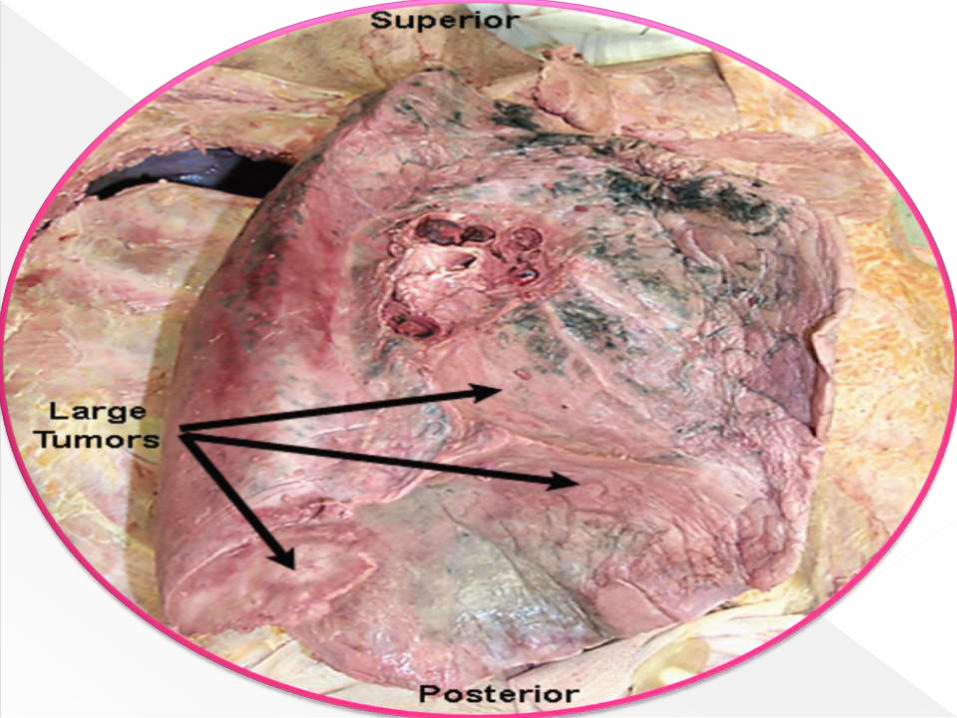
These are life-threatening. Eg. Lung cancer.
Cancers arising from epithelial cells are
referred to as carcinomas, while that from
connective tissue are known as sarcomas.
General & morphological changes:
Shape of cells:
Rounder in shape
Alterations in cell structures:
The cytoskeletal structure of the tumor cells
with regard to actin filament is different.
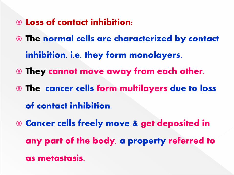
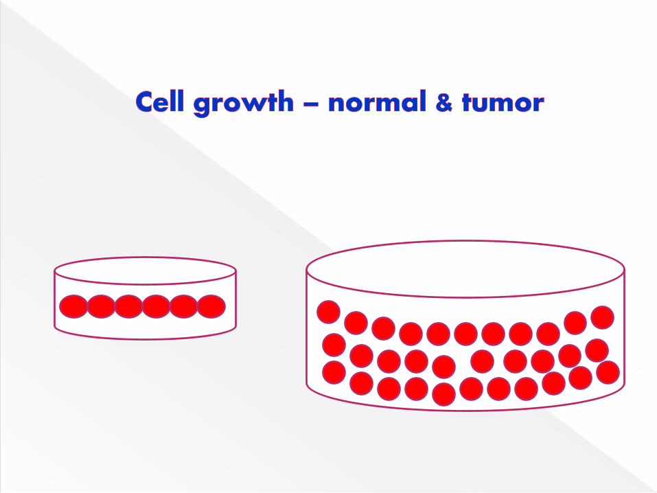
Loss of contact inhibition:
The normal cells are characterized by contact
inhibition, i.e. they form monolayers.
They cannot move away from each other.
The cancer cells form multilayers due to loss
of contact inhibition.
Cancer cells freely move & get deposited in
any part of the body, a property referred to
as metastasis.
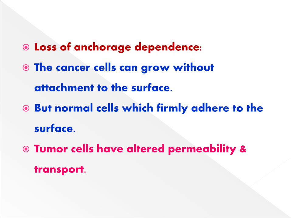
Loss of anchorage dependence:
The cancer cells can grow without
attachment to the surface.
But normal cells which firmly adhere to the
surface.
Tumor cells have altered permeability &
transport.
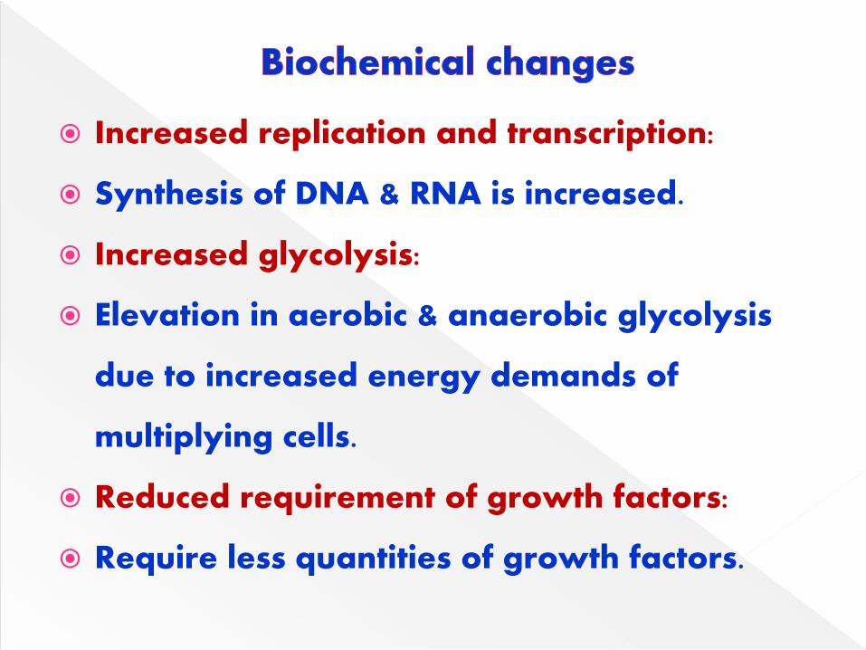
Increased replication and transcription:
Synthesis of DNA & RNA is increased.
Increased glycolysis:
Elevation in aerobic & anaerobic glycolysis
due to increased energy demands of
multiplying cells.
Reduced requirement of growth factors:
Require less quantities of growth factors.
Synthesis of fetal proteins:
During fetal life, certain genes are active,
leading to the synthesis of specific proteins.
These genes are suppressed in adult cells.
Alterations in the structure of molecules:
Changes in the structure of glycoproteins &
glycolipids.
Include physical, chemical, genetic &
environmental factors.
Chemical carcinogens:
Almost 80% of human cancers are caused by
chemical carcinogens in nature.
The chemicals may be organic
(dimethylbenzanthracene) or inorganic
(arsenic, cadmium) in nature.
Occupation e.g. asbestos, benzene.
Diet: e.g aflatoxin B produced by fungus
(Aspergillus flavus) contamination of
foodstuffs, particularly peanuts.
Drugs certain therapeutic drugs can be
carcinogenic e.g. diethylstibesterol.
Life style e.g. cigarette smoking.
Few of the chemicals are directly
carcinogenic.
Majority of them require prior metabolism to
become carcinogenic.
The enzymes cytochrome P450 responsible for
the metabolism of xenobiotics.
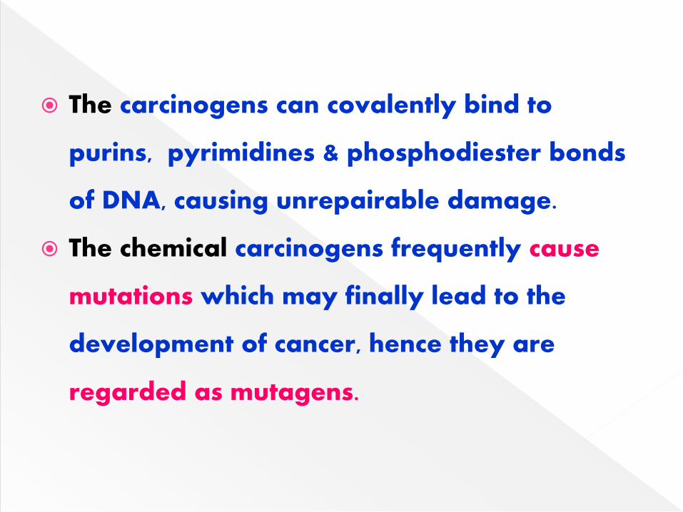
The carcinogens can covalently bind to
purins, pyrimidines & phosphodiester bonds
of DNA, causing unrepairable damage.
The chemical carcinogens frequently cause
mutations which may finally lead to the
development of cancer, hence they are
regarded as mutagens.
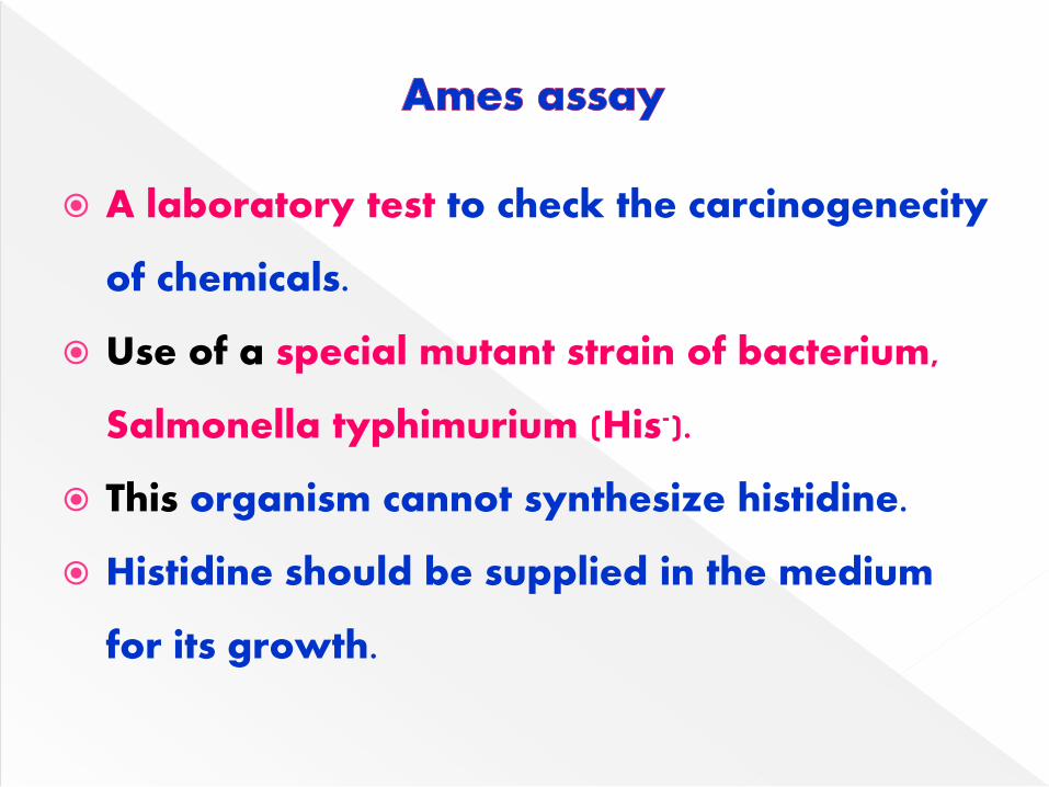
A laboratory test to check the carcinogenecity
of chemicals.
Use of a special mutant strain of bacterium,
Salmonella typhimurium (His-).
This organism cannot synthesize histidine.
Histidine should be supplied in the medium
for its growth.
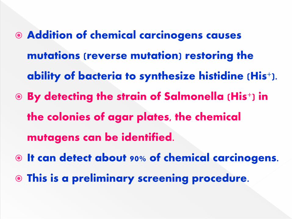
Addition of chemical carcinogens causes
mutations (reverse mutation) restoring the
ability of bacteria to synthesize histidine (His+).
By detecting the strain of Salmonella (His+) in
the colonies of agar plates, the chemical
mutagens can be identified.
It can detect about 90% of chemical carcinogens.
This is a preliminary screening procedure.

Radiation energy:
Ultraviolet rays, x-rays and y-rays have
been proved to be mutagenic in nature
causing cancers.
These rays damage DNA.
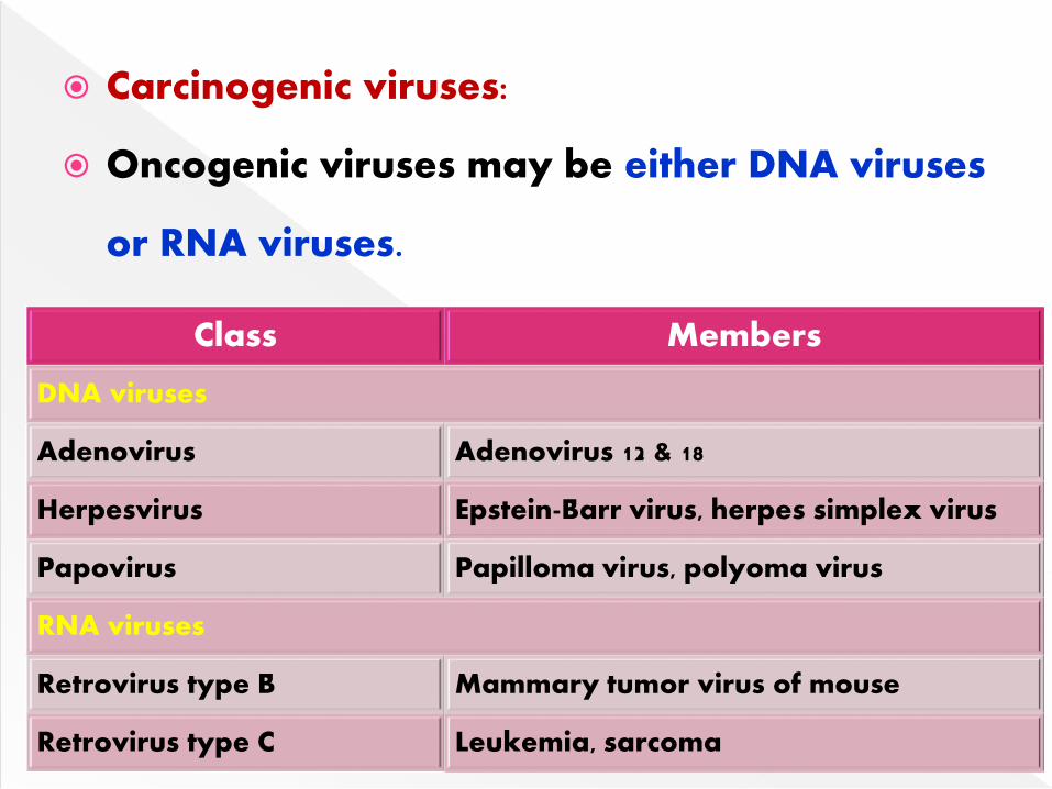
Carcinogenic viruses:
Oncogenic viruses may be either DNA viruses
or RNA viruses.
Class Members
DNA viruses
Adenovirus Adenovirus 12 & 18
Herpesvirus Epstein-Barr virus, herpes simplex virus
Papovirus Papilloma virus, polyoma virus
RNA viruses
Retrovirus type B Mammary tumor virus of mouse
Retrovirus type C Leukemia, sarcoma
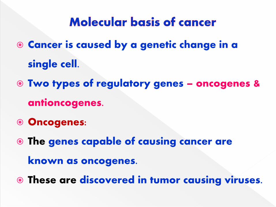
Cancer is caused by a genetic change in a
single cell.
Two types of regulatory genes – oncogenes &
antioncogenes.
Oncogenes:
The genes capable of causing cancer are
known as oncogenes.
These are discovered in tumor causing viruses.
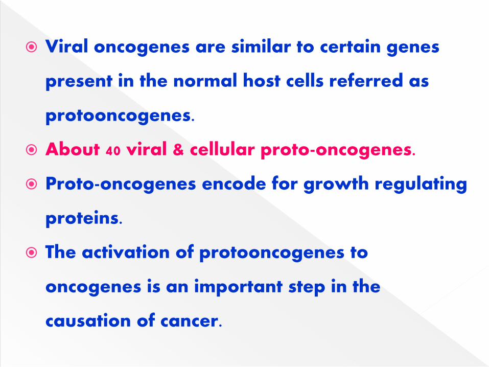
Viral oncogenes are similar to certain genes
present in the normal host cells referred as
protooncogenes.
About 40 viral & cellular proto-oncogenes.
Proto-oncogenes encode for growth regulating
proteins.
The activation of protooncogenes to
oncogenes is an important step in the
causation of cancer.
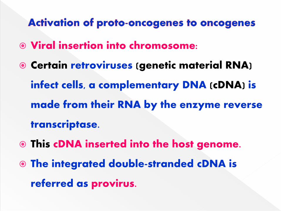
Viral insertion into chromosome:
Certain retroviruses (genetic material RNA)
infect cells, a complementary DNA (cDNA) is
made from their RNA by the enzyme reverse
transcriptase.
This cDNA inserted into the host genome.
The integrated double-stranded cDNA is
referred as provirus.
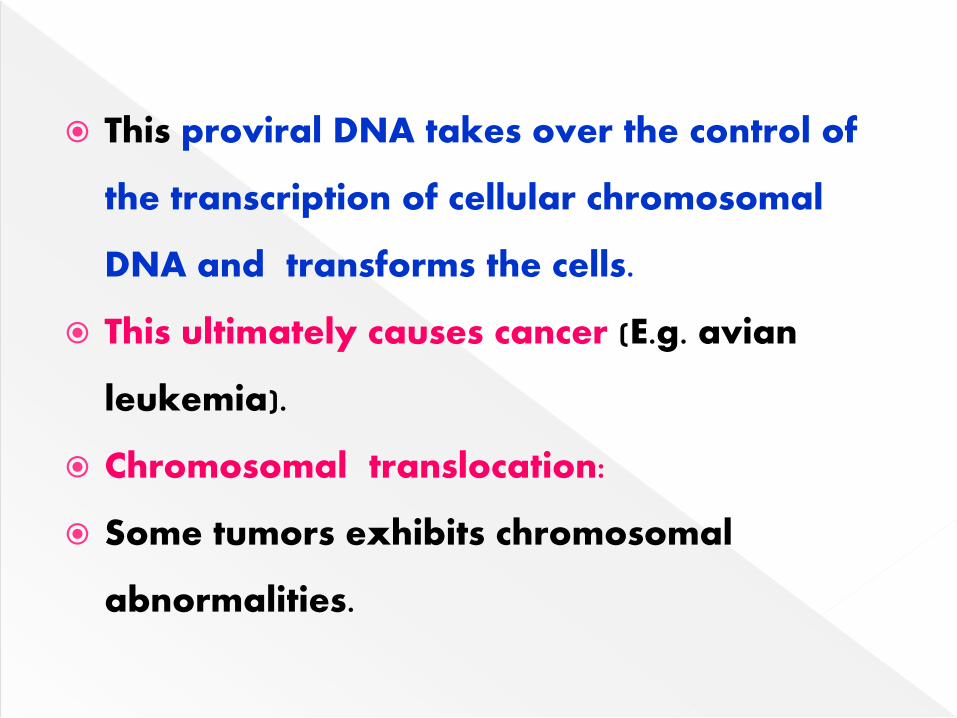
This proviral DNA takes over the control of
the transcription of cellular chromosomal
DNA and transforms the cells.
This ultimately causes cancer (E.g. avian
leukemia).
Chromosomal translocation:
Some tumors exhibits chromosomal
abnormalities.
This due to rearrangement of genetic
material (DNA) by chromosomal
translocation i.e. splitting off a small
fragment of chromosome which is joined to
another chromosome.
It usually results in over expression of proto-
oncogenes.
Burkitt’s lymphoma, a cancer of B-
lymphocytes.
Gene amplification:
Several fold amplifications of certain DNA
sequences are observed in some cancers.
Administration of anticancer drugs
methotrexate (inhibitor of dihydrofolate
reductase) is associated with gene
amplification.
The drug becomes inactive due to gene
amplification resulting in a several fold
increase in the activity of dihydrofolate
reductase.
Point mutations:
The replacement of one base pair by another
results in point mutation.
Growth factors:
Cell proliferation is stimulated by growth factors.
Growth factor binds to protein receptor on the
plasma membrane.
This binding activates cytoplasmic protein kinases
leading to the phosphorylation of intracellular
target proteins.
The phosphorylated proteins act as intracellular
messengers to stimulate cell division.
Transforming growth factor (TGF-α) is a
protein synthesized & required for the
growth of epithelial cells.
TGF - α is produced in high concentration in
individuals suffering from psoriasis, a
disease characterized by excessive
proliferation of epidermal cells.
Some oncogenes encoding growth factor
receptors.
Overexpression and/or structural
alterations in growth factor receptors are
associated with carcinogenesis.
These are a group of signal transducing
proteins.
Guanosine triphosphate (GTP) – binding
proteins are found in 30% of human cancers.
Mutation of ras protooncogene is the single-
most dominant cause of many human tumors.
A special category of genes – cancer
suppressor genes or antioncogenes.
The products of these genes apply breaks &
regulate cell proliferation.
The loss of these suppressor genes removes
the growth control of cells & a key factor in
the development of several tumors. E.g.
retinoblastoma, carcinoma of lung.
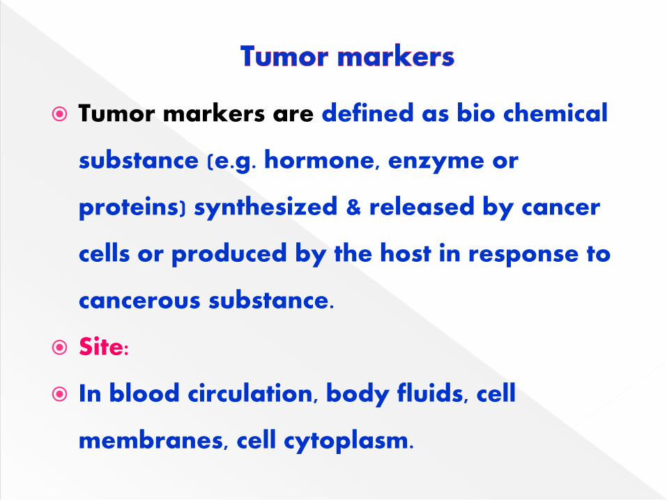
Tumor markers are defined as bio chemical
substance (e.g. hormone, enzyme or
proteins) synthesized & released by cancer
cells or produced by the host in response to
cancerous substance.
Site:
In blood circulation, body fluids, cell
membranes, cell cytoplasm.
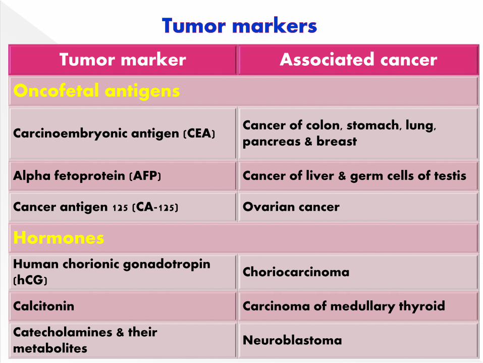
Tumor marker Associated cancer
Oncofetal antigens
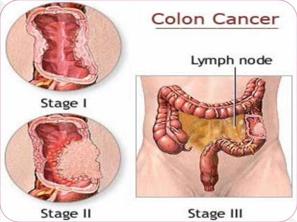
Carcinoembryonic antigen (CEA)Cancer of colon, stomach, lung, pancreas & breast
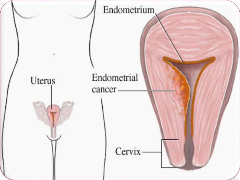
Alpha fetoprotein (AFP) Cancer of liver & germ cells of testis
Cancer antigen 125 (CA-125) Ovarian cancer
Hormones
Human chorionic gonadotropin(hCG)
Choriocarcinoma
Calcitonin Carcinoma of medullary thyroid
Catecholamines & their metabolites
Neuroblastoma
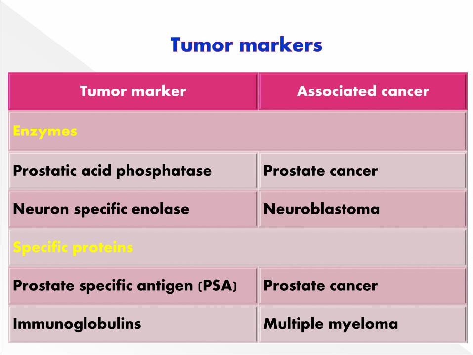
Tumor marker Associated cancer
Enzymes
Prostatic acid phosphatase Prostate cancer
Neuron specific enolase Neuroblastoma
Specific proteins
Prostate specific antigen (PSA) Prostate cancer
Immunoglobulins Multiple myeloma
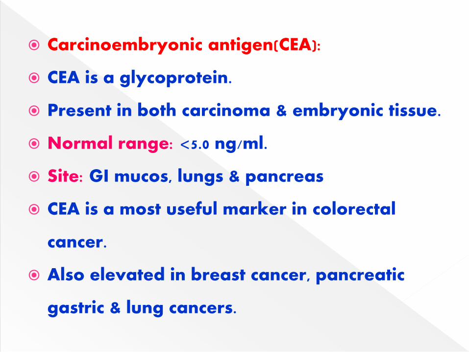
Carcinoembryonic antigen(CEA):
CEA is a glycoprotein.
Present in both carcinoma & embryonic tissue.
Normal range: <5.0 ng/ml.
Site: GI mucos, lungs & pancreas
CEA is a most useful marker in colorectal
cancer.
Also elevated in breast cancer, pancreatic
gastric & lung cancers.
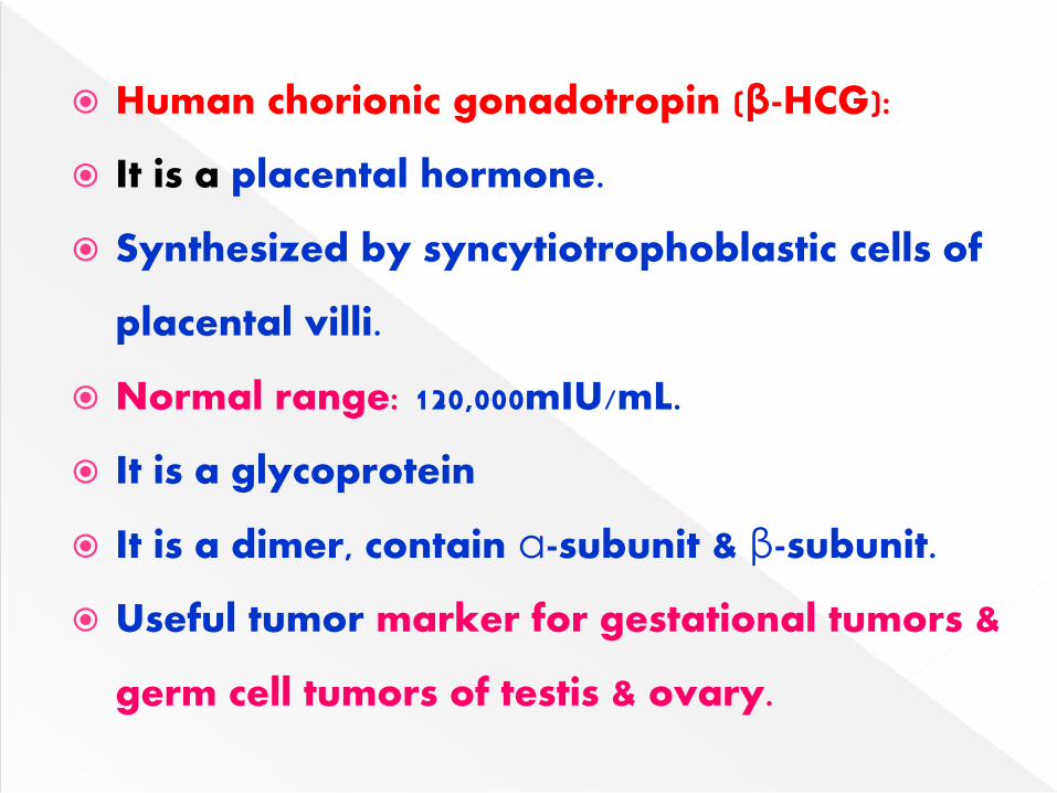
Human chorionic gonadotropin (β-HCG):
It is a placental hormone.
Synthesized by syncytiotrophoblastic cells of
placental villi.
Normal range: 120,000mIU/mL.
It is a glycoprotein
It is a dimer, contain α-subunit & β-subunit.
Useful tumor marker for gestational tumors &
germ cell tumors of testis & ovary.
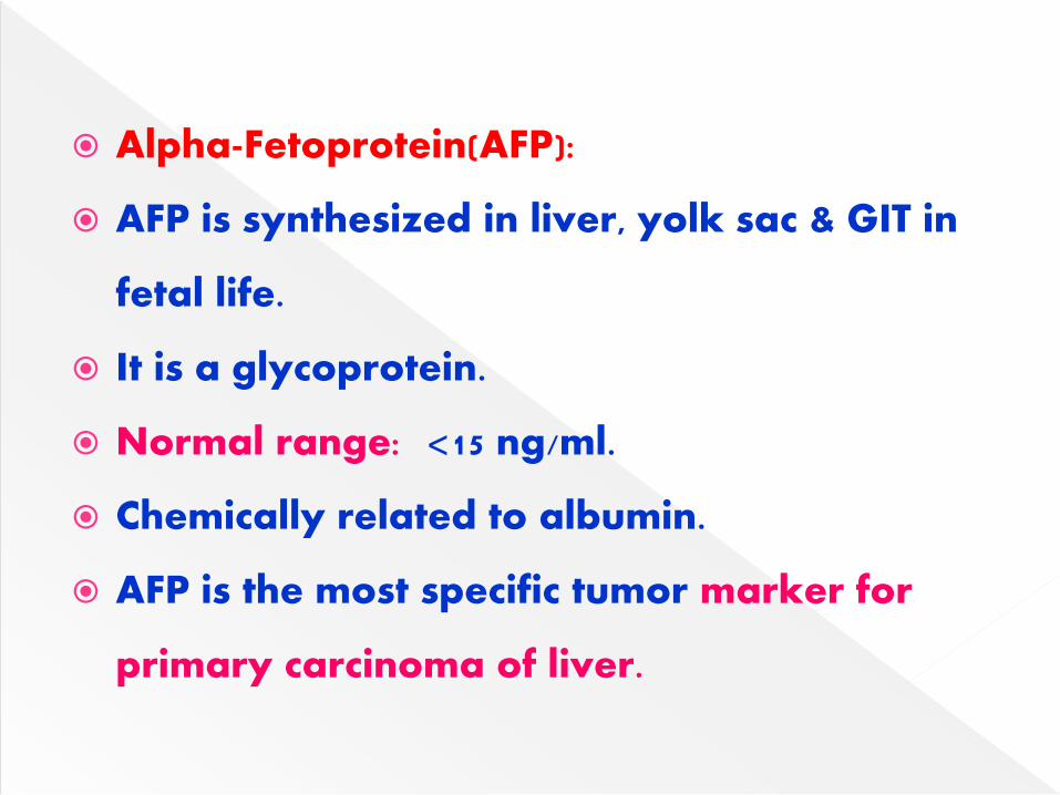
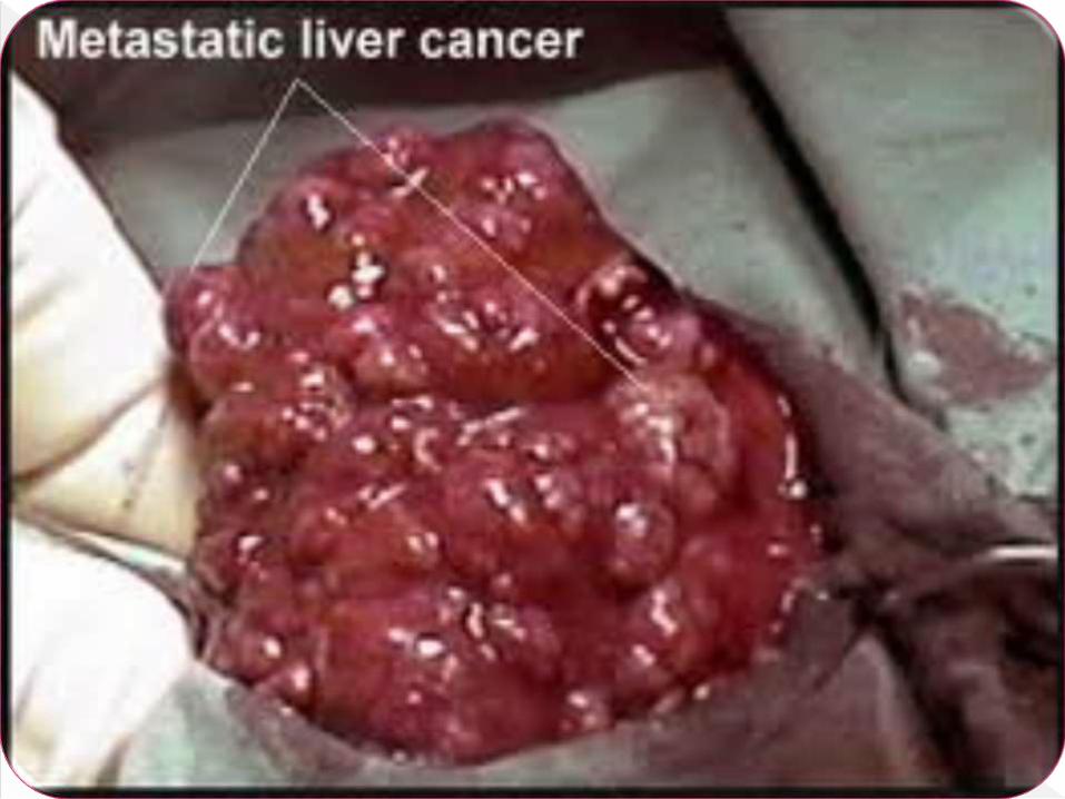
Alpha-Fetoprotein(AFP):
AFP is synthesized in liver, yolk sac & GIT in
fetal life.
It is a glycoprotein.
Normal range: <15 ng/ml.
Chemically related to albumin.
AFP is the most specific tumor marker for
primary carcinoma of liver.
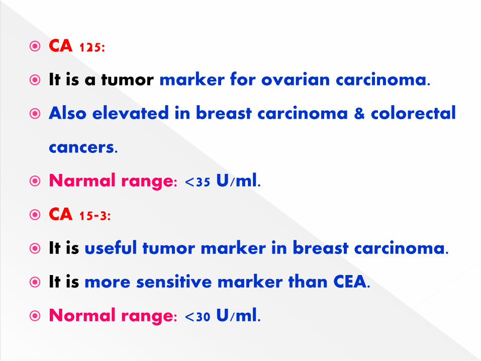
CA 125:
It is a tumor marker for ovarian carcinoma.
Also elevated in breast carcinoma & colorectal
cancers.
Narmal range: <35 U/ml.
CA 15-3:
It is useful tumor marker in breast carcinoma.
It is more sensitive marker than CEA.
Normal range: <30 U/ml.
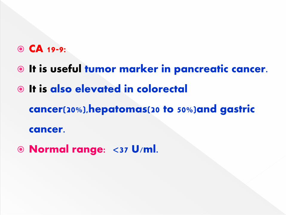
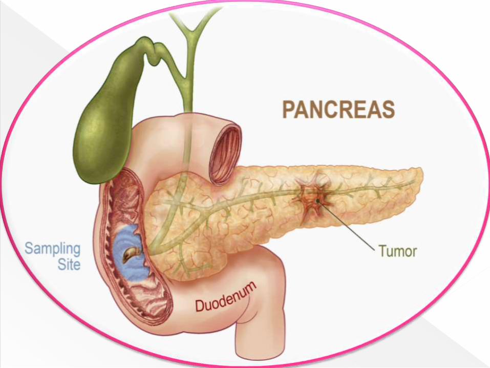
CA 19-9:
It is useful tumor marker in pancreatic cancer.
It is also elevated in colorectal
cancer(20%),hepatomas(20 to 50%)and gastric
cancer.
Normal range: <37 U/ml.
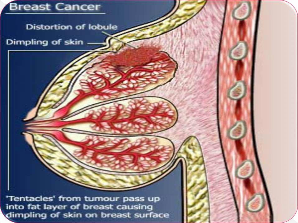
These are identified in breast cancer.
MCA (Mucin-like carcinoma associated
antigen)
It is a mucin glycoprotein.
MAM6:
It is a epithelial membrane antigen.
Present on ductal & alveolar epithelial cells.
MSA (mammary serum antigen):
It is marker of intraductal breast cancer.
More sensitive marker then CA 15-3.
MAP (Mitogen activated protein) kinase:
It is a new breast cancer marker.
MAP kinase levels are 5 to 20 times higher in
breast cancer as compared to normal breast
tissue.
Prostatic acid phosphatase (PAP):
It is a tumor marker for prostate cancer.
Normal range: <2.5ng/ml.
Prostate specific antigen (PSA):
It is an organ specific.
Localized to prostatic ductal cells.
Normal range: 4.5 µg/L
Neuron –specific Enolase (NSE):
It is a specific tumor marker of neuro-
endocrine origin.
It is useful marker in neuroblastomas & lung
cancer.
NSE:
Also elevated in small-cell carcinoma of lung
The upper reference limit: 12.5 µg/L
Textbook of Biochemistry – U Satyanarayana
Textbook of Biochemistry – DM Vasudevan
Textbook of Biochemistry – MN Chatterjea

Related Documents


