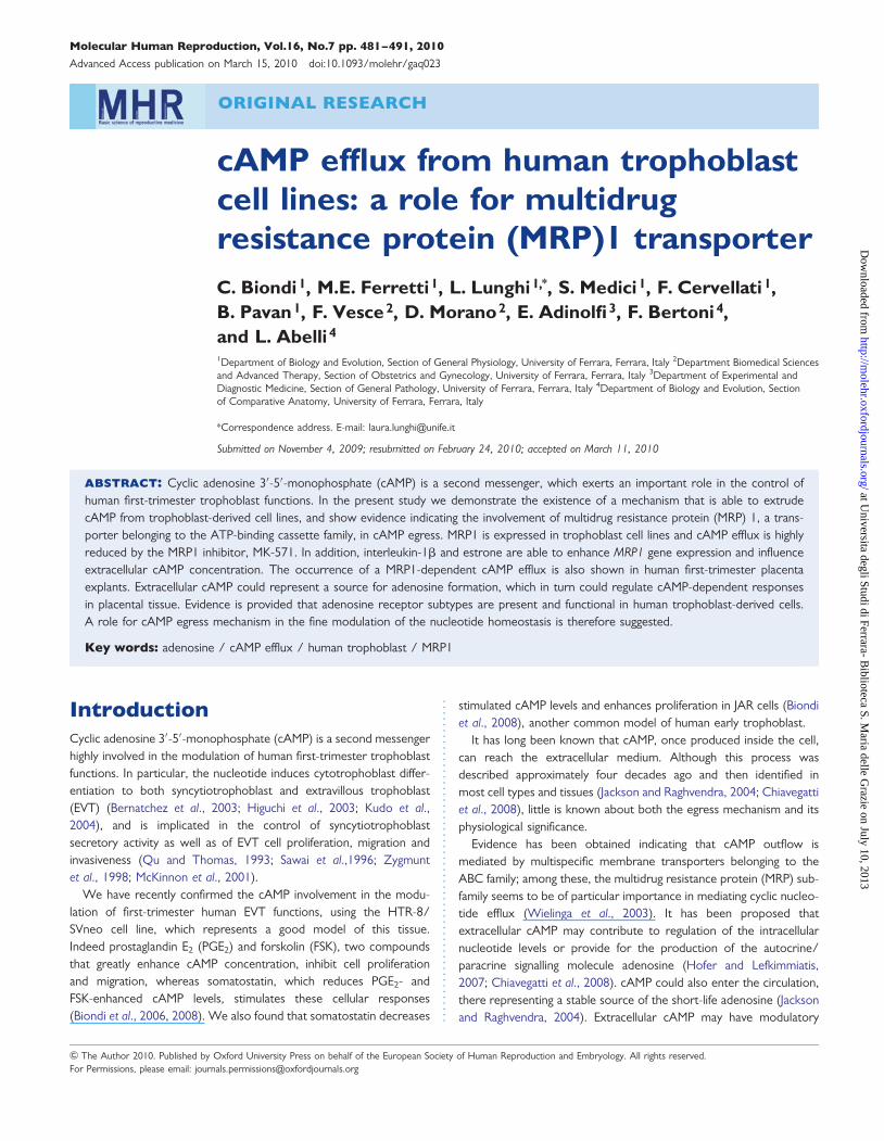
ORIGINAL RESEARCH cAMP efflux from human trophoblast cell lines: a role for multidrug resistance protein (MRP)1 transporter C. Biondi 1 , M.E. Ferretti 1 , L. Lunghi 1, * , S. Medici 1 , F. Cervellati 1 , B. Pavan 1 , F. Vesce 2 , D. Morano 2 , E. Adinolfi 3 , F. Bertoni 4 , and L. Abelli 4 1 Department of Biology and Evolution, Section of General Physiology, University of Ferrara, Ferrara, Italy 2 Department Biomedical Sciences and Advanced Therapy, Section of Obstetrics and Gynecology, University of Ferrara, Ferrara, Italy 3 Department of Experimental and Diagnostic Medicine, Section of General Pathology, University of Ferrara, Ferrara, Italy 4 Department of Biology and Evolution, Section of Comparative Anatomy, University of Ferrara, Ferrara, Italy *Correspondence address. E-mail: [email protected] Submitted on November 4, 2009; resubmitted on February 24, 2010; accepted on March 11, 2010 abstract: Cyclic adenosine 3 ′ -5 ′ -monophosphate (cAMP) is a second messenger, which exerts an important role in the control of human first-trimester trophoblast functions. In the present study we demonstrate the existence of a mechanism that is able to extrude cAMP from trophoblast-derived cell lines, and show evidence indicating the involvement of multidrug resistance protein (MRP) 1, a trans- porter belonging to the ATP-binding cassette family, in cAMP egress. MRP1 is expressed in trophoblast cell lines and cAMP efflux is highly reduced by the MRP1 inhibitor, MK-571. In addition, interleukin-1b and estrone are able to enhance MRP1 gene expression and influence extracellular cAMP concentration. The occurrence of a MRP1-dependent cAMP efflux is also shown in human first-trimester placenta explants. Extracellular cAMP could represent a source for adenosine formation, which in turn could regulate cAMP-dependent responses in placental tissue. Evidence is provided that adenosine receptor subtypes are present and functional in human trophoblast-derived cells. A role for cAMP egress mechanism in the fine modulation of the nucleotide homeostasis is therefore suggested. Key words: adenosine / cAMP efflux / human trophoblast / MRP1 Introduction Cyclic adenosine 3 ′ -5 ′ -monophosphate (cAMP) is a second messenger highly involved in the modulation of human first-trimester trophoblast functions. In particular, the nucleotide induces cytotrophoblast differ- entiation to both syncytiotrophoblast and extravillous trophoblast (EVT) (Bernatchez et al., 2003; Higuchi et al., 2003; Kudo et al., 2004), and is implicated in the control of syncytiotrophoblast secretory activity as well as of EVT cell proliferation, migration and invasiveness (Qu and Thomas, 1993; Sawai et al.,1996; Zygmunt et al., 1998; McKinnon et al., 2001). We have recently confirmed the cAMP involvement in the modu- lation of first-trimester human EVT functions, using the HTR-8/ SVneo cell line, which represents a good model of this tissue. Indeed prostaglandin E 2 (PGE 2 ) and forskolin (FSK), two compounds that greatly enhance cAMP concentration, inhibit cell proliferation and migration, whereas somatostatin, which reduces PGE 2 - and FSK-enhanced cAMP levels, stimulates these cellular responses (Biondi et al., 2006, 2008). We also found that somatostatin decreases stimulated cAMP levels and enhances proliferation in JAR cells (Biondi et al., 2008), another common model of human early trophoblast. It has long been known that cAMP, once produced inside the cell, can reach the extracellular medium. Although this process was described approximately four decades ago and then identified in most cell types and tissues (Jackson and Raghvendra, 2004; Chiavegatti et al., 2008), little is known about both the egress mechanism and its physiological significance. Evidence has been obtained indicating that cAMP outflow is mediated by multispecific membrane transporters belonging to the ABC family; among these, the multidrug resistance protein (MRP) sub- family seems to be of particular importance in mediating cyclic nucleo- tide efflux (Wielinga et al., 2003). It has been proposed that extracellular cAMP may contribute to regulation of the intracellular nucleotide levels or provide for the production of the autocrine/ paracrine signalling molecule adenosine (Hofer and Lefkimmiatis, 2007; Chiavegatti et al., 2008). cAMP could also enter the circulation, there representing a stable source of the short-life adenosine (Jackson and Raghvendra, 2004). Extracellular cAMP may have modulatory & The Author 2010. Published by Oxford University Press on behalf of the European Society of Human Reproduction and Embryology. All rights reserved. For Permissions, please email: [email protected] Molecular Human Reproduction, Vol.16, No.7 pp. 481– 491, 2010 Advanced Access publication on March 15, 2010 doi:10.1093/molehr/gaq023 at Universita degli Studi di Ferrara- Biblioteca S. Maria delle Grazie on July 10, 2013 http://molehr.oxfordjournals.org/ Downloaded from

Welcome message from author
This document is posted to help you gain knowledge. Please leave a comment to let me know what you think about it! Share it to your friends and learn new things together.
Transcript
ORIGINAL RESEARCH
cAMP efflux from human trophoblastcell lines: a role for multidrugresistance protein (MRP)1 transporterC. Biondi1, M.E. Ferretti1, L. Lunghi1,*, S. Medici1, F. Cervellati 1,B. Pavan1, F. Vesce2, D. Morano2, E. Adinolfi3, F. Bertoni4,and L. Abelli 4
1Department of Biology and Evolution, Section of General Physiology, University of Ferrara, Ferrara, Italy 2Department Biomedical Sciencesand Advanced Therapy, Section of Obstetrics and Gynecology, University of Ferrara, Ferrara, Italy 3Department of Experimental andDiagnostic Medicine, Section of General Pathology, University of Ferrara, Ferrara, Italy 4Department of Biology and Evolution, Sectionof Comparative Anatomy, University of Ferrara, Ferrara, Italy
*Correspondence address. E-mail: [email protected]
Submitted on November 4, 2009; resubmitted on February 24, 2010; accepted on March 11, 2010
abstract: Cyclic adenosine 3′-5′-monophosphate (cAMP) is a second messenger, which exerts an important role in the control ofhuman first-trimester trophoblast functions. In the present study we demonstrate the existence of a mechanism that is able to extrudecAMP from trophoblast-derived cell lines, and show evidence indicating the involvement of multidrug resistance protein (MRP) 1, a trans-porter belonging to the ATP-binding cassette family, in cAMP egress. MRP1 is expressed in trophoblast cell lines and cAMP efflux is highlyreduced by the MRP1 inhibitor, MK-571. In addition, interleukin-1b and estrone are able to enhance MRP1 gene expression and influenceextracellular cAMP concentration. The occurrence of a MRP1-dependent cAMP efflux is also shown in human first-trimester placentaexplants. Extracellular cAMP could represent a source for adenosine formation, which in turn could regulate cAMP-dependent responsesin placental tissue. Evidence is provided that adenosine receptor subtypes are present and functional in human trophoblast-derived cells.A role for cAMP egress mechanism in the fine modulation of the nucleotide homeostasis is therefore suggested.
Key words: adenosine / cAMP efflux / human trophoblast / MRP1
IntroductionCyclic adenosine 3′-5′-monophosphate (cAMP) is a second messengerhighly involved in the modulation of human first-trimester trophoblastfunctions. In particular, the nucleotide induces cytotrophoblast differ-entiation to both syncytiotrophoblast and extravillous trophoblast(EVT) (Bernatchez et al., 2003; Higuchi et al., 2003; Kudo et al.,2004), and is implicated in the control of syncytiotrophoblastsecretory activity as well as of EVT cell proliferation, migration andinvasiveness (Qu and Thomas, 1993; Sawai et al.,1996; Zygmuntet al., 1998; McKinnon et al., 2001).
We have recently confirmed the cAMP involvement in the modu-lation of first-trimester human EVT functions, using the HTR-8/SVneo cell line, which represents a good model of this tissue.Indeed prostaglandin E2 (PGE2) and forskolin (FSK), two compoundsthat greatly enhance cAMP concentration, inhibit cell proliferationand migration, whereas somatostatin, which reduces PGE2- andFSK-enhanced cAMP levels, stimulates these cellular responses(Biondi et al., 2006, 2008). We also found that somatostatin decreases
stimulated cAMP levels and enhances proliferation in JAR cells (Biondiet al., 2008), another common model of human early trophoblast.
It has long been known that cAMP, once produced inside the cell,can reach the extracellular medium. Although this process wasdescribed approximately four decades ago and then identified inmost cell types and tissues (Jackson and Raghvendra, 2004; Chiavegattiet al., 2008), little is known about both the egress mechanism and itsphysiological significance.
Evidence has been obtained indicating that cAMP outflow ismediated by multispecific membrane transporters belonging to theABC family; among these, the multidrug resistance protein (MRP) sub-family seems to be of particular importance in mediating cyclic nucleo-tide efflux (Wielinga et al., 2003). It has been proposed thatextracellular cAMP may contribute to regulation of the intracellularnucleotide levels or provide for the production of the autocrine/paracrine signalling molecule adenosine (Hofer and Lefkimmiatis,2007; Chiavegatti et al., 2008). cAMP could also enter the circulation,there representing a stable source of the short-life adenosine (Jacksonand Raghvendra, 2004). Extracellular cAMP may have modulatory
& The Author 2010. Published by Oxford University Press on behalf of the European Society of Human Reproduction and Embryology. All rights reserved.For Permissions, please email: [email protected]
Molecular Human Reproduction, Vol.16, No.7 pp. 481–491, 2010
Advanced Access publication on March 15, 2010 doi:10.1093/molehr/gaq023
at Universita degli Studi di Ferrara- B
iblioteca S. Maria delle G
razie on July 10, 2013http://m
olehr.oxfordjournals.org/D
ownloaded from
effects per se; indeed, although specific receptors for this nucleotidehave been characterized only in primitive organisms (Lee et al.,2005), their presence has been reported in mammalian cells too(Sorbera and Morad, 1991).
Only a few investigations have been carried out on cAMP outflowfrom fetal and/or maternal tissues. For example, Grealy andSreenan (1999) found an efflux of cAMP from preimplantation cattleblastocysts and proposed that the nucleotide may have a role inembryo-maternal signalling and/or in the regulation of embryoniccell proliferation. cAMP egress was also evidenced in bovine oviductcells, where it could regulate sperm function, fertilization, and earlyembryo development (Cometti et al., 2003). To our best knowledge,no report exists regarding cAMP efflux from human trophoblast;however, this tissue could indeed secrete cAMP, since the expressionof putative cyclic nucleotide transporters has been demonstrated inhuman placenta and cultured trophoblast cells (Meyer zu Schwabedis-sen et al., 2005; Evseenko et al., 2006).
In the present study we utilized HTR-8/SVneo cell line to verifywhether intracellular cAMP, produced in basal conditions and uponaddition of receptorial and non-receptorial agonists, flows to theextracellular medium. In order to obtain insights about the mechan-isms involved in this process, we used compounds able to influenceboth activity and expression of putative cyclic nucleotide transporters.To corroborate results obtained in HTR-8/SVneo cells, we also uti-lized JAR cells and human first-trimester placenta explants. The poss-ible role of extracellular cAMP in the modulation of human trophoblastfunctions is discussed.
Materials and Methods
Chemicals(G-3H)adenosine 3′-5′-monophosphate (specific activity 27 Ci/mmol) waspurchased from Amersham Italia Srl (Milan, Italy). cAMP, isobutylmethyl-xanthine (IBMX), PGE2, FSK, probenecid, progesterone, cyclopentyl-adenosine (CPA), 4-aminobenzyl-5′-N-methylcarboxamidoadenosine(AB-MECA), estrone, 1,3-dipropyl-8-p-sulphophenilxanthine (DPSPX),propidium iodide and Folin-Ciocalteu’s phenol reagent were from SigmaChemical Co. (St. Louis, MO). Ro 201724 and CGS 21 680 were pur-chased from Tocris (Avonmouth, UK). Interleukin (IL)-1b was fromEndogen (Woburn, MA, USA). MK-571 was from Calbiochem(Germany). Tissue culture media, sera, RNA extraction and RT–PCRreagents were purchased from Invitrogen (Paisley, Scotland, UK). Therabbit polyclonal antiserum H-70 raised against amino acids 1–70 ofhuman MRP1 was from Santa Cruz Biotechnology (CA, USA). Themouse monoclonal antibody (mAb) QCRL-1 against human MRP1(linear internal epitope S918SYSGDI924) was from Alexis Biochemicals(Lausen, Switzerland). The mouse mAb MEM-G1 against recombinanthuman HLA-G denaturated heavy chain was from Exbio (Prague, CzechRep.). The fluorescein isothiocyanate (FITC)-labeled affinity-purified goatanti-rabbit IgG serum and the Texas Red-labeled horse anti-mouse IgGserum were from Vector Laboratories (Burlingame CA, USA).
qPCR human reference cDNA was purchased from Clontech Labora-tories (Mountain View, CA, USA). All other chemicals were the highestreagent grades commercially available.
Cell culturesThe HTR-8/SVneo trophoblast cell line, obtained from human first-trimester placental explant cultures and immortalized using SV40 large T
antigen, was kindly provided by Dr. CH Graham, Queen’s University,Kingston, Ontario (Canada). The human choriocarcinoma JAR cellswere obtained from Istituto Zooprofilattico, Brescia (Italy). Cells were cul-tured at 378C in an atmosphere of 5% CO2/95% air in RPMI 1640medium containing 10% fetal bovine serum, 100 U/ml penicillin and100 mg/ml streptomycin.
Placental tissuePlacental tissue, obtained from a consenting patient undergoing spontaneousabortion at the 16th week of gestation, was fixed and utilized for immunohis-tochemistry. First-trimester placenta explants (n ¼ 3) were obtained fromconsenting patients undergoing elective termination of pregnancy (9–12weeks of gestation) and freshly used for cAMP level determination.
cAMP level determinationCells were grown to confluence (2–3 days), then the medium was removedand replaced by serum-free RPMI. The incubation was carried out in theabsence or in the presence of phosphodiesterase (PDE) inhibitors andtest substances, for the indicated times. Media were then collected andimmediately frozen at 2708C until cAMP levels were measured. Ice-cold0.1 N HCl (0.25 ml) was added to the cells and, after centrifugation at12 500 × g for 10 min, supernatants were neutralized adding 0.5 MTrizma base (0.05 ml) and utilized for intracellular cAMP determination.
Placental explants were incubated in serum-free RPMI in the presenceof 1023 M IBMX and test substances for 60 min. Media were then col-lected and immediately frozen at 2708C until extracellular cAMP levelswere measured. For intracellular cAMP determination, tissues were trans-ferred to 5% cold trichloroacetic acid (0.5 ml) and maintained at 48C over-night. Samples were homogenized and centrifuged at 2000 × g for10 min, then supernatants were utilized for cAMP determination afterextraction with aqueous ethyl-ether.
Intracellular and extracellular cAMP were measured by the method ofBrown et al. (1972) and the nucleotide levels were expressed as pmoles/106 cells/time or as pmoles/mg protein/time. Protein content of placen-tal explants was determined according to Lowry et al. (1951) on samplesdissolved in 1 N NaOH. Assay sensitivity was 0.5 pmol/106 cells, and theintra- or inter-assay coefficients of variations were ,10%.
RT–PCRRNA was extracted by a single-step guanidine thiocyanate method, usingTrizol reagent, from HTR-8/SVneo cells (grown to 90% confluence).Total RNA was dissolved in diethyl pyrocarbonate-treated H2O and quan-tified by spectrophotometry. Three micrograms of total RNA was reverse-transcribed using 200 units of Moloney murine leukemia virus reverse tran-scriptase and 1.5 mg of random primers, in a 20 ml reaction volume at378C for 1 h. One microlitre was used as a template for PCR. The ampli-fication reaction was performed using 0.5 units of Platinum Taq DNA poly-merase in a 25 ml reaction volume containing 10 mM Tris–HCl, pH 8.3,50 mM KCl, 1.5 mM MgCl2, 0.2 mM dNTPs and 0.4 mM of each primer.To check the RNA quality and quantity, RT–PCRs were performedusing specific primers for 18S mRNA (Selvey et al., 2001) or ribosomalprotein L 13A (RPL 13A) (Pattyn et al., 2006), two constitutivelyexpressed genes. PCR amplification from reverse transcribed cDNA wascarried out using specific primers (Table I). Each reaction cycle consistedof an initial denaturation step at 948C for 40 s (MRP1) and 30 s (adenosinereceptors, 18S and RPL 13A); an annealing step for 30 s at 648C (MRP1),588C (adenosine receptors), 57.38C (18S) and for 60 s at 608C (RPL13A); an elongation step for 40 s at 728C (MRP1 and RPL 13A), for90 s at 688C (adenosine receptors) and for 45 s at 728C (18S). Twenty-four cycles of PCR were performed for MRP1, 35 for adenosine receptorsas well as RPL 13A and 22 for 18S.
482 Biondi et al.
at Universita degli Studi di Ferrara- B
iblioteca S. Maria delle G
razie on July 10, 2013http://m
olehr.oxfordjournals.org/D
ownloaded from
The PCR products were separated by electrophoresis on 2% (w/v)agarose gels containing 0.05% ethidium bromide using 100 bp DNAladder as size marker. Relative amounts of amplification products wereestimated by digital densitometry, using the ‘Gel Doc 2000’ video imagesystem (Bio-Rad Laboratories, Hercules, CA).
Localization of MRP1 in cell linesHTR-8/SVneo and JAR cells were incubated in serum-free medium for 24 h,then cells (1.5 × 10⁶/ml) were allowed to adhere onto glass coverslipsovernight. Cell viability was checked by Trypan blue exclusion test beforefixation. Adherent cells were prefixed with 1% paraformaldehyde (PFA)for 3 min, washed in phosphate-buffered salt solution (PBS; 0.01 M,pH 7.2) and labelled with the anti-human MRP1 rabbit polyclonal antiserumH-70 (200 mg/ml undiluted or 2 mg/ml diluted up to 1:100 inPBS-containing 0.05% BSA and 0.1% sodium azide) or the mouse mAbQCRL-1 (20 mg/ml undiluted) for 1 h at room temperature (RT) withoutany cell permeabilization. For double labelling experiments, the antiserumH-70 (diluted 1:10) was used together with the anti-human HLA-Gmouse mAb MEM-G1 (diluted 1:100). Cells were then washed in PBSand incubated with FITC-labelled goat anti-rabbit IgG serum (diluted1:100 in PBS) and/or Texas Red-labelled horse anti-mouse IgG serum(diluted 1:300 in PBS) for 1 h at RT in the dark. Controls included labellingwith secondary antibodies alone in the absence of primary antibodies.
Cells were washed in PBS, post-fixed with 3% PFA for 3 min, rinsed in PBS,mounted in Vectashield (Vector) anti-fading in the dark and observed (FITCand Texas Red fluorescence excitation at 488 and 543 nm, respectively)with a laser scanning confocal microscope (LSCM; Carl Zeiss LSM 510,Arese, Italy) equipped with 40× and plan apochromat 63× oil immersionobjectives and Argon/HeNE laser sources. Superimposition of red andgreen fluorescence, not specifically due to co-localization, was excluded byapplying to the green channel a beam path admitting the acquisition of fluor-escence between 505 and 550 nm and to the red channel a light path exclud-ing all fluorescence ,560 nm. All images were obtained at an 8% laserpotency and with pinhole diameters of 73 (40 × ) and 104 mm (63 × ).Amplifier and detector optimizing parameters were maintained constantfor all the experiments. Digital magnification, cell and stacks dimension par-ameters were obtained with LSM confocal software (Zeiss).
In single labelling experiments with the anti-human MRP1 antiserumH-70, the nuclei of post-fixed HTR-8/SVneo and JAR cells were counter-stained with propidium iodide (0.1 mM, 5 min in the dark).
Localization of MRP1 in human placentaFreshly collected human placenta was fixed in Bouin’s liquid for 7 h at 48C,dehydrated in graded cold ethanol series and embedded in paraffin wax.Serial sections of 7 mm thickness were de-waxed with toluene, rehydratedin graded cold ethanol series and put in 0.01 M, pH 7.2 PBS. Some sec-tions were stained with Giemsa for general histology. Immunohistochem-istry was performed by double immunofluorescence staining. Multiple setsof sections were placed in PBS containing 0.1% Triton X-100 for 30 min,washed in PBS and preincubated for 30 min with 5% normal human serum.Sections were then incubated overnight at 48C with the anti-human MRP1rabbit polyclonal antiserum H-70 (diluted 1:10) and the anti-humanHLA-G mouse mAb MEM-G1 (diluted 1:100). Primary antisera werediluted in PBS containing 0.05% BSA and 0.1% sodium azide. Negativecontrols were made by omission of the primary antibodies, or their sub-stitution by normal rabbit and mouse sera. Serum prior rabbit immuniz-ation was used as a negative control for H-70 immunostaining.Thereafter, sections were washed in PBS and incubated for 1 h at RT inthe dark with FITC-labelled goat anti-rabbit IgG serum (1:100) andTexas Red-labelled horse anti-mouse IgG serum (1:100). Secondary anti-sera were diluted in PBS containing BSA and sodium azide. Sections werewashed in PBS, rinsed in distilled water, mounted in Vectashield anti-fadingin the dark and observed with the LSCM, as described for cell lines.
Statistical analysisNumerical results were analysed by ANOVA followed by Bonferroni’smultiple comparison test, Dunnett’s comparison post-test or two-tail Stu-dent’s t-test, as appropriate, using the GraphPad Prism 2.1 software stat-istical package. The level for accepted statistical significance was P , 0.05.
Results
In vitro experiments carried out in HTR-8/SVneo cellsIntra- and extra-cellular cAMP levels stimulated by PGE2 and FSKIn a first series of experiments, we measured intracellular cAMP levelsin HTR-8/SVneo cells, incubated up to 60 min in the absence and inthe presence of 1026 M PGE2 or FSK. In basal conditions, cAMP con-centrations remained almost constant at all tested times (around 6.0
.............................................................................................................................................................................................
Table I Primer sequences for RT–PCR.
Gene Primer (5′ �3′) Base pairs References
MRP1 F: cGGAAACCATCCACGACCCTAATC 294 Alcorn et al., 2002R: aCCTCCTCATTCGCATCCACCTTG
A1 F: gGTGGAATTCTCCATCTCAGCTTTCCAGGC 525 Phelps et al., 2006R: gGTGAAGCTTTCGAACTCGCACTTGATCAC
A2a F:gGTGGAATTCAACAACTGCGGTCAGCCAAA 544 Phelps et al., 2006R: gGTGAAGCTTCAGCTGCCTTGAAAGGTTCT
A2b F:gGTGGAATTCGAACCACGAATGAAAGCTGC 491 Phelps et al., 2006R: gGTGAAGCTTTGACCATTCCCACTCTTGAC
A3 F: aACGTGCTGGTCATCTGCGTGGTC 440 Phelps et al., 2006R: gTAGTCCATTCTCATGACGGAAAC
18S F: tCAAGAACGAAAGTCGGAGG 489 Selvey et al., 2001R: gGACATCTAAGGGCATCACA
RPL 13A F: cCTGGAGGAGAAGAGGAAAGAGA 126 Pattyn et al., 2006R: tTGAGGACCTCTGTGTATTTGTCAA
cAMP efflux from human trophoblast cells 483
at Universita degli Studi di Ferrara- B
iblioteca S. Maria delle G
razie on July 10, 2013http://m
olehr.oxfordjournals.org/D
ownloaded from

pmoles/106 cells, not shown). In the presence of PGE2 or FSK, intra-cellular cAMP production increased as a function of incubation time upto 15 min (38- and 50-fold, respectively), thereafter a reduction of thenucleotide level was observed (Fig. 1A and B). A decrease of cAMPcontent due to its degradation appears unlikely since these exper-iments were carried out in the presence of 1023 M IBMX, a broadspectrum inhibitor of cAMP PDE (Chiavegatti et al., 2008). Thereforewe evaluated the possibility of cAMP egress, quantifying the nucleotideconcentration in the extracellular medium. In basal conditions theextracellular cAMP levels were similar to the intracellular ones andquite constant at all tested times (not shown). In the presence ofPGE2 or FSK, they progressively increased in a time-dependentfashion at least up to 60 min (Fig. 1A and B).
Effect of cAMP efflux inhibitorsWe then tested the effect of progesterone and probenecid, twoagents known to inhibit cyclic nucleotide efflux in several cell types(Florio et al., 1999; Godinho and Costa, 2003; Wielinga et al.,2003), on cAMP levels stimulated by 1026 M FSK or PGE2. After60 min of incubation with FSK, both compounds significantlyreduced (249%, P , 0.01 for progesterone; 262%, P , 0.01 for
probenecid) extracellular cAMP concentration at the highest dosetested (Fig. 2A). Similar results were obtained in the presence ofPGE2 (260%, P , 0.01 for progesterone; 250%, P , 0.01 for probe-necid) (Fig. 2B). Otherwise, no significant alteration of the intracellularnucleotide level was found (not shown).
We then utilized MK-571, an inhibitor of MRP1 (Olson et al., 2001),which is a well-characterized transporter in human placenta(Nagashige et al., 2003; Evseenko et al., 2006; Parry and Zhang,2007). In cells incubated for 60 min with 1026 M FSK or PGE2, extra-cellular cAMP levels decreased in the presence of MK-571 dosesvarying between 1026 and 1024 M the maximal effect being observedat 1025 M (not shown). When we used 1025 M MK-571 in time-course experiments, an evident reduction of extracellular cAMP wasfound at all tested times (Fig. 2C and D), the maximal effect beingobserved after 60 min of incubation (272% for PGE2- and 251%for FSK-stimulated cAMP). In contrast, the intracellular cAMPconcentrations did not vary significantly at any of the times tested(Fig. 2C and D).
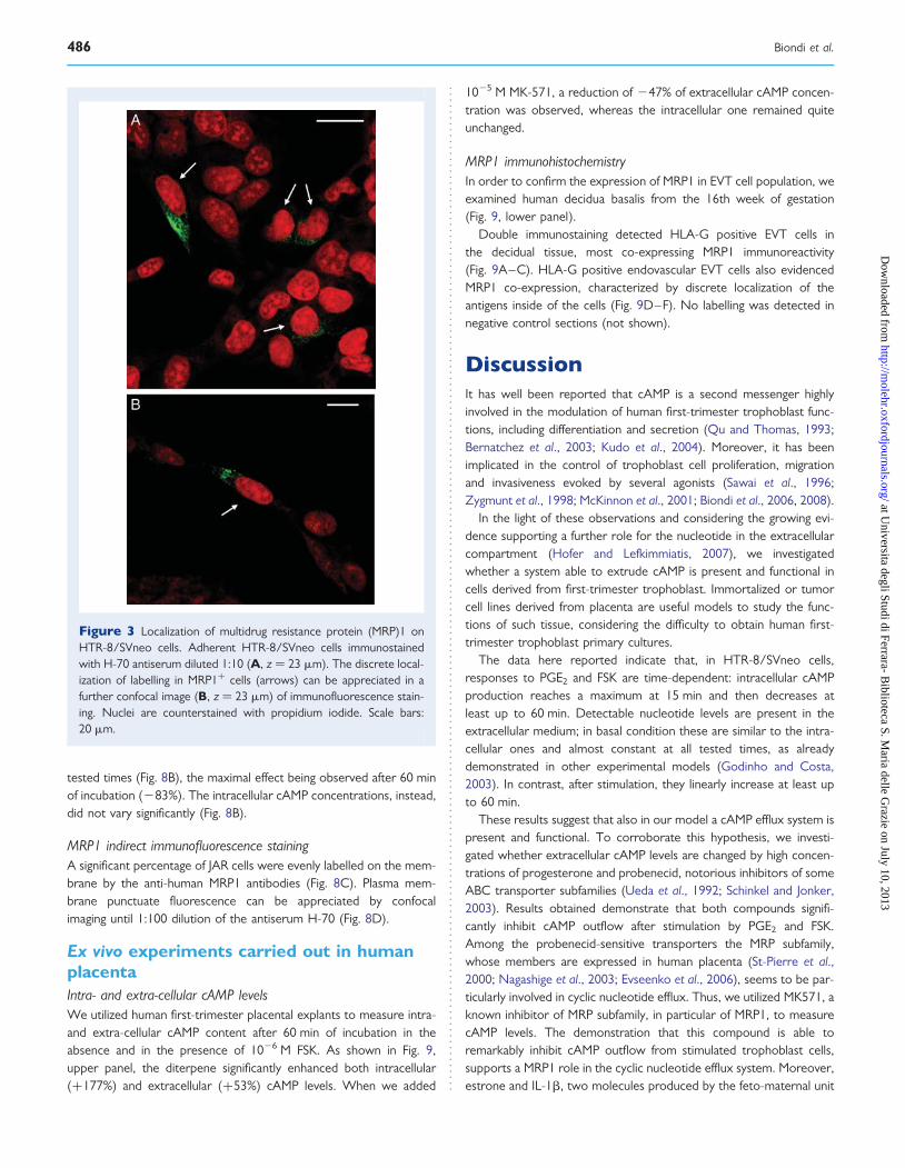
Indirect immunofluorescence staining of MRP1Cells were fixed and incubated with the antibodies raisedagainst the human MRP1. Confocal microscopy was used toexamine the plasma membrane versus intracellular localization ofthe immunostaining.
A percentage of HTR-8/SVneo cells were labelled on plasma mem-brane by the anti-human MRP1 antibodies (Fig. 3A). Interestingly,plasma membrane immunostaining was discretely localized in a poleof most positive cells (Fig. 3B).
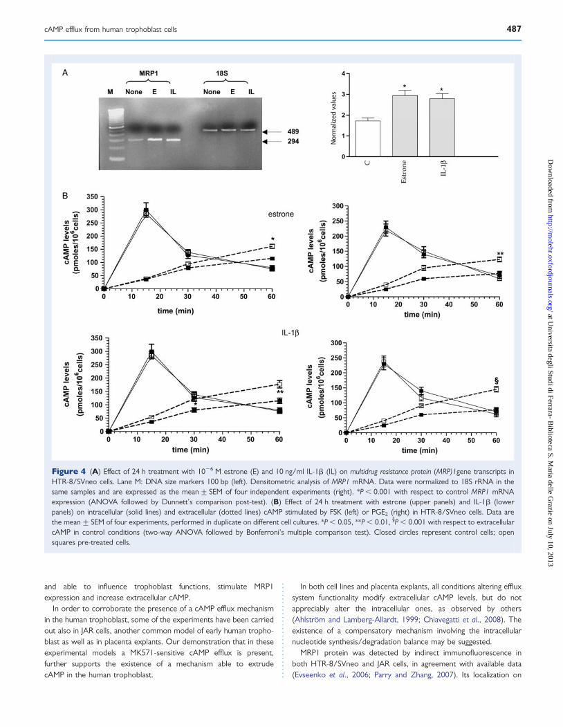
Effect of estrone and IL-1b treatmentSince it has been reported that mRNA expression for ABC transpor-ters is modulated by cytokines and steroid hormones in human tro-phoblast (Evseenko et al., 2007; Keating et al., 2007), we tested byRT–PCR the effect of a 24 h treatment with 1026 M estrone and10 ng/ml IL-1b, on MRP1 gene transcripts. Both estrone and IL-1bsignificantly enhanced (+61% and +55%, respectively, P , 0.001)the expression of MRP1 mRNA (Fig. 4A).
The effect of the same treatments was investigated also on intra-and extra-cellular cAMP levels, stimulated by 1026 M FSK or PGE2
(Fig. 4B). In both instances, the extracellular cAMP levels significantlyincreased after 60 min of treatment with estrone (+53% and +88%in the presence of FSK and PGE2, respectively) and IL-1b (+39%and +60% in the presence of FSK and PGE2, respectively). In allcases, the intracellular cAMP levels did not change significantly.
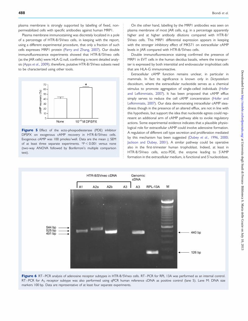
Analysis of exogenous cAMP degradationTo verify whether extracellular cAMP is metabolized by trophoblastcells, we added exogenous cAMP (100 pmoles/well) to the cellmedium in the absence and in the presence of 1024 M DPSPX, aselective inhibitor of ecto-PDE (Chiavegatti et al., 2008); in the col-lected medium we then determined cAMP level recovery after15 min of incubation. As shown in Fig. 5, in the absence of the druga recovery of about 40% was obtained. This value significantlyenhanced in the presence of DPSPX.
Figure 1 Time courses of intracellular (solid lines) and extracellular(dotted lines) cAMP levels in HTR-8/SVneo cells. (A) Cells incubatedwith 1026 M prostaglandin (PG)E2; (B) Cells incubated with 1026 Mforskolin (FSK). Data are the mean+ SEM of at least four exper-iments, performed in duplicate on different cell cultures.
484 Biondi et al.
at Universita degli Studi di Ferrara- B
iblioteca S. Maria delle G
razie on July 10, 2013http://m
olehr.oxfordjournals.org/D
ownloaded from

Analysis of adenosine receptor expression and functionalityEvaluating the presence of mRNAs for adenosine receptor subtypes,we found transcripts for A1, A2a, A2b, but not for A3 receptor. Thecommercially available human reference cDNA, used as positivecontrol for A3 receptor, showed a band of 440 bp corresponding tothe amplification product of interest, under conditions used forHTR-8/SVneo cells (Fig. 6).
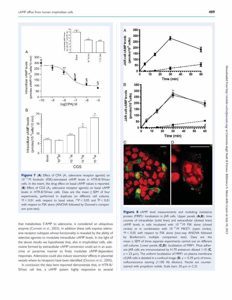
In order to verify the receptor functionality, we measured cAMPlevels in cells incubated in the absence or in the presence ofstable and selective agonists for the different adenosine receptorsubtypes. Since IBMX is a known adenosine receptor antagonist(Wu et al., 1982), we used 1024 M Ro 20-1724 as cAMP PDE inhibi-tor. In Fig. 7 the dose–response curves for CPA and CGS 21 680,selective A1 and A2 receptor agonists respectively, are reported.CPA dose dependently inhibited basal and FSK-stimulated intracellu-lar cAMP (Fig. 7A). Its effect on FSK-stimulated cAMP levels reachedstatistical significance at 1028 M and became maximal (274%) at1025 M. On the contrary, CGS 21 680 dose dependently stimulated
the nucleotide production and its effect became statistically signifi-cant (+88%) at 1026 M (Fig. 7B). Coherently with the PCRresults, the A3 receptor agonist, AB-MECA, unaltered cAMP pro-duction (not shown).
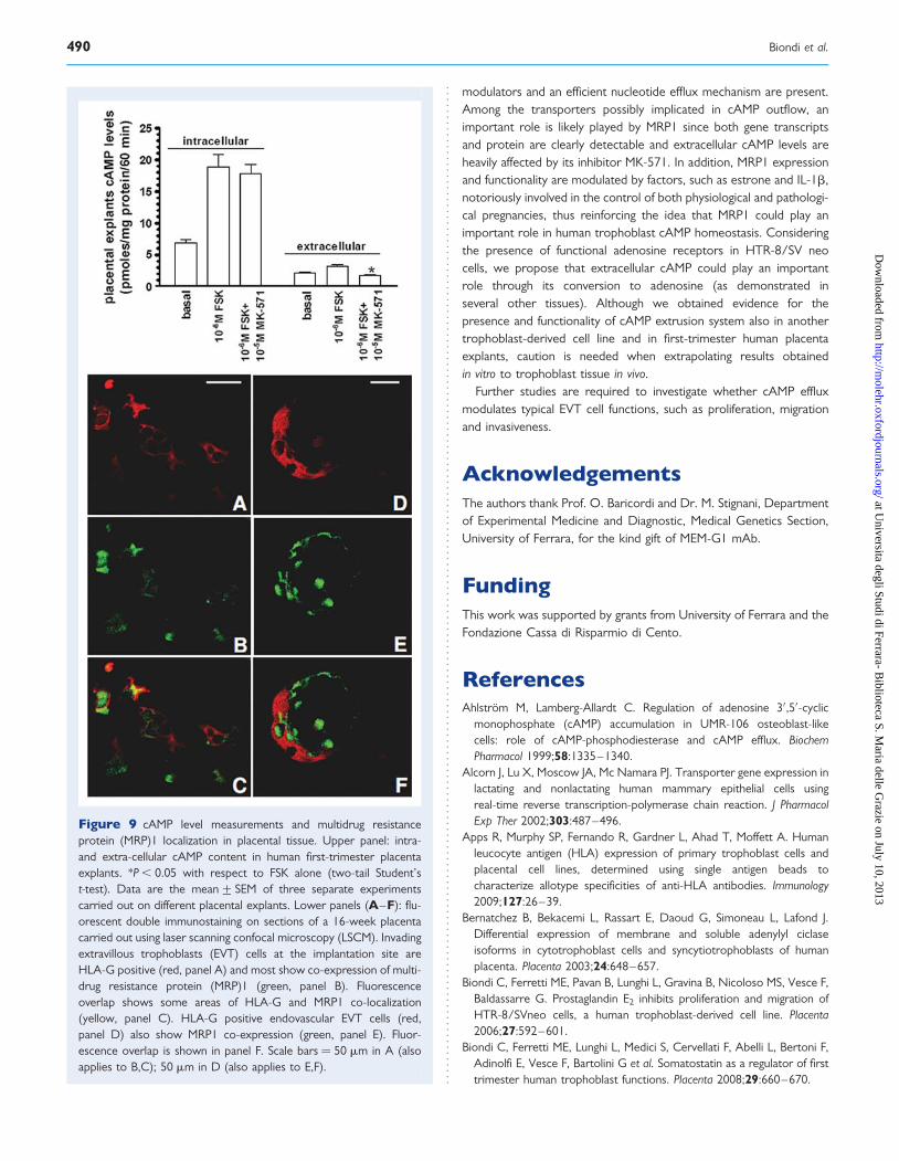
In vitro experiments carried out in JAR cellsIntra- and extra-cellular cAMP levelsIn JAR cells incubated up to 60 min, the basal intracellular cAMP levelsdid not significantly vary (around 9.0 pmoles/106 cells, not shown); inaddition, 1026 M FSK induced a stimulation reaching a maximum at30 min (31-fold). The extracellular cAMP levels did not change signifi-cantly in basal conditions (not shown), whereas they linearly increasedup to 60 min following FSK stimulation (Fig. 8A).
In cells incubated for 60 min with 1026 M FSK, extracellular cAMPlevels decreased in the presence of MK-571 doses varying between1026 and 1024 M the maximal effect being observed at 1025 M(not shown). When we used 1025 M MK-571 in time-course exper-iments, an evident reduction of extracellular cAMP was found at all
Figure 2 Effect of cAMP efflux inhibitors in HTR-8/SVneo cells. Upper: effects of progesterone (P2) or probenecid (Prob) on forskolin(FSK)-stimulated (A) or prostaglandin (PG)E2-stimulated (B) extracellular cAMP levels in cells incubated for 60 min. Data are the mean+ SEM ofat least four experiments, performed in duplicate on different cell cultures. *P , 0.01 with respect to FSK or PGE2 alone (ANOVA followed byDunnett’s comparison post-test). Bottom: effect of MK-571 on intracellular (solid lines) and extracellular (dotted lines) cAMP levels. (C) Cells incu-bated with 1026 M FSK; (D) Cells incubated with 1026 M PGE2. Closed squares represent 1026 M PGE2 alone; open squares represent 1026 MPGE2+1025 M MK571; closed circles 1026 M FSK alone; open circles 1026 M FSK + 1025 M MK571. Data are the mean+ SEM of at least fourexperiments, performed in duplicate on different cell cultures. *P , 0.05 with respect to PGE2 or FSK alone; **P , 0.01 with respect to PGE2
alone (two-way ANOVA followed by Bonferroni’s multiple comparison test).
cAMP efflux from human trophoblast cells 485
at Universita degli Studi di Ferrara- B
iblioteca S. Maria delle G
razie on July 10, 2013http://m
olehr.oxfordjournals.org/D
ownloaded from
tested times (Fig. 8B), the maximal effect being observed after 60 minof incubation (283%). The intracellular cAMP concentrations, instead,did not vary significantly (Fig. 8B).
MRP1 indirect immunofluorescence stainingA significant percentage of JAR cells were evenly labelled on the mem-brane by the anti-human MRP1 antibodies (Fig. 8C). Plasma mem-brane punctuate fluorescence can be appreciated by confocalimaging until 1:100 dilution of the antiserum H-70 (Fig. 8D).
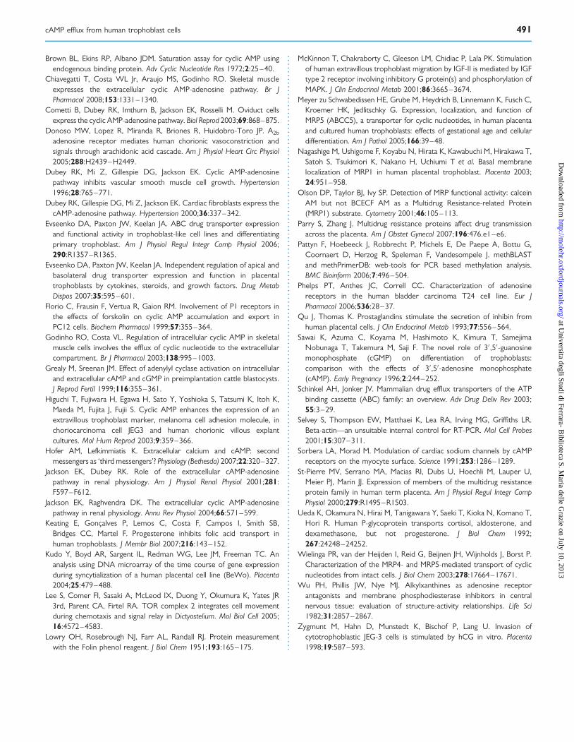
Ex vivo experiments carried out in humanplacentaIntra- and extra-cellular cAMP levelsWe utilized human first-trimester placental explants to measure intra-and extra-cellular cAMP content after 60 min of incubation in theabsence and in the presence of 1026 M FSK. As shown in Fig. 9,upper panel, the diterpene significantly enhanced both intracellular(+177%) and extracellular (+53%) cAMP levels. When we added
1025 M MK-571, a reduction of 247% of extracellular cAMP concen-tration was observed, whereas the intracellular one remained quiteunchanged.
MRP1 immunohistochemistryIn order to confirm the expression of MRP1 in EVT cell population, weexamined human decidua basalis from the 16th week of gestation(Fig. 9, lower panel).
Double immunostaining detected HLA-G positive EVT cells inthe decidual tissue, most co-expressing MRP1 immunoreactivity(Fig. 9A–C). HLA-G positive endovascular EVT cells also evidencedMRP1 co-expression, characterized by discrete localization of theantigens inside of the cells (Fig. 9D–F). No labelling was detected innegative control sections (not shown).
DiscussionIt has well been reported that cAMP is a second messenger highlyinvolved in the modulation of human first-trimester trophoblast func-tions, including differentiation and secretion (Qu and Thomas, 1993;Bernatchez et al., 2003; Kudo et al., 2004). Moreover, it has beenimplicated in the control of trophoblast cell proliferation, migrationand invasiveness evoked by several agonists (Sawai et al., 1996;Zygmunt et al., 1998; McKinnon et al., 2001; Biondi et al., 2006, 2008).
In the light of these observations and considering the growing evi-dence supporting a further role for the nucleotide in the extracellularcompartment (Hofer and Lefkimmiatis, 2007), we investigatedwhether a system able to extrude cAMP is present and functional incells derived from first-trimester trophoblast. Immortalized or tumorcell lines derived from placenta are useful models to study the func-tions of such tissue, considering the difficulty to obtain human first-trimester trophoblast primary cultures.
The data here reported indicate that, in HTR-8/SVneo cells,responses to PGE2 and FSK are time-dependent: intracellular cAMPproduction reaches a maximum at 15 min and then decreases atleast up to 60 min. Detectable nucleotide levels are present in theextracellular medium; in basal condition these are similar to the intra-cellular ones and almost constant at all tested times, as alreadydemonstrated in other experimental models (Godinho and Costa,2003). In contrast, after stimulation, they linearly increase at least upto 60 min.
These results suggest that also in our model a cAMP efflux system ispresent and functional. To corroborate this hypothesis, we investi-gated whether extracellular cAMP levels are changed by high concen-trations of progesterone and probenecid, notorious inhibitors of someABC transporter subfamilies (Ueda et al., 1992; Schinkel and Jonker,2003). Results obtained demonstrate that both compounds signifi-cantly inhibit cAMP outflow after stimulation by PGE2 and FSK.Among the probenecid-sensitive transporters the MRP subfamily,whose members are expressed in human placenta (St-Pierre et al.,2000; Nagashige et al., 2003; Evseenko et al., 2006), seems to be par-ticularly involved in cyclic nucleotide efflux. Thus, we utilized MK571, aknown inhibitor of MRP subfamily, in particular of MRP1, to measurecAMP levels. The demonstration that this compound is able toremarkably inhibit cAMP outflow from stimulated trophoblast cells,supports a MRP1 role in the cyclic nucleotide efflux system. Moreover,estrone and IL-1b, two molecules produced by the feto-maternal unit
Figure 3 Localization of multidrug resistance protein (MRP)1 onHTR-8/SVneo cells. Adherent HTR-8/SVneo cells immunostainedwith H-70 antiserum diluted 1:10 (A, z ¼ 23 mm). The discrete local-ization of labelling in MRP1+ cells (arrows) can be appreciated in afurther confocal image (B, z ¼ 23 mm) of immunofluorescence stain-ing. Nuclei are counterstained with propidium iodide. Scale bars:20 mm.
486 Biondi et al.
at Universita degli Studi di Ferrara- B
iblioteca S. Maria delle G
razie on July 10, 2013http://m
olehr.oxfordjournals.org/D
ownloaded from
and able to influence trophoblast functions, stimulate MRP1expression and increase extracellular cAMP.
In order to corroborate the presence of a cAMP efflux mechanismin the human trophoblast, some of the experiments have been carriedout also in JAR cells, another common model of early human tropho-blast as well as in placenta explants. Our demonstration that in theseexperimental models a MK571-sensitive cAMP efflux is present,further supports the existence of a mechanism able to extrudecAMP in the human trophoblast.
In both cell lines and placenta explants, all conditions altering effluxsystem functionality modify extracellular cAMP levels, but do notappreciably alter the intracellular ones, as observed by others(Ahlstrom and Lamberg-Allardt, 1999; Chiavegatti et al., 2008). Theexistence of a compensatory mechanism involving the intracellularnucleotide synthesis/degradation balance may be suggested.
MRP1 protein was detected by indirect immunofluorescence inboth HTR-8/SVneo and JAR cells, in agreement with available data(Evseenko et al., 2006; Parry and Zhang, 2007). Its localization on
Figure 4 (A) Effect of 24 h treatment with 1026 M estrone (E) and 10 ng/ml IL-1b (IL) on multidrug resistance protein (MRP)1gene transcripts inHTR-8/SVneo cells. Lane M: DNA size markers 100 bp (left). Densitometric analysis of MRP1 mRNA. Data were normalized to 18S rRNA in thesame samples and are expressed as the mean+ SEM of four independent experiments (right). *P , 0.001 with respect to control MRP1 mRNAexpression (ANOVA followed by Dunnett’s comparison post-test). (B) Effect of 24 h treatment with estrone (upper panels) and IL-1b (lowerpanels) on intracellular (solid lines) and extracellular (dotted lines) cAMP stimulated by FSK (left) or PGE2 (right) in HTR-8/SVneo cells. Data arethe mean+ SEM of four experiments, performed in duplicate on different cell cultures. *P , 0.05, **P , 0.01, §P , 0.001 with respect to extracellularcAMP in control conditions (two-way ANOVA followed by Bonferroni’s multiple comparison test). Closed circles represent control cells; opensquares pre-treated cells.
cAMP efflux from human trophoblast cells 487
at Universita degli Studi di Ferrara- B
iblioteca S. Maria delle G
razie on July 10, 2013http://m
olehr.oxfordjournals.org/D
ownloaded from
plasma membrane is strongly supported by labelling of fixed, non-permeabilized cells with specific antibodies against human MRP1.
Plasma membrane immunostaining was discretely localized in a poleof a percentage of HTR-8/SVneo cells, in keeping with the report,using a different experimental procedure, that only a fraction of suchcells expresses MRP1 protein (Parry and Zhang, 2007). Our doubleimmunofluorescence experiments showed that HTR-8/SVneo cells(as the JAR cells) were HLA-G null, confirming a recent detailed analy-sis (Apps et al., 2009); therefore, putative HTR-8/SVneo subsets needto be characterized using other tools.
On the other hand, labelling by the MRP1 antibodies was seen onplasma membrane of most JAR cells, e.g. in a percentage apparentlyhigher and at higher antibody dilutions compared with HTR-8/SVneo cells. This MRP1 differential expression appears in keepingwith the stronger inhibitory effect of MK571 on extracellular cAMPlevels in JAR compared with HTR-8/SVneo cells.
Double immunofluorescence staining confirmed the presence ofMRP1 in EVT cells in the human decidua basalis, where the transpor-ter is expressed by both interstitial and endovascular trophoblast cellsthat are HLA-G immunoreactive.
Extracellular cAMP function remains unclear, in particular inmammals. In fact its significance is known only in Dictyosteliumdiscoideum, where the extracellular nucleotide serves as a chemicalstimulus to promote aggregation of single-celled individuals (Hoferand Lefkimmiatis, 2007). It has been proposed that cAMP effluxsimply serves to reduce the cell cAMP concentration (Hofer andLefkimmiatis, 2007). Our data demonstrating intracellular cAMP stea-diness though in the presence of an altered efflux, are not in line withthis hypothesis, but support the idea that nucleotide egress could rep-resent an additional arm of cAMP pathway able to evoke regulatoryactions. Some experimental evidence indicates that a plausible physio-logical role for extracellular cAMP could involve adenosine formation.A regulation of different cell type secretion and proliferation mediatedby this mechanism has been suggested (Dubey et al., 1996, 2000;Jackson and Dubey, 2001). A similar pathway could be operativealso in the first-trimester human trophoblast. Indeed, at least inHTR-8/SVneo cells, ecto-PDE, the enzyme leading to 5′AMPformation in the extracellular medium, is functional and 5′nucleotidase,
Figure 6 RT–PCR analysis of adenosine receptor subtypes in HTR-8/SVneo cells. RT–PCR for RPL 13A was performed as an internal control.RT–PCR for A3 receptor subtype was also performed using qPCR human reference cDNA as positive control (lane 5). Lane M: DNA sizemarkers 100 bp. Data are representative of at least four separate experiments.
Figure 5 Effect of the ecto-phospodiesterase (PDE) inhibitorDPSPX on exogenous cAMP recovery in HTR-8/SVneo cells.Exogenous cAMP was 100 pmoles/well. Data are the mean+ SEMof at least three separate experiments. *P , 0.001 versus none(two-way ANOVA followed by Bonferroni’s multiple comparisontest).
488 Biondi et al.
at Universita degli Studi di Ferrara- B
iblioteca S. Maria delle G
razie on July 10, 2013http://m
olehr.oxfordjournals.org/D
ownloaded from
that metabolizes 5′AMP to adenosine, is considered an ubiquitousenzyme (Cometti et al., 2003). In addition these cells express adeno-sine receptor subtypes whose functionality is revealed by the ability ofselective agonists to modulate intracellular cAMP levels. In the light ofthe above results we hypothesize that, also in trophoblast cells, ade-nosine formed by extracellular cAMP conversion could act in an auto-crine or paracrine manner to finely modulate cAMP-dependentresponses. Adenosine could also induce vasomotor effects in placentalvessels where its receptors have been identified (Donoso et al., 2005).
In conclusion the data here reported demonstrate that, in HTR-8/SVneo cell line, a cAMP system highly responsive to several
Figure 7 (A) Effect of CPA (A1 adenosine receptor agonist) on1025 M forskolin (FSK)-stimulated cAMP levels in HTR-8/SVneocells. In the insert, the drug effect on basal cAMP values is reported.(B) Effect of CGS (A2 adenosine receptor agonist) on basal cAMPlevels in HTR-8/SVneo cells. Data are the mean+ SEM of fourexperiments, performed in duplicate on different cell cultures.*P , 0.01 with respect to basal value; **P , 0.05 and §P , 0.01with respect to FSK alone (ANOVA followed by Dunnett’s compari-son post-test).
Figure 8 cAMP level measurements and multidrug resistanceprotein (MRP)1 localization in JAR cells. Upper panels (A,B): timecourses of intracellular (solid lines) and extracellular (dotted lines)cAMP levels in cells incubated with 1026 M FSK alone (closedcircles) or in combination with 1025 M MK571 (open circles).*P , 0.05 with respect to FSK alone (two-way ANOVA followedby Bonferroni’s multiple comparison test). Data are themean+ SEM of three separate experiments carried out on differentcell cultures. Lower panels (C,D): localization of MRP1. Most adher-ent JAR cells are immunostained by H-70 antiserum diluted 1:10 (C,z ¼ 23 mm). The uniform localization of MRP1 on plasma membraneof JAR cells is detailed in a confocal image (D, z ¼ 0.39 mm) of immu-nofluorescence staining (1:100 Ab dilution). Nuclei are counter-stained with propidium iodide. Scale bars: 20 mm in C,D.
cAMP efflux from human trophoblast cells 489
at Universita degli Studi di Ferrara- B
iblioteca S. Maria delle G
razie on July 10, 2013http://m
olehr.oxfordjournals.org/D
ownloaded from
modulators and an efficient nucleotide efflux mechanism are present.Among the transporters possibly implicated in cAMP outflow, animportant role is likely played by MRP1 since both gene transcriptsand protein are clearly detectable and extracellular cAMP levels areheavily affected by its inhibitor MK-571. In addition, MRP1 expressionand functionality are modulated by factors, such as estrone and IL-1b,notoriously involved in the control of both physiological and pathologi-cal pregnancies, thus reinforcing the idea that MRP1 could play animportant role in human trophoblast cAMP homeostasis. Consideringthe presence of functional adenosine receptors in HTR-8/SV neocells, we propose that extracellular cAMP could play an importantrole through its conversion to adenosine (as demonstrated inseveral other tissues). Although we obtained evidence for thepresence and functionality of cAMP extrusion system also in anothertrophoblast-derived cell line and in first-trimester human placentaexplants, caution is needed when extrapolating results obtainedin vitro to trophoblast tissue in vivo.
Further studies are required to investigate whether cAMP effluxmodulates typical EVT cell functions, such as proliferation, migrationand invasiveness.
AcknowledgementsThe authors thank Prof. O. Baricordi and Dr. M. Stignani, Departmentof Experimental Medicine and Diagnostic, Medical Genetics Section,University of Ferrara, for the kind gift of MEM-G1 mAb.
FundingThis work was supported by grants from University of Ferrara and theFondazione Cassa di Risparmio di Cento.
ReferencesAhlstrom M, Lamberg-Allardt C. Regulation of adenosine 3′,5′-cyclic
monophosphate (cAMP) accumulation in UMR-106 osteoblast-likecells: role of cAMP-phosphodiesterase and cAMP efflux. BiochemPharmacol 1999;58:1335–1340.
Alcorn J, Lu X, Moscow JA, Mc Namara PJ. Transporter gene expression inlactating and nonlactating human mammary epithelial cells usingreal-time reverse transcription-polymerase chain reaction. J PharmacolExp Ther 2002;303:487–496.
Apps R, Murphy SP, Fernando R, Gardner L, Ahad T, Moffett A. Humanleucocyte antigen (HLA) expression of primary trophoblast cells andplacental cell lines, determined using single antigen beads tocharacterize allotype specificities of anti-HLA antibodies. Immunology2009;127:26–39.
Bernatchez B, Bekacemi L, Rassart E, Daoud G, Simoneau L, Lafond J.Differential expression of membrane and soluble adenylyl ciclaseisoforms in cytotrophoblast cells and syncytiotrophoblasts of humanplacenta. Placenta 2003;24:648–657.
Biondi C, Ferretti ME, Pavan B, Lunghi L, Gravina B, Nicoloso MS, Vesce F,Baldassarre G. Prostaglandin E2 inhibits proliferation and migration ofHTR-8/SVneo cells, a human trophoblast-derived cell line. Placenta2006;27:592–601.
Biondi C, Ferretti ME, Lunghi L, Medici S, Cervellati F, Abelli L, Bertoni F,Adinolfi E, Vesce F, Bartolini G et al. Somatostatin as a regulator of firsttrimester human trophoblast functions. Placenta 2008;29:660–670.
Figure 9 cAMP level measurements and multidrug resistanceprotein (MRP)1 localization in placental tissue. Upper panel: intra-and extra-cellular cAMP content in human first-trimester placentaexplants. *P , 0.05 with respect to FSK alone (two-tail Student’st-test). Data are the mean+ SEM of three separate experimentscarried out on different placental explants. Lower panels (A–F): flu-orescent double immunostaining on sections of a 16-week placentacarried out using laser scanning confocal microscopy (LSCM). Invadingextravillous trophoblasts (EVT) cells at the implantation site areHLA-G positive (red, panel A) and most show co-expression of multi-drug resistance protein (MRP)1 (green, panel B). Fluorescenceoverlap shows some areas of HLA-G and MRP1 co-localization(yellow, panel C). HLA-G positive endovascular EVT cells (red,panel D) also show MRP1 co-expression (green, panel E). Fluor-escence overlap is shown in panel F. Scale bars ¼ 50 mm in A (alsoapplies to B,C); 50 mm in D (also applies to E,F).
490 Biondi et al.
at Universita degli Studi di Ferrara- B
iblioteca S. Maria delle G
razie on July 10, 2013http://m
olehr.oxfordjournals.org/D
ownloaded from
Brown BL, Ekins RP, Albano JDM. Saturation assay for cyclic AMP usingendogenous binding protein. Adv Cyclic Nucleotide Res 1972;2:25–40.
Chiavegatti T, Costa WL Jr, Araujo MS, Godinho RO. Skeletal muscleexpresses the extracellular cyclic AMP-adenosine pathway. Br JPharmacol 2008;153:1331–1340.
Cometti B, Dubey RK, Imthurn B, Jackson EK, Rosselli M. Oviduct cellsexpress the cyclic AMP-adenosine pathway. Biol Reprod 2003;69:868–875.
Donoso MW, Lopez R, Miranda R, Briones R, Huidobro-Toro JP. A2b
adenosine receptor mediates human chorionic vasoconstriction andsignals through arachidonic acid cascade. Am J Physiol Heart Circ Physiol2005;288:H2439–H2449.
Dubey RK, Mi Z, Gillespie DG, Jackson EK. Cyclic AMP-adenosinepathway inhibits vascular smooth muscle cell growth. Hypertension1996;28:765–771.
Dubey RK, Gillespie DG, Mi Z, Jackson EK. Cardiac fibroblasts express thecAMP-adenosine pathway. Hypertension 2000;36:337–342.
Evseenko DA, Paxton JW, Keelan JA. ABC drug transporter expressionand functional activity in trophoblast-like cell lines and differentiatingprimary trophoblast. Am J Physiol Regul Integr Comp Physiol 2006;290:R1357–R1365.
Evseenko DA, Paxton JW, Keelan JA. Independent regulation of apical andbasolateral drug transporter expression and function in placentaltrophoblasts by cytokines, steroids, and growth factors. Drug MetabDispos 2007;35:595–601.
Florio C, Frausin F, Vertua R, Gaion RM. Involvement of P1 receptors inthe effects of forskolin on cyclic AMP accumulation and export inPC12 cells. Biochem Pharmacol 1999;57:355–364.
Godinho RO, Costa VL. Regulation of intracellular cyclic AMP in skeletalmuscle cells involves the efflux of cyclic nucleotide to the extracellularcompartment. Br J Pharmacol 2003;138:995–1003.
Grealy M, Sreenan JM. Effect of adenylyl cyclase activation on intracellularand extracellular cAMP and cGMP in preimplantation cattle blastocysts.J Reprod Fertil 1999;116:355–361.
Higuchi T, Fujiwara H, Egawa H, Sato Y, Yoshioka S, Tatsumi K, Itoh K,Maeda M, Fujita J, Fujii S. Cyclic AMP enhances the expression of anextravillous trophoblast marker, melanoma cell adhesion molecule, inchoriocarcinoma cell JEG3 and human chorionic villous explantcultures. Mol Hum Reprod 2003;9:359–366.
Hofer AM, Lefkimmiatis K. Extracellular calcium and cAMP: secondmessengers as ‘third messengers’? Physiology (Bethesda) 2007;22:320–327.
Jackson EK, Dubey RK. Role of the extracellular cAMP-adenosinepathway in renal physiology. Am J Physiol Renal Physiol 2001;281:F597–F612.
Jackson EK, Raghvendra DK. The extracellular cyclic AMP-adenosinepathway in renal physiology. Annu Rev Physiol 2004;66:571–599.
Keating E, Goncalves P, Lemos C, Costa F, Campos I, Smith SB,Bridges CC, Martel F. Progesterone inhibits folic acid transport inhuman trophoblasts. J Membr Biol 2007;216:143–152.
Kudo Y, Boyd AR, Sargent IL, Redman WG, Lee JM, Freeman TC. Ananalysis using DNA microarray of the time course of gene expressionduring syncytialization of a human placental cell line (BeWo). Placenta2004;25:479–488.
Lee S, Comer FI, Sasaki A, McLeod IX, Duong Y, Okumura K, Yates JR3rd, Parent CA, Firtel RA. TOR complex 2 integrates cell movementduring chemotaxis and signal relay in Dictyostelium. Mol Biol Cell 2005;16:4572–4583.
Lowry OH, Rosebrough NJ, Farr AL, Randall RJ. Protein measurementwith the Folin phenol reagent. J Biol Chem 1951;193:165–175.
McKinnon T, Chakraborty C, Gleeson LM, Chidiac P, Lala PK. Stimulationof human extravillous trophoblast migration by IGF-II is mediated by IGFtype 2 receptor involving inhibitory G protein(s) and phosphorylation ofMAPK. J Clin Endocrinol Metab 2001;86:3665–3674.
Meyer zu Schwabedissen HE, Grube M, Heydrich B, Linnemann K, Fusch C,Kroemer HK, Jedlitschky G. Expression, localization, and function ofMRP5 (ABCC5), a transporter for cyclic nucleotides, in human placentaand cultured human trophoblasts: effects of gestational age and cellulardifferentiation. Am J Pathol 2005;166:39–48.
Nagashige M, Ushigome F, Koyabu N, Hirata K, Kawabuchi M, Hirakawa T,Satoh S, Tsukimori K, Nakano H, Uchiumi T et al. Basal membranelocalization of MRP1 in human placental trophoblast. Placenta 2003;24:951–958.
Olson DP, Taylor BJ, Ivy SP. Detection of MRP functional activity: calceinAM but not BCECF AM as a Multidrug Resistance-related Protein(MRP1) substrate. Cytometry 2001;46:105–113.
Parry S, Zhang J. Multidrug resistance proteins affect drug transmissionacross the placenta. Am J Obstet Gynecol 2007;196:476.e1–e6.
Pattyn F, Hoebeeck J, Robbrecht P, Michels E, De Paepe A, Bottu G,Coornaert D, Herzog R, Speleman F, Vandesompele J. methBLASTand methPrimerDB: web-tools for PCR based methylation analysis.BMC Bioinform 2006;7:496–504.
Phelps PT, Anthes JC, Correll CC. Characterization of adenosinereceptors in the human bladder carcinoma T24 cell line. Eur JPharmacol 2006;536:28–37.
Qu J, Thomas K. Prostaglandins stimulate the secretion of inhibin fromhuman placental cells. J Clin Endocrinol Metab 1993;77:556–564.
Sawai K, Azuma C, Koyama M, Hashimoto K, Kimura T, SamejimaNobunaga T, Takemura M, Saji F. The novel role of 3′,5′-guanosinemonophosphate (cGMP) on differentiation of trophoblasts:comparison with the effects of 3′,5′-adenosine monophosphate(cAMP). Early Pregnancy 1996;2:244–252.
Schinkel AH, Jonker JV. Mammalian drug efflux transporters of the ATPbinding cassette (ABC) family: an overview. Adv Drug Deliv Rev 2003;55:3–29.
Selvey S, Thompson EW, Matthaei K, Lea RA, Irving MG, Griffiths LR.Beta-actin—an unsuitable internal control for RT-PCR. Mol Cell Probes2001;15:307–311.
Sorbera LA, Morad M. Modulation of cardiac sodium channels by cAMPreceptors on the myocyte surface. Science 1991;253:1286–1289.
St-Pierre MV, Serrano MA, Macias RI, Dubs U, Hoechli M, Lauper U,Meier PJ, Marin JJ. Expression of members of the multidrug resistanceprotein family in human term placenta. Am J Physiol Regul Integr CompPhysiol 2000;279:R1495–R1503.
Ueda K, Okamura N, Hirai M, Tanigawara Y, Saeki T, Kioka N, Komano T,Hori R. Human P-glycoprotein transports cortisol, aldosterone, anddexamethasone, but not progesterone. J Biol Chem 1992;267:24248–24252.
Wielinga PR, van der Heijden I, Reid G, Beijnen JH, Wijnholds J, Borst P.Characterization of the MRP4- and MRP5-mediated transport of cyclicnucleotides from intact cells. J Biol Chem 2003;278:17664–17671.
Wu PH, Phillis JW, Nye MJ. Alkylxanthines as adenosine receptorantagonists and membrane phosphodiesterase inhibitors in centralnervous tissue: evaluation of structure-activity relationships. Life Sci1982;31:2857–2867.
Zygmunt M, Hahn D, Munstedt K, Bischof P, Lang U. Invasion ofcytotrophoblastic JEG-3 cells is stimulated by hCG in vitro. Placenta1998;19:587–593.
cAMP efflux from human trophoblast cells 491
at Universita degli Studi di Ferrara- B
iblioteca S. Maria delle G
razie on July 10, 2013http://m
olehr.oxfordjournals.org/D
ownloaded from
Related Documents