elifesciences.org RESEARCH ARTICLE Calcium specificity signaling mechanisms in abscisic acid signal transduction in Arabidopsis guard cells Benjamin Brandt †‡ , Shintaro Munemasa †§ , Cun Wang, Desiree Nguyen, Taiming Yong, Paul G Yang, Elly Poretsky, Thomas F Belknap, Rainer Waadt, Fernando Alem ´ an, Julian I Schroeder* Division of Biological Sciences, Cell and Developmental Biology Section, University of California, San Diego, San Diego, United States Abstract A central question is how specificity in cellular responses to the eukaryotic second messenger Ca 2+ is achieved. Plant guard cells, that form stomatal pores for gas exchange, provide a powerful system for in depth investigation of Ca 2+ -signaling specificity in plants. In intact guard cells, abscisic acid (ABA) enhances (primes) the Ca 2+ -sensitivity of downstream signaling events that result in activation of S-type anion channels during stomatal closure, providing a specificity mechanism in Ca 2+ -signaling. However, the underlying genetic and biochemical mechanisms remain unknown. Here we show impairment of ABA signal transduction in stomata of calcium-dependent protein kinase quadruple mutant plants. Interestingly, protein phosphatase 2Cs prevent non-specific Ca 2+ -signaling. Moreover, we demonstrate an unexpected interdependence of the Ca 2+ -dependent and Ca 2+ -independent ABA-signaling branches and the in planta requirement of simultaneous phosphorylation at two key phosphorylation sites in SLAC1. We identify novel mechanisms ensuring specificity and robustness within stomatal Ca 2+ -signaling on a cellular, genetic, and biochemical level. DOI: 10.7554/eLife.03599.001 Introduction Cytosolic calcium ([Ca 2+ ] cyt ) functions as key cellular second messenger in a plethora of crucial processes in plants and other eukaryotes (Hetherington and Woodward, 2003; Clapham, 2007; McAinsh and Pittman, 2009; Berridge, 2012; Charpentier and Oldroyd, 2013; Webb, 2013). Elucidation of the mechanisms mediating specificity in Ca 2+ signaling is fundamental to understanding signal transduction (Berridge et al., 2003; Hetherington and Woodward, 2003; Clapham, 2007; Webb, 2013). In a few cases, the biochemical and cellular mechanisms mediating Ca 2+ signaling specificity have been revealed (e.g. De Koninck and Schulman, 1998; Dolmetsch et al., 1998; Oancea and Meyer, 1998; Dolmetsch et al., 2001; Bradshaw et al., 2003; Rellos et al., 2010; Chao et al., 2011). More than one (non- exclusive) mechanism is likely to contribute to specificity in Ca 2+ signal transduction (Berridge et al., 2003; Dodd et al., 2010). However, characterization of the combined cellular, biochemical, and genetic mechanisms underlying Ca 2+ specificity in a single cell type has not been achieved to our knowledge. The genome of the plant Arabidopsis thaliana encodes over 200 EF-hand Ca 2+ -binding proteins (Day et al., 2002), with many of these genes co-expressed in the same cell types (Harmon et al., 2000; McCormack et al., 2005; Schmid et al., 2005; Winter et al., 2007), illustrating the need for Ca 2+ specificity signaling mechanisms in plants. Two guard cells form a stomatal pore representing the gateway for CO 2 influx, which is inevitably accompanied by plant water loss. The aperture of stomatal pores is consequently tightly regulated by the guard cells. Intracellular Ca 2+ represents a key second messenger in stomatal closing (McAinsh et al., 1990; MacRobbie, 2000; Hetherington, 2001; Hetherington and Woodward, 2003; Hubbard et al., 2012), but intracellular Ca 2+ also functions in *For correspondence: [email protected] † These authors contributed equally to this work Present address: ‡ Structural Plant Biology Laboratory, Department for Botany and Plant Biology, University of Geneva, Geneva, Switzerland; § Graduate School of Environmental and Life Science, Okayama University, Okayama, Japan Competing interests: The authors declare that no competing interests exist. Funding: See page 20 Received: 05 June 2014 Accepted: 18 June 2015 Published: 20 July 2015 Reviewing editor: Detlef Weigel, Max Planck Institute for Developmental Biology, Germany Copyright Brandt et al. This article is distributed under the terms of the Creative Commons Attribution License, which permits unrestricted use and redistribution provided that the original author and source are credited. Brandt et al. eLife 2015;4:e03599. DOI: 10.7554/eLife.03599 1 of 25

Welcome message from author
This document is posted to help you gain knowledge. Please leave a comment to let me know what you think about it! Share it to your friends and learn new things together.
Transcript

elifesciences.org
RESEARCH ARTICLE
Calcium specificity signaling mechanismsin abscisic acid signal transduction inArabidopsis guard cellsBenjamin Brandt†‡, Shintaro Munemasa†§, Cun Wang, Desiree Nguyen,Taiming Yong, Paul G Yang, Elly Poretsky, Thomas F Belknap, Rainer Waadt,Fernando Aleman, Julian I Schroeder*
Division of Biological Sciences, Cell and Developmental Biology Section, University ofCalifornia, San Diego, San Diego, United States
Abstract A central question is how specificity in cellular responses to the eukaryotic second
messenger Ca2+ is achieved. Plant guard cells, that form stomatal pores for gas exchange, provide
a powerful system for in depth investigation of Ca2+-signaling specificity in plants. In intact guard
cells, abscisic acid (ABA) enhances (primes) the Ca2+-sensitivity of downstream signaling events that
result in activation of S-type anion channels during stomatal closure, providing a specificity
mechanism in Ca2+-signaling. However, the underlying genetic and biochemical mechanisms remain
unknown. Here we show impairment of ABA signal transduction in stomata of calcium-dependent
protein kinase quadruple mutant plants. Interestingly, protein phosphatase 2Cs prevent non-specific
Ca2+-signaling. Moreover, we demonstrate an unexpected interdependence of the Ca2+-dependent
and Ca2+-independent ABA-signaling branches and the in planta requirement of simultaneous
phosphorylation at two key phosphorylation sites in SLAC1. We identify novel mechanisms ensuring
specificity and robustness within stomatal Ca2+-signaling on a cellular, genetic, and biochemical level.
DOI: 10.7554/eLife.03599.001
IntroductionCytosolic calcium ([Ca2+]cyt) functions as key cellular second messenger in a plethora of crucial processes
in plants and other eukaryotes (Hetherington and Woodward, 2003; Clapham, 2007; McAinsh and
Pittman, 2009; Berridge, 2012; Charpentier and Oldroyd, 2013; Webb, 2013). Elucidation of the
mechanisms mediating specificity in Ca2+ signaling is fundamental to understanding signal transduction
(Berridge et al., 2003; Hetherington and Woodward, 2003; Clapham, 2007; Webb, 2013). In a few
cases, the biochemical and cellular mechanisms mediating Ca2+ signaling specificity have been revealed
(e.g. De Koninck and Schulman, 1998; Dolmetsch et al., 1998; Oancea and Meyer, 1998; Dolmetsch
et al., 2001; Bradshaw et al., 2003; Rellos et al., 2010; Chao et al., 2011). More than one (non-
exclusive) mechanism is likely to contribute to specificity in Ca2+ signal transduction (Berridge et al.,
2003; Dodd et al., 2010). However, characterization of the combined cellular, biochemical, and genetic
mechanisms underlying Ca2+ specificity in a single cell type has not been achieved to our knowledge.
The genome of the plant Arabidopsis thaliana encodes over 200 EF-hand Ca2+-binding proteins
(Day et al., 2002), with many of these genes co-expressed in the same cell types (Harmon et al.,
2000; McCormack et al., 2005; Schmid et al., 2005; Winter et al., 2007), illustrating the need for
Ca2+ specificity signaling mechanisms in plants. Two guard cells form a stomatal pore representing the
gateway for CO2 influx, which is inevitably accompanied by plant water loss. The aperture of stomatal
pores is consequently tightly regulated by the guard cells. Intracellular Ca2+ represents a key second
messenger in stomatal closing (McAinsh et al., 1990; MacRobbie, 2000; Hetherington, 2001;
Hetherington and Woodward, 2003; Hubbard et al., 2012), but intracellular Ca2+ also functions in
*For correspondence:
†These authors contributed
equally to this work
Present address: ‡Structural
Plant Biology Laboratory,
Department for Botany and Plant
Biology, University of Geneva,
Geneva, Switzerland; §Graduate
School of Environmental and Life
Science, Okayama University,
Okayama, Japan
Competing interests: The
authors declare that no
competing interests exist.
Funding: See page 20
Received: 05 June 2014
Accepted: 18 June 2015
Published: 20 July 2015
Reviewing editor: Detlef Weigel,
Max Planck Institute for
Developmental Biology,
Germany
Copyright Brandt et al. This
article is distributed under the
terms of the Creative Commons
Attribution License, which
permits unrestricted use and
redistribution provided that the
original author and source are
credited.
Brandt et al. eLife 2015;4:e03599. DOI: 10.7554/eLife.03599 1 of 25
stomatal opening (Irving et al., 1992; Shimazaki et al., 1992; Curvetto et al., 1994; Shimazaki et al.,
1997; Cousson and Vavasseur, 1998; Young et al., 2006), raising the question how cytosolic free Ca2+
concentration ([Ca2+]cyt) elevations trigger a specific cellular response. The underlying mechanisms
mediating specificity in guard cell Ca2+ signaling are not well understood. The development of
genetic, electrophysiological, and cell signaling tools for the dissection of Ca2+ signaling within this
model cell type renders guard cells a powerful system for the investigation of specificity mechanisms
within Ca2+ signal transduction. Recent studies including analyses in intact Arabidopsis (Young et al.,
2006) and Vicia faba (Chen et al., 2010) guard cells, have shown that stomatal closing stimuli
including abscisic acid (ABA) and CO2 enhance the [Ca2+]cyt sensitivity of downstream signaling
mechanisms, switching them from an inactivated state to an enhanced Ca2+-responsive ‘primed’ state,
thus tightly controlling specificity in Ca2+ responsiveness (Young et al., 2006;Munemasa et al., 2007;
Siegel et al., 2009; Chen et al., 2010; Xue et al., 2011). A rise of [Ca2+]cyt from resting to elevated
levels alone does not trigger the full ion channel regulation and stomatal response (Young et al.,
2006; Munemasa et al., 2007; Siegel et al., 2009; Chen et al., 2010; Xue et al., 2011). Similarly,
a recent study of pathogen-associated molecular pattern (PAMP) signaling suggests that prior PAMP
signaling enhances the sensitivity to intracellular Ca2+ during signal transduction (Kadota et al., 2014),
indicating that this principle for Ca2+ specificity priming may be more widely used in plants. The
biological closing stimulus has to be present for the guard cell to react to physiological Ca2+ elevation.
However, the biochemical and genetic mechanisms mediating Ca2+ sensitivity priming remain unknown.
SLAC1 represents the major anion channel mediating S-type anion currents in guard cells (Negi
et al., 2008; Vahisalu et al., 2008) and Ca2+ activation of S-type anion currents is an early and crucial
step in stomatal closure (Schroeder and Hagiwara, 1989; McAinsh et al., 1990; Siegel et al., 2009;
Chen et al., 2010). Ca2+-independent SnRK2 protein kinases (Li et al., 2000; Mustilli et al., 2002;
Yoshida et al., 2002), most importantly OST1, have been shown to activate SLAC1 in Xenopus leavis
oocytes (Geiger et al., 2009; Lee et al., 2009; Brandt et al., 2012). The full length Ca2+-dependent
eLife digest Plant leaves have tiny openings or pores called stomata, which allow carbon
dioxide, water vapor and other gases to diffuse in and out of the plant. Two cells called guard cells
surround each stoma and control the opening and closing of the pore. If a plant is losing excessive
amounts of water, for example during a drought, the plant produces a hormone called abscisic acid
that promotes the closure of its stomata.
When abscisic acid is present, the guard cells are sensitive to changes in their internal
concentration of calcium ions so that calcium ions can activate a protein called SLAC1. This leads to
responses in the guard cells that close the stoma. The calcium ions activate SLAC1 by stimulating
enzymes called calcium-dependent protein kinases (CPKs). However, abscisic acid can also trigger
other enzymes that can activate SLAC1 independently of the calcium ions.
Calcium ions are also reported to be involved in the opening of stomata, when abscisic acid is not
present. Therefore, it is not clear how abscisic acid works to specifically ‘prime’ guard cells to close
the stomata in response to increases in calcium ions during drought. Brandt, Munemasa et al. studied
stomata in a plant called Arabidopsis thaliana. The experiments show that, in the presence of abscisic
acid, mutant plants that lack four different CPK enzymes are impaired in the activation of SLAC1 and
the closing of stomata in response to increases in calcium ions.
Further experiments found that other enzymes called the PP2Cs—which are switched off by
abscisic acid—are responsible for regulating the Ca2+ sensitivity of guard cells. Switching off PP2Cs
enables closing of the stomata in response to calcium ions. It has been suggested previously that the
CPKs and the calcium-independent enzymes are involved in two separate pathways that promote the
closure of stomata. However, Brandt, Munemasa et al. found that the calcium-independent enzymes
are required for calcium ions to activate SLAC1 in guard cells, revealing that these two pathways are
linked.
Brandt, Munemasa et al.’s findings reveal how abscisic acid is able to specifically prime guard cells
to close stomata in response to calcium ions. The next challenge is to understand how the CPKs and
calcium-independent enzymes work together during the closure of stomata.
DOI: 10.7554/eLife.03599.002
Brandt et al. eLife 2015;4:e03599. DOI: 10.7554/eLife.03599 2 of 25
Research article Plant biology

protein kinases 6, 21, and 23 (CPK6, CPK21, and CPK23) also activate SLAC1 in oocytes (Geiger et al.,
2010; Brandt et al., 2012). Presently, the Ca2+-dependent and Ca2+–independent branches are
considered to function independently (e.g. Li et al., 2006; Kim and et al., 2010; Roelfsema et al.,
2012). The activation of SLAC1 by OST1 or CPK6 is inhibited by the clade A protein phosphatase 2Cs
(PP2Cs) ABI1, ABI2, or PP2CA in oocytes (Geiger et al., 2009; Lee et al., 2009; Brandt et al., 2012).
The cytosolic ABA-receptors pyrabactin resistance (PYR)/PYR-like (PYL)/regulatory component of ABA
receptor (RCAR) (Ma et al., 2009; Park and et al., 2009) have been shown to inhibit PP2C activity in
the presence of ABA (Ma et al., 2009; Park and et al., 2009; Santiago et al., 2009; Nishimura et al.,
2010; Szostkiewicz et al., 2010). Reconstitution of ABA activation of SLAC1 in Xenopus oocytes has
been shown by co-expression of the ABA-receptor PYR1 together with SLAC1, PP2Cs, and either Ca2+
-independent OST1 or Ca2+-dependent CPK6 protein kinases (Brandt et al., 2012). However, whether
the Ca2+-dependent and–independent branches in ABA signal transduction are functionally linked and
depend on one-another in planta remains to be investigated using higher order genetic mutants. Here
we present biochemical, genetic and cellular signaling findings that describe mechanisms underlying
specificity and robustness in Ca2+ signaling within a single cell type and demonstrate an unexpected
strong dependence of the Ca2+-dependent signal transduction branch on the Ca2+-independent
pathway in guard cells. Moreover our results suggest that in contrast to OST1 (Umezawa et al., 2009;
Vlad et al., 2009), calcium-dependent protein kinases (CPKs) are not directly deactivated by PP2Cs,
but these PP2Cs rapidly deactivate both of the Ca2+-dependent and Ca2+–independent branches by
directly dephosphorylating the protein kinase target SLAC1.
Results
CPK requirement for ABA activation of anion channelsPrevious studies have shown that A. thaliana single or double mutants in CPKs cause partial ABA-
insensitivities in guard cell signaling (Mori et al., 2006; Zhu et al., 2007; Hubbard et al., 2012). We
addressed the question whether higher order CPK gene disruption mutant plants display more
strongly impaired ABA responses. CPK6 and CPK23 were shown to activate SLAC1 in Xenopus
oocytes and disruption of the corresponding genes in plants leads to a partial reduction of S-type
anion current activation in guard cells (Mori et al., 2006; Geiger et al., 2010; Brandt et al., 2012).
The closest homolog to CPK6, CPK5, is associated with reactive oxygen species signaling (Boudsocq
et al., 2010; Dubiella et al., 2013). CPK5 also activates SLAC1 in oocytes (Figure 1—figure
supplement 1A,B). Whole-cell patch-clamp analysis showed that mutation of CPK5 alone does not
substantially disrupt ABA-activation of S-type anion channels (Figure 1—figure supplement 1C,D),
consistent with findings of over-lapping gene functions in this response (Mori et al., 2006; Hubbard
et al., 2012). CPK11 is highly expressed in guard cells and involved in ABA responses (Zhu et al.,
2007; Geiger et al., 2009). We isolated cpk5/6/11/23 quadruple T-DNA insertion mutant plants and
investigated ABA-induced S-type anion channel current regulation. Either ABA treatment (Siegel
et al., 2009) or by-passing ABA signaling by exposure of guard cells to a high external Ca2+ shock
(Allen et al., 2002) renders wildtype (Col0) guard cells sensitive to physiological [Ca2+]cyt increases.
Notably, even when previously exposed to ABA or a high external Ca2+ shock, 2 μM [Ca2+]cyt did not
result in S-type anion current activation in cpk5/6/11/23 quadruple mutant guard cells in contrast to
WT plants (Figure 1A–D). These results show an important role of these calcium sensing protein
kinases in ABA-dependent S-type anion channel activation in guard cells. We further investigated
ABA-induced stomatal movement responses. Application of 5 μM ABA to WT leaves significantly
decreased stomatal apertures compared to mock-treated control stomatal apertures (Figure 1E; p < 0.05).
In the cpk5/6/11/23 mutant, however, 5 μM ABA-induced stomatal closing was not significant (Figure 1E;
p = 0.51). When the ABA concentration was increased to 10 μM, ABA-induced stomatal closure was
weakened in cpk5/6/11/23 mutant leaves (Figure 1F; p = 0.07; 0 min ABA-exposed cpk5/6/11/23 mutant
leaves compared to 60 min ABA-exposed cpk5/6/11/23 mutant leaves). The partial ABA response at the
higher ABA concentration may be linked to parallel activation of R-type anion channels (see ‘Discussion’).
Constitutive [Ca2+]cyt activation of S-type anion channels and primedCa2+-dependent stomatal closure in pp2c quadruple mutant guard cellsMembers of the clade A of the PP2C class play important roles as negative regulators of ABA
signaling (Cutler et al., 2010) and were shown to inhibit CPK-activation of SLAC1 in oocytes
Brandt et al. eLife 2015;4:e03599. DOI: 10.7554/eLife.03599 3 of 25
Research article Plant biology
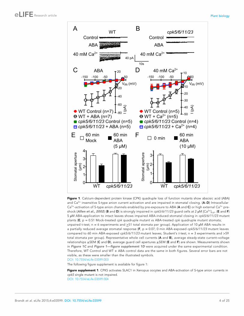
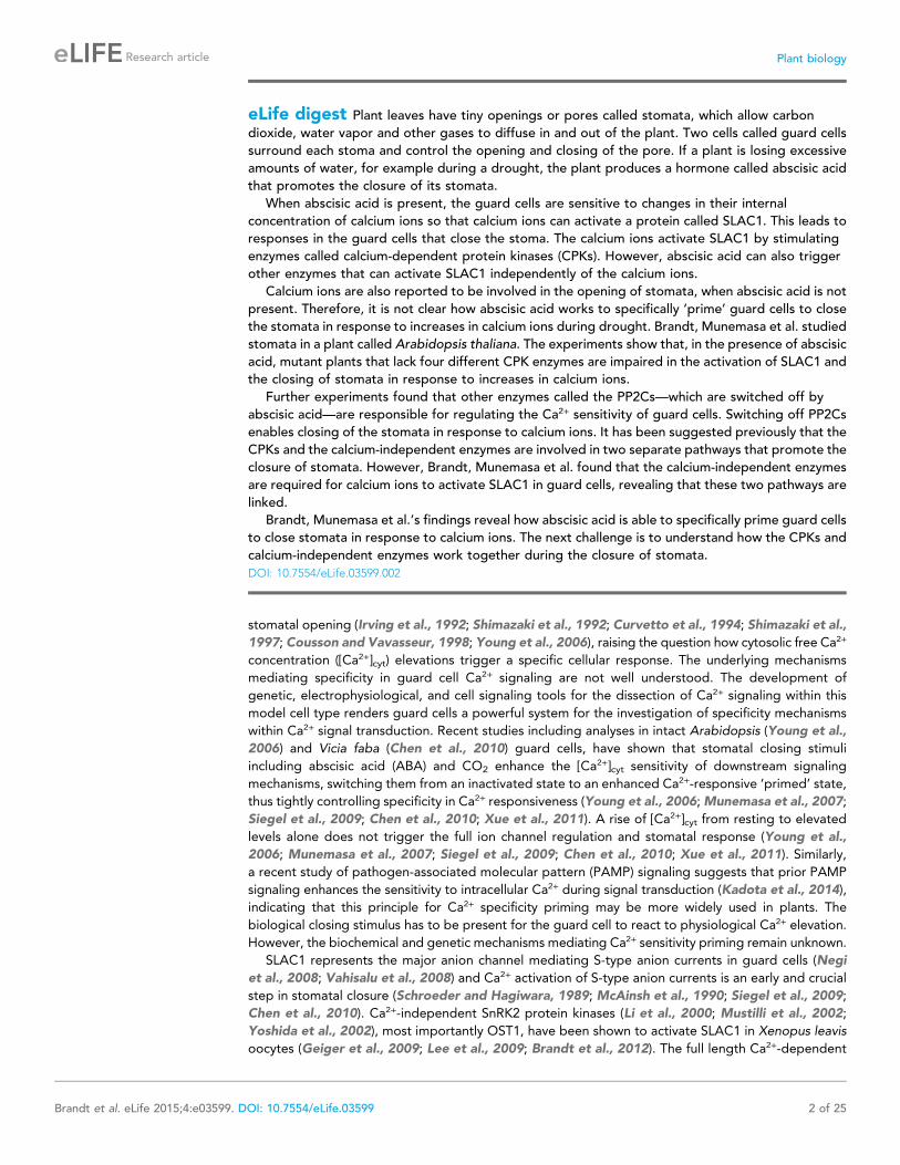
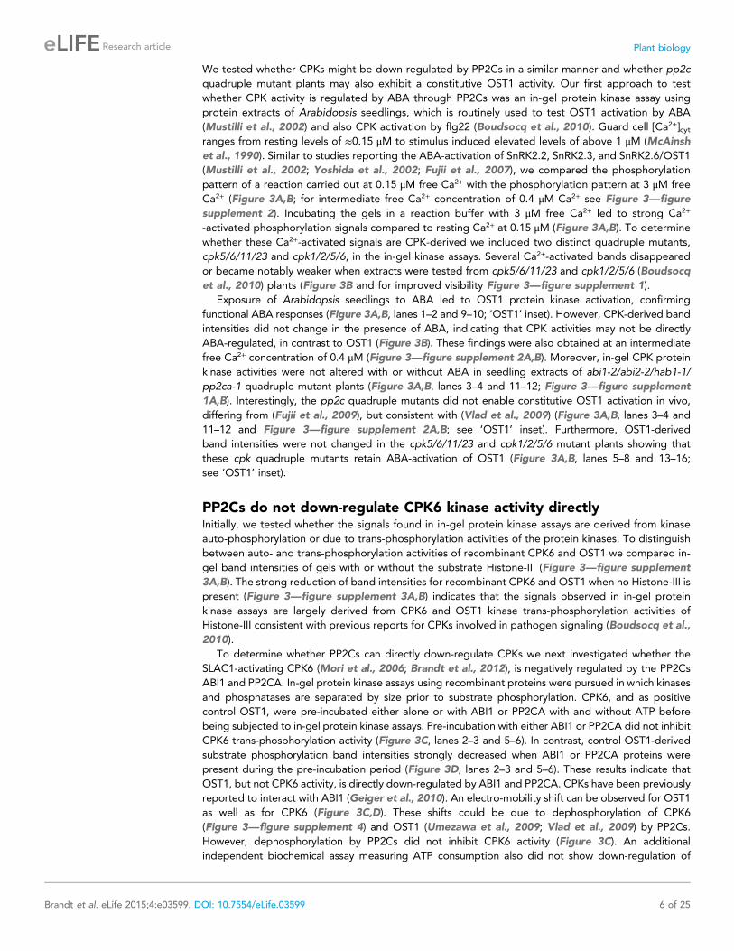
Figure 1. Calcium-dependent protein kinase (CPK) quadruple loss of function mutants show abscisic acid (ABA)
and Ca2+ insensitive S-type anion current activation and are impaired in stomatal closing. (A–D) Intracellular
Ca2+-activation of S-type anion channels enabled by pre-exposure to ABA (A and C) or high external Ca2+ pre-
shock (Allen et al., 2002) (B and D) is strongly impaired in cpk5/6/11/23 guard cells at 2 μM [Ca2+]cyt. (E and F)
5 μM ABA-application to intact leaves shows impaired ABA-induced stomatal closing in cpk5/6/11/23 mutant
plants (E; p = 0.51 Mock-treated cpk quadruple mutant vs ABA-treated cpk quadruple mutant stomata;
unpaired t-test; n = 6 experiments and >51 total stomata per group). Application of 10 μM ABA results in
a partially reduced average stomatal response (F, p = 0.07; 0 min ABA-exposed cpk5/6/11/23 mutant leaves
compared to 60 min ABA-exposed cpk5/6/11/23 mutant leaves; Student’s t-test; n = 3 experiments and >59total stomata per group). Representative whole cell currents (A and B), average steady-state current–voltage
relationships ±SEM (C and D), average guard cell apertures ±SEM (E and F) are shown. Measurements shown
in Figure 1C and Figure 1—figure supplement 1D were acquired under the same experimental condition.
Therefore, WT Control and WT + ABA control data are the same in both figures. Several error bars are not
visible, as these were smaller than the illustrated symbols.
DOI: 10.7554/eLife.03599.003
The following figure supplement is available for figure 1:
Figure supplement 1. CPK5 activates SLAC1 in Xenopus oocytes and ABA-activation of S-type anion currents in
cpk5 single mutant is not impaired.
DOI: 10.7554/eLife.03599.004
Brandt et al. eLife 2015;4:e03599. DOI: 10.7554/eLife.03599 4 of 25
Research article Plant biology

(Geiger et al., 2010; Brandt et al., 2012). To determine whether these PP2Cs function in the ABA-
triggered enhancement of the [Ca2+]cyt-sensitivity in guard cells, we performed whole-cell patch-clamp
analysis using a plant line carrying T-DNA insertion mutations in the key ABA signaling PP2Cs ABI1,
ABI2, HAB1, and PP2CA (abi1-2/abi2-2/hab1-1/pp2ca-1). Surprisingly, in abi1-2/abi2-2/hab1-1/
pp2ca-1 quadruple mutant guard cells, strong Ca2+-activated S-type anion currents were observed
even without pre-exposure to ABA (Figure 2A–D). At low 0.1 μM [Ca2+]cyt S-type anion channels did
not show significant activation in the pp2c quadruple mutant compared to WT (Figure 2—figure
supplement 1A,B; p = 0.294 at −145 mV). These findings provide genetic evidence for first genes that
are essential for the ABA-triggered Ca2+ sensitivity priming in guard cells and show that these PP2Cs
provide a mechanism ensuring specificity in Ca2+ signal transduction.
CPK activities are not directly ABA-regulated and disruption of PP2Csdoes not cause constitutive activation of OST1Based on the above results we sought to determine the biochemical mechanisms by which PP2Cs
down-regulate Ca2+ sensitivity in the absence of ABA. The main SLAC1-activating protein kinase in the
Ca2+-independent branch, OST1 (Mustilli et al., 2002; Yoshida et al., 2002), is directly inactivated by
PP2Cs through de-phosphorylation of the activation loop (Umezawa et al., 2009; Vlad et al., 2009).
Figure 2. In protein phosphatase 2C (PP2C) quadruple mutant plants, Ca2+ activation of S-type anion currents is
constitutively primed. (A and C) 2 μM [Ca2+]cyt activates S-type anion currents in WT if the guard cells were
pre-exposed to ABA. (B and D) In PP2C quadruple mutant guard cells ABA pre-exposure is not required for 2 μM[Ca2+]cyt-activation of S-type anion currents. Average steady-state current–voltage relationships ±SEM, guard cell
numbers (C and D), and representative whole cell currents (A and B) are presented. Several error bars are not visible,
as these were smaller than the illustrated symbols.
DOI: 10.7554/eLife.03599.005
The following figure supplement is available for figure 2:
Figure supplement 1. Analysis of ABA activation of S-type anion currents in PP2C quadruple mutant guard cells at
low [Ca2+]cyt.
DOI: 10.7554/eLife.03599.006
Brandt et al. eLife 2015;4:e03599. DOI: 10.7554/eLife.03599 5 of 25
Research article Plant biology
We tested whether CPKs might be down-regulated by PP2Cs in a similar manner and whether pp2c
quadruple mutant plants may also exhibit a constitutive OST1 activity. Our first approach to test
whether CPK activity is regulated by ABA through PP2Cs was an in-gel protein kinase assay using
protein extracts of Arabidopsis seedlings, which is routinely used to test OST1 activation by ABA
(Mustilli et al., 2002) and also CPK activation by flg22 (Boudsocq et al., 2010). Guard cell [Ca2+]cytranges from resting levels of ≈0.15 μM to stimulus induced elevated levels of above 1 μM (McAinsh
et al., 1990). Similar to studies reporting the ABA-activation of SnRK2.2, SnRK2.3, and SnRK2.6/OST1
(Mustilli et al., 2002; Yoshida et al., 2002; Fujii et al., 2007), we compared the phosphorylation
pattern of a reaction carried out at 0.15 μM free Ca2+ with the phosphorylation pattern at 3 μM free
Ca2+ (Figure 3A,B; for intermediate free Ca2+ concentration of 0.4 μM Ca2+ see Figure 3—figure
supplement 2). Incubating the gels in a reaction buffer with 3 μM free Ca2+ led to strong Ca2+
-activated phosphorylation signals compared to resting Ca2+ at 0.15 μM (Figure 3A,B). To determine
whether these Ca2+-activated signals are CPK-derived we included two distinct quadruple mutants,
cpk5/6/11/23 and cpk1/2/5/6, in the in-gel kinase assays. Several Ca2+-activated bands disappeared
or became notably weaker when extracts were tested from cpk5/6/11/23 and cpk1/2/5/6 (Boudsocq
et al., 2010) plants (Figure 3B and for improved visibility Figure 3—figure supplement 1).
Exposure of Arabidopsis seedlings to ABA led to OST1 protein kinase activation, confirming
functional ABA responses (Figure 3A,B, lanes 1–2 and 9–10; ‘OST1’ inset). However, CPK-derived band
intensities did not change in the presence of ABA, indicating that CPK activities may not be directly
ABA-regulated, in contrast to OST1 (Figure 3B). These findings were also obtained at an intermediate
free Ca2+ concentration of 0.4 μM (Figure 3—figure supplement 2A,B). Moreover, in-gel CPK protein
kinase activities were not altered with or without ABA in seedling extracts of abi1-2/abi2-2/hab1-1/
pp2ca-1 quadruple mutant plants (Figure 3A,B, lanes 3–4 and 11–12; Figure 3—figure supplement
1A,B). Interestingly, the pp2c quadruple mutants did not enable constitutive OST1 activation in vivo,
differing from (Fujii et al., 2009), but consistent with (Vlad et al., 2009) (Figure 3A,B, lanes 3–4 and
11–12 and Figure 3—figure supplement 2A,B; see ‘OST1’ inset). Furthermore, OST1-derived
band intensities were not changed in the cpk5/6/11/23 and cpk1/2/5/6 mutant plants showing that
these cpk quadruple mutants retain ABA-activation of OST1 (Figure 3A,B, lanes 5–8 and 13–16;
see ‘OST1’ inset).
PP2Cs do not down-regulate CPK6 kinase activity directlyInitially, we tested whether the signals found in in-gel protein kinase assays are derived from kinase
auto-phosphorylation or due to trans-phosphorylation activities of the protein kinases. To distinguish
between auto- and trans-phosphorylation activities of recombinant CPK6 and OST1 we compared in-
gel band intensities of gels with or without the substrate Histone-III (Figure 3—figure supplement
3A,B). The strong reduction of band intensities for recombinant CPK6 and OST1 when no Histone-III is
present (Figure 3—figure supplement 3A,B) indicates that the signals observed in in-gel protein
kinase assays are largely derived from CPK6 and OST1 kinase trans-phosphorylation activities of
Histone-III consistent with previous reports for CPKs involved in pathogen signaling (Boudsocq et al.,
2010).
To determine whether PP2Cs can directly down-regulate CPKs we next investigated whether the
SLAC1-activating CPK6 (Mori et al., 2006; Brandt et al., 2012), is negatively regulated by the PP2Cs
ABI1 and PP2CA. In-gel protein kinase assays using recombinant proteins were pursued in which kinases
and phosphatases are separated by size prior to substrate phosphorylation. CPK6, and as positive
control OST1, were pre-incubated either alone or with ABI1 or PP2CA with and without ATP before
being subjected to in-gel protein kinase assays. Pre-incubation with either ABI1 or PP2CA did not inhibit
CPK6 trans-phosphorylation activity (Figure 3C, lanes 2–3 and 5–6). In contrast, control OST1-derived
substrate phosphorylation band intensities strongly decreased when ABI1 or PP2CA proteins were
present during the pre-incubation period (Figure 3D, lanes 2–3 and 5–6). These results indicate that
OST1, but not CPK6 activity, is directly down-regulated by ABI1 and PP2CA. CPKs have been previously
reported to interact with ABI1 (Geiger et al., 2010). An electro-mobility shift can be observed for OST1
as well as for CPK6 (Figure 3C,D). These shifts could be due to dephosphorylation of CPK6
(Figure 3—figure supplement 4) and OST1 (Umezawa et al., 2009; Vlad et al., 2009) by PP2Cs.
However, dephosphorylation by PP2Cs did not inhibit CPK6 activity (Figure 3C). An additional
independent biochemical assay measuring ATP consumption also did not show down-regulation of
Brandt et al. eLife 2015;4:e03599. DOI: 10.7554/eLife.03599 6 of 25
Research article Plant biology

Figure 3. CPK activity is not changed by ABA or hyper-activated in pp2c quadruple mutants at defined Ca2+
concentrations. (A and B) In-gel kinase assays with Histone-III as substrate for whole plant protein extracts show
(B) 3 μM Ca2+-activated trans-phosphorylation kinase activities independent of application of 50 μM ABA (lanes 9
and 10). In contrast, ABA activation of OST1 is clearly visible (lanes 1–2 and 9–10 at ∼41 kDa; lower ‘OST1’ inset
shows the same signal optimized autoradiography at the ∼41 kDa region and the corresponding gel regions are
indicated by blue lines; see ‘Materials and methods’). Disruption of four PP2C genes (ABI1, ABI2, HAB1, and PP2CA)
does not result in constitutive Ca2+-activated and OST1 kinase activities (lanes 3–4 and 11–12). In-gel kinase activities
of two independent CPK quadruple mutant lines indicate that the Ca2+-activated kinase signals are CPK-derived
(compare lanes 9–10 with 13–16 in B and see Figure 3—figure supplement 1); predicted MWs for CPK1, CPK2,
CPK5, CPK6, CPK11, and CPK23 are 68.3 kDa, 72.3 kDa, 62.1 kDa, 61.1 kDa, 55.9 kDa, 58.7 kDa, respectively.
(C and D) In-gel protein kinase assays with recombinant proteins show that incubation of the protein kinases with the
PP2Cs ABI1 and PP2CA does (C) not change CPK6 activity while (D) OST1 activity is strongly down-regulated by
PP2Cs. Each experiment has been repeated at least three times with similar results.
DOI: 10.7554/eLife.03599.007
The following figure supplements are available for figure 3:
Figure supplement 1. Close up view of Ca2+-activated kinase activities.
DOI: 10.7554/eLife.03599.008
Figure 3. continued on next page
Brandt et al. eLife 2015;4:e03599. DOI: 10.7554/eLife.03599 7 of 25
Research article Plant biology

CPK6 activity in the presence of ABI1 and PP2CA (Figure 3—figure supplement 5), further underlining
no direct down-regulation of CPK6 activity by these three PP2Cs, in contrast to OST1 controls.
PP2Cs interact with and rapidly dephosphorylate SLAC1Our results suggest that PP2Cs neither down-regulate CPK6 activity directly in vitro (Figure 3C,D and
Figure 3—figure supplement 5) nor that CPK activities are strongly ABA-regulated independent of
[Ca2+] changes in native plant protein extracts (Figure 3A,B). We next investigated the kinetics and
specificity of PP2C down-regulation of SLAC1 activation by CPKs through dephosphorylation of the
SLAC1 channel, a mechanism reported for CPK-dependent transcription factor regulation (Lynch
et al., 2012) and consistent with previous findings (Brandt et al., 2012). First, we determined whether
SLAC1 interacts with the PP2C ABI1 in planta using bimolecular fluorescence complementation (BiFC).
We observed clear BiFC signals for full length SLAC1 co-expressed with CPK6 and ABI1 (Figure 4A,B)
while signal intensities of SLAC1 co-expressed with a control protein phosphatase 2A catalytic subunit
5 (PP2AC5) were very low (Figure 4B). Protein–protein interaction of SLAC1 with PP2CA in BiFC
experiments was reported earlier (Lee et al., 2009). As shown in Figure 4C,D, the ABI1-mediated
dephosphorylation of the N-terminus of SLAC1 (SLAC1-NT) previously phosphorylated by CPK6
(Brandt et al., 2012) occurs very rapidly. Already 1 min after the addition of ABI1 a strong decrease of
the phosphorylation signal was observed (Figure 4D, lane 4). This de-phosphorylation was also found
when the PP2C phosphatase PP2CA was added instead of ABI1 (Figure 4C,E, lane 4). To test whether
this is a general phenomenon, we phosphorylated the SLAC1-NT with the SLAC1-activating and
-phosphorylating kinases CPK21, CPK23, and OST1 (Geiger et al., 2009, 2010; Lee et al., 2009) and
analyzed whether ABI1 and PP2CA are able to remove phospho-groups added by these kinases
(Figure 4F–H and Figure 4—figure supplement 1). After inhibiting the kinase with staurosporine,
band intensities decreased only after addition of the PP2C protein phosphatases for all combinations,
showing that this rapid SLAC1 de-phosphorylation is mediated by PP2Cs (Figure 4F–H and
Figure 4—figure supplement 1, lanes 5–6).
Disruption of Ca2+-independent SnRK kinases impairs Ca2+-dependentS-type anion channel regulationThe Ca2+-independent and Ca2+-dependent branches of ABA signal transduction are presently
considered to be independent (e.g., Li et al., 2006; Kim and et al., 2010; Roelfsema et al., 2012),
but this model has not been genetically investigated in Arabidopsis. In the cpk5/6/11/23 quadruple
mutant, ABA-activation of S-type anion currents and stomatal closure were impaired (Figure 1A–E),
providing evidence for a possible interdependence of these signaling branches. The ost1 single gene
disruption mutant in the Col ecotype shows intermediate S-type anion current activation by ABA
(Geiger et al., 2009). Three Ca2+-independent SnRK kinases, SnRK2.2, SnRK2.3, and OST1 can
activate SLAC1 in oocytes (Geiger et al., 2009) and redundantly function in controlling leaf water loss
(Fujii and Zhu, 2009). Interestingly, snrk2.2/snrk2.3/ost1 triple mutants were strongly impaired in ABA
activation and notably also external Ca2+ shock-induced activation of S-type anion channels at 2 μM[Ca2+]cyt (Figure 5A–D). Imposing repetitive cytosolic Ca2+ transients by alternating guard cell
incubation buffers induces a fast Ca2+-reactive stomatal closure response (Allen et al., 2001). We
further analyzed imposed Ca2+ oscillation-induced stomatal closure in snrk2.2/snrk2.3/ost1 triple
mutants. Ca2+ reactive stomatal closure of the snrk triple mutant was impaired compared to wildtype
plants (Figure 5E, p < 0.02 for wildtype vs snrk2.2/snrk2.3/ost1 at 120 min). These data show that
Figure 3. Continued
Figure supplement 2. Protein kinase activities are not altered by ABA-application at 150 nM and 400 nM free Ca2+.
DOI: 10.7554/eLife.03599.009
Figure supplement 3. Signals in in-gel kinase assays are largely derived from kinase trans-phosphorylation activities.
DOI: 10.7554/eLife.03599.010
Figure supplement 4. CPK6 is de-phosphorylated by the PP2Cs ABI1, ABI2, and PP2CA.
DOI: 10.7554/eLife.03599.011
Figure supplement 5. CPK6 kinase activity is not inhibited in the presence of ABI1 or PP2CA.
DOI: 10.7554/eLife.03599.012
Brandt et al. eLife 2015;4:e03599. DOI: 10.7554/eLife.03599 8 of 25
Research article Plant biology

Figure 4. PP2Cs interact with and directly and rapidly dephosphorylate the N-terminus of SLAC1 (SLAC1-NT) when previously phosphorylated by several
SLAC1-activating CPK and OST1 protein kinases. (A) Bimolecular fluorescence complementation (BiFC) experiments in Nicotiana benthamiana leaves
show YFP-derived fluorescence signals of YC-SLAC1 co-expressed with CPK6-YN and YN-ABI1. (B) Quantification of BiFC-mediated YFP-fluorescence
shows that SLAC1 interacts with CPK6 and ABI1 but not with the control catalytic protein phosphatase 2A subunit C5 (PP2AC5). YFP signals of positive
control YN-PP2AC5 with protein phosphatase 2A regulatory subunit A3 fused to YC (YC-PP2AA3) confirm expression of PP2AC5. Data shown in (B)
represent the average fluorescence intensity of randomly picked leaf areas (n = 40; ±SEM) and these data are also included in Figure 6—figure
supplement 5. (C–E) CPK6-phosphorylated SLAC1-NT is rapidly de-phosphorylated by ABI1 and PP2CA. SLAC1-NT phosphorylation by CPK6 (D and E,
lane 1) is strongly inhibited if the PP2C protein phosphatase was added before starting the reaction (D and E, lane 2), but remains stable after addition of
elution buffer (Elu.) and kinase inhibitor staurosporine (Stau.) with subsequent 10 min incubation (D and E, lane 3). If (D) ABI1 or (E) PP2CA together with
staurosporine are added after the initial 10 min CPK6 mediated phosphorylation period, the SLAC1-NT phosphorylation signal rapidly decreases within
1 min (D and E, lanes 4–7). Staurosporine pre-exposure control inhibits SLAC1-NT phosphorylation by CPK6 (D and E, lane 8). (F–H) PP2Cs de-
phosphorylate the SLAC1-NT which was phosphorylated by major SLAC1-activating kinases CPK23 and OST1. The SLAC1-NT is phosphorylated by CPK23
(G, lane 1) and OST1 (H, lane 1) which is inhibited when the PP2Cs ABI1 and PP2CA are added before starting the reactions (G and H, lanes 2–3). When
adding staurosporine and elution buffer after the initial phosphorylation period and incubating for 10 min the signal does not change (G and H, lane 4).
Addition of ABI1 or PP2CA after supplementing the reaction with staurosporine leads to rapid (10 min) dephosphorylation of the SLAC1-NT previously
phosphorylated by the OST1 and CPK23 protein kinases (G and H, lanes 5–6).
DOI: 10.7554/eLife.03599.013
The following figure supplement is available for figure 4:
Figure supplement 1. When previously phosphorylated by CPK21, the SLAC1-NT is de-phosphorylated by the PP2Cs ABI1 and PP2CA.
DOI: 10.7554/eLife.03599.014
Brandt et al. eLife 2015;4:e03599. DOI: 10.7554/eLife.03599 9 of 25
Research article Plant biology

Figure 5. Both, ABA- and high external Ca2+-activation of S-type anion currents at elevated [Ca2+]cyt and imposed
Ca2+-oscillation-triggered stomatal closure are impaired in snrk2.2/2.3/ost1 triple mutant guard cells while the
ABA-activation of ICa currents is intact. (A–D) Whole-cell patch-clamp experiments reveal that [Ca2+]cyt-activation of
S-type anion currents is disrupted in snrk2.2/2.3/ost1 triple mutant guard cells even if pre-incubated with high
external Ca2+ shock (A and B) or ABA (C and D). Note that pre-incubation with high external Ca2+ shock by passes
early ABA signaling (Allen et al., 1999a; Allen et al., 2002). Typical current responses (A and C), average steady-
state current–voltage relationships ±SEM, and the number of measured cells are presented (B and D). In (B) data for
snrk2.2/2.3/ost1 triple mutants with and without ABA overlap with WT controls. (E) Imposed Ca2+ oscillation-induced
stomatal closure is impaired in Ca2+-independent protein kinase snrk2.2/2.3/ost1 triple mutant leaves, providing
further evidence for an interdependence of these responses. Four 5-min extracellular Ca2+-pulses were applied in
10-min intervals from time = 0 to 35 min. Average individually tracked stomatal apertures were normalized to the
stomatal apertures at time zero. The averages of the normalized apertures ±SEM and the number of independent
genotype-blind experiments (n = 4) are shown (>40 total stomata per group). Average stomatal apertures at time
zero were 4.61 ± 0.44 μm in WT (n = 4) and 5.51 ± 0.87 μm in the snrk2.2/2.3/ost1 triple mutant (n = 4). (F) Patch
clamp experiments reveal that ABA activation of ICa currents is not impaired in snrk2.2/2.3/ost1 triple mutant guard
cells. Average steady-state current–voltage relationships ±SEM, and the number of measured cells are presented in
(F). Representative whole cell current traces for (F) are presented in Figure 5—figure supplement 1. Several error
bars are not visible, as these were smaller than the illustrated symbols.
DOI: 10.7554/eLife.03599.015
Figure 5. continued on next page
Brandt et al. eLife 2015;4:e03599. DOI: 10.7554/eLife.03599 10 of 25
Research article Plant biology

disruption of Ca2+-independent signaling in snrk2 triple mutants also impairs Ca2+-dependent
stomatal responses. Thus these findings investigating S-type anion channel regulation and stomatal
movements both provide genetic evidence for an unexpected interdependence of the Ca2+-dependent
and -independent branches of the guard cell signaling network.
The Ca2+-independent OST1 protein kinase affects Ca2+ signaling in Landsberg erecta guard cells
via regulation of plasma membrane-localized Ca2+-permeable channels (ICa) (Acharya et al., 2013). To
test whether the functional linkage of the Ca2+-dependent and Ca2+-independent branch is due to the
regulation of the ICa channels by SnRK2 protein kinases in the Columbia ecotype, we performed patch
clamp analyses measuring plasma membrane ICa channel currents in snrk2.2/snrk2.3/ost1 triple
mutant guard cells. However, ABA activation of ICa channels remained intact in snrk2.2/snrk2.3/ost1
triple mutant guard cells (Figure 5F and Figure 5—figure supplement 1). In positive control
experiments, ABA receptor pyr1/pyl1/2/4 quadruple mutant guard cells showed clear impairment of
ABA activation of ICa channels (Data not shown, n = 5; control vs ABA, p = 0.96; Student’s t-test),
consistent with previous findings (Wang et al., 2013).
ABA-dependent stomatal responses are impaired in non-phosphorylatable SLAC1 serine 59 and serine 120 double mutant plantsIn addition to possible direct cross-regulation of CPKs and SnRK2s, another non-mutually exclusive
potential mechanism for the requirement of both SnRK and CPK kinases for ABA activation of S-type
anion channels could be that SLAC1 serves as coincidence detector through differential
phosphorylation by protein kinases of the Ca2+-dependent and -independent branches. The amino
acid residue serine 120 of SLAC1 has been shown to be required for OST1, but not for CPK23
activation of SLAC1 in Xenopus oocytes (Geiger et al., 2009, 2010). A different site, serine 59, has
been shown to be required for SLAC1 activation by CPK6 (Brandt et al., 2012). Thus we investigated
whether several CPKs can activate the SLAC1 S120A mutant in oocytes and whether the SLAC1 S59A
mutant is activated by OST1 and other CPKs in oocytes. CPK5, CPK6, and CPK23 activation of SLAC1
S120A was similar to WT SLAC1 activation (Figure 6A–C and Figure 6—figure supplement 1A,B). In
contrast, SLAC1 S59A activation by these CPKs was strongly impaired (Figure 6A–C and
Figure 6—figure supplement 1A,B). Interestingly however, OST1 was able to activate SLAC1
S59A (Figure 6D–F), which was confirmed in multiple independent experimental sets under the
imposed conditions. These results suggest that S59 is required for strong activation by protein kinases
of the Ca2+-dependent CPK branch, while S120 represents a crucial amino acid for strong activation by
the Ca2+-independent branch of the ABA signaling core. To avoid spurious phosphorylation by high
protein kinase concentrations in oocytes, effects of co-expression of CPK6 and OST1 at low levels that
do not fully activate SLAC1 were investigated. These experiments show a clear enhanced SLAC1
activation in oocytes when both kinases are co-expressed (Figure 6—figure supplement 2A–D). This
enhancement of SLAC1 activation by OST1 became less clear when an inactive OST1 protein kinase
(OST1 D140A) was analyzed (Figure 6—figure supplement 2E).
To more directly investigate S-type anion channel regulation in planta, we established slac1-1 plant
lines which express SLAC1 WT, S59A, S120A, and S59A/S120A fused to mVenus under the native
SLAC1 promoter and carried out patch clamp analyses. Expression of wildtype SLAC1-mVenus in
slac1-1 guard cells resulted in recovery of S-type anion channels (Figure 6G and Figure 6—figure
supplement 3A). Unexpectedly, expression of the single site SLAC1 mutants, SLAC1 S59A or SLAC1
S120A in slac1-1 guard cells restored ABA regulation of S-type anion currents (Figure 6G and
Figure 6—figure supplement 3A). However, expression of the double phosphorylation site SLAC1
mutant, SLAC1 S59A/S120A did not restore ABA activation of S-type anion channels (Figure 6G and
Figure 6—figure supplement 3A). Furthermore, ABA-induced stomatal closing responses in these
complementation lines confirmed the need to mutate both the S59 and S120 sites to alanine to
Figure 5. Continued
The following figure supplement is available for figure 5:
Figure supplement 1. snrk2.2/2.3/ost1 triple mutant guard cells show intact ABA activation of Ca2+-permeable ICa
currents.
DOI: 10.7554/eLife.03599.016
Brandt et al. eLife 2015;4:e03599. DOI: 10.7554/eLife.03599 11 of 25
Research article Plant biology

Figure 6. Ca2+-dependent protein kinase and OST1 protein kinase activation of SLAC1 in oocytes requires serine 59
or serine 120, respectively while in planta ABA-dependent S-type anion current activation and stomatal closing are
only impaired in SLAC1 S59A/S120A double amino acid mutants. (A–C) SLAC1 activation by CPK6 in Xenopus
oocytes was abolished when serine 59 is mutated to alanine (S59A) (A and C) (Brandt et al., 2012) but was
comparable to wild type SLAC1 activation for the SLAC1 S120A mutated version (B and C). (D–F) OST1 activation of
SLAC1 was abolished in the SLAC1 S120A mutant (E and F) (Geiger et al., 2009), while OST1 robustly activated
SLAC1 S59A (D and F). (G) In whole-cell patch-clamp experiments, slac1-1 guard cells show impaired ABA-activation
of S-type anion currents. Expression of SLAC1 WT, S59A, and S120A in slac1-1 plants restores ABA activation of
S-type anion currents in guard cells, but expression of SLAC1 S59A/S120A does not. (H) The ABA-insensitive
phenotype of slac1-1 stomata was recovered by expression of SLAC1 WT, S59A, and S120A, but not by expression
of S59A/S120A. Note that SLAC1 WT, S59A, S120A, and S59A/S120A are expressed as C-terminal mVenus fusion
proteins under native SLAC1 promoter (see ‘Materials and methods’). Representative current traces are depicted in
(A, B, D and E) and average current voltage relationships are shown (C and F; ±SEM). Average steady-state current
responses ±SEM at −145 mV are plotted in (G) and average stomatal apertures ±SEM in (H). * indicates p < 0.05;
unpaired Student’s t-test. Exact p-values and number of individual experiments for (G and H) can be found in
Figure 6—figure supplement 4. Note that WT (Col0) and slac1-1 control measurements shown in (G and H) are the
same control data as those shown in Figure 6—figure supplement 3A,B as all lines were investigated under the
same conditions. Several error bars are not visible, as these were smaller than the illustrated symbols.
DOI: 10.7554/eLife.03599.017
The following source data and figure supplements are available for figure 6:
Source data 1. Statistical data and number of repeats (n) for the (Table 1) patch clamp measurements shown in
Figure 6G and Figure 6—figure supplement 3A and (Table 2) for measurements of stomatal apertures presented
in Figure 6H and Figure 6—figure supplement 3B (n = 3 experiments and >45 total stomata per group).
DOI: 10.7554/eLife.03599.018
Figure supplement 1. SLAC1 serine 59 but not serine 120 is required for CPK5 or CPK23 activation in Xenopus
oocytes.
DOI: 10.7554/eLife.03599.019
Figure supplement 2. SLAC1 exhibits enhanced activity by co-expression of CPK6 and OST1 in Xenopus oocytes.
DOI: 10.7554/eLife.03599.020
Figure supplement 3. ABA-induced S-type anion currents and stomatal closure responses are impaired when both
SLAC1 S59 and S120 are substituted with alanine in independent double amino acid mutant line.
DOI: 10.7554/eLife.03599.021
Figure supplement 4. Analysis of expression and subcellular localization of SLAC1-WT, SLAC1S59A, S120A, and
S59A/S120A in slac1-1 complementation lines.
DOI: 10.7554/eLife.03599.022
Figure 6. continued on next page
Brandt et al. eLife 2015;4:e03599. DOI: 10.7554/eLife.03599 12 of 25
Research article Plant biology

significantly impair ABA-induced stomatal closing in planta (Figure 6H and Figure 6—figure
supplement 3B). The above described patch clamp and stomatal movement experiments were
conducted with two independent complementation lines (Figure 6G,H and Figure 6—figure
supplement 3A,B). To ensure that the impaired ABA-activation of S-type anion currents and stomatal
closure in the SLAC1 S59A/S120A mutant was not due to non-expressed protein we investigated
the mVenus-derived fluorescence in all complementation lines. All SLAC1 complementation lines
expressed SLAC1-mVenus driven by the native SLAC1 promoter to a similar degree (Figure 6—figure
supplement 4).
We examined putative roles of the two phosphorylation sites in SLAC1 for interaction of SLAC1
with CPK6 and ABI1 by BiFC analysis. Reconstituted YFP fluorescence intensity of CPK6-YN co-
expressed with YC-SLAC1-S59A, YC-SLAC1-S120A, or YC-SLAC1-S59A/S120A was significantly lower
than that of CPK6-YN co-expressed with YC-SLAC1-WT (Figure 6—figure supplement 5). YN-ABI1
co-expression with the YC-SLAC1-S59A mutant did not significantly change the YFP fluorescence
intensity while YN-ABI1 co-expression with YC-SLAC1-S120A or YC-SLAC1-S59A/S120A resulted
in lower YFP fluorescence intensity when compared to YN-ABI1 co-expression with YC-SLAC1-WT
(p < 0.005; unpaired t-test; Figure 6—figure supplement 5). These results point to the need for
future research to determine whether these phosphorylation sites in SLAC1 might contribute to
promotion of CPK6 kinase and ABI1 phosphatase interaction strength with the SLAC1 channel
(Figure 6—figure supplement 5).
DiscussionDissection of Ca2+ signaling specificity mechanisms can be advanced through characterization of the
combined cellular, genetic, and biochemical mechanisms in a single cell type. Biochemical and
cellular mechanisms that function in Ca2+ specificity, notably those mediated by the mammalian
Ca2+/calmodulin-dependent kinase II, have been characterized (De Koninck and Schulman, 1998;
Bradshaw et al., 2003; Rellos et al., 2010; Chao et al., 2011). Genome analyses in plants have
revealed the existence of more than 200 genes encoding for proteins containing Ca2+-binding EF-
hands in the Arabidopsis genome alone (Day et al., 2002) with overlapping expression of many
genes in the same cell type, including guard cells (Harmon et al., 2000; McCormack et al., 2005;
Schmid et al., 2005; Winter et al., 2007). This plethora of Ca2+ signaling proteins and the many
responses in plants mediated by Ca2+ (Dodd et al., 2010) calls for robust mechanisms mediating
specificity in Ca2+ signaling.
Ca2+ is a major hub within the signaling network of plant guard cells (MacRobbie, 2000;
Hetherington, 2001; Hetherington and Woodward, 2003), but the biochemical mechanisms
mediating Ca2+ specificity have remained unknown. In guard cells, stomatal closing stimuli, including
ABA and CO2, enhance (prime) [Ca2+]cyt-sensitivity, as also shown in intact Arabidopsis and V. faba
guard cells (Young et al., 2006; Munemasa et al., 2007; Siegel et al., 2009; Chen et al., 2010; Xue
et al., 2011). Calcium sensitivity priming could provide a key mechanism contributing to specificity in
Ca2+ signaling, as this response switches between a state of reduced Ca2+ sensitivity to a Ca2+-re-
sponsive ‘primed’ state, thus tightly controlling Ca2+ responsiveness (Allen et al., 2002; Young et al.,
2006;Munemasa et al., 2007; Siegel et al., 2009; Chen et al., 2010; Xue et al., 2011). However, the
genetic and biochemical mechanisms mediating Ca2+ sensitivity priming have remained unknown.
Here we report genetic, biochemical and signaling network mechanisms that underpin this cellular
response. In the absence of ABA, Ca2+ responsiveness is inhibited by PP2Cs, thereby preventing
responses to unrelated stomatal opening-mediating stimuli (Irving et al., 1992; Shimazaki et al.,
1992; Curvetto et al., 1994; Shimazaki et al., 1997; Cousson and Vavasseur, 1998; Young et al.,
2006) and also spontaneous Ca2+ elevations (Young et al., 2006; Siegel et al., 2009). As PP2Cs
inhibit OST1 and also down-regulate SLAC1 directly, this network not only enables stimulus specific
activation of SLAC1, but also provides a tight off switch via PP2C-catalyzed dephosphorylation of
Figure 6. Continued
Figure supplement 5. BiFC fluorescence intensities are altered for CPK6 and ABI1 co-expression with SLAC1-WT,
SLAC1S59A, S120A, and S59A/S120A.
DOI: 10.7554/eLife.03599.023
Brandt et al. eLife 2015;4:e03599. DOI: 10.7554/eLife.03599 13 of 25
Research article Plant biology

SLAC1 (Figure 7). This mechanism could also prevent SLAC1 activation by CPK23 which exhibits
a moderate Ca2+ sensitivity (Geiger et al., 2010). Moreover, as PP2Cs control Ca2+ signaling
specificity downstream of the CPK Ca2+ sensors (Figure 7), the same CPK isoforms remain capable of
fulfilling other signaling roles, consistent with several studies (Boudsocq et al., 2010; Munemasa
et al., 2011; Dubiella et al., 2013; Gao et al., 2013). Similarly, the same MAP kinase genes have been
shown to function in multiple plant signaling pathways and unknown mechanisms mediating specificity
are required (Rodriguez et al., 2010; Xu and Zhang, 2015). It was reported that ABI1 is not able to
remove phosphate groups from SLAC1 after OST1 phosphorylation (Geiger et al., 2009; Scherzer
et al., 2012). In contrast, the present study and other recent research shows a clear
dephosphorylation of SLAC1 by PP2Cs (Brandt et al., 2012; Maierhofer et al., 2014). Here we
demonstrate that ABI1 and PP2CA not only dephosphorylate the SLAC1 N-terminus, but these PP2Cs
are able to very rapidly remove the OST1- and CPK-mediated phosphorylation of SLAC1
(Figure 4C–H and Figure 4—figure supplement 1).
The Ca2+-dependent and Ca2+-independent ABA-signaling branches are presently considered to
function independent of one another (e.g., Li et al., 2006; Kim et al., 2010; Roelfsema et al., 2012).
However, this model has not yet been investigated using higher order genetic mutants. In the present
study we unexpectedly have found that snrk2.2/snrk2.3/ost1 triple mutant plants in the Ca2+-inde-
pendent ABA signal transduction pathway, also abrogate the ‘by pass’ (Allen et al., 1999a) Ca2+
-induced [Ca2+]cyt activation of S-type anion channels and Ca2+ oscillation-induced stomatal closing in
planta (Figure 5A,B,E). These data show an unexpected dependence of Ca2+-dependent stomatal
closing on the Ca2+-independent SnRK2 protein kinase signaling branch. Moreover, we have identified
the cpk5/6/11/23 quadruple mutations to impair ABA activation of S-type anion channels and
stomatal closure (Figure 1). Notably, this impairment occurs despite an intact SnRK2 signaling branch.
Note that a weakened ABA-induced stomatal closing response in cpk mutant plants, as found here
when applying 10 μM ABA (Figure 1F), is likely the result of parallel ABA activation of R-type anion
channels (Meyer et al., 2010; Sasaki et al., 2010; Imes et al., 2013) and a possible less-stringent CPK
regulation of the SnRK2 signaling branch. In cpk5/6/11/23 quadruple mutant plants signal
Figure 7. Simplified schematic model for Ca2+-specificity mechanism within ABA-dependent SLAC1 activation in
guard cells. (A) Without ABA, Ca2+ elevations that can also function in stomatal opening responses (Irving et al.,
1992; Shimazaki et al., 1992; Curvetto et al., 1994; Shimazaki et al., 1997; Cousson and Vavasseur, 1998;
Young et al., 2006) and spontaneous or un-specifically induced Ca2+ transients (Allen et al., 1999b; Klusener
et al., 2002; Young et al., 2006; Yang et al., 2008; Siegel et al., 2009) do not lead to S-type anion channel
(SLAC1) activation as PP2C protein phosphatases directly negatively regulate SLAC1 activation. (B) In the presence
of ABA this SLAC1 inhibition is released, OST1 and CPKs phosphorylate, and thereby activate the channel. ABA also
causes [Ca2+]cyt elevation via PP2C inhibition (Allen et al., 1999a; Murata et al., 2001). Data indicate cross-talk
between Ca2+-dependent and -independent ABA-activation of SLAC1 which may be mediated through
a combination of protein kinase cross regulation and additive activation via differential affinities for SLAC1
phosphorylation sites by OST1 and CPKs.
DOI: 10.7554/eLife.03599.024
Brandt et al. eLife 2015;4:e03599. DOI: 10.7554/eLife.03599 14 of 25
Research article Plant biology

transduction via the Ca2+-independent SnRK2 pathway appears to partially prevail at higher ABA
concentrations. Together these data indicate an unexpected dependence of the Ca2+-dependent
signal transduction pathway on the Ca2+-independent SnRK2 protein kinase-mediated pathway
(Figure 5). Furthermore, the present results together indicate that the output of the Ca2+-dependent
signaling pathway may affect the output of the SnRK2 signaling branch.
The presented combined genetic, cell signaling and physiological response analyses provide
strong evidence for a concomitant requirement of both the Ca2+-dependent and Ca2+-independent
branches to trigger a robust (Hetherington, 2001) downstream stomatal closing response (Figure 7).
One model for cross talk of SnRK2-induced signaling with Ca2+ signaling could be that OST1 causes
the activation of the Ca2+-permeable plasma membrane ICa channels (Hamilton et al., 2000; Pei
et al., 2000). However, our data clearly show that triple knock out of the Ca2+-independent SnRK2
kinases, OST1, SnRK2.2, and SnRK2.3 in the Columbia accession, does not impair ABA activation of ICachannels (Figure 5F and Figure 5—figure supplement 1). Interestingly however, cpk mutants show
impairment in ABA activation of ICa channels in guard cells (Mori et al., 2006).
The present study suggests that the integration of signals via differential phosphorylation of SLAC1
by the kinases of the Ca2+-dependent and Ca2+-independent branches could contribute to the
interdependence of both signaling branches. In Xenopus oocytes, SLAC1 S59 is required for the
activation by CPKs while SLAC1 S120 is required for the activation by the Ca2+-independent kinase
OST1 in oocytes (Figure 6A–F and Figure 6—figure supplement 1). Additionally, SLAC1 activation is
enhanced by co-expression of (non-split YFP moieties) non-saturating OST1 and CPK6 activities
(Figure 6—figure supplement 2). However, in planta analyses of slac1-1 plants expressing single
SLAC1 S59A or SLAC1 S120A mutants under the control of the SLAC1 promoter unexpectedly display
intact ABA-responses indicating that the phosphorylation of either amino acid residue, together with
phosphorylation of other amino acids, is sufficient for ABA-induced stomatal closing in intact stomata
and ABA activation of S-type anion channels (Figure 6G,H and Figure 6—figure supplement 3).
Furthermore, simultaneous mutation of both residues in SLAC1 (S59A and S120A) caused a strong
impairment in ABA activation of S-type anion channels and stomatal closing in planta, illustrating the
combined key functions of these residues in the intact guard cell system.
It should be noted that although SLAC1 S120, but not S59, is crucial for the activation by OST1 in
Xenopus oocytes (Figure 6E,F) (Geiger et al., 2009), phosphorylation of SLAC1 S59 by OST1 is also
found in vitro (Vahisalu et al., 2010). In addition, although the S120A mutation does not affect CPK6
activation of SLAC1 in Xenopus oocyte system (Figure 6B,C), our LC-MS/MS analyses reveal that the
S120 can be also phosphorylated by CPK6 in vitro (data not shown). Combined with these in vitro
data, our present in planta findings suggest that the SnRK2 and CPK protein kinases may have distinct
affinities for the S59 and S120 phospho-sites of SLAC1, which could contribute to the in-
terdependence of the Ca2+-dependent and -independent branches of the ABA signaling network.
In addition, crosstalk regulation mechanisms of these protein kinase responses may exist in planta and
will require further investigation (Figure 7).
Note that, similar to the slac1-1 mutation, mutation of SLAC1 S120 to phenylalanine (slac1-7) can
impair ozone-induced stomatal closing (Vahisalu et al., 2010). It is plausible that a phenylalanine
residue at this position causes more significant structural changes that impair SLAC1 function compared
to alanine. When both S59 and S120 are mutated to alanine simultaneously however, ABA-triggered
S-type anion current activation and stomatal closure were abrogated, highlighting the importance of
these two residues for ABA-signaling in planta. The results gained in planta also highlight that data
gained in oocytes, though helpful, are simplified and, not surprisingly, do not necessarily represent the
situation in the complex plant system. Over-expression of the components, including activating protein
kinases, to a high abundance in oocytes is well-suited to test several possible mechanisms in ion channel
regulation, and can guide follow up investigation in the native environment in plant cells.
ConclusionsIn summary, the present study reveals a first genetic mechanism that mediates Ca2+ sensitivity
priming. Ca2+ sensitivity is demonstrated here to be constitutively primed in pp2c quadruple mutant
guard cells, showing that PP2Cs ensure Ca2+ signaling specificity. Interestingly, PP2Cs do not directly
down-regulate CPK activity, in contrast to direct PP2C down-regulation of the SnRK2 protein kinases.
Rather PP2Cs very rapidly down-regulate signaling targets downstream of CPKs, which could enable
Brandt et al. eLife 2015;4:e03599. DOI: 10.7554/eLife.03599 15 of 25
Research article Plant biology

the same CPKs to function in more than one pathway. We have further identified a cpk quadruple
mutant here that for the first time strongly abrogates ABA activation of S-type anion channels. This
abrogation occurs despite an intact Ca2+-independent SnRK2 signaling branch. Furthermore,
disruption of the Ca2+-independent signaling branch in snrk2 protein kinase triple mutant plants
abrogates Ca2+ signaling. Thus, unexpectedly genetic analyses reveal a dependence of the Ca2+
-sensitive ABA signaling branch on the Ca2+-insensitive branch in planta. The control of ABA-triggered
stomatal closure by parallel interdependent Ca2+-dependent and–independent mechanisms could
contribute to the robustness of the stomatal ABA signaling network (Hetherington, 2001).
Unexpectedly, in planta studies show that the S59 and S120 phosphorylation sites in SLAC1 are
together required for intact ABA-induced stomatal closing in vivo. The Ca2+ sensitivity priming
mechanism described here could represent a more general principle present in plants contributing to
Ca2+ specificity within cellular signal transduction pathways, while also maintaining the availability of
Ca2+ sensors for distinct Ca2+-dependent signaling outputs.
Materials and methods
Mutant plant linesAll A. thaliana plants used in this study are in the Col0 ecotype. cpk5/6/11/23 quadruple T-DNA
insertion mutant plants were established by crossing cpk5/6/11 (sail_657C06/salk_025460/
salk_054495) kindly provided by Dr Jen Sheen (Harvard Medical School) (Boudsocq et al., 2010)
with cpk23-1 (salk_007958) obtained from ABRC (Ma and Wu, 2007; Geiger et al., 2010). Dr Ping He
(Texas A&M University) shared cpk1/2/5/6 (salk_096452/salk_059237/sail_657C06/salk_025460)
mutant seeds (Gao et al., 2013). The PP2C quadruple knock-out plants (abi1-2/abi2-2/hab1-1/
pp2ca-1; salk_072009/salk_015166/salk_002104/salk_028132) and snrk2.2/2.3/ost1 (GABI-Kat_807G04/
salk_107315/salk_008068) were kindly provided by Dr Pedro L Rodriguez (University of Valencia)
(Antoni et al., 2013). A second independent snrk2.2/2.3/ost1 (GABI-Kat_807G04/salk_107315/
salk_008068) line was established by crossing snrk2.2/2.3 supplied by Dr Jian-Kang Zhu (Shanghai
Center for Plant Stress Biology) with ost1-3. To establish SLAC1 complementation lines a 4.4 kb
fragment including 1.63 kb of the 5′-UTR, the genomic SLAC1 gene region and 0.9 kb of the 3′-UTR(Negi et al., 2008) was amplified using the PfuX7 polymerase (Norholm, 2010). The fragment was
cloned into a modified pGreenII (Hellens et al., 2000) vector lacking a promoter and being
compatible with USER-cloning. Employing USER cloning (Nour-Eldin et al., 2006; Bitinaite et al.,
2007; Geu-Flores et al., 2007) the point mutations were introduced and SLAC1 was fused with
mVenus (C-terminally) (Nagai et al., 2004). These pGreenII constructs were transformed into
Agrobacterium tumefaciens GV3101(pMP90) RG (Koncz and Schell, 1986). slac1-1 mutant plants
were then transformed by the floral dipping method (Clough and Bent, 1998) and propagated until
the T-DNA insertion was confirmed to be homozygous.
Patch clamp analysesArabidopsis plants were grown on soil in the growth chamber at 21˚C under a 16-hr-light/8-hr-dark
photoperiod with a photon flux density of 80 μmol/(m2 × s). The plants were watered from bottom
trays with deionized water once or twice per week and sprayed with deionized water every day. The
growth chamber humidity was 50–70%.
Arabidopsis guard cell protoplasts were isolated enzymatically as previously described (Pei et al.,
1997). One or two rosette leaves of 4- to 5-week-old plants were blended in a blender with deionized
water at room temperature (RT) for approximately 30 s. For isolation of guard cell protoplasts from
snrk2.2/snrk2.3/ost1 triple mutants, four or five rosette leaves were used. Epidermal tissues were
collected using a 100-μm nylon mesh and rinsed well with deionized water. The epidermal tissues
were then incubated in 10 ml of enzyme solution containing 1% (wt/vol) Cellulase R-10 (Yakult, Japan),
0.5% (wt/vol) Macerozyme R-10 (Yakult, Japan), 0.1 mM KCl, 0.1 mM CaCl2, 500 mM D-mannitol, 0.5%
(wt/vol) BSA, 0.1% (wt/vol) kanamycin sulfate, and 10 mM ascorbic acid for 16 hr at 25˚C on a circular
shaker at 40 rpm. Guard cell protoplasts were then collected by filtering through a 20-μm nylon mesh.
Subsequently, the protoplasts were washed twice with washing solution containing 0.1 mM KCl,
0.1 mM CaCl2, and 500 mM D-sorbitol (pH 5.6 with KOH) by centrifugation for 10 min at 200×g. Theguard cell protoplast suspension was kept on ice before use.
Brandt et al. eLife 2015;4:e03599. DOI: 10.7554/eLife.03599 16 of 25
Research article Plant biology

To investigate ABA activation of S-type anion channels, the guard cell protoplast suspension was
pre-incubated with 10 μM (Figure 1A,C, Figure 1—figure supplement 1C,D, Figure 6G, and
Figure 6—figure supplement 3A) or 50 μM (Figure 2A–D and Figure 2—figure supplement 1A,B as
well as Figure 5C,D) ± ABA (Sigma, St. Louis, MO) for 30 min. S-type anion channel currents in guard
cell protoplasts were recorded by the whole-cell patch-clamp technique as previously described (Pei
et al., 1997; Vahisalu et al., 2008; Siegel et al., 2009). The pipette solution contained 150 mM CsCl,
2 mMMgCl2, 5 mM Mg-ATP, 6.7 mM EGTA, and 10 mM Hepes-Tris (pH 7.1). To obtain a free [Ca2+]cytof 2 μM and 110 nM, 5.86 mM and 1.79 mM of CaCl2 were added to the pipette solution, respectively.
Osmolality of the pipette solution was adjusted to 500 mmol/l using D-sorbitol. The bath solution
contained 30 mM CsCl, 2 mM MgCl2, 1 mM CaCl2, and 10 mM MES-Tris (pH 5.6). Osmolality of the
bath solution was adjusted to 485 mmol/l using D-sorbitol. To investigate external Ca2+ activation of
S-type anion channels, guard cell protoplasts were pre-incubated with the bath solution containing 40
mM CaCl2, instead of 1 mM CaCl2 for 30 min. Whole-cell currents were recorded 3–5 min after
achieving the whole-cell configuration. The seal resistance was no less than 10 GΩ. The voltage was
decreased from +35 mV to −145 mV with 30 mV decrements and the holding potential was +30 mV.
To investigate ABA activation of Ca2+-permeable ICa channels, the pipette solution contained
10 mM BaCl2, 4 mM EGTA, and 10 mM HEPES-Tris (pH 7.1). 5 mM NADPH was freshly added to the
pipette solution before experiments. The bath solution contained 100 mM BaCl2, and 10 mM MES-
Tris (pH 5.6). 0.1 mM DTT was freshly added to the bath solution before experiments. Osmolarity was
adjusted to 500 mmol/l for the pipette solution and 485 mmol/l for the bath solution with D-sorbitol.
A ramp voltage protocol from +20 to −180 mV (holding potential, 0 mV; ramp speed, 200 mV/s) was
used for ICa recordings (Pei et al., 2000). The seal resistance was no less than 10 GΩ. Data were
filtered at 3 kHz. Initial control whole-cell currents were recorded 10 times with a 1 min interval
between each recording 1–3 min after achieving whole-cell configurations. The average current
obtained from the 10 current traces per cell at 0, −30, −60, −90, −120, −150, and −180 mV was
determined for IV curves. After control current recordings, ABA was added to the bath solution by
perfusion, and guard cell protoplasts were incubated with ABA in the bath solution for 3 min. Then,
ABA-activated ICa currents were recorded 10 times for another 10 min and the average current
obtained from the 10 traces was determined for IV curves.
Stomatal aperture analyses2-week-old plate-grown plants were transferred to soil and grown in >70% relative humidity under
16 hr light/8 hr dark. Rosette leaves from 4- to 5-week-old plants were detached and incubated in
stomatal opening buffer (5 mM KCl, 50 μM CaCl2, 10 mM MES and pH 5.6 with Tris base) for 2.5 hr, in
150–180 μmol/(m2 × s) light. Next, leaves were treated with either 5 μM ABA or 0.05% ethanol for an
additional 1 hr incubation. After the incubation period, leaves were blended and fragments were
collected with a 100 μm nylon mesh (Figure 1E, Figure 6H and Figure 6—figure supplement 3B)
except for Figure 1F. In Figure 1F, epidermal peels were prepared using a perforated-tape epidermal
detachment method (Ibata et al., 2013). Images of stomata from the abaxial side of the leaves were
collected by microscopy. Stomatal aperture analyses were conducted as single-blind experiments in
which the experimenter did not know the plant genotypes during measurements (Figure 1E,F) or as
double-blind experiments in which the experimenter did not know both the ABA concentration and
the plant genotypes (Figure 6H and Figure 6—figure supplement 3B).
Imposed Ca2+ pulse-regulated stomatal apertures of individuallymapped stomataStomatal aperture analyses for imposed Ca2+ pulses were performed as previously described (Allen
et al., 2001; Mori et al., 2006; Siegel et al., 2009). Stomatal apertures of individually mapped
stomata were measured at the indicated time points after the start of imposed Ca2+ pulses. The lower
epidermis of rosette leaves from 4- to 5-week-old plants was attached onto a coverslip using medical
adhesive (Hollister). Then mesophyll layers of the leaf were carefully removed using a razor blade until
only the epidermal layer remained. The lower epidermis was incubated in depolarizing buffer (50 mM
KCl and 10 mM MES-Tris [pH 5.6]) for 3 hr under white light (150–180 μmol/(m2 × s)) to open stomata.
Depolarizing buffer was changed to hyperpolarizing buffer (1 mM KCl, 1 mM CaCl2, and 10 mM MES-
Tris at pH 5.6). Four 5-min extracellular Ca2+ pulses were applied in 5-min intervals in the first 35 min.
Brandt et al. eLife 2015;4:e03599. DOI: 10.7554/eLife.03599 17 of 25
Research article Plant biology
Stomatal aperture analyses were conducted as blind experiments in which the experimenter did not
know the plant genotypes during measurements (Figure 5E).
Recombinant protein isolationOver-expression and purification of recombinant proteins were performed as described in Brandt
et al. (2012) with minor adjustments: For the isolation of the PP2C proteins ABI1, ABI2, and PP2C
additionally 5 mM MgCl2 and 5% Glycerol were added to the buffer in which the bacterial pellet were
re-suspended (buffer W in IBA manual). Also, all proteins except SLAC1-NT were eluted in elution
buffer supplemented with 20% Glycerol instead of 10% and stored at −80˚C instead of −20˚C. Toassess protein concentrations, several volumes of the eluates were loaded on a gel together with
several defined bovine serum albumin (BSA) protein amounts. After separating the proteins by SDS-
PAGE (Laemmli, 1970), the proteins were stained with coomassie brilliant blue R-250, dried between
two sheets of cellophane, and then scanned. BSA and recombinant protein band intensities were
measured using Fiji (Schindelin et al., 2012). After subtracting the background signal, BSA band signal
intensities were used to plot a standard curve. Concentrations of isolated recombinant proteins were
then calculated based on the equation resulting from the linear regression of the BSA standard curve.
Whole plant protein extractionSeeds were sterilized by incubation in sterilization medium (70% ethanol and 0.04% (wt/vol) SDS) for
15 min followed by three washes in 100% ethanol. After drying, the seeds for all genotypes were
plated on one plate with ½ Murashige and Skoog Basal Medium (MS; Sigma–Aldrich, St. Louis, MO)
and 0.8% phyto-agar. The plate was then stored at 4˚C for >3 days and subsequently transferred to
a growth cabinet (16/8 light/dark and 22˚C). After a growth phase of 10–14 days >10 seedlings per
genotype were floated on liquid ½ MS and equilibrated for 60–90 min in the growth cabinet. Either ±ABA (Sigma) to a final concentration of 50 μM (indicated by + in the figure) or the same volume of
solvent control (ethanol; indicated by—in the figure) was added to the floating seedlings. After
30 min the seedlings were removed from the ½ MS and flash frozen in liquid nitrogen. Plant tissue was
disrupted by shaking the frozen seedlings together with steel balls in a shaker (Retsch) for three times
30 s at 30 Hz in pre-cooled mountings. Subsequently, extraction buffer: 100 mM HEPES-NaOH pH 7.5,
5 mM EDTA, 5 mM EGTA, 0.5% (vol/vol) Triton X-100, 150 mM NaCl, 0.5 mM DTT, 10 mM NaF, 0.5%
(vol/vol) protease inhibitor (Sigma–Aldrich), 0.5% (vol/vol) phosphatase inhibitor 2 (Sigma–Aldrich),
0.5% (vol/vol) phosphatase inhibitor 3 (Sigma–Aldrich), 5 mM Na3VO4, and 5 mM β-Glycerophosphate
disodium salt hydrate was added. The samples were then treated in a sonication water bath (Fisher
Scientific) with ice added to the water for 30 s. Cell debris was removed via centrifugation at 20,000×gand 4˚C for 40 min. Protein concentrations of the supernatants were measured using the BCA Protein
Assay Kit (Pierce). 20 μg of total protein for each genotype and treatment were subjected to SDS-
PAGE (Laemmli, 1970) under denaturing conditions (see in-gel kinase assay).
In vitro protein kinase activity analysesThe reaction buffer consisted of 100 mM HEPES-NaOH pH 7.5, 10 mM MgCl2, 2 mM DTT, 1 mM
EGTA, and CaCl2 was added to get a final concentration of 2.5 μM free Ca2+ for all assays except the
assay depicted in Figure 3—figure supplement 4 for which free Ca2+ was adjusted to 5 μM(calculated with http://www.stanford.edu/∼cpatton/webmaxc/webmaxcE.htm). Note that the pH of
the reaction buffer dropped to pH 7.3 after adding all components and free Ca2+ calculations were
performed accordingly. The flow charts in the respective figures indicate the components which were
added subsequently in sequence (from top to bottom) and the respective incubation times. For the
reactions shown in Figure 3—figure supplement 4 0.5 μg of CPK6 and 1 μg of the PP2Cs ABI1, ABI2,
and PP2CA were used. The addition of EGTA for reactions shown in Figure 3—figure supplement 4
lanes 2–4 resulted in a free Ca2+ concentration <10 nM (calculated with http://www.stanford.edu/
∼cpatton/webmaxc/webmaxcE.htm). For the experiments shown in Figure 4D–H and
Figure 4—figure supplement 1, SLAC1-NT (1.5 μg) was mixed together with 200 nM of the protein
kinases CPK6, CPK23, OST1, and CPK21 in reaction buffer. Staurosporine was added to a final
concentration of 100 μM and the final concentration of the PP2Cs ABI1 and PP2CA was 600 nM. To
start all in vitro kinase reactions, 5 μCi of [γ-32P]-ATP (Perkin–Elmer) was added and the reactions were
incubated at RT for 10 min. The final volumes were 20 μl and the reactions were stopped by the
Brandt et al. eLife 2015;4:e03599. DOI: 10.7554/eLife.03599 18 of 25
Research article Plant biology

addition of 4 μl of 6× loading dye with subsequent incubation at 95˚C for 5 min. The proteins were
then separated by SDS polyacrylamide gel electrophoresis (SDS-PAGE, Laemmli, 1970) in 4–20%
acryl amide gradient gels (Biorad). After, the proteins were stained with coomassie brilliant blue R-250
(Sigma). To visualize incorporated 32P-derived radioactive signals, gels were exposed to a storage
phosphor screen (Molecular Dynamics; Figure 4D–H and Figure 4—figure supplement 1) or HyBlot
CL autoradiography films (Denville Scientific; Figure 3—figure supplement 4). The phosphor storage
screen was read out using a Typhoon scanner (Amersham Bioscience).
To compare CPK6 activities by measuring ATP consumption (Figure 3—figure supplement 5) with
or without the PP2Cs ABI1 and PP2CA, 0.5 μM of the protein kinase was incubated at RT for
7.5 min either alone or with 1 μM of PP2C protein in the above mentioned reaction buffer
supplemented with 10 μM ATP and ∼150 μM Histone III-S (Sigma). The reactions were stopped by the
addition of staurosporine. Residual ATP levels were quantified using the Kinase-Glo kit (Promega)
according to the manufacturer’s instructions resulting in luminescence signals measured in a plate
reader (Berthold Mithras LB 940) (Latz et al., 2012). ATP consumption was calculated by first
assessing the maximum range (ΔRmax) of luminescence by subtracting the signal intensity of the
background (no ATP added; RB) from the maximum signal (no kinase added; Rmax). To calculate the
ATP consumption, signal intensities derived from the residual ATP in the reactions (Rx) were
subtracted from the maximum signal (Rmax) and then related to the maximum range (ΔRmax) and
plotted in per cent ([(Rmax − Rx)/ΔRmax] × 100).
In-gel kinase assaysFor in-gel protein kinase assays using recombinant proteins shown in Figure 3C,D, 500 ng of OST1
and CPK6 kinase and the PP2Cs ABI1 and PP2CA in a 1:3 molar ration were mixed in reaction buffer
with 2.5 μM free Ca2+ (for buffer composition see in vitro kinase assay section). The reactions labelled
with ‘(ATP)’ were additionally supplemented with 100 μM ATP. All samples were incubated at RT for
20 min and stopped by adding SDS loading dye and heating at 95˚C for 5 min. For the assay depicted
in Figure 3—figure supplement 3, 1250 ng of OST1 and 500 ng of CPK6 were used.
These samples as well as the samples described in the ‘whole plant protein extraction’ section were
subjected to SDS-PAGE (Laemmli, 1970). The 10% SDS acryl amide resolving gels were supplemented
with 0.25–0.5 mg/ml Histone III-S (Sigma–Aldrich) (Figure 3, Figure 3—figure supplement 2 and
Figure 3—figure supplement 3) or without Histone III-S (Figure 3—figure supplement 3). After
electrophoresis, the gel was washed three times with washing buffer (25 mM Tris–HCl pH 8.0, 0.5 mM
DTT, 0.1 mMNa3VO4, 5 mMNaF, 0.5 mg/ml BSA, and 0.1% (vol/vol) Triton X-100) for 30 min each at RT,
followed by two washes with renaturation buffer (25 mM Tris–HCl pH 8.0, 1 mM DTT, 0.1 mM Na3VO4,
and 5 mM NaF) for 30 min each at RT and one wash at 4˚C overnight. Then, the gels were equilibrated
with reaction buffer (see in vitro kinase assay) for 30–45 min at RT and incubated in 20 ml of reaction
buffer supplemented with 50 μCi [γ-32P]-ATP (Perkin–Elmer). The reaction times were: Figure 3A,B,
Figure 3—figure supplement 2, and Figure 3—figure supplement 3: 90 min; Figure 3C: 60 min;
Figure 3D: 120 min. To stop the reactions and to remove background signals the gels were subsequently
extensively washed with a solution containing 5% (vol/vol) trichloroacetic acid and 1% (vol/vol)
phosphoric acid for at least six times for 15 min each. The gels were then stained with coomassie brilliant
blue R-250, dried on Whatman 3MM paper, and exposed to a storage phosphor screen (Molecular
Dynamics). The storage phosphor screen was scanned with a Typhoon reader (Amersham Bioscience).
For the in-gel kinase assays shown in Figure 3A,B and Figure 3—figure supplement 2 all steps
except the equilibration in reaction buffer and the reactions were carried out together and exactly the
same way which allows the autoradiographs to be compared. The image files given by the Typhoon
reader software are automatically adjusted to best display the bands with the highest intensity. Ca2+
-activated kinase signals are stronger than OST1-derived bands which renders OST1 bands hardly
visible by the Typhoon reader software. To better visualize OST1 activity, the signal intensity of the
∼41 kDa regions (blue box) in Figure 3A,B and Figure 3—figure supplement 1 were adjusted as
described in the following: The output files (.gel) of the Typhoon scanner software was opened using
Fiji (Schindelin et al., 2012) and in order to enhance the visibility of OST1-derived bands the
maximum signal was adjusted for the entire image including controls and the two gels which are
compared in accordance with publication policies (http://jcb.rupress.org/content/166/1/11.full).
Subsequently, the regions around 41 kDa were saved as .jpg file which was used for the preparation
Brandt et al. eLife 2015;4:e03599. DOI: 10.7554/eLife.03599 19 of 25
Research article Plant biology

of the figures. The parallel adjustment of the whole image showing both gels which are depicted in
either Figure 3A,B or Figure 3—figure supplement 1 allows the comparisons of band intensities
within each figure. Additionally, in Figure 3—figure supplement 1 several lanes of the same gel have
been cut out indicated by the black line as explained in http://jcb.rupress.org/content/166/1/11.full.
Quantitative bimolecular fluorescence complementationQuantitative BiFC experiments were carried out as described in (Waadt et al., 2008) with changes
explained in the following: BIFC vectors were altered to be USER cloning (Nour-Eldin et al., 2006)
compatible (indicated by the ‘u’ addition to the vector name) as described in Nour-Eldin et al. (2010).
SLAC1-WT, SLAC1 S59A, SLAC1 S120A, SLAC1 S59A/S120A, CPK6, and ABI1 coding sequences
were amplified using the PfuX7 polymerase (Norholm, 2010). SLAC1-WT, SLAC1 S59A, SLAC1
S120A, and SLAC1 S59A/S120A, were USER-cloned into pSPYCE(MR)u while CPK6 and ABI1 were
inserted into pSPYNE173u, and pSPYNE(R)173u, respectively. Coding sequences of PP2AA3
(AT1G13320) and PP2AC5 (AT1G69960) were PCR-amplified using Phusion DNA polymerase
(Invitrogen) and inserted SpeI/XmaI into a modified pUC19 vector including the pUBQ10 promoter
(pUC-pUBQ10; [Waadt et al., 2014]). PP2AA3 was subcloned into the BiFC vectors pSPYCE(MR) and
PP2AC5 into pSPYNE(R)173. Spinning disc confocal microscopy was performed using the following
setup: Nikon Eclipse TE2000-U microscope with Nikon Plan 20×/0.40 ∞/0.17 WD; 1.3 and Plan Apo
60×/1.20 WI ∞/0.15–0.18 WD; 0.22 objectives. Attached were a CL-2000 diode pumped crystal laser
(LaserPhysics Inc.), and a LS 300 Kr/Ar laser (Dynamic Laser), a Photometrics CascadeII 512 camera,
a QLC-100 spinning disc (VisiTech international), and a MFC2000 z-motor (Applied Scientific
Instruments). The software used to acquire the images was Metamorph (version 7.7.7.0; Molecular
Devices). Figure 4A images depict maximum projections of z-stacks.
Electrophysiological measurements in Xenopus laevis oocytesTwo electrode voltage clamp measurements in X. leavis oocytes were carried out as described
previously (Brandt et al., 2012) with adjustments listed in the following. The recording solution
contained 10 mM MES/Tris (pH 5.6), 1 mM CaCl2, 1 mM MgCl2, 2 mM KCl, 24 mM NaCl, and 70 mM
Na-gluconate. Osmolality was adjusted to 220 mM using D-sorbitol. Oocytes were held at a holding
potential of 0 mV, and subjected to voltage pulse from +40 mV to −120 mV, −140 mV, or −160 mV in
−20 mV decrements. The amounts of injected cRNA were 10 ng except for Figure 6—figure
supplement 2A,E. For the experiments shown in Figure 6—figure supplement 2A,E the amounts of
injected cRNA were 5 ng of SLAC1, 0.5 ng of CPK6, and 7.5 ng of OST1.
AcknowledgementsWe thank Drs Jen Sheen (cpk5/6/11), Pedro L Rodriguez (abi1-2/abi2-2/hab1-1/pp2ca-1 and snrk2.2/
2.3/ost1), and Ping He (cpk1/2/5/6) for supplying mutant seeds, Dr Stephan Clemens for BiFC vector
construction, Dr David G Mendoza-Cozatl for pGreenII_u vector construction and members of the
Schroeder laboratory, in particular Dr Felix Hauser, for comments and discussions. This work was
supported by the National Institute of Health (GM060396-ES010337), National Science Foundation
(MCB1414339), and protein–protein interaction studies were supported by the Division of Chemical,
Geo, and Biosciences, Office of Basic Energy Sciences, US Department of Energy (DE-FG02-
03ER15449) to JIS. BB received initial support from a German academic exchange service PhD
fellowship, and SM received support in part from a Japan Society for the Promotion of Science (JSPS)
Postdoctoral Fellowship for Research Abroad.
Additional information
Funding
Funder Grant reference Author
National Institutes of Health (NIH) GM060396-ES010337 Julian I Schroeder
National ScienceFoundation (NSF)
MCB1414339 Julian I Schroeder
Brandt et al. eLife 2015;4:e03599. DOI: 10.7554/eLife.03599 20 of 25
Research article Plant biology
Funder Grant reference Author
German Academic ExchangeService
PhD Fellowship Benjamin Brandt
Japan Society for the Promotionof Science (JSPS)
Postdoctoral fellowship Shintaro Munemasa
U.S. Department of Energy DE-FG02-03ER15449 Julian I Schroeder
The funders had no role in study design, data collection and interpretation, or thedecision to submit the work for publication.
Author contributions
BB, SM, Conception and design, Acquisition of data, Analysis and interpretation of data, Drafting or
revising the article, Contributed unpublished essential data or reagents; CW, DN, TY, PGY, EP, TFB,
Acquisition of data, Analysis and interpretation of data; RW, FA, Conception and design, Analysis
and interpretation of data, Contributed unpublished essential data or reagents; JIS, Conception and
design, Analysis and interpretation of data, Drafting or revising the article
ReferencesAcharya BR, Jeon BW, Zhang W, Assmann SM. 2013. Open Stomata 1 (OST1) is limiting in abscisic acid responsesof Arabidopsis guard cells. New Phytologist 200:1049–1063. doi: 10.1111/nph.12469.
Allen GJ, Chu SP, Harrington CL, Schumacher K, Hoffmann T, Tang YY, Grill E, Schroeder JI. 2001. A defined range ofguard cell calcium oscillation parameters encodes stomatal movements.Nature 411:1053–1057. doi: 10.1038/35082575.
Allen GJ, Kuchitsu K, Chu SP, Murata Y, Schroeder JI. 1999a. Arabidopsis abi1-1 and abi2-1 phosphatasemutations reduce abscisic acid–induced cytoplasmic calcium rises in guard cells. The Plant Cell 11:1785–1798.
Allen GJ, Kwak JM, Chu SP, Llopis J, Tsien RY, Harper JF, Schroeder JI. 1999b. Cameleon calcium indicator reportscytoplasmic calcium dynamics in Arabidopsis guard cells. Plant Journal 19:735–747. doi: 10.1046/j.1365-313x.1999.00574.x.
Allen GJ, Murata Y, Chu SP, Nafisi M, Schroeder JI. 2002. Hypersensitivity of abscisic acid–induced cytosoliccalcium increases in the Arabidopsis Farnesyltransferase mutant era1-2. The Plant Cell 14:1649–1662. doi: 10.1105/tpc.010448.
Antoni R, Gonzalez-Guzman M, Rodriguez L, Peirats-Llobet M, Pizzio GA, Fernandez MA, De Winne N, De JaegerG, Dietrich D, Bennett MJ, Rodriguez PL. 2013. PYRABACTIN RESISTANCE1-LIKE8 plays an important role forthe regulation of abscisic acid signaling in root. Plant Physiology 161:931–941. doi: 10.1104/pp.112.208678.
Berridge MJ. 2012. Calcium signalling remodelling and disease. Biochemical Society Transactions 40:297–309.doi: 10.1042/BST20110766.
Berridge MJ, Bootman MD, Roderick HL. 2003. Calcium signalling: dynamics, homeostasis and remodelling.Nature Reviews Molecular Cell Biology 4:517–529. doi: 10.1038/nrm1155.
Bitinaite J, Rubino M, Varma KH, Schildkraut I, Vaisvila R, Vaiskunaite R. 2007. USER friendly DNA engineering andcloning method by uracil excision. Nucleic Acids Research 35:1992–2002. doi: 10.1093/nar/gkm041.
Boudsocq M, Willmann MR, McCormack M, Lee H, Shan L, He P, Bush J, Cheng SH, Sheen J. 2010. Differentialinnate immune signalling via Ca2+ sensor protein kinases. Nature 464:418–422. doi: 10.1038/nature08794.
Bradshaw JM, Kubota Y, Meyer T, Schulman H. 2003. An ultrasensitive Ca2+/calmodulin-dependent protein kinaseII-protein phosphatase 1 switch facilitates specificity in postsynaptic calcium signaling. Proceedings of theNational Academy of Sciences of USA 100:10512–10517. doi: 10.1073/pnas.1932759100.
Brandt B, Brodsky DE, Xue S, Negi J, Iba K, Kangasjarvi J, Ghassemian M, Stephan AB, Hu H, Schroeder JI. 2012.Reconstitution of abscisic acid activation of SLAC1 anion channel by CPK6 and OST1 kinases and branched ABI1PP2C phosphatase action. Proceedings of the National Academy of Sciences of USA 109:10593–10598. doi: 10.1073/pnas.1116590109.
Chao LH, Stratton MM, Lee IH, Rosenberg OS, Levitz J, Mandell DJ, Kortemme T, Groves JT, Schulman H, KuriyanJ. 2011. A mechanism for tunable autoinhibition in the structure of a human Ca2+/calmodulin- dependent kinase IIholoenzyme. Cell 146:732–745. doi: 10.1016/j.cell.2011.07.038.
Charpentier M, Oldroyd GED. 2013. Nuclear calcium signaling in plants. Plant Physiology 163:496–503. doi: 10.1104/pp.113.220863.
Chen ZH, Hills A, Lim CK, Blatt MR. 2010. Dynamic regulation of guard cell anion channels by cytosolic free Ca2+
concentration and protein phosphorylation. Plant Journal 61:816–825. doi: 10.1111/j.1365-313X.2009.04108.x.Clapham DE. 2007. Calcium signaling. Cell 131:1047–1058. doi: 10.1016/j.cell.2007.11.028.Clough SJ, Bent AF. 1998. Floral dip: a simplified method forAgrobacterium-mediated transformation ofArabidopsis thaliana. The Plant Journal 16:735–743. doi: 10.1046/j.1365-313x.1998.00343.x.
Cousson A, Vavasseur A. 1998. Putative involvement of cytosolic Ca2+ and GTP-binding proteins in cyclic-GMP-mediated induction of stomatal opening by auxin in Commelina communis L. Planta 206:308–314. doi: 10.1007/s004250050405.
Curvetto N, Darjania L, Delmastro S. 1994. Effect of 2 cAMP analogs on stomatal opening in Vicia faba. Possiblerelationship with cytosolic calcium-concentration. Plant Physiology and Biochemistry 32:365–372.
Brandt et al. eLife 2015;4:e03599. DOI: 10.7554/eLife.03599 21 of 25
Research article Plant biology

Cutler SR, Rodriguez PL, Finkelstein RR, Abrams SR. 2010. Abscisic acid: emergence of a Core signaling network.Annual Review of Plant Biology 61:651–679. doi: 10.1146/annurev-arplant-042809-112122.
Day IS, Reddy VS, Shad Ali G, Reddy AS. 2002. Analysis of EF-hand-containing proteins in Arabidopsis. GenomeBiology 3:RESEARCH0056.
De Koninck P, Schulman H. 1998. Sensitivity of CaM kinase II to the frequency of Ca2+ oscillations. Science 279:227–230. doi: 10.1126/science.279.5348.227.
Dodd AN, Kudla J, Sanders D. 2010. The language of calcium signaling. Annual Review of Plant Biology 61:593–620. doi: 10.1146/annurev-arplant-070109-104628.
Dolmetsch RE, Pajvani U, Fife K, Spotts JM, Greenberg ME. 2001. Signaling to the nucleus by an L-type calciumchannel-calmodulin complex through the MAP kinase pathway. Science 294:333–339. doi: 10.1126/science.1063395.
Dolmetsch RE, Xu KL, Lewis RS. 1998. Calcium oscillations increase the efficiency and specificity of geneexpression. Nature 392:933–936. doi: 10.1038/31960.
Dubiella U, Seybold H, Durian G, Komander E, Lassig R, Witte CP, Schulze WX, Romeis T. 2013. Calcium-dependent protein kinase/NADPH oxidase activation circuit is required for rapid defense signal propagation.Proceedings of the National Academy of Sciences of USA 110:8744–8749. doi: 10.1073/pnas.1221294110.
Fujii H, Chinnusamy V, Rodrigues A, Rubio S, Antoni R, Park SY, Cutler SR, Sheen J, Rodriguez PL, Zhu JK. 2009.In vitro reconstitution of an abscisic acid signalling pathway. Nature 462:660–664. doi: 10.1038/nature08599.
Fujii H, Verslues PE, Zhu JK. 2007. Identification of two protein kinases required for abscisic acid regulation of seedgermination, root growth, and gene expression in Arabidopsis. The Plant Cell 19:485–494. doi: 10.1105/tpc.106.048538.
Fujii H, Zhu JK. 2009. Arabidopsis mutant deficient in 3 abscisic acid-activated protein kinases reveals critical rolesin growth, reproduction, and stress. Proceedings of the National Academy of Sciences of USA 106:8380–8385.doi: 10.1073/pnas.0903144106.
Gao X, Chen X, Lin W, Chen S, Lu D, Niu Y, Li L, Cheng C, McCormack M, Sheen J, Shan L, He P. 2013. Bifurcationof Arabidopsis NLR immune signaling via Ca2+-dependent protein kinases. PLOS Pathogen 9:e1003127. doi: 10.1371/journal.ppat.1003127.
Geiger D, Scherzer S, Mumm P, Marten I, Ache P, Matschi S, Liese A, Wellmann C, Al-Rasheid KA, Grill E, RomeisT, Hedrich R. 2010. Guard cell anion channel SLAC1 is regulated by CDPK protein kinases with distinct Ca2+
affinities. Proceedings of the National Academy of Sciences of USA 107:8023–8028. doi: 10.1073/pnas.0912030107.
Geiger D, Scherzer S, Mumm P, Stange A, Marten I, Bauer H, Ache P, Matschi S, Liese A, Al-Rasheid KA, Romeis T,Hedrich R. 2009. Activity of guard cell anion channel SLAC1 is controlled by drought-stress signaling kinase-phosphatase pair. Proceedings of the National Academy of Sciences of USA 106:21425–21430. doi: 10.1073/pnas.0912021106.
Geu-Flores F, Nour-Eldin HH, Nielsen MT, Halkier BA. 2007. USER fusion: a rapid and efficient method forsimultaneous fusion and cloning of multiple PCR products.Nucleic Acids Research 35:e55. doi: 10.1093/nar/gkm106.
Hamilton DW, Hills A, Kohler B, Blatt MR. 2000. Ca2+ channels at the plasma membrane of stomatal guard cells areactivated by hyperpolarization and abscisic acid. Proceedings of the National Academy of Sciences of USA 97:4967–4972. doi: 10.1073/pnas.080068897.
Harmon AC, Gribskov M, Harper JF. 2000. CDPKs – a kinase for every Ca2+ signal? Trends in Plant Science 5:154–159. doi: 10.1016/S1360-1385(00)01577-6.
Hellens RP, Edwards EA, Leyland NR, Bean S, Mullineaux PM. 2000. pGreen: a versatile and flexible binary Tivector for Agrobacterium-mediated plant transformation. Plant Molecular Biology 42:819–832. doi: 10.1023/A:1006496308160.
Hetherington AM. 2001. Guard cell signaling. Cell 107:711–714. doi: 10.1016/S0092-8674(01)00606-7.Hetherington AM, Woodward FI. 2003. The role of stomata in sensing and driving environmental change. Nature424:901–908. doi: 10.1038/nature01843.
Hubbard KE, Siegel RS, Valerio G, Brandt B, Schroeder JI. 2012. Abscisic acid and CO2 signalling via calciumsensitivity priming in guard cells, new CDPK mutant phenotypes and a method for improved resolution ofstomatal stimulus–response analyses. Annals of Botany 109:5–17. doi: 10.1093/aob/mcr252.
Ibata H, Nagatani A, Mochizuki N. 2013. Perforated-tape Epidermal Detachment (PED): a simple and rapidmethod for isolating epidermal peels from specific areas of Arabidopsis leaves. Plant Biotechnology 30:497–502.doi: 10.5511/plantbiotechnology.13.0903b.
Imes D, Mumm P, Bohm J, Al-Rasheid KA, Marten I, Geiger D, Hedrich R. 2013. Open STomata Kinase OST1 controlsR-type anion channel QUAC1 in Arabidopsis guard cells. Plant Journal 34:372–382. doi: 10.1111/tpj.12133.
Irving HR, Gehring CA, Parish RW. 1992. Changes in cytosolic pH and calcium of guard cells precede stomatalmovements. Proceedings of the National Academy of Sciences of USA 89:1790–1794. doi: 10.1073/pnas.89.5.1790.
Kadota Y, Sklenar J, Derbyshire P, Stransfeld L, Asai S, Ntoukakis V, Jones JD, Shirasu K, Menke F, Jones A, ZipfelC. 2014. Direct regulation of the NADPH oxidase RBOHD by the PRR-associated kinase BIK1 during plantimmunity. Molecular Cell 54:43–55. doi: 10.1016/j.molcel.2014.02.021.
Kim TH, Bohmer M, Hu H, Nishimura N, Schroeder JI. 2010. Guard cell signal transduction network: advances inunderstanding abscisic acid, CO2, and Ca2+ signaling. Annual Review of Plant Biology 61:561–591. doi: 10.1146/annurev-arplant-042809-112226.
Klusener B, Young JJ, Murata Y, Allen GJ, Mori IC, Hugouvieux V, Schroeder JI. 2002. Convergence of calciumsignaling pathways of Pathogenic Elicitors and abscisic acid in Arabidopsis guard cells. Plant Physiology 130:2152–2163. doi: 10.1104/pp.012187.
Brandt et al. eLife 2015;4:e03599. DOI: 10.7554/eLife.03599 22 of 25
Research article Plant biology

Koncz C, Schell J. 1986. The promoter of TL-DNA gene 5 controls the tissue-specific expression of chimaeric genescarried by a novel type of Agrobacterium binary vector. Molecular and General Genetics 204:383–396. doi: 10.1007/BF00331014.
Laemmli UK. 1970. Cleavage of structural proteins during assembly of head of bacteriophage-T4. Nature 227:680–685. doi: 10.1038/227680a0.
Latz A, Mehlmer N, Zapf S, Mueller TD, Wurzinger B, Pfister B, Csaszar E, Hedrich R, Teige M, Becker D. 2012. Saltstress triggers phosphorylation of the Arabidopsis vacuolar K+ channel TPK1 by calcium dependent proteinkinases (CDPKs). Molecular Plant 6:1274–1289. doi: 10.1093/mp/sss158.
Lee SC, Lan W, Buchanan BB, Luan S. 2009. A protein kinase-phosphatase pair interacts with an ion channel toregulate ABA signaling in plant guard cells. Proceedings of the National Academy of Sciences of USA 106:21419–21424. doi: 10.1073/pnas.0910601106.
Li S, Assmann SM, Albert R. 2006. Predicting essential components of signal transduction networks: a dynamicmodel of guard cell abscisic acid signaling. PLOS Biology 4:1732–1748. doi: 10.1371/journal.pbio.0040312.
Li J, Wang XQ, Watson MB, Assmann SM. 2000. Regulation of abscisic acid-induced stomatal closure and anionchannels by guard cell AAPK kinase. Science 287:300–303. doi: 10.1126/science.287.5451.300.
Lynch T, Erickson BJ, Finkelstein R. 2012. Direct interactions of ABA-insensitive(ABI)-clade protein phosphatase(PP)2Cs with calcium-dependent protein kinases and ABA response element-binding bZIPs may contribute to turningoff ABA response. Plant Molecular Biology 80:647–658. doi: 10.1007/s11103-012-9973-3.
Ma Y, Szostkiewicz I, Korte A, Moes D, Yang Y, Christmann A, Grill E. 2009. Regulators of PP2C phosphataseactivity function as abscisic acid sensors. Science 324:1064–1068. doi: 10.1126/science.1172408.
Ma SY, Wu WH. 2007. AtCPK23 functions in Arabidopsis responses to drought and salt stresses. Plant MolecularBiology 65:511–518. doi: 10.1007/s11103-007-9187-2.
MacRobbie EA. 2000. ABA activates multiple Ca2+ fluxes in stomatal guard cells, triggering vacuolar K+(Rb+)release. Proceedings of the National Academy of Sciences of USA 97:12361–12368. doi: 10.1073/pnas.220417197.
Maierhofer T, Diekmann M, Offenborn JN, Lind C, Bauer H, Hashimoto K, S Al-Rasheid K, ALuan S, Kudla J, GeigerD, Hedrich R. 2014. Site- and kinase-specific phosphorylation-mediated activation of SLAC1, a guard cell anionchannel stimulated by abscisic acid. Science Signaling 7. doi: 10.1126/scisignal.2005703.
McAinsh MR, Brownlee C, Hetherington AM. 1990. Abscisic acid-induced elevation of guard cell cytosolic Ca2+
precedes stomatal closure. Nature 343:186–188. doi: 10.1038/343186a0.McAinsh MR, Pittman JK. 2009. Shaping the calcium signature.New Phytologist 181:275–294. doi: 10.1111/j.1469-8137.2008.02682.x.
McCormack E, Tsai YC, Braam J. 2005. Handling calcium signaling: Arabidopsis CaMs and CMLs. Trends in PlantScience 10:383–389. doi: 10.1016/j.tplants.2005.07.001.
Meyer S, Mumm P, Imes D, Endler A, Weder B, Al-Rasheid KA, Geiger D, Marten I, Martinoia E, Hedrich R. 2010.AtALMT12 represents an R-type anion channel required for stomatal movement in Arabidopsis guard cells. ThePlant Journal 63:1054–1062. doi: 10.1111/j.1365-313X.2010.04302.x.
Mori IC, Murata Y, Yang Y, Munemasa S, Wang YF, Andreoli S, Tiriac H, Alonso JM, Harper JF, Ecker JR, Kwak JM,Schroeder JI. 2006. CDPKs CPK6 and CPK3 function in ABA regulation of guard cell s-type anion- and Ca2+-permeable channels and stomatal closure. PLOS Biology 4:e327. doi: 10.1371/journal.pbio.0040327.
Munemasa S, Hossain MA, Nakamura Y, Mori IC, Murata Y. 2011. The Arabidopsis calcium-dependent proteinkinase, CPK6, functions as a positive regulator of methyl jasmonate signaling in guard cells. Plant Physiology 155:553–561. doi: 10.1104/pp.110.162750.
Munemasa S, Oda K, Watanabe-Sugimoto M, Nakamura Y, Shimoishi Y, Murata Y. 2007. The coronatine-insensitive 1 mutation reveals the hormonal signaling interaction between abscisic acid and methyl Jasmonate inArabidopsis guard cells. Specific impairment of ion channel activation and second messenger production. PlantPhysiology 143:1398–1407. doi: 10.1104/pp.106.091298.
Murata Y, Pei ZM, Mori IC, Schroeder J. 2001. Abscisic acid activation of plasma membrane Ca2+ channels in guardcells requires cytosolic NAD(P)H and is differentially disrupted upstream and downstream of reactive oxygenspecies production in abi1-1 and abi2-1 protein phosphatase 2C mutants. The Plant Cell 13:2513–2523.
Mustilli AC, Merlot S, Vavasseur A, Fenzi F, Giraudat J. 2002. Arabidopsis OST1 protein kinase mediates theregulation of stomatal aperture by abscisic acid and acts upstream of reactive oxygen species production.The Plant Cell 14:3089–3099. doi: 10.1105/tpc.007906.
Nagai T, Yamada S, Tominaga T, Ichikawa M, Miyawaki A. 2004. Expanded dynamic range of fluorescent indicatorsfor Ca2+ by circularly permuted yellow fluorescent proteins. Proceedings of the National Academy of Sciences ofUSA 101:10554–10559. doi: 10.1073/pnas.0400417101.
Negi J, Matsuda O, Nagasawa T, Oba Y, Takahashi H, Kawai-Yamada M, Uchimiya H, Hashimoto M, Iba K. 2008.CO2 regulator SLAC1 and its homologues are essential for anion homeostasis in plant cells. Nature 452:483–486.doi: 10.1038/nature06720.
Nishimura N, Sarkeshik A, Nito K, Park SY, Wang A, Carvalho PC, Lee S, Caddell DF, Cutler SR, Chory J, Yates JR,Schroeder JI. 2010. PYR/PYL/RCAR family members are major in-vivo ABI1 protein phosphatase 2C-interactingproteins in Arabidopsis. The Plant Journal 61:290–299. doi: 10.1111/j.1365-313X.2009.04054.x.
Norholm M. 2010. A mutant Pfu DNA polymerase designed for advanced uracil-excision DNA engineering. BMCBiotechnology 10:21. doi: 10.1186/1472-6750-10-21.
Nour-Eldin HH, Geu-Flores F, Halkier BA. 2010. USER cloning and USER fusion: the ideal cloning techniques forsmall and big laboratories. In: FettNeto AG, editor. Plant secondary metabolism engineering: methods andapplications. p. 185–200.
Brandt et al. eLife 2015;4:e03599. DOI: 10.7554/eLife.03599 23 of 25
Research article Plant biology
Nour-Eldin HH, Hansen BG, Nørholm MH, Jensen JK, Halkier BA. 2006. Advancing uracil-excision based cloningtowards an ideal technique for cloning PCR fragments.Nucleic Acids Research 34:e122. doi: 10.1093/nar/gkl635.
Oancea E, Meyer T. 1998. Protein kinase C as a molecular machine for decoding calcium and diacylglycerol signals.Cell 95:307–318. doi: 10.1016/S0092-8674(00)81763-8.
Park SY, Fung P, Nishimura N, Jensen DR, Fujii H, Zhao Y, Lumba S, Santiago J, Rodrigues A, Chow TF, Alfred SE,Bonetta D, Finkelstein R, Provart NJ, Desveaux D, Rodriguez PL, McCourt P, Zhu JK, Schroeder JI, Volkman BF,Cutler SR. 2009. Abscisic acid inhibits type 2C protein phosphatases via the PYR/PYL family of start proteins.Science 324:1068–1071. doi: 10.1126/science.1173041.
Pei ZM, Kuchitsu K, Ward JM, Schwarz M, Schroeder JI. 1997. Differential abscisic acid regulation of guard cellSlow anion channels in Arabidopsis wild-type and abi1 and abi2 mutants. The Plant Cell 9:409–423. doi: 10.1105/tpc.9.3.409.
Pei ZM, Murata Y, Benning G, Thomine S, Klusener B, Allen GJ, Grill E, Schroeder JI. 2000. Calcium channelsactivated by hydrogen peroxide mediate abscisic acid signalling in guard cells. Nature 406:731–734. doi: 10.1038/35021067.
Rellos P, Pike AC, Niesen FH, Salah E, Lee WH, von Delft F, Knapp S. 2010. Structure of the CaMKIIδ/Calmodulincomplex reveals the molecular mechanism of CaMKII kinase activation. PLOS Biology 8:e1000426. doi: 10.1371/journal.pbio.1000426.
Rodriguez MC, Petersen M, Mundy J. 2010. Mitogen-activated protein kinase signaling in plants. Annual Review ofPlant Biology 61:621–649. doi: 10.1146/annurev-arplant-042809-112252.
Roelfsema MR, Hedrich R, Geiger D. 2012. Anion channels: master switches of stress responses. Trends in PlantScience 17:221–229. doi: 10.1016/j.tplants.2012.01.009.
Santiago J, Rodrigues A, Saez A, Rubio S, Antoni R, Dupeux F, Park SY, Marquez JA, Cutler SR, Rodriguez PL.2009. Modulation of drought resistance by the abscisic acid receptor PYL5 through inhibition of clade A PP2Cs.The Plant Journal 60:575–588. doi: 10.1111/j.1365-313X.2009.03981.x.
Sasaki T, Mori IC, Furuichi T, Munemasa S, Toyooka K, Matsuoka K, Murata Y, Yamamoto Y. 2010. Closing plantstomata requires a homolog of an aluminum-activated malate transporter. Plant & Cell Physiology 51:354–365.doi: 10.1093/pcp/pcq016.
Scherzer S, Maierhofer T, Al-Rasheid KA, Geiger D, Hedrich R. 2012. Multiple calcium-dependent kinasesmodulate ABA-activated guard cell anion channels. Molecular Plant 5:1409–1412. doi: 10.1093/mp/sss084.
Schindelin J, Arganda-Carreras I, Frise E, Kaynig V, Longair M, Pietzsch T, Preibisch S, Rueden C, Saalfeld S,Schmid B, Tinevez JY, White DJ, Hartenstein V, Eliceiri K, Tomancak P, Cardona A. 2012. Fiji: an open-sourceplatform for biological-image analysis. Nature Methods 9:676–682. doi: 10.1038/nmeth.2019.
Schmid M, Davison TS, Henz SR, Pape UJ, Demar M, Vingron M, Scholkopf B, Weigel D, Lohmann JU. 2005. A geneexpression map of Arabidopsis thaliana development. Nature Genetics 37:501–506. doi: 10.1038/ng1543.
Schroeder JI, Hagiwara S. 1989. Cytosolic calcium regulates ion channels in the plasma membrane of Vicia fabaguard cells. Nature 338:427–430. doi: 10.1038/338427a0.
Shimazaki KI, Kinoshita T, Nishimura M. 1992. Involvement of calmodulin and calmodulin-dependent Myosin lightChain kinase in blue light-dependent H+ pumping by guard cell protoplasts from Vicia faba L. Plant Physiology99:1416–1421. doi: 10.1104/pp.99.4.1416.
Shimazaki KI, Tominaga M, Shigenaga A. 1997. Inhibition of the stomatal blue light response by Verapamil at highconcentration. Plant & Cell Physiology 38:747–750. doi: 10.1093/oxfordjournals.pcp.a029230.
Siegel RS, Xue S, Murata Y, Yang Y, Nishimura N, Wang A, Schroeder JI. 2009. Calcium elevation-dependent andattenuated resting calcium-dependent abscisic acid induction of stomatal closure and abscisic acid-inducedenhancement of calcium sensitivities of S-type anion and inward-rectifying K+ channels in Arabidopsis guard cells.The Plant Journal 59:207–220. doi: 10.1111/j.1365-313X.2009.03872.x.
Szostkiewicz I, Richter K, Kepka M, Demmel S, Ma Y, Korte A, Assaad FF, Christmann A, Grill E. 2010. Closelyrelated receptor complexes differ in their ABA selectivity and sensitivity. The Plant Journal 61:25–35. doi: 10.1111/j.1365-313X.2009.04025.x.
Umezawa T, Sugiyama N, Mizoguchi M, Hayashi S, Myouga F, Yamaguchi-Shinozaki K, Ishihama Y, Hirayama T, ShinozakiK. 2009. Type 2C protein phosphatases directly regulate abscisic acid-activated protein kinases in Arabidopsis.Proceedings of the National Academy of Sciences of USA 106:17588–17593. doi: 10.1073/pnas.0907095106.
Vahisalu T, Kollist H, Wang YF, Nishimura N, Chan WY, Valerio G, Lamminmaki A, Brosche M, Moldau H, DesikanR, Schroeder JI, Kangasjarvi J. 2008. SLAC1 is required for plant guard cell S-type anion channel function instomatal signalling. Nature 452:487–491. doi: 10.1038/nature06608.
Vahisalu T, Puzorjova I, Brosche M, Valk E, Lepiku M, Moldau H, Pechter P, Wang YS, Lindgren O, Salojarvi J, LoogM, Kangasjarvi J, Kollist H. 2010. Ozone-triggered rapid stomatal response involves the production of reactiveoxygen species, and is controlled by SLAC1 and OST1. The Plant Journal 62:442–453. doi: 10.1111/j.1365-313X.2010.04159.x.
Vlad F, Rubio S, Rodrigues A, Sirichandra C, Belin C, Robert N, Leung J, Rodriguez PL, Lauriere C, Merlot S. 2009.Protein phosphatases 2C regulate the activation of the Snf1-related kinase OST1 by abscisic acid in Arabidopsis.The Plant Cell 21:3170–3184. doi: 10.1105/tpc.109.069179.
Waadt R, Hitomi K, Nishimura N, Hitomi C, Adams SR, Getzoff ED, Schroeder JI. 2014. FRET-based reporters forthe direct visualization of abscisic acid concentration changes and distribution in Arabidopsis. eLife 3. doi: 10.7554/eLife.01739.
Waadt R, Schmidt LK, Lohse M, Hashimoto K, Bock R, Kudla J. 2008. Multicolor bimolecular fluorescencecomplementation reveals simultaneous formation of alternative CBL/CIPK complexes in planta. Plant Journal 56:505–516. doi: 10.1111/j.1365-313X.2008.03612.x.
Brandt et al. eLife 2015;4:e03599. DOI: 10.7554/eLife.03599 24 of 25
Research article Plant biology
Wang Y, Chen ZH, Zhang B, Hills A, Blatt MR. 2013. PYR/PYL/RCAR abscisic acid receptors regulate K+ and Cl−
channels through reactive oxygen species-mediated activation of Ca2+ channels at the plasma membrane ofintact Arabidopsis guard cells. Plant Physiology 163:566–577. doi: 10.1104/pp.113.219758.
Webb AA. 2013. Calcium signaling. Plant Physiology 163:457–458. doi: 10.1104/pp.113.900472.Winter D, Vinegar B, Nahal H, Ammar R, Wilson GV, Provart NJ. 2007. An ‘Electronic Fluorescent Pictograph’browser for exploring and analyzing large-scale biological data sets. PLOS ONE 2:e718. doi: 10.1371/journal.pone.0000718.
Xu J, Zhang S. 2015. Mitogen-activated protein kinase cascades in signaling plant growth and development.Trends in Plant Science 20:56–64. doi: 10.1016/j.tplants.2014.10.001.
Xue S, Hu H, Ries A, Merilo E, Kollist H, Schroeder JI. 2011. Central functions of bicarbonate in S-type anionchannel activation and OST1 protein kinase in CO2 signal transduction in guard cell. EMBO Journal 30:1645–1658. doi: 10.1038/emboj.2011.68.
Yang Y, Costa A, Leonhardt N, Siegel RS, Schroeder JI. 2008. Isolation of a strong Arabidopsis guard cell promoterand its potential as a research tool. Plant Methods 4:6. doi: 10.1186/1746-4811-4-6.
Yoshida R, Hobo T, Ichimura K, Mizoguchi T, Takahashi F, Aronso J, Ecker JR, Shinozaki K. 2002. ABA-activatedSnRK2 protein kinase is required for dehydration stress signaling in Arabidopsis. Plant & Cell Physiology 43:1473–1483. doi: 10.1093/pcp/pcf188.
Young JJ, Mehta S, Israelsson M, Godoski J, Grill E, Schroeder JI. 2006. CO2 signaling in guard cells: calciumsensitivity response modulation, a Ca2+-independent phase, and CO2 insensitivity of the gca2 mutant.Proceedings of the National Academy of Sciences of USA 103:7506–7511. doi: 10.1073/pnas.0602225103.
Zhu SY, Yu XC, Wang XJ, Zhao R, Li Y, Fan RC, Shang Y, Du SY, Wang XF, Wu FQ, Xu YH, Zhang XY, Zhang DP.2007. Two calcium-dependent protein kinases, CPK4 and CPK11, regulate abscisic acid signal transduction inArabidopsis. The Plant Cell 19:3019–3036. doi: 10.1105/tpc.107.050666.
Brandt et al. eLife 2015;4:e03599. DOI: 10.7554/eLife.03599 25 of 25
Research article Plant biology
Related Documents