CAD/CAM international magazine of digital dentistry 2 2016 issn 1616-7390 Vol. 7 • Issue 2/2016 case report Digital planning for full mouth reconstruction opinion The 3-D difference: CBCT diagnostics to enhance treatment practice management Seven dental marketing mistakes... and how to avoid them

Welcome message from author
This document is posted to help you gain knowledge. Please leave a comment to let me know what you think about it! Share it to your friends and learn new things together.
Transcript
CAD/CAM international magazine of digital dentistry
22016
issn 1616-7390 Vol. 7 • Issue 2/2016
case reportDigital planning for full mouth reconstruction
opinionThe 3-D difference: CBCT diagnostics to enhance treatment
practice managementSeven dental marketing mistakes... and how to avoid them
€
Interact
with
customers
Earn C
.E. credits
Manage patients & inventory Learn about the latest trends in dentistry
Read the latest
denta
l ne
ws
Tr
ack
orde
rs
W
atch
w
ebin
ars
Ann
ounc
e ne
w p
rodu
cts,
spe
cial
offe
rs &
dis
coun
ts
Generate leads
Present your company profile Increase revenue
Communicate with labs
Order products
Brow
se products & com
pare prices
FOR DENTAL PROFESSIONALS Manage patients & inventory Browse products & compare prices Place orders & track delivery status Collaborate with labs & colleagues Read the latest international dental news & research
Watch webinars & earn C.E. credits
FOR VENDORS List products & special off ers Announce new products & discounts Generate leads & increase sales Release & distribute articles, videos & tutorials Present your company profi le Interact with your clients
The most comprehensive resource in dentistry
The New Digital Marketplace
CAD/CAM editorial |
03CAD/CAM2 2016
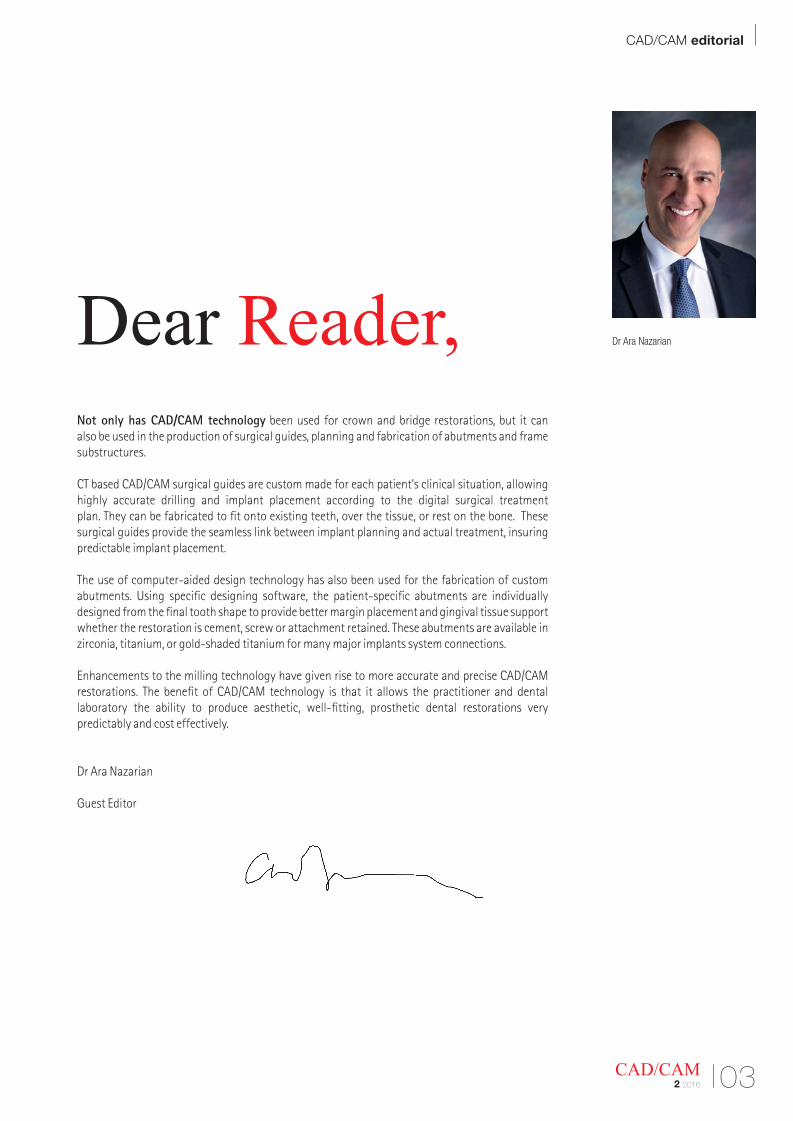
Not only has CAD/CAM technology been used for crown and bridge restorations, but it can also be used in the production of surgical guides, planning and fabrication of abutments and frame substructures.
CT based CAD/CAM surgical guides are custom made for each patient’s clinical situation, allowing highly accurate drilling and implant placement according to the digital surgical treatment plan. They can be fabricated to fit onto existing teeth, over the tissue, or rest on the bone. These surgical guides provide the seamless link between implant planning and actual treatment, insuring predictable implant placement.
The use of computer-aided design technology has also been used for the fabrication of custom abutments. Using specific designing software, the patient-specific abutments are individually designed from the final tooth shape to provide better margin placement and gingival tissue support whether the restoration is cement, screw or attachment retained. These abutments are available in zirconia, titanium, or gold-shaded titanium for many major implants system connections.
Enhancements to the milling technology have given rise to more accurate and precise CAD/CAM restorations. The benefit of CAD/CAM technology is that it allows the practitioner and dental laboratory the ability to produce aesthetic, well-fitting, prosthetic dental restorations very predictably and cost effectively.
Dr Ara Nazarian
Guest Editor
Dr Ara NazarianDear Reader,
| content
page 06
page 30
page 14
page 42
page 20
page 46
© Robert Kneschke/S/S/SSShShSutte
utterstorstockck.com
| editorial
03 Dear ReaderDr Ara Nazarian, Guest Editor
| practice management
06 Seven dental marketing mistakes... and how to avoid themCarolyn S. Dean
| feature
10 “Transfer of knowledge at eye level” An interview with Dr Fred Bergmann, President of the Deutsche Gesellschaft für Orale Implantologie (DGOI), the German society for oral implantology
| case report
14 Abutment fracture in a bridge supported by natural teeth and implantsDrs Gregory-George Zafiropoulos, Giorgio Deli & Rainer Valentin
20 Fixed aesthetic restorationsDr Dario Žujic, DT Velimir Žujic & DT Dragan Stolica
26 Digital planning for full mouth reconstructionDr Ara Nazarian
| opinion
30 The 3-D difference: CBCT diagnostics to enhance treatment—Part 2Dr Anthony Ramirez
| industry news
40 Manufacturer news
44 “The future of dentistry is digital and focused on prevention”An interview with Curaden CEO Ueli Breitschmid
| meetings
46 IMAGINA Dental receives high praise as foremost technology congress
48 International Events
| about the publisher
49 submission guidelines50 imprint
04 CAD/CAM2 2016
CAD/CAM international magazine of digital dentistry
22016
issn 1616-7390 Vol. 7 • Issue 2/2016
case reportDigital planning for full mouth reconstruction
opinionThe 3-D difference: CBCT diagnostics to enhance treatment
practice managementSeven dental marketing mistakes... and how to avoid them
Cover image courtesy of Nobel Biocare Services AG (www.nobelbiocare.com).
VCONCEPT is a universal approach that takes into consideration an entire structure: The innovative V3 implant, the advanced prosthetic appliance and their affect on a greater volume of bone and soft tissue. To learn more about VCONCEPT visit: www.vconcept.com
©MIS Corporation. All Rights Reserved
in a place that has no borders, creativity goes unbound...
By
SET THE VOLUME
| practice management marketing mistakes
06 CAD/CAM2 2016
Seven dental marketing mistakes...and how to avoid themAuthor: Carolyn S. Dean, Australia
As a dental professional, you face unfamiliar chal-lenges in running and marketing your practice. You are confronted with increased competition (both locally and abroad), an oversupply of dentists, ever rising practice operating costs, and more marketing-savvy patients. On top of this, your potential patients are becoming more discerning about where they go for dental treatment, with many heading overseas.
In order to achieve practice success, it is essential to build long-term relationships with patients and prospects. Long-term patients are more likely to feel satisfied. It is they who welcome the opportunity to refer others to you and who will continue to use your services in the future.
Over my years working with hundreds of dentists as a marketing consultant, I have observed the common mistakes that prevent them being able to market their practices successfully.
1. Not knowing your numbers and not tracking them
One of the most common mistakes that I see is that many dental practices just do not track their numbers. There is a saying that “if you fail to plan, you plan to fail”. It is critical that you track all of the metrics in your busi-ness, and your marketing spend is no exception. The sig-nificant numbers that you need to know and track are:
· average lifetime value of a patient · marketing return on investment · new patients · patient loss.
2. Not knowing your ideal patient
One of the cornerstones of any marketing campaign is knowing who your ideal patient is. Many practices
make the mistake of not identifying this in their ea-gerness to go ahead with their marketing campaign as soon as possible. You need to stop and think about whom your marketing will be directed to, what this group of patients wants, what problems they have, and what solutions they need. The key to implement-ing a strategic marketing plan is identifying your practice’s ideal patient or target patient profile. Once you know your market, you need to establish how best to communicate with them.
3. Wanting a silver bullet
Marketing your dental practice to attract the right kind of patients, keep them active and encourage them to refer you to their contacts is no easy task. Many practices think (and hope) that there is a silver bullet to solve their marketing issues. This leaves them open to unscrupulous sales people and to disillusion-ment and frustration when their marketing efforts fail. The companies trying to sell you the marketing silver bullet that will solve all your marketing worries are constantly calling. Well-meaning friends, col-leagues and patients may give you advice on what they think you should do to market your practice. The range of marketing media is evolving, and the rapid changes in online marketing make it almost impos-sible to keep up.
4. Taking a scatter-gun approach
I speak to many dentists who tell me that they have tried many different types of marketing and they have all failed and nothing has worked for them. When I dig deeper, I discover that they have tried many different approaches, but nearly all of these have been done in a haphazard way and in short bursts. I call this a “scatter-gun approach” to marketing. It does not work to try one approach for a month or two in an inconsistent manner without tracking the results or
© koya979/Shutterstock.com
“Many practices think (and hope)
that there is a silver bullet
to solve their marketing issues.”
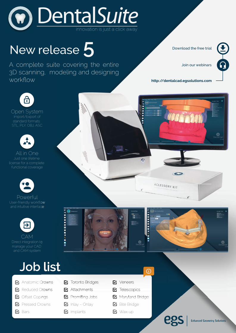
innovation is just a click away
Download the free trial
Join our webinars
http://dentalcad.egsolutions.com
New release 5A complete suite covering the entire 3D scanning, modeling and designing workflow
All in OneJust one lifetime
license for a complete functional coverage
PowerfulUser-friendly workflowand intuitive interface
Open SystemImport/Export ofstandard formats:
STL, PLY, OBJ, ASC
CAMDirect integration tomanage your CAD and CAM system
Job list
User-friendly workflowand intuitive interface
Direct integration toyour CAD
Job listJob list
| practice management marketing mistakes
08 CAD/CAM2 2016
refi ning the campaign. This will always end in failure. It has been shown that it can take between six and eleven repetitions for patients to see or hear a mes-sage before they act on it. Do you know how many ways and how many times you communicate with your patients?
5. Doing it all by yourself
You have to remember that patients are more savvy than ever before. They are constantly exposed to a huge amount of marketing and their expectations of what is and is not professional are continually in-creasing. The reality is that when you are competing against the corporates, you need to ensure that your marketing is up to scratch.
It is very common for practices to have their branding and logo professionally designed and then decide to take it over, producing home-made brochures and other marketing collateral that use different colours, fonts and even versions of the logo. If you are not consistent, your attempts at establishing a brand will be in effective.
6. Procrastinating
There are just so many things for you to think about when it comes to your dental marketing. How can you fi x your website that is not effective? Should you be engaging with your patients on social media and how to start? You know that you need to educate your patients on a regular basis, but what are the best ways to do this? You need reactivation and referral cam-paigns, but you have no idea how to carry this out in a
professional and consistent manner. It is not uncom-mon to be so confused and overwhelmed that you spend your time procrastinating and doing nothing.
7. Not getting the right advice
When you own or run a dental practice, in fact any kind of business, there is no shortage of marketing advice to follow; there is an overwhelming amount of advice out there. You may have had the experience of wasting time or money on poor advice. The problem is that many dentists are not getting the right dental marketing advice. They may listen to many different sources and form opinions based on advice from peo-ple who may not understand the business of dentistry.
8. Summary
There is no magic when it comes to marketing your practice successfully. Quite simply, it comes down to:
· picking the aspects of marketing you want to use, wisely and with due care and thought,
· ensuring that, whatever marketing activities you decide to undertake, you perform to the best of your ability and budget,
· being consistent, · tracking your results—setting your goals and re-
viewing or refi ning them on a regular basis, · getting good advice from trusted experts in the
area of marketing you are undertaking.
It takes time, but the effort that you put in will be re-warded by more patients, increased production, bet-ter relationships with your team and patients, and a sense of control when it comes to your marketing. It is now time for you to focus on your marketing. By marketing well, doing it consistently, and avoiding the scatter-gun approach, you can avoid making the common mistakes that many practices make._
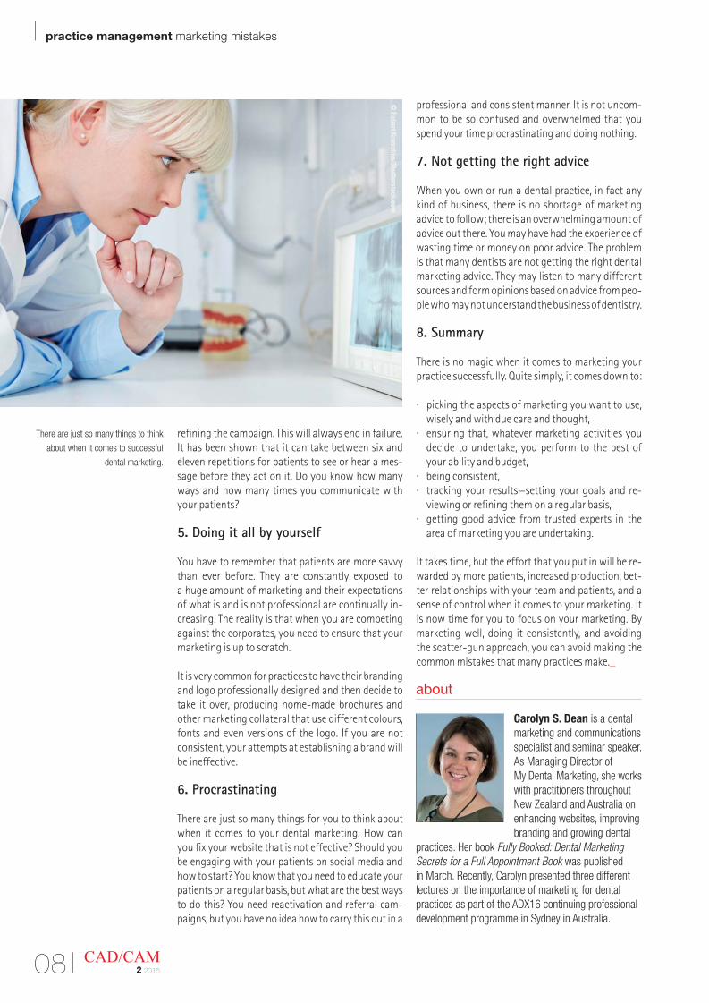
There are just so many things to think
about when it comes to successful
dental marketing.
about
Carolyn S. Dean is a dental marketing and communications specialist and seminar speaker. As Managing Director of My Dental Marketing, she works with practitioners throughout New Zealand and Australia on enhancing websites, improving branding and growing dental
practices. Her book Fully Booked: Dental Marketing Secrets for a Full Appointment Book was published in March. Recently, Carolyn presented three different lectures on the importance of marketing for dental practices as part of the ADX16 continuing professional development programme in Sydney in Australia.
exocad.com/partners
exocad DentalCAD.Flexible, robust and powerful.
Dental CAD/CAM has never been so easy! Contact your local dealer for the latest dental CAD/CAM so�ware generation!
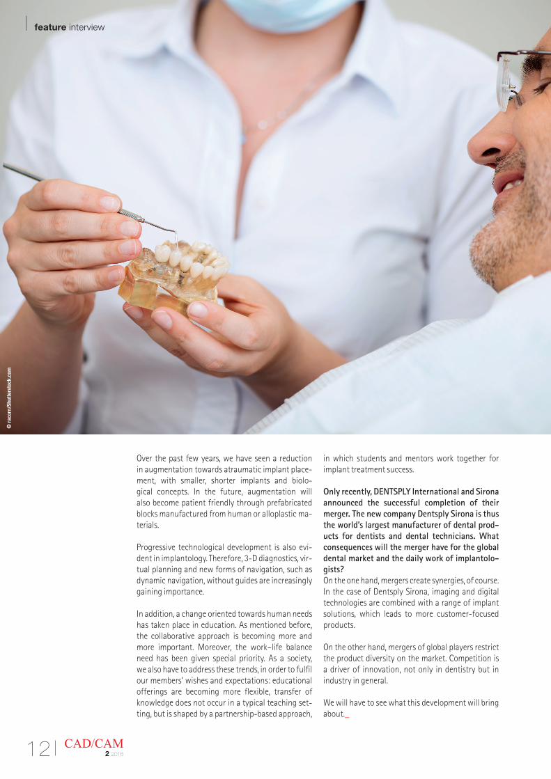
| feature interview
10 CAD/CAM2 2016
“ Transfer of knowledge at eye level”
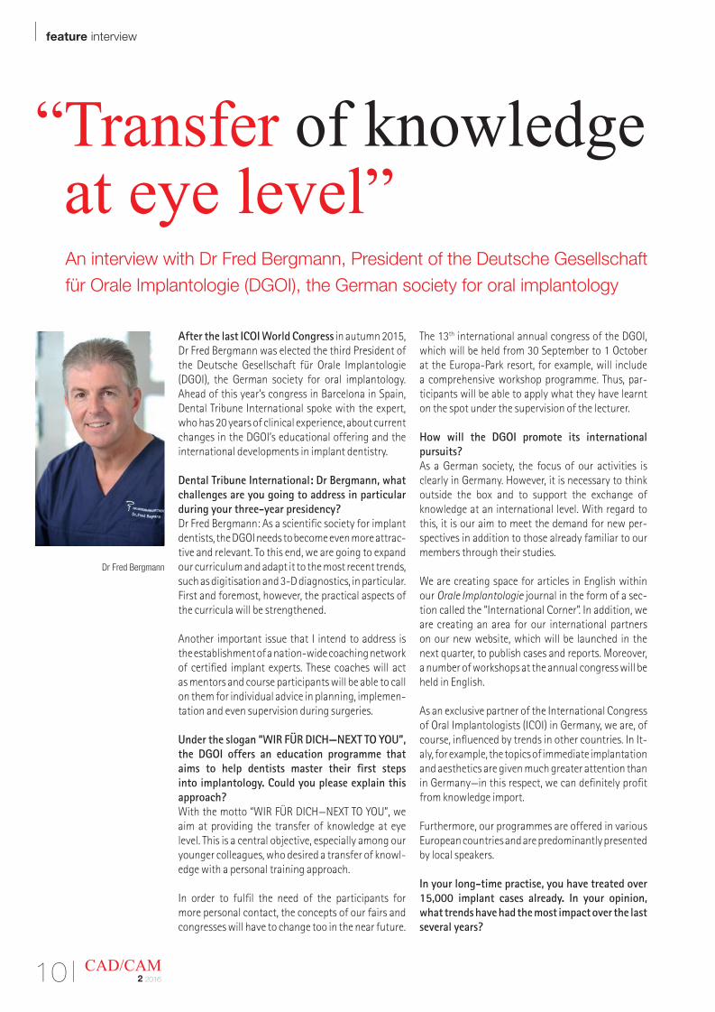
An interview with Dr Fred Bergmann, President of the Deutsche Gesellschaft für Orale Implantologie (DGOI), the German society for oral implantology
After the last ICOI World Congress in autumn 2015, Dr Fred Bergmann was elected the third President of the Deutsche Gesellschaft für Orale Implantologie (DGOI), the German society for oral implantology. Ahead of this year’s congress in Barcelona in Spain, Dental Tribune International spoke with the expert, who has 20 years of clinical experience, about current changes in the DGOI’s educational offering and the international developments in implant dentistry.
Dental Tribune International: Dr Bergmann, what challenges are you going to address in particular during your three-year presidency? Dr Fred Bergmann: As a scientific society for implant dentists, the DGOI needs to become even more attrac-tive and relevant. To this end, we are going to expand our curriculum and adapt it to the most recent trends, such as digitisation and 3-D diagnostics, in particular. First and foremost, however, the practical aspects of the curricula will be strengthened.
Another important issue that I intend to address is the establishment of a nation-wide coaching network of certified implant experts. These coaches will act as mentors and course participants will be able to call on them for individual advice in planning, implemen-tation and even supervision during surgeries.
Under the slogan “WIR FÜR DICH—NEXT TO YOU”, the DGOI offers an education programme that aims to help dentists master their first steps into implantology. Could you please explain this approach?With the motto “WIR FÜR DICH—NEXT TO YOU”, we aim at providing the transfer of knowledge at eye level. This is a central objective, especially among our younger colleagues, who desired a transfer of knowl-edge with a personal training approach.
In order to fulfil the need of the participants for more personal contact, the concepts of our fairs and congresses will have to change too in the near future.
The 13th international annual congress of the DGOI, which will be held from 30 September to 1 October at the Europa-Park resort, for example, will include a comprehensive workshop programme. Thus, par-ticipants will be able to apply what they have learnt on the spot under the supervision of the lecturer.
How will the DGOI promote its international pursuits? As a German society, the focus of our activities is clearly in Germany. However, it is necessary to think outside the box and to support the exchange of knowledge at an international level. With regard to this, it is our aim to meet the demand for new per-spectives in addition to those already familiar to our members through their studies.
We are creating space for articles in English within our Orale Implantologie journal in the form of a sec-tion called the “International Corner”. In addition, we are creating an area for our international partners on our new website, which will be launched in the next quarter, to publish cases and reports. Moreover, a number of workshops at the annual congress will be held in English.
As an exclusive partner of the International Congress of Oral Implantologists (ICOI) in Germany, we are, of course, influenced by trends in other countries. In It-aly, for example, the topics of immediate implantation and aesthetics are given much greater attention than in Germany—in this respect, we can definitely profit from knowledge import.
Furthermore, our programmes are offered in various European countries and are predominantly presented by local speakers.
In your long-time practise, you have treated over 15,000 implant cases already. In your opinion, what trends have had the most impact over the last several years?
Dr Fred Bergmann
More than a lab partner.True ambition to increase
your efficiency.
At Straumann we are fully committed to taking care of you and the success of your business. We stand for highest quality, and our passion is to continu-ously shape our portfolio offering with innovative products & services that simplify your workflows and increase your efficiency. Find out what’s in it for you!
www.straumann.com/dentallab
| feature interview
12 CAD/CAM2 2016
Over the past few years, we have seen a reduction in augmentation towards atraumatic implant place-ment, with smaller, shorter implants and biolo- gical concepts. In the future, augmentation will also become patient friendly through prefabricated blocks manufactured from human or alloplastic ma-terials.
Progressive technological development is also evi-dent in implantology. Therefore, 3-D diagnostics, vir-tual planning and new forms of navigation, such as dynamic navigation, without guides are increasingly gaining importance.
In addition, a change oriented towards human needs has taken place in education. As mentioned before, the collaborative approach is becoming more and more important. Moreover, the work–life balance need has been given special priority. As a society, we also have to address these trends, in order to fulfil our members’ wishes and expectations: educational offerings are becoming more flexible, transfer of knowledge does not occur in a typical teaching set-ting, but is shaped by a partnership-based approach,
in which students and mentors work together for implant treatment success.
Only recently, DENTSPLY International and Sirona announced the successful completion of their merger. The new company Dentsply Sirona is thus the world’s largest manufacturer of dental prod-ucts for dentists and dental technicians. What consequences will the merger have for the global dental market and the daily work of implantolo-gists?On the one hand, mergers create synergies, of course. In the case of Dentsply Sirona, imaging and digital technologies are combined with a range of implant solutions, which leads to more customer-focused products.
On the other hand, mergers of global players restrict the product diversity on the market. Competition is a driver of innovation, not only in dentistry but in industry in general.
We will have to see what this development will bring about._
© ra
corn
/Shu
tters
tock
.com
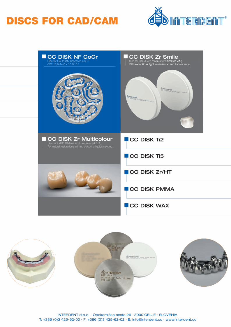
CC DISK NF CoCrDisc for CAD/CAM based on CoCr.CTE 13,9-14,0 x 10⁻⁶K10-1
INTERDENT d.o.o. · Opekarniška cesta 26 · 3000 CELJE · SLOVENIAT: +386 (0)3 425-62-00 · F: +386 (0)3 425-62-02 · E: [email protected] · www.interdent.cc
CC DISK Ti2
DISCS FOR CAD/CAM
CC DISK Zr MulticolourDisc for CAD/CAM made of pre-sintered ZrO
2.
For natural restorations with no colouring liquids needed.
CC DISK Zr SmileDisc for CAD/CAM made of pre-sintered ZrO
2.
With exceptional light transmission and translucency.
CC DISK Ti5
CC DISK Zr/HT
CC DISK PMMA
CC DISK WAX
Disc for CAD/CAM made of pre-sintered ZrO2.
With exceptional light transmission and translucency.
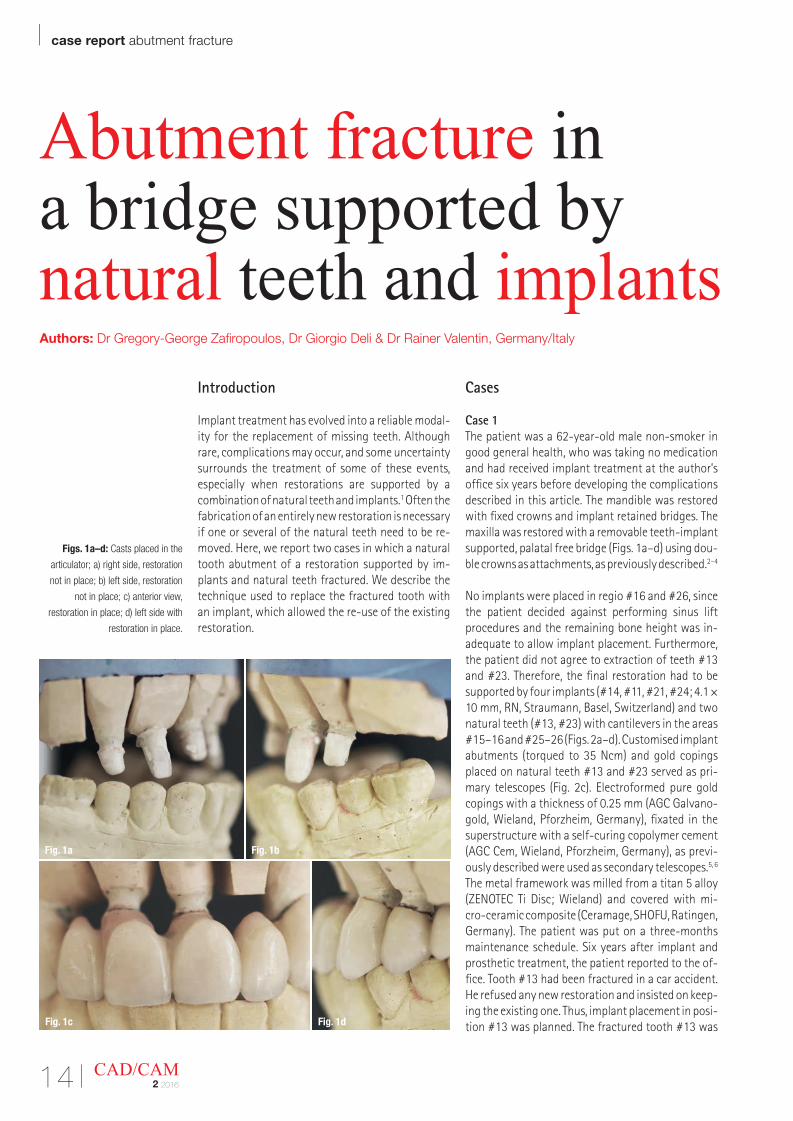
Introduction
Implant treatment has evolved into a reliable modal-ity for the replacement of missing teeth. Although rare, complications may occur, and some uncertainty surrounds the treatment of some of these events, especially when restorations are supported by a combination of natural teeth and implants.1 Often the fabrication of an entirely new restoration is necessary if one or several of the natural teeth need to be re-moved. Here, we report two cases in which a natural tooth abutment of a restoration supported by im-plants and natural teeth fractured. We describe the technique used to replace the fractured tooth with an implant, which allowed the re-use of the existing restoration.
Cases
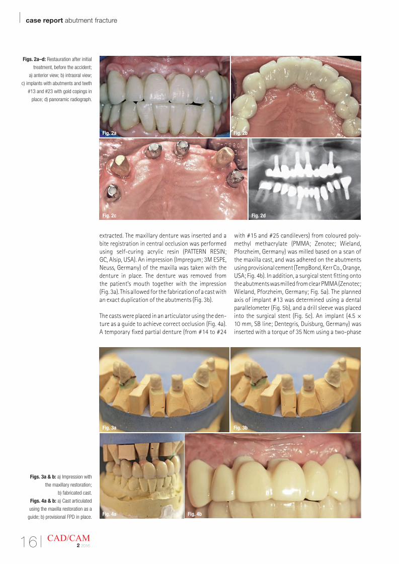
Case 1The patient was a 62-year-old male non-smoker in good general health, who was taking no medication and had received implant treatment at the author’s office six years before developing the complications described in this article. The mandible was restored with fixed crowns and implant retained bridges. The maxilla was restored with a removable teeth-implant supported, palatal free bridge (Figs. 1a–d) using dou-ble crowns as attachments, as previously described.2–4
No implants were placed in regio #16 and #26, since the patient decided against performing sinus lift procedures and the remaining bone height was in-adequate to allow implant placement. Furthermore, the patient did not agree to extraction of teeth #13 and #23. Therefore, the final restoration had to be supported by four implants (#14, #11, #21, #24; 4.1 × 10 mm, RN, Straumann, Basel, Switzerland) and two natural teeth (#13, #23) with cantilevers in the areas #15–16 and #25–26 (Figs. 2a–d). Customised implant abutments (torqued to 35 Ncm) and gold copings placed on natural teeth #13 and #23 served as pri-mary telescopes (Fig. 2c). Electroformed pure gold copings with a thickness of 0.25 mm (AGC Galvano-gold, Wieland, Pforzheim, Germany), fixated in the superstructure with a self-curing copolymer cement (AGC Cem, Wieland, Pforzheim, Germany), as previ-ously described were used as secondary telescopes.5, 6 The metal framework was milled from a titan 5 alloy (ZENOTEC Ti Disc; Wieland) and covered with mi-cro-ceramic composite (Ceramage, SHOFU, Ratingen, Germany). The patient was put on a three-months maintenance schedule. Six years after implant and prosthetic treatment, the patient reported to the of-fice. Tooth #13 had been fractured in a car accident. He refused any new restoration and insisted on keep-ing the existing one. Thus, implant placement in posi-tion #13 was planned. The fractured tooth #13 was
| case report abutment fracture
14 CAD/CAM2 2016
Abutment fracture in a bridge supported by natural teeth and implantsAuthors: Dr Gregory-George Zafiropoulos, Dr Giorgio Deli & Dr Rainer Valentin, Germany/Italy
Figs. 1a–d: Casts placed in the
articulator; a) right side, restoration
not in place; b) left side, restoration
not in place; c) anterior view,
restoration in place; d) left side with
restoration in place.
Fig. 1a Fig. 1b
Fig. 1c Fig. 1d
Beyond CAD/CAMPatient-specific prosthetic solutions for all major implant systems
In order to provide truly ideal solutions, you need restorative versatility, flexibility within your workflow and design options that are as individual as each patient. By choosing ATLANTIS, you get the freedom, esthetics, simplicity and reliability that go beyond CAD/CAM.
DEN
TSPL
Y Im
plan
ts d
oes
not w
aive
any
rig
ht to
its
trade
mar
ks b
y no
t usi
ng th
e sy
mbo
ls ®
or ™
. 32
6708
48-U
SX-1
512
© 2
015
DEN
TSPL
Y Im
plan
ts. A
ll rig
hts
rese
rved
Follow DENTSPLY Implants
www.dentsplyimplants.com
32670848-USX-1512 ATLANTIS Ad_A4.indd 1 2016-01-14 08:54
| case report abutment fracture
16 CAD/CAM2 2016
extracted. The maxillary denture was inserted and a bite registration in central occlusion was performed using self-curing acrylic resin (PATTERN RESIN; GC, Alsip, USA). An impression (Impregum; 3M ESPE, Neuss, Germany) of the maxilla was taken with the denture in place. The denture was removed from the patient’s mouth together with the impression (Fig. 3a). This allowed for the fabrication of a cast with an exact duplication of the abutments (Fig. 3b).
The casts were placed in an articulator using the den-ture as a guide to achieve correct occlusion (Fig. 4a). A temporary fixed partial denture (from #14 to #24
with #15 and #25 candilevers) from coloured poly-methyl methacrylate (PMMA; Zenotec; Wieland, Pforzheim, Germany) was milled based on a scan of the maxilla cast, and was adhered on the abutments using provisional cement (TempBond, Kerr Co., Orange, USA; Fig. 4b). In addition, a surgical stent fitting onto the abutments was milled from clear PMMA (Zenotec; Wieland, Pforzheim, Germany; Fig. 5a). The planned axis of implant #13 was determined using a dental parallelometer (Fig. 5b), and a drill sleeve was placed into the surgical stent (Fig. 5c). An implant (4.5 × 10 mm, SB line; Dentegris, Duisburg, Germany) was inserted with a torque of 35 Ncm using a two-phase
Figs. 2a–d: Restauration after initial
treatment, before the accident;
a) anterior view; b) intraoral view;
c) implants with abutments and teeth
#13 and #23 with gold copings in
place; d) panoramic radiograph.
Figs. 3a & b: a) Impression with
the maxillary restoration;
b) fabricated cast.
Figs. 4a & b: a) Cast articulated
using the maxilla restoration as a
guide; b) provisional FPD in place.
Fig. 2a Fig. 2b
Fig. 2c Fig. 2d
Fig. 3a Fig. 3b
Fig. 4a Fig. 4b
abutment fracture case report |
17CAD/CAM2 2016
Figs. 5a–c:_a) Surgical stent;
b) determination of the axial direction;
c) drill sleeve was placed into
the surgical stent.
Fig. 6: Panoramic radiograph
obtained after implant insertion
at position #6.
Fig. 7: Impression post in the
surgical guide.
Figs. 8a & b: a) Transfer of the
implant position to mounted cast;
b) implant analog in cast.
Figs. 9a & b: a) Fabricated
customised abutment; b) try-in of
the abutment in the restoration.
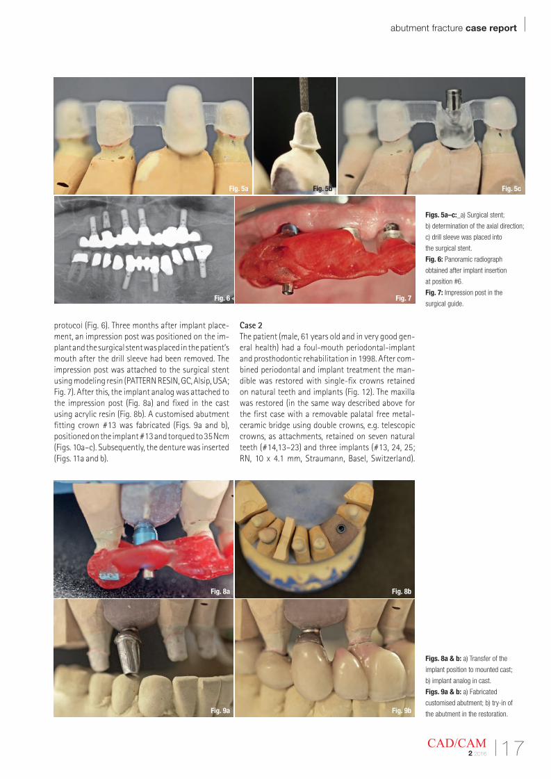
protocol (Fig. 6). Three months after implant place-ment, an impression post was positioned on the im-plant and the surgical stent was placed in the patient’s mouth after the drill sleeve had been removed. The impression post was attached to the surgical stent using modeling resin (PATTERN RESIN, GC, Alsip, USA; Fig. 7). After this, the implant analog was attached to the impression post (Fig. 8a) and fixed in the cast using acrylic resin (Fig. 8b). A customised abutment fitting crown #13 was fabricated (Figs. 9a and b), positioned on the implant #13 and torqued to 35 Ncm (Figs. 10a–c). Subsequently, the denture was inserted (Figs. 11a and b).
Case 2The patient (male, 61 years old and in very good gen-eral health) had a foul-mouth periodontal-implant and prosthodontic rehabilitation in 1998. After com-bined periodontal and implant treatment the man-dible was restored with single-fix crowns retained on natural teeth and implants (Fig. 12). The maxilla was restored (in the same way described above for the first case with a removable palatal free metal- ceramic bridge using double crowns, e.g. telescopic crowns, as attachments, retained on seven natural teeth (#14,13–23) and three implants (#13, 24, 25; RN, 10 x 4.1 mm, Straumann, Basel, Switzerland).
Fig. 5a Fig. 5b Fig. 5c
Fig. 6 Fig. 7
Fig. 8a Fig. 8b
Fig. 9a Fig. 9b
Fig. 2a
| case report abutment fracture
18 CAD/CAM2 2016
Because the patient did not consent to a sinus aug-mentation, no implants were placed in regio #16 and #26. Tooth #14 was treated endodontically and was used as the last abutment (Fig. 12).
Thirteen years after prosthetic rehabilitation, the patient reported to the office with a root fracture in tooth #15. The tooth was extracted, the secondary
telescopic crown in regio #15 was removed, and the supraconstruction was temporarily filled with a pho-tocured, highly elastic temporary material (Fermit, Ivoclar Vivadent, Ellwangen, Germany). The patient again refused a sinus lift and therefore the immediate implant placement regio #15 was scheduled. The axis of the tooth #15, the fabrication of the transfer key and the implant placement, were performed as pre-
Figs. 10a–c: a & b) Abutment
mounted on the implant; c) panoramic
radiograph of custom-made
abutment #13 in place.
Figs. 11a & b: Denture in place; a)
close-up; b) anterior view.
Fig. 12: Orthopatomograph taken
at the completion of the perio-
implant-prosthodontic treatment.
Figs. 13a & b: a) Surgical guide for
implant placement #15; b) X-ray after
implant placement.
Fig. 10a Fig. 10b Fig. 10c
Fig. 11a Fig. 11b
Fig. 13a Fig. 13b
Fig. 12
abutment fracture case report |
19CAD/CAM2 2016
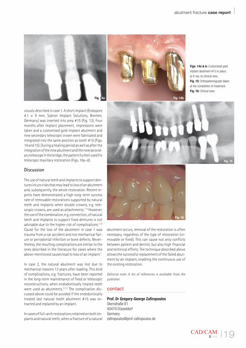
viously described in case 1. A short implant (Endopore 4.1 × 9 mm, Sybron Implant Solutions, Bremen, Germany) was inserted into area #15 (Fig. 13). Four months after implant placement, impressions were taken and a customised gold implant abutment and new secondary telescopic crown were fabricated and integrated into the same position as tooth #15 (Figs. 14 and 15). During a healing period as well as after the integration of the new abutment and the new second-ary telescope in the bridge, the patient further used his telescopic maxillary restoration (Figs. 16a–d).
Discussion
The use of natural teeth and implants to support den-tures incurs risks that may lead to loss of an abutment and, subsequently, the whole restoration. Recent re-ports have demonstrated a high long-term success rate of removable restorations supported by natural teeth and implants when double crowns, e.g. tele-scopic crowns, are used as attachments.7–9 However, the use of the combination, e.g. connection, of natural teeth and implants to support fixed dentures is not advisable due to the higher risk of complications.1,10 Cause for the loss of the abutment in case 1 was trauma from a car accident and not mechanical fail-ure or periodontal infection or bone defects. Never-theless, the resulting complications are similar to the ones described in the literature for cases where the above-mentioned causes lead to loss of an implant.1
In case 2, the natural abutment was lost due to mechanical reasons 13 years after loading. This kind of complications, e.g. fractures, have been reported in the long-term maintenance of fixed or telescopic reconstructions, when endodontically treated teeth were used as abutments.11,12 The complication dis-cussed above could be avoided if the endodontically treated last natural tooth abutment #15 was ex-tracted and replaced by an implant.
In cases of full-arch restorations retained on both im-plants and natural teeth, when a fracture of a natural
abutment occurs, removal of the restoration is often necessary, regardless of the type of restoration (re-movable or fixed). This can cause not only conflicts between patient and dentist, but also high financial and technical efforts. The technique described above allows the successful replacement of the failed abut-ment by an implant, enabling the continuous use of the existing restoration.
Editorial note: A list of references is available from the publisher.
Figs. 14a & b: Customised gold
implant abutment #15 in place;
a) X-ray; b) clinical view.
Fig. 15: Orthopatomograph taken
at the completion of treatment.
Fig. 16: Clinical view.
contact
Prof. Dr Gregory-George ZafiropoulosSternstraße 6140479 Dü[email protected]
Fig. 14a Fig. 14b
Fig. 15
Fig. 16
| case report aesthetic restorations
20 CAD/CAM2 2016
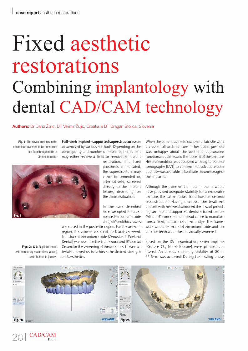
Fixed aesthetic restorationsCombining implantology with dental CAD/CAM technologyAuthors: Dr Dario Žujic, DT Velimir Žujic, Croatia & DT Dragan Stolica, Slovenia
Full-arch implant-supported superstructures can be achieved by various methods. Depending on the bone quality and number of implants, the patient may either receive a fixed or removable implant
restoration. If a fixed prosthesis is indicated, the superstructure may either be cemented or, alternatively, screwed directly to the implant fixture, depending on the clinical situation.
In the case described here, we opted for a ce-mented zirconium oxide bridge. Monolithic crowns
were used in the posterior region. For the anterior region, the crowns were cut back and veneered. Translucent zirconium oxide (Zenostar T, Wieland Dental) was used for the framework and IPS e.max Ceram for the veneering of the anteriors. These ma-terials allowed us to achieve the desired strength and aesthetics.
When the patient came to our dental lab, she wore a classic full-arch denture in her upper jaw. She was unhappy about the aesthetic appearance, functional qualities and the loose fit of the denture. Her oral condition was assessed with digital volume tomography (DVT) to confirm that adequate bone quantity was available to facilitate the anchorage of the implants.
Although the placement of four implants would have provided adequate stability for a removable denture, the patient asked for a fixed all-ceramic reconstruction. Having discussed the treatment options with her, we abandoned the idea of provid-ing an implant-supported denture based on the “All-on-4” concept and instead chose to manufac-ture a fixed, implant-retained bridge. The frame-work would be made of zirconium oxide and the anterior teeth would be individually veneered.
Based on the DVT examination, seven implants (Replace CC, Nobel Biocare) were planned and placed. An adequate primary stability of 30 to 35 Ncm was achieved. During the healing phase,
Fig. 1: The seven implants in the
edentulous jaw were to be connected
to a fixed bridge made of
zirconium oxide.
Figs. 2a & b: Digitized model
with temporary restorations (above)
and abutments (below).
Fig. 1
Fig. 2a Fig. 2b
aesthetic restorations case report |
21CAD/CAM2 2016
the patient wore the existing denture that had been relined with soft silicone.
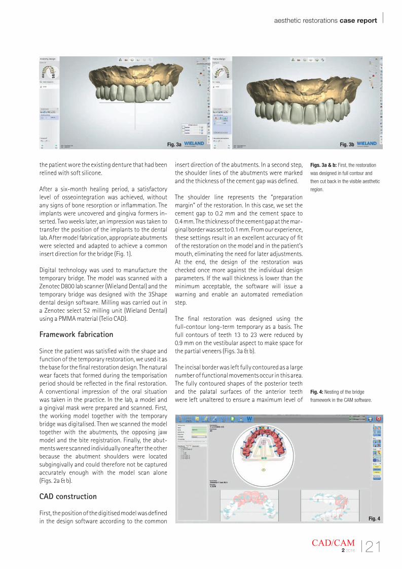
After a six-month healing period, a satisfactory level of osseointegration was achieved, without any signs of bone resorption or inflammation. The implants were uncovered and gingiva formers in-serted. Two weeks later, an impression was taken to transfer the position of the implants to the dental lab. After model fabrication, appropriate abutments were selected and adapted to achieve a common insert direction for the bridge (Fig. 1).
Digital technology was used to manufacture the temporary bridge. The model was scanned with a Zenotec D800 lab scanner (Wieland Dental) and the temporary bridge was designed with the 3Shape dental design software. Milling was carried out in a Zenotec select S2 milling unit (Wieland Dental) using a PMMA material (Telio CAD).
Framework fabrication
Since the patient was satisfied with the shape and function of the temporary restoration, we used it as the base for the final restoration design. The natural wear facets that formed during the temporisation period should be reflected in the final restoration. A conventional impression of the oral situation was taken in the practice. In the lab, a model and a gingival mask were prepared and scanned. First, the working model together with the temporary bridge was digitalised. Then we scanned the model together with the abutments, the opposing jaw model and the bite registration. Finally, the abut-ments were scanned individually one after the other because the abutment shoulders were located subgingivally and could therefore not be captured accurately enough with the model scan alone (Figs. 2a & b).
CAD construction
First, the position of the digitised model was defined in the design software according to the common
insert direction of the abutments. In a second step, the shoulder lines of the abutments were marked and the thickness of the cement gap was defined.
The shoulder line represents the “preparation margin” of the restoration. In this case, we set the cement gap to 0.2 mm and the cement space to 0.4 mm. The thickness of the cement gap at the mar-ginal border was set to 0.1 mm. From our experience, these settings result in an excellent accuracy of fit of the restoration on the model and in the patient’s mouth, eliminating the need for later adjustments. At the end, the design of the restoration was checked once more against the individual design parameters. If the wall thickness is lower than the minimum acceptable, the software will issue a warning and enable an automated remediation step.
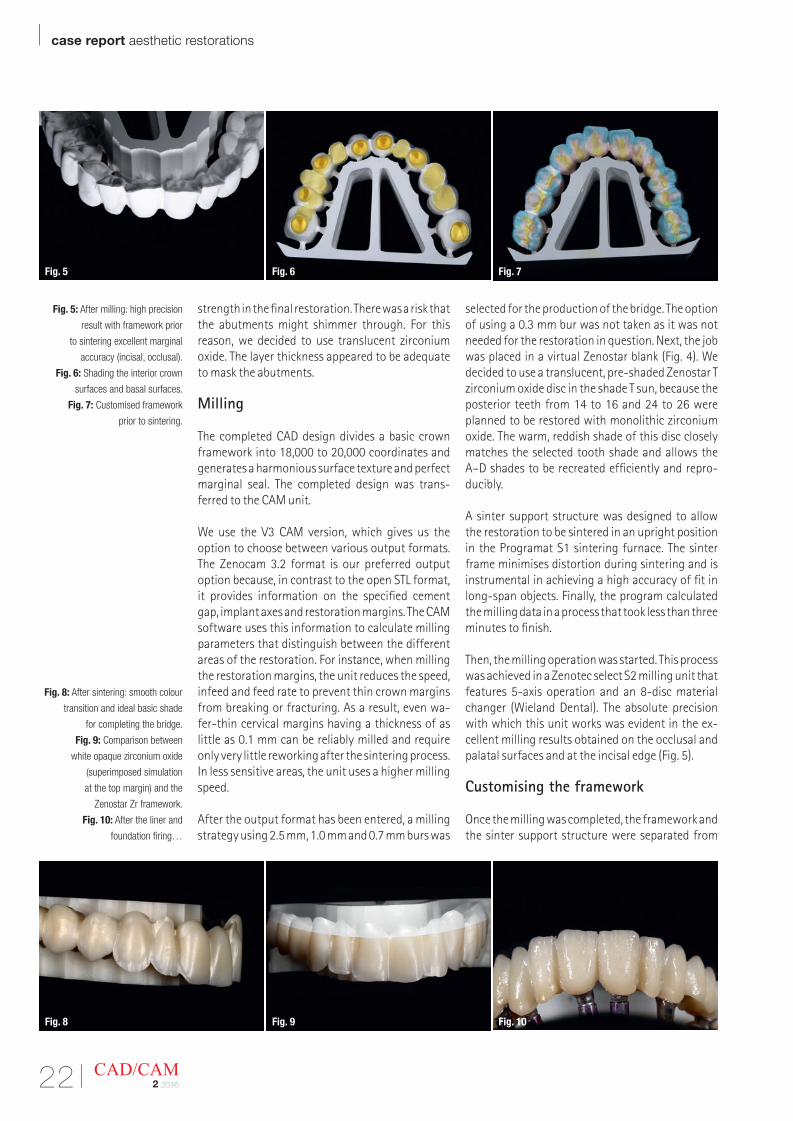
The final restoration was designed using the full-contour long-term temporary as a basis. The full contours of teeth 13 to 23 were reduced by 0.9 mm on the vestibular aspect to make space for the partial veneers (Figs. 3a & b).
The incisal border was left fully contoured as a large number of functional movements occur in this area. The fully contoured shapes of the posterior teeth and the palatal surfaces of the anterior teeth were left unaltered to ensure a maximum level of
Figs. 3a & b: First, the restoration
was designed in full contour and
then cut back in the visible aesthetic
region.
Fig. 4: Nesting of the bridge
framework in the CAM software.
Fig. 3a Fig. 3b
Fig. 4
| case report aesthetic restorations
22 CAD/CAM2 2016
strength in the final restoration. There was a risk that the abutments might shimmer through. For this reason, we decided to use translucent zirconium oxide. The layer thickness appeared to be adequate to mask the abutments.
Milling
The completed CAD design divides a basic crown framework into 18,000 to 20,000 coordinates and generates a harmonious surface texture and perfect marginal seal. The completed design was trans-ferred to the CAM unit.
We use the V3 CAM version, which gives us the option to choose between various output formats. The Zenocam 3.2 format is our preferred output option because, in contrast to the open STL format, it provides information on the specified cement gap, implant axes and restoration margins. The CAM software uses this information to calculate milling parameters that distinguish between the different areas of the restoration. For instance, when milling the restoration margins, the unit reduces the speed, infeed and feed rate to prevent thin crown margins from breaking or fracturing. As a result, even wa-fer-thin cervical margins having a thickness of as little as 0.1 mm can be reliably milled and require only very little reworking after the sintering process. In less sensitive areas, the unit uses a higher milling speed.
After the output format has been entered, a milling strategy using 2.5 mm, 1.0 mm and 0.7 mm burs was
selected for the production of the bridge. The option of using a 0.3 mm bur was not taken as it was not needed for the restoration in question. Next, the job was placed in a virtual Zenostar blank (Fig. 4). We decided to use a translucent, pre-shaded Zenostar T zirconium oxide disc in the shade T sun, because the posterior teeth from 14 to 16 and 24 to 26 were planned to be restored with monolithic zirconium oxide. The warm, reddish shade of this disc closely matches the selected tooth shade and allows the A–D shades to be recreated efficiently and repro-ducibly.
A sinter support structure was designed to allow the restoration to be sintered in an upright position in the Programat S1 sintering furnace. The sinter frame minimises distortion during sintering and is instrumental in achieving a high accuracy of fit in long-span objects. Finally, the program calculated the milling data in a process that took less than three minutes to finish.
Then, the milling operation was started. This process was achieved in a Zenotec select S2 milling unit that features 5-axis operation and an 8-disc material changer (Wieland Dental). The absolute precision with which this unit works was evident in the ex-cellent milling results obtained on the occlusal and palatal surfaces and at the incisal edge (Fig. 5).
Customising the framework
Once the milling was completed, the framework and the sinter support structure were separated from
Fig. 5: After milling: high precision
result with framework prior
to sintering excellent marginal
accuracy (incisal, occlusal).
Fig. 6: Shading the interior crown
surfaces and basal surfaces.
Fig. 7: Customised framework
prior to sintering.
Fig. 8: After sintering: smooth colour
transition and ideal basic shade
for completing the bridge.
Fig. 9: Comparison between
white opaque zirconium oxide
(superimposed simulation
at the top margin) and the
Zenostar Zr framework.
Fig. 10: After the liner and
foundation firing…
Fig. 5 Fig. 6 Fig. 7
Fig. 8 Fig. 9 Fig. 10
aesthetic restorations case report |
23CAD/CAM2 2016
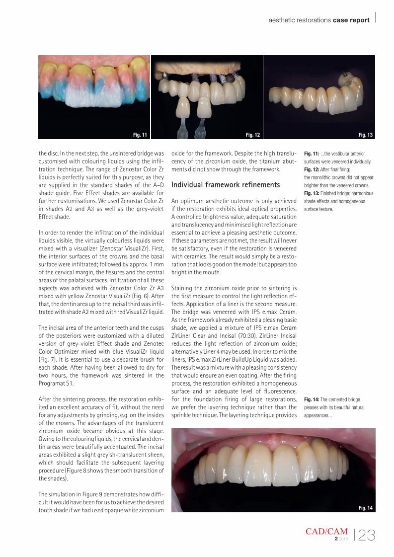
the disc. In the next step, the unsintered bridge was customised with colouring liquids using the infil-tration technique. The range of Zenostar Color Zr liquids is perfectly suited for this purpose, as they are supplied in the standard shades of the A–D shade guide. Five Effect shades are available for further customisations. We used Zenostar Color Zr in shades A2 and A3 as well as the grey-violet Effect shade.
In order to render the infiltration of the individual liquids visible, the virtually colourless liquids were mixed with a visualizer (Zenostar VisualiZr). First, the interior surfaces of the crowns and the basal surface were infiltrated; followed by approx. 1 mm of the cervical margin, the fissures and the central areas of the palatal surfaces. Infiltration of all these aspects was achieved with Zenostar Color Zr A3 mixed with yellow Zenostar VisualiZr (Fig. 6). After that, the dentin area up to the incisal third was infil-trated with shade A2 mixed with red VisualiZr liquid.
The incisal area of the anterior teeth and the cusps of the posteriors were customized with a diluted version of grey-violet Effect shade and Zenotec Color Optimizer mixed with blue VisualiZr liquid (Fig. 7). It is essential to use a separate brush for each shade. After having been allowed to dry for two hours, the framework was sintered in the Programat S1.
After the sintering process, the restoration exhib-ited an excellent accuracy of fit, without the need for any adjustments by grinding, e.g. on the insides of the crowns. The advantages of the translucent zirconium oxide became obvious at this stage. Owing to the colouring liquids, the cervical and den-tin areas were beautifully accentuated. The incisal areas exhibited a slight greyish-translucent sheen, which should facilitate the subsequent layering procedure (Figure 8 shows the smooth transition of the shades).
The simulation in Figure 9 demonstrates how diffi-cult it would have been for us to achieve the desired tooth shade if we had used opaque white zirconium
oxide for the framework. Despite the high translu-cency of the zirconium oxide, the titanium abut-ments did not show through the framework.
Individual framework refinements
An optimum aesthetic outcome is only achieved if the restoration exhibits ideal optical properties. A controlled brightness value, adequate saturation and translucency and minimised light reflection are essential to achieve a pleasing aesthetic outcome. If these parameters are not met, the result will never be satisfactory, even if the restoration is veneered with ceramics. The result would simply be a resto-ration that looks good on the model but appears too bright in the mouth.
Staining the zirconium oxide prior to sintering is the first measure to control the light reflection ef-fects. Application of a liner is the second measure. The bridge was veneered with IPS e.max Ceram. As the framework already exhibited a pleasing basic shade, we applied a mixture of IPS e.max Ceram ZirLiner Clear and Incisal (70:30). ZirLiner Incisal reduces the light reflection of zirconium oxide; alternatively Liner 4 may be used. In order to mix the liners, IPS e.max ZirLiner BuildUp Liquid was added. The result was a mixture with a pleasing consistency that would ensure an even coating. After the firing process, the restoration exhibited a homogeneous surface and an adequate level of fluorescence. For the foundation firing of large restorations, we prefer the layering technique rather than the sprinkle technique. The layering technique provides
Fig. 11: ...the vestibular anterior
surfaces were veneered individually.
Fig. 12: After final firing:
the monolithic crowns did not appear
brighter than the veneered crowns.
Fig. 13: Finished bridge: harmonious
shade effects and homogeneous
surface texture.
Fig. 14: The cemented bridge
pleases with its beautiful natural
appearances...
Fig. 11 Fig. 12 Fig. 13
Fig. 14
| case report aesthetic restorations
24 CAD/CAM2 2016
better adhesion and optical effects (wash firing: Deep Dentin A2, A1, DA2, A1 and T-Neutral) (Fig. 10). The individual vestibular surfaces can be easily veneered.
The tooth shape was given and the framework was used as the basic shade (veneering: Dentin A2, A1, T-Neutral, OE1, OE2, I1) (Fig. 11). After the firing pro-cess was completed, the value, saturation and light reflection effects looked as desired. The shade effect of the restoration is identical in intensive light, in normal light and in the shade and matches the chosen A–D tooth shade.
Shade characterisations (Shades, Stains) are applied to the monolithic portions before dentin firing. We continued to apply thin “soft” coatings of colour and used IPS e.max Glaze Fluo for the glaze firing process.
After the final firing, the restoration exhibited har-monious shade effects. The bridge satisfied all functional and aesthetic criteria. The monolithic portions did not appear brighter than the veneered parts (Fig. 12). Finally, we polished the bridge and
ensured that the conditions for optimum oral hygiene were in place. Smooth sur-faces are essential to prevent the excel-lent biocompatibility of zirconium oxide from being diminished and undesirable wear from occurring in the opposing jaw. After a final check, the restoration was forwarded to the dental practice (Fig. 13).
Conclusion
After the preparations were completed, the bridge was cemented in place. The ceramic restoration looks three-dimen-sional. Even without layering, the poste-rior teeth demonstrated a natural colour depth. With their vibrant internal shade effects and lifelike warm translucency, the anterior teeth demonstrated impres-sive aesthetic properties (Fig. 14).
The combination of cutting-edge milling technol-ogy and high-quality veneering ceramics provides an efficient route to achieving aesthetically pleas-ing, reliable and long-lasting treatment results. The goal of the prosthetic treatment team is to see a happy patient with a beautiful natural smile (Fig. 15)._
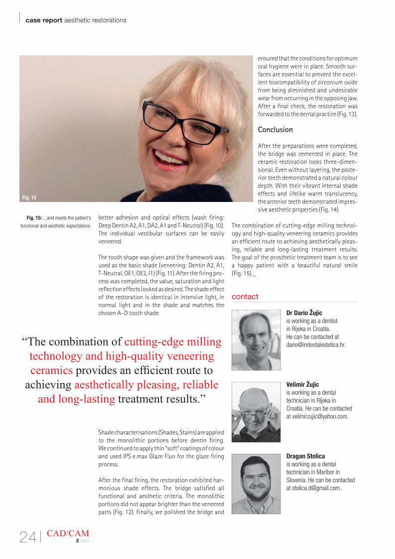
Fig. 15: ...and meets the patient’s
functional and aesthetic expectations.
contact
Dr Dario Žujic is working as a dentist in Rijeka in Croatia. He can be contacted at [email protected].
Velimir Žujic is working as a dental technician in Rijeka in Croatia. He can be contacted at [email protected].
Dragan Stolica is working as a dental technician in Maribor in Slovenia. He can be contact ed at [email protected].
“The combination of cutting-edge milling technology and high-quality veneering ceramics provides an efficient route to
achieving aesthetically pleasing, reliable and long-lasting treatment results.”
Fig. 15
OPEN 3D DENTAL SCANNER
Maestro 3D DENTAL SystemInnovative solutions for dental applications
www.maestro3d.com
Attachment designer
Label designer
Brackets module
IPRInterproximal reduction
Models Builder module Clear aligner module Crown & Bridge
www.maestro3d.com
| case report digital planning
26 CAD/CAM2 2016
Digital planning for full mouth reconstructionAuthor: Dr Ara Nazarian, USA
With greater public awareness about cosmetic dental reconstructions, dentists are often chal- lenged with greater demands from the patient. This increased demand for aesthetic restorative treatment challenges the dentist, the laboratory technician and dental manufacturers to develop techniques and materials to satisfy the discerning patient. Utilising digital planning, modern materials and effective techniques, the restorative team can succeed in restoring a smile to proper form, func-tion, and health. The case presented in this article demonstrates the significance of a systematic ap-proach to planning, preparation and material se-lection in full mouth reconstruction of a patient’s dentition.
Case presentation
A man in his late 30s was referred to my practice by his dental provider because he was dissatisfied with the appearance of his smile. The patient commented that he felt that his existing teeth and restorations were unattractive because of recurrent decay, wear and colour (Figs. 1 & 2). Most im portantly, he men-tioned that he was suffering from tension head-aches, grinding and a limited range of function.
Initial diagnostic evaluation at the first appoint-ment consisted of a series of digital images with study casts, a centric relation bite record, a face bow transfer and a full mouth set of X-rays.
In the maxillary arch, the patient had several teeth that had worn composite restorations as well as abfractions with cervical decay. Tooth #5 had an existing crown on an implant.
In the lower arch, several existing composite resto-rations had wear as well as decay on the facial cer-vical areas. Although there were no re storations present in the anterior mandibular teeth, there was severe wear in the incisal edges due to possible grinding, parafunction and end-to-end bite.
Planning
After reviewing the clinical findings as well as the mounted models, the patient was diagnosed with a restricted envelope of function and decreased ver-tical dimension from continuous wear. To develop a treatment plan and determine if the vertical dimen-sion could be increased, a diagnostic 3-D White Wax-Up (Arrowhead Dental Lab) was fabricated (Fig. 3). With this service, the dental provider also receives a Preparation Guide as well as a Tempo-risation Fabrication Template (Fig. 4). The vertical dimension was increased by 1.5 mm. Based on infor-mation gathered from the initial consult and digital images, it was determined that the maxillary cen-trals could be lengthened by 1.3 mm to improve the aesthetics. The canines would also be lengthened to restore canine guidance in lateral excursions. In regards to his lower anterior teeth, the goal was
to correct the length to width ratio and create a less worn appearance. As a result of the information gathered from the diagnostic wax-up, it was determined that aesthetics and function could be enhanced by restor-ing the entire dentition. Since tooth #31 was already missing and tooth #2 already had a root canal, core and crown restoration, it was decided to
Fig. 1: Preoperative
retracted view biting.
Fig. 2: Preoperative
retracted view open.
Fig. 2Fig. 1
digital planning case report |
27CAD/CAM2 2016
not remove this restoration since it did not oppose a lower tooth and it was not visible when the patient smiled. The final treatment plan would consist of crown restorations, placing composite cores where needed from teeth #3–15 in the upper arch and teeth #18–30 in the lower arch.
The material of choice for these crown restorations would be Zenostar (Wieland, Ivoclar Vivadent). According to the manufacturer, this translucent zirconia material combines excellent flexural strength with the aesthetics of natural tooth shades. Zeno star is especially suitable for making mono-lithic restora tions but can also be used as anaes-thetic framework material.
Preparation
When informed consent was obtained from the patient, treatment was initiated. After anaesthetic was administered, any existing crown restorations were removed and the teeth cored with composite if any old amalgam cores were present or there was any indication of recurrent decay remaining in the tooth using a Midwest MultiPrep Carbide Bur (DENTSPLY). Adhese Universal bonding agent (Ivoclar Vivadent) was applied following the man-ufacturer’s protocol and cured using the Demi Ultra (Kerr) curing light. Using Multicore Flow Light (Ivoclar Vivadent), build-ups were accomplished on any teeth requiring cores. A Clear Reduction Guide (Arrowhead Dental Lab) provided with the 3-D White Wax-Up was used to insure adequate reduc-tion for the definitive restorations. In other words, the Clear Reduction Guide allows the dental pro-vider the ability to work quickly and comfortably knowing exactly how much to prepare each tooth for the best result.
Using a coarse grit chamfer diamond bur 856 (Axis), the entire dentition was prepared for Zenostar crowns starting from teeth #3–15 and then teeth #18–30. Once these teeth were prepared, a sequen-tial bite was obtained using Blu-Mousse VPS (Parkell) bite registration material. A stump shade (Ivoclar Vivadent) was selected for shade matching
of the preparations to assist the laboratory techni-cian in creating natural looking restorations.
Utilising Expasyl (Kerr) we not only controlled hae-morrhaging, but also achieved gingival retraction. After approximately two minutes in the sulcus, the Expasyl was rinsed off thoroughly with copious amounts of water.
A full arch impression was taken using Instant Custom C&B Trays (Goodfit). Made of a proprietary material (PMMA —polymethyl methacrylate) that becomes adjustable when heated in boiling water, these trays provided a quick, efficient way of cap-turing a dimensionally accurate impression with uniform thickness of impression material.
Once molded and customised to the patient’s ma- xilla and mandible, full arch impressions were taken using a heavy and light polyvinylsiloxane impres-sion material (Take One Advance, Kerr).
After the impressions were completed, a bite rela-tions jig fabricated on the 3-D White Wax-Up models from Arrowhead Dental Lab was tried in the mouth. Light body impression material (Take One Advance, Kerr) was placed into the relations jig and seated into the patient’s mouth on to the prepared teeth (Fig. 5). The patient was asked to bite into the re-lations jig until he reached the vertical stops and the material set. Instructions for the size, shape, and colour of the final restorations was forwarded to the dental laboratory (Arrowhead Dental Lab) as well as the 3-D White Wax-Up models.
Provisionalisation
A provisional restoration, which would aid in deter-mining the best size, shape, colour and position for the definitive restorations, was made from a Siltec (Ivoclar Vivadent) impression of the 3-D White Wax-Up provided by the dental lab. Using a B1 shade of Structur 3 (VOCO America) temporary material, the Siltec mold was quickly filled and placed on the patient’s prepared dentition. Within minutes, the provisionals were fab ricated and effortlessly
Fig. 5Fig. 3 Fig. 4
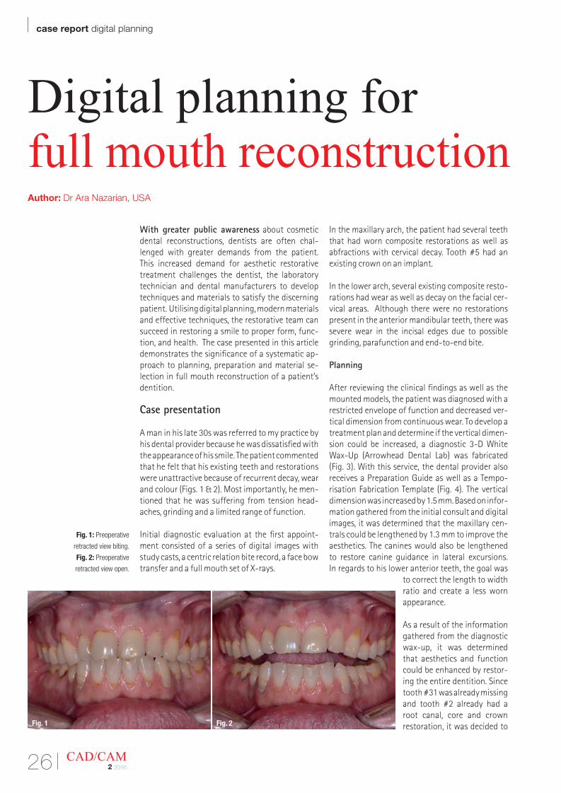
Fig. 3: 3-D White Wax-Up.
Fig. 4: Preparation guides
and temporary guides.
Fig. 5: Bite jig relined
capturing full arch bite.
| case report digital planning
28 CAD/CAM2 2016
trimmed with trimming burs and discs (Axis). Once the teeth were desensitised with Systemp desensi-tiser (Ivoclar Vivadent) and dried, the provisionals were temporarily cemented using Temp Bond Clear (Kerr). The patient was instructed about their care and use in eating, speaking and biting.
A few weeks later, the patient returned for evalua-tion of aesthetics, phonetics, and bite. Already he exhibited excitement and confidence with his pro-visional restorations, commenting that all his co-workers noticed he looked younger and happier. Most importantly, the patient said he no longer ex-perienced discomfort in his TMJ and that his bite never felt better. Since no adjustment or modifica-tion of the temporary was needed, the dental lab was instructed to replicate the 3-D White Wax-Up when fabricating the definitive restorations.
Laboratory considerations
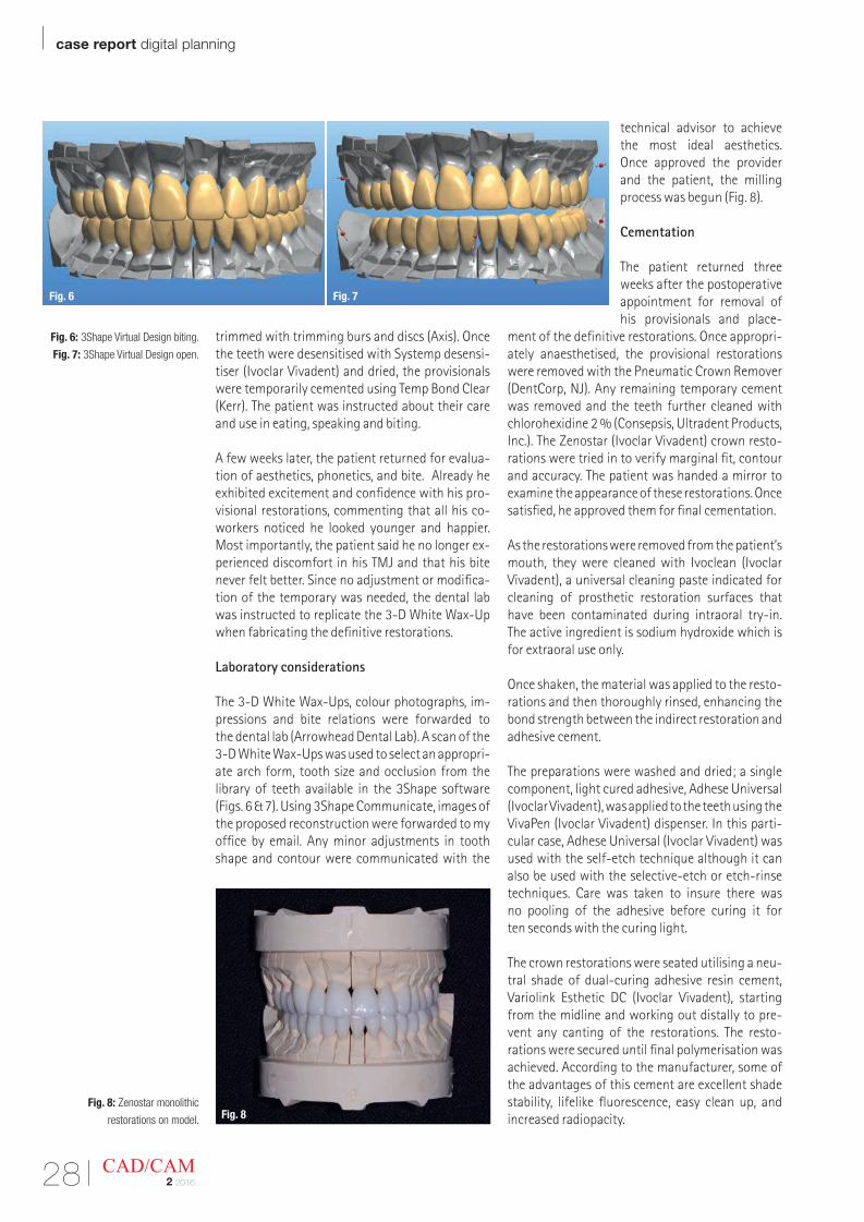
The 3-D White Wax-Ups, colour photographs, im-pressions and bite relations were forwarded to the dental lab (Arrowhead Dental Lab). A scan of the 3-D White Wax-Ups was used to select an appropri-ate arch form, tooth size and occlusion from the library of teeth available in the 3Shape software (Figs. 6 & 7). Using 3Shape Communicate, images of the proposed reconstruction were forwarded to my office by email. Any minor adjustments in tooth shape and contour were communicated with the
technical advisor to achieve the most ideal aesthetics. Once approved the provider and the patient, the milling process was begun (Fig. 8).
Cementation
The patient returned three weeks after the postoperative appointment for removal of his provisionals and place-
ment of the definitive restorations. Once appropri-ately anaesthetised, the provisional restorations were removed with the Pneumatic Crown Remover (DentCorp, NJ). Any remaining temporary cement was removed and the teeth further cleaned with chlorohexidine 2 % (Consepsis, Ultradent Products, Inc.). The Zenostar (Ivoclar Vivadent) crown resto-rations were tried in to verify marginal fit, contour and accuracy. The patient was handed a mirror to examine the appearance of these restorations. Once satisfied, he approved them for final cementation.
As the restorations were removed from the patient’s mouth, they were cleaned with Ivoclean (Ivoclar Vivadent), a uni versal cleaning paste indicated for cleaning of prosthetic restoration surfaces that have been contaminated during intraoral try-in. The active ingredient is sodium hydroxide which is for extraoral use only.
Once shaken, the material was applied to the resto-rations and then thoroughly rinsed, enhancing the bond strength between the indirect restoration and adhesive cement.
The preparations were washed and dried; a single component, light cured adhesive, Adhese Universal (Ivoclar Vivadent), was applied to the teeth using the VivaPen (Ivoclar Vivadent) dispenser. In this parti-cular case, Adhese Universal (Ivoclar Vivadent) was used with the self-etch technique although it can also be used with the selective-etch or etch-rinse techniques. Care was taken to insure there was no pooling of the adhesive before curing it for ten seconds with the curing light.
The crown restorations were seated utilising a neu-tral shade of dual-curing adhesive resin cement, Variolink Esthetic DC (Ivoclar Vivadent), starting from the midline and working out distally to pre- vent any canting of the restorations. The resto-rations were secured until final polymerisation was achieved. According to the manufacturer, some of the advantages of this cement are excellent shade stability, lifelike fluorescence, easy clean up, and increased radiopacity.Fig. 8
Fig. 6: 3Shape Virtual Design biting.
Fig. 7: 3Shape Virtual Design open.
Fig. 8: Zenostar monolithic
restorations on model.
Fig. 6 Fig. 7
www.DTStudyClub.comADA CERP is a service of the American Dental Association to assist dental professionals in identifying qualityproviders of continuing dental education. ADA CERP does not approve or endorse individual courses or instructors,nor does it imply acceptance of credit hours by boards of dentistry.
register for
FREE
– education everywhereand anytime
– live and interactivewebinars
– more than 1,000 archivedcourses
– a focused discussionforum
– free membershipno travel costs
– no time away fromthe practice
– interaction withcolleagues and expertsacross the globe
– a growing databaseof scientific articlesand case reports
– ADA CERP-recognizedcredit administration
Dental Tribune Study Club
Join the largesteducational
networkin dentistry!
digital planning case report |
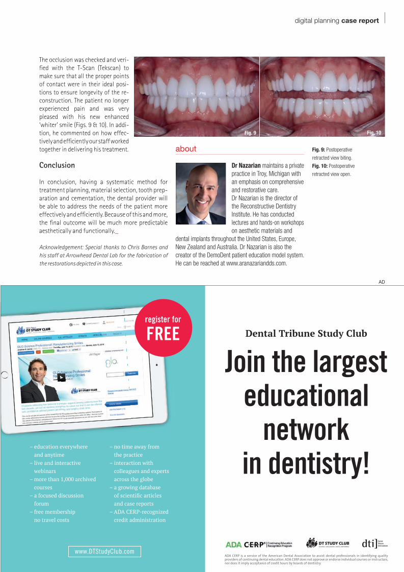
The occlusion was checked and veri-fied with the T-Scan (Tekscan) to make sure that all the proper points of contact were in their ideal posi-tions to ensure longevity of the re-construction. The patient no longer experienced pain and was very pleased with his new enhanced ‘whiter’ smile (Figs. 9 & 10). In addi-tion, he commented on how effec-tively and efficiently our staff worked together in delivering his treatment.
Conclusion
In conclusion, having a systematic method for treatment planning, material selection, tooth prep-aration and cementation, the dental provider will be able to address the needs of the patient more effectively and efficiently. Because of this and more, the final outcome will be much more predictable aesthetically and functionally._
Acknowledgement: Special thanks to Chris Barnes and his staff at Arrowhead Dental Lab for the fabrication of the restorations depicted in this case.
Fig. 10Fig. 9
www.DTStudyClub.com ADA CERP is a service of the American Dental Association to assist dental professionals in identifying quality providers of continuing dental education. ADA CERP does not approve or endorse individual courses or instructors, nor does it imply acceptance of credit hours by boards of dentistry.
register for
FREE
– education everywhere and anytime
– live and interactive webinars
– more than 1,000 archived courses
– a focused discussion forum
– free membership no travel costs
– no time away from the practice
– interaction with colleagues and experts across the globe
– a growing database of scientifi c articles and case reports
– ADA CERP-recognized credit administration
Dental Tribune Study Club
Join the largest educational
networkin dentistry!
AD
Fig. 9: Postoperative
retracted view biting.
Fig. 10: Postoperative
retracted view open.
about
Dr Nazarian maintains a private practice in Troy, Michigan with an emphasis on comprehensive and restorative care. Dr Nazarian is the director of the Reconstructive Dentistry Institute. He has conducted lectures and hands-on workshops on aesthetic materials and
dental implants throughout the United States, Europe, New Zealand and Australia. Dr Nazarian is also the creator of the DemoDent patient education model system. He can be reached at www.aranazariandds.com.
| opinion 3-D technology
30 CAD/CAM2 2016
The 3-D Difference: Cone Beam CT diagnostics to enhance treatment—Part 2Author: Dr Anthony Ramirez, USA
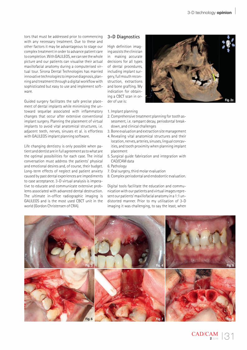
This article will deliberate advanced implant den-tistry using the 3-D difference to diagnose and treatment plan the failing maxillary arch in the third dimension. What I know now can be directly at-tributed to GALILEOS Cone Beam and can be applied to any advanced dental case. Treatment planning the decimated and devastated dentition necessi-tates the prerequisite for enhanced 3-D imaging. Of the cases presented here, all come diagnostically driven with the 3-D imaging of GALILEOS, ensuring optimal treatment outcomes. I practice life chang-ing dentistry and have been doing so for over eight years with GALILEOS.
The technology I have invested in has facilitated these treatments, as well as afforded patients the assurance of my judgement as they see first-hand
the benefit of having these technologies. In short, the outcome is unmatched and how they advance my treatment planning and treatment skills in all cases. We can reveal, identify and present more care with this ra-diographic modality. We do dentistry differ-ently and utilise advanced treatment modal-ities to increase the longevity of treatment rendered. Using careful 3-D analysis and guided implantology where possible, we can safely and precisely place restoratively driven implants. We will use cone beam com-
puted tomography (CBCT) imaging to confirm or al-ter our diagnosis when conventional 2-D imaging is limited in its diagnostic value.
I perform complete site assessment, tooth by tooth, and rapidly transfer the information to the patient chair-side. 3-D imaging improves and elevates our diagnostic ability and can facilitate the removal of diseased tissue—i.e. hopeless carious or periodon-tally involved teeth—and expose unseen periapical or inter-radicular pathology.
We dentists strive to improve patients’ lives, but sometimes we are faced with hopeless dentitions requiring enhanced diagnostics and many dental disciplines to treat globally. Psychological complex-ities with medical and financial capabilities are fac-
Fig. 2a Fig. 2b
Fig. 1a Fig. 1b
3-D technology opinion |
31CAD/CAM2 2016
tors that must be addressed prior to commencing with any necessary treatment. Due to these and other factors it may be advantageous to stage our complex treatment in order to advance patient care to completion. With GALILEOS, we can see the whole picture and our patients can visualise their actual maxillofacial anatomy during a computerised vir-tual tour. Sirona Dental Technologies has married innovative technologies to improve diagnosis, plan-ning and treatment through a digital workflow with sophisticated but easy to use and implement soft-ware.
Guided surgery facilitates the safe precise place-ment of dental implants while minimising the un-toward sequelae associated with inflammatory changes that occur after extensive conventional implant surgery. Planning the placement of virtual implants to avoid vital anatomical structures, i.e. adjacent teeth, nerves, sinuses et al. is effortless with GALILEOS implant planning software.
Life changing dentistry is only possible when pa-tient and dentist are in full agreement as to what are the optimal possibilities for each case. The initial conversation must address the patients’ physical and emotional desires and, of course, their budget. Long-term effects of neglect and patient anxiety caused by past dental experiences are impediments to case acceptance. 3-D virtual analysis is impera-tive to educate and communicate extensive prob-lems associated with advanced dental destruction. The ultimate in-office radiographic imaging is GALILEOS and is the most used CBCT unit in the world (Gordon Christensen of CRA).
3-D Diagnostics
High definition imag-ing assists the clinician in making accurate decisions for all types of dental procedures, including implant sur-gery, full mouth recon-struction, extractions and bone grafting. My indication for obtain-ing a CBCT scan in or-der of use is:
1. Implant planning2. Comprehensive treatment planning for tooth as-
sessment, i.e. rampant decay, periodontal break-down, and clinical challenges
3. Bone evaluation and extraction site management4. Revealing vital anatomical structures and their
location, nerves, arteries, sinuses, lingual concav-ities, and tooth proximity when planning implant placement
5. Surgical guide fabrication and integration with CAD/CAM data
6. Pathology 7. Oral surgery, third molar evaluation8. Complex periodontal and endodontic evaluation.
Digital tools facilitate the education and commu-nication with our patients and virtual images repre-sent our patients’ maxillofacial anatomy in a 1:1 un-distorted manner. Prior to my utilisation of 3-D imaging it was challenging, to say the least, when
Fig. 2c
Fig. 6 Fig. 7 Fig. 8
Fig. 3 Fig. 4 Fig. 5
| opinion 3-D technology
32 CAD/CAM2 2016
communicating the extent of my patients’ condi-tions and the comprehensive nature as to what is necessary to return them to a full complement of teeth and oral health. Patients unaware of the de-struction caused by bacterial infection, poor den-tistry and neglect are very difficult to convince that they require extensive dental treatment to improve their oral health. Since I have the benefit of 3-D im-aging, their conditions can be displayed on a large computer screen, which facilitates an interactive conversation which generally results in their under-standing their problems and moving forward with the necessary treatment presented. It is especially useful when evaluating bone deficiencies, periapi-cal pathology, evaluating existing periodontal sup-port and residual bone volume in edentulous areas.
All of the following cases benefited via advanced 3-D imaging to gain a complete understanding of the enormity of the obstacles these patients faced prior to commencing with treatment. The case presenta-tion visit explained clearly how advanced implant dentistry and proper sequencing would provide each patient with their desired result. The goal for each was to rehabilitate these patients with an aesthetic and fully functioning maxillary arch of new teeth. They all were given options to replace hopeless max-illary arches with the guarantee being that their prosthetic replacements would look and function much better than their failing natural dentition.
What follows is how I diagnosed, treatment planned, case presented and treated three decimated maxil-
lary arches. As you read my applications of the 3-D difference and the eventual treatments, think about how these cases were positively impacted by the use of 3-D imaging and how its use led to case accep-tance and facilitated positive patient experiences, even though these cases were as complex and chal-lenging as exist in the dentistry today.
Where do we begin?
All three patients presented with a common prob-lem, hopeless maxillary dentitions due to years of neglect, fear and poor maintenance of their natural dentitions. These types of situations are complex and challenging to treat. Each case required the re-moval of all maxillary natural teeth, but psycholog-ical complexities made for the unique treatment plans developed. It took a lot of courage for these patients to finally decide that they were ready to proceed with a treatment to improve their condi-tions. The decimated dentition requires managing the whole patient and the 3-D difference increases the ability for my patients to understand and accept treatment. My office has a distinct advantage over offices lacking the technology.
The first patient accepted her fate of an immediate complete upper denture, prior to guided implant placement and replacing her teeth with an implant retained metal reinforced horseshoe CUD overden-ture. The second patient was not ready to accept the immediate complete upper denture so she would be transitioned into a four unit implant retained re-
Fig. 12a Fig. 12b
Fig. 9 Fig. 10 Fig. 11
3-D technology opinion |
33CAD/CAM2 2016
movable over-denture. I elected to retain a number of natural teeth while bone regenerated and im-plants were placed as bone volume allowed. The natural teeth would retain a fixed provisional as long as possible. The third case would be treated with ‘teeth in a day’ approach. All maxillary teeth would be extracted and six immediate implants would be placed and restored with a fixed screw- retained prosthesis during the same visit. These three case reports will illustrate what is possible when advanced technologies are implemented in the modern dental practice. The 3-D difference was employed to assess each case on a tooth-by-tooth basis. The diagnosis that each tooth had a poor to hopeless prognosis made it clear that ‘HERODONTICS’ would be uncalled for.
Case 1: ‘I want to smile again’
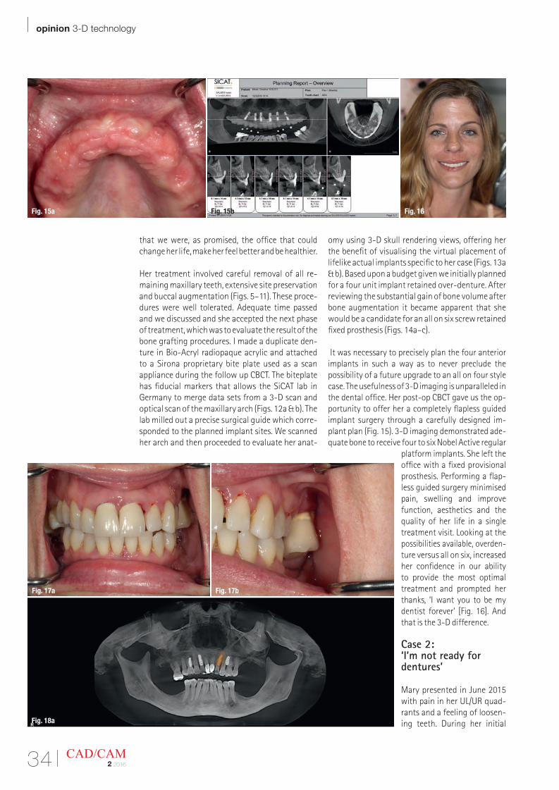
Christina, 37, who was seeking to improve her smile and improve her confidence. She had been treated previously at another dental office with little suc-cess and it was obvious that critical care was neces-sary to improve the current state of her maxillary arch. The initial visit was to decide upon a course of action to remove her discomfort and provide a fully functioning arch of teeth. Christina presented with extensive decay in all of the remaining maxillary natural teeth (Figs.1a & b). All conventional diag-nostic, clinical and radiographic procedures were completed and reviewed during the first fifteen minutes. I recommended and she accepted an en-hanced 3-D CBCT scan which would be used to per-form a complete dental examination (Figs. 2a–c).
During co-diagnosis she made it clear that she would accept extraction of all the remaining maxil-lary teeth and replacement with an immediate CUD. This would serve as an interim prosthesis that would allow her to begin to feel better about her smile and give her confidence to socialise and eat without discomfort. Options for a more definitive treatment were formulated between us and facilitated by the virtual 3-D imaging and 3-D analysis and review of the maxillofacial anatomy that existed.
There is no such thing as a cookie-cutter approach to diagnosis and treatment planning in complex dentistry. The clinician must be a good listener, be respectful of the patients’ desires and be decisive in providing solutions to their problems. We have to manage the entire patient from start to finish and manage and exceed their expectations.
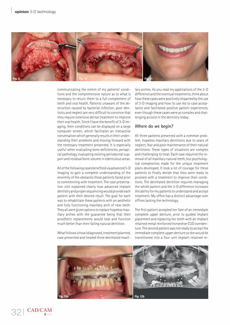
Her treatment began with extraction and bone grafting the UR/UL molars #15 and #2 (Figs. 3 & 4). She accepted an immediate CUD as an interim pros-thesis. This would provide her with a beautiful nat-ural smile as fast as possible and commit her to the development of a more definitive treatment plan. Extraction of all maxillary teeth would remove the cause of her embarrassment and give her the confi-dence to eat more comfortably and socialise more readily. I impressed the maxillary arch after a couple of weeks of healing so the posterior portion of the denture could seat on a firm base. Christina devel-oped a trust in us and was grateful to smile again. With this came a decreased level of anxiety and she was given an emotional lift when she came to realise
Fig. 13a Fig. 13b
Fig. 14a Fig. 14b Fig. 14c
| opinion 3-D technology
34 CAD/CAM2 2016
that we were, as promised, the office that could change her life, make her feel better and be healthier.
Her treatment involved careful removal of all re-maining maxillary teeth, extensive site preservation and buccal augmentation (Figs. 5–11). These proce-dures were well tolerated. Adequate time passed and we discussed and she accepted the next phase of treatment, which was to evaluate the result of the bone grafting procedures. I made a duplicate den-ture in Bio-Acryl radiopaque acrylic and attached to a Sirona proprietary bite plate used as a scan appliance during the follow up CBCT. The biteplate has fiducial markers that allows the SiCAT lab in Germany to merge data sets from a 3-D scan and optical scan of the maxillary arch (Figs. 12a & b). The lab milled out a precise surgical guide which corre-sponded to the planned implant sites. We scanned her arch and then proceeded to evaluate her anat-
omy using 3-D skull rendering views, offering her the benefit of visualising the virtual placement of lifelike actual implants specific to her case (Figs. 13a & b). Based upon a budget given we initially planned for a four unit implant retained over-denture. After reviewing the substantial gain of bone volume after bone augmentation it became apparent that she would be a candidate for an all on six screw retained fixed prosthesis (Figs. 14a–c).
It was necessary to precisely plan the four anterior implants in such a way as to never preclude the possibility of a future upgrade to an all on four style case. The usefulness of 3-D imaging is unparalleled in the dental office. Her post-op CBCT gave us the op-portunity to offer her a completely flapless guided implant surgery through a carefully designed im-plant plan (Fig. 15). 3-D imaging demonstrated ade-quate bone to receive four to six Nobel Active regular
platform implants. She left the office with a fixed provisional prosthesis. Performing a flap-less guided surgery minimised pain, swelling and improve function, aesthetics and the quality of her life in a single treatment visit. Looking at the possibilities available, overden-ture versus all on six, increased her confidence in our ability to provide the most optimal treatment and prompted her thanks, ‘I want you to be my dentist forever’ [Fig. 16]. And that is the 3-D difference.
Case 2: ‘I’m not ready for dentures’
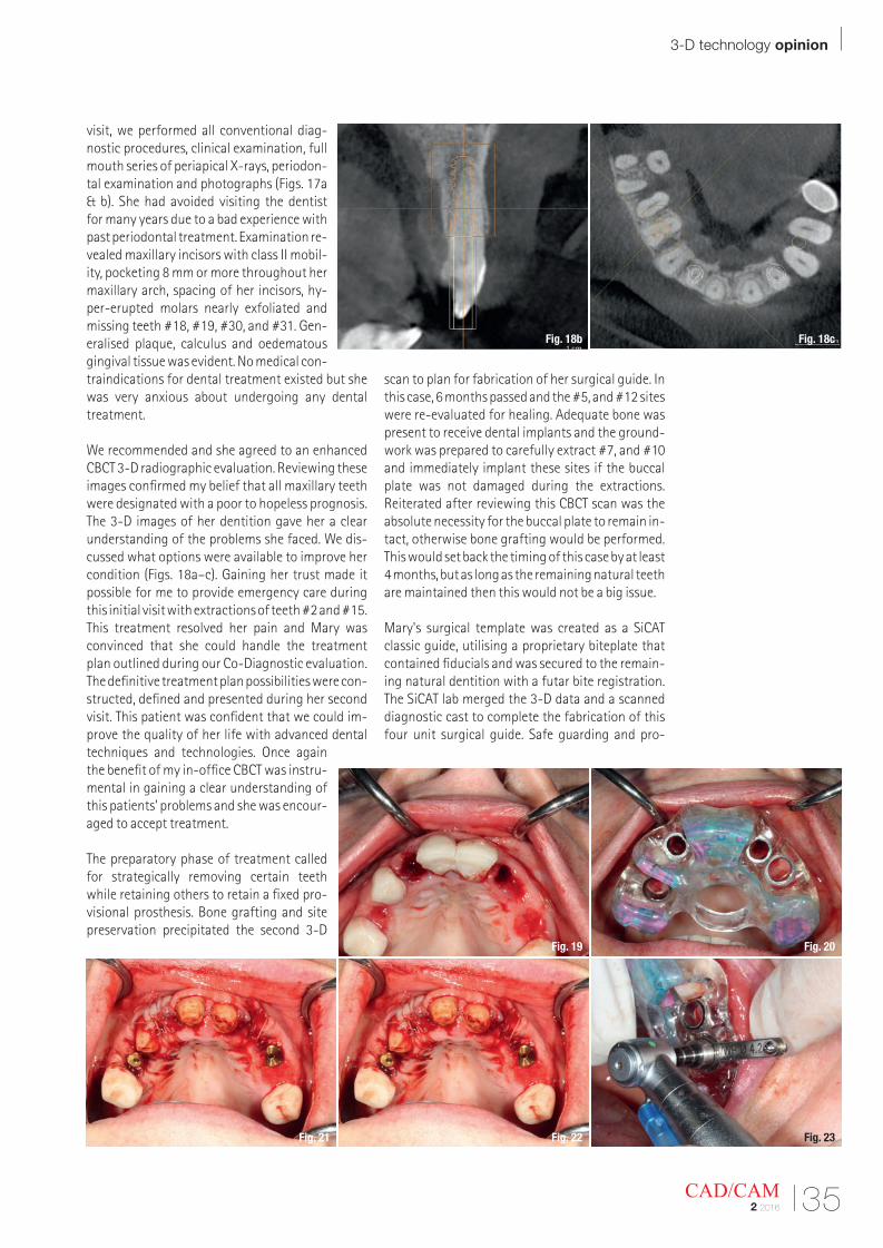
Mary presented in June 2015 with pain in her UL/UR quad-rants and a feeling of loosen-ing teeth. During her initial Fig. 18a
Fig. 17a Fig. 17b
Fig. 16Fig. 15a Fig. 15b
3-D technology opinion |
35CAD/CAM2 2016
visit, we performed all conventional diag-nostic procedures, clinical examination, full mouth series of periapical X-rays, periodon-tal examination and photographs (Figs. 17a & b). She had avoided visiting the dentist for many years due to a bad experience with past periodontal treatment. Examination re-vealed maxillary incisors with class II mobil-ity, pocketing 8 mm or more throughout her maxillary arch, spacing of her incisors, hy-per-erupted molars nearly exfoliated and missing teeth #18, #19, #30, and #31. Gen-eralised plaque, calculus and oedematous gingival tissue was evident. No medical con-traindications for dental treatment existed but she was very anxious about undergoing any dental treatment.
We recommended and she agreed to an enhanced CBCT 3-D radiographic evaluation. Reviewing these images confirmed my belief that all maxillary teeth were designated with a poor to hopeless prognosis. The 3-D images of her dentition gave her a clear understanding of the problems she faced. We dis-cussed what options were available to improve her condition (Figs. 18a–c). Gaining her trust made it possible for me to provide emergency care during this initial visit with extractions of teeth #2 and #15. This treatment resolved her pain and Mary was convinced that she could handle the treatment plan outlined during our Co-Diagnostic evaluation. The definitive treatment plan possibilities were con-structed, defined and presented during her second visit. This patient was confident that we could im-prove the quality of her life with advanced dental techniques and technologies. Once again the benefit of my in-office CBCT was instru-mental in gaining a clear understanding of this patients’ problems and she was encour-aged to accept treatment.
The preparatory phase of treatment called for strategically removing certain teeth while retaining others to retain a fixed pro-visional prosthesis. Bone grafting and site preservation precipitated the second 3-D
scan to plan for fabrication of her surgical guide. In this case, 6 months passed and the #5, and #12 sites were re-evaluated for healing. Adequate bone was present to receive dental implants and the ground-work was prepared to carefully extract #7, and #10 and immediately implant these sites if the buccal plate was not damaged during the extractions. Reiterated after reviewing this CBCT scan was the absolute necessity for the buccal plate to remain in-tact, otherwise bone grafting would be performed. This would set back the timing of this case by at least 4 months, but as long as the remaining natural teeth are maintained then this would not be a big issue.
Mary's surgical template was created as a SiCAT classic guide, utilising a proprietary biteplate that contained fiducials and was secured to the remain-ing natural dentition with a futar bite registration. The SiCAT lab merged the 3-D data and a scanned diagnostic cast to complete the fabrication of this four unit surgical guide. Safe guarding and pro-
Fig. 18b Fig. 18c
Fig. 19 Fig. 20
Fig. 21 Fig. 22 Fig. 23
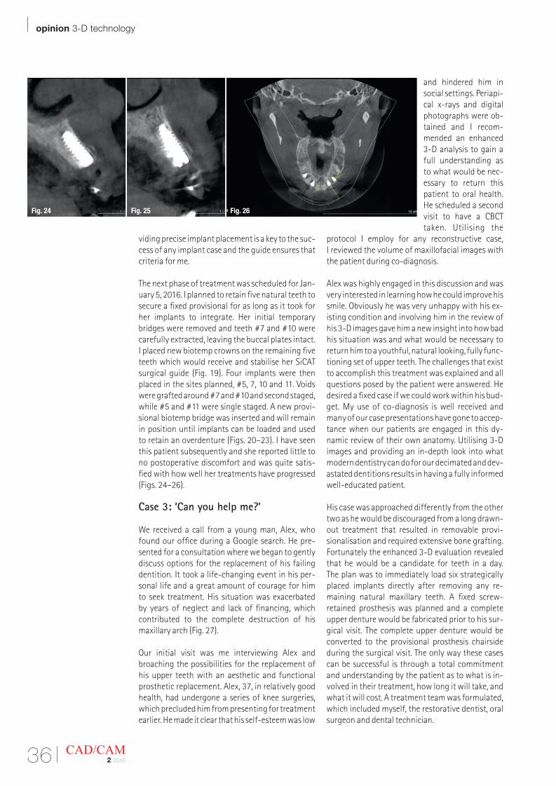
viding precise implant placement is a key to the suc-cess of any implant case and the guide ensures that criteria for me.
The next phase of treatment was scheduled for Jan-uary 5, 2016. I planned to retain five natural teeth to secure a fixed provisional for as long as it took for her implants to integrate. Her initial temporary bridges were removed and teeth #7 and #10 were carefully extracted, leaving the buccal plates intact. I placed new biotemp crowns on the remaining five teeth which would receive and stabilise her SiCAT surgical guide (Fig. 19). Four implants were then placed in the sites planned, #5, 7, 10 and 11. Voids were grafted around #7 and #10 and second staged, while #5 and #11 were single staged. A new provi-sional biotemp bridge was inserted and will remain in position until implants can be loaded and used to retain an overdenture (Figs. 20–23). I have seen this patient subsequently and she reported little to no postoperative discomfort and was quite satis- fied with how well her treatments have progressed (Figs. 24–26).
Case 3: ‘Can you help me?’
We received a call from a young man, Alex, who found our office during a Google search. He pre-sented for a consultation where we began to gently discuss options for the replacement of his failing dentition. It took a life-changing event in his per-sonal life and a great amount of courage for him to seek treatment. His situation was exacerbated by years of neglect and lack of financing, which contributed to the complete destruction of his maxillary arch (Fig. 27).
Our initial visit was me interviewing Alex and broaching the possibilities for the replacement of his upper teeth with an aesthetic and functional prosthetic replacement. Alex, 37, in relatively good health, had undergone a series of knee surgeries, which precluded him from presenting for treatment earlier. He made it clear that his self-esteem was low
and hindered him in social settings. Periapi-cal x-rays and digital photographs were ob-tained and I recom-mended an enhanced 3-D analysis to gain a full understanding as to what would be nec-essary to return this patient to oral health. He scheduled a second visit to have a CBCT taken. Utilising the
protocol I employ for any reconstructive case, I reviewed the volume of maxillofacial images with the patient during co-diagnosis.
Alex was highly engaged in this discussion and was very interested in learning how he could improve his smile. Obviously he was very unhappy with his ex-isting condition and involving him in the review of his 3-D images gave him a new insight into how bad his situation was and what would be necessary to return him to a youthful, natural looking, fully func-tioning set of upper teeth. The challenges that exist to accomplish this treatment was explained and all questions posed by the patient were answered. He desired a fixed case if we could work within his bud-get. My use of co-diagnosis is well received and many of our case presentations have gone to accep-tance when our patients are engaged in this dy-namic review of their own anatomy. Utilising 3-D images and providing an in-depth look into what modern dentistry can do for our decimated and dev-astated dentitions results in having a fully informed well-educated patient.
His case was approached differently from the other two as he would be discouraged from a long drawn-out treatment that resulted in removable provi-sionalisation and required extensive bone grafting. Fortunately the enhanced 3-D evaluation revealed that he would be a candidate for teeth in a day. The plan was to immediately load six strategically placed implants directly after removing any re-maining natural maxillary teeth. A fixed screw- retained prosthesis was planned and a complete upper denture would be fabricated prior to his sur-gical visit. The complete upper denture would be converted to the provisional prosthesis chairside during the surgical visit. The only way these cases can be successful is through a total commitment and understanding by the patient as to what is in-volved in their treatment, how long it will take, and what it will cost. A treatment team was formulated, which included myself, the restorative dentist, oral surgeon and dental technician.
| opinion 3-D technology
36 CAD/CAM2 2016
Fig. 24 Fig. 25 Fig. 26
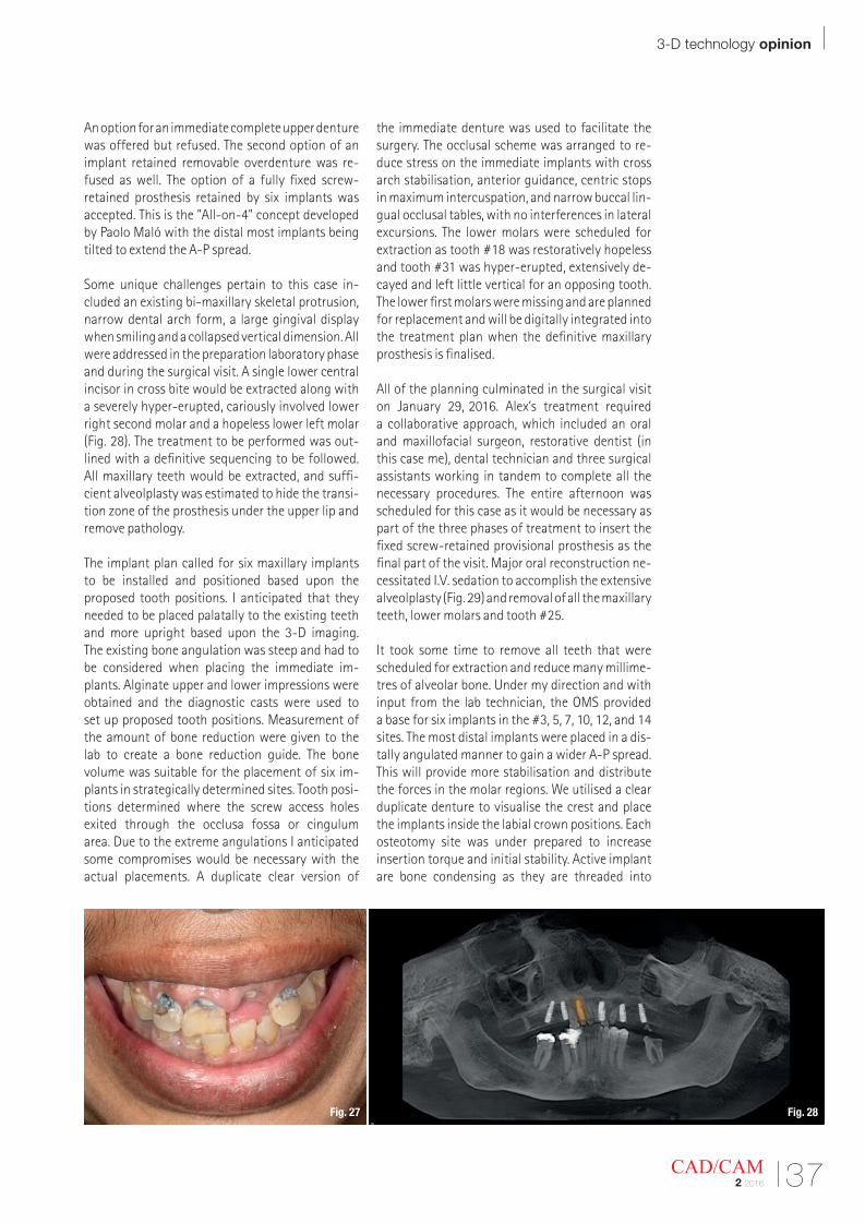
3-D technology opinion |
37CAD/CAM2 2016
An option for an immediate complete upper denture was offered but refused. The second option of an implant retained removable overdenture was re-fused as well. The option of a fully fixed screw- retained prosthesis retained by six implants was accepted. This is the "All-on-4" concept developed by Paolo Maló with the distal most implants being tilted to extend the A-P spread.
Some unique challenges pertain to this case in-cluded an existing bi-maxillary skeletal protrusion, narrow dental arch form, a large gingival display when smiling and a collapsed vertical dimension. All were addressed in the preparation laboratory phase and during the surgical visit. A single lower central incisor in cross bite would be extracted along with a severely hyper-erupted, cariously involved lower right second molar and a hopeless lower left molar (Fig. 28). The treatment to be performed was out-lined with a definitive sequencing to be followed. All maxillary teeth would be extracted, and suffi-cient alveolplasty was estimated to hide the transi-tion zone of the prosthesis under the upper lip and remove pathology.
The implant plan called for six maxillary implants to be installed and positioned based upon the proposed tooth positions. I anticipated that they needed to be placed palatally to the existing teeth and more upright based upon the 3-D imaging. The existing bone angulation was steep and had to be considered when placing the immediate im-plants. Alginate upper and lower impressions were obtained and the diagnostic casts were used to set up proposed tooth positions. Measurement of the amount of bone reduction were given to the lab to create a bone reduction guide. The bone volume was suitable for the placement of six im-plants in strategically determined sites. Tooth posi-tions determined where the screw access holes exited through the occlusa fossa or cingulum area. Due to the extreme angulations I anticipated some compromises would be necessary with the actual placements. A duplicate clear version of
the immediate denture was used to facilitate the surgery. The occlusal scheme was arranged to re-duce stress on the immediate implants with cross arch stabilisation, anterior guidance, centric stops in maximum intercuspation, and narrow buccal lin-gual occlusal tables, with no interferences in lateral excursions. The lower molars were scheduled for extraction as tooth #18 was restoratively hopeless and tooth #31 was hyper-erupted, extensively de-cayed and left little vertical for an opposing tooth. The lower first molars were missing and are planned for replacement and will be digitally integrated into the treatment plan when the definitive maxillary prosthesis is finalised.
All of the planning culminated in the surgical visit on January 29, 2016. Alex’s treatment required a collaborative approach, which included an oral and maxillofacial surgeon, restorative dentist (in this case me), dental technician and three surgical assistants working in tandem to complete all the necessary procedures. The entire afternoon was scheduled for this case as it would be necessary as part of the three phases of treatment to insert the fixed screw-retained provisional prosthesis as the final part of the visit. Major oral reconstruction ne-cessitated I.V. sedation to accomplish the extensive alveolplasty (Fig. 29) and removal of all the maxillary teeth, lower molars and tooth #25.
It took some time to remove all teeth that were scheduled for extraction and reduce many millime-tres of alveolar bone. Under my direction and with input from the lab technician, the OMS provided a base for six implants in the #3, 5, 7, 10, 12, and 14 sites. The most distal implants were placed in a dis-tally angulated manner to gain a wider A-P spread. This will provide more stabilisation and distribute the forces in the molar regions. We utilised a clear duplicate denture to visualise the crest and place the implants inside the labial crown positions. Each osteotomy site was under prepared to increase insertion torque and initial stability. Active implant are bone condensing as they are threaded into
Fig. 27 Fig. 28
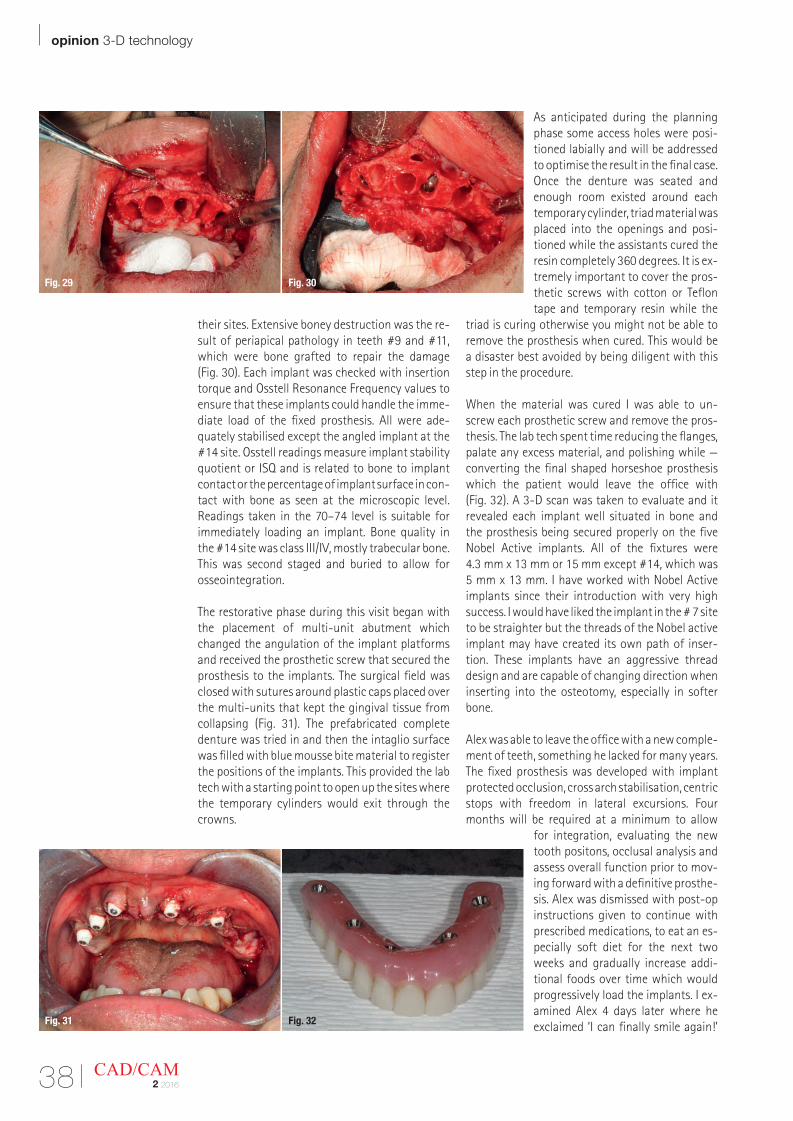
| opinion 3-D technology
38 CAD/CAM2 2016
their sites. Extensive boney destruction was the re-sult of periapical pathology in teeth #9 and #11, which were bone grafted to repair the damage (Fig. 30). Each implant was checked with insertion torque and Osstell Resonance Frequency values to ensure that these implants could handle the imme-diate load of the fixed prosthesis. All were ade-quately stabilised except the angled implant at the #14 site. Osstell readings measure implant stability quotient or ISQ and is related to bone to implant contact or the percentage of implant surface in con-tact with bone as seen at the microscopic level. Readings taken in the 70–74 level is suitable for immediately loading an implant. Bone quality in the #14 site was class III/IV, mostly trabecular bone. This was second staged and buried to allow for osseointegration.
The restorative phase during this visit began with the placement of multi-unit abutment which changed the angulation of the implant platforms and received the prosthetic screw that secured the prosthesis to the implants. The surgical field was closed with sutures around plastic caps placed over the multi-units that kept the gingival tissue from collapsing (Fig. 31). The prefabricated complete denture was tried in and then the intaglio surface was filled with blue mousse bite material to register the positions of the implants. This provided the lab tech with a starting point to open up the sites where the temporary cylinders would exit through the crowns.
As anticipated during the planning phase some access holes were posi-tioned labially and will be addressed to optimise the result in the final case. Once the denture was seated and enough room existed around each temporary cylinder, triad material was placed into the openings and posi-tioned while the assistants cured the resin completely 360 degrees. It is ex-tremely important to cover the pros-thetic screws with cotton or Teflon tape and temporary resin while the
triad is curing otherwise you might not be able to remove the prosthesis when cured. This would be a disaster best avoided by being diligent with this step in the procedure.
When the material was cured I was able to un- screw each prosthetic screw and remove the pros-thesis. The lab tech spent time reducing the flanges, palate any excess material, and polishing while —converting the final shaped horseshoe prosthesis which the patient would leave the office with (Fig. 32). A 3-D scan was taken to evaluate and it revealed each implant well situated in bone and the prosthesis being secured properly on the five Nobel Active implants. All of the fixtures were 4.3 mm x 13 mm or 15 mm except #14, which was 5 mm x 13 mm. I have worked with Nobel Active implants since their introduction with very high success. I would have liked the implant in the # 7 site to be straighter but the threads of the Nobel active implant may have created its own path of inser- tion. These implants have an aggressive thread design and are capable of changing direction when inserting into the osteotomy, especially in softer bone.
Alex was able to leave the office with a new comple-ment of teeth, something he lacked for many years. The fixed prosthesis was developed with implant protected occlusion, cross arch stabilisation, centric stops with freedom in lateral excursions. Four months will be required at a minimum to allow
for integration, evaluating the new tooth positons, occlusal analysis and assess overall function prior to mov-ing forward with a definitive prosthe-sis. Alex was dismissed with post-op instructions given to continue with prescribed medications, to eat an es-pecially soft diet for the next two weeks and gradually increase addi-tional foods over time which would progressively load the implants. I ex-amined Alex 4 days later where he exclaimed ‘I can finally smile again!’
Fig. 29 Fig. 30
Fig. 31 Fig. 32
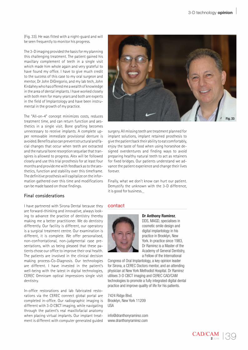
3-D technology opinion |
39CAD/CAM2 2016
(Fig. 33). He was fitted with a night-guard and will be seen frequently to monitor his progress.
The 3-D imaging provided the basis for my planning this challenging treatment. The patient gained his maxillary complement of teeth in a single visit which made him whole again and very grateful to have found my office. I have to give much credit to the success of this case to my oral surgeon and mentor, Dr John DiGregorio, and my lab tech, John Kirdahey who has offered me a wealth of knowledge in the area of dental implants. I have worked closely with both men for many years and both are experts in the field of Implantology and have been instru-mental in the growth of my practice.
The “All-on-4” concept minimizes costs, reduces treatment time, and can return function and aes-thetics in a single visit. Bone grafting becomes unne cessary to receive implants. A complete up- per removable immediate provisional denture is avoided. Benefits also can prevent structural and fa-cial changes that occur when teeth are extracted and the natural bone resorption sequelae that tran-spires is allowed to progress. Alex will be followed closely and use this trial prosthesis for at least four months and provide me with feedback as to the aes-thetics, function and stability over this timeframe. The definitive prosthesis will capitalize on the infor-mation gathered over this time and modifications can be made based on those findings.
Final considerations
I have partnered with Sirona Dental because they are forward-thinking and innovative, always look-ing to advance the practice of dentistry thereby making me a better practitioner. We do dentistry differently. Our facility is different, our operatory is a surgical treatment centre. Our examination is different, it is complete. We offer personalised, non-confrontational, non-judgmental case pre-sentations, with us being pleased that these pa-tients chose our office to improve their oral health. The patients are involved in the clinical decision making process-Co-Diagnosis. Our technologies are different. I have invested in the patient’s well-being with the latest in digital technologies, CEREC Omnicam optical impressions single visit dentistry.
In-office restorations and lab fabricated resto-rations via the CEREC connect global portal are completed in-office. Our radiographic imaging is different with 3-D CBCT imaging, while navigating through the patient’s real maxillofacial anatomy when placing virtual implants. Our implant treat-ment is different with computer generated guided
surgery. All missing teeth are treatment planned for implant solutions, implant retained prosthesis to give the patient back their ability to eat comfortably, enjoy the taste of food when using horseshoe de-signed overdentures and finding ways to avoid preparing healthy natural teeth to act as retainers for fixed bridges. Our patients understand we ad-vance the patient experience and change their lives forever.
Finally, what we don’t know can hurt our patient. Demystify the unknown with the 3-D difference, it is good for business._
contact
Dr Anthony Ramirez, DDS, MAGD, specialises in cosmetic smile design and digital implantology in his practice in Brooklyn, New York. In practice since 1983, Dr Ramirez is a Master of the Academy of General Dentistry, a Fellow of the International
Congress of Oral Implantology, a key opinion leader for Sirona, a CEREC Doctors mentor, and an attending physician at New York Methodist Hospital. Dr Ramirez utilises 3-D CBCT imaging and CEREC CAD/CAM technologies to promote a fully integrated digital dental practice and improve quality of life for his patients.
7424 Ridge Blvd.Brooklyn, New York 11209USA
Fig. 33
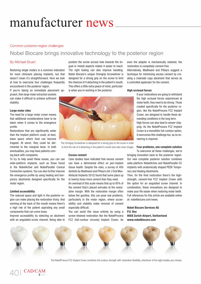
Common posterior-region challenges
Nobel Biocare brings innovative technology to the posterior region
By Michael Stuart
Restoring single molars is a common indication for most clinicians placing implants, but that doesn’t mean it’s straightforward. Here we look at how to overcome four challenges frequently encountered in the posterior region. If you’re taking an immediate placement ap-proach, then large molar extraction sockets can make it difficult to achieve sufficient stability.
Large molar sitesThe need for a large molar crown means that additional considerations have to be taken when it comes to the emergence profile. Restorations that are significantly wider than the implant platform could, at best, leave space where food can become trapped. At worst, they could be det-rimental to the marginal bone. In both eventualities, you may have patients com-ing back with complaints.To try to help avoid these issues, you can use wide-platform implants, such as those found in the NobelActive and NobelParallel Conical Connection systems. You can also further improve the emergence profile by using healing and tem-porary abutments designed specifically for the molar region.
Limited accessibilityThe reduced space and light in the posterior re-gion can make placing the restoration tricky. And working at the back of the mouth means there’s a high risk of the patient aspirating any small components that can come loose.Improve accessibility by selecting an abutment with an angulated screw channel. Being able to
position the screw access hole towards the lin-gual or mesial aspects makes it easier to reach. The right tooling can also improve handling. Nobel Biocare’s unique Omnigrip Screwdriver is designed for a strong grip on the screw to limit the chances of it detaching in the patient’s mouth. This offers a little extra peace of mind, particular-ly when you’re working in the posterior.
Excess cementCase studies have indicated that excess cement can have a detrimental effect on peri-implant tissue health. Despite the risks, a survey of 400 dentists by Wadhwani and Piñeyro (Int J Oral Max-illofacial Implants 2012) found that some place up to twenty times more cement than they need. An overload of this scale means that up to 95% of the cement that’s placed extrudes at the restor-ative margin. With the restorative margin often below the gumline, this can pose real problems, particularly in the molar region, where acces-sibility and visibility make removal of cement especially difficult.You can avoid this issue entirely by using a screw-retained restoration like the NobelProcera FCZ (full-contour zirconia) Implant Crown. As
even the adapter is mechanically retained, the restoration is completely cement-free. Alternatively, Wadhwani and Piñeyro suggest a technique for minimizing excess cement by cre-ating a chairside copy abutment that serves as a controlled applicator for the cement.
High occlusal forcesIf your restorations are going to withstand the high occlusal forces experienced at molar teeth, they need to be strong. Those created specifically for the posterior re-gion, like the NobelProcera FCZ Implant Crown, are designed to handle these de-manding conditions in the long term.High forces can also lead to veneer chip-ping. As the NobelProcera FCZ Implant Crown is a monolithic full-contour option, it overcomes this challenge too, as no ve-neering is required.
Four problems, one complete solutionTo overcome all these challenges, we’re
bringing innovation back to the posterior region. Our new complete posterior solution combines wide-platform NobelActive and NobelParallel CC implants with anatomically shaped PEEK Tempo-rary and Healing Abutments. Then, for the final restoration there’s the high-strength, cement-free FCZ Implant Crown with the option for an angulated screw channel. In combination, these innovations are designed to make your life easier when restoring molar teeth.Full references for this article are available online at: nobelbiocare.com/news.
Nobel Biocare Services AGP.O. Box8058 Zurich-Airport, Switzerlandwww.nobelbiocare.com
manufacturer news
40 CAD/CAM2 2016
The Omnigrip Screwdriver is designed for a strong grip on the screw in order
to limit the risk of it detaching in the patient’s mouth (see also cover image).
The NobelProcera FCZ Implant Crown combines full-contour strength with restorative flexibility, whichever of the eight shades you choose.
DentalSuite 5 is a complete and open solution that covers the entire 3-D scanning, design and modell-ing workflow in an integrated and functional way. It is composed by DScan, 3-D scanner that combines performance, reliability and accuracy (up to 15 mi-crons), and the proprietary software DentalCad.
DScan, the latest generation of scanners with a structured blue LED light technology for the 3-D acquisition of dental models, multidies, impres-sions and verticulator. It is supplied with a special plate that enables optics and axes calibration in automatic in just 5 minutes. Thanks to its high quality mechanical and electronic components, DScan allows a faster and more accurate scan-ning ensuring accuracy and reliability.
On the software side, DentalCAD, EGS’ software for dental modell-ing, has been designed to be ideal for both first users and experienced users. Thanks to the new interface, it is easy to use and it features a num-ber of automatic tools that can suggest optimal parameters to guide the user through the modelling. It presents a lifetime license with a complete func-
tional coverage (crowns, implants, veneers, wax-up, attachments, toronto bridge, inlay-onlay etc.).
Moreover it features:· Automatic updates (free for 1 year and optional
starting from the 2nd one) to ensure the product best performance;
· Bars for edentulous patients: thanks to the new function it will be possible to realise bars with different attachment libraries managed by a new and more performing algorithm;
· Implants with gum: it allows the implant acqui-sition directly on the gum using the margin line also as functional reference;
· Locators scanning with a simple and automatic managing tool throughout the order manager;
· Direct integration with Digital Smile System (DSS) software allows to import the rehibilita-tion project in a CAD environment and to realise the mock-up in order to use it as reference for the final project realisation. This feature’s ad-vantages are clear not only for dentists and den-tal technicians, who gets a fluid workflow which keeps steady the patient’s expected result, but also for patients themselves who get preview about the final aesthetic outcome;
· Abutment management: it is now possible to realise the abutment lower part in a fast and simple way avoiding the margin line process.
Efficient and with a recognisable interface, Dental CAD 5 has got a renowed look: modern and charming and it also allows users to manage ex-ternal CAM systems.
EGS SrlVia Speranza 19/4 40068 S. Lazzaro di Savena (BO), Italywww.egsolutions.com
41CAD/CAM2 2016
There are moments in life when you want a con-nection to be extremely reliable. So far hybrid den-ture attachment systems may have been facing their limits in challenging implant situations. Now, there’s the Straumann Novaloc Retentive System.
The Straumann Novaloc Retentive System for hy-brid dentures offers an innovative carbon-based abutment coating (ADLC, amorphous diamond- like carbon) with excellent wear resistance, over-coming up to 60 degrees implant divergence. As the name implies, “amorphous diamond-like carbon” (ADLC) is a class of carbon bonds which displays several of the desirable qualities of a di-amond. ADLC coatings are commonly used in the medical device field (e.g. hip joints) and reduce abrasive wear prolonging the lifetime of the med-ical appliance.Both a straight and a 15 degrees angled abut-ment, available in various gingiva heights, cover a broad range of clinical implant situations. Together with its durable PEEK (Polyether ether ketone) matrices, the Novaloc Retentive System provides a reliable connection that endures. This results in low maintenance and high patient com-fort. Let your patients profit from the endurance of a reliable treatment solution. Learn more: http://starget.straumann.com/tag/novaloc
Institut Straumann AGPostfach4002 Basel, Switzerlandwww.straumann.com
Digital solutions
DentalSuite 5: one system, full coverage
Hybrid denture attachment systems
Straumann Novaloc
Retentive System:
a reliable connection
As a result of growing demand, dental laboratories that can provide high-quality implant bars to sup-port overdentures will find new business knocking at their doors. Ramping up implant bar production, however, can require a significant investment in equipment, time and staff training, which many labs simply cannot afford. Labs can instead receive a range of high-quality, precision-milled implant bars, simply by sending a model to NobelProcera. This flexible approach to outsourcing offers many benefits for labs. Primarily, it means they can offer precision-fitting bars in NobelProcera’s renowned high quality without needing to invest in a Nobel-Procera CAD system or purchase and maintain expensive production technology.
By removing the need for investments and offering unrivaled results, NobelProcera’s Scan and Design Service lets labs take advantage of requests for high-quality implant bars that they might otherwise be forced to pass up. In other words, it affords labs the flexibility to take opportunities that they can’t afford to miss.
Nobel Biocare Services AGP.O. Box8058 Zurich-Airport, Switzerlandwww.nobelbiocare.com
Nobel Biocare
CAD/CAM implant bars
on demand
This is how it works. NobelProcera’s Scan and Design
Service makes it possible for labs to take advantage of
requests for high-quality implant bars that they might
otherwise be forced to pass up.
MIS
New digital guided surgery solution
The new Conical Connection MGUIDE Surgical Kits include all the drills and tools necessary for a simpler surgical procedure. These new kits are compatible with both C1 and V3 conical connec-tion implants and complete the existing line of surgical kits, which includes MGUIDE Internal Hex. Surgical Set for standard sleeve as well as the Narrow Sleeve MGUIDE Kit. Unlike all other guided surgery services offered by other manufacturers, MIS workflow is created and followed by only one provider. All steps in the procedure are handled by one source. MIS MCENTERs around the world take
the patient information sent to them and transform it into all that is necessary for the dental practi-tioners to perform implant surgery and provide patients with a “full smile solution”. Having all components of the process and workflow in one place, enables the MCENTER team to view each case in a “top down design”.
MIS Implants Technologies GmbHSimeonscarré 232423 Minden, Germanywww.mis-implants.com
Planmeca
Fast scanning—also in colour
The Planmeca FIT™ system for chairside CAD/CAM dentistry provides clinics with a completely digital workflow from start to finish. It seamlessly integrates intraoral scanning, 3-D designing and on-site milling into one software platform, allow-ing clinics to produce restorations in a single visit. Scanning with the system is now 40 % faster than before, with colour scanning also offered.The Planmeca FIT system is all about inte - grated efficiency. Comprised of the Planmeca PlanScan® scanner, the Planmeca PlanCAD®
Easy software and the Planmeca PlanMill® 40 milling unit, the system enables dental clinics to create high-quality restorations that fit perfectly. Clinics can either choose to perform the entire CAD/CAM workflow in-house with the system, or flexibly outsource parts of it.
Planmeca OyAsentajankatu 600880 Helsinki, Finlandwww.planmeca.com
manufacturer news
42 CAD/CAM2 2016
Europe Clinical MastersTM Programin Esthetic and Restorative Dentistry
Three sessions with live patient treatment, hands-on practice, plus online training under the Masters’ supervision.
Learn from the Masters of Esthetic and Restorative Dentistry:
12 days of intensive live training with the Masters in Geneva (CH), Athens (GR), Milan (IT)
12 days of live training with the Mastersin Geneva (CH) , Athens (GR), Milan (IT) + self study
Curriculum fee: €9,900(Based on your schedule, you can register for this program one session at a time.)
Registration information:
contact us at tel.: +49-341-484-74134email: [email protected]
Details on www.TribuneCME.com
Online access to our library of Lectures & Clinical Videos
Tribune Group GmbH is an ADA CERP provider. ADA CERP is a service of the American Dental Association to assist dental professionals in identifying quality providers of continuing dental education. ADA CERP does not approve or endorse individual courses or instructors, nor does it imply acceptance of credit hours by boards of dentistry.
Tribune Group GmbH is designated as an Approved PACE Program Provider by the Academy of General Dentistry. The formal continuing dental education programs of this program provider are accepted by AGD for Fellowship, Mastership and membership maintenance credit. Approval does not imply acceptance by a state or province board of dentistry or AGD endorsement. The current term of approval extends from 7/1/2014 to 6/30/2016. Provider ID# 355051.
Collaborate on your casesand access hours of premium video training and live webinars
University of the Pacificthis course is created in collaboration with University of the Pacific
100C.E.CREDITS
Certificates will be awarded upon completion
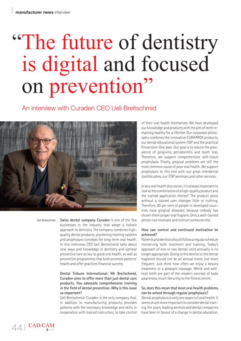
| manufacturer news interview
44 CAD/CAM2 2016
“ The future of dentistry is digital and focused on prevention”
An interview with Curaden CEO Ueli Breitschmid
Swiss dental company Curaden is one of the few businesses in the industry that adopt a holistic approach to dentistry. The company combines high- quality dental products, pioneering training systems and prophylaxis concepts for long-term oral health. In this interview, CEO Ueli Breitschmid talks about new ways and knowledge in dentistry and optimal preventive care as key to good oral health, as well as prevention programmes that both promote patients’ health and offer practices financial success.
Dental Tribune International: Mr Breitschmid, Curaden aims to offer more than just dental care products. You advocate comprehensive training in the field of dental prevention. Why is this issue so important?Ueli Breitschmid: Curaden is the only company that, in addition to manufacturing products, provides patients with the necessary knowledge and skills, in cooperation with trained instructors, to take control
of their oral health themselves. We have developed our knowledge and products with the aim of teeth re-maining healthy for a lifetime. Our corporate philos-ophy combines the innovative CURAPROX products, our dental educational system iTOP and the practical Prevention-One plan. Our goal is to reduce the prev-alence of gingivitis, periodontitis and tooth loss. Therefore, we support comprehensive soft-tissue prophylaxis. Finally, gingival problems are still the most common cause of poor oral health. We support prophylaxis to this end with our great interdental toothbrushes, our iTOP seminars and other services.
In any oral health discussion, it is always important to look at the combination of a high-quality product and the trained application thereof. The product alone without a trained user changes little or nothing. Therefore, 80 per cent of people in developed coun-tries have gingival diseases; because nobody has shown them proper oral hygiene. Only a well-trained person can motivate and instruct someone else.
How can control and continued motivation be achieved?Patients and dentists should follow a regular schedule concerning both treatment and training. Today’s approach of one or two dental visits annually is no longer appropriate. Going to the dentist or the dental hygienist should not be an annual event, but more frequent. Just think how often we enjoy a beauty treatment or a pleasant massage. White and well-kept teeth are part of the modern concept of body awareness, much like a trip to the fitness centre.
So, does this mean that most oral health problems can be solved through regular prophylaxis? Dental prophylaxis is only one aspect of oral health. It seems much more important to consider dental train-ing. For years, leading dentists and dental companies have been in favour of a change in dental education.
Ueli Breitschmid
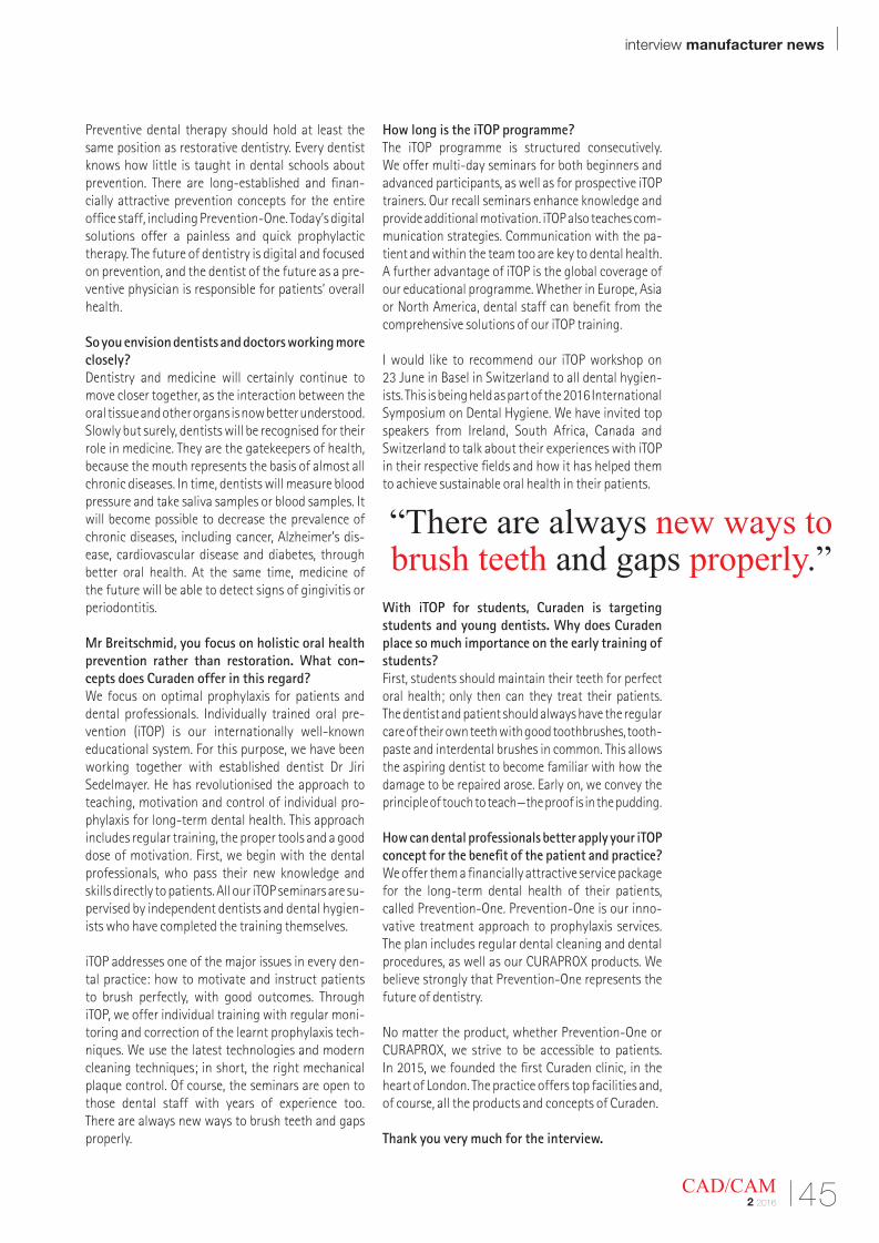
interview manufacturer news |
45CAD/CAM2 2016
Preventive dental therapy should hold at least the same position as restorative dentistry. Every dentist knows how little is taught in dental schools about prevention. There are long-established and finan-cially attractive prevention concepts for the entire office staff, including Prevention-One. Today’s digital solutions offer a painless and quick prophylactic therapy. The future of dentistry is digital and focused on prevention, and the dentist of the future as a pre-ventive physician is responsible for patients’ overall health.
So you envision dentists and doctors working more closely?Dentistry and medicine will certainly continue to move closer together, as the interaction between the oral tissue and other organs is now better understood. Slowly but surely, dentists will be recognised for their role in medicine. They are the gatekeepers of health, because the mouth represents the basis of almost all chronic diseases. In time, dentists will measure blood pressure and take saliva samples or blood samples. It will become possible to decrease the prevalence of chronic diseases, including cancer, Alzheimer’s dis-ease, cardiovascular disease and diabetes, through better oral health. At the same time, medicine of the future will be able to detect signs of gingivitis or periodontitis.
Mr Breitschmid, you focus on holistic oral health prevention rather than restoration. What con-cepts does Curaden offer in this regard?We focus on optimal prophylaxis for patients and dental professionals. Individually trained oral pre-vention (iTOP) is our internationally well-known educational system. For this purpose, we have been working together with established dentist Dr Jiri Sedelmayer. He has revolutionised the approach to teaching, motivation and control of individual pro-phylaxis for long-term dental health. This approach includes regular training, the proper tools and a good dose of motivation. First, we begin with the dental professionals, who pass their new knowledge and skills directly to patients. All our iTOP seminars are su-pervised by independent dentists and dental hygien-ists who have completed the training themselves.
iTOP addresses one of the major issues in every den- tal practice: how to motivate and instruct patients to brush perfectly, with good outcomes. Through iTOP, we offer individual training with regular moni-toring and correction of the learnt prophylaxis tech-niques. We use the latest technologies and modern cleaning techniques; in short, the right mechanical plaque control. Of course, the seminars are open to those dental staff with years of experience too. There are always new ways to brush teeth and gaps properly.
How long is the iTOP programme?The iTOP programme is structured consecutively. We offer multi-day seminars for both beginners and advanced participants, as well as for prospective iTOP trainers. Our recall seminars enhance knowledge and provide additional motivation. iTOP also teaches com-munication strategies. Communication with the pa-tient and within the team too are key to dental health. A further advantage of iTOP is the global coverage of our educational programme. Whether in Europe, Asia or North America, dental staff can benefit from the comprehensive solutions of our iTOP training.
I would like to recommend our iTOP workshop on 23 June in Basel in Switzerland to all dental hygien-ists. This is being held as part of the 2016 International Symposium on Dental Hygiene. We have invited top speakers from Ireland, South Africa, Canada and Switzerland to talk about their experiences with iTOP in their respective fields and how it has helped them to achieve sustainable oral health in their patients.
With iTOP for students, Curaden is targeting students and young dentists. Why does Curaden place so much importance on the early training of students?First, students should maintain their teeth for perfect oral health; only then can they treat their patients. The dentist and patient should always have the regular care of their own teeth with good toothbrushes, tooth-paste and interdental brushes in common. This allows the aspiring dentist to become familiar with how the damage to be repaired arose. Early on, we convey the principle of touch to teach—the proof is in the pudding.
How can dental professionals better apply your iTOP concept for the benefit of the patient and practice?We offer them a financially attractive service package for the long-term dental health of their patients, called Prevention-One. Prevention-One is our inno-vative treatment approach to prophylaxis services. The plan includes regular dental cleaning and dental procedures, as well as our CURAPROX products. We believe strongly that Prevention-One represents the future of dentistry.
No matter the product, whether Prevention-One or CURAPROX, we strive to be accessible to patients. In 2015, we founded the first Curaden clinic, in the heart of London. The practice offers top facilities and, of course, all the products and concepts of Curaden.
Thank you very much for the interview.
“There are always new ways to brush teeth and gaps properly.”
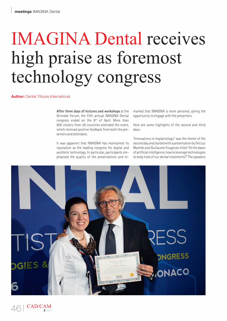
| meetings IMAGINA Dental
46 CAD/CAM2 2016
IMAGINA Dental receives high praise as foremost technology congressAuthor: Dental Tribune International
After three days of lectures and workshops at the Grimaldi Forum, the fifth annual IMAGINA Dental congress ended on the 9th of April. More than 600 visitors from 26 countries attended the event, which received positive feedback from both the pre-senters and attendees.
It was apparent that IMAGINA has maintained its reputation as the leading congress for digital and aesthetic technology. In particular, participants em-phasised the quality of the presentations and re-
marked that IMAGINA is more personal, giving the opportunity to engage with the presenters.
Here are some highlights of the second and third days:
“Innovations in implantology” was the theme of the second day and started with a presentation by Drs Luc Manhès and Guillaume Fougerais titled “At the dawn of artificial intelligence, how to leverage technologies to keep hold of our dental treatments?” The speakers
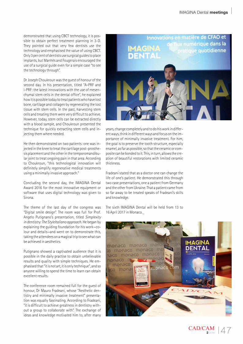
IMAGINA Dental meetings |
47CAD/CAM2 2016
demonstrated that using CBCT technology, it is pos-sible to obtain perfect treatment planning in 3-D. They pointed out that very few dentists use the technology and emphasised the value of using CBCT. Only 3 per cent of dentists use surgical guides to place implants, but Manhès and Fougerais encouraged the use of a surgical guide even for a simple case “to see the technology through”.
Dr Joseph Choukroun was the guest of honour of the second day. In his presentation, titled “A-PRF and i-PRF: the latest innovations with the use of mesen-chymal stem cells in the dental office”, he explained how it is possible today to treat patients who have lost bone, cartilage and collagen by regenerating the lost tissue with stem cells. In the past, harvesting stem cells and treating them were very difficult to achieve. However, today, stem cells can be extracted directly with a blood sample, and Choukroun presented the technique for quickly extracting stem cells and in-jecting them where needed.
He then demonstrated on two patients: one was in-jected in the knee to treat the cartilage post-prosthe-sis placement and the other in the temporomandibu-lar joint to treat ongoing pain in that area. According to Choukroun, “this technological innovation will definitely simplify regenerative medical treatments using a minimally invasive approach.”
Concluding the second day, the IMAGINA Dental Award 2016 for the most innovative equipment or software that uses digital technology was given to Sirona.
The theme of the last day of the congress was “Digital smile design”. The room was full for Prof. Angelo Putignano’s presentation, titled Simplexity in dentistry: The StyleItaliano approach. He began by explaining the guiding foundation for his work—co-lour and details—and went on to demonstrate this, taking the attendees on a magical trip to see what can be achieved in aesthetics.
Putignano showed a captivated audience that it is possible in the daily practise to obtain unbelievable results and quality with simple techniques. He em-phasised that “it is not art, it is only technique”, and so anyone willing to spend the time to learn can obtain excellent results.
The conference room remained full for the guest of honour, Dr Mauro Fradeani, whose “Aesthetic den-tistry and minimally invasive treatment” presenta-tion was equally fascinating. According to Fradeani, “it is difficult to achieve greatness in dentistry with-out a group to collaborate with”. The exchange of ideas and knowledge motivated him to, after many
years, change completely and to do his work in differ-ent ways, think in different ways and focus on the im-portance of minimally invasive treatment. For him, the goal is to preserve the tooth structure, especially enamel, as far as possible, so that the ceramic or com-posite can be bonded to it. This, in turn, allows the cre-ation of beautiful restorations with limited ceramic thickness.
Fradeani stated that as a doctor one can change the life of one’s patient. He demonstrated this through two case presentations, one a patient from Germany and the other from Ukraine. That a patient came from so far away to be treated speaks of Fradeani’s skills and knowledge.
The sixth IMAGINA Dental will be held from 13 to 16 April 2017 in Monaco._
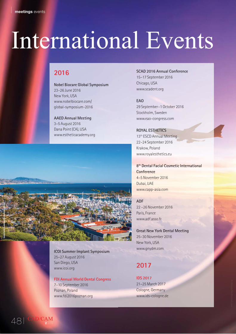
| meetings events
International Events2016
Nobel Biocare Global Symposium23–26 June 2016New York, USAwww.nobelbiocare.com/ global-symposium-2016
AAED Annual Meeting 3–5 August 2016Dana Point (CA), USAwww.estheticacademy.org
ICOI Summer Implant Symposium25–27 August 2016San Diego, USAwww.icoi.org
FDI Annual World Dental Congress7–10 September 2016Poznan, Polandwww.fdi2016poznan.org
SCAD 2016 Annual Conference15–17 September 2016Chicago, USAwww.scadent.org
EAO29 September–1 October 2016Stockholm, Swedenwww.eao-congress.com
ROYAL ESTHETICS 13th ESCD Annual Meeting22–24 September 2016Krakow, Polandwww.royalesthetics.eu
8th Dental Facial Cosmetic International Conference4–5 November 2016Dubai, UAEwww.capp-asia.com
ADF 22 –26 November 2016Paris, Francewww.adf.asso.fr
Great New York Dental Meeting25–30 November 2016New York, USAwww.gnydm.com
2017
IDS 201721–25 March 2017Cologne, Germanywww.ids-cologne.de
ananaaanananaananaananana
Danaan
aan
ananaanaanaDa
naanaDa
nananaaDa
nannnannDanaaaD
Pi
Poii
Poi
Poi
Poi
Poi
Poi
Poi
Poi
PoioiPoi
Poi
Poi
Poiii
Poii
Poiii
PooPoPoPoPoPoPoPoPooPoPPPPPt ©©©©©©nt
©nt
©nt
©t ©©©nt ©©
nt ©
nt ©©©©t ©nt ©©t ©©©©nt©t©©©©©t©nt ©©©©
nt©
nt ©©©©
nt
ntnttnttnttntntttttntnnnnnnnnonJo
nJo
nJo
nJo
nJo
nJo
nJo
nJo
nJo
nJJJJJJJJJ
Bilo
Bilo
Bilo
Bilo
Bilo
Bilo
Bilo
Bilo
Bilo
Bilo
Bilo
Bilo
Bilo
Bilo
Bilo
Bilo
Bilo
Bilo
Bilo
Bilo
Bilo
Bilo
Bilo
Bilo
Bilo
Biloo
Bilo
Bil
BililBil
BililllilBil
Bil
Bil
BiiiiBiiBiBBBBBBBBBus
/S/Sus
/Sus
/Sus
/S/Sus
/Sus
/SSus
/Sus
/Sus
/S/Sus
/SS/Sus
/S/Ss/S
us/Ss/S
us/Ss/S
s/S
us/
us/s/s///s/usssusssssussssssusssusussssusususssssuuuuuuuu
tthu
tttutt
hutttt
hutt
hutt
hutt
hutt
hutt
hutttt
hutt
hutt
hutt
hutt
huttt
huttttuttttt
hut
huttt
hutututhut
hututhuhuhuuuhuhuhuhuhuhuuuhuhhhhh
rststrsttstttttstrst
rstt
rstt
rst
ersttrstttt
rststtrsttttt
ersttttttt
erstrststst
ersttttstrst
erst
erststst
erstst
erss
erssssssrsers
ers
ers
ers
ers
ers
ersrserrrerreeeeeee
k.k.k.oc
k.oc
k.oc
k.oc
k.oc
k.oc
k.oc
k..kk.k.oc
k.k.oc
k.ck.k....k.
ock..
ockckkckkckckock
ockk
ockk
ockkkkkkk
ock
ockk
ock
ock
occccccccccoccocococcocococococcooooooooooooooooooooooooooooooooco
mmomcom
com
comomcommm
com
com
comomcomommcomm
com
comm
comm
com
comommmoocococoococoocoocoooococococococococococococcccccccccccccccccccc
©©©06ph06phpphhoto/
otoooShuttters
tersterssstock
toctoctoctoctock.co
.commmm
48 CAD/CAM2 2016
submission guidelines about the publisher |
submission guidelines:Please note that all the textual components of your submission must be combined into one MS Word document. Please do not submit multiple files for each of these items:
· the complete article; · all the image (tables, charts, photographs, etc.) captions; · the complete list of sources consulted; and · the author or contact information (biographical sketch,
mailing address, e-mail address, etc.).
In addition, images must not be embedded into the MS Word document. All images must be submitted separately, and details about such submission follow below under image requirements.
Text lengthArticle lengths can vary greatly—from 1,500 to 5,500 words— depending on the subject matter. Our approach is that if you need more or less words to do the topic justice, then please make the article as long or as short as necessary.
We can run an unusually long article in multiple parts, but this usually entails a topic for which each part can stand alone be-cause it contains so much information.
In short, we do not want to limit you in terms of article length, so please use the word count above as a general guideline and if you have specific questions, please do not hesitate to contact us.
Text formattingWe also ask that you forego any special formatting beyond the use of italics and boldface. If you would like to emphasise certain words within the text, please only use italics (do not use underli-ning or a larger font size). Boldface is reserved for article headers. Please do not use underlining.
Please use single spacing and make sure that the text is left jus - tified. Please do not centre text on the page. Do not indent para-graphs, rather place a blank line between paragraphs. Please do not add tab stops.
Should you require a special layout, please let the word processing programme you are using help you do this formatting automati-cally. Similarly, should you need to make a list, or add footnotes or endnotes, please let the word processing programme do it for you automatically. There are menus in every programme that will enable you to do so. The fact is that no matter how carefully done, errors can creep in when you try to number footnotes yourself.
Any formatting contrary to stated above will require us to remove such formatting before layout, which is very time-consuming. Please consider this when formatting your document.
Image requirementsPlease number images consecutively throughout the article by using a new number for each image. If it is imperative that certain images are grouped together, then use lowercase letters to designate these in a group (for example, 2a, 2b, 2c).
Please place image references in your article wherever they are appropriate, whether in the middle or at the end of a sentence. If you do not directly refer to the image, place the reference at the end of the sentence to which it relates enclosed within brackets and before the period.
In addition, please note:
· We require images in TIF or JPEG format. · These images must be no smaller than 6 x 6 cm in size at 300 DPI. · These image files must be no smaller than 80 KB in size (or they
will print the size of a postage stamp!).
Larger image files are always better, and those approximately the size of 1 MB are best. Thus, do not size large image files down to meet our requirements but send us the largest files available. (The larger the starting image is in terms of bytes, the more lee-way the designer has for resizing the image in order to fill up more space should there be room available.)
Also, please remember that images must not be embedded into the body of the article submitted. Images must be submitted separately to the textual submission.
You may submit images via e-mail, via our FTP server or post a CD containing your images directly to us (please contact us for the mailing address, as this will depend upon the country from which you will be mailing).
Please also send us a head shot of yourself that is in accordance with the requirements stated above so that it can be printed with your article.
AbstractsAn abstract of your article is not required.
Author or contact informationThe author’s contact information and a head shot of the author are included at the end of every article. Please note the exact information you would like to appear in this section and for-mat it according to the requirements stated above. A short biographical sketch may precede the contact information if you provide us with the necessary information (60 words or less).
Questions?Magda Wojtkiewicz (Managing Editor)[email protected]
49CAD/CAM2 2016
| about the publisher imprint
Publisher/President/CEOTorsten R. Oemus [email protected]
Managing EditorMagda [email protected]
Designer Franziska Dachsel
Copy EditorsSabrina RaaffHans Motschmann
International Administration
Chief Financial OfficerDan Wunderlich
Business Development ManagerClaudia Salwiczek-Majonek
Project Manager Online Tom Carvalho
Event Manager Lars Hoffmann
International PR & Project Manager Marc Chalupsky
Marketing & Sales Services Nicole Andrä
Event Services Esther Wodarski
Executive Producer Gernot Meyer
Accounting Services Karen HamatschekAnja MaywaldManuela Hunger
Media Sales Managers
Matthias Diessner (Key Accounts)Melissa Brown (International)Antje Kahnt (International)Peter Witteczek (Asia Pacific)Weridiana Mageswki (Latin America)Maria Kaiser (North America)Hélène Carpentier (Europe)Barbora Solarova (Eastern Europe)
International Offices
Dental Tribune InternationalHolbeinstr. 29, 04229 Leipzig, GermanyTel.: +49 341 48474-302Fax: +49 341 [email protected]
Dental Tribune Asia Pacific Ltd.Room A, 20/F, Harvard Commercial Building,105–111 Thomson Road, Wanchai, Hong KongTel.: +852 3113 6177Fax: +852 3113 6199
Tribune America, LLC116 West 23rd Street, Ste. 500, New York, NY 10011, USATel.: +1 212 244 7181Fax: +1 212 244 7185
Editorial BoardProf. Albert Mehl, SwitzerlandProf. Gerwin Arnetzl, Austria
Dr Stefan Holst, GermanyHans Geiselhöringer, GermanyDr Ansgar Cheng, Singapore
Printed byLöhnert DruckHandelsstraße 1204420 Markranstädt, Germany
Copyright RegulationsCAD/CAM international magazine of digital dentistry is published by Dental Tribune International (DTI) and appears in 2016 with four issues. The magazine
and all articles and illustrations therein are protected by copyright. Any utilisation without the prior consent of editor and publisher is in admissible and liable to prosecution. This applies in particular to duplicate copies, translations, microfilms, and storage and processing in electronic systems.
Reproductions, including extracts, may only be made with the permission of the publisher. Given no statement to the contrary, any submissions to the editorial department are understood to be in agreement with a full or partial publishing of said submission. The editorial department reserves the right to check all submitted articles for formal errors and factual authority, and to make amendments if necessary. No responsibility shall be taken for unsolicited books and manuscripts. Articles bearing symbols other than that of the editorial department, or which are distinguished by the name of the author, represent the opinion of the afore-mentioned, and do not have to comply with the views of DTI. Responsibility for such articles shall be borne by the author. Responsibility for advertisements and other specially labeled items shall not be borne by the editorial department. Likewise, no responsibility shall be assumed for information published about associations, companies and commercial markets. All cases of consequential liability arising from inaccurate or faulty representation are excluded. General terms and conditions apply. Legal venue is Leipzig, Germany.
CAD/CAMinternational magazine of digital dentistry
50 CAD/CAM2 2016
Nobel Biocare Global SymposiumJune 23–26, 2016 – New York
© Nobel Biocare Services AG, 2016. All rights reserved. Nobel Biocare, the Nobel Biocare logotype and all other trademarks are, if nothing else is stated or is evident from the context in a certain case, trade-marks of Nobel Biocare. Please refer to nobelbiocare.com/trademarks for more information. Product images are not necessarily to scale. Disclaimer: Some products may not be regulatory cleared/released for sale in all markets. Please contact the local Nobel Biocare sales office for current product assortment and availability.
nobelbiocare.com/global-symposium-2016
An experience beyond the ordinary
The Nobel Biocare Global Symposium 2016 program will offer unparalleled clinical and scientific education, as well as in-depth hands-on training. Held at the Waldorf Astoria in New York, it’s a unique opportunity to experience how innovation can come to life in your daily work.
Design your own learning experience
Choose from numerous lectures, forums, master classes and hands-on sessions – from over 150 of the best speakers and presenters in the world. This must-attend event will cover a vast variety of techniques and treatment solutions, from diagnosis to treatment completion. Don’t miss this opportunity. Sign up now and we’ll see you in New York!
Where innovation comes to life
Registernow
Read more about the Symposium
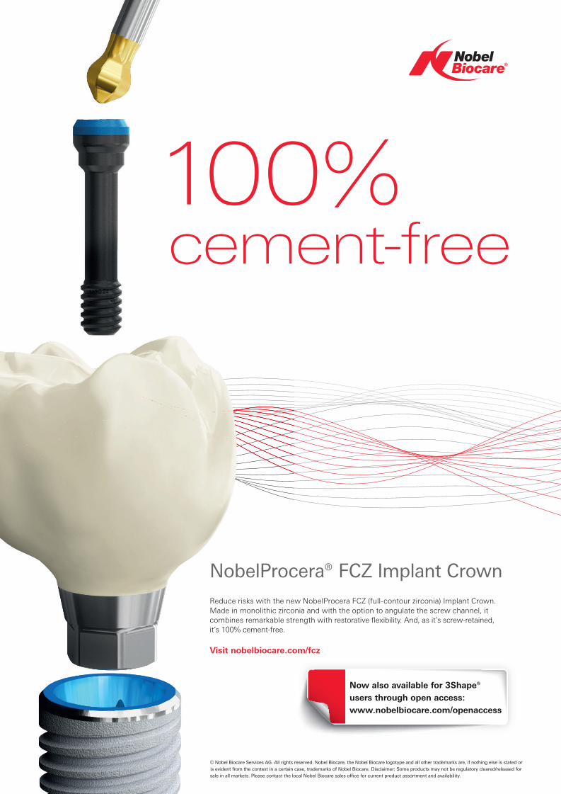
cement-free100%
© Nobel Biocare Services AG. All rights reserved. Nobel Biocare, the Nobel Biocare logotype and all other trademarks are, if nothing else is stated or is evident from the context in a certain case, trademarks of Nobel Biocare. Disclaimer: Some products may not be regulatory cleared/released for sale in all markets. Please contact the local Nobel Biocare sales offi ce for current product assortment and availability.
Reduce risks with the new NobelProcera FCZ (full-contour zirconia) Implant Crown. Made in monolithic zirconia and with the option to angulate the screw channel, it combines remarkable strength with restorative fl exibility. And, as it’s screw-retained, it’s 100% cement-free.
Visit nobelbiocare.com/fcz
NobelProcera® FCZ Implant Crown
Now also available for 3Shape® users through open access: www.nobelbiocare.com/openaccess
Related Documents