Molecular Vision 2007; 13:443-56 <http://www.molvis.org/molvis/v13/a47/> Received 5 May 2006 | Accepted 23 February 2007 | Published 27 March 2007 The retinal pigment epithelium (RPE) is a monolayer of pigmented cells that closely interacts with photoreceptors to maintain their structural integrity and excitability [1-4]. A changed growth factor secretion by the RPE is believed to be involved in the etiology of proliferative eye diseases,such as choroidal neovascularization (CNV) [5-8] in age-related macu- lar degeneration (AMD), which is the most common cause for legal blindness in industrialized countries [9]. Mainly a changed growth factor secretion by the RPE seems to be of importance for initiating CNV: increased secretion of pro-an- giogenic factors and decreased secretion of anti-angiogenic factors. CNV, representing the most severe complication in AMD, originates in choroidal blood vessels that grow through Bruch’s membrane into the sub RPE as well as the subretinal space. In many studies, but especially in recent clinical trials, vascular endothelial growth factor (VEGF) appears to be the major angiogenic factor in this process. Several studies using cultured or freshly isolated RPE cells from various species demonstrated the expression of L-type Ca 2+ channels [10-15]. L-type Ca 2+ channels represent a group of high-voltage activated Ca 2+ channels [16,17]. Studies ex- amining the regulation of L-type channels of the RPE sug- gested that these Ca 2+ channels provide a Ca 2+ -influx pathway involved in growth factor-dependent intracellular signaling [12,15]. Activation of L-type channels in the RPE by the cy- tosolic subtype tyrosine kinase pp60 c-src shifts the voltage-de- pendent activation to a more negative voltage-range, closer to the resting potential of RPE cells [12,18]. This leads to a higher number of active channels and, thus, an increase in intracellu- lar free Ca 2+ . In this way, high-voltage-activated Ca 2+ chan- nels can contribute to intracellular signaling in epithelial cells. A voltage-dependent activation in a rather negative voltage- range has been discussed as a property of L-type channels composed of Ca v 1.3 α-subunits [19-21]. The expression of Ca v 1.3 subunits has been demonstrated in rat RPE cells [12,15]. Thus, L-type channels in RPE cells can participate in intracel- lular signaling which could mediate changes in a yet unknown RPE cell function. L-type channels composed of the Ca v 1.3 α-channel-sub- unit belong to the neuroendocrine subtype, because these chan- nels are known to regulate the insulin secretion by β-islet cells in the pancreas [16]. The RPE is known to secrete a variety of growth factors like insulin like growth factor-1 (IGF-1), basic fibroblast growth factor (bFGF or FGF2), VEGF or pigment epithelium-derived factor (PEDF) [7,22-30]. In order to show that L-type channels might regulate secretion rates in RPE cells we studied voltage-dependent Ca 2+ channels and the regu- ©2007 Molecular Vision Ca 2+ channels in retinal pigment epithelial cells regulate vascular endothelial growth factor secretion rates in health and disease Rita Rosenthal, 1 Heinrich Heimann, 2 Hansjürgen Agostini, 3 Gottfried Martin, 3 Lutz Lothar Hansen, 3 Olaf Strauss 1,4 1 Institut für Klinische Physiologie, Charité-Universitatsmedizin Berlin, Campus Benjamin Franklin, Berlin, Germany, 2 St. Pauls Eye Unit, Royal Liverpool Hospital, Liverpool, United Kingdom, 3 Augenklinik des Universitätsklinikums Freiburg, Albert-Ludwigs- Universität Freiburg, Freiburg, 4 Experimentelle Ophthalmologie, Klinik und Poliklinik für Augenheilkunde, Universitätsklinikum Hamburg-Eppendorf, Germany Purpose: Choroidal neovascularization (CNV) is the most severe complication in age-related macular degeneration. The major angiogenic factor involved is vascular endothelial growth factor (VEGF) secreted by the retinal pigment epithelium (RPE). Since RPE cells express neuroendocrine L-type Ca 2+ channels we investigated their involvement in VEGF secre- tion in normal RPE cells and RPE cells from patients with CNV. Methods: Freshly isolated and cultured RPE cells were studied using the patch-clamp technique and ELISA-based secre- tion assays. Results: Both freshly isolated and cultured cells showed whole-cell Ba 2+ currents with properties of L-type Ca 2+ currents: high activation threshold, sensitivity to dihydropyridines (10 μM nifedipine) and slow inactivation. VEGF-A secretion was elevated by BayK8644 (10 μM) or basic fibroblast growth factor (bFGF, 10 ng/ml), both of which are able to activate L-type channels. Cells from CNV tissue also showed nifedipine-sensitive Ba 2+ currents, which displayed a voltage-depen- dent activation at more negative potentials, faster inactivation and changed regulation by tyrosine kinase pp60 c-src . The CNV RPE cells showed higher VEGF secretion rates which were reduced by nifedipine. Conclusions: Thus, L-type Ca 2+ channels in normal RPE cells regulate the secretion of VEGF. RPE cells from eyes with CNV maintain a VEGF secretion regulated by nifedipine-sensitve Ca 2+ channels which might be of importance for the development of CNV. Correspondence to: Olaf Strauss, Experimentelle Ophthalmologie, Klinik und Poliklinik für Augenheilkunde, Universitätsklinikum Hamburg-Eppendorf, Martinistrasse 52, 20246 Hamburg, Germany; Phone: +49 40 42803 9469; FAX: +49 40 42803 5017; email: [email protected] 443

Ca channels in retinal pigment epithelial cells regulate ... · Phone: +49 40 42803 9469; FAX: +49 40 42803 5017; email: [email protected] 443. lation of VEGF-A secretion
Feb 20, 2019
Welcome message from author
This document is posted to help you gain knowledge. Please leave a comment to let me know what you think about it! Share it to your friends and learn new things together.
Transcript

Molecular Vision 2007; 13:443-56 <http://www.molvis.org/molvis/v13/a47/>Received 5 May 2006 | Accepted 23 February 2007 | Published 27 March 2007
The retinal pigment epithelium (RPE) is a monolayer ofpigmented cells that closely interacts with photoreceptors tomaintain their structural integrity and excitability [1-4]. Achanged growth factor secretion by the RPE is believed to beinvolved in the etiology of proliferative eye diseases,such aschoroidal neovascularization (CNV) [5-8] in age-related macu-lar degeneration (AMD), which is the most common causefor legal blindness in industrialized countries [9]. Mainly achanged growth factor secretion by the RPE seems to be ofimportance for initiating CNV: increased secretion of pro-an-giogenic factors and decreased secretion of anti-angiogenicfactors. CNV, representing the most severe complication inAMD, originates in choroidal blood vessels that grow throughBruch’s membrane into the sub RPE as well as the subretinalspace. In many studies, but especially in recent clinical trials,vascular endothelial growth factor (VEGF) appears to be themajor angiogenic factor in this process.
Several studies using cultured or freshly isolated RPE cellsfrom various species demonstrated the expression of L-typeCa2+ channels [10-15]. L-type Ca2+ channels represent a groupof high-voltage activated Ca2+ channels [16,17]. Studies ex-
amining the regulation of L-type channels of the RPE sug-gested that these Ca2+ channels provide a Ca2+-influx pathwayinvolved in growth factor-dependent intracellular signaling[12,15]. Activation of L-type channels in the RPE by the cy-tosolic subtype tyrosine kinase pp60c-src shifts the voltage-de-pendent activation to a more negative voltage-range, closer tothe resting potential of RPE cells [12,18]. This leads to a highernumber of active channels and, thus, an increase in intracellu-lar free Ca2+. In this way, high-voltage-activated Ca2+ chan-nels can contribute to intracellular signaling in epithelial cells.A voltage-dependent activation in a rather negative voltage-range has been discussed as a property of L-type channelscomposed of Ca
v1.3 α-subunits [19-21]. The expression of
Cav1.3 subunits has been demonstrated in rat RPE cells [12,15].
Thus, L-type channels in RPE cells can participate in intracel-lular signaling which could mediate changes in a yet unknownRPE cell function.
L-type channels composed of the Cav1.3 α-channel-sub-
unit belong to the neuroendocrine subtype, because these chan-nels are known to regulate the insulin secretion by β-islet cellsin the pancreas [16]. The RPE is known to secrete a variety ofgrowth factors like insulin like growth factor-1 (IGF-1), basicfibroblast growth factor (bFGF or FGF2), VEGF or pigmentepithelium-derived factor (PEDF) [7,22-30]. In order to showthat L-type channels might regulate secretion rates in RPEcells we studied voltage-dependent Ca2+ channels and the regu-
©2007 Molecular Vision
Ca2+ channels in retinal pigment epithelial cells regulate vascularendothelial growth factor secretion rates in health and disease
Rita Rosenthal,1 Heinrich Heimann,2 Hansjürgen Agostini,3 Gottfried Martin, 3 Lutz Lothar Hansen,3 OlafStrauss1,4
1Institut für Klinische Physiologie, Charité-Universitatsmedizin Berlin, Campus Benjamin Franklin, Berlin, Germany, 2St. PaulsEye Unit, Royal Liverpool Hospital, Liverpool, United Kingdom, 3Augenklinik des Universitätsklinikums Freiburg, Albert-Ludwigs-Universität Freiburg, Freiburg, 4Experimentelle Ophthalmologie, Klinik und Poliklinik für Augenheilkunde, UniversitätsklinikumHamburg-Eppendorf, Germany
Purpose: Choroidal neovascularization (CNV) is the most severe complication in age-related macular degeneration. Themajor angiogenic factor involved is vascular endothelial growth factor (VEGF) secreted by the retinal pigment epithelium(RPE). Since RPE cells express neuroendocrine L-type Ca2+ channels we investigated their involvement in VEGF secre-tion in normal RPE cells and RPE cells from patients with CNV.Methods: Freshly isolated and cultured RPE cells were studied using the patch-clamp technique and ELISA-based secre-tion assays.Results: Both freshly isolated and cultured cells showed whole-cell Ba2+ currents with properties of L-type Ca2+ currents:high activation threshold, sensitivity to dihydropyridines (10 µM nifedipine) and slow inactivation. VEGF-A secretionwas elevated by BayK8644 (10 µM) or basic fibroblast growth factor (bFGF, 10 ng/ml), both of which are able to activateL-type channels. Cells from CNV tissue also showed nifedipine-sensitive Ba2+ currents, which displayed a voltage-depen-dent activation at more negative potentials, faster inactivation and changed regulation by tyrosine kinase pp60c-src. TheCNV RPE cells showed higher VEGF secretion rates which were reduced by nifedipine.Conclusions: Thus, L-type Ca2+ channels in normal RPE cells regulate the secretion of VEGF. RPE cells from eyes withCNV maintain a VEGF secretion regulated by nifedipine-sensitve Ca2+ channels which might be of importance for thedevelopment of CNV.
Correspondence to: Olaf Strauss, Experimentelle Ophthalmologie,Klinik und Poliklinik für Augenheilkunde, UniversitätsklinikumHamburg-Eppendorf, Martinistrasse 52, 20246 Hamburg, Germany;Phone: +49 40 42803 9469; FAX: +49 40 42803 5017; email:[email protected]
443

lation of VEGF-A secretion by human RPE cells and by RPEcells from surgically excised CNV tissues of AMD patients.We found that VEGF secretion is dependent on the activity ofvoltage-dependent Ca2+ channels, which might provide newtargets to prevent CNV by interventions at the source forVEGF.
METHODSHuman tissue: For the use of human material, tenets of theDeclaration of Helsinki were followed, informed consent wasobtained, and Institutional Human Experimentation Commit-tee approval was granted for the studies. CNV membraneswere obtained directly after eye surgery of patients with AMD.The mean and the range of the age of the patients are given inTable 1. Patients with severe systemic diseases were not in-cluded in the study. The human tissue was obtained about onehour after eye surgery or enucleation and immediately pre-pared for the experiments The detailed information is listed inTable 1.
Patch-clamp recordings with freshly isolated cells: A por-tion of the CNV membranes was dissolved into single cellsuspension by enzymatic digestion using protease (10%) andtrypsin (0.1%). Cells from donor eyes without CNV wereplaced into single-cell suspension by the same method. Cellsuspensions were placed onto a poly-lysine-coated glass coverslip and allowed to settle. The cover slips were placed into theperfusion chamber on the stage of an inverted microscope andconstantly superfused. The bath solution contained the fol-lowing (given in mM): 136.4 NaCl, 1.1 NaH
2PO
4, 4.2 NaHCO
3,
0.9 MgCl2, 0.95 CaCl
2, 10.0 BaCl
2, 5.8 TEACl, 25.0 HEPES,
and 11.1 glucose. Perforated-patch recordings were performedwith patch-pipettes of 3-5 mega-Ω resistance, using a pipettesolution that contained the following (given in mM): 100.0CsCl, 10.0 NaCl, 2.0 MgSO
4, 0.5 CaCl
2, 5.5 EGTA, 10.0
HEPES, nystatin (150 mg/ml). For perforated-patch record-ings from pigmented cells, we added an antibody against classIII β-tubulin (Sigma, Deisenhofen, Germany) to the pipettesolution. Currents were measured using an EPC-7 patch-clampamplifier in conjunction with TIDA hardware and software
(HEKA, Lamprecht, Germany). The freshly isolated cellsshowed a membrane capacitance of 43.3±6.9 pF (n=10), andaccess resistance of 12.5±2.0 mega-Ω (n=10). Access resis-tance was compensated to values lower than 10 mega-Ω. Sealswere stable for approximately 3-7 min.
For stimulation of Ba2+ currents through L-type channelscells were depolarized from a holding potential of -70 mV by9 voltage-steps of 50 ms duration up to +20 mV (Figure 1A).Measurements of maximal current density and kinetic param-eters occurred at +10 mV to ensure data comparison at equalbiophysical driving forces.
Patch-clamp recordings from cultured cells: CNV mem-brane pieces not used for in situ investigations were employedfor growing RPE cell cultures, using the method of Schluncket al. [31]. Cultures from eyes without CNV were establishedusing the method of Aronson [32]. Recordings from culturedcells were performed under the same conditions with the samepipette and bath solutions as used for freshly isolated cells.Purity of cell cultures was confirmed by immunohistochemi-cal staining for class III β-tubulin, vimentin, cytokeratin 18,glial fibrillary acidic protein (GFAP), and endothelial factor8. Sub confluent cell cultures of both control RPE and CNVRPE from the 5th to the 12th passage were used for patch-clampexperiments. Mean membrane capacitance of cultured cellswas 128.9±17.8 pF (n=19); access resistance was 16.9±2.1mega-Ω (n=19).
Intracellular application of pp60c-src: During the wholecell recording, 30 U/ml pp60c-src (>90% purity) was appliedvia the patch-pipette employing a procedure described by Wangand Salter [33]. In addition to pp60c-src (Biomol, Hamburg,Germany), the pipette solution contained ATP (4x10⁻³ M) andthe extracellular solution DIDS (5x10⁻⁴ M) to block Cl- chan-nels possibly activated by the application of ATP [34]. Afterthe patch was opened into the whole-cell configuration, themaximal Ba2+ current amplitude was measured every 1 minand plotted over the experimental time normalized to the am-plitude measured directly after breaking into the whole-cellconfiguration. As a control, a series of the same experimentswere performed with heat-inactivated pp60c-src (30 min at 95°C).
Immunoblotting: Membrane proteins were separated bypolyacrylamide gel electrophoresis (8.5% polyacrylamide;Mini-Protean cells Bio-Rad Life Science Group, Hercules, CA;for 1 h at 150 V), and 30 mg of total protein were loaded ineach lane. The proteins were blotted to nitrocellulose filterscreens (Polyscreen, NEN®, Life Science Products Boston,MA) for 1 h at 100 V. Blots were blocked in phosphate buff-ered saline (PBS/Tween) with nonfat dry milk (10%) for 2 hand with bovine serum albumin (BSA; 5%) for 4 h at roomtemperature. They were then probed overnight at 4 °C withantibodies against Ca
v1.2 and Ca
v1.3 channel subunits and with
the peroxidase-conjugated secondary antibody for 1 h at roomtemperature. Next, blots were visualized using a chemilumi-nescence kit (Amersham Pharmacia Biotech, Braunschweig,Germany) and digitized using the LAS-1000 Image Analyzer(Fujifilm, Berlin, Germany) and the AIDA 2.0 software(Raytest, Berlin, Germany). Specific staining was verified by
©2007 Molecular VisionMolecular Vision 2007; 13:443-56 <http://www.molvis.org/molvis/v13/a47/>
TABLE 1. ORIGIN OF HUMAN TISSUE
Patient information RPE CNV-------------------- -------- --------Number of patients 7 18Mean age of patients 69.0±1.6 70.7±4.0Male 3 7Female 4 11
Retinal pigment epithelium (RPE) was isolated from eyes withoutchoroidal neovascularization (CNV). These eyes were obtained frommulti organ donors after removal of cornea for transplantation. CNVis derived from choroidal neovascular tissue of patients with age-related macular degeneration (AMD); removal of CNV tissue wasindicated by individuell clinical picture. All AMD patients sufferedfrom wet AMD. Since only 46% of RPE cells in CNV tissue expressvoltage-dependent channels, a larger number of patients is includedin this study.
444

©2007 Molecular VisionMolecular Vision 2007; 13:443-56 <http://www.molvis.org/molvis/v13/a47/>
stripping blots and staining them a second time using the sameantibody together with the corresponding blocking peptide(antibody: blocking peptide, 1:1).
Vascular endothelial growth factor secretion by retinalpigment epithelium cells: Confluent cell cultures were usedto study VEGF secretion by RPE cells. For this assay 100,000cells were kept in a chamber of a 12-well plate. Every cham-ber was equipped with 500 µl test solution (Dulbecco’s modi-fied Eagle medium with 0.5% fetal calf serum). Beginningwith the last change of test medium, the concentration ofVEGF-165 was measured every 4 h in the test medium usinga commercially available ELISA kit (Biosource International,Solingen, Germany) according to the manufacturer’s instruc-tions. The number of experiments (n) includes the mean val-ues of two repeated measurements of VEGF concentration inthe medium in one experiment.
Immunohistochemistry: Freshly isolated and culturedcells were stained with anti-class III β-tubulin antibody(Sigma). The following procedure was used for freshly iso-lated cells. After the perforated-patch recording of a pigmentedcell the patch was opened into a classical whole-cell configu-ration, and negative voltage was applied to facilitate diffusionof the antibody from the pipette into the cell. The patch-pi-pette was removed from the cell, and the cell stayed attachedto the ground and could be subjected to immuno-staining. Inthe first step, the cells underwent methanol-based fixation fol-lowed by permeabilization with Triton X-100 (0.5% in PBS).Then cells were washed several times and incubated with thesecondary fluorescence conjugated antibody. Cultured cellsunderwent methanol-based fixation and permeabilization be-fore they were incubated with the first antibody for 1 h. Afterbeing placed through a series of washes cells were next incu-bated with the secondary fluorescence conjugated antibody.Fluorescence staining was visualized by analysis (Soft Imag-ing System, Muenster, Germany).
Confluent cultures, which had been subjected to VEGF-A secretion assay, were tested for their homogeneity by meansof immunohistochemistry methods as previously described inreference [31]. In brief, cultures were rinsed with PBS andfixed with acetone/PBS 1:1 followed with iced acetone. Cul-tures were incubated with antibodies against cytokeratin 18(Sigma), vimentin, glial fibrillary acidic protein (GFAP), en-dothelial factor 8 (all from DAKO, Hamburg, Germany). Cellnuclei were counter-stained with hematoxylin. Primary anti-bodies were stained with alkaline phosphatase- or peroxidase-labeled secondary antibodies (Sigma) with fast red as sub-strate.
Statistics: Numbers of experiments (n) with freshly iso-lated cells correspond to numbers of individual AMD patientsor eyes without CNV. Data from cultured cells from CNVmembranes were obtained from three different cultures fromthree different patients. All data are stated as mean±SEM. Datawere tested for statistical significance by ANOVA (Sigma Plot,Systat Software, Inc., Point Richmond, CA). Significance wasconsidered at p-values lower than 0.05.
RESULTSVoltage-dependent Ca2+ channels in freshly isolated cells:Cells were identified as RPE cells by subsequent immunohis-tochemical staining and fluorescence microscopy (Figure 1G,Figure 2). For this purpose, the solution of the recording pi-pette contained antibodies against class III β-tubulin, a markerfor RPE cells in both healthy and several forms of disease[35]. Once the membrane currents were recorded, the anti-body was intracellularly applied via dialysis by the solution inthe patch-pipette.
Freshly isolated RPE cells showed voltage-dependent in-ward currents in the absence of potassium. With Ba2+ as chargecarrier, these cells responded to depolarization from a holdingpotential of -70 mV (Figure 1B) to values more positive than-30 mV with fast activating and inactivating inward currents(Figure 1B). The currents showed a maximal current densityat +10 mV (Figure 1C) with 1.9±0.6 pApF-1 with a range from1.4 to 2.8 pApF-1 (n=3; Figure 1F), a potential of the half maxi-mal activation of -9.7±1.9 mV (n=3; Figure 3B), a time topeak of 9.7±2.8 ms (n=3), and an inactivation time constantof 53.9±10.3 ms (n=3).
In six out of 13 (46%) investigated CNV membranes, RPEcells showed voltage-dependent Ba2+ currents (Figure 1D-G).The mean current density of the maximal amplitude at +10mV was 7.0±3.4 pApF-1 (n=6; current density ranged at +10mV from 3.5 pApF-1 to 24.4 pApF-1), which was reached after2.8±0.4 ms (n=6). This was faster than in cells from controleyes (p=0.024). The potential of the half maximal activationwas -15.4±1.3 mV (n=6) which was significantly more nega-tive than in cells from control eyes (p=0.0254). The currentsinactivated with a time constant of 12.6±2.3 ms (n=6). Thus,inactivation was faster than in cells from control eyes(p=0.00094).
The activation threshold at about -30 mV indicates thatthe Ba2+ currents were in freshly isolated control cells and incells from CNV tissue currents through high-voltage-activated(HVA) Ca2+ channels. Dihydropyridine nifedipine, a blockerof an HVA channel subtype, the L-type Ca2+channels, inhib-ited maximal current amplitudes to 39.4±6.3% (n=3) of con-trol in freshly isolated RPE cells from eyes without CNV (Fig-ure 3A,B,E). Application of nifedipine (10-5 M, Figure 3F,D,E)reduced maximal current amplitudes to 20.7±7.5% (n=2) ofthe control values in freshly isolated cells from CNV tissues.The voltage-dependence of Ba2+ currents was not changed inthe presence of nifedipine in cells from CNV tissues as wellas in cells from eyes without CNV (Figure 3B,D).
Ba2+ currents in cultured cells: To further examine thecellular physiological characteristics of RPE cells, we devel-oped cultures from CNV tissues by following the methods ofSchlunck et al. [31]. In order to test these cultures on theirorigin and homogeneity, cultures were labeled with antibod-ies against vimentin, cytoskeratin 18, endothelial factor 8, andglial acidic fibrillary protein (GFAP). Cultures were homoge-neously stained with antibodies against vimentin or cytokeratin18 (Figure 2). No staining was observed using antibodiesagainst GFAP or endothelial factor 8. Identity of cells used for
445

©2007 Molecular VisionMolecular Vision 2007; 13:443-56 <http://www.molvis.org/molvis/v13/a47/>
Figure 1. Voltage-dependent Ba2+ currents in freshly isolated cells. A: Pattern of electrical stimulation to activate voltage-dependent Ba2+
currents. B: Ba2+ inward currents in a human retinal pigment epithelium (RPE) cell freshly isolated from an eye without choroidalneovascularization (CNV). C: Current/voltage plot of currents from three freshly isolated cells from eyes without CNV D: Ba2+ inwardcurrents in a human RPE cell freshly isolated from CNV tissue. E: Current/voltage plot of currents from six freshly isolated cells from CNVmembranes. F: Comparison of maximal Ba2+ densities at +10 mV in freshly isolated cells. G: Class III β-tubulin staining of a freshly isolatedcell from CNV membrane after patch-clamp recording.
446

patch-clamp recordings was confirmed by staining againstclass III β-tubulin (Figure 1G).
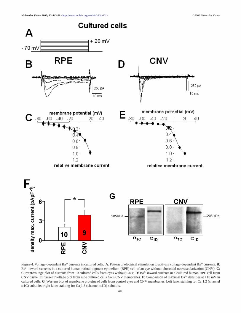
RPE cells maintain the activity of voltage-dependent Ca2+
channels in culture (Figure 4 and Figure 5). The maximal cur-rent density at +10 mV in cultured RPE cells from CNV mem-branes was 3.9±0.8 pApF-1 (n=9) and in cultured RPE cellsfrom eyes without CNV (2.0±0.4 pApF-1, n=10; p=0.049).Cells from CNV membranes had a time to peak of with3.3±0.45 ms (n=9), which was faster than in cells from eyeswithout CNV (11.9±1.4 ms, n=10; p=0.00004). Western blotanalysis of membrane proteins revealed the expression ofCa
v1.3 subunits in cultured RPE cells from eyes without CNV
and in cells from CNV membranes. A weak expression ofCa
v1.2 subunits could only be detected in RPE cells from eyes
without CNV (Figure 4G).The currents in cultured cells were sensitive to the
dihydropyridine nifedipine, too (Figure 5). Application ofnifedipine reduced the maximal current amplitude measuredat +10 mV to 53.1±8.7% (n=4) in cells from CNV tissues andto 60.1±4.8% (n=4) in cells from eyes without CNV. Further-more, as in freshly isolated cells, the voltage-dependence ofBa2+ currents was not influenced by nifedipine in both cellsfrom CNV tissues and cells from control eyes.
Vascular endothelial growth factor secretion: CulturedRPE cells release VEGF into the medium, a process that couldbe stimulated by a test medium containing 0.5% fetal calf se-rum. Under these conditions, we found that VEGF concentra-tion increased with time in the test medium in both RPE cellsfrom eyes without CNV and in RPE cells from CNV mem-branes. However, the VEGF concentration increased signifi-cantly faster and reached higher concentrations in culturescontaining cells from CNV membranes when compared tocultures of cells from eyes without CNV (Figure 6A). Thisresulted in a higher VEGF secretion rate of 12.8 ± 1.4 pg/h(versus no CNV: 5.4±0.9 pg/h; n=4; p=0.0475). Inhibition ofCa2+ channels by nifedipine (10-5 M) reduced VEGF secretionby both cell populations (Figure 6B,C). To confirm the con-clusion that activation of voltage-dependent Ca2+ channelsleads to secretion of VEGF, we used the L-type channel Ca2+
channel modulator BayK8644 (10-5 M; Figure 6B,C) as (-)enantiomer, which activates L-type channels and as the (+)enantiomer, which inhibits L-type channels. The (-) BayK8644increased the VEGF secretion rate by RPE cells from eyeswithout CNV (Figure 6B), whereas it only slightly increasedthe VEGF-A secretion rate in cells from CNV membranes(Figure 6C). The latter effect was not statistically significant.
©2007 Molecular VisionMolecular Vision 2007; 13:443-56 <http://www.molvis.org/molvis/v13/a47/>
Figure 2. Analysis of retinal pigment epithelium cell cultures from CNV membranes. Sub-confluent cultures of retinal pigment epithelium(RPE) cells from choroidal neovascularization (CNV) tissues used for patch-clamp analysis were immunostained against class III β-tubulin.Confluent RPE cell cultures from CNV tissues were stained against glial acidic fibrillary protein (GFAP), endothelial factor 8, cytokeratin 18,or vimentin.
447

©2007 Molecular VisionMolecular Vision 2007; 13:443-56 <http://www.molvis.org/molvis/v13/a47/>
Figure 3. Effect of the dihydropyridine compound nifedipine on volt-age-dependent Ba2+ currents of freshly isolated RPE cells. A: Ba2+
currents induced by a 50 ms voltage-step from -70 mV to +10 mV ina retinal pigment epithelium (RPE) cell freshly isolated from an eyewithout choroidal neovascularization (CNV) under control condi-tions and in the presence of nifedipine (10-5 M). B: Curves of volt-age-dependent activation from six freshly isolated cells from eyeswithout CNV: three cells under control conditions and three cells inthe presence of nifedipine. Currents were normalized to the maximalcurrent amplitude and fitted using the Boltzmann equation. C: Ba2+
currents induced by a 50 ms voltage-step from -70 mV to +10 mV inan RPE cell freshly isolated from a CNV membrane under controlconditions and in the presence of nifedipine (10-5 M). D: Curves ofvoltage-dependent activation from eight cells freshly isolated fromCNV membranes: six cells under control conditions and two cells inthe presence of nifedipine. Currents were normalized to the maximalcurrent amplitude and fitted using the Boltzmann equation. E: Com-parison of the inhibitory effect of nifedipine in freshly isolated RPEcells from eyes without CNV and from CNV tissues. Maximal cur-rents at +10 mV under control conditions were set as 100%.
448
©2007 Molecular VisionMolecular Vision 2007; 13:443-56 <http://www.molvis.org/molvis/v13/a47/>
Figure 4. Voltage-dependent Ba2+ currents in cultured cells. A: Pattern of electrical stimulation to activate voltage-dependent Ba2+ currents. B:Ba2+ inward currents in a cultured human retinal pigment epithelium (RPE) cell of an eye without choroidal neovascularization (CNV). C:Current/voltage plot of currents from 10 cultured cells from eyes without CNV. D: Ba2+ inward currents in a cultured human RPE cell fromCNV tissue. E: Current/voltage plot from nine cultured cells from CNV membranes. F: Comparison of maximal Ba2+ densities at +10 mV incultured cells. G: Western blot of membrane proteins of cells from control eyes and CNV membranes. Left lane: staining for Ca
v1.2 (channel
α1C) subunits; right lane: staining for Cav1.3 (channel α1D) subunits.
449

©2007 Molecular VisionMolecular Vision 2007; 13:443-56 <http://www.molvis.org/molvis/v13/a47/>
Figure 5. Effect of the dihydropyridine compound nifedipine on volt-age-dependent Ba2+ currents of cultured retinal pigment epitheliumcells. A: Ba2+ currents induced by a 50 ms voltage-step from -70 mVto +10 mV (upper panel) in a cultured retinal pigment epithelium(RPE) cell from an eye without choroidal neovascularization (CNV)under control conditions and in the presence of nifedipine (10-5 M).B: Curves of voltage-dependent activation from 14 cultured cells fromeyes without CNV: 10 cells under control conditions and four cellsin the presence of nifedipine. Currents were normalized to the maxi-mal current amplitude and fitted using the Boltzmann equation. C:Ba2+ currents induced by a 50 ms voltage-step from -70 mV to +10mV in a cultured RPE cell from a CNV membrane under controlconditions and in the presence of nifedipine (10-5 M). D: Curves ofvoltage-dependent activation from 13 cultured cells from CNV mem-branes: nine cells under control conditions and four cells in the pres-ence of nifedipine. Currents were normalized to the maximal currentamplitude and fitted using the Boltzmann equation. E: Comparisonof the inhibitory effect of nifedipine in cultured RPE cells from eyeswithout CNV and from CNV tissues. Maximal currents at +10 mVunder control conditions were set as 100%.
450

The (+) BayK8644 reduced the VEGF-A secretion rate in bothRPE cells from CNV membranes (Figure 6C) as well as incells from eyes without CNV (Figure 6B). To achieve a morerealistic stimulation of RPE cells, we used a test medium thatcontained bFGF (10 ng/ml). In both, cells from CNV tissueand cells from eyes without CNV, bFGF increased the VEGFsecretion rate. Further inhibition of voltage-dependent Ca2+
channels by nifedipine reduced the bFGF-induced increase inthe VEGF secretion rate (Figure 6D) to control values. In ad-dition, bFGF increased L-type channel activity in RPE cellsfrom CNV membranes as well as in cells from eyes withoutCNV (Figure 7D).
Regulation of voltage-dependent Ca2+ channels: Sincethe voltage-dependent Ca2+ channels mediate growth factor-
dependent secretion of VEGF, we studied the regulation ofthese ion channels to find a possible link to how growth fac-tors stimulate VEGF secretion by stimulation of Ca2+ chan-nels. Intracellular application of the cytosolic subtype of ty-rosine kinase pp60c-src (30 U/ml) during the whole-cell con-figuration via the patch-pipette increased the maximal currentamplitude of L-type channels in RPE (n=4; Figure 7A). Ap-plication of heat-inactivated pp60c-src did not change the L-type channel activity (n=4). Furthermore, intracellular appli-cation of pp60c-src accelerated time-dependent inactivation ofL-type channel currents (reduction of inactivation time con-stant to 22±11% of control measured directly after breakinginto the whole-cell configuration; n=7; p=0.00036 paired test)Thus, pp60c-src subtype of tyrosine kinase is an activator of L-
©2007 Molecular VisionMolecular Vision 2007; 13:443-56 <http://www.molvis.org/molvis/v13/a47/>
Figure 6. Vascular endothelial growth factor secretion. A: Increase of vascular endothelial growth factor (VEGF) concentration in the testsolution over time (each group: n=3 which represent 3 cultures from 3 different patients). B: Effect of L-type Ca2+ channel modulation(inhibition: nifedipine, (+)BayK8644 enantiomer; activation: (-)BayK8644 enantiomer) on VEGF secretion by cells from eyes without CNV(8 h values). C: Effect of L-type channel modulation (inhibition: nifedipine, 10-5 (+)BayK8644 enantiomer; activation: 10-5 (-)BayK 8644enantiomer) on VEGF secretion rate in cells from choroidal neovascularization (CNV) membranes (8 h values). D: Effect of nifedipine onbasic fibroblast growth factor (bFGF) induced rise in the VEGF secretion rate by cells from eyes without CNV (left bars) and from CNVmembranes (right bars; 8 h values).
451

type channel activity and modulator of L-type channel kinet-ics in human RPE cells. Intracellular application of the activepp60c-src to RPE cells from CNV membranes did not increasethe maximal current amplitude (n=3; Figure 7B) or changethe time-dependent inactivation (inactivation time constant67±21% of control; n=4; not significant with paired test).However, inhibition of pp60c-src by overnight incubation withherbimycin A (10-5 M) or by 1 h incubation with SU6656 (10-
6 M, according to [36,37]) reduced the current density to thesame base level in both groups of cells. (Figure 7C). Thus, L-type channels in cells from CNV membranes are activated bysrc-subtype tyrosine kinase, but these channels cannot be fur-ther stimulated by pp60c-src. However, as we demonstratedbFGF can further stimulate L-type channel activity in cellsfrom CNV membranes (Figure 7D).
DISCUSSION The RPE is known to secrete a variety of growth factors [5-7,38]. In this study, we provide insight into the mechanismsthat regulate their secretion rate. We found the rate of VEGFsecretion by human RPE cells is dependent on the activationof L-type Ca2+ channels. Furthermore, RPE cells from CNVtissue also maintained expression of voltage-dependent Ca2+
channels and secreted the angiogenic factor VEGF in responseto activation of these channels. Our observations might be ofimportance to explore new strategies to prevent or reduce CNV.
Freshly isolated human RPE cells showeddihydropyridine-sensitive voltage-dependent Ba2+ inward cur-rents in response to depolarization to potentials more positivethan -30 mV from a holding potential of -70 mV. Thus, freshhuman RPE cells exhibited currents with properties of L-type
©2007 Molecular VisionMolecular Vision 2007; 13:443-56 <http://www.molvis.org/molvis/v13/a47/>
Figure 7. Regulation of L-type channels. A: Intracellular application of pp60c-src (30 U/ml) via the patch pipette in cultured cells from eyeswithout choroidal neovascularization (CNV): change in maximal currents amplitudes by active pp60c-src versus heat-inactivated pp60c-src. B:Intracellular application of pp60c-src via the patch pipette in cultured cells from CNV membranes: change in the maximal current amplitudes byactive pp60c-src versus heat-inactivated pp60c-src. C: Effect of the src-kinase inhibitors herbimycin A (10-5 M) and SU6656 (10-6 M) on currentdensity in cultured cells from eyes without CNV and from CNV membranes. D: Effect of basic fibroblast growth factor (bFGF) application(10 ng/ml) on L-type currents in cultured cells from eye without CNV and from CNV membranes. Control represents current amplitude beforebFGF application, bFGF represents current amplitude after bFGF application.
452

Ca2+ channels. We used a mixture of charge carrier ions of 1mM Ca2+ and 10 mM Ba2+ to enhance seal formation and sta-bility of the perforated-patch configuration. This would leadto an anomalous mole fraction effect [39-41]. This effect arisesfrom the higher affinity of Ca2+ compared to Ba2+ at the selec-tivity filter of the Ca2+ channel pore and leads to changes inthe current properties such as smaller amplitudes or even oc-casional shifts in the voltage-dependence of the currents[39,41]. However, the presence of L-type channels is in ac-cordance with many studies using freshly isolated or culturedRPE cells from various species [10-15,18,42,43]. The L-typechannel currents in freshly isolated human RPE cells exhib-ited fast time-dependent activation, rather negative potentialof half-maximal activation, and were reduced to only 40% ofthe control values in the presence of 10-5 M nifedipine, a ratherlarge concentration. With the low dihydropyridine sensitivity,voltage-dependence, and kinetic behavior, the currents infreshly isolated human RPE cells share characteristics withcurrents exhibited by L-type Ca2+ channel α-subunit Ca
v1.3
investigated in a heterologous expression system [19-21]. Thedihydropyridine block was incomplete in which the currentsdiffer from those in heterologous expression system. How-ever, characterization of Ca
v1.3 channels in native cells re-
vealed that these channels were also only incompletely blockedby dihydropyridines at large concentrations [44]. Thus, thecurrents in freshly isolated RPE cells from eyes without CNVdisplay characteristics of Ca
v1.3 channels by means of volt-
age-dependence, dihydropyridine sensitivity, and activationkinetics. The application of nifedipine did not change the volt-age-dependent activation of the channels. Thus, the L-typechannel blocker did not unmask another Ca2+ channel with adifferent voltage-dependence contributing to the whole-cellBa2+ currents. The expression of the Ca
v1.3 subunit was con-
firmed by Western blot analysis of membrane proteins. Thepredominant expression of Ca
v1.3 subunits in the RPE was
also described in rat and mouse cells [12,15,42,43]. However,the L-type channel currents in human RPE cells showed a fastinactivation with a time constant of 50 ms. Ca
v1.3 subunits in
a heterologous expression system showed almost no inactiva-tion [19,20] except in one study [21]. The reason for this dif-ference remains to be elucidated. It might be due to the ab-sence of an anomalous mole fraction effect in other studiesthat did not use extracellular solutions containing both Ba2+
and Ca2+, to different phosphorylation in native cells, or dif-ferent composition of accessory subunits. Another possibilitymight be the specific splice variant expressed in RPE cells[42]. This splice variant has not been so far described to bepresent in other tissues, and its characteristics are unknown.
The western blot analysis analysis also revealed a weakexpression of Ca
v1.2 (channel α1C) subunits. The time- and
voltage-dependent activation indicates that this subtype of L-type channel might only weakly contribute to the whole-cellBa2+ currents in RPE cells, but it cannot be excluded.
The expression of Cav1.3 (or α1D subunits) known as
neuroendocrine L-type channels [16] in RPE cells implies arole in the control of the secretory activity. Since expressionof L-type Ca2+ channels was maintained in cultured cells, we
analyzed effects of L-type channel modulation on VEGF se-cretion by RPE cells in vitro. Direct modulation of L-type chan-nels by application of the dihydropyridine compoundBayK8644 substantially changed the VEGF secretion rate.Application of the Ca2+ channel activating enantiomer ofBayK8644 increased the VEGF secretion rate. The same wasobserved by stimulation with low concentration of fetal calfserum. Furthermore, application of bFGF stimulated both L-type channel currents and VEGF secretion rate. The growth-factor-stimulated increase in VEGF secretion was reduced byL-type channel inhibition using nifedipine or the Ca2+ channelblocking enantiomer of BayK8644. Thus, either direct orgrowth-factor-dependent stimulation of L-type channels re-sulted in an increase of the VEGF secretion rate. The underly-ing growth-factor-dependent increase in L-type channel ac-tivity might be achieved in two ways. First, the bFGF recep-tor FGFR-2 can stimulate L-type channels by direct interac-tion with the Ca
v1.3 subunit [15]. Second, the Ca
v1.3 subunit
in rat RPE was found to directly interact with the cytosolictyrosine kinase pp60c-src [12]. In human cells pp60c-src inhibi-tion results in a decrease of L-type channel activity, whereasintracellular application of pp60c-src leads to an increase of L-type channel activity. Thus, pp60c-src constitutively maintainsa basic level of L-type channel activity, which can be furtherincreased. Since stimulation by several growth factors resultsin an increase of pp60c-src activity, the growth-factor-depen-dent stimulation of VEGF secretion might result from pp60c-
src-dependent activation of L-type channels.Patch-clamp recordings with freshly isolated cells from
CNV revealed that RPE cells can maintain the expression ofvoltage-dependent Ca2+ channels in disease. In a number ofinvestigated membranes, none of the RPE cells displayed anyvoltage-dependent current. These cells showed a linear cur-rent/voltage relationship with maximal current amplitudes inthe range of +/- 20 pA. This corresponds with observations inhistological investigations of CNV membranes. In these stud-ies, RPE cells in CNV membranes appeared as atrophic cells,a consequence of a disease that starts in the RPE [9]. How-ever, we found that other CNV membranes contained RPEcells appearing to be functional active cells. These cells ex-pressed voltage-dependent Ca2+ channels. The currents acti-vate at relatively positive potentials, showed fast activationkinetics and were inhibited by the dihydropyridine nifedipine.With these characteristics, the currents were comparable withthose in cells from eyes without CNV, mostly L-type channelcurrents. However, currents in cells from CNV membranesshowed unusual fast inactivation kinetics, and the potential ofhalf-maximal activation was shifted to more negative poten-tials. Thus, it could be that in these cells another voltage-de-pendent Ca2+ channel contributes to the observed Ba2+ cur-rents. Since application of nifedipine resulted in significantcurrent inhibition without shifts in the voltage-dependent ac-tivation, the presence of nifedipine did not unmask the contri-bution by another Ca2+ channel with different voltage-depen-dence to the whole-cell currents. Furthermore, we could alsodetect the presence of Ca
v1.3 subunits in RPE cells from CNV
tissues. Thus, it is likely that these L-type channels contrib-
©2007 Molecular VisionMolecular Vision 2007; 13:443-56 <http://www.molvis.org/molvis/v13/a47/>
453

ute, at least in part to the Ba2+ currents in these cells. In allcells from CNV membranes, the voltage-dependent activa-tion was shifted to more negative values, and the currentsreached their maximum more rapidly. Thus, in RPE cells fromCNV membranes, smaller depolarizing shifts in membranepotential led to faster activation of L-type channels, which, inturn, generated larger Ca2+ influxes into the cell. These differ-ences could be again due to an anomalous mole fraction effecton different Ca2+ channel subtypes [39,41]. Another reasonfor this might be a higher pp60c-src-dependent stimulation ofL-type channels. CNV tissue represents a growth-factor-richenvironment [5,6,8,27,45] that should increase the pp60c-src
activity in general. Furthermore, intracellular application ofpp60c-src to RPE cells from control eyes resulted in faster inac-tivation of L-type channel currents and has been found to shiftthe potential of the half-maximal activation to more negativevalues in rat RPE cells [12]. Since intracellular application ofpp60c-src did not further increase L-type channel activity in RPEcells from CNV membranes, it appears that L-type channelsin these cells are maximally stimulated by this tyrosine ki-nase. This is supported by the observation that inhibition ofpp60c-src in cells from CNV membranes reduced the L-typechannel activity to the same level as in cells from control eyes.
RPE cells from CNV membranes not only maintained theexpression of nifedipine-sensitive voltage-dependent Ca2+
channels, but the VEGF secretion rate is also dependent onthe activity of these channels. Application of nifedipine or theCa2+ channel inhibiting (+) enantiomer of BayK8644 signifi-cantly reduced the VEGF-A secretion to the same level. TheCa2+ channel activating (-) enantiomer of BaK8644 slightly,but not significantly, increased VEGF-A secretion by RPE cellsfrom CNV membranes. This is not unlike experiments whereintracellular application of src-kinase was used to stimulateCa2+ channel activities in CNV RPE cells. Here, also, no fur-ther stimulatory effect was observed. Several angiogenic andgrowth factors are present in CNV membranes. Vitronectin[46], a major component of drusen [47,48], VEGF [49] viaKDR/flk-1 receptor expressed by RPE cells [50,51], or IGF-1[25-27,52,53] act via activation of the src-subtype tyrosinekinase, and many are produced by the RPE [5-7,22-27] andbFGF [54]. We propose that the secretion of these factors isregulated by L-type channels at the beginning of the disease.In the progression of the disease growth of CNV tissue is pro-moted by VEGF-A secretion under control of nifedipine-sen-sitive voltage-dependent Ca2+ channels. The CNV membranes,which were found to contain RPE cells with active Ca2+ chan-nels, might reflect a state of active CNV development. Ca2+
channel activity and VEGF secretion rate appears to be higherin cells from CNV tissues compared to cells from eyes with-out CNV. Immunohistological analysis of RPE cell culturesfrom CNV tissues revealed that these cultures contained ho-mogeneous RPE cells. RT-PCR analysis revealed the expres-sion of the RPE marker RPE65 in these cultures [31]. Thepresence of endothelial cells could be excluded by absence ofendothelial factor 8 staining, and the presence of glia cellscould by excluded by absence of GFAP staining. It is unlikelythat cultured photoreceptors or neurons were present.
It is intriguing to argue that this might be a pathophysi-ological effect. However, this could also be an effect resultingfrom a different reaction to the experimental procedures. Incontrast, taking the different Ca2+ channel properties into ac-count it might be that RPE cells in the proliferating CNV tis-sue underwent a transdifferentiation into a different pheno-type, which is characterized by predominant growth factorsecretion. This phenotype is maintained in culture as can beseen by the conserved Ca2+ channel characteristics. As alreadystated, these characteristics could be due to the expression ofadditional Ca2+ channel subtypes.
In summary, we showed for the first time that regulationof the angiogenic factor secretion rate by human RPE cellsinvolves the activation of voltage-dependent L-type Ca2+ chan-nels. Since a changed growth factor secretion by the RPE isthought to play a role in the induction of various eye diseasesinvolving cell proliferation, voltage-dependent Ca2+ channelsor their regulatory proteins could be included as targets in thera-peutic strategies acting directly at the secretion site of angio-genic factors.
ACKNOWLEDGEMENTS The authors thank Sarah Coupland for important helpful dis-cussions and Marianne Boxberger for excellent technical as-sistance. This work was supported by the grants of the DeutscheForschungsgemeinschaft DFG STR480/8-1 (O.S.) and STR480/8-2 (O.S.).
REFERENCES 1. Bok D. The retinal pigment epithelium: a versatile partner in vi-
sion. J Cell Sci Suppl 1993; 17:189-95.2. Steinberg RH. Interactions between the retinal pigment epithe-
lium and the neural retina. Doc Ophthalmol 1985; 60:327-46.3. Thompson DA, Gal A. Vitamin A metabolism in the retinal pig-
ment epithelium: genes, mutations, and diseases. Prog Retin EyeRes 2003; 22:683-703.
4. Strauss O. The retinal pigment epithelium in visual function. PhysiolRev 2005; 85:845-81.
5. Amin R, Puklin JE, Frank RN. Growth factor localization in cho-roidal neovascular membranes of age-related macular degen-eration. Invest Ophthalmol Vis Sci 1994; 35:3178-88.
6. Frank RN. Growth factors in age-related macular degeneration:pathogenic and therapeutic implications. Ophthalmic Res 1997;29:341-53.
7. Witmer AN, Vrensen GF, Van Noorden CJ, Schlingemann RO.Vascular endothelial growth factors and angiogenesis in eye dis-ease. Prog Retin Eye Res 2003; 22:1-29.
8. Campochiaro PA. Retinal and choroidal neovascularization. J CellPhysiol 2000; 184:301-10.
9. Ambati J, Ambati BK, Yoo SH, Ianchulev S, Adamis AP. Age-related macular degeneration: etiology, pathogenesis, and thera-peutic strategies. Surv Ophthalmol 2003; 48:257-93.
10. Ueda Y, Steinberg RH. Voltage-operated calcium channels in freshand cultured rat retinal pigment epithelial cells. InvestOphthalmol Vis Sci 1993; 34:3408-18.
11. Ueda Y, Steinberg RH. Dihydropyridine-sensitive calcium cur-rents in freshly isolated human and monkey retinal pigmentepithelial cells. Invest Ophthalmol Vis Sci 1995; 36:373-80.
12. Strauss O, Buss F, Rosenthal R, Fischer D, Mergler S, Stumpff F,Thieme H. Activation of neuroendocrine L-type channels
©2007 Molecular VisionMolecular Vision 2007; 13:443-56 <http://www.molvis.org/molvis/v13/a47/>
454

(alpha1D subunits) in retinal pigment epithelial cells and brainneurons by pp60(c-src). Biochem Biophys Res Commun 2000;270:806-10.
13. Strauss O, Mergler S, Wiederholt M. Regulation of L-type cal-cium channels by protein tyrosine kinase and protein kinase Cin cultured rat and human retinal pigment epithelial cells. FASEBJ 1997; 11:859-67.
14. Rosenthal R, Strauss O. Ca2+-channels in the RPE. Adv ExpMed Biol 2002; 514:225-35.
15. Rosenthal R, Thieme H, Strauss O. Fibroblast growth factor re-ceptor 2 (FGFR2) in brain neurons and retinal pigment epithe-lial cells act via stimulation of neuroendocrine L-type channels(Ca(v)1.3). FASEB J 2001; 15:970-7.
16. Catterall WA. Structure and regulation of voltage-gated Ca2+channels. Annu Rev Cell Dev Biol 2000; 16:521-55.
17. Striessnig J. Pharmacology, structure and function of cardiac L-type Ca(2+) channels. Cell Physiol Biochem 1999; 9:242-69.
18. Mergler S, Strauss O. Stimulation of L-type Ca(2+) channels byincrease of intracellular InsP3 in rat retinal pigment epithelialcells. Exp Eye Res 2002; 74:29-40.
19. Bell DC, Butcher AJ, Berrow NS, Page KM, Brust PF, NesterovaA, Stauderman KA, Seabrook GR, Nurnberg B, Dolphin AC.Biophysical properties, pharmacology, and modulation of hu-man, neuronal L-type (alpha(1D), Ca(V)1.3) voltage-dependentcalcium currents. J Neurophysiol 2001; 85:816-27.
20. Koschak A, Reimer D, Huber I, Grabner M, Glossmann H, EngelJ, Striessnig J. alpha 1D (Cav1.3) subunits can form l-type Ca2+channels activating at negative voltages. J Biol Chem 2001;276:22100-6.
21. Scholze A, Plant TD, Dolphin AC, Nurnberg B. Functional ex-pression and characterization of a voltage-gated CaV1.3(alpha1D) calcium channel subunit from an insulin-secretingcell line. Mol Endocrinol 2001; 15:1211-21.
22. Adamis AP, Shima DT, Yeo KT, Yeo TK, Brown LF, Berse B,D’Amore PA, Folkman J. Synthesis and secretion of vascularpermeability factor/vascular endothelial growth factor by hu-man retinal pigment epithelial cells. Biochem Biophys ResCommun 1993; 193:631-8.
23. Tanihara H, Inatani M, Honda Y. Growth factors and their recep-tors in the retina and pigment epithelium. Prog Retin Eye Res1997; 16:271-301.
24. Sternfeld MD, Robertson JE, Shipley GD, Tsai J, Rosenbaum JT.Cultured human retinal pigment epithelial cells express basicfibroblast growth factor and its receptor. Curr Eye Res 1989;8:1029-37.
25. Slomiany MG, Rosenzweig SA. Autocrine effects of IGF-I-in-duced VEGF and IGFBP-3 secretion in retinal pigment epithe-lial cell line ARPE-19. Am J Physiol Cell Physiol 2004;287:C746-53.
26. Slomiany MG, Rosenzweig SA. IGF-1-induced VEGF andIGFBP-3 secretion correlates with increased HIF-1 alpha ex-pression and activity in retinal pigment epithelial cell line D407.Invest Ophthalmol Vis Sci 2004; 45:2838-47.
27. Lambooij AC, van Wely KH, Lindenbergh-Kortleve DJ, KuijpersRW, Kliffen M, Mooy CM. Insulin-like growth factor-I and itsreceptor in neovascular age-related macular degeneration. In-vest Ophthalmol Vis Sci 2003; 44:2192-8.
28. King GL, Suzuma K. Pigment-epithelium-derived factor—a keycoordinator of retinal neuronal and vascular functions. N Engl JMed 2000; 342:349-51.
29. Campochiaro PA, Sugg R, Grotendorst G, Hjelmeland LM. Reti-nal pigment epithelial cells produce PDGF-like proteins andsecrete them into their media. Exp Eye Res 1989; 49:217-27.
30. Dawson DW, Volpert OV, Gillis P, Crawford SE, Xu H, BenedictW, Bouck NP. Pigment epithelium-derived factor: a potent in-hibitor of angiogenesis. Science 1999; 285:245-8.
31. Schlunck G, Martin G, Agostini HT, Camatta G, Hansen LL.Cultivation of retinal pigment epithelial cells from human cho-roidal neovascular membranes in age related macular degen-eration. Exp Eye Res 2002; 74:571-6.
32. Aronson JF. Human retinal pigment cell culture. In Vitro 1983;19:642-50.
33. Wang YT, Salter MW. Regulation of NMDA receptors by ty-rosine kinases and phosphatases. Nature 1994; 369:233-5.
34. Strauss O, Steinhausen K, Mergler S, Stumpff F, Wiederholt M.Involvement of protein tyrosine kinase in the InsP3-inducedactivation of Ca2+-dependent Cl- currents in cultured cells ofthe rat retinal pigment epithelium. J Membr Biol 1999; 169:141-53.
35. Vinores SA, Derevjanik NL, Mahlow J, Hackett SF, Haller JA,deJuan E, Frankfurter A, Campochiaro PA. Class III beta-tubu-lin in human retinal pigment epithelial cells in culture and inepiretinal membranes. Exp Eye Res 1995; 60:385-400.
36. Blake RA, Broome MA, Liu X, Wu J, Gishizky M, Sun L,Courtneidge SA. SU6656, a selective src family kinase inhibi-tor, used to probe growth factor signaling. Mol Cell Biol 2000;20:9018-27.
37. Bowman T, Broome MA, Sinibaldi D, Wharton W, Pledger WJ,Sedivy JM, Irby R, Yeatman T, Courtneidge SA, Jove R. Stat3-mediated Myc expression is required for Src transformation andPDGF-induced mitogenesis. Proc Natl Acad Sci U S A 2001;98:7319-24.
38. Hageman GS, Luthert PJ, Victor Chong NH, Johnson LV, Ander-son DH, Mullins RF. An integrated hypothesis that considersdrusen as biomarkers of immune-mediated processes at the RPE-Bruch’s membrane interface in aging and age-related maculardegeneration. Prog Retin Eye Res 2001; 20:705-32.
39. Hess P, Tsien RW. Mechanism of ion permeation through cal-cium channels. Nature 1984; 309:453-6.
40. Lansman JB, Hess P, Tsien RW. Blockade of current throughsingle calcium channels by Cd2+, Mg2+, and Ca2+. Voltageand concentration dependence of calcium entry into the pore. JGen Physiol 1986; 88:321-47.
41. McDonald TF, Pelzer S, Trautwein W, Pelzer DJ. Regulation andmodulation of calcium channels in cardiac, skeletal, and smoothmuscle cells. Physiol Rev 1994; 74:365-507.
42. Rosenthal R, Bakall B, Kinnick T, Peachey N, Wimmers S,Wadelius C, Marmorstein A, Strauss O. Expression ofbestrophin-1, the product of the VMD2 gene, modulates volt-age-dependent Ca2+ channels in retinal pigment epithelial cells.FASEB J 2006; 20:178-80.
43. Wollmann G, Lenzner S, Berger W, Rosenthal R, Karl MO, StraussO. Voltage-dependent ion channels in the mouse RPE: compari-son with Norrie disease mice. Vision Res 2006; 46:688-98.
44. Michna M, Knirsch M, Hoda JC, Muenkner S, Langer P, PlatzerJ, Striessnig J, Engel J. Cav1.3 (alpha1D) Ca2+ currents in neo-natal outer hair cells of mice. J Physiol 2003; 553:747-58.
45. Lopez PF, Sippy BD, Lambert HM, Thach AB, Hinton DR.Transdifferentiated retinal pigment epithelial cells are immu-noreactive for vascular endothelial growth factor in surgicallyexcised age-related macular degeneration-related choroidalneovascular membranes. Invest Ophthalmol Vis Sci 1996;37:855-68.
46. Felsenfeld DP, Schwartzberg PL, Venegas A, Tse R, Sheetz MP.Selective regulation of integrin—cytoskeleton interactions bythe tyrosine kinase Src. Nat Cell Biol 1999; 1:200-6.
©2007 Molecular VisionMolecular Vision 2007; 13:443-56 <http://www.molvis.org/molvis/v13/a47/>
455

47. Hageman GS, Mullins RF, Russell SR, Johnson LV, AndersonDH. Vitronectin is a constituent of ocular drusen and thevitronectin gene is expressed in human retinal pigmented epi-thelial cells. FASEB J 1999; 13:477-84.
48. Crabb JW, Miyagi M, Gu X, Shadrach K, West KA, SakaguchiH, Kamei M, Hasan A, Yan L, Rayborn ME, Salomon RG,Hollyfield JG. Drusen proteome analysis: an approach to theetiology of age-related macular degeneration. Proc Natl AcadSci U S A 2002; 99:14682-7.
49. Chou MT, Wang J, Fujita DJ. Src kinase becomes preferentiallyassociated with the VEGFR, KDR/Flk-1, following VEGFstimulation of vascular endothelial cells. BMC Biochem 2002;3:32.
50. Ohno-Matsui K, Yoshida T, Uetama T, Mochizuki M, Morita I.Vascular endothelial growth factor upregulates pigment epithe-lium-derived factor expression via VEGFR-1 in human retinalpigment epithelial cells. Biochem Biophys Res Commun 2003;303:962-7.
©2007 Molecular VisionMolecular Vision 2007; 13:443-56 <http://www.molvis.org/molvis/v13/a47/>
51. Wada M, Ogata N, Otsuji T, Uyama M. Expression of vascularendothelial growth factor and its receptor (KDR/flk-1) mRNAin experimental choroidal neovascularization. Curr Eye Res1999; 18:203-13.
52. Adams TE, Epa VC, Garrett TP, Ward CW. Structure and func-tion of the type 1 insulin-like growth factor receptor. Cell MolLife Sci 2000; 57:1050-93.
53. Rosenthal R, Wohlleben H, Malek G, Schlichting L, Thieme H,Bowes Rickman C, Strauss O. Insulin-like growth factor-1 con-tributes to neovascularization in age-related macular degenera-tion. Biochem Biophys Res Commun 2004; 323:1203-8.
54. Rosenthal R, Malek G, Salomon N, Peill-Meininghaus M,Coeppicus L, Wohlleben H, Wimmers S, Bowes Rickman C,Strauss O. The fibroblast growth factor receptors, FGFR-1 andFGFR-2, mediate two independent signalling pathways in hu-man retinal pigment epithelial cells. Biochem Biophys ResCommun 2005; 337:241-7.
456
The print version of this article was created on 27 Mar 2007. This reflects all typographical corrections and errata to the article through thatdate. Details of any changes may be found in the online version of the article. α
Related Documents