Journal of Cellular Biochemistry 94:635–644 (2005) C3-Induced 3LL Cell Proliferation Is Mediated by C Kinase Agostina Longo, 1 Roberto Gradini, 1,3 Vincenzo Mattei, 1 Emanuela Morgante, 1 Patrizio Sale, 1 Marco Tafani, 1 Marcella Lipari, 1 Giuseppe M. Pontieri, 1 and Matteo A. Russo 1,2 * 1 Department of Experimental Medicine and Pathology, La Sapienza University, Viale Regina Elena, 324 00161 Roma, Italy 2 San Raffaele, Tosinvest, Roma, Italy 3 INM Neuromed, Pozzilli, Italy Abstract It has been demonstrated that the third component of complement (C3) 1 and its peptides increase normal and tumour cell proliferation. However, the signal cascade responsible for this phenomenon is still unknown. In this study, we elucidate some of the mechanisms involved in the signalling of C3 stimulation of cell proliferation. We have first investigated the in and out traffic of C3 peptides, then we have identified the subcellular localisation of internalised C3 and, finally, we have explored the role of protein phosphorylation in C3 traffic and in the proliferation of the Lewis lung carcinoma (3LL) cells. Our results indicate that traffic of C3 is not dependent on cytoskeletal integrity and requires protein kinase C-dependent phosphorylation. In addition, proliferation of 3LL cells stimulated by C3 depends on both C3 in- ternalisation and protein-kinase C phosphorylation. J. Cell. Biochem. 94: 635–644, 2005. ß 2004 Wiley-Liss, Inc. Key words: C3; cell proliferation; protein kinase C; internalisation It has been shown that C3, different frag- ments of C3, and synthetic peptides of the C3d region of C3 may increase the proliferation rate of normal and neoplastic B cells through a mechanism in which complement receptor CR2 seems to be involved [Hatzfeld et al., 1988]. Previous studies from our and other groups have shown that C3 binds to acceptor sites in Lewis lung carcinoma (3LL) cells without lysing the cells but promoting their proliferation [Kuraya et al., 1990; Luxemburg and Cooper, 1994; Di Renzo et al., 1999]. In a previous study [Di Renzo et al., 1999], we have shown that the internalisation and proliferation effects ob- served in 3LL cells incubated in normal murine serum (NMS) depend on the presence of the third component of complement (C3). In fact, when anti-C3 antibodies were added to the NMS medium, the proliferation of 3LL cells was abrogated. We have also suggested that 3LL cells do not synthesise C3 and appear to possess a mechanism for clearing potentially harmful C3 molecules from their surface by translocat- ing them into the cell and using them as a growth signal [Morgante et al., 1997; Di Renzo et al., 1999]. However, a number of major questions re- main to be addressed. First, what is the mech- anism by which C3 is translocated into the cytoplasm from the cell surface? Second what, if any, is the role of the cytoskeleton in this internalisation? Finally, what is the pathway that mediates the C3-dependent proliferation of 3LL cells? In this article, we provide evidence that: (a) internalisation of C3 does not dependent on actin-based and tubulin-based contractile sys- tems; (b) protein kinase C-dependent phosphory- lation is required in the translocation of C3 ß 2004 Wiley-Liss, Inc. Abbreviations used: C3 1 , third component of complement; NMS, normal murine serum; 3LL, Lewis lung carcinoma; TPA, 12-O-tetradecanoylphorbol 13-acetate; PBS, phos- phate buffered saline; PKA, protein kinase A; PKC, protein kinase C. Grant sponsor: Ministry of Health 2002. *Correspondence to: Prof. Matteo A. Russo, Department of Experimental Medicine and Pathology, La Sapienza Uni- versity, Viale Regina Elena, 324 00161 Roma, Italy. E-mail: [email protected] Received 23 July 2004; Accepted 14 September 2004 DOI 10.1002/jcb.20336

Welcome message from author
This document is posted to help you gain knowledge. Please leave a comment to let me know what you think about it! Share it to your friends and learn new things together.
Transcript

Journal of Cellular Biochemistry 94:635–644 (2005)
C3-Induced 3LL Cell Proliferation Is Mediatedby C Kinase
Agostina Longo,1 Roberto Gradini,1,3 Vincenzo Mattei,1 Emanuela Morgante,1 Patrizio Sale,1
Marco Tafani,1 Marcella Lipari,1 Giuseppe M. Pontieri,1 and Matteo A. Russo1,2*1Department of Experimental Medicine and Pathology, La Sapienza University,Viale Regina Elena, 324 00161 Roma, Italy2San Raffaele, Tosinvest, Roma, Italy3INM Neuromed, Pozzilli, Italy
Abstract It has been demonstrated that the third component of complement (C3)1 and its peptides increase normaland tumour cell proliferation.However, the signal cascade responsible for this phenomenon is still unknown. In this study,we elucidate some of the mechanisms involved in the signalling of C3 stimulation of cell proliferation. We have firstinvestigated the in and out traffic of C3 peptides, then we have identified the subcellular localisation of internalised C3and, finally, we have explored the role of protein phosphorylation in C3 traffic and in the proliferation of the Lewis lungcarcinoma (3LL) cells. Our results indicate that traffic of C3 is not dependent on cytoskeletal integrity and requires proteinkinase C-dependent phosphorylation. In addition, proliferation of 3LL cells stimulated by C3 depends on both C3 in-ternalisation and protein-kinase C phosphorylation. J. Cell. Biochem. 94: 635–644, 2005. � 2004 Wiley-Liss, Inc.
Key words: C3; cell proliferation; protein kinase C; internalisation
It has been shown that C3, different frag-ments of C3, and synthetic peptides of the C3dregion of C3 may increase the proliferation rateof normal and neoplastic B cells through amechanism in which complement receptor CR2seems to be involved [Hatzfeld et al., 1988].Previous studies from our and other groupshave shown that C3 binds to acceptor sites inLewis lung carcinoma (3LL) cells without lysingthe cells but promoting their proliferation[Kuraya et al., 1990; Luxemburg and Cooper,1994; Di Renzo et al., 1999]. In a previous study
[Di Renzo et al., 1999], we have shown that theinternalisation and proliferation effects ob-served in 3LL cells incubated in normal murineserum (NMS) depend on the presence of thethird component of complement (C3). In fact,when anti-C3 antibodies were added to theNMSmedium, the proliferation of 3LL cells wasabrogated. We have also suggested that 3LLcells do not synthesise C3 and appear to possessa mechanism for clearing potentially harmfulC3 molecules from their surface by translocat-ing them into the cell and using them as agrowth signal [Morgante et al., 1997; Di Renzoet al., 1999].
However, a number of major questions re-main to be addressed. First, what is the mech-anism by which C3 is translocated into thecytoplasm from the cell surface? Second what,if any, is the role of the cytoskeleton in thisinternalisation? Finally, what is the pathwaythatmediates theC3-dependent proliferation of3LL cells?
In this article, we provide evidence that: (a)internalisation of C3 does not dependent onactin-based and tubulin-based contractile sys-tems; (b) proteinkinaseC-dependentphosphory-lation is required in the translocation of C3
� 2004 Wiley-Liss, Inc.
Abbreviations used: C31, third component of complement;NMS, normal murine serum; 3LL, Lewis lung carcinoma;TPA, 12-O-tetradecanoylphorbol 13-acetate; PBS, phos-phate buffered saline; PKA, protein kinase A; PKC, proteinkinase C.
Grant sponsor: Ministry of Health 2002.
*Correspondence to: Prof. Matteo A. Russo, Department ofExperimental Medicine and Pathology, La Sapienza Uni-versity, Viale Regina Elena, 324 00161 Roma, Italy.E-mail: [email protected]
Received 23 July 2004; Accepted 14 September 2004
DOI 10.1002/jcb.20336

into the cytoplasm and in the increase in cellproliferation; (c) protein kinase A activity is notinvolved in the C3 traffic and appears irrelev-ant for cell proliferation; (d) internalised C3becomes compartmentalised mainly into thecytosol with a minor amount entering thenucleoplasm, and does not appear to be seques-tered in small or large vesicles.
We suggest that C3 activates a proliferationsignal through a protein kinase C dependentpathway.
MATERIALS AND METHODS
Media and Reagents
Pooled normal mouse serum (NMS), obtainedfrom healthy C57Bl/6 mice, aliquoted in smallvolumes and stored at �708C, was used as asource of complement.
12-O-tetradecanoylphorbol 13-acetate (TPA,20 nM), cytochalasin D (5 mM), NH4Cl (10 mM),chloroquine (10 mM), monensin (10 mM), andtrypsin were obtained from Sigma-AldrichS.r.l., Milano, Italy, staurosporine (50 nM),genistein (70 mM), bisindolylmaleimide (5 mM),forskolin (50 mM), okadaic acid (1 mM), and A3(20 nM) were obtained from ICN Biomedical,S.r.l, Milano, Italy.
NMS was treated, when indicated, with: (a)methylamine hydrochloride (100 mM) (Sigma-Aldrich) at 378C for 30 min, followed by dialysisagainst phosphate buffered saline (PBS) at 48C(MA-NMS) toavoid covalent binding ofC3; or (b)with zymosan (Sigma) (2 mg/ml) for 45 min at378C (Z-NMS) in order to allow receptor bindingof C3-fragments.
In order to quantify internalised C3, sur-face bound C3 was removed by incubationwith 0.25% trypsin for 10 min at 378C. Thisprocedure removes all surface bound ligand
[Owensby et al., 1989] as determined by cyto-fluorimetric analysis.
Cell Cultures
3LL cells were obtained from Lewis lungcarcinoma as previously described [Lipari et al.,1988]. Cells were cultured at 378C, 5% CO2, inRPMI-1640 medium, heat-inactivated foetalcalf serum (FCS, 10%, v/v) and L-glutamine2 mM (Gibco, Grand Island, NY). Subconfluentcells were washed and cultured for 24 h inserum-free AIM-V medium (Gibco) before ex-periments. When indicated, reagents listed inTable I were added to the cultures. Each agentwas used at the optimal concentration as esta-blished by preliminary dose–response experi-ments. At the end of each treatment, cellviability was checked by trypan blue exclusiontest; acceptable viability was considered above90%.
Cytofluorimetric Analysis
Cells (5� 105) were washed, resuspended inPBS buffer, and incubated for 30 min on ice inthe presence of FITC-labelled goat anti mouseC3 antibody (Cappel, MP Biomedicals, Orange-burg, NY) diluted 1:20 in PBS.
Samples resuspended in 200 ml of cold FACSfixing solution were analysed in a cytofluori-meter (Epics XL MCL Coulter Electronics,Hialeah, FL).
ELISA for C3 Detection
ELISA was used to determine the concentra-tion of C3, in both cell-free supernatants andcell lysates (1� 106 cells/ml). Nunc immuno-microwells Maxi-Sorp (Nunc, Cophenaghen,Denmark)were coatedwith polyclonal goat antimouse C3 (Cappel). After blocking with BSA,samples (100 ml) or different dilutions of purified
TABLE I. Reagents Used to Activate or Inhibit PKC and PKA
Reagents (final concentration) Function
TPA phorbol 12-myristate-13-acetate (20 nM) Activates protein kinase C in vivo and in vitro, activatesCa2þ-ATPase
Staurosporine (50 nM) A potent inhibitor of protein kinases, inhibits CaMkinase protein kinase A, protein kinase C
Genistein (70 mM) Inhibits protein kinases by acting as competitiveinhibitor of ATP
Bisindolylmameimide (5 mM) A highly selective cell-permeable protein kinase Cinhibitor, acts as competitive inhibitor for the ATPbinding site of PKC
Forskolin (50 mM) Activator of protein kinase AOkadaic acid (1 mM) Potent inhibitor of protein phosphatase 1 and 2AA3 [N-(2 aminoethyl)-5-chloronaphthalene-1-
sulfonamide hydrochloride] (20 nM)Inhibitor of protein kinase A
636 Longo et al.

C3 (concentrations ranging from 1 mg/ml to3.9 ng/ml) were added and incubated at roomtemperature for 1 h. After washing with PBScontaining 0.05% (v/v) Tween 20, the plateswere subsequently kept for 45 min at roomtemperature with 100 ml horseradish peroxi-dase-conjugated rabbit anti-mouse C3 (Cappel)diluted 1:1,000 in PBS and with 100 ml of o-phenylendiamine (Sigma-Aldrich) (0.4 mg/mlin citrate buffer pH 5.0, containing 4 mg/ml of3% [v/v or w/v] hydrogen peroxide).After 30 min, the reaction was terminated by
the addition of 5 ml of 4NH2S04 and absorbanceat 492 nm measured with a Titertek Multiskan4SLT (Labinstrument, Town, Austria).
SDS and Immunoblotting Analysis
Supernatants and cell lysates (1� 106 cells)were obtained from 3LL cultures at differenttimes. Protein contentwas determined byBrad-ford assayusing bovine serumalbumin (BSA) asa standard (Bio-RadLab. Richmond,CA). Equalamounts of protein were separated in 6% (celllysate) and 7.5% (cell supernatant) SDS–PAGEin non reducing conditions by a Mini-ProteanII Dual Slab Cell (Bio-Rad Lab.) and blottedonto nitrocellulose sheets. The blots were thensaturated with PBS containing skimmed milk(5%, w/v) and Triton X-100 (0.1%) and incu-bated with peroxidase-conjugated polyclonalgoat anti-mouse C3 antibody (Cappel) (diluted1:1,000). Bound antibodies were visualizedusing an enhanced chemiluminescent (ECL) de-tectionsystem(AmershamInt.PLC,Amersham,Bucks., UK).
Proliferation Assay
5� 104/100 ml 3LL cells were seeded in tripli-cate into 96-well flat microtitre plates andcultured for 24hat 378C,5%CO2.Microcultureswere pulsed for the last 16 h with 1 mCi/well of[3H]-thymidine (specific activity 126 Ci/mmol)(Amersham). The different treatments did notaffect cell viability, as detected by trypan blueexclusion at the end of the [3H]-thymidinepulse period. Cells were harvested onto glassfibre filters and radioactivity was measured ina liquid scintillation counter (Wallac, MiltonKeynes, Loughborough, UK).
Laser Scanning Microscopy
Cells (1� 105), grown on coverslips, were fixedin acetone-methanol (1:1) at �208C for 10 min.After washing in PBS (3� 10 min), cells were
incubated for 1 h at room temperature (RT)withan affinity-purified polyclonal goat anti-mouseC3 (200mg/ml inPBS) (Cappel).Afterwashing inPBS (3� 10 min), the bound antibodieswere visualised with rabbit anti-goat IgG FITC(400 mg/ml in PBS) (Cappel), 45min at RT. Afterwashing in PBS (3� 10 min), cells were incu-batedwith propidium iodide (10 mg/ml) at RT for20 min. Following PBS washing (3� 10 min)the coverslips were mounted with glycerol(600 ml)þTris-HCl 0.1M pH 9.2 (400 ml).The samples were analysed by a Zeiss LaserScan Microscope LSM 510 (Zeiss, Oberkochen,Germany).
Electron Microscopy: Cryoultramicrotomyand Immunolabeling
Cells were embedded as pellets in PBS 0.1M,pH 7.4, containing gelatin (3% w/v) to obtainsmall blocks (<1 mm3) that were suspended in2.3M sucrose and frozen in liquid nitrogen.Ultrathin cryosections were cut on a Reichertcryoultramicrotome, according to Griffits et al.[1984] and Slot et al. [1988]. Immunogoldlabel-ingwasperformedby incubating frozen sectionsfor 60 min at room temperature with polyclonalgoat IgG fraction to mouse C3 (from Cappel)and subsequently with rabbit antibodies raisedagainst goat immunoglobulin. These antibod-ies were conjugated with 10 nm gold particles(Biocell Co., Town, UK). Two controls wereused: the first antibody was either omitted orreplaced by goat IgG to mouse IgG (Sigma).Sections were protected and stained by embed-ding in 2% methyl cellulose containing 0.2%uranyl acetate andfinally studiedwith aPhilipsCM-10 electron microscope.
Statistical Evaluation
Mean (M) and standard deviation (SD) werecalculated after samples were shown to behomogeneous by calculation of variance coeffi-cients. Significance of difference was determin-ed by Student’s t-paired test.
RESULTS
Internalisation and SubsequentRecycling of C3
3LL cells were incubated in the presence ofNMS for up to 60 min to allow C3 interna-lisation. After each incubation time any ex-ternal ligand was eliminated as described in‘‘Materials andMethods.’’ Figure 1A shows that
C3-Induced 3LL Cell Proliferation 637

the internalisation process was time-dependentand reached a plateau at 15 min.
To study the fate of internalisedC3, 3LL cells,previously treated with NMS for 15 min, wereincubated again in the same medium, in theabsence of any external ligand. At intervals of15 min C3 was determined in cell lysates andsupernatants. Figure 1B shows that the amountof intracellular C3 decreased with time as partof it was released in the supernatant.
Immunoblotting analysis of released C3(Fig. 2A) shows bands of approximately 190and 140 kDa as seen with NMS alone. The 190and 140 kDa bands may represent C3/C3b andC3c, respectively. This result suggests thatpreviously internalised and then released C3,was not degraded in lysosomal compartments.Figure 2B shows immunoblotting analysis,under non reducing conditions, of internalised
C3 in 3LL cells. Several bands higher than190 kDa are present in the extracts. Aspreviously reported form our group [Di Renzoet al., 1999] these bands represent C3 and/or itsfragments covalently bound to 3LL cell acceptorsites. Chloroquine, which interrupts recyclingof receptors, did not alter C3 release (data notshown).
Effect of Various Agents on C3 Internalisation
The integrity of cytoskeleton is required forvesicle-mediated traffic of proteins; in parti-cular, the actin-based cytoskeleton and micro-tubular cytoplasmic meshwork appear to benecessary for endocytosis as well as for thevesicle-mediated secretory pathway or exo-cytosis [Kelly, 1985; Allen, 1995; Qualmanet al., 2000]. For this reason, we have examinedwhether one or both of these two systems wereinvolved in the internalisation of C3 and itssubsequent release in the culture medium. Weused cytochalasin D to disrupt actin filamentsand colchicine to disorganise microtubules.
Table IIA (column 2) shows that treatment of3LL cells with either colchicine or cytochalasinD did not affect C3 internalisation.
C3 internalisation could be alsoNaþ or protontransport-coupled. To test, at least in part, thishypothesis, we have used monensin that ex-changes Naþ for protons at plasma membranelevel. The excess of cytosolic Naþ induces anosmotic enlargement of the vesicular compart-ment, while the normally acidic pH of some vac-uoles rises. In this case also the amount ofinternalised C3 was not affected (Table IIA). Asimilar result was obtained in the presence ofNH4Cl, which also alters the pH of various sub-cellular compartments (Table IIA).
Similarly, C3 release, which could occureither by exocytosis or by a facilitated Naþ orproton-dependent transport mechanism [Kelly,1985], was not affected by the presence of bothcytochalasin D or colchicine and was also unaf-fected by treatment with monensin or NH4Cl(Table IIB).
Role for Protein Kinases in C3 InternalisationWithin 3LL Cells
In a previous study we have shown that,incubation of 3LL cells with C3 molecules fromNMS, results in internalisation of C3 with asubsequent enhancement of cell growth [DiRenzo et al., 1999].
Fig. 1. A: Internalisation of C3. Lewis lung carcinoma (3LL)cells were incubated with normal murine serum (NMS) at 378Cfor 60 min. Duplicate aliquots taken over time and after washingtotal bound ligand and internalised ligand were determined bythe ELISA technique. A standard curve was obtained usingdifferent dilutions of purified C3. This curve was then used tomeasure the amount of C3 in the samples. Data representmean� SD from four experiments. B: Release of internalised C3.3LL cells were allowed to internalise C3 for 15min at 378C, thentreated with trypsin. Cells were then re-incubated at 378C inRPMI and sampled at different times to assay for residual C3. Thepresence of C3 in the supernatants (}) and in the cell lysates (.)was determined by ELISA as reported in 1A.
638 Longo et al.

Phosphorylation of a number of proteins isrequired for peptide translocation across mem-branes and compartments. Therefore, to studythe role of protein phosphorylation on C3 inter-nalisation several reagents were used. Table Isummarizes such reagents. They can be divid-ed in four groups: (1) activators of proteinkinases C and A (TPA and forskolin, respec-tively); (2) selective inhibitors of protein kinasesC and A (bisindolylmaleimide and A3, respec-tively); (3) non-selective inhibitors (staurospor-ine, genistein); and (4) a phosphatases inhibitor(okadaic acid). Confocal microscopy and fluor-escent anti-C3 antibodies was used to follow C3traffic.Table III and Figure 3C show that activation
of PKC with TPA did not alter C3 interna-lisation. In fact, the C3 fluorescence was similarto that of the cells treated with NMS alone
(Table III and Fig. 3B). Importantly, in the pre-sence of bysindolylmaleimide and staurospor-ine, twowell known protein kinase C inhibitors,the internalisation of C3 was completely pre-vented (Table III and Fig. 3D,E).
On the other hand, both an activator (for-skolin) and an inhibitor (A3) of PKA did notchange the C3 traffic (data not shown).
In addition, okadaic acid, which increases thelevel of protein phosphorylation by inhibitingphosphatases, did not interferewith the process(Table III and Fig. 3F).
Finally, binding of C3b to the plasma mem-brane surface was not significantly altered byany of the above agents, suggesting that proteinkinases may act on the internalisation processrather than on a preliminary phase of binding,as also shown by cytofluorimetric analysis (datanot shown).
Fig. 2. A: Immunoblotting analysis of C3 released in super-natants. 3LL cells were allowed to internalise C3 for 15 min at378C, then treated with trypsin. Cells were then re-incubated at378C in RPMI and sampled at different times to assay for residualC3. C3 was recovered from 3LL cell supernatants after 10 min(lane a), 300 (lane b), and 600 (lane c) and analysed on a 7.5%
SDS–PAGE. Lane d: NMS without cells. Lane e: Supernatantfrom control cells cultured in the absence of NMS. B: Immuno-blotting analysis of internalised C3. C3 was recovered fromcell lysates of 3LL cells after 10 min (lane g), 300 (lane h), and 600
(lane i) and analysed on a 6% SDS–PAGE. Lane f: Lysate of 3LLcells cultured in the absence of NMS.
TABLE II. Treatment of 3LL Cells
Treatment
A: Effect of different agents onC3 internalisation
B: Release ofinternalised C3
C3 (bound on 3LL)Cell ng/1� 106
C3(internalised)
ng C3residual
ng C3released
/ 230�12 46� 7 26� 2.4 27�2.2Cytochalasin D 5 mM 300�15 45.5� 8 24� 2.7 23�2.5Colchicine 10 mM 260�16 46.6� 4 22� 2.9 28�3.5NH4Cl 10 mM 260�14 41.8� 5 25� 2.3 22�2.5Monensin 10 mM 240� 9 40.0� 5 20� 2.5 22�2.8
C3-Induced 3LL Cell Proliferation 639

Cell Proliferation in Response to ProteinKinase C Inhibitors
The potential involvement of protein kinasesin the proliferation response of 3LL cells afterincubation with NMS was investigated bytreating the cells with activators or inhibitorsof protein kinases. When 3LL cells were treated
with TPA (20 nM) in the presence of C3 a signi-ficant stimulation of cell proliferation (1.5-fold)was observed as compared to cells treated withC3 alone (Fig. 4). Importantly, TPA alone didnot effect the proliferation of 3LL cells (data notshown). PKC inhibitors prevented this in-crease in proliferation. When cells were treat-ed with staurosporine, bisindolylmaleimide orgenistein, the proliferation reached valuessimilar to those of the control untreated cells(Fig. 4). None of these inhibitors affected thebasal proliferation of the cells (Fig. 4). In con-trast, selective activation of PKA with forskolinor inhibition with A3 had no effect on C3-induced proliferation of 3LL cells, suggestingthat PKA may be not involved in the prolifera-tion (Fig. 4).
To determine whether phosphatases modu-lated 3LL cells proliferation okadaic acid wasused. Figure 4 shows that, in the presence of
TABLE III. C3 Internalisation as Seen byConfocal Microscope
Treatment Fluorescence
3LL cells�no NMS �3LL cellsþNMS þþþ3LL cellsþNMSþTPA þþþ3LL cellsþNMSþ bisindolylmaleimide �3LL cellsþNMSþ staurosporine �3LL cellsþNMSþ okadaic acid þþ
�, negative;þ, low positive fluorescence;þþ, moderate positivefluorescence; þþþ, intense positive fluorescence.
Fig. 3. Laser scanning confocalmicroscopy images of 3LL cellstreated with FITC-labelled antibodies against C3. 3LL cells wereincubated as described in Figure 1A in the presence or absenceofdifferent agents. The green fluorescence indicating the presenceof C3 increases or disappears after treatment with different
agents. A: 3LL cells�no NMS. B: 3LL cellsþNMS. C: 3LLcellsþNMSþTPA. D: 3LL cellsþNMSþbisindolylmaleimide.E: 3LL cellsþNMSþ staurosporine. F: 3LL cellsþNMSþ okadaicacid. Propidium iodide was used to evidence cell nuclei (redstaining).
640 Longo et al.

okadaic acid, the extent of cell proliferation wasnot altered.All results obtained with [3H]-Tdr incorpora-
tion analysis were confirmed by direct count-ing techniques and by the trypan blue exclusiontest.
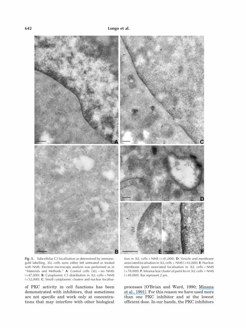
Subcellular Localisation of C3
Details of C3 subcellular distribution andtraffic have been obtained by immunoelectronmicroscopic techniques. As previously shown[Di Renzo et al., 1999] surface particles werenot clustered as in classical coated pits, butthey were adherent to and scattered along theplasma membrane. When studying the cyto-plasm (Fig. 5B–D), gold particles appeared freein the cytosol and not sequestered in small orlarge vesicles. Sometimes they were associatedwith the cytosolic side of a vesicular mem-brane (Fig. 5D) or arranged in small clusters(Fig. 5C,E). Finally, a few particles were alsodetected in association with the nuclear mem-brane and in the nucleoplasm (Fig. 5C,E,F).
DISCUSSION
We have shown that 3LL cells, which do notexpress complement receptors and do not pro-duce C3 [Di Renzo et al., 1999], are able tointernalise the C3 molecules from the externalcompartment. We have also provided evidencethat C3 is translocated by a mechanism that isnot cytoskeleton- and/or vesicle-dependent butdepends on PKC-specific phosphorylation. In3LL cells, PKC seems able to modulate prolif-eration through signals that are dependent onC3 internalisation.
Protein kinases C consists of a family of Caþþ
and phospholipid-dependent serine-threonineprotein kinases [Liu et al., 1992; Mellor andParker, 1998; Nakashima, 2002]. These enzy-mes play an important role in cell-signal trans-duction during cell growth and differentiationas well as in tumour promotion, oncogene acti-vation, and carcinogenesis. There are severalreports that correlate PKC activity and cellgrowth rate [Nishizuka, 1984; Clemons et al.,1992; Matsuzaki et al., 2004]. The involvement
Fig. 4. C3 stimulated proliferation of 3LL cells is mediated by the activity of PKC, but not PKA. 3LL cellswere incubated with medium alone and treated with NMS for 150 in the presence or absence of severalagents. Cell proliferation was measured as reported in ‘‘Materials and Methods.’’
C3-Induced 3LL Cell Proliferation 641
of PKC activity in cell functions has beendemonstrated with inhibitors, that sometimesare not specific and work only at concentra-tions that may interfere with other biological
processes [O’Brian and Ward, 1990; Minanaet al., 1991]. For this reason we have used morethan one PKC inhibitor and at the lowestefficient dose. In our hands, the PKC inhibitors
Fig. 5. Subcellular C3 localisation as determined by immuno-gold labelling. 3LL cells were either left untreated or treatedwith NMS. Electron microscopy analysis was performed as in‘‘Materials and Methods.’’ A: Control cells (3LL�no NMS)(�47,000). B: Cytoplasmic C3 distribution in 3LL cellsþNMS(�52,000). C: Small cytoplasmic clusters and nuclear localisa-
tion in 3LL cellsþNMS (�41,000). D: Vesicle and membraneassociated localisation in 3LL cellsþNMS (�43,000). E: Nuclearmembrane (pore) associated localisation in 3LL cellsþNMS(�78,000). F: Intranuclear cluster of particles in 3LL cellsþNMS(�49,000). Bar represent 2 mm.
642 Longo et al.

prevented the proliferation of 3LL cells inducedby C3. Another strategy for demonstrating thePKC involvement in C3-induced cell prolifera-tion was its stimulation by TPA. PKC acts asreceptor for phorbol esters and other tumourpromoters [Blood and Zetter, 1989]. This is oneof the mechanisms by which PKC is thought tointeract in the cellular response to growth fac-tors [Castagna et al., 1982; Nishizuka, 1984;Blood and Zetter, 1989]. In fact, when 3LL cellswere treated with TPA in the presence of C3,we observed a significant stimulation of cellproliferation.In contrast, neither selective activation nor
inhibition of PKA as used by Mueller et al.[1997] resulted in significant changes of C3internalisation and proliferation of 3LL cells.Similarly protein phosphatases [Young et al.,1991; Lucocq, 1992] did not seem to transducebiochemical signals required for C3 internalisa-tion and proliferation.Our results establish that, while PKC is
obligatory for C3-induced proliferation of 3LLcells, PKA activation might not be necessary.The mechanism of C3 internalisation was
investigated using inhibitors of the processesthat are normally involved in it. The persistentinternalisation after treatment of cells withcytochalasin and colchicine demonstrated thatthe internalisation process does not require adynamic reorganization of the actin and tubulincytoskeleton. On the other hand there are con-flicting reports on the extent to which theseagents inhibit endocytosis [Qualman et al.,2000].An interesting point in the C3 endocytosis
is that a certain amount of these moleculesappears to be recycled by the cell and released inan active form in the medium. The rapid C3exocytosis in an intact active form suggests thatC3 recycling does not involve the lysosomalhydrolase-containing compartment: this find-ing suggests that cells can promptly use C3again.The role of C3 in enhancing growth has been
demonstrated in normal lymphocytes, in Rajicells and in many other cell lines [Kuraya et al.,1990]. We have shown here that C3 internalisa-tion is an essential step for mediating thisimportant function and that PKCmediates thisevent.A final interesting observation may be worth
of noting: the translocation and clustering of C3into the nucleus, which seems to be stimulated
by PKC activity. At present, we don’t have aplausible explanation for this phenomenon thatcould be either related to the signalling forproliferation gene activation, or be just a fortu-ity and anecdotic event.
In conclusion, we have confirmed that C3is able to stimulate 3LL cell proliferation, andwe have provided evidences for its signallingwithin the cell. First, its rapid traffic seems to bedependent on PKC and independent on thecytoskeleton; second, the increase in cell growthand internalisation are coupled and dependenton PKC, but not on PKA.
REFERENCES
Allen V. 1995. Membrane traffic motors. FEBS Lett 396:101–106.
Blood HC, Zetter BR. 1989. Membrane-bound proteinkinase C modulates receptor affinity and chemotacticresponsiveness of Lewis lung carcinoma sublines toan elastin-derived peptide. J Biol Chem 264:10614–10620.
Castagna M, Takai Y, Kaibuchi K, Samo K, KikkawaV, Nishizuka Y. 1982. Direct activation of calcium-activated, phospholipid-dependent protein kinase bytumour-promoting phorbol esther. J Biol Chem 257:7847–7851.
ClemonsMJ, Trayner I, Menaya J. 1992. The role of proteinKinase C isoenzymes in the regulation of cell prolifera-tion and differentiation. J Cell Sci 103:881–887.
Di Renzo L, Longo A, Morgante E, Mardente S, ProdingerMW, Russo MA, Pontieri GM, Lipari M. 1999. C3 molec-ules internalize and enhance the growth of Lewis lungcarcinoma cells. Immunobiol 200:92–105.
Griffits G, McDowall A, Back R, Dubochet J. 1984. On thepreparation of cryosections for immunocytochemistry.J Utrastruct Res 89:65–77.
Hatzfeld A, Fisher E, Levesque JP, Perrin R, Hatzfeld J,Kazatchkine MD. 1988. Binding of C3 and C3dg to theCR2 complement receptor induced growth of an Epstein–Barr virus-positive human B cell line. J Immunol 140:170–175.
Kelly RB. 1985. Pathway of protein secretion in eukaryotes.Science 230:25–32.
KurayaM,Nilsson B, Nilsson-Ekdahl K, Klein E. 1990. C3dmediated negative and positive signals on the prolifera-tion of the human B cells separated from blood. ImmunolLett 26:51–58.
Lipari M, Di Renzo L, Zicari A, Schulz TF, Magliocca A,Mardente S, Dietrich MP, Pontieri GM. 1988. Lewislung carcinoma cells enhance the synthesis of C3 andopsonized by C3 secreted from murine macrophages.Immunobiol 177:233–244.
Liu B, Renaud C, Nelson KK, Chen YQ, Bazaz R, KowiniaJ, Timar J, Diglio AC, Honn KV. 1992. Protein-Kinase-Cinhibitor calphostin C reduces B16 amelanotic melanomacell adhesion to endothelium and lung colonization. Int JCancer 52:147–152.
Lucocq J. 1992. Mimicking mitotic Golgi disassembly usingokadaic acid. J Cell Sci 103:875–880.
C3-Induced 3LL Cell Proliferation 643

Luxemburg AT, Cooper NR. 1994. Modulation of signalingvia the B cell antigen receptor by CD21, the receptor forC3dg and EBV. J Immunol 153:4448–4457.
Matsuzaki Y, Takaoka Y, Hitomi T, Nishino H, Sakai T.2004. Activation of protein kinase C promotes humancancer cell growth through downregulation of p18(INK4c).Oncogene 23:5409–5414.
Mellor H, Parker PJ. 1998. The extended protein kinase Csuperfamily. Biochem J 332:281–292.
Minana MD, Felipo V, Cortes F, Grislia F. 1991. Inhibitionof protein kinase C arrests proliferation of humantumours. FEBS Lett 284:60–62.
Morgante E, Di Renzo L, Longo A, Cunsolo MG, Lipari M,Pontieri GM, Russo M. 1997. Compartmentalization ofthe third component of Complement (C3) in 3LL murinecarcinoma cells, studied by immunogold labeling. In:Motta PM, editor. Recent adv in microscopy of cells,tissues and organs. Roma: Antonio Delfino. pp 135–139.
Mueller H, Liu R, David F, Eppenberger U. 1997. Selec-tive modulation of protein kinase A and proteinkinase C activities in epidermal growth factor (EGF)-stimulated MCF-7 breast cancer cells. Biol Chem 378:1023–1029.
Nakashima S. 2002. Protein kinase C alpha (PKC alpha):Regulation and biological function. J Biochem (Tokyo)132:669–675.
Nishizuka Y. 1984. The role of protein kinase C in cell-surface signal transduction and tumour promotion.Nature 308:693–698.
O’Brian CA, Ward NE. 1990. Staurosporine: A prototipe ofa novel class of inhibitors of tumour cell invasion? J NatCancer Inst 82:1734–1735.
Owensby DA, Morton PA, Schwartz AL. 1989. Quantitativeevaluation of receptor-mediated endocytosis. MethodsCell Biol 32:305–328.
Qualman B, Lessel MM, Kelly RB. 2000. Molecular linksbetween endocytosis and actin cytoskeleton. J Cell Biol150:111–116.
Slot JW, Geuze HJ, Weerkamp AJ. 1988. Localisation ofmacromolecular components by applications of the im-munogold technique on cryosectioned bacteria. MethMicrobiol 20:211–235.
Young MRI, Young ME, Lozano Y, Coogan M. 1991. Reg-ulation of protein kinase A activation and prostaglandinE2-stimulated migration of Lewis lung carcinoma clones.Int J Cancer 49:150–155.
644 Longo et al.
Related Documents