CERVICAL SPINE INJURY Narain Chotirosniramit MD. Trauma and critical care unit Department of surgery Faculty of medicine Chiangmai University

Welcome message from author
This document is posted to help you gain knowledge. Please leave a comment to let me know what you think about it! Share it to your friends and learn new things together.
Transcript

CERVICAL SPINE INJURY
Narain Chotirosniramit MD.
Trauma and critical care unit
Department of surgery
Faculty of medicine Chiangmai University

NEUROLOGICAL ASSESSMENT
AIRWAY MANEUVERS ON C-SPINE MOVEMENT
WAY TO ACHIEVE TRACHEAL INTUBATION
BREATHING AND CIRCULATION
CLINICAL CRITERIA FOR CLEARING C-SPINE
CERVICAL SPINE IMMOBILIZATION
C-SPINE CLEARANCE GUIDELINE
CORTICOSTEROIDS WITH SCI

INTRODUCTION
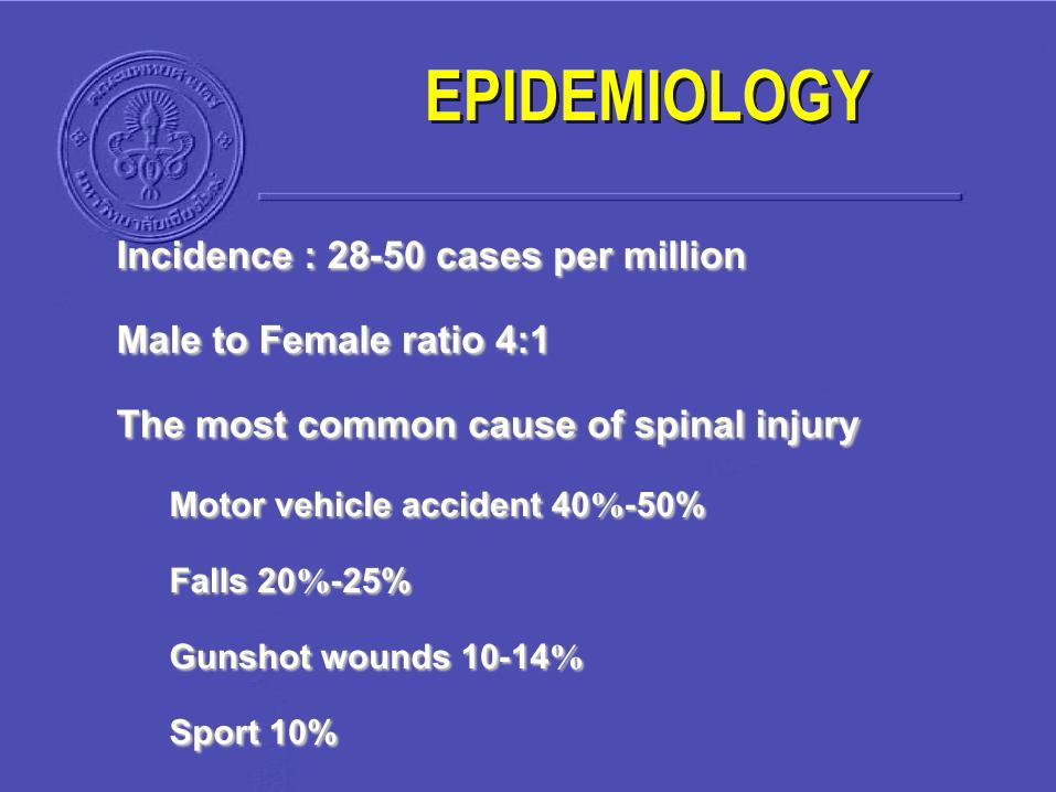
EPIDEMIOLOGY
Incidence : 28-50 cases per million
Male to Female ratio 4:1
The most common cause of spinal injuryMotor vehicle accident 40%-50%
Falls 20%-25%
Gunshot wounds 10-14%
Sport 10%

Level of injury ,commonly
Cervical 55%
Thoracic 30%
Lumbar 15%
95% one spinal region
Two thirds: cervical
EPIDEMIOLOGY
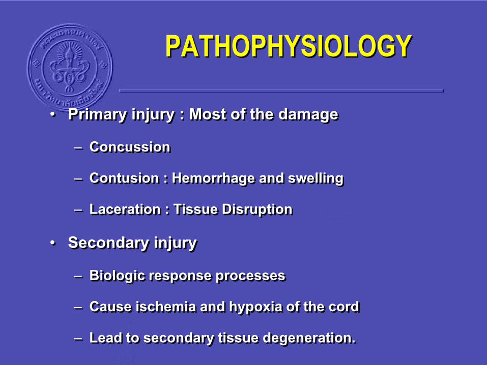
PATHOPHYSIOLOGY
• Primary injury : Most of the damage– Concussion
– Contusion : Hemorrhage and swelling
– Laceration : Tissue Disruption
• Secondary injury – Biologic response processes
– Cause ischemia and hypoxia of the cord
– Lead to secondary tissue degeneration.
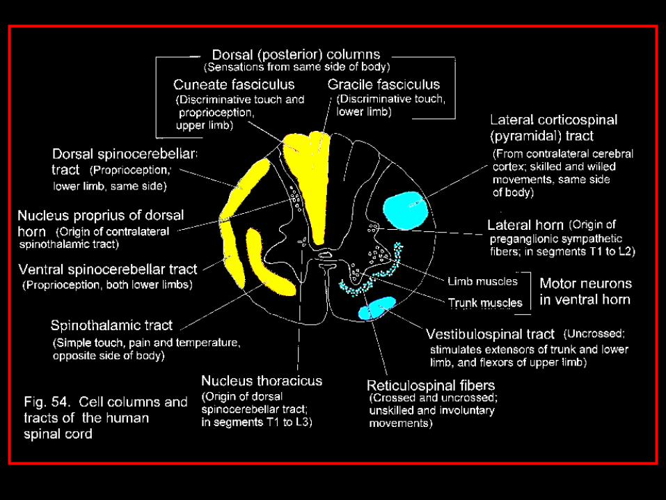
NEUROLOGICAL ASSESSMENT
AIRWAY MANEUVERS ON C-SPINE MOVEMENT
WAY TO ACHIEVE TRACHEAL INTUBATION
BREATHING AND CIRCULATION
CLINICAL CRITERIA FOR CLEARING C-SPINE
CERVICAL SPINE IMMOBILIZATION
C-SPINE CLEARANCE GUIDELINE
CORTICOSTEROIDS WITH SCI

NEUROLOGICAL ASSESSMENT
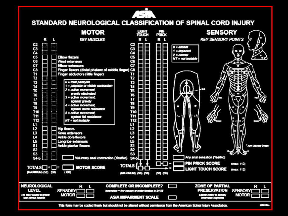
• The examination should include :
Sensory
Motor
Proprioception
Perianal sensation
Rectal sphincter tone
Bulbocavernous reflex

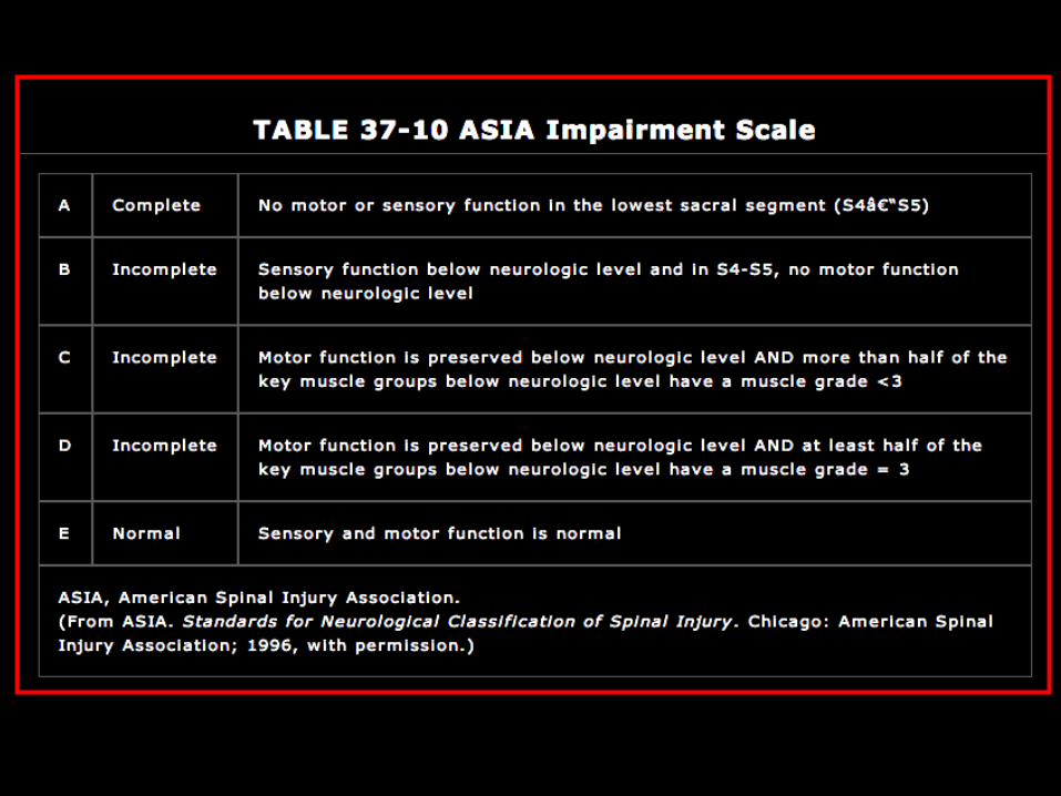
Simple & acceptable classification of SCI :
A. Complete absence of motor and sensory function.
B. Sensation present but no motor function
C. Sensation + motor function 2–3/5.
D. Sensation present with motor function of 4/5.
E. Normal sensory and motor functionBrowner BD, Jupiter JB, Levine AM, et al. Skeletal Trauma: Fractures, Dislocations,
Ligamentous Injuries. Philadelphia: WB Saunders, 1998
Frankel classification

• To assess the patient : must be defined:
1. Complete SCI:
No motor or sensory function caudal to the level of injury
The bulbocavernous reflex is present.
NEUROLOGICAL ASSESSMENT
NEUROLOGICAL ASSESSMENT
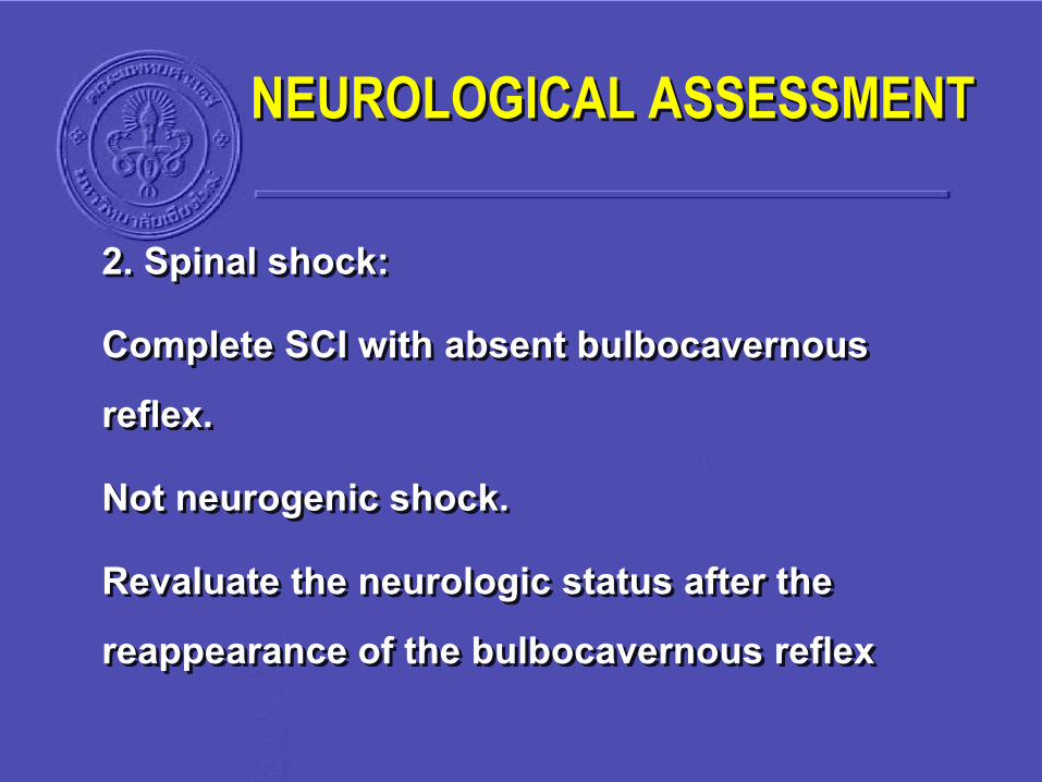
2. Spinal shock:
Complete SCI with absent bulbocavernous reflex.
Not neurogenic shock.
Revaluate the neurologic status after the reappearance of the bulbocavernous reflex

3. Incomplete SCI (ICSCI):
Some motor or sensory function below the level of injury.
NEUROLOGICAL ASSESSMENT

• Central cord syndrome
Most common ICSCI
Quadriplegia with perianal & sacral sparing.
75% : partial recovery of the motor function.
Formal Types of ICSCI

• Brown-Sequard syndrome Unilateral SCI (usually due
to penetration)
Motor deficit ipsilateral to the injury combined with contralateral sensory deficit.
Most : gain partial recovery with bowel and bladder continence & usually walking ability.
Formal Types of ICSCI
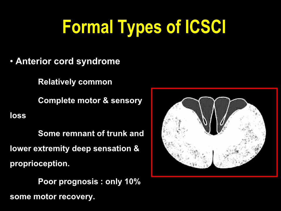
• Anterior cord syndrome
Relatively common
Complete motor & sensory loss
Some remnant of trunk and lower extremity deep sensation & proprioception.
Poor prognosis : only 10% some motor recovery.
Formal Types of ICSCI

• Posterior cord syndrome
Rare ICSCI
Loss of proprioception & deep sensation
Intact motor functioning.
“tabes dorsalis gait”.
Formal Types of ICSCI

NEUROLOGICAL ASSESSMENT
AIRWAY MANEUVERS ON C-SPINE MOVEMENT
WAY TO ACHIEVE TRACHEAL INTUBATION
BREATHING AND CIRCULATION
CLINICAL CRITERIA FOR CLEARING C-SPINE
CERVICAL SPINE IMMOBILIZATION
C-SPINE CLEARANCE GUIDELINE
CORTICOSTEROIDS WITH SCI

AIRWAY MANEUVERS
• Both basic and advanced airway maneuver : cause movement in different segments of the cervical spine.
• Even chin lift and jaw thrust : cause movements cervical spine.

• Advanced airway : Blind NT intubation & direct laryngoscopy & OT intubation (DLOI)
Cause relative segmental cervical spine movement
Atlanto-occipital and atlantoaxial joints : most oftenAprahamian C, et al. Ann Emerg Med 1984; 13: 584–7.
Sawin PD, et al. Anesthesiology 1996; 85: 26–36
AIRWAY MANEUVERS

No significant different in movement was found between curved or straight laryngoscope blades.
Gerling MC, et al. Ann Emerg Med 2000; 36: 293–300.
AIRWAY MANEUVERS
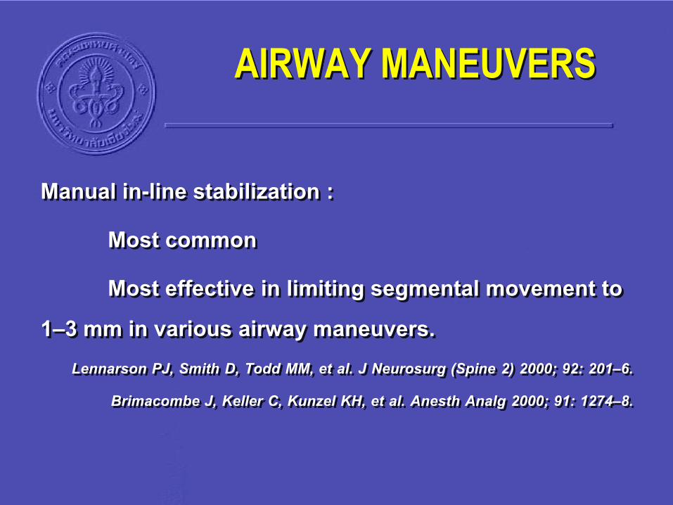
Manual in-line stabilization :
Most common
Most effective in limiting segmental movement to 1–3 mm in various airway maneuvers.
Lennarson PJ, Smith D, Todd MM, et al. J Neurosurg (Spine 2) 2000; 92: 201–6.
Brimacombe J, Keller C, Kunzel KH, et al. Anesth Analg 2000; 91: 1274–8.
AIRWAY MANEUVERS
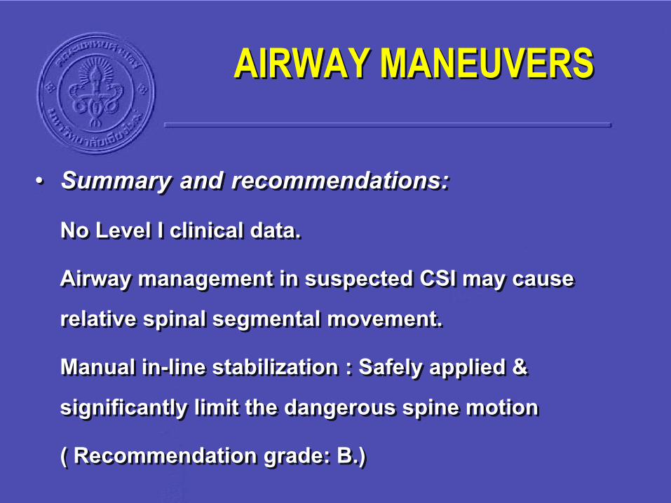
• Summary and recommendations: No Level I clinical data.
Airway management in suspected CSI may cause relative spinal segmental movement.
Manual in-line stabilization : Safely applied & significantly limit the dangerous spine motion
( Recommendation grade: B.)
AIRWAY MANEUVERS

NEUROLOGICAL ASSESSMENT
AIRWAY MANEUVERS ON C-SPINE MOVEMENT
WAY TO ACHIEVE TRACHEAL INTUBATION
BREATHING AND CIRCULATION
CLINICAL CRITERIA FOR CLEARING C-SPINE
CERVICAL SPINE IMMOBILIZATION
C-SPINE CLEARANCE GUIDELINE
CORTICOSTEROIDS WITH SCI
TRACHEAL INTUBATION
12 retrospective series :
395 DLOI in patients with CSI
(most of them unstable)
Only 2 : Neurological deterioration (not attributed to the airway intervention)
Crosby ET. Anesthesiology 2006; 104: 1293–318.

Awake nasotracheal intubation :
Many anesthesiologists prefered for definitive airway control in suspected CSI patients.
Rosenblatt WH, et al. Anesth Analg 1998; 87: 153–7.
TRACHEAL INTUBATION
• Fiber optic endoscope.
Minimal spine movement
Maintaining airway protective reflexes
Disadvantages : Slow learning curve that causes many doctors to be uncomfortable with the procedure
Ezri T, et al. J Clin Anesth 2003; 15: 418–22.Potential for desaturation : might aggravate secondary cord injury.
Fuchs G, et al. J Neurosurg Anesth 1999; 11: 11–16.
TRACHEAL INTUBATION
• Summary and recommendations: Both DLOI and fiber optic awake NT intubation are safe & effective options for securing the airway in a trauma patient with suspected CSI.
(Recommendation grade: B).
DLOI : No special equipment or advanced expertise
Preferred in emergency situations
Fiber optic : elective procedures.
(Recommendation grade: C.)
TRACHEAL INTUBATION

NEUROLOGICAL ASSESSMENT
AIRWAY MANEUVERS ON C-SPINE MOVEMENT
WAY TO ACHIEVE TRACHEAL INTUBATION
BREATHING AND CIRCULATION
CLINICAL CRITERIA FOR CLEARING C-SPINE
CERVICAL SPINE IMMOBILIZATION
C-SPINE CLEARANCE GUIDELINE
CORTICOSTEROIDS WITH SCI

BREATHING AND CIRCULATION
• Cervical spinal cord injury : May have respiratory failure and hemodynamic compromise.
• Hypoxemia & hypotension : increase the chance for secondary cord injury and worsening the neurological outcome.

• Risk for ventilatory failure : based on the level and completeness of injury.
• Ventilatory support : majority of patients > C5 injuries
> C3 injuries. Adequate fluid resuscitation & hemodynamic improvement : correlated to better neurological outcome
Vale FL, Burns J, Jackson AB, et al. J Neurosurg 1997; 87: 239–46.
BREATHING AND CIRCULATION

• High SCI (above T6) :
Disruption of sympathetic chain
Hypotension & bradycardia. (neurogenic shock)
Found to be 19.3% Guly HR, Bouamra O, Lecky FE. Resuscitation 2008; 76: 57–62.
BREATHING AND CIRCULATION
• If SBP < 90 mmHg, MABP < 85 mmHg.
• Early administration of vasoactive drug should be considered.
Hadley MN, et al. Neurosurgery 2002; 50(suppl): 58–62.
BREATHING AND CIRCULATION

NEUROLOGICAL ASSESSMENT
AIRWAY MANEUVERS ON C-SPINE MOVEMENT
WAY TO ACHIEVE TRACHEAL INTUBATION
BREATHING AND CIRCULATION
CLINICAL CRITERIA FOR CLEARING C-SPINE
CERVICAL SPINE IMMOBILIZATION
C-SPINE CLEARANCE GUIDELINE
CORTICOSTEROIDS WITH SCI

CLINICAL CRITERIA
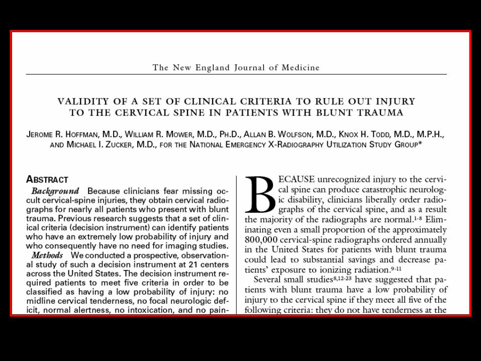
• The NEXUS study : 34,069 patients.
• 5 criteria for the definition a low probability of CSI:
1. No midline cervical tenderness
2. No focal neurological deficit
3. Normal alertness
4. No intoxication
5. No painful, distracting injuryHoffman JR, Mower WR, Wolfson AB, et al. N Engl J Med 2000; 343: 94–9.


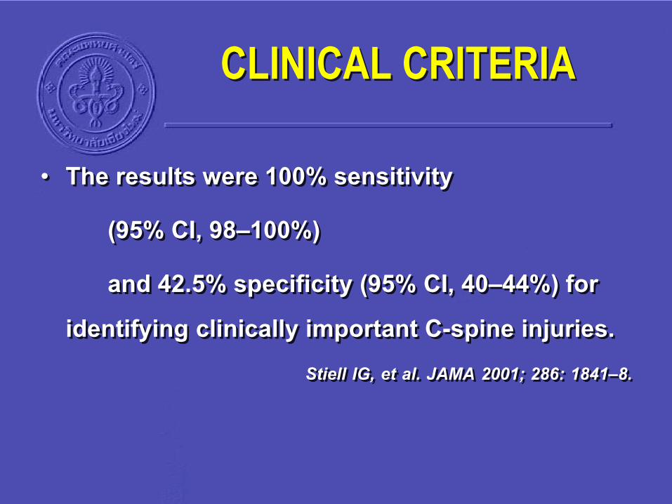
• The results were 100% sensitivity
(95% CI, 98–100%)
and 42.5% specificity (95% CI, 40–44%) for identifying clinically important C-spine injuries.
Stiell IG, et al. JAMA 2001; 286: 1841–8.
CLINICAL CRITERIA


The Canadian C-spine rule :
More sensitive than the NEXUS (99.4% versus 90.7%, p < 0.001)
More specific (45.1% versus 36.8%, p < 0.001)
Lower radiography rates.Stiell IG, Clement CM, McKnight RD, et al. N Engl J Med 2003; 349: 2510–18.
CLINICAL CRITERIA

NEUROLOGICAL ASSESSMENT
AIRWAY MANEUVERS ON C-SPINE MOVEMENT
WAY TO ACHIEVE TRACHEAL INTUBATION
BREATHING AND CIRCULATION
CLINICAL CRITERIA FOR CLEARING C-SPINE
CERVICAL SPINE IMMOBILIZATION
C-SPINE CLEARANCE GUIDELINE
CORTICOSTEROIDS WITH SCI

IMMOBILIZATION
Cervical spine injuries : may be impaired by pathological motion of the injured vertebrae.
3 to 25% of SCI : Occur during transit or early in the course of management .
Brunette DD, et al. J Trauma 27:445–447, 1987.
Burney RE, et al. J Trauma 29:1497–1499, 1989.
Geisler WO, et al. Med Serv J Can 22:512–523, 1966.
Hachen HJ. Paraplegia 12:33–37, 1974.
Prasad VS, et al. Spinal Cord 37:560–568, 1999
Totten VY, et al. Prehosp Emerg Care 3:347–352, 1999.
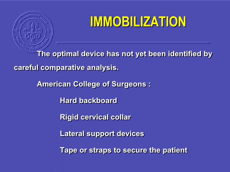
The optimal device has not yet been identified by careful comparative analysis.
American College of Surgeons :
Hard backboard
Rigid cervical collar
Lateral support devices
Tape or straps to secure the patient
IMMOBILIZATION

Occipital padding combined with a rigid backboard : a better neutral position than a flat backboard alone
Schriger DL, et al. Ann Emerg Med 20:878–881, 1991.
Stauffer ES. Clin Orthop 102: 92–99, 1974.
IMMOBILIZATION

Extension Neutral Flexion

• Compare immobilization : Soft collar
Hard collar
Extrication collar
Philadelphia collar
Bilateral sandbags with 3-inch cloth tape across forehead
Combination of sandbags, tape, and a Philadelphia collar.Podolsky S, et al.J Trauma 23:461–465, 1983.
IMMOBILIZATION

• Hard foam & hard plastic collars were better at limiting cervical spine motion than soft foam collars
• Neither collars alone nor sandbags and tape in combination provided satisfactory restriction of cervical spine motion
• Sandbags and tape combined with a rigid cervical collar were the best
Podolsky S, et al.J Trauma 23:461–465, 1983.
IMMOBILIZATION
• Spine immobilization increases the risk of pressure sores.
• Pressure sores were associated with immobilization (patients who were not turned during the first 2 hours after injury).
Linares HA, et al. Orthopedics 10:571–573, 1987
IMMOBILIZATION

• Summary :
Immobilization of the entire spinal column is necessary until a spinal column injury has been excluded, or until appropriate treatment has been initiated
IMMOBILIZATION

• Summary :
It seems that a combination of rigid cervical collar with supportive blocks on a rigid backboard with straps is effective at achieving safe, effective spine immobilization for transport.
IMMOBILIZATION
NEUROLOGICAL ASSESSMENT
AIRWAY MANEUVERS ON C-SPINE MOVEMENT
WAY TO ACHIEVE TRACHEAL INTUBATION
BREATHING AND CIRCULATION
CLINICAL CRITERIA FOR CLEARING C-SPINE
CERVICAL SPINE IMMOBILIZATION
C-SPINE CLEARANCE GUIDELINE
CORTICOSTEROIDS WITH SCI
Practice management guidelines for
identification of cervical spine injuries
following trauma
2009
update from the Eastern Association for the Surgery of Trauma
Practice
Management Guidelines Committee

C-spine clearance
• Search from PubMed
• Articles regarding the identification of CS injury from 1998-2007
78 articles were identified.
52 articles were selected

C-spine clearance
• The questions posed were:
1. Who needs CS imaging
2. What imaging should be obtained;
3. When should CT, MRI, or F/E radiographs be used.
4. How is significant ligamentous injury excluded in the comatose patient?

RECOMMENDATIONS

A. Removal of cervical collars:
Cervical collars should be removed as soon as feasible after trauma (level 3)
C-spine clearance

A. Removal of cervical collars
• Early removal of cervical collars may decrease :
Collar-related decubitus ulceration
Incidence of increase Intracranial pressure (ICP)
Ventilator days
Intensive care unit (ICU) and hospital days
The incidence of delirium and pneumonia.

A. Removal of cervical collars
• Chendrasekhar and colleagues
38% : Collar-related decubitus ulceration in head-injured patients who survived greater than 24 hours.
• A significantly longer duration of cervical collar use than those who did not
Chendrasekhar A, Moorman DW, Timberlake GA.
An evaluation of the effects of semirigid cervical collars in patients with severe closed head injury.
Am Surg 1998; 64:604-606

A. Removal of cervical collars
• Powers et al
Skin breakdown in 6.8% of ICU patients (with a cervical collar >24 hours).
Most significant predictor of breakdown was time in a cervical collar.
Powers J, Daniels D, McGuire C, et al.
The incidence of skin breakdown associated with the use of cervical collars.
J Trauma Nurs 2006; 13:198-200

A. Removal of cervical collars
Hunt and co-workers applied cervical collars to patients with traumatic brain injury and found a significant rise from the baseline ICP when the collars were applied
Hunt K, Hallworth S, Smith M. Anaesthesia 2001; 56:511-513
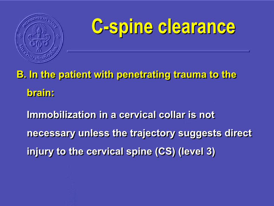
B. In the patient with penetrating trauma to the brain:
Immobilization in a cervical collar is not necessary unless the trajectory suggests direct injury to the cervical spine (CS) (level 3)
C-spine clearance

B. Penetrating trauma to the brain
• Retrospective studies
105 patients with GSW to the cranium : no CS injury
Kennedy FR, Gonzalez P, Beitler A, et al. South Med J 1994; 87:621-623.

B. Penetrating trauma to the brain
• Kaups and co-workers :
Reviewed 215 patients with a GSW to the head : no patient sustained indirect (blast or fall-related) spinal column injury
J Trauma 1998; 44:865-867.

C. In awake, alert trauma patients without neurologic deficit or distracting injury who have no neck pain or tenderness with full range of motion of the CS:
CS imaging is not necessary and the cervical collar may be removed
C-spine clearance

C. Awake, alert trauma patients • National Emergency X-Radiography Utilization Study (NEXUS)
Required patients to have
1) No midline cervical tenderness
2) No focal neurologic deficit,
3) Normal alertness
4) No intoxication
5) No painful distracting injury.Hoffman JR, Mower WR, Wolfson AB, et al.
Validation of a set of clinical criteria to rule out injury to the cervical spine in patients with blunt trauma. N Engl J Med 2000; 343:94-99.

D. All other patients in whom CS injury is suspected must have radiographic evaluation
1. The primary screening modality is axial computed tomography (CT) from the occiput to T1 with sagittal and coronal reconstructions
2. Plain radiographs contribute no additional information and should not be obtained
C-spine clearance

D. CS injury is suspected
• In the past : initial radiographic screening test was
A 3-view ( lateral, AP & odontoid views)
CS series supplemented by swimmer’s views and CT CS for poorly-visualized areas.

D. CS injury is suspected
• A prospective study of 58 blunt trauma patients with CS imaging and a CT of another body region.
• Both plain radiography and CT CS.
20 patients (34.4%) : CS injuries.
Plain radiography : missed 8 injuries (3 unstable)
CT CS : missed only 2 injuries (stable).
• The sensitivity for plain CS : 60%, CT CS : 90% Berne JD, Velmahos GC, El-Tawil Q, et al. J Trauma 1999; 47:896-903

D. CS injury is suspected
• Cohort of 1,199 blunt trauma patients with posterior neck tenderness, altered mental status, or neurologic deficit that underwent both plain films and CT CS for CS evaluation.
• 116 patients : CS injury.
Detected by both plain films & CT CS : 75 patients.
Detected by CT CS but missed by plain radiography : 41 patients�
• CT CS missed no injuries.
• There was no apparent role for screening with plain CS radiography.Griffen MM, Frykberg ER, Kerwin AJ, et al.
J Trauma 2003; 55:222-227.
D. CS injury is suspected
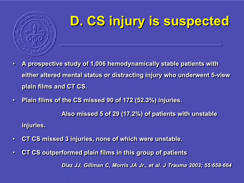
• A prospective study of 1,006 hemodynamically stable patients with either altered mental status or distracting injury who underwent 5-view plain films and CT CS.
• Plain films of the CS missed 90 of 172 (52.3%) injuries.
Also missed 5 of 29 (17.2%) of patients with unstable injuries.
• CT CS missed 3 injuries, none of which were unstable.
• CT CS outperformed plain films in this group of patientsDiaz JJ, Gillman C, Morris JA Jr., et al. J Trauma 2003; 55:658-664

D. CS injury is suspected
• 2005 : Holmes and Akkinepalli published a meta-analysis comparing plain films to CT CS.
• The pooled sensitivity
Plain radiography was 52%
CT CS it was 98%.Holmes JF, Akkinepalli R.
Computed tomography versus plain radiography to screen for cervical spine injury: a meta-analysis.
J Trauma 2005; 58:902-905

D. CS injury is suspected
CT CS must :
Include axial images from the occiput to T1
Sagittal and coronal reconstructions.
CT CS :
More accurate than plain radiography
Time, effective, cost effective
Does not require additional plain films
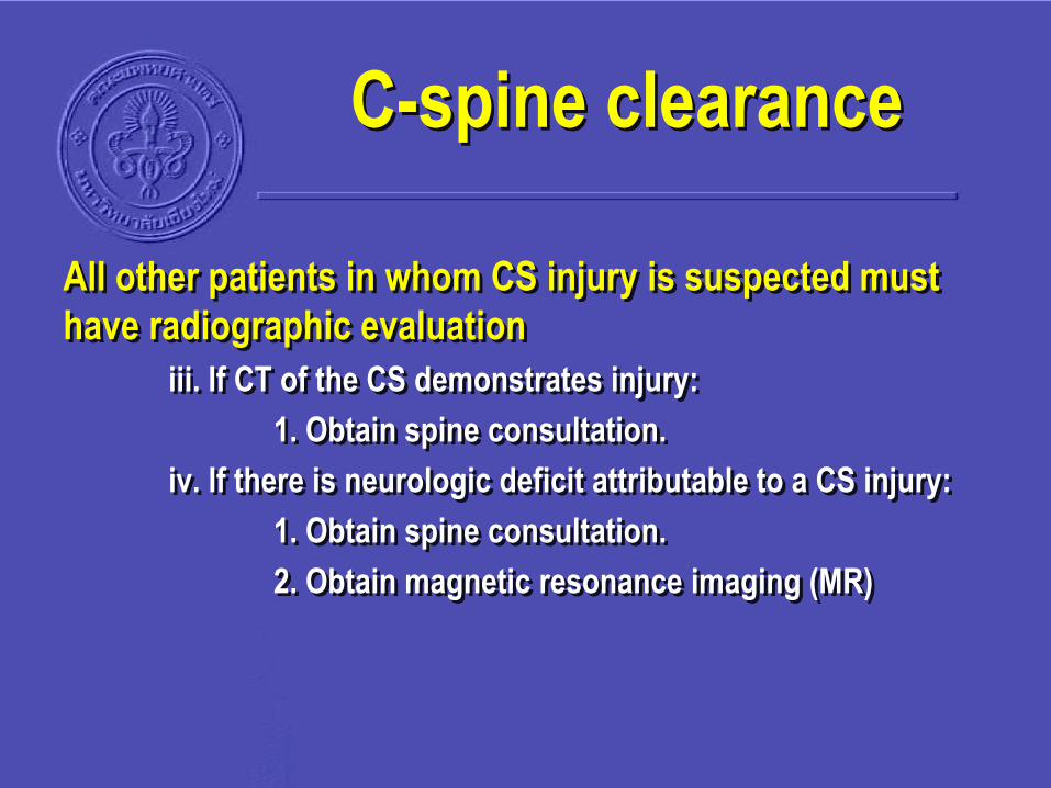
All other patients in whom CS injury is suspected must
have radiographic evaluation
iii. If CT of the CS demonstrates injury:
1. Obtain spine consultation.
iv. If there is neurologic deficit attributable to a CS injury:
1. Obtain spine consultation.
2. Obtain magnetic resonance imaging (MR)
C-spine clearance
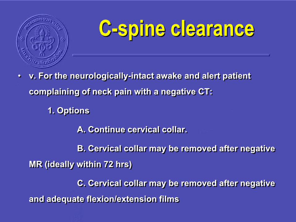
• v. For the neurologically-intact awake and alert patient complaining of neck pain with a negative CT:
1. Options
A. Continue cervical collar.
B. Cervical collar may be removed after negative MR (ideally within 72 hrs)
C. Cervical collar may be removed after negative and adequate flexion/extension films
C-spine clearance

• Vi. Obtunded patient with a negative CT and gross motor function of extremities:
1. Flexion / extension radiography should not be performed
2. The risk / benefit ratio of obtaining MR in addition to CT is not clear, and its use must be individualized in each institution options are:
A. Continue cervical collar immobilization until a clinical exam can be performed.
B. Remove the cervical collar on the basis of CT alone.
C. Obtain MR.
3. If MR is negative, the cervical collar may be safely removed
C-spine clearance

1. F/E radiography should not be performed
• The incidence of ligamentous injury identified by dynamic fluoroscopy in patients with altered mental status was 0.7%.
Davis JW, Kaups KL, Cunningham MA, et al.
Routine evaluation of the cervical spine in head-injured patients with dynamic fluoroscopy: a reappraisal.
J Trauma 2001; 50:1044- 1047
CT vs MR
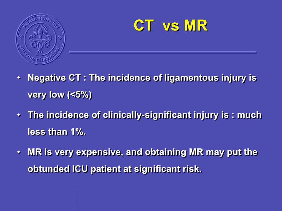
• Negative CT : The incidence of ligamentous injury is very low (<5%)
• The incidence of clinically-significant injury is : much less than 1%.
• MR is very expensive, and obtaining MR may put the obtunded ICU patient at significant risk.
CT vs MR
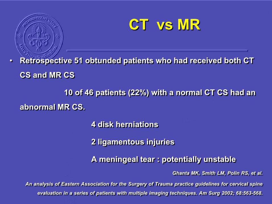
• Retrospective 51 obtunded patients who had received both CT CS and MR CS
10 of 46 patients (22%) with a normal CT CS had an abnormal MR CS.
4 disk herniations
2 ligamentous injuries
A meningeal tear : potentially unstableGhanta MK, Smith LM, Polin RS, et al.
An analysis of Eastern Association for the Surgery of Trauma practice guidelines for cervical spine evaluation in a series of patients with multiple imaging techniques. Am Surg 2002; 68:563-568.

CT vs MR
• 46 obtunded patients with a normal CT CS : All had MR CS.
• An injury was detected by MR CS in 5 patients (11%).
4 : ligamentous injuries
1 : a herniated disk.
None of these injuries required surgery.Sarani B, Waring S, Sonnad S, et al. Magnetic resonance imaging is a useful adjunct in the evaluation
of the cervical spine of injured patients. J Trauma 2007; 63:637-640.

CT vs MR
• MR CS is not reliable for identifying osseous injury. It missed 45% of fractures.
Holmes JF, Mirvis SE, Panacek EA, et al.
Variability in computed tomography and magnetic resonance imaging in patients with cervical spine injuries.
J Trauma 2002; 53:524-530.

CT vs MR• MR CS should only be used to clear the CS in the
obtunded patient after a CT CS has cleared the CS of any bony abnormality.
• MR CS should be obtained within 72 hours of injury
Ability to detect soft-tissue injury may diminish after this time.
D’Alise MD, Benzel EC, Hart BL.
Magnetic resonance imaging evaluation of the cervical spine in the comatose or obtunded trauma patient.
J Neurosurg 1999; 91:54-59.

• Vi. Obtunded patient with a negative CT and gross motor function of extremities:
1. Flexion / extension radiography should not be performed
2. The risk / benefit ratio of obtaining MR in addition to CT is not clear, and its use must be individualized in each institution options are:
A. Continue cervical collar immobilization until a clinical exam can be performed.
B. Remove the cervical collar on the basis of CT alone.
C. Obtain MR.
3. If MR is negative, the cervical collar may be safely removed
C-spine clearance

• Adjunct to primary survey film
CXR
Pelvis AP
(No lateral C-spine film : Just splint)
C-spine clearance

NEUROLOGICAL ASSESSMENT
AIRWAY MANEUVERS ON C-SPINE MOVEMENT
WAY TO ACHIEVE TRACHEAL INTUBATION
BREATHING AND CIRCULATION
CLINICAL CRITERIA FOR CLEARING C-SPINE
CERVICAL SPINE IMMOBILIZATION
C-SPINE CLEARANCE GUIDELINE
CORTICOSTEROIDS WITH SCI

CORTICOSTEROIDS
• A survey of 60 Canadian neurosurgeons and orthopedic spine surgeons :
75% : Routinely prescribe steroids for acute SCI
70% : Fear from litigation or peer criticism.
17% : Believe that steroids actually improve their patient’s neurological outcome.
Hurlbert RJ, Moulton R. Can J Neurol Sci 2002; 29: 236–9.

• Moderate vs low-dose methylprednisolone, 10-day regi-men
1 trial (Bracken 1984/85).
No difference in the neurologic outcome scores at 6 weeks, 6 months
Only wound infection was elevated in the high dose regimen (RR = 3.50, 95% CI 1.18 to 10.41)
CORTICOSTEROIDS

• High-dose methylprednisolone vs placebo or none, 24-hr regimen
3 trials (Bracken 1990/93, Otani 1994, Petitjean 1998).
Analysis restricted to patients treated within 8 hour
High-does methylprednisolone : Greater motor function recovery at 6 wks, 6 mths and the final outcome (WMD= 4.06, 95% CI 0.58 to 7.55).
Pinprick sensation : Significantly improved at 6 mths (WMD = 3.37, 95% CI 0.74 to 6.00) but not at one year
CORTICOSTEROIDS

• High-dose methylprednisolone for 48 versus 24 hours1 trial (Bracken 1997/98).
Patients treated within 3 hours : did not differ in their recovery from 24 or 48-hour methylprednisolone (Bracken 1997/98).
Patients treated within 3 - 8 hours :
Improved motor function if treated with 48-hr
No differences for pinprick or touch sensation
Severe pneumonia & severe sepsis tended to be elevated in the 48-hr but overall mortality at 1 year was not
CORTICOSTEROIDS
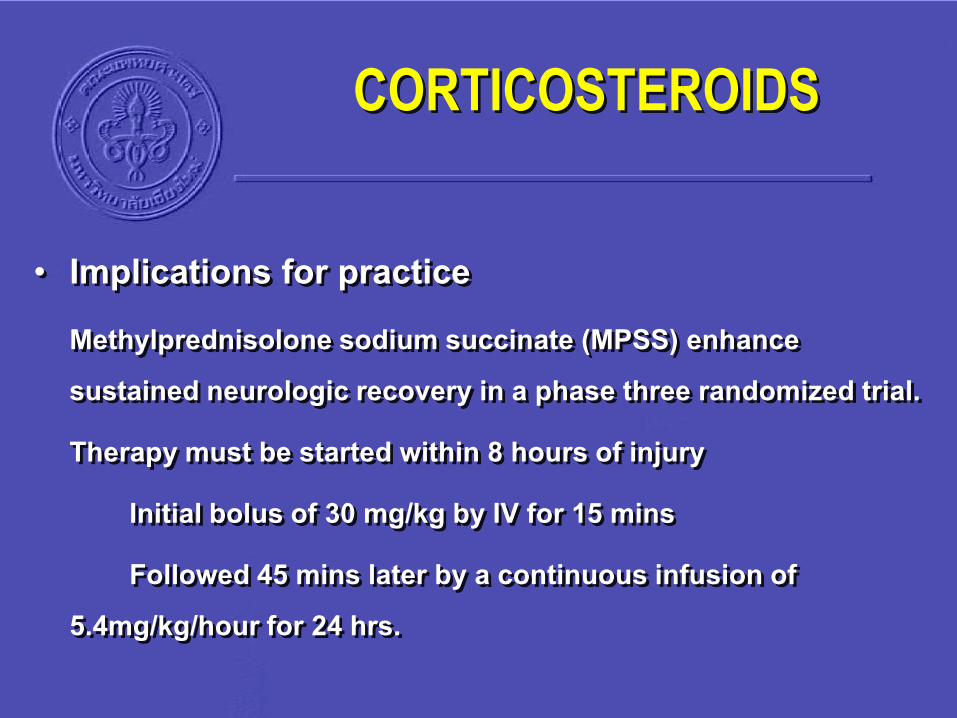
• Implications for practiceMethylprednisolone sodium succinate (MPSS) enhance sustained neurologic recovery in a phase three randomized trial.
Therapy must be started within 8 hours of injury
Initial bolus of 30 mg/kg by IV for 15 mins
Followed 45 mins later by a continuous infusion of 5.4mg/kg/hour for 24 hrs.
CORTICOSTEROIDS

• Implications for practice
Further improvement in motor function recovery when the maintenance therapy is extended for 48 hours.
This is particularly evident when the initial bolus dose could only be administered 3-8 hours after injury.
CORTICOSTEROIDS

CONCLUSIONS

THANK YOU
FOR
YOUR ATTENTIONS
Related Documents