By By Professor Of Pediatrics, Professor Of Pediatrics, Head of Allergy & Clinical Head of Allergy & Clinical Immunology Unit - Mansoura Immunology Unit - Mansoura University University Egypt Egypt

By Professor Of Pediatrics, Head of Allergy & Clinical Immunology Unit - Mansoura University Egypt.
Dec 18, 2015
Welcome message from author
This document is posted to help you gain knowledge. Please leave a comment to let me know what you think about it! Share it to your friends and learn new things together.
Transcript
By By
Professor Of Pediatrics,Professor Of Pediatrics,
Head of Allergy & Clinical Head of Allergy & Clinical Immunology Unit - Mansoura Immunology Unit - Mansoura
University University Egypt Egypt
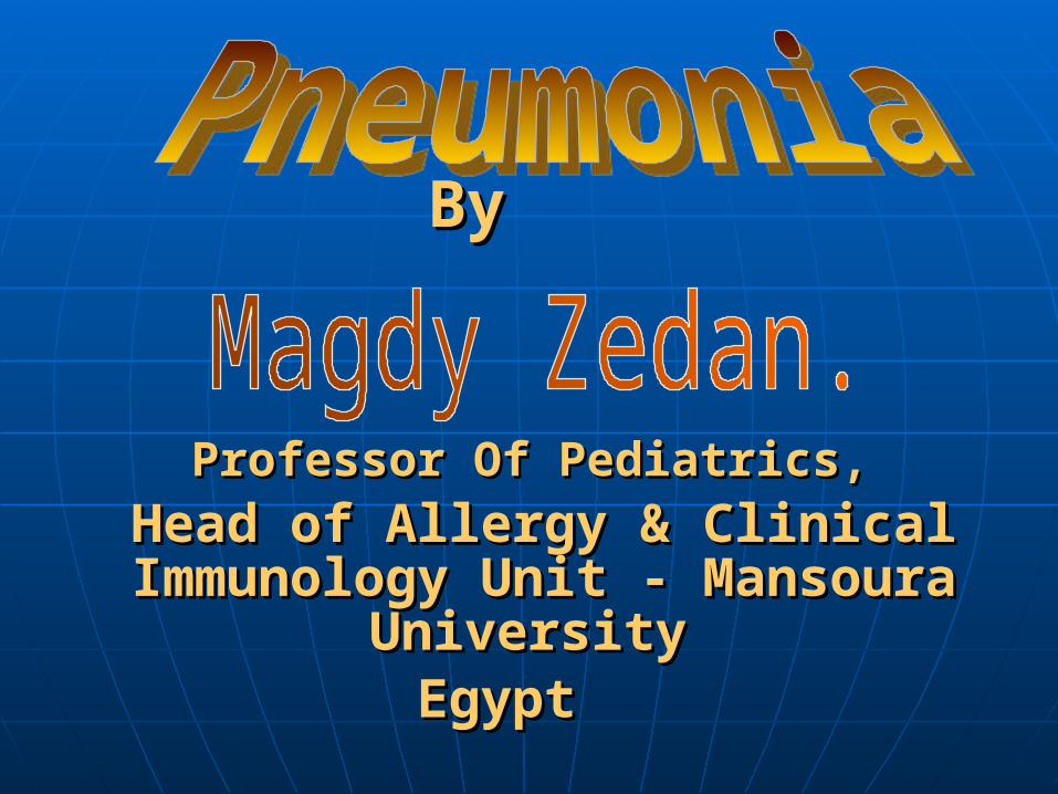
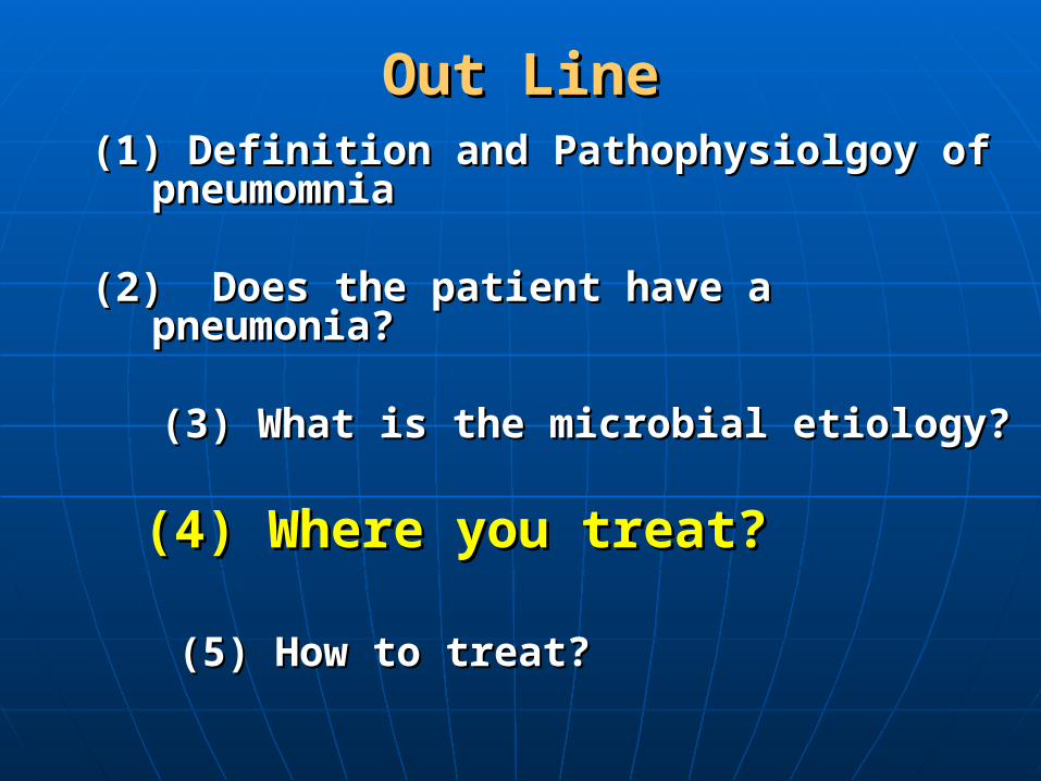
Out LineOut Line
(1) Definition and Pathophysiolgoy of (1) Definition and Pathophysiolgoy of pneumomniapneumomnia
((2) Does the patient have a pneumonia? 2) Does the patient have a pneumonia?
(3) What is the microbial etiology?(3) What is the microbial etiology? (4) Where you treat?(4) Where you treat?
(5) How to treat?(5) How to treat?

DefinitionDefinition
It’s an infection in alveolar spaces It’s an infection in alveolar spaces
(pulmonary parenchyma) leading to their (pulmonary parenchyma) leading to their
Consolidation.Consolidation.
Consolidation meansConsolidation means the alveoli are the alveoli are
filled with exudates and inflammatoryfilled with exudates and inflammatory
cells with loss of their gaseous content.cells with loss of their gaseous content.
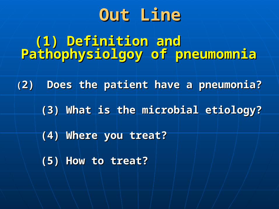
PathophysiologyPathophysiology
A- Development of Pneumonia:A- Development of Pneumonia:• Virulent organism.Virulent organism.• Immune compromised host Immune compromised host LocalLocal
SystemicSystemic
B- Pulmonary host defensesB- Pulmonary host defenses(1)Mechanical and structural:(1)Mechanical and structural:
Cough.Cough. Mucocilliary clearanceMucocilliary clearance Airway branching (configuration)Airway branching (configuration)
continuecontinue
Pulmonary host defensesPulmonary host defenses
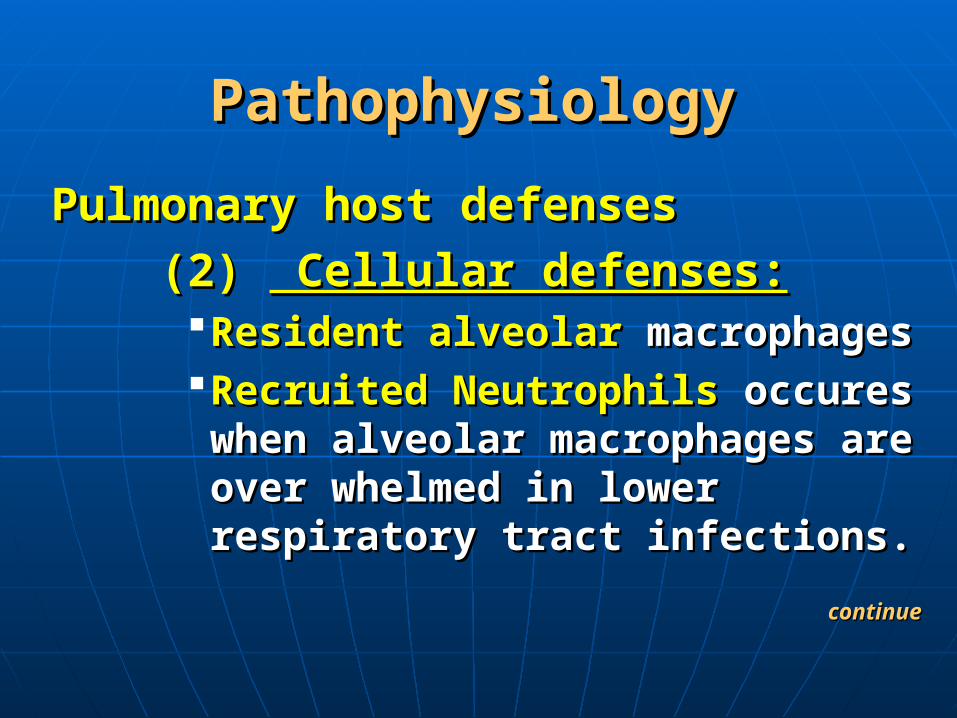
(2) (2) Cellular defenses: Cellular defenses:Resident alveolarResident alveolar macrophages macrophages Recruited NeutrophilsRecruited Neutrophils occures when occures when
alveolar macrophages are over alveolar macrophages are over whelmed in lower respiratory tract whelmed in lower respiratory tract infections.infections.
continuecontinue
PathophysiologyPathophysiology

PathophysiologyPathophysiology
(3)(3)Humoral defensesHumoral defenses IgA in upper airwayIgA in upper airway IgG in lower airwayIgG in lower airway
(4) (4) Inflammatory and molecularInflammatory and molecular defenses:defenses:
AirwayAirway epithelium epithelium through secretion of through secretion of peptides called defensins, and chemokines.peptides called defensins, and chemokines.
CytokinesCytokines as: IL-10,GM- CSF. as: IL-10,GM- CSF.
continuecontinue
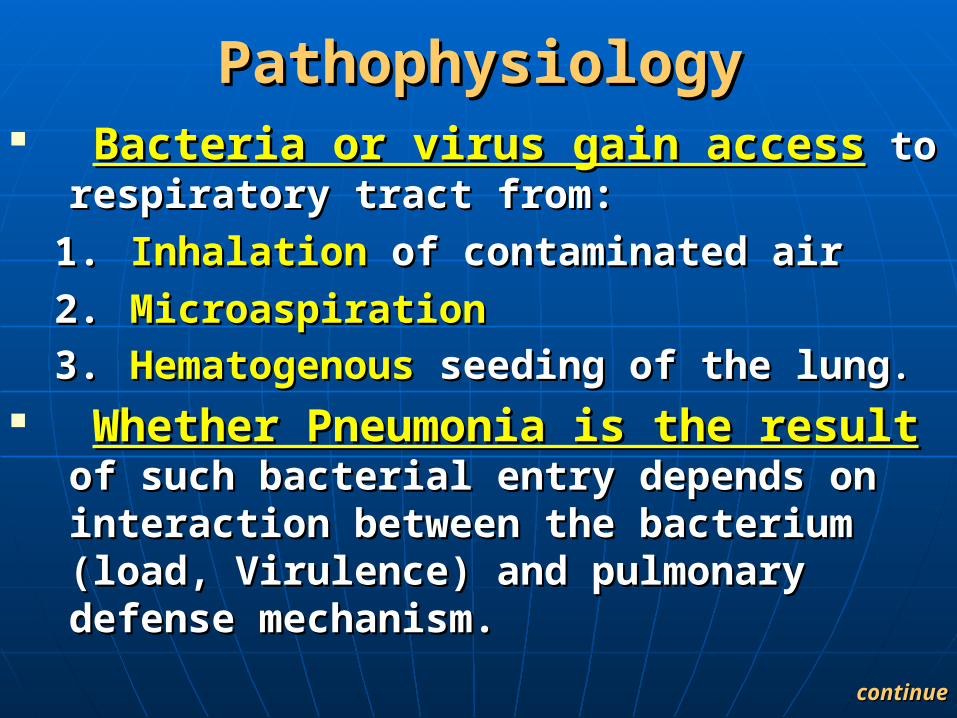
PathophysiologyPathophysiology Bacteria or virus gain accessBacteria or virus gain access to respiratory to respiratory
tract from:tract from:
1.1. InhalationInhalation of contaminated air of contaminated air
2.2. MicroaspirationMicroaspiration
3.3. Hematogenous Hematogenous seeding of the lungseeding of the lung..
Whether Pneumonia is the resultWhether Pneumonia is the result of such of such bacterial entry depends on interaction between bacterial entry depends on interaction between the bacterium (load, Virulence) and pulmonary the bacterium (load, Virulence) and pulmonary defense mechanism. defense mechanism.
continuecontinue
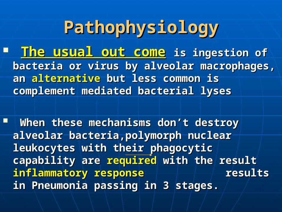
PathophysiologyPathophysiology The usual out comeThe usual out come is ingestion of bacteria or is ingestion of bacteria or
virus by alveolar macrophages, an virus by alveolar macrophages, an alternativealternative but less but less common is complement mediated bacterial lysescommon is complement mediated bacterial lyses
When these mechanisms don’t destroy alveolar When these mechanisms don’t destroy alveolar bacteria,polymorph nuclear leukocytes with their bacteria,polymorph nuclear leukocytes with their phagocytic capability are phagocytic capability are required required with the result with the result inflammatory response inflammatory response results in Pneumonia results in Pneumonia passing in 3 stages.passing in 3 stages.

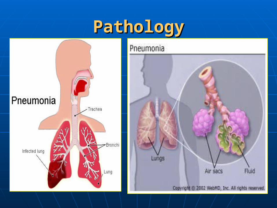
Pathology of Pneumonia:Pathology of Pneumonia:
1- Stage of congestion:1- Stage of congestion: (red hepatization) (red hepatization)
Due to antigen antibody reaction noDue to antigen antibody reaction no
radiological findingsradiological findings..
2- Stage of gray hepatiztion:2- Stage of gray hepatiztion:
Respiratory distress and homogenousRespiratory distress and homogenous
opacity in chest rdiography.opacity in chest rdiography.
3- Stage of resolution:3- Stage of resolution: When immune system takes upper When immune system takes upper
hand, resolution and convalescence.hand, resolution and convalescence.
PathologyPathology
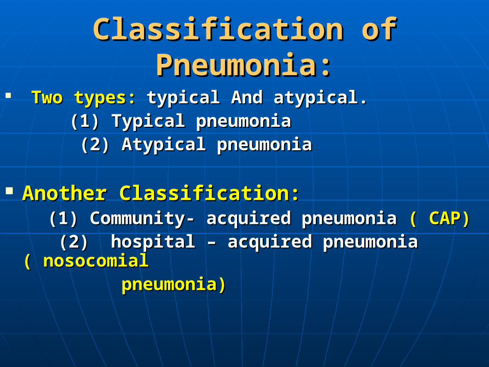
Classification of Pneumonia:Classification of Pneumonia:
Two types:Two types: typical And atypical.typical And atypical. (1) Typical pneumonia(1) Typical pneumonia (2) Atypical pneumonia(2) Atypical pneumonia
Another Classification:Another Classification: (1) Community- acquired pneumonia (1) Community- acquired pneumonia ( CAP)( CAP) (2) hospital – acquired pneumonia (2) hospital – acquired pneumonia (( nosocomialnosocomial pneumonia) pneumonia)

Out LineOut Line
(1) Definition and Pathophysiolgoy of pneumomnia(1) Definition and Pathophysiolgoy of pneumomnia
(2) Does the patient have a pneumonia? (2) Does the patient have a pneumonia?
(3) What is the microbial etiology?(3) What is the microbial etiology? (4) Where you treat?(4) Where you treat?
(5) How to treat?(5) How to treat?

Clinical and Radiological Diagnosis Clinical and Radiological Diagnosis of Pneumoniaof Pneumonia
(1) (1) Respiratory distressRespiratory distress (2) (2) Bronchial breathingBronchial breathing or fine or fine consonating creptation by consonating creptation by auscultation auscultation
(3) (3) Homogenous opacityHomogenous opacity is a cardinal is a cardinal radiological sign radiological sign
Clinical and Radiological Diagnosis Clinical and Radiological Diagnosis of Pneumoniaof Pneumonia
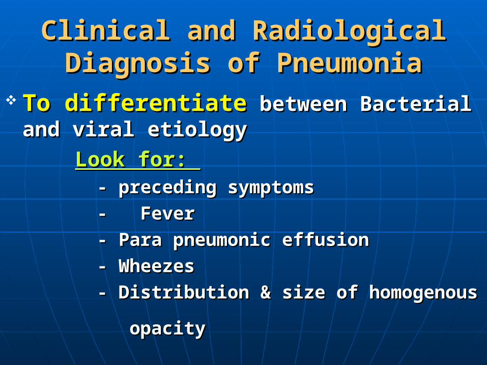
To differentiateTo differentiate between Bacterial and viral between Bacterial and viral etiology etiology
Look for: Look for: - preceding symptoms- preceding symptoms
- Fever- Fever
- Para pneumonic effusion- Para pneumonic effusion
- Wheezes- Wheezes
- Distribution & size of homogenous- Distribution & size of homogenous
opacityopacity

RadiologyRadiology

RadiologyRadiology

Out LineOut Line(1) Definition and Pathophysiolgoy of pneumomnia(1) Definition and Pathophysiolgoy of pneumomnia
(2) Does the patient have a pneumonia? (2) Does the patient have a pneumonia?
(3) What is the microbial etiology?(3) What is the microbial etiology? (4) Where you treat?(4) Where you treat?
(5) How to treat?(5) How to treat?
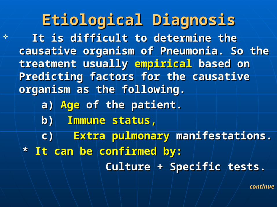
Etiological DiagnosisEtiological Diagnosis It is difficult to determine the causative It is difficult to determine the causative
organism of Pneumonia. So the treatment organism of Pneumonia. So the treatment usually usually empirical empirical based on Predicting factors based on Predicting factors for the causative organism as the following. for the causative organism as the following.
a) a) AgeAge of the patient. of the patient.
b) b) Immune status,Immune status,
c) c) Extra pulmonaryExtra pulmonary manifestations. manifestations.
* * It can be confirmed by:It can be confirmed by:
Culture + Specific tests.Culture + Specific tests.
continuecontinue

Etiological DiagnosisEtiological Diagnosis Age Age as a predictor for eteological diagnosisas a predictor for eteological diagnosis
A- In CAP:A- In CAP: Neonates:Neonates:
Common:-Common:- = Group B B- hemolytic streptococci= Group B B- hemolytic streptococci
= Gram negative enteric bacilli such as = Gram negative enteric bacilli such as Escherichia coli and Klepsella pneumoniae Escherichia coli and Klepsella pneumoniae
Less frequent:Less frequent:
Staph-aurasStaph-auras
P.aeruginosaP.aeruginosa
continuecontinue
Etiological DiagnosisEtiological Diagnosis
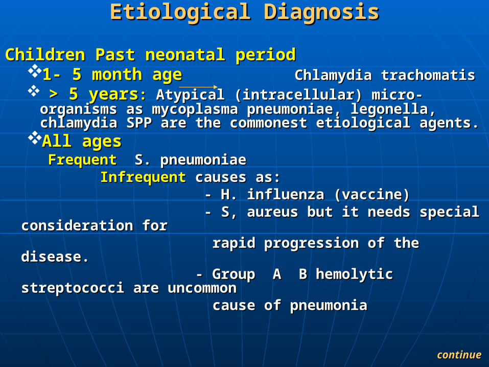
Children Past neonatal period Children Past neonatal period 1-1- 5 month age5 month age Chlamydia trachomatis Chlamydia trachomatis > 5 years> 5 years: : Atypical (intracellular) micro-organisms as Atypical (intracellular) micro-organisms as
mycoplasma pneumoniae, legonella, chlamydia SPP are the mycoplasma pneumoniae, legonella, chlamydia SPP are the commonest etiological agents.commonest etiological agents.
All agesAll ages Frequent Frequent S. pneumoniae S. pneumoniae
Infrequent Infrequent causes as:causes as: - H. influenza (vaccine) - H. influenza (vaccine) - S, aureus but it needs special consideration for - S, aureus but it needs special consideration for rapid progression of the disease.rapid progression of the disease. - Group A B hemolytic streptococci are uncommon - Group A B hemolytic streptococci are uncommon cause of pneumoniacause of pneumonia
continue continue

Etiological DiagnosisEtiological Diagnosis In hospital acquired pneumoniaIn hospital acquired pneumonia
Common:Common: Gram negativeGram negative organism such as: organism such as:
K. pneumonia, Pseudomonas aurugnosia and K. pneumonia, Pseudomonas aurugnosia and serrata spacesserrata spaces
Gram positiveGram positive organisms such as: organisms such as:
S. Aureus most frequent leogionella pneumoniae S. Aureus most frequent leogionella pneumoniae is rareis rare
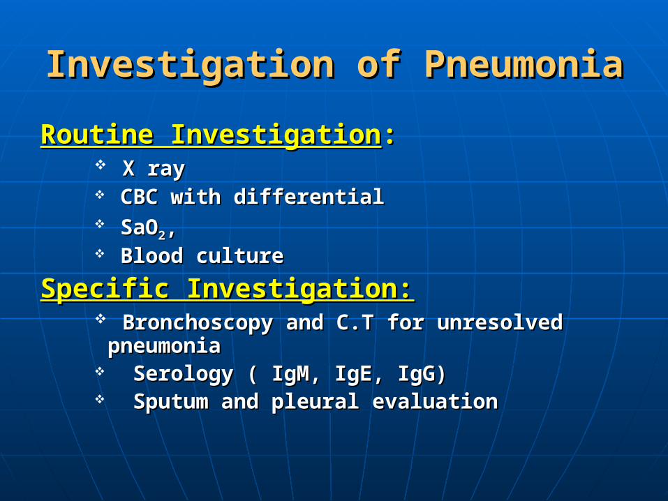
Investigation of PneumoniaInvestigation of Pneumonia
Routine InvestigationRoutine Investigation:: X rayX ray CBC with differentialCBC with differential SaOSaO22, , Blood cultureBlood culture
Specific Investigation:Specific Investigation: Bronchoscopy and C.T for unresolved pneumoniaBronchoscopy and C.T for unresolved pneumonia Serology ( IgM, IgE, IgG)Serology ( IgM, IgE, IgG) Sputum and pleural evaluation Sputum and pleural evaluation
Out LineOut Line(1) Definition and Pathophysiolgoy of pneumomnia(1) Definition and Pathophysiolgoy of pneumomnia
(2) Does the patient have a pneumonia? (2) Does the patient have a pneumonia?
(3) What is the microbial etiology?(3) What is the microbial etiology?
(4) Where you treat?(4) Where you treat?
(5) How to treat?(5) How to treat?
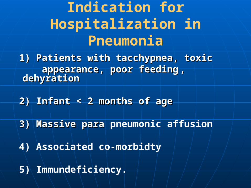
Indication for Hospitalization in Pneumonia
1) Patients with tacchypnea, toxic1) Patients with tacchypnea, toxic appearance, poor feedingappearance, poor feeding , dehyration, dehyration 2) Infant < 2 months of age2) Infant < 2 months of age 3) Massive para 3) Massive para pneumonic affusion
4) Associated co-morbidty
5) Immundeficiency.
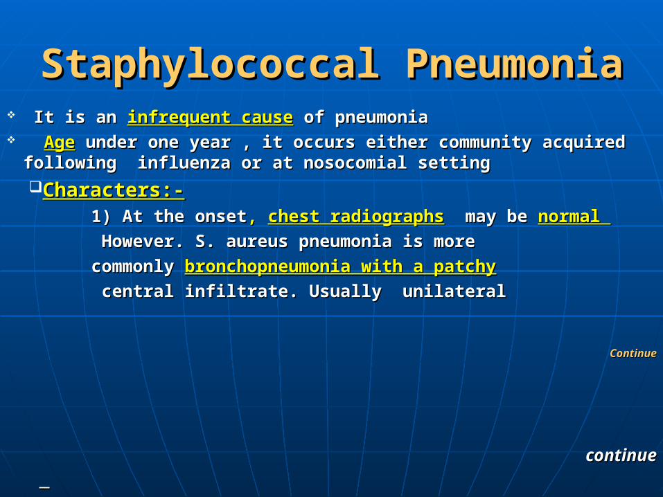
Staphylococcal PneumoniaStaphylococcal Pneumonia It is an It is an infrequent causeinfrequent cause of pneumonia of pneumonia AgeAge under one year , it occurs either community acquired following influenza or under one year , it occurs either community acquired following influenza or
at nosocomial setting at nosocomial setting
Characters:-Characters:- 1) At the onset1) At the onset, , chest radiographschest radiographs may be may be normal normal
However. S. aureus pneumonia is more However. S. aureus pneumonia is more
commonly commonly bronchopneumonia with a patchybronchopneumonia with a patchy
central infiltrate. Usually unilateralcentral infiltrate. Usually unilateral
ContinueContinue
continuecontinue

Staphylococcal PneumoniaStaphylococcal Pneumonia2)2) Rapid progression of the disease fromRapid progression of the disease from Patchy cansolidation to cavity, pneumatocele orPatchy cansolidation to cavity, pneumatocele or
tension pneumothorax should raise the possibility of S. tension pneumothorax should raise the possibility of S. aureus pneumonia, when it occurs aureus pneumonia, when it occurs
3) Effusion or empyema3) Effusion or empyema
Develops in about 90% of patients Develops in about 90% of patients Spontaneous pneumothorax or pyopneumothoraxSpontaneous pneumothorax or pyopneumothorax
Occur in about 25-50% of cases Occur in about 25-50% of cases Pneumatocele Pneumatocele
Occur in > 50% of cases and may change hourly in number or size Occur in > 50% of cases and may change hourly in number or size in acute phase.in acute phase.
continue continue

Staphylococcal PneumoniaStaphylococcal Pneumonia
This previous radiographic picture of S. aureus This previous radiographic picture of S. aureus pneumonia is notpneumonia is not pathognomonic as it occurs with:pathognomonic as it occurs with:
Pneumonia caused by:Pneumonia caused by: Klebsiella species, other gram negative bacteria group Klebsiella species, other gram negative bacteria group
A streptococci and occasionally pneumococciA streptococci and occasionally pneumococci
OROR it may mimic congenital diaphragmatic hernia it may mimic congenital diaphragmatic hernia ..
44) ) Staphylococcal pneumeniaStaphylococcal pneumenia ItIt should be suspected in hospitalized infants (particularly those should be suspected in hospitalized infants (particularly those
in ICU) who develop parenchymal unexplained opaque shadows. in ICU) who develop parenchymal unexplained opaque shadows.
continuecontinue
Staphylococcal PneumoniaStaphylococcal Pneumonia
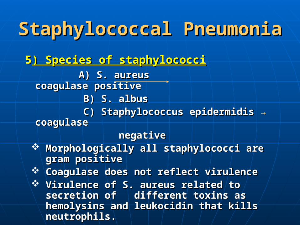
55) Species of staphylococci) Species of staphylococci A) S. aureusA) S. aureus coagulase positive coagulase positive
B) S. albusB) S. albus C) Staphylococcus epidermidis C) Staphylococcus epidermidis → → coagulase coagulase negativenegative
Morphologically all staphylococci are gram Morphologically all staphylococci are gram positivepositive
Coagulase does not reflect virulenceCoagulase does not reflect virulence Virulence of S. aureus related to secretion of Virulence of S. aureus related to secretion of
different toxins as hemolysins and leukocidin different toxins as hemolysins and leukocidin that kills neutrophils. that kills neutrophils.

Treatment of StaphylococcalTreatment of Staphylococcal pneumoniapneumonia
The production of B- lactamase by nearly all S. aureus The production of B- lactamase by nearly all S. aureus isolates rendered ineffective any drug bydrolysed by this isolates rendered ineffective any drug bydrolysed by this enzymeenzyme..
First – line therapy is: Hospital admission,First – line therapy is: Hospital admission,
B lactamase resistant penicillinB lactamase resistant penicillin
such as Methicillinsuch as Methicillin →→ no longer in use no longer in use
its modern analogs i.eits modern analogs i.e ox oxacillin, acillin, clocloxacillin, xacillin, FlucloFlucloxacillin, nafcillinxacillin, nafcillin
MRSA MRSA ( methicillin resistant S. aureus) which was ( methicillin resistant S. aureus) which was found to be resistant to all B lactams and all found to be resistant to all B lactams and all cephalosporin compounds.cephalosporin compounds.

Treatment of StaphylococcalTreatment of Staphylococcal PneumoniaPneumonia
MRSA infections:MRSA infections: that are nosocomially acquired was that are nosocomially acquired was found to be sensitive to found to be sensitive to VancomycinVancomycin
Now MRSA infection became community acquiredNow MRSA infection became community acquired Clindamycin:Clindamycin: indicated in MRSA infection in patients indicated in MRSA infection in patients
allergic to B-lactamsallergic to B-lactams Duration of treatment:Duration of treatment: 2-3 weeks, but 6 weeks 2-3 weeks, but 6 weeks
recommended to decrease the risk of relapserecommended to decrease the risk of relapse Supportive therapy:Supportive therapy: o o2,2, maintenance of, fluid, maintenance of, fluid,
electrolyte and hemoglobin levelselectrolyte and hemoglobin levelscontinuecontinue
continuecontinue

Treatment of StaphylococcalTreatment of Staphylococcal PneumoniaPneumonia
The most important complication isThe most important complication is
Empyema andEmpyema and lung abscesslung abscess, pneumothorax each , pneumothorax each
has a special treatment has a special treatment
Treatment in summaryTreatment in summary B. lactamase – resistant penicillins or first B. lactamase – resistant penicillins or first generation cephalosporin for 2-3 weeks generation cephalosporin for 2-3 weeks MRSA MRSA → → vancomycinvancomycinClindamycin Clindamycin MRSAMRSA
patient allergic to Bpatient allergic to B lactamslactams

HaemophilusInfluenzaType bHaemophilusInfluenzaType b
H. influenza is gram - negative bacilli H. influenza : can lead to invasive infectious in
pediatrics as pneumonia, meningitis, cellulites, epiglottites, septic arthritis, osteomyelitis percarditis and bacteremia
Incidance of H. influenza pneumonia decrease after administration of Hib canjugate vaccine
The virulence of serotype b is related to serotype b capsular polysaccharide

Haemophilus Influenza Type bHaemophilus Influenza Type b
Radiographic findingsRadiographic findings of H. influenza of H. influenza pneumonia vary from:-pneumonia vary from:-
1) 1) Bronchiolctic typeBronchiolctic type with central linear with central linear
infiltrates and over inflationinfiltrates and over inflation
2)2)Bronchopneumonia withBronchopneumonia with patchy patchy
consolidation consolidation with no lobarwith no lobar affection affection
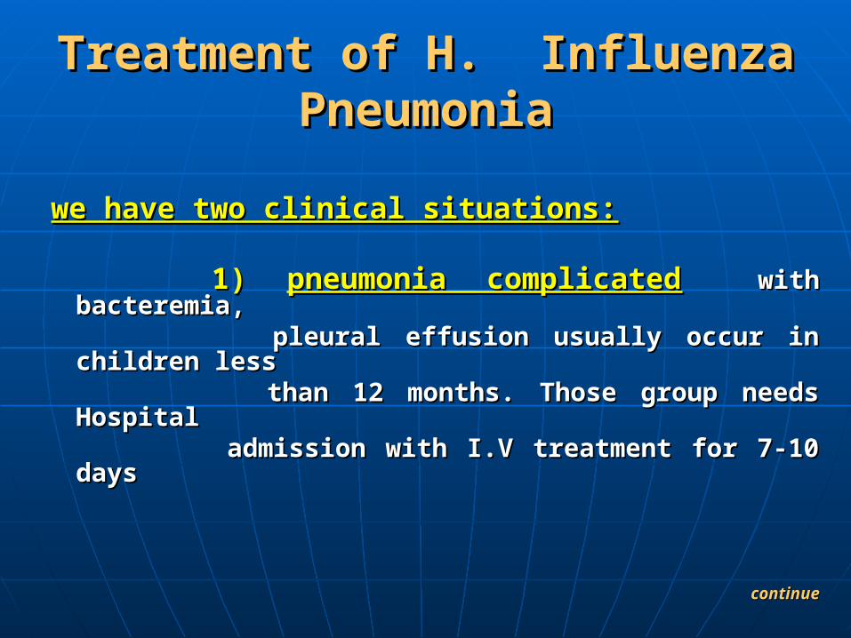
Treatment of H. Influenza Treatment of H. Influenza PneumoniaPneumonia
we have two clinical situations:we have two clinical situations:
1) 1) pneumonia complicatedpneumonia complicated with bacteremia, with bacteremia, pleural effusion usually occur in children lesspleural effusion usually occur in children less than 12 months. Those group needs Hospital than 12 months. Those group needs Hospital admission with I.V treatment for 7-10 daysadmission with I.V treatment for 7-10 days
continuecontinue
Treatment of H. Influenza Treatment of H. Influenza PneumoniaPneumonia
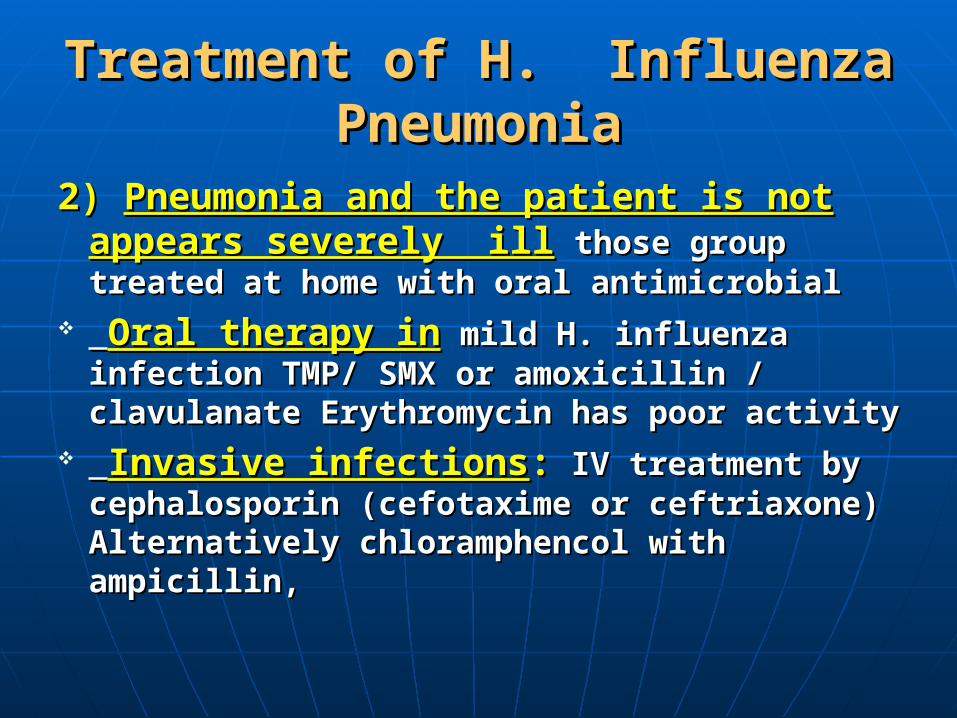
2) 2) Pneumonia and the patient is not appears Pneumonia and the patient is not appears severely illseverely ill those group treated at home with those group treated at home with oral antimicrobialoral antimicrobial
Oral therapy inOral therapy in mild H. influenza infection mild H. influenza infection TMP/ SMX or amoxicillin / clavulanate TMP/ SMX or amoxicillin / clavulanate Erythromycin has poor activity Erythromycin has poor activity
Invasive infectionsInvasive infections:: IV treatment by IV treatment by cephalosporin (cefotaxime or ceftriaxone) cephalosporin (cefotaxime or ceftriaxone) Alternatively chloramphencol with ampicillin,Alternatively chloramphencol with ampicillin,
Mycoplasma PneumoniaMycoplasma Pneumonia
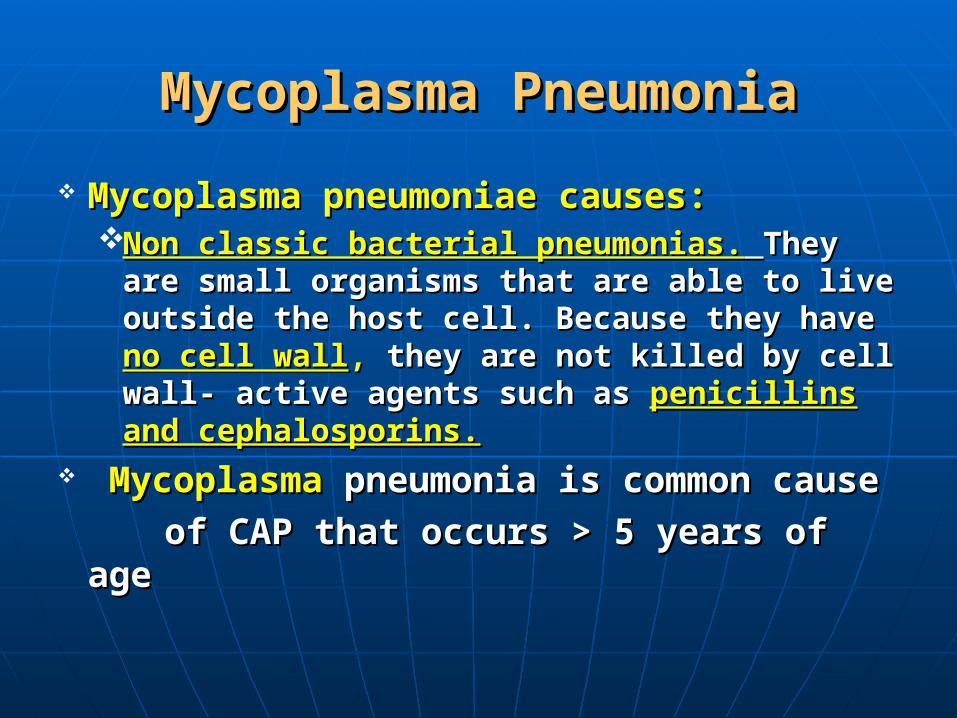
Mycoplasma pneumoniae causes:Mycoplasma pneumoniae causes:Non classic bacterial pneumonias.Non classic bacterial pneumonias. They are They are
small organisms that are able to live outside the small organisms that are able to live outside the host cell. Because they have host cell. Because they have no cell wallno cell wall,, they they are not killed by cell wall- active agents such as are not killed by cell wall- active agents such as penicillins and cephalosporins.penicillins and cephalosporins.
MycoplasmaMycoplasma pneumonia is common cause pneumonia is common cause
of CAP that occurs > 5 years of ageof CAP that occurs > 5 years of age

Pathogenesis of Pulmonary InfectionsPathogenesis of Pulmonary Infections
Infection with M. pneumoniae are acquired via Infection with M. pneumoniae are acquired via respiratory route from droplet infection.respiratory route from droplet infection.
The organism attaches to a receptor on respiratory The organism attaches to a receptor on respiratory epithelium and remains extra cellular causing epithelium and remains extra cellular causing cellular damage.cellular damage.
Specific cell mediated immune responses increase Specific cell mediated immune responses increase with age so it tends to be milder in children than with age so it tends to be milder in children than in adults and also severe in reinfection.in adults and also severe in reinfection.
Diagnosis of Mycoplasma PneumoniaDiagnosis of Mycoplasma Pneumonia
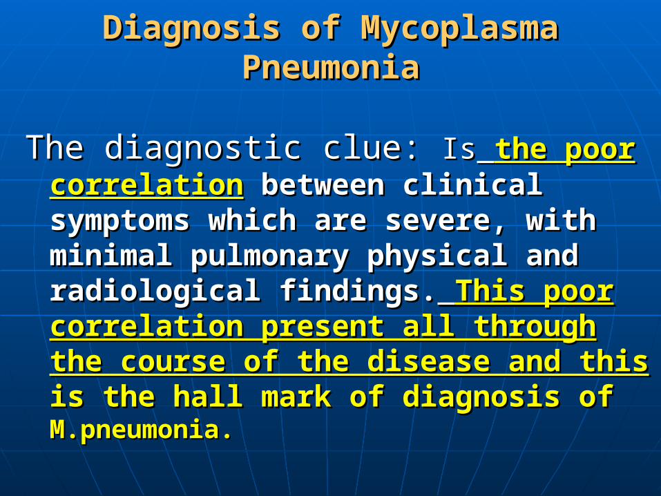
The diagnostic clue: The diagnostic clue: IsIs the poor correlationthe poor correlation between clinical symptoms which are severe, between clinical symptoms which are severe, with minimal pulmonary physical and with minimal pulmonary physical and radiological findings.radiological findings. This poor correlation This poor correlation present all through the course of the disease present all through the course of the disease and thisand this is the hall mark of diagnosis of is the hall mark of diagnosis of M.pneumonia.M.pneumonia.

Clinical PictureClinical Picture
1.1. Typically the patientsTypically the patients present with present with gradual onsetgradual onset of of malaise, headache and fever over 1 week. Cough dry or malaise, headache and fever over 1 week. Cough dry or productive associated with productive associated with symptomssymptoms as vomiting, diarrhea, as vomiting, diarrhea, chest pain, sore throat.chest pain, sore throat.
2.2. Physical findingsPhysical findings are relatively minimal early in the are relatively minimal early in the course of illness no finding on chest examination later on course of illness no finding on chest examination later on bronchial breathing crackles or wheezes can be heard.bronchial breathing crackles or wheezes can be heard.
3- 3- Radiologic findings:Radiologic findings: Usually unilateral in 87% and involves lower lobesUsually unilateral in 87% and involves lower lobes In the early stages the pattern is reticular and interstitialIn the early stages the pattern is reticular and interstitial Later patchy or segmental areas of consolidation are Later patchy or segmental areas of consolidation are noted.noted.
Continue Continue
Clinical PictureClinical Picture
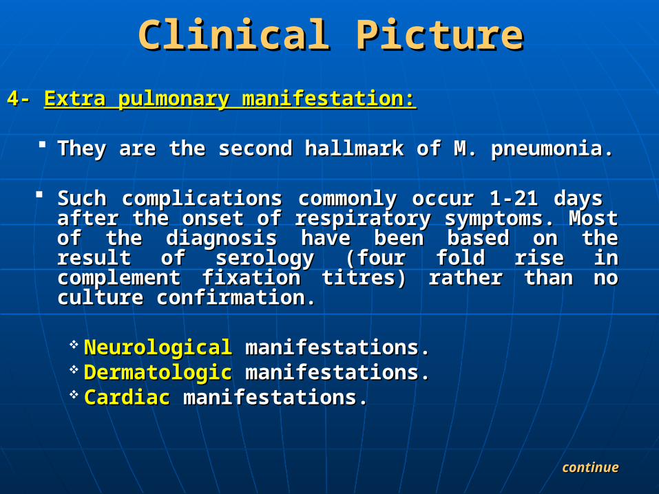
4-4- Extra pulmonary manifestation:Extra pulmonary manifestation:
They are the second hallmark of M. pneumonia.They are the second hallmark of M. pneumonia.
Such complications commonly occur 1-21 days after the Such complications commonly occur 1-21 days after the onset of respiratory symptoms. Most of the diagnosis have onset of respiratory symptoms. Most of the diagnosis have been based on the result of serology (four fold rise in been based on the result of serology (four fold rise in complement fixation titres) rather than no culture complement fixation titres) rather than no culture confirmation.confirmation.
NeurologicalNeurological manifestations.manifestations. DermatologicDermatologic manifestations.manifestations. Cardiac Cardiac manifestations.manifestations.
continuecontinue

Clinical PictureClinical Picture
4- 4- Extra pulmonary manifestation:Extra pulmonary manifestation:
GIT GIT manifestations.manifestations. HematologicHematologic manifestations. manifestations. MusculoskeletalMusculoskeletal manifestations.manifestations. GenitourinaryGenitourinary manifestations. manifestations. ImmunologicImmunologic manifestations. manifestations.

Diagnostic Criteria of Mycoplasma Diagnostic Criteria of Mycoplasma InfectionInfection
Serologic assays are the mainstay for diagnosing mycoplasma Serologic assays are the mainstay for diagnosing mycoplasma infection:-infection:-
1.Cold agglutinin identification1.Cold agglutinin identification
Cold agglutinins usually appear by the end of first Cold agglutinins usually appear by the end of first week and disappear by 2-3 monthsweek and disappear by 2-3 months
Cold agglutinin responses are non specific and Cold agglutinin responses are non specific and consistent consistent mostly mostly IgMIgM
So So IgM IgM lacks specificity and sensitivitylacks specificity and sensitivity
continuecontinue

Diagnostic Criteria of Mycoplasma Diagnostic Criteria of Mycoplasma InfectionInfection
Specific serological test:-Specific serological test:- Complement fixation test has been used as Complement fixation test has been used as standared for diagnosis with titer standared for diagnosis with titer >> 1:321:32 The test measures The test measures IgM antibodiesIgM antibodies This test has 90% sensitivity and 94%This test has 90% sensitivity and 94% specificityspecificity
Polymerase chain Reaction(PCR)Polymerase chain Reaction(PCR) PCR diagnosis M. pneumonia in both throat swab and PCR diagnosis M. pneumonia in both throat swab and
CSF.CSF.

Treatment of M. PneumoniaTreatment of M. Pneumonia
Macrolides are the drug of choice as:-Macrolides are the drug of choice as:-Erythromycins, Azithromycin are specific Erythromycins, Azithromycin are specific
treatment.treatment. Because M. pneumonia has no cell wall, it is Because M. pneumonia has no cell wall, it is
resistant to penicillins, cephalosporins and resistant to penicillins, cephalosporins and
other cell wall active agents. other cell wall active agents.

Pneumococcal PneumoniaPneumococcal Pneumonia
Pneumococcus (Streptococcus Pneumoniae) It is the most common cause of bacterial
pneumonia in children. The organism is gram positive cocci.The virulence of the pneumococci is related to there capsule.
It affects all ages.
Continue

Pneumococcal PneumoniaPneumococcal Pneumonia
Diagnosis:Diagnosis:1)1) It is It is typical pneumoniatypical pneumonia with sudden onset with sudden onset
of fever, cough, chest pain and dyspnea.of fever, cough, chest pain and dyspnea.
2)2) Physical examination:Physical examination:• Pleurtic chest pain my be referred to Pleurtic chest pain my be referred to
abdomen with misleading suspicion of acute abdomen with misleading suspicion of acute appendicitis.appendicitis.
• Symptoms and signs of meningismus may Symptoms and signs of meningismus may be present with upper lobe pneumonia.be present with upper lobe pneumonia.
ContinueContinue
Pneumococcal PneumoniaPneumococcal Pneumonia
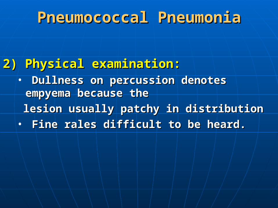
2) Physical examination:2) Physical examination:• Dullness on percussion denotes empyema because the Dullness on percussion denotes empyema because the
lesion usually patchy in distributionlesion usually patchy in distribution• Fine rales difficult to be heard.Fine rales difficult to be heard.
Pneumococcal PneumoniaPneumococcal Pneumonia
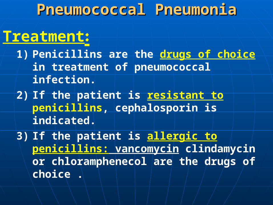
Treatment:1) Penicillins are the drugs of choice in treatment of
pneumococcal infection.
2) If the patient is resistant to penicillins, cephalosporin is indicated.
3) If the patient is allergic to penicillins: vancomycin clindamycin or chloramphenecol are the drugs of choice .

Recurrent or Unresolved PneumoniaRecurrent or Unresolved Pneumonia
I- Multiple lobe affection:- Congential heart disease with excess pulmonary blood
flow.- Immune deficiency either local or general.- Specific infection as T.B and fungal infection
(Aspergillus)
II- Singal lobe affection:- Obstruction : Intraluminal, Extraluminal.- Structural abnormalities as pulmonary
sequestration.
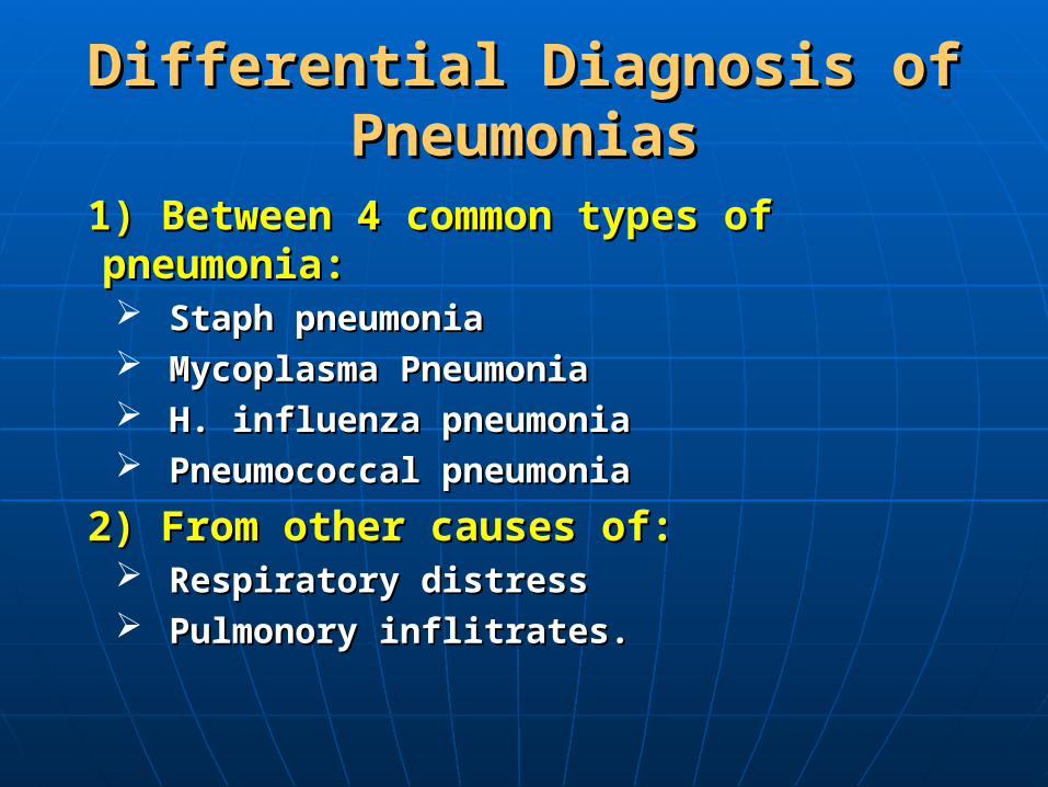
Differential Diagnosis of Differential Diagnosis of PneumoniasPneumonias
1) Between 4 common types of pneumonia:1) Between 4 common types of pneumonia: Staph pneumoniaStaph pneumonia Mycoplasma PneumoniaMycoplasma Pneumonia H. influenza pneumoniaH. influenza pneumonia Pneumococcal pneumoniaPneumococcal pneumonia
2) From other causes of:2) From other causes of: Respiratory distressRespiratory distress Pulmonory inflitrates.Pulmonory inflitrates.

Related Documents