MANAGEMENT OF ACETABULAR FRACTURES BY BLUE TEAM

BY BLUE TEAM. By Dr Kabiru Salisu NOHD INTRODUCTION HISTORY EPIDEMIIOLOGY AETIOLOGY PATHOPHYSIOLOGY SURGICAL ANATOMY CLASSIFICATION.
Dec 29, 2015
Welcome message from author
This document is posted to help you gain knowledge. Please leave a comment to let me know what you think about it! Share it to your friends and learn new things together.
Transcript

MANAGEMENT OF ACETABULAR
FRACTURESBY
BLUE TEAM

PATHOLOGY OF ACETABULAR
FRACTUREBy Dr Kabiru Salisu
NOHD

INTRODUCTION HISTORY EPIDEMIIOLOGY AETIOLOGY PATHOPHYSIOLOGY SURGICAL
ANATOMY CLASSIFICATION
OUTLINE

INTRODUCTION
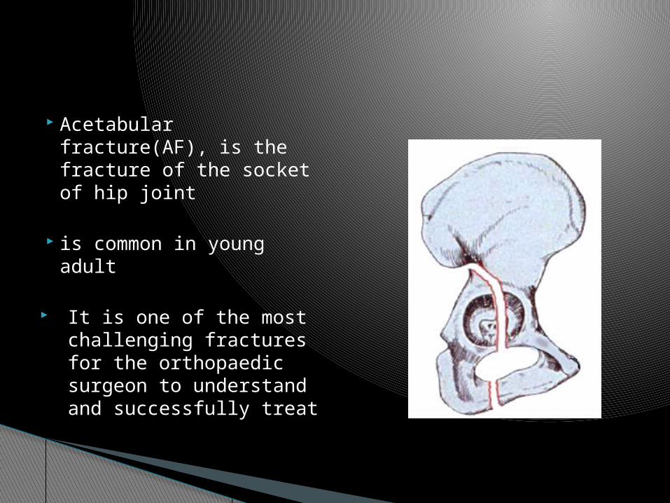
Acetabular fracture(AF), is the fracture of the socket of hip joint
is common in young adult
It is one of the most challenging fractures for the orthopaedic surgeon to understand and successfully treat

These fractures are often associated with other life-threatening injuries
Orthopaedic - Extremity
injury (36%) - Nerve palsy (13%) - Spine injury (4%)
Systemic injuries- head injury (19%) - Chest injury (18%) - Abdominal injury
(8%) - Genitourinary
injury (6%

Acetabular fracture usually result from high energy injury
Anatomic reduction and stable fixation of the fracture, is the treatment goal in these difficult fractures

Fractures of the acetabulum were treated nonoperatively until the middle of the 20th century
The Judets & Emile Letournel study was responsible for popularizing the surgical management
History

With advances in imaging technologies, performing acetabular fracture surgery through smaller incisions is now possible
The exact incidence of acetabular fractures in various parts of the world is not known.
Studies at level I trauma centers have shown an admission rate for pelvic and acetabular fractures of 0.5-7.5%
EPIDEMIOLOGY

Acetabulum fractures usually occur as a result of high-velocity trauma, such as;
- Motor vehicular accidents or
- Falls from heights
AETIOLOGY
AF occur as a result of the force exerted through the head of the femur to the acetabulum.
The femoral head acts like a hammer and is the last link in the chain of forces transmitted from the greater trochanter, knee, or foot to the acetabulum.
The position of the femur at the time of impact and the direction of the force determine the type and displacement of the fracture
PATHOPHYSIOLOGY

◦ Inverted “Y” two column concept (1966)
◦ Columns are connected to the SI joint by a thick area of bone above the greater sciatic notch known as the sciatic buttress
Relevant anatomy


Several classifications for AF do exist, all the classifications are base on the anatomy described by Judets and letournel
CLASSIFICATION

1- Tile universal classification

This is the most widely accepted classification
This system divides fractures of the acetabulum into;
- Five simple (elementary) - Five complex (associated)
2- Letournel and Judets classification


This is a modification of judet & letournel classification
Type A- pertial articular involving only one column
A1- Posterior column fractureA2-posterior wall fractureA3- Anterior column and wall
3- AO Muller classification

Type B- Partial articular, involving transverse component
B1 - pure transverse
B2 - T- shaped
B3 - Anterior column and posterior hemitransverse

Type c- complete articular both columns C1- High variety extending to iliac crest
C2- Low variety extending to the anterior border of the ilium
C3 – Extention into the sacroiliac joint
Related Documents