RESEARCH Open Access Bufotenine is able to block rabies virus infection in BHK-21 cells Hugo Vigerelli 1,2 , Juliana Mozer Sciani 1 , Carlos Jared 3 , Marta Maria Antoniazzi 3 , Graciane Maria Medeiros Caporale 2 , Andréa de Cássia Rodrigues da Silva 2 and Daniel C Pimenta 1* Abstract Background: Rabies is a fatal zoonotic neglected disease that occurs in more than 150 countries, and kills more than 55.000 people every year. It is caused by an enveloped single stranded RNA virus that affects the central nervous system, through an infection initiated by the muscular nicotinic acetylcholine receptor, according to many authors. Alkaloids, such as acetylcholine, are widespread molecules in nature. They are present in numerous biological fluids, including the skin secretion of many amphibians, in which they act (together with proteins, peptides and steroids) as protection agents against predators and/or microorganisms. Among those amphibians that are rich in alkaloids, there is the genus Rhinella. Methods: Bufotenine was isolated from Rhinela jimi skin secretion after a liquid-liquid partition (H 2 O:CH 2 Cl 2 ) and reversed phase high-performance liquid chromatography analyses (RP-HPLC). Bufotenine was also extracted from seeds of Anadenanthera colubrina in acetone solution and purified by RP-HPLC, as well. Structural characterization was performed by mass spectrometry and nuclear magnetic resonance analyses. Cytotoxic tests of bufotenine were performed over baby hamster kidney (BHK-21) cells using MTT test. For the antiviral activity, Rabies virus strain Pasteur vaccine (PV) was used on fluorescence inhibition test and fluorescent foci inhibition test, with both simultaneous and time course treatment of the cells with the virus and bufotenine. Results: In the present work we describe the effects of bufotenine, obtained either from toads or plants, that can inhibit the penetration of rabies virus in mammalian cells through an apparent competitive mechanism by the nicotinic acetylcholine receptor. Moreover, this inhibition was dose- and time-dependent, pointing out to a specific mechanism of action. Conclusions: This work do not present or propose bufotenine as a drug for the treatment of rabies due to the hallucinogen and psychotropic effects of the molecule. However, continued studies in the elucidation of the antiviral mechanism of this molecule, may lead to the choice or development of a tryptamine analogue presenting potential clinical use. Keywords: Rabies, Rhinella, Bufotenine, Alkaloids, Toxins, Mass spectrometry Background According to the World Health Organization (WHO), ra- bies is a zoonotic neglected disease that occurs in more than 150 countries and territories, killing more than 55,000 people every year, mostly in Asia and Africa [1]. It is caused by a virus that affects the central nervous system and, once symptoms have developed, is nearly always fatal [2,3]. Different in vitro experiments showed evidences that some cells receptors, such as the muscular form of the nicotinic acetylcholine receptor (nAChR), are capable of participating on the entry of rabies virus into cells [4-7]. Other molecules – including the p75 neutro- phin, the neuronal cell adhesion molecule (NCAM) and glicosides of the cell membrane – are also capable of par- ticipating in the entry of rabies virus into cells, maybe involved in different stages of the infection [8]. Con- formational studies on an internal tetrapeptide of rabies virus glycoprotein (Asn 194 -Ser 195 -Arg 196 -Gly 197 ) consid- ered this to be an essential part of the binding site of the virus to the acetylcholine receptor and have demonstrated * Correspondence: [email protected] 1 Laboratory of Biochemistry and Biophysics, Butantan Institute, São Paulo, SP, Brazil Full list of author information is available at the end of the article © 2014 Vigerelli et al.; licensee BioMed Central Ltd. This is an Open Access article distributed under the terms of the Creative Commons Attribution License (http://creativecommons.org/licenses/by/4.0), which permits unrestricted use, distribution, and reproduction in any medium, provided the original work is properly credited. The Creative Commons Public Domain Dedication waiver (http://creativecommons.org/publicdomain/zero/1.0/) applies to the data made available in this article, unless otherwise stated. Vigerelli et al. Journal of Venomous Animals and Toxins including Tropical Diseases 2014, 20:45 http://www.jvat.org/content/20/1/45

Welcome message from author
This document is posted to help you gain knowledge. Please leave a comment to let me know what you think about it! Share it to your friends and learn new things together.
Transcript

Vigerelli et al. Journal of Venomous Animals and Toxins including Tropical Diseases 2014, 20:45http://www.jvat.org/content/20/1/45
RESEARCH Open Access
Bufotenine is able to block rabies virus infectionin BHK-21 cellsHugo Vigerelli1,2, Juliana Mozer Sciani1, Carlos Jared3, Marta Maria Antoniazzi3, Graciane Maria Medeiros Caporale2,Andréa de Cássia Rodrigues da Silva2 and Daniel C Pimenta1*
Abstract
Background: Rabies is a fatal zoonotic neglected disease that occurs in more than 150 countries, and kills more than55.000 people every year. It is caused by an enveloped single stranded RNA virus that affects the central nervous system,through an infection initiated by the muscular nicotinic acetylcholine receptor, according to many authors. Alkaloids, suchas acetylcholine, are widespread molecules in nature. They are present in numerous biological fluids, including the skinsecretion of many amphibians, in which they act (together with proteins, peptides and steroids) as protection agentsagainst predators and/or microorganisms. Among those amphibians that are rich in alkaloids, there is the genus Rhinella.
Methods: Bufotenine was isolated from Rhinela jimi skin secretion after a liquid-liquid partition (H2O:CH2Cl2) and reversedphase high-performance liquid chromatography analyses (RP-HPLC). Bufotenine was also extracted from seeds ofAnadenanthera colubrina in acetone solution and purified by RP-HPLC, as well. Structural characterization was performedby mass spectrometry and nuclear magnetic resonance analyses. Cytotoxic tests of bufotenine were performed overbaby hamster kidney (BHK-21) cells using MTT test. For the antiviral activity, Rabies virus strain Pasteur vaccine (PV) wasused on fluorescence inhibition test and fluorescent foci inhibition test, with both simultaneous and time coursetreatment of the cells with the virus and bufotenine.
Results: In the present work we describe the effects of bufotenine, obtained either from toads or plants, that caninhibit the penetration of rabies virus in mammalian cells through an apparent competitive mechanism by thenicotinic acetylcholine receptor. Moreover, this inhibition was dose- and time-dependent, pointing out to a specificmechanism of action.
Conclusions: This work do not present or propose bufotenine as a drug for the treatment of rabies due to thehallucinogen and psychotropic effects of the molecule. However, continued studies in the elucidation of theantiviral mechanism of this molecule, may lead to the choice or development of a tryptamine analogue presentingpotential clinical use.
Keywords: Rabies, Rhinella, Bufotenine, Alkaloids, Toxins, Mass spectrometry
BackgroundAccording to the World Health Organization (WHO), ra-bies is a zoonotic neglected disease that occurs in morethan 150 countries and territories, killing more than55,000 people every year, mostly in Asia and Africa [1].It is caused by a virus that affects the central nervoussystem and, once symptoms have developed, is nearlyalways fatal [2,3]. Different in vitro experiments showed
* Correspondence: [email protected] of Biochemistry and Biophysics, Butantan Institute, São Paulo, SP,BrazilFull list of author information is available at the end of the article
© 2014 Vigerelli et al.; licensee BioMed CentraCommons Attribution License (http://creativecreproduction in any medium, provided the orDedication waiver (http://creativecommons.orunless otherwise stated.
evidences that some cells receptors, such as the muscularform of the nicotinic acetylcholine receptor (nAChR), arecapable of participating on the entry of rabies virus intocells [4-7]. Other molecules – including the p75 neutro-phin, the neuronal cell adhesion molecule (NCAM) andglicosides of the cell membrane – are also capable of par-ticipating in the entry of rabies virus into cells, maybeinvolved in different stages of the infection [8]. Con-formational studies on an internal tetrapeptide of rabiesvirus glycoprotein (Asn194-Ser195-Arg196-Gly197) consid-ered this to be an essential part of the binding site of thevirus to the acetylcholine receptor and have demonstrated
l Ltd. This is an Open Access article distributed under the terms of the Creativeommons.org/licenses/by/4.0), which permits unrestricted use, distribution, andiginal work is properly credited. The Creative Commons Public Domaing/publicdomain/zero/1.0/) applies to the data made available in this article,

Vigerelli et al. Journal of Venomous Animals and Toxins including Tropical Diseases 2014, 20:45 Page 2 of 8http://www.jvat.org/content/20/1/45
that the side chains of asparagine and arginine can, ap-parently, mimic the acetylcholine spatial structure, be-ing responsible for binding the virus to the acetylcholinereceptor [9].The skin secretion of some amphibians, on the other
hand, is rich in alkaloids [10]. Not only that, but also pro-teins, peptides, steroids and amines that act as chemicalprotection agents against predators and/or microorganisms,such as fungi and bacteria [8,11]. Among those amphibiansthat are rich in alkaloids, there is the genus Bufo thatwas recently split into Bufo in the Old World and Rhinellain the New World [12]. Their secretion contain a largenumber of alkaloids including bufotenine – a tryptaminealkaloid used as a defense mechanism due to its toxicity –that is also found in the Leguminosae family [13-16].Taking into account that the rabies virus can bind to the
nicotinic acetylcholine receptor, and that other alkaloids(nicotine, lobeline, cytisine, anabasine etc.) can also bindto these receptors, the aim of this study was to evaluatethe effects of bufotenine as a possible interfering agent inthe process of infection of the rabies virus in mamma-lian cells, using the simplified fluorescent inhibition microt-est (SFIMT) and rapid fluorescent focus inhibition test(RFFIT) techniques (with adaptations). Both are sero-logic tests commonly used in rabies diagnosis that areconsidered viable tools for the evaluation of new naturalcompounds as potential antiviral agents.
MethodsReagentsAll reagents were of analytical grade and were purchasedfrom Sigma Aldrich (USA), unless otherwise stated.
Isolation of bufotenine from Rhinella jimi skin secretionThe collection and housing of Rhinella jimi specimenswere performed under license number 15964-1 from theBrazilian Institute of Environment and Renewable NaturalResources (IBAMA). Skins secretions were collected throughmechanical stimulation of the parotoid macroglands. Aliquid-liquid partition (H2O:CH2Cl2) was performed and theaqueous partition was centrifuged at 4,794 g and the super-natant was analyzed by reversed phase high-performanceliquid chromatography (RP-HPLC), using a binary HPLCsystem (20A Prominence, Shimadzu Co, Japan), coupled toa C18 column (ACE®, 250 mm× 7.75 mm). The mobilephase consisted of solvent A [H2O: trifluoroacetic acid(TFA); 1000:1] and solvent B [acetonitrile (ACN):H2O:TFA;900:100:1] with a linear gradient of B over A (10% to 70%)in 35 minutes, at a constant flow rate of 1.7 mL.min-1 andmonitored by UV absorbance at 214 nm. Fractions weremanually collected and submitted to mass spectrometry(MS and MS2 ESI-IT-TOF) analyses. The fraction contain-ing bufotenine was then purified using a C18 column(ACE®, 250 mm × 4.6 mm), with a linear gradient of B
over A 13% to 15% in 15 minutes, at a constant flow rateof 1.1 mL.min-1, at 4°C, monitored by UV absorbance at214 nm. Bufotenine peak was manually collected, driedand submitted to 1H-NMR spectroscopic analyses and/orbiologic assays.
Isolation of bufotenine from Anadenanthera colubrineseedsSince bufotenine is a long and well known alkaloid thatcan be also obtained from other biological sources,such as plants, we chose to purify this molecule fromAnadenanthera colubrina seeds, which contain up to 2%of the seed dry weight in bufotenine. The seeds were ob-tained from the legitimate supplier Arbocenter Comérciode Sementes Ltda, Birigui, São Paulo (batch 0019), Brazil.The extraction of bufotenine was performed as describedby Stromberg [16], with modifications. Twenty grams ofseeds were powdered and subsequently deionized H2O and5 g of sodium carbonate (Na2CO3) were added until a uni-form mixture was obtained. This solution was then lyophi-lized and ressuspended in 50 mL of acetone (CH3COCH3).This extract was filtered and concentrated in a rotavaporsystem, following RP-HPLC analysis, in a C18 column(ACE®, 250 mm × 4.6 mm), with a linear gradient of Bover A 0% to 100% in 20 minutes, at a constant flow rateof 1 mL/min and monitored by UV absorbance at 214 nm.Bufotenine peak was manually collected and dried for massspectrometry and biologic assays.
Mass spectrometryPreviously dried samples were dissolved into 50% ACNwith 0.5% formic acid for MS and/or MS2 analyses in anESI-IT-TOF mass spectrometer system (Shimadzu Co.,Japan). Typically, 3 μL of samples were manually injectedin a Rheodyne injector under a constant flow of 50 μL/min.The interface voltage used was 4.5 kV and detector voltage,1.76 kV, with source temperature of 200°C. Data were ac-quired under positive mode and instrument control, dataacquisition and processing were performed by the LCMSsolution software (Shimadzu Co., Japan).
NMR analysesNMR screening was used to confirm the bufotenine struc-ture from Rhinella jimi skin secretion. 1H NMR spectrawere recorded on Bruker 500 MHz spectrometer (BrukerCo., Germany), with samples diluted in deuterated chloro-form and achievement of 128 scans. The results were proc-essed in TopSpin 1.3 software (Bruker Co., Germany).
Cells and virusesBaby hamster kidney (BHK-21) cells (ATCC® CCL – 100)were cultured in Eagle’s minimum essential medium, sup-plemented with 10% fetal bovine serum (MEM-10), at 37°Cunder a humidified 5% CO2 atmosphere until the formation

Vigerelli et al. Journal of Venomous Animals and Toxins including Tropical Diseases 2014, 20:45 Page 3 of 8http://www.jvat.org/content/20/1/45
of the cell monolayer. Rabies virus strain Pasteur vac-cine (PV), from Pasteur Institute, Brazil, was used to deter-mine the antiviral activity of bufotenine. The titerswere determined by plate assay in BHK-21 cells andexpressed as 100% infecting dose in cell culture (IDCC100)and 50% focus-forming dose (FFD50) as described byBatista et al. [17].
Cytotoxicity assayThe cytotoxicity evaluation of bufotenine was performedby MTT [3-(4,5-dimethylthiazol-2-yl)-2,5-diphenyltetra-zolium bromide] method, according to Takeuchi et al. [18]and Mosmann [19], with modifications. Briefly, BHK-21cells (5 × 10⁴ cells/well) were deposited in 96-well tissueculture microtiter plates with 50 μL of different bufote-nine concentrations diluted in MEM-10. For positive con-trol of citotoxicity dimethyl sulfoxid (DMSO) 20% was usedand the negative control was MEM-10 alone. After 24hours at 37°C, under a humidified 5% CO2 atmosphere, themedium was removed and 50 μL of MTT (Sigma® 1 mg/mL) solution, prepared in MEM-10, was added to each welland the plates were incubated once more for four hours.After incubation, the MTT solution was removed and 100μL of dimethyl sulfoxide (DMSO) were added to each wellto solubilize the formazan crystals. After gently shaking theplates, the crystals were completely dissolved, and theabsorbances were measured by using a spectrophotom-eter (Molecular Devices®, SpectraMax M2) at 540 nm.The CC50 was defined as the cytotoxic concentration ofbufotenine that reduced the absorbance of treated cells to50% when compared to the control.
Antiviral activityRabies virus does not cause cytopathic effects in cells cul-tured in vitro, being necessary some kind of additional testto monitor cell infection, such as tests with antibodiesbound to fluorescent substances [20]. For this reason,we choose these two tests adapted from commonly sero-logical tests used for antirabies virus titration, being thefluorescence inhibition a qualitative test, for a better visua-lization of both the infection and its inhibition, and thefluorescence focus inhibition comprises a quantitative test,used for the statistical analyses.
Fluorescence inhibition testThis test was based on simplified fluorescent inhibitionmicrotest (SFIMT), acording to Favoretto et al. [21], withmodifications. Briefly, 50 μL of BHK-21 cells (5 × 10⁴ cells/well) were deposited on 96-well tissue culture microtiterplates with 50 μL of each bufotenine dose (from 0.5 to 4mg/mL) and 50 μL of PV virus previously diluted at 1:100(IDCC100). As negative inhibition control, only 50 μL ofMEM-10 was added to the cells and as positive inhibitioncontrol ketamine was used (Dopalen®, 23.4 μM), according
to Lockhart et al. [22]. After a 24-hour period of incuba-tion at 37°C under a humidified 5% CO2 atmosphere, themedium was removed by suction from all wells and themicroplates were cooled on ice. The cells were fixed byadding acetone 80% in water (kept at –20°C). After 15minutes, the plates were emptied by inversion and dried at37°C according to Smith et al. [23] and Chaves et al. [24].The staining was carried out by adding 40 μL of an optimaldilution of the antirabies fluorescent conjugate [25]. Afterone hour of incubation, the microtiter plates were washedby immersion in PBS and then in distilled water. Themicrotiter plates reading was performed in inverted fluor-escence microscope (Leica DMIL, 100× magnification) ina qualitative manner based on the fluorescence inhibitioncompared to negative and positive controls, where thereare 100% and 0% of fluorescence, respectively.
Fluorescent focus inhibition testThis test was based on rapid fluorescent focus inhibitiontest (RFFIT) acording to Smith et al. [23], adapted to mi-crotiter plates as described by Chaves et al. [24], with mod-ifications. Briefly, 100 μL of BHK-21 cells (2.5 × 10⁴ cells/well) were deposited on 96-well tissue culture microtiterplates containing different bufotenine doses (3.9, 1.95, 0.97and 0.48 mg/mL), and 50 μL of PV virus previously dilutedat thirty-fold the FFD50 value. As negative inhibition con-trol, only 50 μL of MEM-10 was added to the cells and aspositive inhibition control ketamine was used (Dopalen®,23.4 μM). After a 20-hour period of incubation at 37°Cunder a humidified 5% CO2 atmosphere, the medium wasremoved by suction from all wells. The procedures of cellsfixation and staining were the same described in “Fluores-cence inhibition test” section. The microtiter plates readingwas quantitatively performed in inverted fluorescencemicroscope (Leica DMIL, 200× magnification), where eachfield with fluorescence were counted, being 0 the mini-mum (positive control) and 18 the maximum (negativecontrol) number of infected fields. The IC50 was de-fined as the inhibition concentration of bufotenine thatreduced the number of fields with fluorescent foci to 50%when compared with the negative control.
Time course studyThe time course effect of bufotenine was examined onPV virus with two minor modifications of the fluorescentfocus inhibition test. First, to test a possible “protectoreffect”, bufotenine was added in different times (1, 3 and6 hours), prior to the addition of the virus and, secondly,to test a possible “treatment effect”, bufotenine was addedin different times (1, 3 and 6 hours), after incubation ofcells and PV virus. All the other procedures were the sameas described in “Fluorescent focus inhibition test” section.

Vigerelli et al. Journal of Venomous Animals and Toxins including Tropical Diseases 2014, 20:45 Page 4 of 8http://www.jvat.org/content/20/1/45
Data analysisThe 50% cytotoxic (CC50) and inhibition (IC50) concentra-tions were calculated from concentration-effect curves afterlinear regression analysis. The results represent the mean ±standard error of the mean values of two different experi-ments (triplicates).
ResultsRP-HPLC and mass spectrometry analysisRP-HPLC analysis of the aqueous partition of Rhinela jimiskin secretion showed the presence of five major HPLCpeaks (Figure 1A). MS analysis showed that fraction 3contained the following m/z values: 205, 219 and 160(Figure 1B). Comparing the MS2 analysis with alreadypublished data by McClean et al. [26] (Figure 1C), it waspossible to characterize the two indole alkaloids containedin this fraction: the 205 m/z correspond to N’,N’-dimethyl5-hydroxytryptamione (bufotenine) and 219 m/z corres-pond to N’,N’,N’-trimethyl 5-hydroxytryptamine (5-HTQ).The 160 m/z ion is a spontaneous fragmentation of bothalkaloids, which also appears on MS2 profile (Additionalfile 1: Figure S1). This two compounds were separated byRP-HPLC yielding pure bufotenine (Additional file 2:Figure S2), as confirmed by NMR analyses (Additional
Figure 1 RP-HPLC analysis of the aqueous partition of Rhinela jimi ski(λ = 214 nm), showing five fractions. (B) MS analysis of fraction 3 showingindole alkaloids bufotenine (205 m/z) and 5-HTQ (219 m/z), resulting in an
file 3: Figure S3). Bufotenine from Anadenanthera colubrinaseeds were purified only after one chromatographic step, fol-lowing the acetone extract, being the major peak on the RP-HPLC chromatogram and appearing pure on mass spectro-metric analyses (Additional file 4: Figure S4).
Cytotoxicity effect on viability of BHK-21 cellsThe 3.9 mg/mL dose of R. jimi bufotenine used on thevirologic tests presented statistically significant cytotoxiceffects on the viability of BHK-21 cells (66% of viable cells)when compared to the negative (cells +MEM-10) andpositive controls (cells + DMSO 20%) (Additional file 5:Figure S5). Subsequent assays were performed with theseeds bufotenine, due to the high availability. In the MTTtest (Additional file 6: Figure S6), it was possible to deter-mine the CC50 value (7.6 mg/mL).
Antiviral activity
� Fluorescence inhibition test
Bufotenine was able to inhibit the infection significantly,showing dose-response effect when tested at a concentra-tion of 0.5 to 3 mg/mL (Figure 2).
n secretion. (A) RP-HPLC profile of Rhinella jimi aqueous partitionthree major molecules of 205, 219 and 160 m/z. (C) Fragmentation ofion of 160 m/z.

Figure 2 Effects of bufotenine from Anadenanthera colubrine on fluorescence inhibition test. (A) positive inhibition control (ketamine);(B) negative control (cells + MEM-10); bufotenine concentrations: (C) 0.5 mg/mL, (D) 1.0 mg/mL, (E) 1.5 mg/mL, (F) 2.0 mg/mL, (G) 2.5 mg/mL,(H) 3.0 mg/mL. Magnification 100×. Insert magnification 200 ×.
Vigerelli et al. Journal of Venomous Animals and Toxins including Tropical Diseases 2014, 20:45 Page 5 of 8http://www.jvat.org/content/20/1/45
� Fluorescent focus inhibition test
Bufotenine was able to inhibit 100% of the infection at3.9 mg.mL-1. It was also possible to observe the dose-response effect. Moreover, there was a time course de-pendency of the effect regarding the moment of PV virusadministration, being bufotenine most effective when addedalong with the cells and PV virus (time 0, Figure 3).
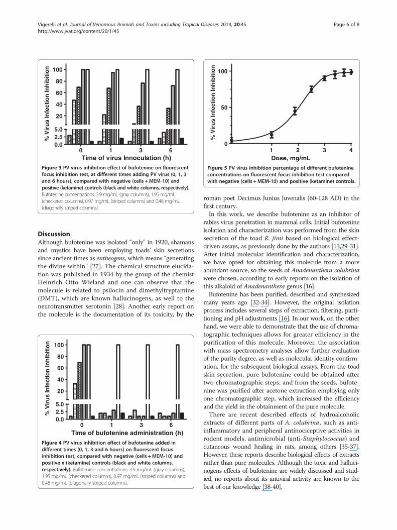
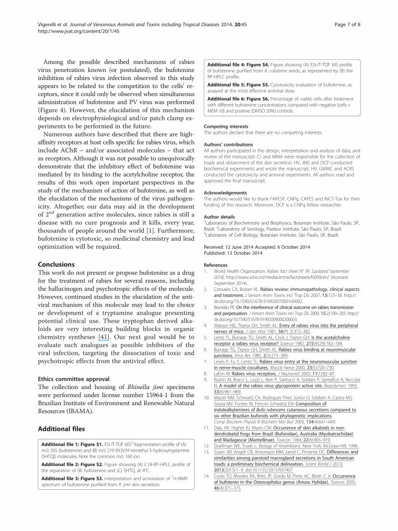
On the other hand, when bufotenine was administeredover the time, the molecule was unable to inhibit theinfection, regardless of the tested concentration, fromone hour onwards (Figure 4). After testing differentbufotenine concentrations in the most effective condi-tions used in the time course study (time 0), it waspossible to determine the IC50 value of (1.57 ± 0.03)mg.mL-1 (Figure 5).
0 1 3 60.02.55.0
20
40
60
80
100
Time of virus Innoculation (h)
% V
iru
s In
fect
ion
Inh
ibiti
on
Figure 3 PV virus inhibition effect of bufotenine on fluorescentfocus inhibition test, at different times adding PV virus (0, 1, 3and 6 hours), compared with negative (cells +MEM-10) andpositive (ketamine) controls (black and white columns, respectively).Bufotenine concentrations: 3.9 mg/mL (gray columns), 1.95 mg/mL(checkered columns), 0.97 mg/mL (striped columns) and 0.48 mg/mL(diagonally striped columns).
1 2 3 40
50
100
Dose, mg/mL
% V
iru
s In
fect
ion
Inh
ibit
ion
Figure 5 PV virus inhibition percentage of different bufotenineconcentrations on fluorescent focus inhibition test comparedwith negative (cells + MEM-10) and positive (ketamine) controls.
Vigerelli et al. Journal of Venomous Animals and Toxins including Tropical Diseases 2014, 20:45 Page 6 of 8http://www.jvat.org/content/20/1/45
DiscussionAlthough bufotenine was isolated “only” in 1920, shamansand mystics have been employing toads’ skin secretionssince ancient times as entheogens, which means “generatingthe divine within” [27]. The chemical structure elucida-tion was published in 1934 by the group of the chemistHeinrich Otto Wieland and one can observe that themolecule is related to psilocin and dimethyltryptamine(DMT), which are known hallucinogens, as well to theneurotransmitter serotonin [28]. Another early report onthe molecule is the documentation of its toxicity, by the
0 1 3 60.02.55.0
20
40
60
80
100
Time of bufotenine administration (h)
% V
iru
s In
fect
ion
Inh
ibiti
on
Figure 4 PV virus inhibition effect of bufotenine added indifferent times (0, 1, 3 and 6 hours) on fluorescent focusinhibition test, compared with negative (cells +MEM-10) andpositive x (ketamine) controls (black and white columns,respectively). Bufotenine concentrations: 3.9 mg/mL (gray columns),1.95 mg/mL (checkered columns), 0.97 mg/mL (striped columns) and0.48 mg/mL (diagonally striped columns).
roman poet Decimus Junius Juvenalis (60-128 AD) in thefirst century.In this work, we describe bufotenine as an inhibitor of
rabies virus penetration in mammal cells. Initial bufotenineisolation and characterization was performed from the skinsecretion of the toad R. jimi based on biological effect-driven assays, as previously done by the authors [13,29-31].After initial molecular identification and characterization,we have opted for obtaining this molecule from a moreabundant source, so the seeds of Anadenanthera colubrinawere chosen, according to early reports on the isolation ofthis alkaloid of Anadenanthera genus [16].Bufotenine has been purified, described and synthesized
many years ago [32-34]. However, the original isolationprocess includes several steps of extraction, filtering, parti-tioning and pH adjustments [16]. In our work, on the otherhand, we were able to demonstrate that the use of chroma-tographic techniques allows for greater efficiency in thepurification of this molecule. Moreover, the associationwith mass spectrometry analyses allow further evaluationof the purity degree, as well as molecular identity confirm-ation, for the subsequent biological assays. From the toadskin secretion, pure bufotenine could be obtained aftertwo chromatographic steps, and from the seeds, bufote-nine was purified after acetone extraction employing onlyone chromatographic step, which increased the efficiencyand the yield in the obtainment of the pure molecule.There are recent described effects of hydroalcoholic
extracts of different parts of A. colubrina, such as anti-inflammatory and peripheral antinociceptive activities inrodent models, antimicrobial (anti-Staphylococcus) andcutaneous wound healing in rats, among others [35-37].However, these reports describe biological effects of extractsrather than pure molecules. Although the toxic and halluci-nogens effects of bufotenine are widely discussed and stud-ied, no reports about its antiviral activity are known to thebest of our knowledge [38-40].
Vigerelli et al. Journal of Venomous Animals and Toxins including Tropical Diseases 2014, 20:45 Page 7 of 8http://www.jvat.org/content/20/1/45
Among the possible described mechanisms of rabiesvirus penetration known (or postulated), the bufotenineinhibition of rabies virus infection observed in this studyappears to be related to the competition to the cells’ re-ceptors, since it could only be observed when simultaneousadministration of bufotenine and PV virus was performed(Figure 4). However, the elucidation of this mechanismdepends on electrophysiological and/or patch clamp ex-periments to be performed in the future.Numerous authors have described that there are high-
affinity receptors at host cells specific for rabies virus, whichinclude AChR – and/or associated molecules – that actas receptors. Although it was not possible to unequivocallydemonstrate that the inhibitory effect of bufotenine wasmediated by its binding to the acetylcholine receptor, theresults of this work open important perspectives in thestudy of the mechanism of action of bufotenine, as well asthe elucidation of the mechanisms of the virus pathogen-icity. Altogether, our data may aid in the developmentof 2nd generation active molecules, since rabies is still adisease with no cure prognosis and it kills, every year,thousands of people around the world [1]. Furthermore,bufotenine is cytotoxic, so medicinal chemistry and leadoptimization will be required.
ConclusionsThis work do not present or propose bufotenine as a drugfor the treatment of rabies for several reasons, includingthe hallucinogen and psychotropic effects of the molecule.However, continued studies in the elucidation of the anti-viral mechanism of this molecule may lead to the choiceor development of a tryptamine analogue presentingpotential clinical use. These tryptophan derived alka-loids are very interesting building blocks in organicchemistry syntheses [41]. Our next goal would be toevaluate such analogues as possible inhibitors of theviral infection, targeting the dissociation of toxic andpsychotropic effects from the antiviral effect.
Ethics committee approvalThe collection and housing of Rhinella jimi specimenswere performed under license number 15964-1 from theBrazilian Institute of Environment and Renewable NaturalResources (IBAMA).
Additional files
Additional file 1: Figure S1. ESI-IT-TOF MS2 fragmentation profile of (A)m/z 205 (bufotenine) and (B) m/z 219 (N’,N’,N’-trimethyl 5-hydroxytryptamine[5HTQ]) molecules. Note the common m/z 160 ion.
Additional file 2: Figure S2. Figure showing (A) C18-RP-HPLC profile ofthe separation of (B) bufotenine and (C) 5HTQ, at 4°C.
Additional file 3: Figure S3. Interpretation and annotation of 1H-NMRspectrum of bufotenine purified from R. jimi skin secretion.
Additional file 4: Figure S4. Figure showing (A) ESI-IT-TOF MS profileof bufotenine purified from A. colubrine seeds, as represented by (B) theRP-HPLC profile.
Additional file 5: Figure S5. Cytotoxicity evaluation of bufotenine, asassayed at the most effective antiviral dose.
Additional file 6: Figure S6. Percentage of viable cells after treatmentwith different bufotenine concentrations compared with negative (cells +MEM-10) and positive (DMSO 20%) controls.
Competing interestsThe authors declare that there are no competing interests.
Authors’ contributionsAll authors participated in the design, interpretation and analysis of data, andreview of the manuscript. CJ and MMA were responsible for the collection oftoads and obtainment of the skin secretion. HV, JMS and DCP conductedbiochemical experiments and wrote the manuscript. HV, GMMC and ACRSconducted the cytotoxicity and antiviral experiments. All authors read andapproved the final manuscript.
AcknowledgementsThe authors would like to thank FAPESP, CNPq, CAPES and INCT-Tox for theirfunding of this research. Moreover, DCP is a CNPq fellow researcher.
Author details1Laboratory of Biochemistry and Biophysics, Butantan Institute, São Paulo, SP,Brazil. 2Laboratory of Serology, Pasteur Institute, São Paulo, SP, Brazil.3Laboratory of Cell Biology, Butantan Institute, São Paulo, SP, Brazil.
Received: 12 June 2014 Accepted: 6 October 2014Published: 13 October 2014
References1. World Health Organization: Rabies fact sheet N° 99. [updated September
2014]. http://www.who.int/mediacentre/factsheets/fs099/en/ (Acessed:September 2014).
2. Consales CA, Bolzan VL: Rabies review: immunopathology, clinical aspectsand treatment. J Venom Anim Toxins incl Trop Dis 2007, 13(1):5–38. http://dx.doi.org/10.1590/S1678-91992007000100002.
3. Brandão PE: On the interference of clinical outcome on rabies transmissionand perpetuation. J Venom Anim Toxins incl Trop Dis 2009, 15(2):190–203. http://dx.doi.org/10.1590/S1678-91992009000200003.
4. Watson HD, Tignor GH, Smith AL: Entry of rabies virus into the peripheralnerves of mice. J Gen Virol 1981, 56(Pt 2):372–382.
5. Lentz TL, Burrage TG, Smith AL, Crick J, Tignor GH: Is the acetylcholinereceptor a rabies virus receptor? Science 1982, 215(4529):182–184.
6. Burrage TG, Tignor GH, Smith AL: Rabies virus binding at neuromuscularjunctions. Virus Res 1985, 2(3):273–389.
7. Lewis P, Fu Y, Lentz TL: Rabies virus entry at the neuromuscular junctionin nerve-muscle cocultures. Muscle Nerve 2000, 23(5):720–730.
8. Lafon M: Rabies virus receptors. J Neurovirol 2005, 11(1):82–87.9. Rustici M, Bracci L, Lozzi L, Neri P, Santucci A, Soldani P, Spreafico A, Niccolai
N: A model of the rabies virus glycoprotein active site. Biopolymers 1993,33(6):961–969.
10. Maciel NM, Schwartz CA, Rodrigues Pires Júnior O, Sebben A, Castro MS,Sousa MV, Fontes W, Ferroni Schwartz EN: Composition ofindolealkylamines of Bufo rubescens cutaneous secretions compared tosix other Brazilian bufonids with phylogenetic implications.Comp Biochem Physiol B Biochem Mol Biol 2003, 134(4):641–649.
11. Daly JW, Highet RJ, Myers CW: Occurrence of skin alkaloids in non-dendrobatid frogs from Brazil (Bufonidae), Australia (Myobatrachidae)and Madagascar (Mantellinae). Toxicon 1984, 22(6):905–919.
12. Duellman WE, Trueb L: Biology of Amphibians. New York: McGraw-Hill; 1996.13. Sciani JM, Angeli CB, Antoniazzi MM, Jared C, Pimenta DC: Differences and
similarities among parotoid macrogland secretions in South Americantoads: a preliminary biochemical delineation. Scient World J 2013,2013(2013):1–9. doi:10.1155/2013/937407.
14. Costa TO, Morales RA, Brito JP, Gordo M, Pinto AC, Blosh C Jr: Occurrenceof bufotenin in the Osteocephalus genus (Anura: Hylidae). Toxicon 2005,46(4):371–375.

Vigerelli et al. Journal of Venomous Animals and Toxins including Tropical Diseases 2014, 20:45 Page 8 of 8http://www.jvat.org/content/20/1/45
15. Smith TA: Tryptamine and related compounds in plants. Phytochemistry1977, 16(2):171–175.
16. Stromberg VL: The isolation of bufotenine from Piptadenia peregrina.J Am Chem Soc 1954, 76(6):1707.
17. Batista AM, Cruz PS, Almeida E, Costa AEB, Scheffer KC, Chaves LB, Silva ACR,Medeiros Caporale GM: Infection of BHK-21 cells cultivated in stationarymonolayers by PV and CVS strains. Bol Epidemiol Paul (Online) 2009, 6(71):4–11.
18. Takeuchi H, Baba M, Shigeta S: An application of tetrazolium (MTT)colorimetric assay for the screening of anti-herpes simplex virus compounds.J Virol Methods 1991, 33(1–2):61–71.
19. Mosmann T: Rapid colorimetric assay for cellular growth and survival:application to proliferation and cytotoxicity assays. J Immunol Methods1983, 65(1–2):55–63.
20. Flores EF, Editor: Vírus da raiva e lyssavirus relacionados. In VirologiaVeterinária. Rio Grande do Sul: UFSM; 2007:700–711.
21. Favoretto SR, Carrieri ML, Tino MS, Zanetti CR, Pereira AO: Simplifiedfluorescent inhibition microtest for the titration of rabies neutralizingantibodies. Rev Inst Med Trop São Paulo 1993, 35(2):171–175.
22. Lockhart BP, Tordo N, Tsiang H: Inhibition of rabies virus transcription inrat cortical neurons with the dissociative anesthetic ketamine. AntimicrobAgents Chemother 1992, 36(8):1750–1755.
23. Smith JS, Yager PA, Baer GM: A rapid reproducible test for determiningrabies neutralizing antibody. Bull World Health Organ 1973, 48(5):535–541.
24. Chaves LB, Mazutti ALC, Medeiros Caporale GM, Scheffer KC, Silva ACR:Comparision of RFFIT performed in lab-tek® and in 96-well microtitreplates. In RITA XVII. Annales of the XVII International Conference on Rabies inthe Americas: 15-20 octuber, 2006; Brasília, Brazil. Brasília: Ministério da Saúde;2006:161.
25. Medeiros Caporale GM, Rodrigues Da Silva A, De C, Peixoto ZM, Chaves LB,Carrieri ML, Vassao RC: First production of fluorescent anti-ribonucleoproteins conjugate for diagnostic of rabies in Brazil.J Clin Lab Anal 2009, 23(1):7–13.
26. McClean S, Robinson RC, Shaw C, Smyth WF: Characterisation anddetermination of indole alkaloids in frog-skin secretions by electrosprayionisation ion trap mass spectrometry. Rapid Commun Mass Spectrom2002, 16(5):346–354.
27. Handovsky H: Ein Alkaloid im Gifte von Bufo vulgaris. Arch Exp PatholPharmakol 1920, 86(1–2):138–158.
28. Blom JD: A Dictionary of Hallucinations. New York: Springer-Verlag; 2010:79–80.29. Prates I, Antoniazzi MM, Sciani JM, Pimenta DC, Toledo LF, Haddad CF, Jared C:
Skin glands, poison and mimicry in dendrobatid and leptodactylidamphibians. J Morphol 2012, 273(3):279–290.
30. Sousa JC, Berto RF, Gois EA, Fontenele-Cardi NC, Honório JE Jr, Konno K,Richardson M, Rocha MF, Camargo AA, Pimenta DC, Cardi BA, Carvalho KM:Leptoglycin: a new Glycine/Leucine-rich antimicrobial peptide isolatedfrom the skin secretion of the South American frog Leptodactyluspentadactylus (Leptodactylidae). Toxicon 2009, 54(1):23–32.
31. Tempone AG, Pimenta DC, Lebrun I, Sartorelli P, Taniwaki NN, De AndradeHF, Jr AMM, Jared C: Antileishmanial and antitrypanosomal activity ofbufadienolides isolated from the toad Rhinella jimi parotoid macroglandsecretion. Toxicon 2008, 52(1):13–21.
32. Evarts EV, Landau W, Freygang W Jr, Marshall WH: Some effects of lysergicacid diethylamide and bufotenine on electrical activity in the cat’s visualsystem. Am J Physiol 1955, 182:594–598.
33. Harley-Mason J, Jackson AH: A new synthesis of bufotenine.Chem Ind 1952, 39:954.
34. Wieland H, Konz W, Mittasch H: Die konstitution von Bufotenin undBufotenidin. Über kröten-Giftstoffc. VII. Justus Liebigs Ann Chem 1934,513(1):1–25.
35. Santos JS, Marinho RR, Ekundi-Valentim E, Rodrigues L, Yamamoto MH, TeixeiraSA, Muscara MN, Costa SK, Thomazzi SM: Beneficial effects of Anadenantheracolubrina (Vell.) Brenan extract on the inflmmatory and nociceptiveresponses in rodent models. J Ethnopharmacol 2013, 148(1):218–222.
36. Da Silva LC, Sandes JM, Paiva MM, De Araújo JM, De Figueiredo RC, Da SilvaMV, Correia MT: Anti-Staphylococcus aureus action of three Caatinga fruitsevaluated by electron microscopy. Nat Prod Res 2012, 27(16):1492–1496.doi:10.1080/14786419.2012.722090.
37. Pessoa WS, Estevão LRM, Simões RS, Barros MEG, De Mendonça F S, Baratella-Evêncio L, Evêncio-Neto J: Effects of angico extract (Anadenanthera colubrinavar. cebil) in cutaneous wound healing in rats. Acta Cir Bras 2012,27(10):655–670.
38. Fabing HD, Hawkins Jr: Intravenous bufotenine injection in the humanbeing. Science 1956, 123(3203):886–887.
39. Raymond-Hamet M: Sur les effects tenseurs et respiratoires de labufoténine basique dissolute dans le diméthyl-acétonyl-carbinol.Seances Acad Sci 1944, 218:54–56.
40. Raymond-Hamet M: Sur les effets vasculaires de la bufoténine introduitedans la circulation générale. CR Hebd Seances Acad Sci 1942, 214:506–508.Available at: http://gallica.bnf.fr/ark:/12148/bpt6k3166b/f506.
41. Stefani HA, Vasconcelos SNS, Souza FB, Manarin F, Zukerman-Sphector J:One-pot three-component synthesis of indole-3-glyoxyl derivatives andindole-3-glyoxyl triazoles. Tetrahedron Lett 2013, 54(43):5821–5825.
doi:10.1186/1678-9199-20-45Cite this article as: Vigerelli et al.: Bufotenine is able to block rabies virusinfection in BHK-21 cells. Journal of Venomous Animals and Toxins includingTropical Diseases 2014 20:45.
Submit your next manuscript to BioMed Centraland take full advantage of:
• Convenient online submission
• Thorough peer review
• No space constraints or color figure charges
• Immediate publication on acceptance
• Inclusion in PubMed, CAS, Scopus and Google Scholar
• Research which is freely available for redistribution
Submit your manuscript at www.biomedcentral.com/submit
Related Documents