BSCB Newsletter AUTUMN 2011 BRITISH SOCIETY FOR CELL BIOLOGY Super resolution microscopy: are there limits? Looking ahead to the Francis Crick Institute Image competition winners

Welcome message from author
This document is posted to help you gain knowledge. Please leave a comment to let me know what you think about it! Share it to your friends and learn new things together.
Transcript

BSCB NewsletterAUTUMN 2011
BRITISH SOCIETY FOR CELL BIOLOGY
Super resolution microscopy: are there limits?Looking ahead to the Francis Crick Institute Image competition winners

Somewhat later than usual, this Autumn issue of theBSCB newsletter should arrive on your desks in timefor the Christmas party season. I don't know aboutyou, but this term for me in University-land has beenbonkers. What with the mock REF exercise, andfiguring out how to improve NSS scores, andpreparation for the era of £9000 student fees, therehas been practically no time to breathe. So, time nowto prepare for sitting by the fire, putting up your feetand reading the latest BSCB newsletter.
Inside you will find feature articles on the developingFrancis Crick Institute in central London, the latest onSuper Resolution Microscopy, and the BSCB ImageCompetition winning images are displayed on page 5.Matthew Ashenden's first prize image – mouse retinalvasculature – adorns the front cover of the newsletter.Our BSCB President – Jordon Raff – presents his firstreport and the BSCB Summer Vacation Studentshipsare once again advertised on page 3. These offerfinancial support for high calibre undergraduatestudents who would like to get research experience incell biology during their summer holidays.
In addition, read the meeting reports of some of the
PhD students and Postdocs who have received HonorFell/Company of Biologist Travel Awards to attendmeetings in far off places such as Canada and Mexico.Also, Kimberley Byron, a PhD student at the MRC-LMCB, UCL, introduces herself as our new PhDstudent representative.
I would like to encourage you all to providenominations for committee members, and/orsuggestions for candidates worthy of the Hooke Medal2013. Holger Gerhardt is announced here on page 2as the 2012 winner of the Hooke Medal.Congratulations to Holger and a very merry Christmasto you all.
Finally, it is with great sadness that we note thepassing of Leonard 'Sammy' Franks on the 11thNovember 2011. Sammy was the first secretary ofthe BSCB when it was founded in 1965. A fullobituary will be in the Spring 2012 issue of thenewsletter.
The Editor: Kate NobesUniversity of [email protected]
AUTUMN 2011
CON
TENTS
BSCB NewsletterNews 2Features 5Book Review 11Meeting Reports 13Postdocs and PhDs 27Forthcoming Meetings 30Society Business 33
Editorial
The cover image is the winningentry in the BSCB 2011 ImageCompetition. Matthew Ashenden'simage shows the vasculature of themouse retina stained for collagen IV.Matthew is a PhD student in thelaboratory of Clare Isacke at theBreakthrough Breast Cancer Centre,Institute of Cancer Research inLondon
Newsletter editor: Kate Nobes Production: Giles Newton Website: www.bscb.org Printer: Hobbs1

We are pleased to announcethat this year’s Hooke Medalwinner is Holger Gerhardtfrom the CRUK's LondonResearch Institute.
The Hooke Medal is awardedeach year to an outstandingUK cell biologist who hasbeen working as anindependent research scientistfor less than 10 years.Previous winners haveincluded scientists such asAnne Ridley, MatthewFreeman and last yearswinner, Alex Gould.
Holger Gerhardt has been anindependent researcher since2004. He currently runs theVascular Biology Laboratory atCRUK’s London ResearchInstitute. He and his team areinvestigating how blood vesselsgrow during normaldevelopment and in disease,
providing tissues with theoxygen and nutrients they needas fuel. Understanding howthis process works - and how itcan be either improved or shutdown – is vital for researchinto cancer. Holger’s lab haspublished a number ofimportant papers dealing withaspects of angiogenesis andvascular development and hiswork has been at the forefrontof developing innovativestrategies in this area ofresearch. His discovery andconceptualization of endothelial“tip cells” has changed theway vascular biologists look atblood vessels, and continues todeliver new insights intoendothelial cell biology.
Holger will be presented withhis medal and will give theHooke Medal lecture at theBSCB/BSDB Spring meeting inApril next year.
Hooke Medal Winner 2012 – Holger Gerhardt
The 2011 Hooke medal wasawarded to Alex Gould at theBSCB Spring Meeting inCanterbury.
Alex started his scientificcareer as a PhD student atCambridge University, in thelaboratory of Rob White.Subsequently, he was a Beitand MRC postdoctoral fellowat NIMR with Robb Krumlaufbefore becoming a programmeleader at NIMR in 1998.
On presenting the HookeMedal to Alex, Clare Isacke,the BSCB President, describedAlex as a worthy winner of theaward who has made anumber of seminalcontributions to the field ofdevelopmental physiologyusing Drosophila as his modelsystem.
In his Medal lecture, Alexdescribed some of the recentwork from his laboratory,which has included thediscovery and dissection of themolecular mechanismsinvolved in regulating thetiming of cell cycle exit in theDrosophila central nervoussystem. In particular, hediscussed his work on lipidmetabolism in Drosophila,which has led to the idea thatdietary nutrients and remoteorgans, as well as local niches,are key regulators of transitionsin stem-cell behaviour.
The BSCB invites nominationsfor the Hooke Medal 2013from any member of thesociety. If you wish tonominate anyone, pleasecontact the BSCB Secretaryproviding a brief supportingstatement.
The Hooke Medal 2011 Presentation
News
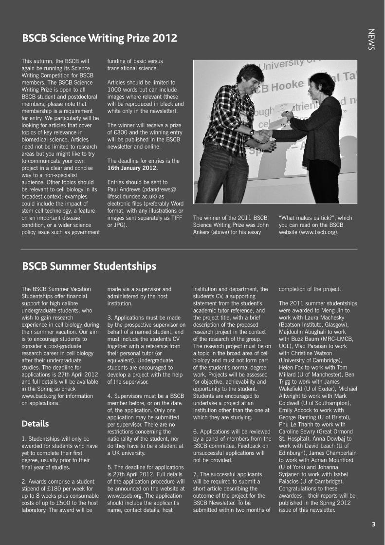
This autumn, the BSCB willagain be running its ScienceWriting Competition for BSCBmembers. The BSCB ScienceWriting Prize is open to allBSCB student and postdoctoralmembers; please note thatmembership is a requirementfor entry. We particularly will belooking for articles that covertopics of key relevance inbiomedical science. Articlesneed not be limited to researchareas but you might like to tryto communicate your ownproject in a clear and conciseway to a non-specialistaudience. Other topics shouldbe relevant to cell biology in itsbroadest context; examplescould include the impact ofstem cell technology, a featureon an important diseasecondition, or a wider sciencepolicy issue such as government
funding of basic versustranslational science.
Articles should be limited to1000 words but can includeimages where relevant (thesewill be reproduced in black andwhite only in the newsletter).
The winner will receive a prizeof £300 and the winning entrywill be published in the BSCBnewsletter and online.
The deadline for entries is the16th January 2012.
Entries should be sent to Paul Andrews ([email protected]) aselectronic files (preferably Wordformat, with any illustrations orimages sent separately as TIFFor JPG).
The winner of the 2011 BSCBScience Writing Prize was JohnAnkers (above) for his essay
“What makes us tick?”, whichyou can read on the BSCBwebsite (www.bscb.org).
The BSCB Summer VacationStudentships offer financialsupport for high calibreundergraduate students, whowish to gain researchexperience in cell biology duringtheir summer vacation. Our aimis to encourage students toconsider a post-graduateresearch career in cell biologyafter their undergraduatestudies. The deadline forapplications is 27th April 2012and full details will be availablein the Spring so checkwww.bscb.org for informationon applications.
Details
1. Studentships will only beawarded for students who haveyet to complete their firstdegree, usually prior to theirfinal year of studies.
2. Awards comprise a studentstipend of £180 per week forup to 8 weeks plus consumablecosts of up to £500 to the hostlaboratory. The award will be
made via a supervisor andadministered by the hostinstitution.
3. Applications must be madeby the prospective supervisor onbehalf of a named student, andmust include the student's CVtogether with a reference fromtheir personal tutor (orequivalent). Undergraduatestudents are encouraged todevelop a project with the helpof the supervisor.
4. Supervisors must be a BSCBmember before, or on the dateof, the application. Only oneapplication may be submittedper supervisor. There are norestrictions concerning thenationality of the student, nordo they have to be a student ata UK university.
5. The deadline for applicationsis 27th April 2012. Full detailsof the application procedure willbe announced on the website atwww.bscb.org. The applicationshould include the applicant’sname, contact details, host
institution and department, thestudent's CV, a supportingstatement from the student’sacademic tutor reference, andthe project title, with a briefdescription of the proposedresearch project in the contextof the research of the group.The research project must be ona topic in the broad area of cellbiology and must not form partof the student’s normal degreework. Projects will be assessedfor objective, achievability andopportunity to the student.Students are encouraged toundertake a project at aninstitution other than the one atwhich they are studying.
6. Applications will be reviewedby a panel of members from theBSCB committee. Feedback onunsuccessful applications willnot be provided.
7. The successful applicantswill be required to submit ashort article describing theoutcome of the project for theBSCB Newsletter. To besubmitted within two months of
completion of the project.
The 2011 summer studentshipswere awarded to Meng Jin towork with Laura Machesky(Beatson Institute, Glasgow),Majdoulin Abughali to workwith Buzz Baum (MRC-LMCB,UCL), Vlad Paraoan to workwith Christine Watson(University of Cambridge),Helen Fox to work with TomMillard (U of Manchester), BenTrigg to work with JamesWakefield (U of Exeter), MichaelAllwright to work with MarkColdwell (U of Southampton),Emily Adcock to work withGeorge Banting (U of Bristol),Phu Le Thanh to work withCaroline Sewry (Great OrmondSt. Hospital), Anna Dowbaj towork with David Leach (U ofEdinburgh), James Chamberlainto work with Adrian Mountford(U of York) and JohannaSyrjanen to work with IsabelPalacios (U of Cambridge).Congratulations to theseawardees – their reports will bepublished in the Spring 2012issue of this newsletter.
BSCB Science Writing Prize 2012
BSCB Summer Studentships
NEW
S
3

NEW
S
Three questions immediatelycame into my mind when ClaireIsacke asked if I would considertaking over from her when sheretired as President of the BSCBthis year. The first was howmuch work it would be to keepthe BSCB running as the well-oiled machine it has been underClaire’s and Liz Smythe’s (theretiring BSCB Secretary) expertguidance. Fortunately, I hadbeen on the BSCB Committeebefore, and so I knew that Iwouldn’t have to do that muchas the very dedicatedCommittee members do a lot ofthe real work. The secondquestion was whether I wouldenjoy doing it. I had reallyenjoyed my time on theCommittee, and so I was prettysure that I would enjoy beingPresident at least as much. Thefinal and most importantquestion was whether I wouldbe any good at the job. Here,only time will tell, but I feelgreatly honoured to have beengiven the chance to find out.
These are very challengingtimes, as we are almostcertainly entering an era ofprolonged financial stress.Besides organizing outstandingmeetings and doling out moneyto students and postdocs toattend meetings or to work in alab for the summer, are thereother useful things that theBSCB can do to help scienceand scientists? For example,how much can the BSCBrealistically hope to influenceGovernment or universityscience policy? Although it maybe a pipe dream to imagine that
it could have any influence atall, I have participated in pro-science political campaigns inthe past and have been amazedat how much can be achieved.Save British Science was veryeffective in the Thatcher years,and the Science is Vitalcampaign has been a veryencouraging recent example.
There was a truly inspiringsession on science activismorganized by BSCB’s PhDstudent representative, JayStone, at the recentBSCB/BSDB meeting inWarwick, which, sadly (and alittle ironically), was quite poorlyattended. Jenny Rohn, (aninitiator of the Science is VitalCampaign) and Rose Wu (fromSense about Science) spoke atthe session, and I hope theyboth will contribute to futureNewsletters and our meetings tohelp us explore what the BSCBmight do in the future in thearea of science politics.
One aspect of science policythat is of great concern to allBSCB members is the fundingof biomedical research by theResearch Councils and majorcharities. These bodiescontinually explore differentways to distribute their funds,but the consequences of theirpolicies are often hard topredict, and, in some cases,they can be devastating forscientists on the front line. It isespecially discouraging thesedays to see so many excellentscientists, including those whohave been running their lab formany years, now struggling to
support their research. Thefunding streams they havepreviously counted on are dryingup, as funding agencies refocustheir spending priorities in waysthat exclude many scientists.Although it is perhaps hard tosee a way that the BSCB canhelp here, we should bechampioning the long held viewof most scientists that the beststrategy for economic success issimply to back the best andbrightest - no matter what theywork on. Trying to force peopleto work only on problems thatothers have identified as beingin the national interest is a triedand trusted recipe for fundingmediocre research. We need tothink of innovative ways ofgetting this message across.
But the central role of the BSCBis surely to improve cell biologyin the UK by ensuring that cellbiologists here can regularlyhear a broad range of world-class scientists talk about theirresearch, and have theopportunity to talk about theirown research to an internationalaudience. To this end, the BSCBhas always organized a largeannual spring meeting (often inassociation with the BSDB) anda smaller, more focused,autumn meeting. It is a realconcern that attendance atthese meetings has been slowlydeclining for the past few years.We must reverse this trend. Wemade a real effort at the lastspring meeting to talk toattendees to try to understandthe reason for the decline.Unsurprisingly, we identifiedseveral factors, including the
large number of other meetings,the cost of attending ourmeetings, the poor attendanceof many of the most senior UKcell biologists, and theattractiveness of the competingannual ASCB meetings. Wehave recently held talks with theBSDB executive and will shortlyannounce some changes thatwe hope will revitalize thespring meeting. The goal mustbe to make the spring meeting a“must attend” meeting for allUK cell biologists, young andold, and we will be workinghard to ensure that you simplycan’t afford to miss it.
I’m looking forward to workingwith BSCB members on someof these issues over the next fewyears. Please feel free to contactme, or any of our committeemembers or Ambassadors, ifyou have any ideas on what weshould be doing and how weshould be doing it.
Jordan Raff, President
4
President’s report
New PhD repHello! My name is Kimberleyand I am delighted to be yournew PhD studentrepresentative. I have juststarted my 3rd year of a 4-yearrotational PhD program at theMRC Laboratory for MolecularCell Biology, in London where Iam part of the Nurrish group,researching neurotransmission
in C. elegans.
My role is to make sure thatthe student community isrepresented within the BSCB.If you have issues that youwish me to address, or ideasfor how the BSCB can betterserve your needs, please doget in touch and I will raisethem at the next committeemeeting. Don't forget that there
is also a Facebook page thatcan be used to stay informedof BSCB events andcompetitions and hopefully inthe near future we will alsohave a Twitter account. I lookforward to meeting many ofyou at the BSCB Springmeeting inWarwick,
Kimberley [email protected]

5
FEATURES
BSCB Image CompetitionWinners 2011
Thank you to all the entrants for the 2011 Imagecompetition for sending in your work, which were
interesting, visually and technically. After the entrieswere anonymised and independently judged by fourcell biologists, several images caught the eye of all thejudges and stood out from the rest. The top five werevery close and the three top scoring images are allgreat. What makes these images outstanding?Predominantly it is quality of the image – thesharpness of focus, quality of staining, samplepreparation and image acquisition. But it is more thanthat because there were technically competent imagesthat didn’t quite make the grade – aesthetic qualitiessuch as composition and colour choice played a partin giving the winning images the edge.
So it gives us great pleasure to be able to announceour 2011 winners:
First prize goes to Matthew Ashenden based atBreakthrough Breast Cancer Research Centre at theInstitute of Cancer Research in London. His beautifuland graphic image showing the vasculature of themouse retina is gracing the cover of this BSCBNewsletter.
Matthew’s image shows Collagen IV staining whichreveals the vasculature of the mouse retina. Initiallyduring development, the superficial plexus (green)expands radially from the centre to cover the retina.Vessels then sprout from the superficial plexus anddescend to form the intermediate (blue) and deepplexus (red).
Second prize goes to Keiran Boyle in theDepartment of Cell & Developmental Biology atUniversity College London. His wonderful image,which looks like an aerial view of a road network atnight, shows a cultured hippocampal neuron in theearly stages of synaptogenesis. The morphology of theneuron is visualised by staining with an antibodyagainst III-tubulin. Incoming axons form synapses ontothe neuron, which are stained with antibodies for thepresynaptic proteins VAMP2 and Synapsin-1.
Third prize goes to Michael Bright from ImperialCollege London for his beautiful space-age scanningelectron microscope image.
Michael’s image shows COS-7 cells ectopicallyexpressing the Fcγ-receptor performing phagocytosison beads opsonised with Immunoglobulin G. His false-colour scanning electron micrograph shows filopodiaand pseudopodia projecting around the beads, whichwill subsequently be fully engulfed. The beads arethree micrometres in diameter.
Please take a look at our prize-winning entries intheir full-colour glory on the BSCB website. Manythanks to all those that entered and if you didn’t getselected this time, or are inspired by what you see,please start collecting some images for next year’scompetition. Happy snapping!
Paul Andrews
Above left: 1st Prize –Mouse Retinal Vasculature(© Matthew Ashenden).Above centre: 2nd Prize –Hippocampal Neuron (© Keiran Boyle)Above right: 3rd Prize –Bead Phagocytosis (© Michael Bright)
6
FEAT
URE
S
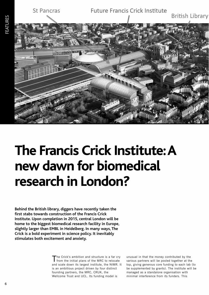
The Crick’s ambition and structure is a far cryfrom the initial plans of the MRC to relocate
and scale down its largest institute, the NIMR. Itis an ambitious project driven by four distinctfounding partners, the MRC, CRUK, theWellcome Trust and UCL. Its funding model is
unusual in that the money contributed by thevarious partners will be pooled together at thetop, giving generous core funding to each lab (tobe supplemented by grants). The institute will bemanaged as a standalone organisation withminimal interference from its funders. This
The Francis Crick Institute: Anew dawn for biomedicalresearch in London?
Behind the British library, diggers have recently taken thefirst stabs towards construction of the Francis CrickInstitute. Upon completion in 2015, central London will behome to the biggest biomedical research facility in Europe,slightly larger than EMBL in Heidelberg. In many ways, TheCrick is a bold experiment in science policy. It inevitablystimulates both excitement and anxiety.

7
arrangement is expected to foster a spirit ofcollaboration that is difficult to achieve in placeswhere funding is balkanised. Indeed the main aim ofthe institute is to foster exchanges between diversedisciplines and thus create unexpected connectionsand research directions. Although translation is animportant aspect of The Crick’s mission, it is clearthat basic research, including cell biology, willfeature heavily in its portfolio. The unashamedambition of Paul Nurse, the director and chiefexecutive, is to make The Crick one of the mostinnovative interdisciplinary research institutes in theworld. The size of the institute and its location arecentral to this aim. The large size is necessary tobring together the diverse disciplines, includingmaths, physics and chemistry that are required totackle modern biomedical problems. Thecosmopolitan nature of London will be an attractionfor scientists from around the world and thetransport hubs around The Crick will facilitateinteractions with scientists from the rest of the UKand beyond.
The potential of The Crick as a researchpowerhouse is clearly generating excitement in manyquarters. However, The Crick is also cause foranxiety at various levels. The institute will cost 600million pounds to build and kit out. One mightwonder whether it is right to spend that muchmoney at a time when research funding is gettingtight and when project grants are being discontinuedby the Wellcome Trust. The Crick’s managementwould argue that a portion of this money will comefrom new sources. Moreover, The Crick is committedto interact with and support research across the UK.Nevertheless, it will be important that individualscientists outside London become convinced thattheir own research will not suffer. There is also someanxiety among scientists currently working withininstitutes of the founding organisations, CRUK’sLondon Research Institute (LRI) and the MRC’s
National Institute for Medical Research (NIMR).Some worry that there might not be enough space tohouse everybody along with new hires and groupsfrom UCL, the Wellcome Trust and the two belatedpartners, Imperial College and Kings College. Theallocation of space is currently under discussion andthe exact composition of The Crick will begin to takeshape during the coming academic year. Theproposed career structure at The Crick has alsosparked a fierce debate. All groups will be given a6+6 years-and-then-you-are-out contract. Only afew senior scientists will be hired and notnecessarily from the junior ranks. There is no doubtthat renewal is important for the dynamism of anyinstitute but we will only find out over time whethera strict renewal policy will provide the stabilityneeded for long term risky research. Time will alsotell whether contracts of strict duration will be anissue for applicants who want to ensure geographicalstability for their families.
If funds were plentiful, no one would question thebenefits of spending new money to reorganise andrenew the research infrastructure in the Londonarea. However, in the current climate, questionsabout the need for the Francis Crick institute willprobably continue to be voiced for some time tocome. For scientists across the UK to accept thatThe Crick is a risk worth taking, they will need to beconvinced that it will not jeopardise, but insteadbenefit, their own research. Hopefully this will occurwhen The Crick reaches steady state and thefunding situation improves.
“If you don't risk anything you risk even more”Erica Jong
Further details can be found on www.crick.ac.uk/
Jean-Paul Vincent, MRC National Institute for Medical Research
FEATURES

8
FEAT
URE
S
Above: Figure 1.Bacteria expressingGFP-FtsZ protein (B.subtilis 2020(amyE::spc Pxyl-gfp-ftsZ) [3] examinedusing (a) conventionalwide field microscopyand (b) superresolution microscopy(N-SIM). Scale bar 5 µm. Images takenusing Nikon N-SIMsuper resolutionsystem, courtesy of D.Adams (Centre forBacterial Cell Biology,Newcastle University).
Right: Figure 2Line profile throughFtsZ band indicated inFigure 1.
Super Resolution Microscopy:are there limits?Every so often a new technique, approach or vision comes along thatchallenges accepted methods and dogma. One may argue that over thepast five years or so HD and now 3D TV have transformed our homeviewing experience. In a similar way, super resolution microscopy, althoughhaving been around for a similar, if not longer period of time, threatens toshake up light microscopy. Like HD TV, super resolution microscopy offersthe potential to visualize structures in greater detail but in this case, thebenefits are not realized simply by filling the image with more pixels.
The resolving power of the standard compoundmicroscope is limited by the wave-like nature of light
such that simply increasing the pixel density of the capturedimage has little effect on the resolving power of the system.Abbe’s principles dictate that even with ‘perfect’ optics, it isonly possible to resolve details half the wavelength of thestudied light. In practice, this means that the lateral (X-Y)resolution limit of GFP-labeled structures is at best around250 nm; axial (Z) resolution is approximately 500 nm.
Armed with novel fluorescent probes and innovativemethods with which to use and visualize them eg.structured illumination and deconvolution techniques,cell biologists and microscopists are pushingconventional light microscopy to its limits. There ishowever, a genuine requirement to probe structures,complexes and individual proteins beyond them.Naturally, this is where EM takes over but not allbiological systems are amenable to EM analysis; it ispractically impossible with EM to image specimens intheir unperturbed state and many EM techniquesthemselves introduce artifacts. Super resolutionmicroscopy promises to extend the resolving power oflight microscopy into that of EM and with it allow theobservation of cellular processes in a different light.Indeed, early adopters have reported the ability to
resolve structures half and in some cases, a tenth of thesize of that possible using conventional light microscopy.
So should we now disregard Abbe’s principles, hasthis diffraction limitation been broken? In a nutshell no,although Abbe I’m sure if he were alive, would have awry smile. Super resolution microscopy techniques havesuccessfully overcome the diffraction limitations either bytaking advantage of the way in which the incidentillumination interacts with the specimen or in othercases exploit the properties of the fluorescent label itself.Over the past decade or so a variety of ‘super resolution’methods have been developed and several have made itto market in partnership with microscope manufacturers.
(a) (b)

9
FEATURES
Right: Figure 3.Overview of SIM,STED and Pointillismsuper resolutionmicroscopy. Figureadapted fromSchermelleh et.al. [1].
References
1. Schermelleh L.,Heintzmann R., andLeonhardt H. (2010).A guide to super-resolution fluorescencemicroscopy. J Cell Biol190, 165-175.2. Toomre D, andBewersdorf J (2010).A new wave of cellularimaging. Annu RevCell Dev Biol 26,285-314.3. Stokes NR, et al.(2005). Novelinhibitors of bacterialcytokinesis identifiedby a cell-basedantibiotic screeningassay. J Biol Chem280, 39709-39715.
There are a number ofrecent, informative anddetailed reviews available[1],[2]. For brevity, I willconcentrate on threemajor approaches: SIM,STED and Pointillism(see figure 3).
Structured IlluminationMicroscopy (SIM) (figure3a, figure 2): Nikon (N-SIM), Zeiss (ELYRA S.1)and Applied PrecisionInc. (OMX) offer thissuper resolutionmicroscopy approach.The technique involvesprojecting a series ofsinusoidal ‘high freq-uency’ striped patterns(optical grating) onto thespecimen. Moiré fringescontaining information relating to the specimen’s subresolution structure develop when this pattern illuminatesfiner labeled structures of the sample. This information isextracted by image processing algorithms and a superresolution image is formed by combining multiple imagescollected from different grating orientations. With SIM,one can expect to roughly increase the resolving powerby a factor of two (~100 nm). As the technique is notreliant on the properties of the fluorescent probe anddoes not requires special sample preparation, it ispossible to image most fluorescent labels.
RESOLFT (Reversible Saturable OpticaLFluorescence Transition) describes a small number ofrelated approaches with which to bypass the diffractionlimitation. STED (STimulated Emission Depletion)Microscopy (figure 3b) and a related technique knownas Ground State Depletion (GSD) microscopy weredeveloped by Stephan Hell in collaboration with Leica;both are now commercially available. STED is theperfect example where novel optics and fluorescentprobe properties have combined to yield diffraction‘breaking’ results. In conventional point-scanningconfocal microscopy, photons in the excitation laserbeam (diffraction limited in size) cause electrons of thedye molecule to become excited from the ground state toa higher energy level. Within a few nanoseconds, beforethese electrons have chance to relax and emit a photon(the basis of fluorescence), a second red-shifteddoughnut-shaped laser beam centered on the sameexcitation spot, is applied. This second beam drivesexcited electrons, except for those located in the centerof the doughnut, back to their groundstate by stimulatingemission of a photon of the same wavelength. Thus,molecules located in the hole can to fluoresce normallywhereas those surrounding cannot. By increasing thepower of the depleting laser, the effective diameter of thehole is reduced and with it, the size of the spot fromwhich molecules are allowed to fluoresce. The result isa fourfold improvement in resolution (~60 nm) with theresults visible in ‘real time’.
Pointillism microscopy (figure 3c): PALM (PhotoActivation Localisation Microscopy), STORM (StochasticOptical Reconstruction Microscopy) and GSD microscopytechniques have been developed in collaboration withZeiss (ELYRA P.1), Nikon (N-STORM) and Leica (SRGSD), respectively. In a similar way to the paintings of
Seurat and other exponents of the pointillism technique,the resultant image is formed from a number ofindividual dots; in this case, each dot represent a singlefluorescing molecule. These approaches exploit theproperties of the fluorophore, in particular its ability tobe photoactivated, bleached or photoswitched. Theessence of the technique is to switch individualfluorescence molecules on and off and to image themusing a camera. The center point of each molecule canthen be calculated computationally and its locationrecorded; the process is repeated hundreds and in mostcases thousands of times to form the final image. Theresults themselves can be impressive; lateral resolutionsof ~20 nm have been claimed.
So just like waiting for a bus, you wait for one superresolution microscopy technique to arrive and threecome at once. But is super resolution a fad? Like theBetamax–VHS battle of the 80s, will one technologydominate over the other? For researchers, the mostcrucial questions are ‘can my sample be imaged in superresolution?’ and ‘what technique is the best?’ At present,there seems to be no clear-cut answers to thesequestions or to the imponderable one of which system toinvest in/adopt.
There is no question that super resolution microscopyhas already had an impact on cell biology. Yes thetechniques offer significant improvements overconventional microscopy, but each approach has itsinherent strengths and weaknesses that influence itsversatility. Pointillism, although offering the bestresolution improvement, is time consuming and requiresthe capture of many hundreds of images. STEDmicroscopy is limited by the availability of compatiblefluorophores and photobleaching issues have beenraised. SIM offers the greatest versatility in terms offluorophore compatibility, but it too requires the captureof multiple images and yields the lowest improvement inresolution of the three methods. Only time will tell ifone technique will champion over the others. Thechallenge will be to make super resolution truly live-cellcompatible; currently both STED microscopy and SIMcan be used with live cells but image capture rates areslow and phototoxicity is an issue.
Alex Laude, Bio-Imaging Unit, Newcastle Universitywww.ncl.ac.uk/bioimaging
(a)
(b)
(c)

11
BOO
K REVIEWS
Book ReviewMolecular Biology, Genes to Proteins,4th Edition BURTON E TROPP.
For me most books fall into one of three categories rather like I considerrestaurant meals.
The first is the traditional ‘Sunday Lunch’ type meal: plenty of goodwholesome food prepared in the way that it has been prepared andpresented down the years. Some might say ‘a bit traditional and heavy’ butone rarely hears complaints about not feeling satisfied afterwards.
The second type of meal is one in which the chef is more of a creativefood artist than a traditional cook. The food is there but often in a morelimited quantity and adorned with sauces drizzled on with varying degreesof artistry and with the addition of interesting, but sometimes distracting,extras such as dried seaweed or flower petals.
Thirdly there is the type of meal that appears acceptable and adequate,satisfies you at the time but is not memorable two hours later.
Using this analogy, Tropp’s Molecular Biology, Genes to Proteins, fourthedition, falls into meal category 1. There is plenty of good wholesomematerial using a ‘recipe’ devised by the author of the first edition, DavidFreifelder in 1983, (the same year that Benjamin Lewin’s Genes I waspublished). Freifelder used a ‘layering approach’, building up from a basicto more complicated level, and in which he ‘emphasised basic molecularprocessing’. Tropp has continued this time tested recipe and layeringapproach by ensuring key concepts and techniques are introduced early inthe first three sections of the book.
The 4th edition content has been thoroughly updated especially in thefields of replication, transcription and translation. A new chapter has beenadded about regulatory RNA and new parts included on RNA structure, theubiquitin proteasome proteolytic pathway, epigenetic programming,imprinting and induced pluripotent stem cells (iPS cells). Some of theseparts are necessarily brief but at least they are included.
Colour printing is used usefully in both tables and diagrams but the bookdoes not have tinted panels or boxes dedicated to specific items as found in
some first edition newer books in the field. Iliked the extensive (twenty-five page) detailedcontents list, written in a declarative style, atthe beginning of the book. These statementsare repeated at the beginning of theappropriate chapter just before the chapteroverview. If you combine the two you have auseful chapter summary. I like having achapter summary and missed this in Tropp,but I found going back to the start of thechapter useful.
I very much liked how the end of chapter‘Further Reading’ suggestions were groupedunder headings such as ‘General’, ‘RNAStructure’, ‘The RNA World Hypothesis’, andso on.
Accessing the Student Companion Websitementioned in the International Edition of thebook is not as direct in the UK as it is in theUSA, but access is available. To obtain anaccess code the reader will need to [email protected] who is thepublisher’s manager in the UK. Althoughindirect, this service means that lecturers can apply for a number of accesscodes for their students even though the readers may be using librarycopies of the book. Unfortunately at the present time there is nothing inthe International Editions on sale in the UK to indicate that this facility isavailable. The reviewer is informed that future publicity material willindicate this availability. A Media CD ROM of Lecture Outline Slides andimages in PowerPoint is available to registered Instructors.
As I found my way round this volume I liked it more and more. It doesnot have the ‘signposting’ that is so good in Lewin’s Genes and you have to‘know the book’ to make best use of it. To use the meal analogy, this bookprovides a good solid nutritional meal and readers will feel well satisfied.
David Archer
Molecular Biology,Genes to Proteins.4th edition.Burton E Tropp.Publishers: Jones &Bartlett Learning Publ. date: April2011 ISBN: 978-1-4496-0092-1 (paperback): 1000 pages. Published price: £39-99 [BSCB memberscan purchase atdiscount, see BSCBwebsite for details.]
Applications are invited for the Royal Microscopical Society (RMS)Medal for Life Sciences. The aim of the award is to celebrate and
mark outstanding scientific achievements applying microscopy in thefield of cell biology. The award is open to researchers who have runtheir own research lab for less than 10 years and will be awardedonce every two years at the RMS MICROSCIENCE Conference andExhibition. As the RMS will be hosting the European MicroscopyCongress (emc2012) in Manchester on 16-21 September 2012instead of MICROSCIENCE 2012, the award will be at emc2012,and then at MICROSCIENCE 2014. Applicants may self-nominate orbe nominated by a colleague or supervisor. The prize is open toapplicants worldwide and will take the form of a certificate andmedal.
Applicants should submit a curriculum vitae and a letter tostate they wish to be considered for the Life Sciences Medal tothe RMS office (Miss Jessica Stanley [email protected]) or
nominators should submit a curriculum vitae for the nominatedcandidate to Jessica at the RMS office. Nominated candidateswill be contacted after the closing date to confirm that they arehappy for their nomination to be considered. The curriculum vitaeshould include a statement (maximum length 1 page) outliningthe merits of the candidate and their suitability for the medal.The RMS Life Sciences committee will consider applications andthe winner will receive complementary registration to theconference and exhibition and be invited to give an oralpresentation at emc2012, where they will be presented with themedal.
Applications should be submitted as soon as possible, with adeadline of 1 March 2012, and the winner will be announced inApril 2012.
For further information on emc2012 visitwww.emc2012.org.uk
RMS Medal for Life Sciences


13
MEETIN
G REPO
RTS
The course was organised by Dr Joshua Brickman (Institute for StemCell Research, Edinburgh, UK), Dr Jennifer Nichols (Wellcome TrustCentre for Stem Cell Research, Cambridge, UK) and local organisersDr Iván Velasco and Dr Diana Escalante (Universidad NacionalAutònoma de México (UNAM), Mexico).
The course aimed to strengthen stem cell research in LatinAmerica by exchanging knowledge and providing protocols and EScell lines from the UK, which is at the forefront of ES cell science.Another objective of the programme was to establish collaborationsbetween Latin America and UK. In order to promote this, sixstudents from the UK were selected to attend the course and trainwith sixteen students from Latin America. I was one of the luckystudents to be selected to takepart in this amazing course andI am going to tell you about myexperience as a participant.
The course lasted over twoweeks and took place inCuernavaca (Mexico). Normallywe had lectures in the morning,practical training in the lab andpreparation of talks in theafternoon and scientificdiscussions in the evenings.After the course we had theopportunity to present our workat the 3rd Latin AmericaNetwork symposium and wehad individually assigned tutorsto help us to improve ourpresentations.
The course started onSunday 27th February with asocial event and assignment ofgroups. The first lecture of thecourse was given by Jenny
Nichols, on 28th February, who delivered an excellent talk aboutmouse pre-implantation development and ES cell derivation. Sheexplained how derivation of ES cells can be improved tremendouslyby using chemically defined media supplemented with MAPK andGSK-3 inhibitors, known as ‘2i and LIF’. The use of 2i mediasupplemented with LIF allowed successful derivation of ES cells fromCBA and NOD mice, which had proved to be difficult in the past andalso allowed ES cells to be derived for the first time from rats.
Another very interesting lecture was given by Prof. AlfonsoMartinez-Arias (University of Cambridge) on 1st March, whodelivered a very interesting talk about signalling and heterogeneity inES cells culture, and introduced the concept of transcriptional noise,
Embryonic stem cells as a model system forembryonic development27 February – 17 March 2011. Cuernavaca, Mexico.
Meeting Reports
“ES cells as a model system for embryonic development” was anintense course that consisted of practical training as well as talksfrom world leading experts in the field. It also included oneoutreach activity and the Latin American Stem Cell Networksymposium. The course was focused on how ES cells and ES celltechnologies can be used to understand mechanisms ofdevelopment and differentiation.
{ }
which led to very interesting discussion among the students. The most relevant lecture for my research was the one given by
Prof. Austin Smith (Wellcome Trust Centre for Stem Cell Research,Cambridge) on 2nd March. He delivered a fascinating talk about EScells pluripotency, explaining the discovery of 2i media, defining theground state of ES cells and then focusing on the molecularmechanisms that may contribute to maintenance of the ground statein 2i. In particular, he provided evidence suggesting that GSK-3inhibition may increase ES cell’s resistance to differentiate by easingTcf3 repression on the pluripotency network. The practical trainingand preparation of talks with our individually assigned tutors alsostarted on the 2nd March.
I was fortunate to be assigned Prof. Austin Smith, Prof. JanetRossant (Hospital for Sick Children Toronto, Ontario, Canada) and DrAlejando Schinder (Leloir Institute, Buenos Aires, Argentina) astutors, who were excellent at giving me advice not only about how toimprove my presentation but also about my project.
During the practical training we learned very useful techniquessuch as flushing morulae for ES cell derivation, morula aggregation,analysis of blastocysts from aggregations, blastocyst injection, ES cellderivation, embryo dissection at different stages of development andseveral methods to differentiate ES cells. Learning these techniqueswas an amazing opportunity for me and being taught by brilliantleading experts such as Jennifer Nichols, Joshua Brickman, JanetRossant and Diana Escalante was a unique and very enjoyableexperience.
On the 4th March another fascinating lecture was presented, thistime by Josh Brickman, who talked about anterior identity andmesendoderm differentiation. He explained how ES cells can modelspecification of mesendoderm in vitro and thus how they can beused to investigate transcriptional events that take place.
We were also involved in an outreach activity that took place on9th March in Mexico City. There was a public lecture where facultymembers spoke about Stem cells: Science, ethics and legislation.During the break we, the participating students, were available toanswer individual questions that the public had regarding any aspectof stem cells. This was a very interesting and pleasant activity.
In between lectures, practicals and tutorials, we were able to enjoy
some cultural activities, including a visit to Xochicalco (MorelosState, Mexico), which is an archaeological site thought to be apolitical, religious and commercial centre founded about 650 AD andit is a UNESCO Heritage site. We also visited the museum of FridaKahlo de Rivera.
One of the last events of the course was the 3rd Symposium of theLatin American Stem Cell Network, which provided a greatopportunity for students to present our work. There were fantastictalks delivered by the students. Ana Hidalgo Sastre (University ofManchester) presented evidence for a crosstalk between Wnt andNotch signalling pathways in mammals and suggested possiblemechanisms that underpin the crosstalk. Another exciting talk wasgiven by Carlos Luzzani (University of Buenos Aires, Argentina) whopresented data on the identification of chromatin modifying factorswhich may be important for maintenance of pluripotency anddifferentiation. One of the most interesting presentations was givenby Sophie Morgani (Institute for Stem Cell Research, Scotland), whowas a teaching assistant in the course. She talked aboutheterogeneity of ES cells and highlighted the fact that Oct4 positiveES cells contain some cells which express Hex1 and are primed toan endoderm fate.
The course finished on the 16th of March with informalpresentations from the students about our laboratory results andgeneral discussion followed by a party, which included salsa dancing!
This course was not only an excellent opportunity to broaden mytheoretical and practical knowledge but also a great chance tointeract with key experts, and to meet like-minded colleagues, withwhom I had great discussions.
I would strongly recommend this course to those of you who areinterested in ES cells and developmental biology as you will have aunique and amazing experience.
I am very thankful to the BSCB for awarding me the Honour FellTravel Award that contributed enormously towards covering the costof my attendance at this exciting course.
Yolanda Sanchez Ripoll, Centre for Regenerative MedicineUniversity of Bath
MEE
TIN
G R
EPO
RTS
14
Stem Cells, Cancer and Metastasis6–11 March 2011. Keystone Resort, Keystone, Colorado, USA.
Organised by Richard J. Gilbertson (St Jude Children’s ResearchHospital, USA) and Daniel A. Haber (Massachusetts GeneralHospital, USA), this meeting focussed on understanding thecellular biology of cancer in order to address important clinicalproblems.
{ }The topics covered included techniques to detect and track stemcells, investigating the cell of origin for different cancers, andpotential therapies for cancers that metastasise or are resistant totherapy.
Overall, the quality of the talks was excellent and several topicshad similarities to my project. I especially enjoyed RichardGilbertson’s talk on homo- and heterogeneity which addressed whysimilar tumours respond differently to the same treatment. I was

MEETIN
G REPO
RTS
15
interested to learn that there is strong evidence that two separatetypes of cells can give rise to the same classification ofMedulloblastoma, a cerebellum tumour. These two distinct cells oforigin formed molecularly different tumours referred to as Wntsubtype and SHH subtype. These two subtypes have mutations intheir corresponding pathways which lead to cancers forming indifferent regions of the brain. MRI and computational analysis ofoverlapping gene expression between the tumours and regions ofexpression in the brain validated this argument by illustrating twodistinct areas where these tumours form. These two regionscomprised of tumours arising in the 4th ventricle compared to thosethat are attached to the dorsal brainstem. Remarkably, this maysuggest that the cell of origin for one subtype of Medulloblastoma,which are currently known as cerebellum tumours, may in fact betumours of the brainstem that invade. I am studying intracranialgerm cell tumours, and I am also investigating the cell of origin forthese tumours. Therefore, Richard Gilbertson’s talk helped me todevelop my own project and gave me several ideas to discuss withmy supervisor.
The morning session of the third day focussed on cancer stemcells, with a specific focus on breast cancer. Professor Max Wicha(University of Michigan, USA) described the effects of stem celldirected chemotherapeutics in the advanced and adjuvant setting i.e.during or post-treatment. Breast cancers that express high levels ofHer2 receptor have been previously shown to be indicative of highlyaggressive cancers. This aggressive nature of cancer is hypothesisedto be linked with Her2 because it is a growth factor receptor.Following this finding, several therapies have been developed totarget and block the Her2 receptor and Trastuzumab, also known asHerceptin, is one such drug. Interestingly however, it appears thattumours that are Her2 negative respond to Trastuzumab with equalefficacy to Her2 positive cancers. I initially thought this finding wascounter-intuitive because blocking the Her2 receptor in normal cellsshould not have an effect on the entire cancer. However, it is nowhypothesised that the cancer stem cells are expressing high levels ofHer2 but the bulk of the tumour where the biopsy would have beentaken are not. Therefore, treatment is more effective because there isno cancer stem cell population left to form another cancer. I foundthis talk fascinating even though my research does not focus oneither cancer stem cells or breast cancer. He concluded with hisplans for clinical trials to investigate therapies that target cancerstem cells given in the adjuvant setting. To complement this, he isalso performing further studies involving the cancer stem cell mousemodel that he has developed.
During the whole meeting there were recurring themes regardingcancer stem cells. One of these themes was the difficulty in finding aconsistent and specific marker for these cancer stem cells in order tobetter understand their role in tumour formation and progression.Several different labs had evidence that they had found suchmarkers; however, these were often contradicted by different labs.One of the inherent difficulties with these studies is that samples ofthe cancers involved are difficult to obtain. During the final session,all researchers had the opportunity to participate in an opendiscussion about several of the themes during the conference, andthis topic was briefly addressed. I think the most practical suggestionwas for each lab to check all the potential markers against all of theirown cancers. I agree that this is the most unbiased way of validatingother labs’ evidence because no one has a bias in validating theirown marker.
Each evening for the first three evenings, researchers were giventhe opportunity to present a poster on the work their labs are doing.The poster I presented described the epigenetic differences betweentwo types of paediatric brain tumour; yolk sac tumours andgerminomas. The researchers interested in my poster ranged fromscientists beginning to investigate methylation, to specialists whooffered feedback. This process of discussion and feedback wasvaluable for my broader scientific understanding.
Some of the areas of research presented during the poster sessionsmirrored aspects of my work. It was very useful to discuss theproblems and solutions to some of the same experiments I am tryingas this gave me a new understanding as well as offering alternativesto other peoples’ problems.
Aside from the fantastic research at the meeting, the beautifulscenery surrounding the accommodation and conference centre washome to one of the best ski resorts in North America. The conferenceschedule allowed for ample time to ski on one of 135 ski slopes atthe resort. These ranged from beginner slopes to some of the mostdifficult The Rocky Mountains had to offer, and this was quiteevident by the increasing number of arm and leg braces as theconference proceeded!
In summary, the Keystone meeting allowed me to network withpotential future employers, examine other researchers’ work, andmature my scientific thinking. I enjoyed the conference enormouslyand I am very grateful to BSCB, BSDB, and The Genetics Society tohave been given the opportunity to attend.
Chris TanUniversity of Nottingham

MEE
TIN
G R
EPO
RTS
16
Keystone meetings are typically held in breathtaking mountainretreats and this year’s ‘Autophagy’ symposium was no different.The meeting was held in the Olympic standard ski resort ofWhistler, Canada, a spectacular 3 hour bus ride through the snowymountains from Vancouver. { }
The conference was organised by Ana Maria Cuervo (Albert EinsteinCollege of Medicine, USA), David C. Rubinsztein (Cambridge Institutefor Medical Research, UK) and Thomas P. Neufeld (University ofMinnesota, USA) and was designed to bring people together from anever growing and ever diversifying autophagy field. Speakers wereinvited to discuss topics from cell biology of autophagy to health anddisease and clinical implications of the work being carried out at the
moment. The first day of the conference started bright and early with
breakfast, giving the attendees the first chance to really interact. Itwas interesting to discover there were attendees from a diverse rangeof scientific disciplines, many relatively new to the autophagy fieldand all very keen to learn. The first day concentrated on novelplayers in autophagy. One talk I particularly enjoyed was by thecharismatic Zvulun Elazar (Weizmann Institute of Science, Israel). Hepresented data showing GATE-16 and LC3, both members of theAtg8 subfamily are sufficient for complete vesicular fusion.Interestingly, the fusion is mediated by an N-terminal region which isalso essential for autophagosome biogenesis.
In addition to the identified fusogenic properties of LC3, the role ofthis protein in autophagy and its regulation is becoming increasinglymore complex. Indeed, an ever growing number of regulatoryproteins have been identified to bind directly to LC3 (discussed by anumber of speakers throughout the week). In addition, DanielKlionsky (University of Michigan, USA) discussed the role of LC3 asa scaffold protein, promoting nucleation of the yeast phagophore anda regulator of autophagosome size. Also of interest is how regulationand roles for the mammalian Atg8 orthologs, GATE-16 andGABARAPs are conserved or distinct as demonstrated by JeannetteMesser (University of Chicago, USA). Data was presented from twolabs identifying novel interplay between a complex of proteins in thephosphorylation and regulation of selective autophagy of bacteria.Ivan Dikic (Goethe University Medical School, Germany) initially tooka biochemical-based approach while Vojo Deretic, (University of NewMexico, USA) carried out a large siRNA-based cell culture screenusing a bacterial killing assay to generate complementary data. Afascinating talk by Xuejun Jiang (Sloan-Kettering Institute, USA) hasidentified that autophagosome fusion to lysosomes occurs via avps16-independent mechanism which is distinct from the process oflate-endosome to lysosome fusion.
The importance of autophagy to cellular homeostasis washighlighted sessions on ‘Autophagy in disease’ and ‘Autophagy, celldeath and cancer’. Andrea Ballabio (Telethon Institute of Geneticsand Medicine, Italy) beautifully presented the research from his labon the role of TFEB, a master regulator of lysosome biogenesis, as akey regulator of autophagy-related genes. Induction of TFEB inmodels of lysosomal storage diseases promotes clearance of thecausative protein aggregates by enhancing autophagosome-lysosome
Keystone symposia: Autophagy27 March – 1 April 2011, Whistler, British Columbia, Canada
17
MEETIN
G REPO
RTS
Organised annually by Cezmi Akdis (The Swiss Institute of Allergyand Asthma Research (SIAF)), the World Immune RegulationMeeting serves as a key event in every regulatory immunologist’scalendar, to hear and discuss the latest developments, in anincreasingly established field.
Nestled amongst the Swiss Alps, Davos is one of the biggest Swissski resorts, with around sixty miles of pistes. The combination ofbreathtaking scenery and brisk mountain air served to create astimulating conference atmosphere, and also gave me the chance totry out skiing for the very first time!
The conference kicked off with a session on innate immunity. As
the session progressed, it became increasingly clear that a very ‘hot’topic at the moment is that of the influence of an individual’s gutmicrobiota upon their immune system, and hence their disposition tovarious diseases. One such talk, by Eric Pamer (Sloan-KetteringInstitute, USA) highlighted the adverse effect of antibiotic treatmentupon the density of gut microbiota, and how this can lead to areduction in production of Reg3γ, an antimicrobial factor producedby intestinal epithelial cells. Alexander Chervonsky (University ofChicago, USA) followed on from this with a talk linking changes incommensal microbes of the gut to the autoimmune disease Type 1Diabetes.
The fifth international conference on immune regulation, with aspecial focus on Innate and Adaptive Immune response and theRole of Tissues in Immune Regulation took place at the CongressCentre amid the beautiful surroundings of the highest city inEurope, Davos. { }
World Immune Regulation Meeting-V 24–27 March, 2011, Davos, Switzerland
fusion. Further talks presented data on the role of autophagy in theregulation or potential therapeutic treatment of diseases includingcancer and tumour development and death (Kevin Ryan, BeatsonInstitute for Cancer Research, and Eileen White, Rutgers University,USA among many others) and Alzheimer’s (Ralph A. Nixon, NYULangone Medical Center/Nathan Kline Institute, USA), to name just acouple.
A key question facing autophagy scientists today is where themembrane for de novo autophagosome formation originates from.There have been many papers and reviews in recent years discussingthis topic and it appears the answer is anything but straightforward.David C. Rubinsztein (Cambridge Institute for Medical Research, UK)presented data published by his lab last year identifying pre-autophagosomal structures that originate from the plasma membranein a clathrin-dependent manner. Jennifer Lippencourt-Swartz alsoshowed a series of stunning live imaging data identifying that insevere starvation conditions, the outer mitochondrial membranelends itself to autophagosome formation. Sharon Tooze, LondonResearch Institute, UK, has also identified that the Golgi andrecycling endosomes contribute to autophagosome formation.
For those not lucky enough to be out enjoying the Olympic-standard skiing, the afternoon workshops were on hand to providevaried and interesting insights into the very forefront of autophagyresearch as well as giving more junior scientists a platform to presenttheir work. The workshops included ‘Novel techniques to trackautophagy’, ‘A clinical point of view’, ‘Advantages and limitations ofnon-mammalian autophagy’, and a series of talks on ‘Large screeningand omics in autophagy’. Each workshop was followed by open andfrank discussions with input from PhD students, post docs and PI’s
and was an excellent opportunity to probe the best minds in thefield. I have not even had a chance to mention the evening postersessions here, but these sessions encouraged more focussed andtechnical discussions. I found the most useful aspect of thesesessions was to see how people addressed questions similar to thoseI am working on with different experimental techniques, clearlyplaying on the strength of the expertise in their labs. I was able toget many ideas for future experiments as well as contacts withpeople who may be able to provide technical and practical help tomy project in the future.
The future of autophagy is an exciting one, many people spoke ofclinical applications for their work. In addition, further expansion ofthe field will allow us to better understand the differences betweendistinct autophagic processes, including starvation-inducedmacroautophagy, selective autophagy and microautophagy.
Overall, the conference was an excellent experience. It offered notonly the opportunity to put a face to all those names you encounterin your research but also the relaxed atmosphere makes it possible tointeract with the very best scientists in the field. Everyone wasfriendly and approachable. It was great to meet people who areworking, and in many cases struggling with the same experiments asyou. I would thoroughly recommend every PhD student to try andattend an international conference at least once during their studies.I would like to thank Keystone and the BSCB for their generousgrants, without which I would not have been able to attend theconference.
Bernadette CarrollImperial College London

18
MEE
TIN
G R
EPO
RTS
Following an afternoon winter sports break, sessions were resumedlate afternoon with various workshops. In each, up and comingspeakers, ranging from PhD students to lab heads, were given sixminutes to present their work. Such brief talks really ensuredspeakers focussed upon the data, and gave an interesting snapshotof many different areas. Graham Britton, from my lab (University ofBristol), gave an interesting talk in which various microscopictechniques were utilised to show the delocalisation of protein kinaseC theta (PKCtheta) from the interface of a regulatory T cell- AntigenPresenting Cell synapse. Also of note, Leona Gabrysova (MRCNational Institute for Medical Research (NIMR)) gave an excellenttalk highlighting the fine boundary in dosage of various stimulatingfactors guiding the differentiation of Foxp3+ regulatory T cells.
The evening session of day one focussed upon immunehomeostasis, and was followed by the first of each evening’s postersessions. The breakdown of each poster session into around eightdifferent categories ensured the two chairs of each category coulddiscuss each poster at detail with the presenter, and increasedaccessibility of the posters to all.
The second day of the conference began with a session on effectorand regulatory T cells. Takashi Saito (RIKEN Research Centre forAllergy and Immunology, Japan) presented beautiful images obtainedusing TIRF microscopy to show the formation of T cell receptormicroclusters (TCR-MC) upon the surface of a T cell upon itsactivation. Following a coffee break, Arne Akbar (University CollegeLondon) showed compelling data to provide a model for the knowndecline in immunity during ageing. In their model, utilising human
samples, it is not T cells whichare defective in olderindividuals, but the activationof T cells, due to reduced TNF-α secretion bymacrophages.
The evening session of thesecond day encompassed adiverse range of talks, rangingfrom the discovery of a novelinnate immune cell ‘nuocyte’which requires the cytokines IL-7 and IL-33 for differentiation(Andrew McKenzie, MRC-Laboratory of MolecularBiology), to the requirement ofthe cytokine IL-2, but not TGF-β, in the development ofinflammatory Th17 cells(Daniel J. Cua, Merck ResearchLaboratories, USA).
Once again, the importanceof infectious agents wasemphasized the followingmorning, with a number of
talks on the immune response to infectious agents. Yasmine Belkaid(National Institute of Health, USA) discussed the importance of thedietary metabolite Retinoic Acid in restoring immune response duringinfection. The downregulation of inflammatory responses by parasiteswas then discussed by Rick Maizels (University of Edinburgh), whohas collected the excretory-secretory products from adult H.polygyrusand used these products in vivo to block the development of airwayallergy. Anne O’Garra (The MRC National Institute for MedicalResearch) then presented an interesting systems biology approach tostudying individuals suffering from tuberculosis (TB), showing a clearblood transcriptional signature for active TB.
I would finally like to mention the work of Maria GraziaRoncarolo’s lab (San Raffaele University, Italy). Prof. Roncarolopresented promising data from three recent clinical trials usingregulatory T cells in allogeneic hematopoietic stem celltransplantation (HSCT). In most cases, regulatory T cells were ableto prevent Graft-versus-host Disease (GvHD) after allo-HSCT.
With so many brilliant talks, I hope the few I have mentioned heregive a taste of the conference. I really enjoyed the chance to discussmy work with so many others, and came away with numerous newideas. I would like to thank the University of Bristol and the BSCBfor the Honor Fell travel award which enabled me to attend thisconference.
Laura Carney University of Bristol
The Cold Spring Harbour 2011 Xenopus course is not just a coursebut an opportunity for members of the Xenopus community toshare their passion for this legendary animal model. It combinesboth intensive laboratory training with daily lectures from someof the world’s leading experts in the Xenopus field. { }
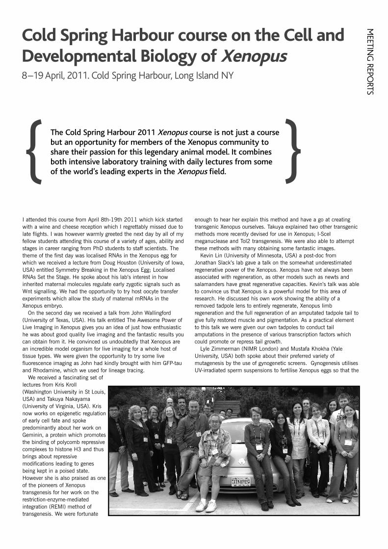
I attended this course from April 8th-19th 2011 which kick startedwith a wine and cheese reception which I regrettably missed due tolate flights. I was however warmly greeted the next day by all of myfellow students attending this course of a variety of ages, ability andstages in career ranging from PhD students to staff scientists. Thetheme of the first day was localised RNAs in the Xenopus egg forwhich we received a lecture from Doug Houston (University of Iowa,USA) entitled Symmetry Breaking in the Xenopus Egg; LocalisedRNAs Set the Stage. He spoke about his lab’s interest in howinherited maternal molecules regulate early zygotic signals such asWnt signalling. We had the opportunity to try host oocyte transferexperiments which allow the study of maternal mRNAs in theXenopus embryo.
On the second day we received a talk from John Wallingford(University of Texas, USA). His talk entitled The Awesome Power ofLive Imaging in Xenopus gives you an idea of just how enthusiastiche was about good quality live imaging and the fantastic results youcan obtain from it. He convinced us undoubtedly that Xenopus arean incredible model organism for live imaging for a whole host oftissue types. We were given the opportunity to try some livefluorescence imaging as John had kindly brought with him GFP-tauand Rhodamine, which we used for lineage tracing.
We received a fascinating set oflectures from Kris Kroll(Washington University in St Louis,USA) and Takuya Nakayama(University of Virginia, USA). Krisnow works on epigenetic regulationof early cell fate and spokepredominantly about her work onGeminin, a protein which promotesthe binding of polycomb repressivecomplexes to histone H3 and thusbrings about repressivemodifications leading to genesbeing kept in a poised state.However she is also praised as oneof the pioneers of Xenopustransgenesis for her work on therestriction-enzyme-mediatedintegration (REMI) method oftransgenesis. We were fortunate
enough to hear her explain this method and have a go at creatingtransgenic Xenopus ourselves. Takuya explained two other transgenicmethods more recently devised for use in Xenopus; I-SceImeganuclease and Tol2 transgenesis. We were also able to attemptthese methods with many obtaining some fantastic images.
Kevin Lin (University of Minnesota, USA) a post-doc fromJonathan Slack’s lab gave a talk on the somewhat underestimatedregenerative power of the Xenopus. Xenopus have not always beenassociated with regeneration, as other models such as newts andsalamanders have great regenerative capacities. Kevin’s talk was ableto convince us that Xenopus is a powerful model for this area ofresearch. He discussed his own work showing the ability of aremoved tadpole lens to entirely regenerate, Xenopus limbregeneration and the full regeneration of an amputated tadpole tail togive fully restored muscle and pigmentation. As a practical elementto this talk we were given our own tadpoles to conduct tailamputations in the presence of various transcription factors whichcould promote or repress tail growth.
Lyle Zimmerman (NIMR London) and Mustafa Khokha (YaleUniversity, USA) both spoke about their preferred variety ofmutagenesis by the use of gynogenetic screens. Gynogenesis utilisesUV-irradiated sperm suspensions to fertilise Xenopus eggs so that the
Cold Spring Harbour course on the Cell andDevelopmental Biology of Xenopus8–19 April, 2011. Cold Spring Harbour, Long Island NY
MEETIN
G REPO
RTS
paternal genome will not contribute to the zygote. This wouldnormally give a generation of unviable haploid embryos, howeverviable diploid embryos can be obtained if these embryos undergo acoldshock to retain their polar bodies before extrusion. Chemicalmutagenesis allows the introduction of single gene defects withresulting phenotypes which can be analysed. Lyle and Mustafa spokeabout some of the remarkably interesting phenotypes they were ableto obtain using this method. These included cyd vicious, one ofLyle’s mutants, which due to a mutation in neural crest regulatorypathways showed a reduction in melanocyte migration resulting in amohican like appearance as pigment cells stay along the back of theembryo. Grinch, one of Mustafa’s mutants, showed a loss of the ciliawhich normally covers the Xenopus surface ectoderm for which hehad some stunning electron microscopy images.
On the last few days we received a talk from a legend in the areaof Xenopus research, Ray Keller (University of Virginia, USA). Hegave a talk on some of his recent work in cell motility, forces andpatterning which occur during gastrulation. Cells will undergoconvergent extension movements due to cell movements andintercalation during gastrulation and Ray is interested in themeasurement of the forces responsible for these processes. Ray
Keller is well known for his skills in grafting with his very own graft,the Keller explant. We were fortunate enough to be taught a varietyof grafting techniques with Ray more than happy to give advice andguidance as we did so.
We finished the course on the exceptional high note that was adelicious steak and lobster banquet. I came away from this fullyequipped with the skills to deshell a lobster, a challenge I had neverpreviously come up against. After this we were fortunate that someof the students and course leaders were musically talented and thuswe were able to have a few drinks and a dance to celebrate the lastnight. I had an amazing time at the course and have found theXenopus community to be a fun, dedicated and welcomingcommunity of which I am proud to be a member. I would like tothank the BSCB for their generous funds which allowed me to attendthe course and Amy Sater and Jerry Thomsen for organising thecourse. I came away with many new friends, fantastic memories anda t-shirt with the take home message of the course “It’s never just afrog thing”.
Victoria HatchUniversity of East Anglia
20
Over 230 participants (including users, prominent scientists andtechnology developers) gathered in Heidelberg for the SixthInternational Congress on Electron Tomography. This meetingdiscussed recent major advances in all things structural – fromsingle proteins at subatomic resolution to entire organism 3Dreconstruction.
{ }Electron microscopy (EM) in European universities appears more
vibrant now than at any time in the past decade or more. Arguably,biological EM came close to extinction as a core technique in the1980s and 90s, as researchers ventured into new techniques in lightmicroscopy and molecular biology. Many departmental facilitiesbecame under-used and several were shut down. EM acquired areputation for being fiddly, costly and – perhaps the greatest of sins –'descriptive'. However, advances in cryo-electron microscopy and 3Delectron tomography, along with a rediscovery of the importance ofthe ultrastructural, has lead to a renaissance in biological EM thatmany university departments are again keen to access. The daily schedule of the Congress consisted of talks from invitedspeakers and oral presentations, followed by a poster session at theend of the afternoon. There was plenty of new and excitinginformation to keep our brains busy through the entire programme.And despite the diversity in applications, questions and models, forme, two dominant trends emerged. One was a bridging of some ofthe gap between light and electron microscopy through the use ofcorrelative techniques. Such techniques varied from fairly 'routine'
registering of images captured by both methods, to engineeringfluorescence capabilities into a cryo-electron microscope, allowingsequential light and high resolution ultrastructural work in one singleinstrument (demonstrated by Abraham Bram Koster, LeidenUniversity Medical Center, Netherlands). The second trend was thehuge increase in the scale of ultrastructural datasets afforded by theuse of high-throughput tomographic reconstructions. The firstelectron tomographic reconstructions of an eukaryote were publishedjust 4 years ago and involved small algae or yeast cells of ~2µm indiameter. Since that time, electron tomography has been used toreconstruct fly whole embryos as well as adult tissues. The growth ofinformation content displayed at this meeting was astonishing, withsingle montaged reconstructions measuring up to 600 Gb. Thebottleneck, however, is still in data analysis, and the development ofmore automated tools is a clear priority for the coming years.Among the talks given by invited speakers, one highlight for me wasThomas Müller-Reichert (University of Technology Dresden,Germany), who is applying light microscopy in combination withelectron tomography (ET) of high pressure frozen material to study
Sixth International Congress on ElectronTomography5–8 May, 2011. EMBL Advanced Training Centre, Heidelberg, Germany.
MEE
TIN
G R
EPO
RTS
21
MEETIN
G REPO
RTS
the very final stages ofcytokinesis. The involvement ofESCRT-III in this constriction wasknown, but Thomas has nowshown its structural side, withESCRT-III forming helicalfilaments that narrow the cortexof the intercellular bridge to asingle stalk. John Briggs (EMBLHeidelberg, Germany), one of theconference organisers, alsotalked about hybrid methods – inthis case used to study coatedvesicle budding – and showedsome very detailed structuralinformation on assembled COPIcoats. Using cryo-electrontomography (cryo-ET) andsubtomogram averaging of areconstituted budding reaction,he showed how subunits of theCOPI coat adopt differentconformations and interact withdifferent stoichiometries so as toaccommodate vesicles ofdifferent sizes and shapes (asopposed to the very regular sizesseeing for clathrin- and COPII-coated vesicles). It wasinteresting to see how thisfundamentally novel basis forvesicle coat assembly sharesfeatures with some viral proteincoats. Takashi Ishikawa (PaulScherrer Institute, Switzerland)showed how ET andsubtomogram averaging could decipher the bending mechanism ofeukaryotic flagella/cilia. A striking feature of cilia/flagella is theconservation of structure displayed in most axonemes, in which nineperipheral microtubule doublets surround two singlet microtubules.Despite this canonical architecture, cilia and flagella can bend inmany different ways. Takashi's incredibly detailed 3D structuralanalysis revealed a series of asymmetries along and showed howthese features would explain different waveforms to be formed incilia and flagella. Sam Li (University of California – San Francisco,USA) then moved us to the base of the cilium, showing the structureof the basal body (BB) at a fantastic 3 nm resolution. By fitting thesolved structure of tubulin into his tomographic reconstructions, heshowed how it was possible to build a pseudo-atomic model of theBB triplet. The 3D density map revealed novel densities thatrepresented non-tubulin proteins attached to the BB. Rather thanaveraging the whole structure, Sam showed us subvolumes atdifferent spatial locations along the BB which, just as for theaxoneme mentioned above, also displayed heterogeneity along itslength, suggesting a sequential and coordinated mechanism for BBassembly. Finally, Wah Chiu (Baylor College of Medicine, USA) gavea fantastic keynote session on cryo-electron tomography singleparticle analysis as an emerging structural technique for imagingindividual macromolecular assemblies close to atomic resolution.Wah Chiu, who was present at the birth of cryoET as a technique,
showed a huge amount of work on bacteriophage structure toillustrate the key concepts behind the method. I found his talk bothhighly informative and enjoyable. My favourite selected oralpresentation was from Wanda Kukulski (John Briggs's lab at EMBLHeidelberg, Germany). She used correlative fluorescence and electrontomography to directly map the signals of ~20 endocytic proteins(Ede1, Sla1 and Rvs167 among others) tagged with GFP or RFP,and gave us a 4D description of the yeast plasma membrane duringthe transition from a plane membrane to tubular invaginations,through formation of a constricted neck followed by abscission of avesicle. Wanda's comprehensive, spatiotemporal description givesnew insights into how protein modules of the endocytosis machinerycoordinate the changes in membrane topology required for vesiclebudding. The meeting was hugely enjoyable and gave me aninvaluable opportunity to see developments in structural cell biology.I presented a poster describing my own work using ET to study howsome human pathogens organise their surface membrane intospecialised domains, and was able to get some great feedback fromsome of the experts in the field. For this, I'm very grateful to theBSCB for awarding me the Honor Fell Travel Award to meet the costsof my travel to Heidelberg.
Catarina Gadelha University of Cambridge

22
MEE
TIN
G R
EPO
RTS
The second abcam: Chromatin, Replication and ChromosomalStability was held in June in Stockholm, organised by Anja Groth(University of Copenhagen), Catherine Green (University ofCambridge) and Camilla Sjögren (Karolinska Institute), followingthe previous successful meeting in 2009 in Copenhagen. { }
I was fortunately able to attend this meeting through BSCB Honor FellTravel Funding, and amazingly my work was selected for oralpresentation; my first talk at a conference.
The conference kicked off with a fascinating broad ranging talk fromone of the keynote speakers, Helen Blau (Stanford University), whohighlighted the importance of a correct demethylation programmeduring reprogramming and the effect of ‘stiffness’ on the regenerativeability of Muscle Stem Cells (MuSCs). Although reprogramming of cellscan also be achieved through iPS cell generation or nuclear transfer, herlab uses the method of cell fusion to investigate the mechanismsinvolved during reprogramming, specifically those of DNAdemethylation, an ‘epigenetic bottleneck’. She described their discoveryof Activation Induced Cytidine Deaminase (AID) expression inheterokaryons, and the subsequent elucidation that demethylationduring reprogramming is achieved through nucleotide base replacementrather than direct demethylation, as was previously thought. Prof. Blauended the talk with an example of how much a cell’s environment canalter its phenotype. She used a Goldilocks analogy to describe howMuSCs grown on the much too ‘stiff’ tissue culture plastic are unable toregenerate in vivo but those grown on a ‘comfy bed’ ofPolyEthyleneGlycol (the same stiffness as muscle), have a greatercapacity for regeneration, thereby conveying some of the previouslyignored 3D requirements of cells.
The first session covered Replication, Chromosome Structure andCellular Memory and I thankfully had an early talk slot. This meant Iwould be able to concentrate fully on the later talks rather thanworrying about my own. Speaking in a session with well-known cellcycle personalities was rather intimidating however, my talk went welland I was able to speak with several people in breaks who askedinteresting questions and offered helpful suggestions. Although it was abit scary, I would definitely recommend pushing yourself as a PhDstudent and trying to get an opportunity to talk about your work.
From this session I found Marcel Méchali’s (Institute of HumanGenetics, CNRS) talk particularly interesting. He described some of thefeatures his group are finding in higher eukaryotic DNA replicationorigins, by mining a large data set. It has long been known that highereukaryotes, unlike yeast, do not have a strict DNA sequence thatspecifies origins. Work from his group showed that origins are enrichedjust before or after transcription start sites but not at the site itself, andthat there is in fact some sequence impact, with origins having aTG/CA bias. He also described their ‘flexible replicon model’ where 4-5origins are grouped in a replicon from which one origin will bestochastically activated and then silence the others in that replicon. Thequestion everyone wanted to know when I spoke to him after was, howdoes this happen? I hope we soon find out!
After lunch, and meeting and chatting with various people, thesecond session on ‘Chromatin Replication and Histone Dynamics’
started. It covered topics from a potential histone modification-basedtherapeutic against Candida infections, to how stalled replication forksare resolved. I particularly enjoyed the talk from Patrick Varga-Weisz(Babraham Institute) telling us about how pericentromeric andcentromeric boundaries are maintained. The heterochromatin found atthese points has a complex combination of specific histonemodifications and recruitment of additional proteins. The correctdisruption and then reassembly of these structures must be undertakenduring each cell cycle and this maintenance is critical for genomicstability and chromosome segregation. Varga-Weisz described the workfrom his lab on the role of the chromatin remodeler SMARCAD1 duringthis process, which appears to complement those roles performed byhistone modifying enzymes such as Histone Deacetylases (HDACs) andHistone Methyltransferases (HMTs). SMARCAD1 interacts with manyproteins, including some involved in DNA replication, repair, silencingand heterochromatin maintenance, and localises to pericentromericheterochromatin during late S-phase when it is replicated. Althoughinitially found to be required for ES cell maintenance, the knock outmouse generated was viable with only a small number of defects. Thisparadox is undergoing further investigation. However, it was clear thatdepletion of SMARCAD1 by siRNA led to a global increase ineuchromatin marks and corresponding decrease in heterochromatinmarks through apparent failure to correctly recruit histone modifyingproteins which interact with SMARCAD1; and led to an increase inmitotic defects. This was the first of several fascinating talks on howchromatin is faithfully maintained after DNA replication.
On the Tuesday, session three covered ‘Initiation, Timing andEpigenetic States’. The first talk by David Gilbert (Florida StateUniversity) made sure we were awake and had brains engaged as hediscussed replication timing. In order to investigate the importance ofwhen particular regions of the genome are replicated (early, mid or lateS-phase) his lab has produced genome wide profiles for replication-timing from a wide number of cell lines, cells at different stages ofdifferentiation and also for various human pathological conditions.Changes in replication-timing can affect half of the genome butsurprisingly, correlated only slightly with transcriptional status andepigenetic marks. The factor that correlated strongest was long-rangechromatin interactions suggesting importance of spatial organisation.Using the huge change in global replication timing between the earlyand late epiblast (which is not accompanied by a significant change inthe transcriptional programme) work from his group showed that mostgenes which change their replication timing at this transition, movefrom early to late replication, and are linked to increased compaction ofchromatin. Echoing the model of DNA fractal globules by ErezLieberman-Aiden and Nynke van Berkum, early and late replicatinggenomic regions appear to segregate, with like associating with like.Consistent with this, regions of the genome that change in timing of
abcam: Chromatin, Replication andChromosomal Stability20–21 June 2011. Werner Gren Centre, Stockholm, Sweden.

23
replication are of the size 400-800kb suggesting this is the domain sizefor a region of the genome which is replicated at the same time.Interestingly the genes which change replication timing statuscorrelated with genes which are difficult to reprogramme, clearlyimpacting on attempts to improve iPS efficiency and further highlightingthe importance of spatial organisation.
The final session was entitled ‘Replisome Structure, Fork Progressionand Repair’ and covered some of the recent data about a wide range ofmechanisms involved in maintaining faithful DNA replication. I foundthe talk on how DNA replication machinery deals with the predictedbulky DNA tertiary structure at G-quadruplex (G4) motifs by VirginiaZakian (Princetown University) particularly fascinating. She presentedwork that replication through large protein complexes or DNA structuressuch as G-quadruplexes are facilitated by the helicases Rrm3 and Pif1(S. cerevisiae) (or related Pfh1 in S. pombe and Pif1 in H. sapiens).From genome wide ChIP, ~25% of G4 motifs were bound by Pif1 andDNA replication dramatically slowed in and around these regions inpif1 mutated cells. Startlingly, knocking down Pif1 by RNAi also led toa huge mutation rate at these sites, 20% of Gs became mutated and97% of these sites were no longer predicted to form G4 structures. Thepredicted G4 structures therefore do appear to form in vivo and beresolved by Pif1 to prevent them causing problems for DNA replicationmachinery and subsequent fork stalling, breakage and mutations
For the last talk of the day, the second keynote speaker, MichaelO’Donnell (Rockefeller University and HHMI), gave us a differentperspective, focussing on the bacterial replisome. It was fascinating tohear the story of the third polymerase, about the flexibility of
polymerases and how the replisome varies its composition as required.In vitro di-polymerase and tri-polymerase replisomes have similar ratesfor DNA synthesis but the three polymerase version has severaladvantages. Firstly the processivity is much greater due to more contactwith the lagging strand, also no gaps are left on the lagging strandunlike those seen when only two polymerases are permitted. Thespecific polymerases found in the replisome however, varieddramatically, with pol III found under normal conditions but replaced bypol II and pol IV during times of DNA damage. These alternativepolymerases slow the helicase dramatically and are stable, presumablyallowing time for DNA repair. It is sometimes too easy to ignorebacteria within the cell biology field, but this definitely showed howmuch we can learn about mechanism from bacteria.
As well as attending this illuminating conference and meeting otherscientists from across the world I also managed to have a look aroundStockholm. The city is beautiful, spread across 14 islands, so there areboats and bridges everywhere; not without reason is it known as theVenice of the North. I also saw some of the distinctive, colourful andvery pretty wooden houses on the 13,000 islands of the archipelagoand of course went to the Nobel Museum and saw one of the famousmedals!
I would like to thank the organisers for a fantastic conference andalso the BSCB for their generous funding which allowed me to attendthis stimulating event.
Rosemary H C Wilson , University of York
MEETIN
G REPO
RTS
The first session of the meeting, Specification of Pluripotency, waschaired by Jenny Nichols. The first talks by Kat Hadjantonakis (Sloan-Kettering Institute, New York) and Berenika Plusa (University ofManchester) both covered the topic of lineage specification of theprimitive endoderm from the pluripotent inner cell mass. Theypresented data outlining roles for growth factors PDGF and Fgf4 in theearly embryo. The session was finished by Takashi Hiiragi (EMBL,Heidelberg) who shared his exciting research using fluorescence-basedgene-trap mouse lines to visualise embryonic patterning, and single cellexpression profiling in the embryo.
After a coffee break the session was continued by a talk from HitoshiNiwa (RIKEN Center for Developmental Biology, Japan) on the role ofSox2 in the maintenance of pluripotency in both embryonic and
trophoblast stem cells. He presented data indicating an evolutionaryconservation of Sox protein function, with Drosophila Sox protein beingable to maintain embryonic stem cell pluripotency. Further talks in thesession were given by Alfonso Martinez Arias (Wellcome Trust CancerResearch, Gurdon Institute, University of Cambridge), AoifeO’Shaughnessy (Wellcome Trust Centre for Stem Cell Research,University of Cambridge) and Claire Chazaud (Genetique, Reproductionet Development, France) who introduced us to the roles of Wntsignalling in mouse embryonic stem cells, chromatin remodeller Mi-2�in lineage decisions of mouse embryos and gave further insight into theprimitive endoderm differentiation and the roles of Fgf4 and Nanog inearly mouse development respectively. The evening was then continuedby the poster session with drinks until the sound of the gong called the
The inaugural Cambridge Stem Cell Symposium took place overtwo sunny days in early July at Downing College. Organisers DrJenny Nichols and Dr Brian Hendrich (Wellcome Trust Centre forStem Cell Research, University of Cambridge) had broughttogether a large number of experts in the fields of pluripotencyand development with talks covering topics from lineage fatedecisions in the early mouse embryo to mesoderm differentiationin human embryonic stem cells.
{ }
Inaugural Cambridge Stem Cell Symposium:Pluripotency and Development6–7 July 2011, Downing College, Cambridge
MEE
TIN
G R
EPO
RTS
24
BSCB Sponsored Meeting: 8th North ofEngland Cell Biology Forum9 September 2011. University of Sheffield
The North of England Cell Biology Forum is a one-day annualforum, which brings together molecular cell biologists, located inthe North of England, working in the areas of membranetrafficking, cytoskeleton, molecular motors and signal transduction.We are fortunate that within this region there is a critical mass ofscientists with related interests in these fields. With strongsupport for the meeting from PIs, the scientific programme of talksand posters is delivered and chaired entirely by PhD students andpost-docs. The meeting has become a well-established part of thecalendar for cell biologists in this area and the 8th meeting washeld at The Edge Conference Facilities at the University of Sheffieldon Friday, September 9th.
As always it was a most enjoyable and stimulating day with 13talks presented by PhD students and postdocs in 4 sessions, allchaired by postdocs. The talks covered a diverse range of cellbiological topics including chromatin structure, secretion, ER,nuclear and chloroplast translocation, and endocytosis in differentcontexts. All were of an extremely high standard and first prize wasawarded to Dr Mark Morgan (University of Manchester) for his talk
on ‘Syndecan-4 Phosphorylation: a critical control point regulatingintegrin recycling and cell migration’. Second prize went to AnnaWillox, a postdoc from Steve Royle’s lab at the University ofLiverpool for her talk on 'Stonin 2 is the major adaptor for clathrin-mediated synaptic vesicle retrieval'. Additionally there was alunchtime poster session and the prize for best poster went to LizGranger from Viki Allan’s lab at the University of Manchester forher poster describing proteins that interact with dynein.
There were 94 registered delegates from the Universities ofSheffield, Sheffield Hallam, Manchester, Hull, York, Leeds andLiverpool at the meeting and, as in previous years, to encourage asbroad an audience as possible, there were no registration fees andcosts were met by sponsorship alone. We are therefore extremelygrateful to BSCB for its generous sponsorship of this event. Itscontribution, together with sponsorship from the BiochemicalSociety and various commercial companies, was essential for us tohold this successful event which so nicely showcased the work ofthe next generation of young cell biologists. Elizabeth Smythe, University of Sheffield
conference participants to the dinner that was served in the formal hallof Downing College.
The second day of the conference started with a session titled:Perdurance of Pluripotency, chaired by Brian Hendrich. The first talk byPhilip Avner (Institut Pasteur, France) was the EMBO talk, and he gavea very extensive overview of the X-inactivation process and its lineagedependency and developmental programming. This talk was followedby Ian Adams (Institute of Genetics and Molecular Medicine, Universityof Edinburgh), who shed light on the important role of Tex19.1 inprotection against aneuploidy and suppression of retrotransposons inthe germline cycle. The morning talks were finished by Amanda Fisher(Clinical Sciences Centre, Imperial College London) who shared datafrom her experiments with the heterokaryon reprogramming method.
Following a brief break for coffee the talks were continued by AntoinePeters (Friedrich Miescher Institute for Biomedical Research,Switzerland). He introduced us to epigenetic reprogramming bymembers of the Polycomb Group of proteins, and how they regulateinheritance of epigenetic information between generations. YusukeMiyanari (Institute of Genetics and Molecular Biology, France) thencontinued the talks by sharing his interesting findings on the expressionof Nanog in the early mouse embryo and how it is regulated on thelevel of individual alleles. By creating dual colour system with GFP andmCherry linked to each allele respectively, he showed datademonstrating real time fluctuations of Nanog expression in the earlyembryo. The final talk of the session was given by Anne Helness(Institute of Reproductive and Developmental Biology, Imperial CollegeLondon) on bivalent chromatin domains. Interestingly their datademonstrated the existence of bivalent domains in both the ICM andthe newly formed trophoectoderm in vivo and high-lighted mutuallyexclusive roles for Ring1b and Suv39h1 in regulating distinct chromatinstates at key developmental genes.
After a quick lunch, to catch up with the time table, we started thefinal session of the conference, Exit from Pluripotency, chaired by Prof
Austin Smith. The first talk was given by Shinichi Nishikawa (RIKENCenter for Developmental Biology, Japan) detailing the developmentalpathway of hematopoietic stem cells. Joshua Brickman (Institute ofGenetics and Molecular Medicine, University of Edinburgh) presentedhis groups findings about heterogeneity of anterior/primitive endodermmarker expression in self-renewing ES cell cultures. We also heard talksfrom Jerome Collignon (Institut Jaques Monod, France), who presenteddata about the role of Nodal in the early mouse embryo and pluripotentstem cells, and from Dean S. Griffiths (Department of Haematology,University of Cambridge), who outlined a role for JAK/STAT signalling inmouse ES cells parallel to LIF. This role involves the control of HP1�binding through histone phosphorylation.
The talks of the final part of the conference were started by ValerieWilson (Institute for Stem Cell Research, University of Edinburgh). Hertalk dealt with the timing of loss of pluripotency in the postimplantationembryo and events regulating it. This was followed by Anton Wutz(Wellcome Trust Centre for Stem Cell Research, University ofCambridge) who returned to the topic of control of stem cell identityand differentiation by Polycomb group complexes. He showed data onPrc1 and Prc2 deficient ES cells, demonstrating that both Prc1 and 2contribute to the maintenance of the epigenetic identity of stem cells.The final talk of the conference was given by Roger Pedersen, whocovered mechanisms of mesoderm differentiation in pluripotent stemcells, demonstrating data indicating a key role for Brachyury in thelineage differentiation.
I would like to congratulate the organisers for a successful andstimulating conference. Keep your eyes out for the 2nd AnnualCambridge Stem Cell Symposium next year. I would also like to expressmy gratitude to the BSCB for generously providing funding for me toattend the conference.
Matias Ilmari Autio, IRDB, Dept Surgery & Cancer, Imperial College London
25
MEETIN
G REPO
RTS
This conference in the peaceful seaside town of Roscoff was openedwith the Plenary talk by Kim Nasmyth, University of Oxford. Hedescribed his laboratory’s findings that the Rec8 kleisin subunit, asopposed to scc1, holds together the cohesin rings that maintainattachment of bivalent chromosomes during female meiosis from birthto ovulation. At the point of fertilisation the place of Rec8 is taken bythe scc1 subunit, prior to the first mitosis.
Among the highlights were the two EMBO Young Investigatorlectures. Monica Gotta, University of Geneva, Switzerland, described arole for SPAT-1, the C.elegans homologue of Bora, in regulation of bothcell polarity and cell cycle progression during asymmetric division. Thisis achieved in conjunction with Plk1 and Aurora A kinases. The secondwas from Philippe Pasero (Institut de Génétique Humaine, France),who has found that the known budding yeast replication stress-responsive kinases, Mec1 and Rad53, are also activated during anormal S phase at sites where transcription interferes with replication.
Sometimes multiple groups were approaching similar questions, forexample how sister chromatid cohesion by the cohesin complex isregulated through S phase and into mitosis. During S phase thereplication forks need to progress past the cohesin, while the sisterchromatids must remain attached. Prasad Jallepalli (Memorial Sloan-Kettering Cancer Center, USA) showed that the RFC-Ctf18 complexregulates positioning and velocity of replication forks, and is required foracetylation of the smc3 subunit of cohesin. This acetylation is requiredfor replication fork progression. In mammals, Sororin may then bindand stabilise cohesion rings post-replication. Work described by Jan-Michael Peters (Research Institute of Molecular Pathology, Austria),found that Sororin competes with the cohesin cofactor WAP1 forbinding to the cohesin complex. His group found that loss of WAP1 inmice caused an excessive cohesion, while loss of Sororin caused theopposite effect. Sororin binds cohesin early in S phase, but this isreduced as cells enter prophase, when cohesion is lost along thechromosome arms in a WAP1-dependent manner. These findings wereelaborated upon by Tomoko Nishiyama from the Peters laboratory inher poster, which described that Sororin association with cohesins isdependent upon cohesin acetylation following DNA replication, amechanism which is conserved in Drosophila.
On the second day we moved on to mitosis. Tarun Kapoor(Rockefeller University, USA) described elegant in vitro experimentsrevealing that when a PRC1 homodimer interacts with a singlemicrotubule it adopts a flexible conformation, while binding of bothsubunits to a pair of antiparallel microtubules forms a defined bridge.These bridges do not significantly slow the rate of microtubule slidingby kinesin 5, suggesting that PRC1 acts as an antiparallel microtubule
tip tracker. Daniel Gerlich (Swiss Federal Institute of Tehnology Zurich,Switzerland) presented a purse-string model for abscission of cellsduring cytokinesis. In this model, spastin-mediated microtubuledisassembly at the midbody facilitates contraction of the intercellularbridge by ESCRTIII complex-rich filaments that underlie the cell cortex.
In the talks on spindle assembly, Helder Maiato (Instituto de BiologiaMolecular e Cellular, Portugal) spoke about the still controversial spindlematrix. He has found that the Drosophila nuclear-pore complex proteinMegator and the spindle checkpoint protein Mad2 form a conservedcomplex that localises to a spindle matrix. Megator is proposed to actas a spatial regulator of the spindle assembly checkpoint here, byensuring efficient loading of Mad2 onto unattached kinetochores.
The following day Sue Biggins (Fred Hutchinson Cancer ResearchCentre, USA) described the successful purification of functionalkinetochores from yeast. These kinetochore particles were able to bindmicrotubules and remained attached to dynamic microtubule tips in amanner that was stabilised by tension. Furthermore the kinetochoresdecreased microtubule catastrophe events showing that microtubule tipdynamics are altered. Later Marina Bacac (University HospitalLousanne CHUV, Switzerland) introduced a novel interphase role formammalian securin and separase, during which they associate withcell membranes. Depletion of these proteins disrupts morphology andfunction of the Golgi Apparatus and endosomes.
One of the speakers on the last morning was Jon Pines (University ofCambridge) who described how the spindle assembly checkpointregulates the choice of substrates degraded by the APC/C, by regulatingthe site on the APC/C to which the APC/C co-activator cdc20 binds.
This meeting was characterised throughout by a convivialatmosphere, lively scientific discussion and celebration of thefascinating cell cycle research being undertaken around the world. Thequality of the work being presented was excellent, and the enthusiasmof each delegate infectious. Particularly striking was the eagerness ofeveryone, even the most experienced principal investigators, to meet allthe other attendees and hear about their work. I left the meetinginspired, and with useful feedback from my own poster. Even thetraditional airport workers strike causing cancellation of my return flightwas unable to dampen my enthusiasm! I am very grateful to the BSCBfor contributing to the cost of my attendance at this meeting. I wouldencourage any other members who have the opportunity to attend thisconference in future years to grasp it with both hands; it was the bestmeeting I have attended.
Fiona Hood, Physiological LaboratoryUniversity of Liverpool
This meeting covered a broad range of cell cycle research topics.The talks were divided into ten sessions on asymmetric celldivision, DNA replication and chromosome cohesion, modellingthe cell cycle, late mitosis and cytokinesis, spindle assembly,spindle dynamics, organelles in mitosis, mitotic progression,nuclear dynamics and meiosis, and the spindle assemblycheckpoint.
{ }
Jacques Monod Conference on cell divisionin time and space11–15 September, 2010. Roscoff, France.
MEE
TIN
G R
EPO
RTS
26
BSCB Sponsored Meeting: British YeastGroup 2011 annual meeting23–25 March 2011. Brighton
At this meeting, 140 delegates enjoyed the beautiful springweather which set the scene for an exciting and interactivemeeting. There were 32 platform presentations from leaders in thefield, new investigators, postdocs and students.
In a plenary session, Sir Paul Nurse described his labs’ recentdata on stripping down the cell cycle machinery to its bareminimum. Surprisingly, this reveals a new layer of homeostasisregulated by cell size which now becomes amenable to geneticanalysis. He was followed by Phil Zegerman (Cambridge), whodescribed work that aims to delineate what establishes thetemporal timing of DNA replication in an unperturbed cell cycle.
In the Genomics and Evolution session, Rick Dunn (Manchester)discussed the role of metabolomics in systems biology and the useof flux analysis for studying carbon metabolism. Ken Wolfe (Dublin)introduced the evolutionary conservation of the arrangement of themating type locus of yeast, and how this has also influenced theevolution of the adjacent chromosome arm. Tim Levine (UCL)explained how the use of the most recent homology search enginessuch as HHpred allowed the identification of distant homologies,which can help assign function to apparently orphan proteins. Hegave the example of how BLOC-1 complex subunits were identifiedin S. cerevisiae, thus demonstrating conservation of a pathway inthis organism that was originally thought to be missing.
In the Chromosome and their Dynamics session, Robin Allshire(Edinburgh) discussed the complex role of histone modifications inthe regulation of heterochromatin and centromere function. AdeleMarston (Edinburgh) described the role of PPA2-Cdc55 phospatasein helping to co-ordinate the various changes to chromosomedynamics and function that define the dual chromosome separationevens of meiosis. Jonathan Baxter (Sussex) introduced us to theconcept that positive supercoiling of DNA during mitosis isnecessary to drive decatenation by topoisomerase II in a mannerdependent on mitotic spindle attachment to kinetochores and thecondensation of DNA via the condensin complex. Further talks onthe analysis of histone modifications emphasized the session’scommon theme of the complexity of understanding chromosomefunction and the subtle roles played by a myriad of interactinggenes and pathways.
The importance of yeasts as model systems for understand basicbiological questions was evident throughout the meeting, but in theShape and Morphogenesis session an additional layer of interestwas added because of the links between growth modalities andpathogenicity in C. albicans. Peter Sudbery (Sheffield) describedelegant cell biological approaches to understanding how cellspolarize growth to the hyphal tip and explored the role ofphosphorylation by cell cycle kinases in the regulation of growthpolarity, a topic expanded upon by Jamie Correa-Bordes (Badajoz,Spain) in his analysis of Mob2 phosphorylation. Alexandra Brand(Aberdeen) discussed how cells respond to their environment byregulating the GTP cycling activity of the Cdc42 polarity complexand showed some beautiful examples of live microscopy wherecells respond to contact with an obstacle in specialized growthcambers. Equally impressive microscopy was presented by JamesDodgson (Cambridge), who imaged cells “end on” to reveal afurther level of physical organization at the growing cell tip. Finally,this session contained the prize presentation by Michelle Leach
(Aberdeen), who elegantly linked mathematical fitting of her data tonew predictions and experimental verification of these whilediscussing how Candida albicans uses its chaperones to regulate aheatshock response essential for virulence.
In the Cell Biology and Signaling session Fritz Muhlschlegel(Kent) described the linking of carbon dioxide signaling withvirulence of C. albicans through the Zn2+ finger transcription factorRca1, the activity of carbonic anyhdrase and the regulation ofadenylyl cyclase. Andrew McAinsh (Warwick) then introduced us tothe self-organization of complex systems and the in vitro study ofmechanisms by which microtubules and their motors organize theinterphase microtubule arrays. The intriguing complexity ofbiological systems was further emphasized by a number of talksrelating to DNA damage and damage signaling including ananalysis of ATR activation in S. pombe by Chris Wardlaw (Sussex)and an intriguing presentation by Thomas Caspari (Bangor) whoreported a novel heat-induced translational initiation in the S.pombe rad9 gene. Finally, last years organizer, Tim Humphrey(Oxford), discussed his genome wide screen for factors thatinfluence chromosome rearrangements in response to a DNAdouble strand break, which identified a number of unexpectedgenes that clustered together with homologous recombinationfactors to define an “HR gene set”.
The Modeling Processes session consisted of talks emphasizingthe role of yeast research in modeling complicated and conservedevents and pathways. The presentations included a discussion byAlan Morgan (Liverpool) on how dietary restriction of yeast extendslifespan and the identification of roles in this process for theheatshock proteins Hsp12 and Hsp16. Katherine Ayscough(Sheffield) explained her work which identified the importance ofactin in various steps of endocytosis, including a role incounteracting the turgor pressure of yeast to allow the invaginationof the membrane as well as the more defined role in constrictingthe membrane to pinch off the vesicle. Campbell Gourley (Kent)explained new roles for the actin regulatory protein Cofilin in stressand mitochondrial regulation, showing that one protein canparticipate in apparently unrelated processes. Jakai Wen(Birmingham) presented work which aims to understand themechanism of nonsense mediated decay, a phenomenon by whichmRNAs with a mis-positioned stop codon are preferentiallydegraded. Mike Stark (Dundee) told us how the potentiallymisnamed elongator complex is responsible for tRNA wobbleuridine modifications and not for directly mediating transcriptionelongation.
The meeting finished with three presentations relating to meiosis.Jesus Carballo (NIMR) led us through the complex regulation ofDNA double strand break formation in meiosis, concentrating onthe role of the ATM and ATR kinases and on their targets within theSpo11 complex. Alastair Goldman (Sheffield) and Valerie Garcia(Sussex) explored how DNA double strand breaks formed inmeiosis are processed collaboratively by Mre11 and ExoI. Themodel that emerges may explain why the exonuclease activity ofMre11 is 3’-5’ and not the originally expected 5’-3’.
Tony Carr, Genome Damage and Stability Centre, University of Sussex

27
POSTD
OCS AN
D PH
DS
Did you know that less than 7%of scientists below the age of 35will get a tenure track position inAmerica? Unfortunately, I expectthat this percentage will be verysimilar here in the UK and maypossibly get worse as fundingstreams dry up due togovernment austerity measures.Throughout the years, articleshave regularly appeared in majorscientific journals lamenting thestate of the science career pathand especially the plight ofpostdocs.
The current career progressionin science has been compared toa nail on an ironing board withthe ironing board resembling themany PhD students andpostdocs vying for the minutenumber of permanentindependent positions available.Amazingly however it seems thatwe still do not appreciate theobscenity of this system. The2011 Careers in ResearchOnline Survey 2011(1) showsthat 80% of scientists still heldfixed-term contracts with somestill on these even after 5previous contracts at the sameinstitution. Only 40% ofrespondents felt there wereenough opportunities forprogression or promotion at theirinstitution. Yet, 75% stillaspired to work within the highereducation sector either teaching,performing research or a mix of
both. Striking, no? Are wecollectively sticking our heads inthe sand holding thumbs that wewill be the lucky ones that doget that coveted PI position?The reality this observationunderlines is that most of us willhave to give up our dream andcommitment to our chosencareer and settle for a, in ourown opinion, second rate careeroption.
Can the scientific endeavourallow this wastage of young,committed and ambitious talentwithout any consequence? Itseems at the moment that it canbut make no mistake the reallybright young ones are good atsizing up their options andopting out of the science careerfor something more beneficialleaving science with fewer andfewer people to choose from.
How can this deficiency beaddressed? One of the solutionswas proposed by Jennifer Rohnin an article published in Naturerecently (2). She argues thatpostdoc careers should beprofessionalized. This means apermanent position on a levelsomewhere between scientificofficer and the PI. While thiswould be good for people notwilling or able to lead a researchgroup it will be limited in itsscope and will not help thosestill intent on achieving somescientific independence. It can
form part of a package ofsolutions which could includethe following:
Firstly, funding agenciesshould allow and activelypromote PIs to apply for grantstogether with their postdocs in acollaborative setup. Both partieswill be named on the grantapplication so both can takecredit for it. Next, postdocsshould have input into who isemployed on the grant moneyand they will supervise theseemployees. Any publicationsarising will have the PI andpostdoc as co-last authors. Thissetup would allow a PI to have anumber of these subgroups inhis lab which would bebeneficial for him since there aremore people working in his labwhile has to supervise fewerpeople. The postdocs, on theother hand, can establishthemselves as independentscientists and get used torunning a small group.
Secondly, science studentnumbers should be restricted.Selection of the best studentsshould begin earlier making thereduction in numbers moregradual and giving students thatdid not make the cut theopportunity to pursue anothercareer before having invested toomuch in their education. Thereshould also be honest andrealistic career advice given to
students early on to inform themof the obstacles of academicresearch and the possibilities foralternative careers.
In the end nothing will changeunless we as postdocs start tomake an effort to inform theparties involved that we wantchange. Joining efforts fromcampaign groups such as‘Science is Vital’ is essential andI implore you to take the time toadd your comments on theirwebsite which they are using tocompile a report for the ministerof State for Universities andScience, David Willets. Vitaeand The Concordat are alsolooking after researcher issues.Furthermore, a number ofuniversities have postdocsocieties that may have somesay in university issues.
Repairing this broken systemwill be beneficial for postdocsbut also for science as a wholeand all those involved in itsendeavours.
References:Careers in Research OnlineSurvey (CROS) 2011 Analysis ofUK aggregate results, Vitae,2011Rohn, Jennifer. Give postdocs acareer, not empty promises.Nature 471, pg 7, 2011
BSCB postdocs
Postdocs forever? How can we mend this broken system?Iman van den Bout
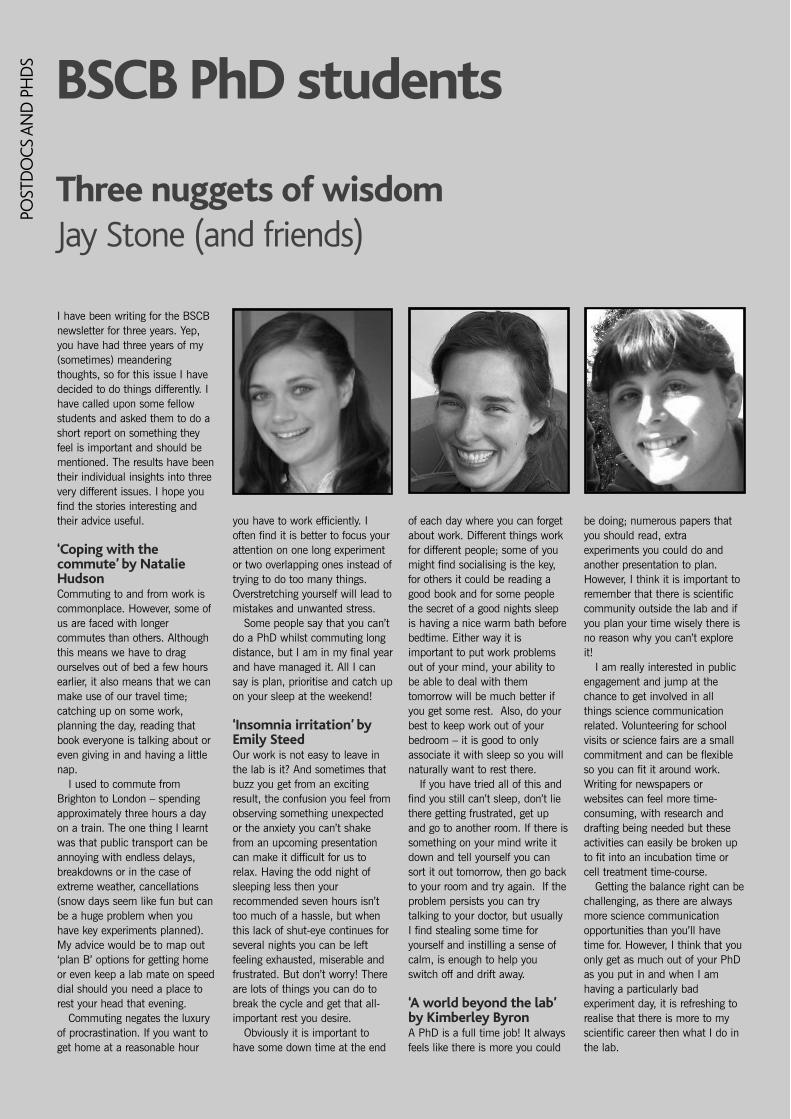
I have been writing for the BSCBnewsletter for three years. Yep,you have had three years of my(sometimes) meanderingthoughts, so for this issue I havedecided to do things differently. Ihave called upon some fellowstudents and asked them to do ashort report on something theyfeel is important and should bementioned. The results have beentheir individual insights into threevery different issues. I hope youfind the stories interesting andtheir advice useful.
‘Coping with thecommute’ by NatalieHudson Commuting to and from work iscommonplace. However, some ofus are faced with longercommutes than others. Althoughthis means we have to dragourselves out of bed a few hoursearlier, it also means that we canmake use of our travel time;catching up on some work,planning the day, reading thatbook everyone is talking about oreven giving in and having a littlenap.
I used to commute fromBrighton to London – spendingapproximately three hours a dayon a train. The one thing I learntwas that public transport can beannoying with endless delays,breakdowns or in the case ofextreme weather, cancellations(snow days seem like fun but canbe a huge problem when youhave key experiments planned).My advice would be to map out‘plan B’ options for getting homeor even keep a lab mate on speeddial should you need a place torest your head that evening.
Commuting negates the luxuryof procrastination. If you want toget home at a reasonable hour
you have to work efficiently. Ioften find it is better to focus yourattention on one long experimentor two overlapping ones instead oftrying to do too many things.Overstretching yourself will lead tomistakes and unwanted stress.
Some people say that you can’tdo a PhD whilst commuting longdistance, but I am in my final yearand have managed it. All I cansay is plan, prioritise and catch upon your sleep at the weekend!
‘Insomnia irritation’ byEmily SteedOur work is not easy to leave inthe lab is it? And sometimes thatbuzz you get from an excitingresult, the confusion you feel fromobserving something unexpectedor the anxiety you can’t shakefrom an upcoming presentationcan make it difficult for us torelax. Having the odd night ofsleeping less then yourrecommended seven hours isn’ttoo much of a hassle, but whenthis lack of shut-eye continues forseveral nights you can be leftfeeling exhausted, miserable andfrustrated. But don’t worry! Thereare lots of things you can do tobreak the cycle and get that all-important rest you desire.
Obviously it is important tohave some down time at the end
of each day where you can forgetabout work. Different things workfor different people; some of youmight find socialising is the key,for others it could be reading agood book and for some peoplethe secret of a good nights sleepis having a nice warm bath beforebedtime. Either way it isimportant to put work problemsout of your mind, your ability tobe able to deal with themtomorrow will be much better ifyou get some rest. Also, do yourbest to keep work out of yourbedroom – it is good to onlyassociate it with sleep so you willnaturally want to rest there.
If you have tried all of this andfind you still can’t sleep, don’t liethere getting frustrated, get upand go to another room. If there issomething on your mind write itdown and tell yourself you cansort it out tomorrow, then go backto your room and try again. If theproblem persists you can trytalking to your doctor, but usuallyI find stealing some time foryourself and instilling a sense ofcalm, is enough to help youswitch off and drift away.
‘A world beyond the lab’by Kimberley ByronA PhD is a full time job! It alwaysfeels like there is more you could
be doing; numerous papers thatyou should read, extraexperiments you could do andanother presentation to plan.However, I think it is important toremember that there is scientificcommunity outside the lab and ifyou plan your time wisely there isno reason why you can’t exploreit!
I am really interested in publicengagement and jump at thechance to get involved in allthings science communicationrelated. Volunteering for schoolvisits or science fairs are a smallcommitment and can be flexibleso you can fit it around work.Writing for newspapers orwebsites can feel more time-consuming, with research anddrafting being needed but theseactivities can easily be broken upto fit into an incubation time orcell treatment time-course.
Getting the balance right can bechallenging, as there are alwaysmore science communicationopportunities than you’ll havetime for. However, I think that youonly get as much out of your PhDas you put in and when I amhaving a particularly badexperiment day, it is refreshing torealise that there is more to myscientific career then what I do inthe lab.
Three nuggets of wisdomJay Stone (and friends)
BSCB PhD studentsPO
STD
OCS
AN
D P
HD
S
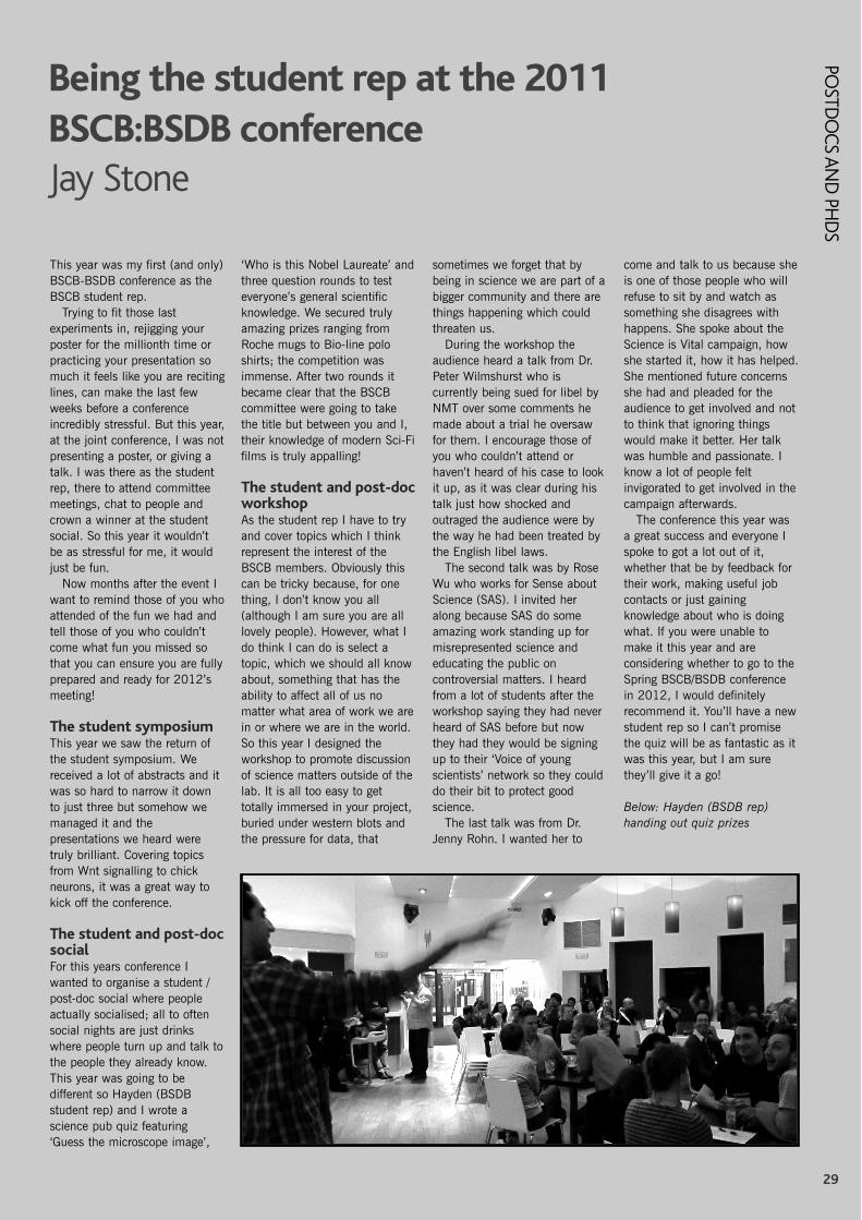
29
POSTD
OCS AN
D PH
DS
This year was my first (and only)BSCB-BSDB conference as theBSCB student rep.
Trying to fit those lastexperiments in, rejigging yourposter for the millionth time orpracticing your presentation somuch it feels like you are recitinglines, can make the last fewweeks before a conferenceincredibly stressful. But this year,at the joint conference, I was notpresenting a poster, or giving atalk. I was there as the studentrep, there to attend committeemeetings, chat to people andcrown a winner at the studentsocial. So this year it wouldn’tbe as stressful for me, it wouldjust be fun.
Now months after the event Iwant to remind those of you whoattended of the fun we had andtell those of you who couldn’tcome what fun you missed sothat you can ensure you are fullyprepared and ready for 2012’smeeting!
The student symposiumThis year we saw the return ofthe student symposium. Wereceived a lot of abstracts and itwas so hard to narrow it downto just three but somehow wemanaged it and thepresentations we heard weretruly brilliant. Covering topicsfrom Wnt signalling to chickneurons, it was a great way tokick off the conference.
The student and post-docsocial For this years conference Iwanted to organise a student /post-doc social where peopleactually socialised; all to oftensocial nights are just drinkswhere people turn up and talk tothe people they already know.This year was going to bedifferent so Hayden (BSDBstudent rep) and I wrote ascience pub quiz featuring‘Guess the microscope image’,
‘Who is this Nobel Laureate’ andthree question rounds to testeveryone’s general scientificknowledge. We secured trulyamazing prizes ranging fromRoche mugs to Bio-line poloshirts; the competition wasimmense. After two rounds itbecame clear that the BSCBcommittee were going to takethe title but between you and I,their knowledge of modern Sci-Fifilms is truly appalling!
The student and post-docworkshop As the student rep I have to tryand cover topics which I thinkrepresent the interest of theBSCB members. Obviously thiscan be tricky because, for onething, I don’t know you all(although I am sure you are alllovely people). However, what Ido think I can do is select atopic, which we should all knowabout, something that has theability to affect all of us nomatter what area of work we arein or where we are in the world.So this year I designed theworkshop to promote discussionof science matters outside of thelab. It is all too easy to gettotally immersed in your project,buried under western blots andthe pressure for data, that
sometimes we forget that bybeing in science we are part of abigger community and there arethings happening which couldthreaten us.
During the workshop theaudience heard a talk from Dr.Peter Wilmshurst who iscurrently being sued for libel byNMT over some comments hemade about a trial he oversawfor them. I encourage those ofyou who couldn’t attend orhaven’t heard of his case to lookit up, as it was clear during histalk just how shocked andoutraged the audience were bythe way he had been treated bythe English libel laws.
The second talk was by RoseWu who works for Sense aboutScience (SAS). I invited heralong because SAS do someamazing work standing up formisrepresented science andeducating the public oncontroversial matters. I heardfrom a lot of students after theworkshop saying they had neverheard of SAS before but nowthey had they would be signingup to their ‘Voice of youngscientists’ network so they coulddo their bit to protect goodscience.
The last talk was from Dr.Jenny Rohn. I wanted her to
come and talk to us because sheis one of those people who willrefuse to sit by and watch assomething she disagrees withhappens. She spoke about theScience is Vital campaign, howshe started it, how it has helped.She mentioned future concernsshe had and pleaded for theaudience to get involved and notto think that ignoring thingswould make it better. Her talkwas humble and passionate. Iknow a lot of people feltinvigorated to get involved in thecampaign afterwards.
The conference this year wasa great success and everyone Ispoke to got a lot out of it,whether that be by feedback fortheir work, making useful jobcontacts or just gainingknowledge about who is doingwhat. If you were unable tomake it this year and areconsidering whether to go to theSpring BSCB/BSDB conferencein 2012, I would definitelyrecommend it. You’ll have a newstudent rep so I can’t promisethe quiz will be as fantastic as itwas this year, but I am surethey’ll give it a go!
Below: Hayden (BSDB rep)handing out quiz prizes
Being the student rep at the 2011BSCB:BSDB conferenceJay Stone

30
FORT
HCO
MIN
G M
EETI
NG
S
30
BSCB / BSDB / JSDB Joint Spring MeetingWarwick University, 15–18 April 2012.
The Joint Spring Meeting of the BSCB, BSDB and JSDB is to takeplace in Warwick University between the 15th and 18th April 2012.The meeting is an exciting blend of cell and developmental biologyand, for the first time, co-organized with the Japanese Society ofDevelopmental Biologists (JSDB).
The BSCB programme will be kicked off by the plenary lecture givenby Professor J. Richard McIntosh (University of Colorado), a world-renowned cell biologist who has pioneered biophysical cytology usinginnovative methods. He has made several groundbreaking discoveriesabout the mitotic spindle organization and kinetochore-microtubuleinteractions.
As always, at this flagship meeting, the speaker line up is excellentand the sessions include: DNA Replication, Cell Division, CellGrowth/ Differentiation, Chromosome Structure/Organization, and CellDeath/Senescence. The BSCB Hooke Medal winner of this year willalso give a talk in this meeting. There will be a call for abstracts topresent short talks that will be interspersed between invited speakersand, of course, plenty of poster slots to fill.
Warwick University accommodates a fantastic conference facility andseveral social events will be arranged to facilitate informalcommunication between meeting participants. Details on speakers,venue, bookings and so on can be found by visiting the website(www.bscb.org). We look forward to welcoming you in April.
Scientific organizers (BSCB programme)Tomoyuki Tanaka, Helfrid Hochegger, Andrew McAinsh
2012 BSCB Programme Outline:15th Sunday
Evening Plenary Lecture: J. Richard McIntosh (University of Colorado)
16th MondayAM: DNA ReplicationHiroyuki Araki (NIG, Mishima)Helle Ulrich (CRUK London)Anindya Dutta (University of Virginia)Julian Blow (University of Dundee) Plus 2 short talks selected from abstracts
PM: Cell DivisionToru Hirota (Cancer centre Tokyo) Andrea Musacchio (MPI Dortmund) Jan Loewe (LMB Cambridge) Monica Bettencourt-Dias (IGC Portugal)Plus 2 short talks selected from abstracts
Evening: BSCB Hooke Medal Talk
17th TuesdayAM: Cell Growth/DifferentiationNorio Nakatsuji (Kyoto University) Denise Barlow (CEMM Vienna)Anton Wutz (CSCR Cambridge)Arp Schnittger (MPI Koern) Plus 2 short talks selected from abstracts
PM: Chromosome Structure/OrganizationTatsuya Hirano (RIKEN Wako) Robin Allshire (University of Edinburgh) Ana Pombo (MRC CSC) Juri Rappsilber (University of Edinburgh) Plus 2 short talks selected from abstracts
18th WednesdayAM: Cell Death/SenescenceTamotsu Yoshimori (Osaka University) Anton Gartner (University of Dundee) Andreas Villunger (Innsbruck Med Univ) Fabrizio d'Adda di Fagagna (IFOM Milan) Plus 2 short talks selected from abstracts
Note that BSCB programme integrates JSDB speakers.
31
FORTH
COM
ING
MEETIN
GS
BSCB Calendar of related meetings in 2012
Gordon Conference: Autophagyin Stress, Development &DiseaseMarch 11–16, 2012Four Points Sheraton / HolidayInn ExpressVentura, CA http://www.grc.org/programs.aspx?year=2012&program=autophagy
Keystone Conference: MolecularBasis of Vascular Inflammationand AtherosclerosisMarch 25–30, 2012Big Sky, Montanahttp://www.keystonesymposia.org/Meetings/ViewMeetings.cfm?MeetingID=1140
Keystone conference: CellBiology of Virus Entry,Replication and PathogenesisMar 26 – 31, 2012Whistler, British Columbiahttp://www.keystonesymposia.org/Meetings/ViewMeetings.cfm?MeetingID=1167
ESF-EMBO SymposiumCell Polarity and MembraneTraffic31 March - 5 April, 2012 Polonia Castle in Pultusk, Polandhttp://www.esf.org/index.php?id=9163
Gordon Conference: FibroblastGrowth Factors in Development& DiseaseMay 13-18, 2012Les Diablerets Conference CenterLes Diablerets, Switzerland http://www.grc.org/programs.aspx?year=2012&program=fgf
EMBO Conference SeriesMicrotubules: Structure,Regulation and FunctionsEMBL Heidelberg, Germany. May 23-26, 2012 http://www.embl.de/training/events/2012/MSF12-01/index.html
EMBO Conference seriesCellular Signaling & MolecularMedicine May 25–29, 2012 Cavtat – Dubrovnikhttp://events.embo.org/12-signaling-molmed/speakers.html
Gordon Conference:Intermediate FilamentsJune 17-22, 2012Bates CollegeLewiston, ME http://www.grc.org/programs.aspx?year=2012&program=intermed
Gordon Conference: Lysosomes& EndocytosisJune 17-22, 2012Proctor AcademyAndover, NH http://www.grc.org/programs.aspx?year=2012&program=lysosomes
Gordon Conference: Cell Biologyof the NeuronJune 24-29, 2012Waterville Valley ResortWaterville Valley, NHhttp://www.grc.org/programs.aspx?year=2012&program=cellneuron
Gordon Conference: NotchSignaling in Development,Regeneration & DiseaseAugust 12-17, 2012Bates CollegeLewiston, ME http://www.grc.org/programs.aspx?year=2012&program=notchsig
Biochemical SocietyConference: G-protein-coupled-receptors: from structuralinsights to functionalmechanisms September 12—14, 2012Monash University Prato Centre,Italyhttp://www.biochemistry.org/Conferences/AllConferences/tabid/379/Page/3/MeetingNo/SA124/view/Conference/Default.aspx
The EMBO Meeting 2012 –Advancing the life science. September 22-25, 2012Nice.www.the-embo-meeting.org/
Biochemical Society AnnualSymposium: Epigeneticmechanisms in developmentand disease December 11–13, 2012University of Leeds, UKhttp://www.biochemistry.org/Conferences/AllConferences/tabid/379/Page/3/MeetingNo/SA141/view/Conference/Default.aspx


33
The British Society for Cell BiologyStatement of Financial Activities for the year to 31 December 2009
2009 2008Unrestricted Restricted Total Total
£ £ £ £Incoming ResourcesIncoming resources from generating funds:
Voluntary income 30,000 27,500 57,500 57,500Incoming resources from charitable activities:
Meetings 2,264 – 2,264 48,023Subscriptions 31,443 – 31,443 20,084
Investment income:Bank interest 782 – 782 6,547
Other incoming resources 177 8,358 8,535 2,461Total incoming resources 64,666 35,858 100,524 134,615
Resources ExpendedCharitable Activities:Grants payable:
CoB/Honor Fell travel awards 27,016 27,016 33,776Other grants 611 – 611 –
Studentship 9,709 – 9,709 11,590Costs of meetings 39,876 – 39,876 66,556Newsletter costs 5,139 – 5,139 5,450Website expenses 7,295 – 7,295 2,180Governance costs 6,808 – 6,808 7,462Bad Debt – – – 900Total resources expended 69,438 27,016 96,454 127,914
Net movement in funds for the year (4,772) 8,842 4,070 6,701
Reconciliation of funds
Funds brought forward at 1 January 225,096 – 225,096 218,395
Funds carried forward at 31 December 220,324 8,842 229,166 225,096
2009 2008£ £ £ £
Current AssetsDebtors:
Prepayments and accrued income 433 406Cash at bank and in hand:
National Savings Investment Account 71,635 71,314HSBC Bank Accounts 159,951 156,126
232,019 227,846Less: Creditors falling due within one year
Creditors and accruals 2,853 2,7502,853 2,750
Net Assets 229,166 225,096
FundsRestricted 8,842Unrestricted 220,324 225,096
229,166 225,096

34
BSCB
CO
MM
ITTE
E Committee Members 2011
PresidentProfessor Jordan RaffSir William Dunn School ofPathologyUniversity of OxfordSouth Parks RoadOxford OX1 3RETel: +44 (0) 1865 275533Email: [email protected]
SecretaryDr Grant WheelerSchool of Biological SciencesThe University of East AngliaNorwich NR4 7TJTel: +44 (0) 1603 593988Email: [email protected]
TreasurerProfessor Adrian HarwoodCardiff School of Biosciences Biomedical BuildingMuseum AvenueCardiff CF10 3AXTel: +44 (0) 29 879358Email: [email protected]
Meetings SecretaryDr Andrew McAinshCentre for Mechanochemical CellBiologyWarwick Medical SchoolThe University of WarwickCoventry, CV4 7ALTel: +44 (0) 2476 151167Email:[email protected]
Membership SecretaryProfessor Dan CutlerMRC Laboratory for MolecularCell BiologyUniversity College LondonGower StreetLondonWC1E 6BTTel: +44 (0) 20 7679 7806Email: [email protected]
Newsletter editorDr Kate NobesSchool of BiochemistryUniversity of Bristol,Medical Sciences BuildingUniversity Walk,Bristol BS8 1TD Tel: +44 (0) 117 331 2229Email:[email protected](to whom material should be
sent– see guidelines for contributors)
Website CoordinatorDr Paul. D. Andrews Cellartis AB 1Wurzburg CourtDundee DD2 1FBTel: +44 (0) 1382 569987Email: [email protected]
Sponsorship secretary Dr Richard GroseCentre for Tumour BiologyInstitute of Cancer and the CR-UK Clinical CentreBarts and The London School ofMedicine and DentistryGround Floor, John Vane ScienceCentreCharterhouse SquareLondon EC1M 6BQTel +44 (0)207 014 0415Email: [email protected]
Honor fell/COB Travel AwardSecretary Dr Ewald HettemaDept of Molecular Biology andBiotechnologyUniversity of SheffieldFirth Court, Western BankSheffield S10 2TNTel: +44 (0)114 222 273Email:[email protected]
Committee members
Dr Buzz BaumMRC Laboratory of MolecularCell BiologyUniversity College LondonTel: +44 (0)20 7679 3040Email: [email protected]
Professor Iain HaganDepartment of Biochemistry andApplied Molecular Biology University of Manchester, andCell Division Group Paterson Institute for CancerResearchChristie HospitalWilmslow RoadWithingtonManchester M20 4BXEmail: [email protected]
Professor Patrick HusseySchool of Biological andBiomedical SciencesDurham UniversityEmail: [email protected]
Dr Jean-Paul VincentMRC National Institute forMedical ResearchThe Ridgeway, Mill Hill, London NW7 1AAEmail: [email protected]
Dr Caroline AustinInstitute for Cell and MolecularBiosciencesThe Medical SchoolUniversity of Newcastle uponTyneFramlington PlaceNewcastle upon Tyne NE2 4HHEmail:[email protected]
Dr Steve RoyleThe Physiological Laboratory, School of Biomedical Sciences, Crown Street,University of Liverpool,Liverpool L69 3BXEmail: [email protected]
Non-elected (co-opted)members
PhD student repKimberley BryonMRC Laboratory of MolecularCell BiologyUniversity College LondonTel: +44 (0)20 7679 Email:[email protected]
Postdoc repDr Iman van den Bout,Paterson Institute for CancerResearch The University of ManchesterWilmslow RoadManchester M20 4BXEmail:[email protected]
BSCB assistant Margaret ClementsBSCB Assistant The Company of Biologists Ltd.140 Cowley RoadCambridge CB4 0DLEmail [email protected]
Schools Liaison OfficerDavid Archer43 Lindsay Gardens,St. Andrews, Fife, KY16 8XD Email: [email protected]

35
BSCB AMBASSAD
ORS
BSCB Ambassadors 2011
City/ Institute Ambassador Contact
Aberdeen Anne Donaldson [email protected] University Eustace Johnson [email protected] Paul Whitley [email protected] James Murray [email protected] John Heath, Feydor Berditchevski [email protected], [email protected] Jason Gill [email protected] John Armstrong [email protected] Harry Mellor [email protected] Joanna Bridger [email protected] Jon Pines, Scottie Robinson [email protected], [email protected]
Simon Cook, Gillian Griffiths [email protected], [email protected] Martin Carden, Dan Mulvihill [email protected], [email protected] Morris Hallet, Adrian Harwood [email protected], [email protected] Hall Simon Boulton [email protected] Angus Lamond, Inke Nathke [email protected], [email protected] Roy Quinlan [email protected] Bill Earnshaw, Ian Chambers [email protected], [email protected]
Margarete Heck, Wendy Bickmore [email protected], [email protected] Nia Bryant, Karen Vousden [email protected], [email protected] Klaus Ersfeld [email protected] Clare Isacke [email protected] Vania Braga, Mandy Fisher [email protected], [email protected]/Guys Simon Hughes [email protected] Michelle Peckham [email protected] Andrew Fry, Colin Ockleford [email protected], [email protected] Giampietro Schiavo [email protected] Daimark Bennett, Sylvie Urbe [email protected], [email protected] Anne Ridley [email protected] Charles Streuli, Iain Hagan [email protected], [email protected]
Viki Allan [email protected] Michael Whittaker [email protected] Peter Rosenthal, Jean-Paul Vincent [email protected], [email protected] Grant Wheeler, Tom Wileman [email protected], [email protected] John Mayer [email protected] Chris Hawes, James Wakefield [email protected], [email protected]
Jordan Raff [email protected] Mary Mark Turner [email protected] Jonathan Gibbins [email protected] Liz Smythe, Andy Grierson [email protected], [email protected] Malcolm East, Paul Townsend [email protected], [email protected]
Jane Collins [email protected] Andrews Jo Parish [email protected] Georges David Winterbourne [email protected] John Carroll, Patricia Salinas [email protected], [email protected] College Nigel Goode [email protected] Anne Straube, Andrew McAinsh [email protected], [email protected] Dawn Coverly [email protected]
The BSCB Ambassadors are the people to ask about sponsoring youfor membership.
Anyone who wishes to volunteer to become a BSCB ambassador atany Institutes not represented in the list below please contact theBSCB.

36
The BSCB newsletter is published twice a year.
SubmissionIf you have an idea for an article please e-mail the editor a brief outlinefirst.
It is preferable to send all articles, reports and images by e-mail (thoughalternatives can be arranged after contacting the editor).
Attachments for text can be in txt, rtf or doc format. Please send images as300dpi JPEG, TIFF or PSD files.
Submission of articles and images should be made to
Dr Kate NobesSchool of Biochemistry,Medical Sciences Building, University Walk,Bristol BS8 1TD Tel: 0117 331 2229Email: [email protected]
Advertising InformationSingle advertisement:
Back cover Black and White £275; Colour £425Inside front cover Black and White £275Full inside page, black and white only £2201/2 Inside page, black and white only £1101/4 Inside page, black and white only £55
Four advertisements, to cover two years: Costs are reduced by 30%.
Advertisements can by supplied on CD or by email. Please send as JPG,TIF or PSD at 300dpi, or as PDF (with fonts embedded). Page size A4: 210x297mm.
There is no charge to advertise a scientific or educational meeting. Pleasecontact the editor with details of any meeting you wish to advertise.
For further information on commercial advertising contact: Dr Richard Grose,Centre for Tumour Biology,Institute of Cancer and the CR-UK Clinical Centre,Barts and The London School of Medicine and Dentistry,Charterhouse Square, London EC1M 6BQEmail: [email protected]
BSCB Subscription informationPaying by direct debit:
Regular member £35Student, school teacher, retired member £15
If you are still paying by standing order, please cancel it and set-up directdebit. Those members who do not wish to pay by direct debit or do nothave a UK bank account should contact Margaret [email protected] for advice.
New members should complete an online application form atwww.bscb.org.
Postmaster and General InquiriesSend changes of address, amendments and general queries to:
Margaret ClementsThe Company of Biologists Ltd.140 Cowley RoadCambridge CB4 0DLTel: 01223 425525E-mail: [email protected]
InvoicesSend to:
Professor Adrian HarwoodCardiff School of Biosciences Biomedical BuildingMuseum AvenueCardiff CF10 3US
JournalsBSCB members are entitled to a range of discounts from journal and bookpublishers. These are correct at the time of going to press but membersshould check www.bscb.org for the latest information.
Offers include a 25% discount from the individual subscription rate to alljournals published by the Company of Biologists, and other discounts fromother publishers. To take advantage of this offer, quote your BSCBmembership number when ordering your subscription.
Company of Biologists discounted prices: Journal of Cell Science: paper only £172/$295; online only £45/$77;paper and online £215/$365Journal of Experimental Biology: paper only £158/$270; online only£44/$75; paper and online £200/$340.Development: paper only £187/$325; online only £46/£80; paper andonline £232/$400
The following journals from John Wiley & Sons have discounts of 25–65%(https://secure.interscience.wiley.com/order_forms/bscb.html)
Journal BSCB rate Standard rateThe Anatomical Record $150 *BioEssays $99 $160Cell Motility and the Cytoskeleton $150 $425Developmental Dynamics $125 $165Genesis $60 $99Journal of Cellular Biochemistry $350 *Journal of Morphology $175 *Microscopy Research and Technique $295 $595
* No standard individual rate available; only available to institutionsNB: The price for the Journal of Morphology is now $175. If there areany members who have ordered the journal at the $150 rate, thoseorders will be honored.
Traffic discounted prices:Print and online: $155 / EUR144 Online only: $147 / EUR137

Related Documents