Bright Fluorescent Streptococcus pneumoniae for Live-Cell Imaging of Host-Pathogen Interactions Morten Kjos, a Rieza Aprianto, a Vitor E. Fernandes, b Peter W. Andrew, b Jos A. G. van Strijp, c Reindert Nijland, c * Jan-Willem Veening a Molecular Genetics Group, Groningen Biomolecular Sciences and Biotechnology Institute, Center for Synthetic Biology, University of Groningen, Groningen, The Netherlands a ; Department of Infection, Immunity and Inflammation, University of Leicester, Leicester, United Kingdom b ; Department of Medical Microbiology, University Medical Center Utrecht, Utrecht, The Netherlands c Streptococcus pneumoniae is a common nasopharyngeal resident in healthy people but, at the same time, one of the major causes of infectious diseases such as pneumonia, meningitis, and sepsis. The shift from commensal to pathogen and its interaction with host cells are poorly understood. One of the major limitations for research on pneumococcal-host interactions is the lack of suit- able tools for live-cell imaging. To address this issue, we developed a generally applicable strategy to create genetically stable, highly fluorescent bacteria. Our strategy relies on fusing superfolder green fluorescent protein (GFP) or a far-red fluorescent protein (RFP) to the abundant histone-like protein HlpA. Due to efficient translation and limited cellular diffusion of these fu- sions, the cells are 25-fold brighter than those of the currently best available imaging S. pneumoniae strain. These novel bright pneumococcal strains are fully virulent, and the GFP reporter can be used for in situ imaging in mouse tissue. We used our re- porter strains to study the effect of the polysaccharide capsule, a major pneumococcal virulence factor, on different stages of in- fection. By dual-color live-cell imaging experiments, we show that unencapsulated pneumococci adhere significantly better to human lung epithelial cells than encapsulated strains, in line with previous data obtained by classical approaches. We also con- firm with live-cell imaging that the capsule protects pneumococci from neutrophil phagocytosis, demonstrating the versatility and usability of our reporters. The described imaging tools will pave the way for live-cell imaging of pneumococcal infection and help further understanding of the mechanisms of pneumococcal pathogenesis. S treptococcus pneumoniae is a major cause of morbidity and mortality worldwide, and pneumococcal infections (e.g., pneumonia, septicemia, and meningitis) kill more than 1 million people every year (1). Pneumococci are also quiescent colonizers of the upper respiratory tract, particularly in children, but little is known about the mechanisms underlying the transition from commensal to pathogen. It is therefore of crucial importance to understand the entire pneumococcal pathogenesis cycle in detail. The polysaccharide capsule covering the cell surface is the most central virulence factor of S. pneumoniae. The involvement of the capsule in pneumococcal pathogenesis has been appreciated since Griffith in 1928 (2) published his famous transformation experi- ment with rough and smooth strains of S. pneumoniae. Today, it is known that the bulky capsule, which is either negatively charged or neutral, contributes to pathogenesis by protecting pneumo- cocci against the human immune system. For example, the cap- sule hinders phagocytosis and inhibits complement activity (3–6). Over 90 different pneumococcal serotypes have been identified to date (7), and the different serotypes vary in how well they protect the bacteria against phagocytosis (6). Furthermore, the amount of capsule differs between bacteria, and it has been shown, for exam- ple, that strains with thinner capsule adhere better than strains with thick capsule during the initial nasopharyngeal colonization (8). While the capsule is an important virulence factor, molecular epidemiology studies have also shown that nontypeable (unen- capsulated) strains are abundant within human populations and act as “hubs” for recombination between pneumococci, driving antibiotic resistance and serotype switching (9). Direct observa- tions of encapsulated and unencapsulated pneumococci in live host-pathogen assays are lacking, and it thus remains unclear how much the capsule contributes to the virulence cycle. Most of the knowledge concerning pneumococcal interactions with host cells and host tissue we have today has been obtained in vitro by biochemical or immunological assays and in vivo by tra- ditional postinfection plating and CFU counts, as well as by elec- tron microscopy of fixed samples of clinical isolates of S. pneu- moniae. To further extend our knowledge about pneumococcal pathogenicity, the key method would be the possibility of imaging interactions between bacteria and host cells in real time. Such a technique will provide understanding of the localization and dy- namics of S. pneumoniae during the course of infection and in that manner unravel factors important for the infection process. It will also open up the possibilities to study the role of the capsule in isogenic pneumococci during host attachment and immune eva- sion. Imaging of bacteria interacting with host cells and host tissue requires labeling to discriminate the bacteria from other cells and the surroundings. In vivo imaging of S. pneumoniae is typically done today using immunofluorescence, where antibodies bound Received 14 August 2014 Accepted 9 December 2014 Accepted manuscript posted online 15 December 2014 Citation Kjos M, Aprianto R, Fernandes VE, Andrew PW, van Strijp JAG, Nijland R, Veening J-W. 2015. Bright fluorescent Streptococcus pneumoniae for live-cell imaging of host-pathogen interactions. J Bacteriol 197:807–818. doi:10.1128/JB.02221-14. Editor: O. Schneewind Address correspondence to Jan-Willem Veening, [email protected]. * Present address: Reindert Nijland, Laboratory of Phytopathology, Wageningen University, Wageningen, The Netherlands. Supplemental material for this article may be found at http://dx.doi.org/10.1128 /JB.02221-14. Copyright © 2015, American Society for Microbiology. All Rights Reserved. doi:10.1128/JB.02221-14 March 2015 Volume 197 Number 5 jb.asm.org 807 Journal of Bacteriology on June 25, 2016 by guest http://jb.asm.org/ Downloaded from

Welcome message from author
This document is posted to help you gain knowledge. Please leave a comment to let me know what you think about it! Share it to your friends and learn new things together.
Transcript
Bright Fluorescent Streptococcus pneumoniae for Live-Cell Imaging ofHost-Pathogen Interactions
Morten Kjos,a Rieza Aprianto,a Vitor E. Fernandes,b Peter W. Andrew,b Jos A. G. van Strijp,c Reindert Nijland,c* Jan-Willem Veeninga
Molecular Genetics Group, Groningen Biomolecular Sciences and Biotechnology Institute, Center for Synthetic Biology, University of Groningen, Groningen, TheNetherlandsa; Department of Infection, Immunity and Inflammation, University of Leicester, Leicester, United Kingdomb; Department of Medical Microbiology, UniversityMedical Center Utrecht, Utrecht, The Netherlandsc
Streptococcus pneumoniae is a common nasopharyngeal resident in healthy people but, at the same time, one of the major causesof infectious diseases such as pneumonia, meningitis, and sepsis. The shift from commensal to pathogen and its interaction withhost cells are poorly understood. One of the major limitations for research on pneumococcal-host interactions is the lack of suit-able tools for live-cell imaging. To address this issue, we developed a generally applicable strategy to create genetically stable,highly fluorescent bacteria. Our strategy relies on fusing superfolder green fluorescent protein (GFP) or a far-red fluorescentprotein (RFP) to the abundant histone-like protein HlpA. Due to efficient translation and limited cellular diffusion of these fu-sions, the cells are 25-fold brighter than those of the currently best available imaging S. pneumoniae strain. These novel brightpneumococcal strains are fully virulent, and the GFP reporter can be used for in situ imaging in mouse tissue. We used our re-porter strains to study the effect of the polysaccharide capsule, a major pneumococcal virulence factor, on different stages of in-fection. By dual-color live-cell imaging experiments, we show that unencapsulated pneumococci adhere significantly better tohuman lung epithelial cells than encapsulated strains, in line with previous data obtained by classical approaches. We also con-firm with live-cell imaging that the capsule protects pneumococci from neutrophil phagocytosis, demonstrating the versatilityand usability of our reporters. The described imaging tools will pave the way for live-cell imaging of pneumococcal infection andhelp further understanding of the mechanisms of pneumococcal pathogenesis.
Streptococcus pneumoniae is a major cause of morbidity andmortality worldwide, and pneumococcal infections (e.g.,
pneumonia, septicemia, and meningitis) kill more than 1 millionpeople every year (1). Pneumococci are also quiescent colonizersof the upper respiratory tract, particularly in children, but little isknown about the mechanisms underlying the transition fromcommensal to pathogen. It is therefore of crucial importance tounderstand the entire pneumococcal pathogenesis cycle in detail.
The polysaccharide capsule covering the cell surface is the mostcentral virulence factor of S. pneumoniae. The involvement of thecapsule in pneumococcal pathogenesis has been appreciated sinceGriffith in 1928 (2) published his famous transformation experi-ment with rough and smooth strains of S. pneumoniae. Today, it isknown that the bulky capsule, which is either negatively chargedor neutral, contributes to pathogenesis by protecting pneumo-cocci against the human immune system. For example, the cap-sule hinders phagocytosis and inhibits complement activity (3–6).Over 90 different pneumococcal serotypes have been identified todate (7), and the different serotypes vary in how well they protectthe bacteria against phagocytosis (6). Furthermore, the amount ofcapsule differs between bacteria, and it has been shown, for exam-ple, that strains with thinner capsule adhere better than strainswith thick capsule during the initial nasopharyngeal colonization(8). While the capsule is an important virulence factor, molecularepidemiology studies have also shown that nontypeable (unen-capsulated) strains are abundant within human populations andact as “hubs” for recombination between pneumococci, drivingantibiotic resistance and serotype switching (9). Direct observa-tions of encapsulated and unencapsulated pneumococci in livehost-pathogen assays are lacking, and it thus remains unclear howmuch the capsule contributes to the virulence cycle.
Most of the knowledge concerning pneumococcal interactions
with host cells and host tissue we have today has been obtained invitro by biochemical or immunological assays and in vivo by tra-ditional postinfection plating and CFU counts, as well as by elec-tron microscopy of fixed samples of clinical isolates of S. pneu-moniae. To further extend our knowledge about pneumococcalpathogenicity, the key method would be the possibility of imaginginteractions between bacteria and host cells in real time. Such atechnique will provide understanding of the localization and dy-namics of S. pneumoniae during the course of infection and in thatmanner unravel factors important for the infection process. It willalso open up the possibilities to study the role of the capsule inisogenic pneumococci during host attachment and immune eva-sion. Imaging of bacteria interacting with host cells and host tissuerequires labeling to discriminate the bacteria from other cells andthe surroundings. In vivo imaging of S. pneumoniae is typicallydone today using immunofluorescence, where antibodies bound
Received 14 August 2014 Accepted 9 December 2014
Accepted manuscript posted online 15 December 2014
Citation Kjos M, Aprianto R, Fernandes VE, Andrew PW, van Strijp JAG, Nijland R,Veening J-W. 2015. Bright fluorescent Streptococcus pneumoniae for live-cellimaging of host-pathogen interactions. J Bacteriol 197:807–818.doi:10.1128/JB.02221-14.
Editor: O. Schneewind
Address correspondence to Jan-Willem Veening, [email protected].
* Present address: Reindert Nijland, Laboratory of Phytopathology, WageningenUniversity, Wageningen, The Netherlands.
Supplemental material for this article may be found at http://dx.doi.org/10.1128/JB.02221-14.
Copyright © 2015, American Society for Microbiology. All Rights Reserved.
doi:10.1128/JB.02221-14
March 2015 Volume 197 Number 5 jb.asm.org 807Journal of Bacteriology
on June 25, 2016 by guesthttp://jb.asm
.org/D
ownloaded from
to fluorescent dyes are used to target S. pneumoniae (10, 11), or bylive/dead staining (12). However, these techniques do not permitimaging of live cells. Alternatively, bacteria can be stained in vitroprior to the experiment using membrane-permeable fluorescentdyes (13–15). This permits live-cell imaging, but the method islimited by the potential toxicity of the dyes and dilution of thefluorescent signal over time due to either secretion or cell division.
A better solution for live imaging is therefore to use strains thatexpress fluorescence or bioluminescence. In vivo imaging withbioluminescent luciferase (lux) reporters has been used to followthe course of infection of S. pneumoniae in mice (16–19). This is apowerful strategy that allows monitoring of the infection in realtime using in vivo imaging systems (IVIS). One of the limitationsusing this approach is that rather high concentrations of bacteriaare required for detection, and single-cell detection is not possible(20). Another important aspect is that luciferase signals, whichdepend on the expression of five genes (luxCDABE), are emittedonly from metabolically active cells. This may be an advantagesince only living cells are detected; on the other hand, lux reporterscannot thus be detected after fixation and embedding of animaltissues. The method of choice would therefore be to have strainsexpressing fluorescent proteins, yet there are only very few exam-ples of such imaging of S. pneumoniae published. These examplesinclude the work of Kadioglu et al. (21), who studied pneumococ-cal invasion of bronco-epithelial cells in mice, and Ribes et al. (22),who imaged pneumococcal interactions with murine microglialcells. In both of these cases, the S. pneumoniae strains contained agreen fluorescent protein (GFP) expressed from a multicopy plas-mid. The likely reason for limited use of these strains is the lack ofa homogenous and sufficiently bright fluorescent signal beingemitted from the pneumococcal cells.
Here, we present bright fluorescent and genetically stablestrains of S. pneumoniae constructed using a generally applicable
strategy. We show that these fluorescent strains are fully virulentin a mouse model and that they are highly suitable for live imagingof bacterium-host cell interactions. By comparing a wild-type en-capsulated strain with an unencapsulated mutant, we show thatthe polysaccharide capsule protects pneumococci against humanneutrophils, but at the same time we show that the encapsulatedstrain is less efficient in adhering to human epithelial cells. Thisprovides further evidence for the role of the capsule in pneumo-coccal infection and confirms that, in vivo, capsule productionand display must be tightly controlled to provide successful colo-nization of S. pneumoniae within the human body.
MATERIALS AND METHODSBacterial growth conditions and transformation. S. pneumoniae wasgrown in liquid casein-based medium with yeast extract (C�Y medium)(23) at 37°C and plated in Columbia agar (Oxoid, Basingstoke, UnitedKingdom) supplemented with 2% (vol/vol) defibrinated sheep blood(Johnny Rottier, Kloosterzade, The Netherlands). For selection, 4.5 �g/mlchloramphenicol or 250 �g/ml kanamycin was added to the plates. Strainsand oligonucleotides are listed in Tables 1 and 2, respectively.
For transformation, S. pneumoniae was grown in C�Y medium (pH6.8) at 37°C until the optical density at 600 nm (OD600) reached 0.1; then100 ng/ml synthetic competence-stimulating peptide 1 (CSP-1) wasadded, and cells were incubated for 12 min at 37°C to activate the trans-formation machinery. DNA was added to the activated cells, and a 20-minincubation at 30°C followed. Cells were subsequently diluted 10 times infresh C�Y medium and incubated for 1.5 h at 37°C. Transformants wereselected by plating in Columbia blood agar containing the appropriateantibiotics.
Growth curves were monitored using 96-wells plates in a Tecan Infi-nite 200 PRO microtiter plate reader essentially as described before (24).
Construction of bacterial strains. (i) JWV500 (PhlpA-hlpA-gfp_Camr).JWV500 (PhlpA-hlpA-gfp_Camr) (in the genotype, the hyphen indicates atranslational fusion, whereas the underscore indicates a transcriptionalfusion) expresses the histone-like protein HlpA with a superfolder fusedto its C-terminal end from the hlpA locus (Fig. 1A). A domain-breakinglinker (RGSGGEAAAKAGTS) was inserted between HlpA and super-folder GFP (sfGFP) to give structural flexibility (25). A fragment contain-ing a chloramphenicol resistance gene (cat) was amplified from genomicDNA of strain sPG6 (26) with primers cam-F�BamHI�SpeI and cam-R�BlpI and ligated into the SpeI and BlpI sites of plasmid pKB01 harboringthe sfgfp from Bacillus subtilis [pKB01_sfgfp(Bs)] (27) to obtain plasmidpJWV503 with cat located downstream of sfgfp(Bs). The sfgfp(Bs)-cat frag-ment was then amplified from pJWV503 using primers GFP_DSM-link-F�BamHI (linker sequence introduced in this primer) and sPG12-cam-R. hlpA and the upstream region (hlpA-up) were amplified fromgenomic DNA of S. pneumoniae D39 using primers hlpA-up-F and hlpA-up-R�BamHI. The region downstream of hlpA (hlpA-down) was ampli-fied from genomic DNA of D39 using primers hlpA-down-F�NotI and
TABLE 1 S. pneumoniae strains used in this study
Strain Descriptiona Reference or source
D39 Serotype 2, encapsulated 41JWV500 D39 hlpA-gfp_Camr This studyMK119 D39 hlpA_hlpA-rfp_Camr 28MK127 JWV500 �cps2E::Kanr This studyMK128 MK119 �cps2E::Kanr This studyMK147 D39 hlpA_gfp_Camr This studyP92 D39 pGFP1 (multicopy GFPmut3 gene) 21a Transcriptional and translational fusions are indicated by an underscore and ahyphen, respectively.
TABLE 2 Oligonucleotides used in this study
Oligonucleotide name Sequence (5=¡ 3=)a
cam-F�BamHI�SpeI GCGTGGATCCACTAGTAGGAGGCATATCAAATGAACTTTAcam-R�BlpI AGCTGCTCAGCTTATAAAAGCCAGTCATTAGGgfp-dsm-link-F�BamHI CGATGGATCCGGATCTGGTGGAGAAGCTGCAGCTAAAGGATCAAAAGGAGAAGAGCTGTTCACAGGsPG12_camR�NotI ACGTGCGGCCGCTTATAAAAGCCAGTCATTAGhlpA-up-F AACAAGTCAGCCACCTGTAGhlpA-up-R�BamHI CTGCGGATCCTTTAACAGCGTCTTTAAGAGCTTTACCAGChlpA-down-F�NotI GACGCGGCCGCACTCAGTCTTTAAAAAGCCTATTGTAThlpA-down-R CGTGGCTGACGATAATGAGGhlpA-R-SphI CGCGCATGCAGACTGATTATTTAACAGCGTCgfp(dsm)_F_rbshlpA_SphI CGCGCATGCTGGAGGAATCATTAACATGTCAAAAGGAGAAGAGCTGTTCACAGGa Restriction sites are underlined.
Kjos et al.
808 jb.asm.org March 2015 Volume 197 Number 5Journal of Bacteriology
on June 25, 2016 by guesthttp://jb.asm
.org/D
ownloaded from
hlpA-down-R. The hlpA-up fragment was then cut with BamHI, thesfgfp(Bs)-cam fragment was cut with BamHI and NotI, and the hlpA-downfragment was cut with NotI. The three fragments were ligated and trans-formed into S. pneumoniae D39. Transformants were selected on Colum-bia blood agar with chloramphenicol. Correct transformants were verifiedby PCR and sequencing.
(ii) MK119 (PhlpA-hlpA_hlpA-rfp_Camr). Construction of strainMK119 is described elsewhere (28). This strain contains the gene hlpAfused to the far-red fluorescent protein (RFP) mKate2 (hlpA-mKate2;here called hlpA-rfp) and a chloramphenicol resistance gene immediatelydownstream of the native hlpA gene. The same ribosomal binding site ispresent upstream of both versions of hlpA.
(iii) MK127 (PhlpA-hlpA-gfp_Camr �cps2E::Kanr) and MK128(PhlpA-hlpA_hlpA-rfp_Camr �cps2E::Kanr). The strains MK127 andMK128, containing a deletion mutation in the capsule locus, were madeby transforming the strains JWV500 and MK119, respectively, with a PCRproduct causing replacement of the gene cps2E (encoding a glucose phos-photransferase which initiates capsule synthesis) with a kanamycin resis-tance gene (M. G. Jørgensen and J.-W. Veening, unpublished data). Thisdeletion is similar to a deletion that was described previously by Ramos-Montañez et al. (29).
(iv) MK147 (PhlpA-hlpA_gfp). In strain MK147, sfgfp(Bs)was inserteddownstream of hlpA on the same transcriptional unit. The sequence up-stream of sfgfp(Bs) (including the ribosomal binding site) is identical tothe upstream sequence of hlpA. The hlpA gene and its upstream regionwere amplified from genomic DNA of S. pneumoniae D39 using primershlpA-up-F and hlpA-R-SphI. Furthermore, sfgfp(Bs), cat, and the regiondownstream of hlpA were amplified from genomic DNA of strain JWV500using primers gfp(dsm)_F_rbshlpA_SphI and hlpA-down-R. The twofragments were digested with SphI, ligated, and transformed into S. pneu-moniae D39. Transformants were selected on Columbia blood agar withchloramphenicol. Correct transformants were verified by PCR and se-quencing.
Microscopy of pneumococcal cells. (i) Epifluorescence microscopy.Epifluorescence microscopy of pneumococcal cells was performed as de-
scribed previously (28). Briefly, S. pneumoniae was grown to exponentialphase (OD600 of 0.15) and spotted onto agarose slides. Microscopy wasperformed using a DV Elite microscope (Applied Precision, USA) withSolid-State Illumination (Applied Precision) using a scientific comple-mentary metal-oxide-semiconductor (sCMOS) camera with a 100� oilimmersion objective. To visualize red fluorescence, an mCherry filter setwith 562- to 588-nm excitation and 602- to 648-nm emission wavelengthswas used with a quad polychroic mirror (mCherry, 580 to 630 nm). Tovisualize GFP fluorescence, GFP/fluorescein isothiocyanate (FITC) filterswith excitation at 461 to 489 nm and emission at 501 to 559 nm was usedwith a quad polychroic mirror (GFP, 480 to 540 nm). For comparison ofGFP signals between strains, images were acquired using Softworx (Ap-plied Precision) with an exposure time of 0.2 s with 50% excitation light.Quantification of fluorescence signals was done using ImageJ (http://rsb.info.nih.gov/ij/). Relative standard deviation was defined as �p/�p�,where �p� is the mean fluorescence and �p is the standard deviation, andwas used as a measure of cell-to-cell variability (30).
(ii) Time-lapse fluorescence microscopy. Time-lapse fluorescencemicroscopy of S. pneumoniae was performed with a DV Elite microscope(Applied Precision) as described previously (28, 31). Images were modi-fied for publication using Softworx and ImageJ. Where appropriate, im-ages were deconvolved using Softworx.
(iii) FRAP. S. pneumoniae cells were grown to an OD600 of 0.2 andimmobilized on 1.5% (wt/vol) agarose pads. A fluorescence recovery afterphotobleaching (FRAP) experiment was performed using a DV Elite mi-croscope (Applied Precision) with a 100� objective equipped with ansCMOS camera and a X4 laser module. The cells were imaged three timesbefore photobleaching and then bleached at 488 nm (50 mW) for 5 mswith 10% laser power. Cells were imaged every second after bleaching withepifluorescence microscopy. Images were analyzed using ImageJ.
Western blotting. Cells were grown in 4-ml cultures and harvested bycentrifugation at 8,500 � g for 5 min when the OD600 reached 0.2. Cellswere lysed by resuspending the pellet in 100 �l of SEDS lysis buffer (32)containing 0.02% (wt/vol) SDS, 15 mM EDTA, 0.01% (wt/vol) deoxy-cholate, and 150 mM NaCl, and the cell suspension was incubated at 37°C
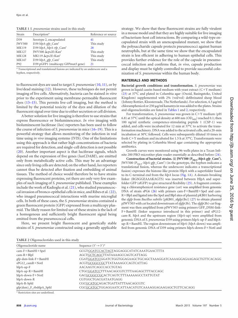
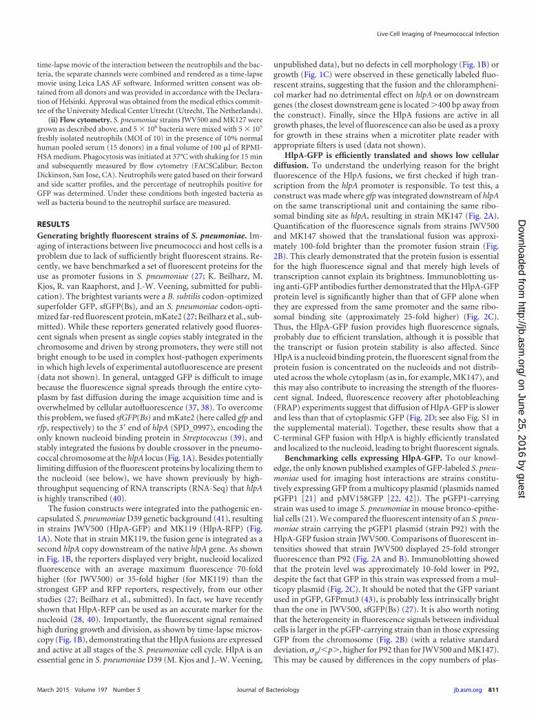
FIG 1 Generating bright fluorescent pneumococci. (A) Schematic representation of the conserved chromosomal locus of hlpA (SPD_0997) and the reporterstrains expressing hlpA-gfp and hlpA-rfp. The chloramphenicol acetyltransferase (cat) gene provides easy selection of chloramphenicol-resistant transformants.The hlpA-promoter (PhlpA) and the transcriptional terminators (lollipops) are indicated. Note that we were not able to generate a strain with hlpA-rfp as the onlycopy of hlpA, indicating that this fusion is not functional (28). (B) Time-lapse fluorescence microscopy of strains JWV500 and MK119, showing that HlpA-GFPand HlpA-RFP are stably expressed throughout growth and division. Images are overlays of fluorescence signal and phase-contrast micrographs. Scale bar, 5 �m.(C) Growth curves of cells grown in C�Y medium at 37°C showing that growth of the modified S. pneumoniae strains JWV500 and MK119 is similar to that ofthe wild-type strain D39 in vitro. Averages of three replicates are shown. Error bars show standard deviations.
Live-Cell Imaging of Pneumococcal Infection
March 2015 Volume 197 Number 5 jb.asm.org 809Journal of Bacteriology
on June 25, 2016 by guesthttp://jb.asm
.org/D
ownloaded from
for 5 min. Proteins from whole-cell extract were separated by SDS-PAGE(12% [wt/vol] polyacrylamide). One gel was stained with Coomassie toverify that similar protein quantities were loaded for each sample. Pro-teins were then blotted onto a polyvinylidene fluoride (PVDF) mem-brane, and GFP proteins and GFP fusion proteins were detected usingpolyclonal anti-GFP from rabbit (Invitrogen, The Netherlands) as theprimary antibody and horseradish peroxidase (HRP)-conjugated anti-rabbit IgG antibody (GE Healthcare, The Netherlands) as the secondaryantibody. Protein bands were quantified using ImageLab software (Bio-Rad, USA).
Studies of S. pneumoniae in mouse lung tissue. (i) Ethics statement.All animal experiments were done at the University of Leicester and wereconducted in strict accordance with guidelines of the Home Office of theUnited Kingdom. The University of Leicester Ethical Committee and theHome Office of the United Kingdom approved the protocol. All micewere scored for signs of disease using the method described by Mortonand Griffiths (33). Any mouse that became severely lethargic was culled, inaccordance with the Home Office License.
(ii) Mice. Female MF1 mice were purchased from Charles RiverLaboratories (United Kingdom) and were acclimatized for 1 weekprior to use. Mice used for infection experiments were between 9 and10 weeks of age.
(iii) Infection. For intranasal infection, animals were lightly anesthe-tized with a mixture of O2 and 2.5% (vol/vol) isoflurane (Abbott Labora-tories, Maidenhead, United Kingdom) and infected intranasally with aninoculum of 1 � 106 CFU in 50 �l of phosphate-buffered saline (PBS)(34) unless stated otherwise. Mice were regularly monitored for clinicalsigns of disease (33) and were culled at predetermined time points or ifthey became severely lethargic. Blood was taken after 24 h from the tailvein for CFU counts.
(iv) Preparation of lung tissue sections. At necropsy, whole lungswere completely immersed in 22-oxacalcitriol (OCT) embedding matrix(CellPath, Powys, United Kingdom) to slowly freeze (21). Tissue sections(15 �m) were cut using a microtome blade (Bright, Huntingdon, UnitedKingdom). Unstained sections were permanently preserved with a drop ofDPX mounting resin (BDH, Poole, United Kingdom) and a coverslip.
(v) Imaging of lung tissue sections. Microscopy of lung tissue sectionswas performed using a DV Elite microscope (Applied Precision) with ansCMOS camera using Solid-State Illumination (Applied Precision)through a 100� oil immersion objective (phase contrast; 1.30 numericalaperture [NA]). Phase-contrast images and GFP fluorescence images(0.5-s exposure time with 100% excitation light) were acquired as z-stacks(28 slices with 0.2 �m between each slice) using Softworx (Applied Pre-cision). Images were modified for publication using Softworx and ImageJ(http://rsb.info.nih.gov/ij/).
(vi) Statistical analysis. GraphPad Prism, version 5.0, software wasused to analyze the data. A log rank (Mantel-Cox) test and a Gehan-Breslow-Wilcoxon test were used to analyze the survival data. An un-paired t test was used to analyze the CFU counts from blood. Results wereconsidered significant at P values of �0.05.
Studies of interactions between S. pneumoniae and human epithe-lial cells. (i) Cell culture. The human type II lung epithelial cell line A549(ATCC CCL-185) was routinely cultured in Dulbecco’s modified Eaglemedium–nutrient mixture F-12 with GlutaMAX (Life Technologies, TheNetherlands) supplemented with 10% (vol/vol) fetal bovine serum (FBS;VWR, The Netherlands) and maintained at 37°C in a humidified 5%(vol/vol) CO2 atmosphere.
(ii) Coincubation of S. pneumoniae and the cell line for fluorescenceimaging. A coincubation experiment was performed as described by Mla-cha et al. (35) with some modifications. A549 cells were plated on eight-chamber microscopy slides (�-slide; Ibidi, Germany), and the monolayerconfluence was confirmed by phase-contrast microscopy. Prior to coin-cubation, the layer was rinsed twice with phosphate-buffered saline (PBS).S. pneumoniae strains were grown in C�Y medium (pH 6.8) until mid-logarithmic phase (OD600 of 0.2) and then centrifuged and resuspended
in RPMI 1640 medium without phenol red (Life Technologies, The Neth-erlands) but supplemented with 1% (vol/vol) PBS. Prior to mixing unen-capsulated and encapsulated strains (1:1), suspensions of S. pneumoniae(JWV500, MK128, MK119, and MK127) were adjusted to the multiplici-ties of infection (MOI) of 10 (i.e., 10 bacteria for every A549 cell) and thenadded onto the A549 monolayer. In order to optimize cell interaction, theslides were centrifuged (at 1,400 � g for 5 min) and then incubated at 37°Cin 5% (vol/vol) CO2 for 2 h. To remove nonadhering bacteria, the super-natants were aspirated, and subsequently the slides were washed twicewith RPMI 1640 medium supplemented with 1% (vol/vol) FBS.
(iii) Fluorescence imaging. During the process of imaging, the slideswere incubated at 37°C under a humidified 5% (vol/vol) CO2 atmosphere.Imaging was performed on a DV Elite microscope (Applied Precision)with an sCMOS camera using Solid-State Illumination (Applied Preci-sion) through a 60� oil immersion objective (bright field; 1.42 NA; work-ing distance [WD], 0.15 mm). The images were generated by first focusingon the monolayer of A549 using bright-field microscopy and then imag-ing on the FITC channel (excitation, 475 nm; emission, 523 nm) andAlexa 594 channel (excitation, 575 nm; emission, 625 nm). In order toquantify fluorescence intensity for adherent bacteria, microscopy stackswere split into the three channels: bright field, GFP, and RFP. Next, back-ground signals were removed from the GFP and RFP channels by adjust-ing their thresholds in ImageJ. Arbitrary values (RFP, minimum 200;GFP, minimum 250) were chosen to remove background fluorescencewhile retaining signals from bacterial cells. This redefinition of thresholdconverted the channels into binary images. In each channel, the amountof signal was calculated by multiplying the mean signal value by the area.For each image, the ratio of the RFP to GFP channel was calculated bydividing the RFP signal by GFP signal. Images were modified for publica-tion using softWoRx, version 6.1 (Applied Precision), and ImageJ.
(iv) Coincubation of S. pneumoniae and A549 for CFU counts. Co-incubation of S. pneumoniae mutant strains with the lung epithelial cellline for CFU counting was performed similarly to the coincubation pro-tocol for fluorescence imaging, except that A549 cells were plated on a24-well plate. After 2 h of incubation, the supernatant was removed, andthe cell layer was washed twice with PBS to remove nonadherent pneu-mococci. Trypsin-EDTA solution was added, and samples were incubatedat 37°C for 5 min to dislodge the epithelial layer along with adherent S.pneumoniae. The suspension was centrifuged (at 1,400 � g for 5 min) andresuspended in C�Y medium, diluted, and plated in 2% (vol/vol) bloodColumbia agar. After incubation at 37°C overnight, colonies werecounted manually. The data shown in Fig. 5B are a collection from threedifferent experiments performed on different days. Statistical analysis wasperformed using an unpaired two-tailed t test (GraphPad Prism 6).
Studies of interactions between S. pneumoniae and neutrophils. (i)Growth and imaging conditions. On the day of the experiment, S. pneu-moniae strains JWV500, MK119, MK127, and MK128 were inoculatedfrom a plate and grown in C�Y medium until the OD600 was 0.3. Bacteriawere washed and resuspended in RPMI 1640 medium containing 25 mMHEPES, L-glutamine (Lonza Biowhittaker, Basel, Switerland), and 0.05%human serum albumin (HSA; Sanquin, Amsterdam, The Netherlands)(RPMI-HSA). Strain JWV500 was mixed 1:1 with strain MK128, andstrain MK119 was mixed 1:1 with strain MK127. Bacteria were diluted inRPMI-HSA medium to a final concentration of 5 � 106 bacteria/ml. Wellsof an eight-well Lab-Tek II chambered cover glass (Thermo Scientific,Rochester, USA) were loaded with 200 �l of RPMI-HSA medium contain-ing the mixed S. pneumoniae strains. Subsequently, 5 � 105 freshly iso-lated neutrophils (36) were added (MOI of 10) to the well, and imagingwas started immediately while the neutrophils were settling at the coverglass bottom of the well. Microscopic image acquisition was performedusing a Leica TSC SP5 inverted microscope equipped with an HCX PLAPO 40� 0.85 objective (Leica Microsystems, The Netherlands). Themicroscope was encased in a dark-environment chamber that was main-tained at 37°C. The cells and bacteria were monitored for GFP (GFP ETfilter cube), RFP (N21 filter cube), and bright field every 10 s. To create a
Kjos et al.
810 jb.asm.org March 2015 Volume 197 Number 5Journal of Bacteriology
on June 25, 2016 by guesthttp://jb.asm
.org/D
ownloaded from
time-lapse movie of the interaction between the neutrophils and the bac-teria, the separate channels were combined and rendered as a time-lapsemovie using Leica LAS AF software. Informed written consent was ob-tained from all donors and was provided in accordance with the Declara-tion of Helsinki. Approval was obtained from the medical ethics commit-tee of the University Medical Center Utrecht (Utrecht, The Netherlands).
(ii) Flow cytometry. S. pneumoniae strains JWV500 and MK127 weregrown as described above, and 5 � 106 bacteria were mixed with 5 � 105
freshly isolated neutrophils (MOI of 10) in the presence of 10% normalhuman pooled serum (15 donors) in a final volume of 100 �l of RPMI-HSA medium. Phagocytosis was initiated at 37°C with shaking for 15 minand subsequently measured by flow cytometry (FACSCalibur; BectonDickinson, San Jose, CA). Neutrophils were gated based on their forwardand side scatter profiles, and the percentage of neutrophils positive forGFP was determined. Under these conditions both ingested bacteria aswell as bacteria bound to the neutrophil surface are measured.
RESULTSGenerating brightly fluorescent strains of S. pneumoniae. Im-aging of interactions between live pneumococci and host cells is aproblem due to lack of sufficiently bright fluorescent strains. Re-cently, we have benchmarked a set of fluorescent proteins for theuse as promoter fusions in S. pneumoniae (27; K. Beilharz, M.Kjos, R. van Raaphorst, and J.-W. Veening, submitted for publi-cation). The brightest variants were a B. subtilis codon-optimizedsuperfolder GFP, sfGFP(Bs), and an S. pneumoniae codon-opti-mized far-red fluorescent protein, mKate2 (27; Beilharz et al., sub-mitted). While these reporters generated relatively good fluores-cent signals when present as single copies stably integrated in thechromosome and driven by strong promoters, they were still notbright enough to be used in complex host-pathogen experimentsin which high levels of experimental autofluorescence are present(data not shown). In general, untagged GFP is difficult to imagebecause the fluorescence signal spreads through the entire cyto-plasm by fast diffusion during the image acquisition time and isoverwhelmed by cellular autofluorescence (37, 38). To overcomethis problem, we fused sfGFP(Bs) and mKate2 (here called gfp andrfp, respectively) to the 3= end of hlpA (SPD_0997), encoding theonly known nucleoid binding protein in Streptococcus (39), andstably integrated the fusions by double crossover in the pneumo-coccal chromosome at the hlpA locus (Fig. 1A). Besides potentiallylimiting diffusion of the fluorescent proteins by localizing them tothe nucleoid (see below), we have shown previously by high-throughput sequencing of RNA transcripts (RNA-Seq) that hlpAis highly transcribed (40).
The fusion constructs were integrated into the pathogenic en-capsulated S. pneumoniae D39 genetic background (41), resultingin strains JWV500 (HlpA-GFP) and MK119 (HlpA-RFP) (Fig.1A). Note that in strain MK119, the fusion gene is integrated as asecond hlpA copy downstream of the native hlpA gene. As shownin Fig. 1B, the reporters displayed very bright, nucleoid localizedfluorescence with an average maximum fluorescence 70-foldhigher (for JWV500) or 35-fold higher (for MK119) than thestrongest GFP and RFP reporters, respectively, from our otherstudies (27; Beilharz et al., submitted). In fact, we have recentlyshown that HlpA-RFP can be used as an accurate marker for thenucleoid (28, 40). Importantly, the fluorescent signal remainedhigh during growth and division, as shown by time-lapse micros-copy (Fig. 1B), demonstrating that the HlpA fusions are expressedand active at all stages of the S. pneumoniae cell cycle. HlpA is anessential gene in S. pneumoniae D39 (M. Kjos and J.-W. Veening,
unpublished data), but no defects in cell morphology (Fig. 1B) orgrowth (Fig. 1C) were observed in these genetically labeled fluo-rescent strains, suggesting that the fusion and the chlorampheni-col marker had no detrimental effect on hlpA or on downstreamgenes (the closest downstream gene is located �400 bp away fromthe construct). Finally, since the HlpA fusions are active in allgrowth phases, the level of fluorescence can also be used as a proxyfor growth in these strains when a microtiter plate reader withappropriate filters is used (data not shown).
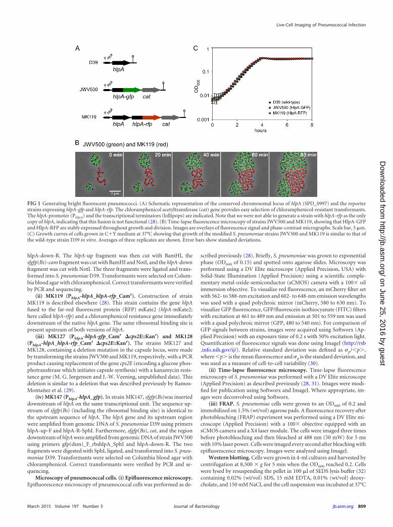
HlpA-GFP is efficiently translated and shows low cellulardiffusion. To understand the underlying reason for the brightfluorescence of the HlpA fusions, we first checked if high tran-scription from the hlpA promoter is responsible. To test this, aconstruct was made where gfp was integrated downstream of hlpAon the same transcriptional unit and containing the same ribo-somal binding site as hlpA, resulting in strain MK147 (Fig. 2A).Quantification of the fluorescence signals from strains JWV500and MK147 showed that the translational fusion was approxi-mately 100-fold brighter than the promoter fusion strain (Fig.2B). This clearly demonstrated that the protein fusion is essentialfor the high fluorescence signal and that merely high levels oftranscription cannot explain its brightness. Immunoblotting us-ing anti-GFP antibodies further demonstrated that the HlpA-GFPprotein level is significantly higher than that of GFP alone whenthey are expressed from the same promoter and the same ribo-somal binding site (approximately 25-fold higher) (Fig. 2C).Thus, the HlpA-GFP fusion provides high fluorescence signals,probably due to efficient translation, although it is possible thatthe transcript or fusion protein stability is also affected. SinceHlpA is a nucleoid binding protein, the fluorescent signal from theprotein fusion is concentrated on the nucleoids and not distrib-uted across the whole cytoplasm (as in, for example, MK147), andthis may also contribute to increasing the strength of the fluores-cent signal. Indeed, fluorescence recovery after photobleaching(FRAP) experiments suggest that diffusion of HlpA-GFP is slowerand less than that of cytoplasmic GFP (Fig. 2D; see also Fig. S1 inthe supplemental material). Together, these results show that aC-terminal GFP fusion with HlpA is highly efficiently translatedand localized to the nucleoid, leading to bright fluorescent signals.
Benchmarking cells expressing HlpA-GFP. To our knowl-edge, the only known published examples of GFP-labeled S. pneu-moniae used for imaging host interactions are strains constitu-tively expressing GFP from a multicopy plasmid (plasmids namedpGFP1 [21] and pMV158GFP [22, 42]). The pGFP1-carryingstrain was used to image S. pneumoniae in mouse bronco-epithe-lial cells (21). We compared the fluorescent intensity of an S. pneu-moniae strain carrying the pGFP1 plasmid (strain P92) with theHlpA-GFP fusion strain JWV500. Comparisons of fluorescent in-tensities showed that strain JWV500 displayed 25-fold strongerfluorescence than P92 (Fig. 2A and B). Immunoblotting showedthat the protein level was approximately 10-fold lower in P92,despite the fact that GFP in this strain was expressed from a mul-ticopy plasmid (Fig. 2C). It should be noted that the GFP variantused in pGFP, GFPmut3 (43), is probably less intrinsically brightthan the one in JWV500, sfGFP(Bs) (27). It is also worth notingthat the heterogeneity in fluorescence signals between individualcells is larger in the pGFP-carrying strain than in those expressingGFP from the chromosome (Fig. 2B) (with a relative standarddeviation, �p/�p�, higher for P92 than for JWV500 and MK147).This may be caused by differences in the copy numbers of plas-
Live-Cell Imaging of Pneumococcal Infection
March 2015 Volume 197 Number 5 jb.asm.org 811Journal of Bacteriology
on June 25, 2016 by guesthttp://jb.asm
.org/D
ownloaded from
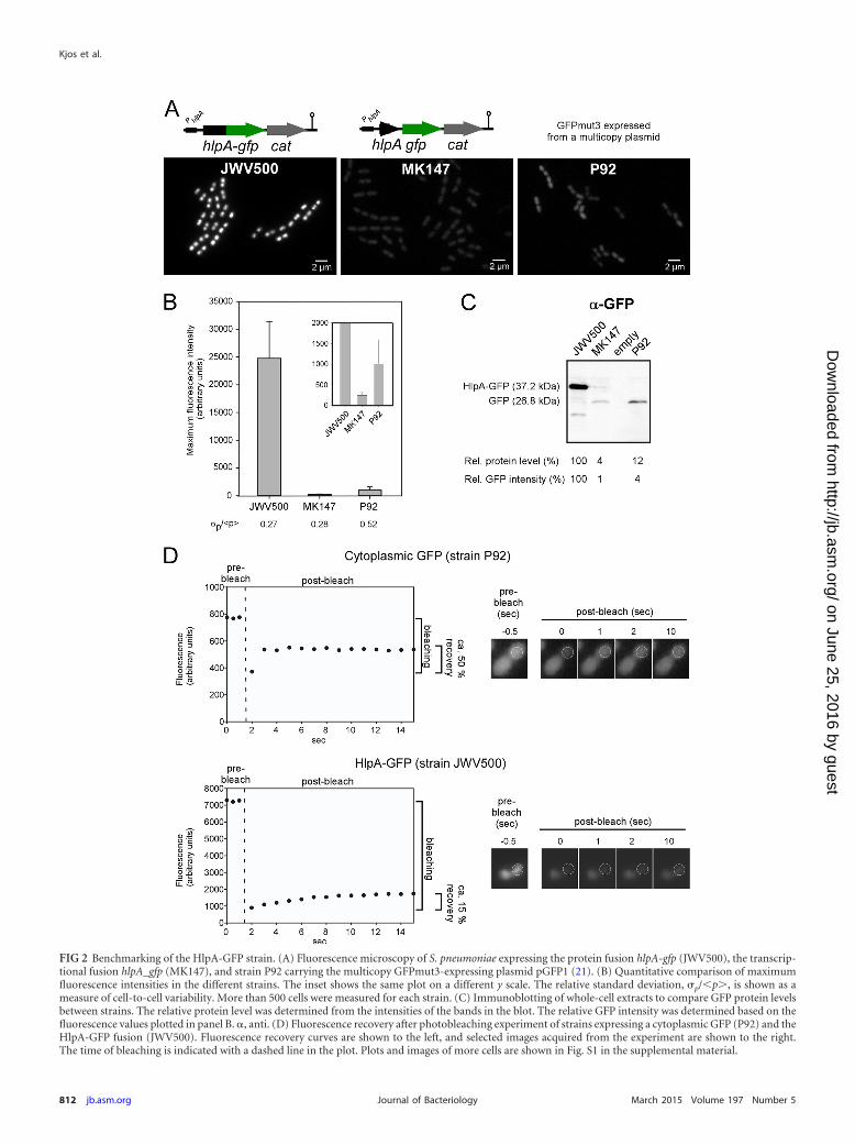
FIG 2 Benchmarking of the HlpA-GFP strain. (A) Fluorescence microscopy of S. pneumoniae expressing the protein fusion hlpA-gfp (JWV500), the transcrip-tional fusion hlpA_gfp (MK147), and strain P92 carrying the multicopy GFPmut3-expressing plasmid pGFP1 (21). (B) Quantitative comparison of maximumfluorescence intensities in the different strains. The inset shows the same plot on a different y scale. The relative standard deviation, �p/�p�, is shown as ameasure of cell-to-cell variability. More than 500 cells were measured for each strain. (C) Immunoblotting of whole-cell extracts to compare GFP protein levelsbetween strains. The relative protein level was determined from the intensities of the bands in the blot. The relative GFP intensity was determined based on thefluorescence values plotted in panel B. , anti. (D) Fluorescence recovery after photobleaching experiment of strains expressing a cytoplasmic GFP (P92) and theHlpA-GFP fusion (JWV500). Fluorescence recovery curves are shown to the left, and selected images acquired from the experiment are shown to the right.The time of bleaching is indicated with a dashed line in the plot. Plots and images of more cells are shown in Fig. S1 in the supplemental material.
Kjos et al.
812 jb.asm.org March 2015 Volume 197 Number 5Journal of Bacteriology
on June 25, 2016 by guesthttp://jb.asm
.org/D
ownloaded from
mids inside the cells; however, other factors, such as poor GFPfolding, may also contribute to such heterogeneity (30). Takentogether, the fluorescent and genetic properties of HlpA-GFP-expressing S. pneumoniae are superior to the system previouslydescribed.
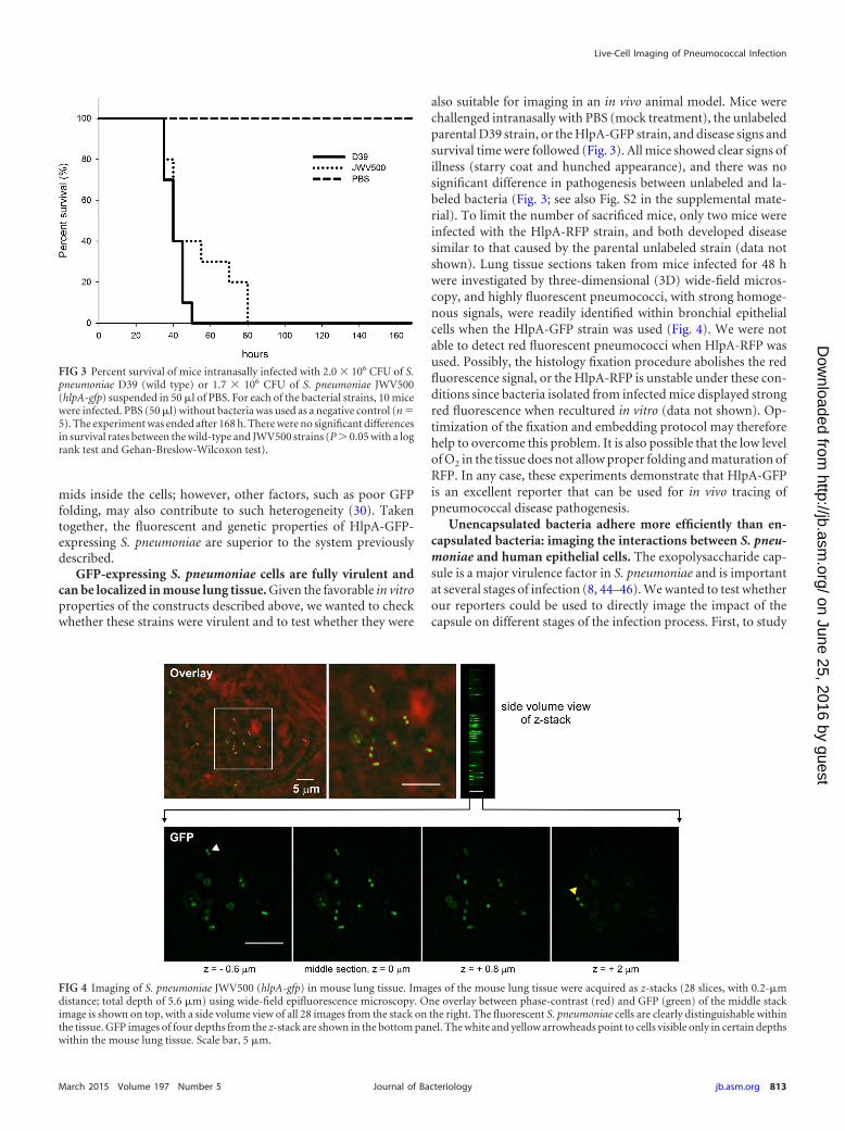
GFP-expressing S. pneumoniae cells are fully virulent andcan be localized in mouse lung tissue. Given the favorable in vitroproperties of the constructs described above, we wanted to checkwhether these strains were virulent and to test whether they were
also suitable for imaging in an in vivo animal model. Mice werechallenged intranasally with PBS (mock treatment), the unlabeledparental D39 strain, or the HlpA-GFP strain, and disease signs andsurvival time were followed (Fig. 3). All mice showed clear signs ofillness (starry coat and hunched appearance), and there was nosignificant difference in pathogenesis between unlabeled and la-beled bacteria (Fig. 3; see also Fig. S2 in the supplemental mate-rial). To limit the number of sacrificed mice, only two mice wereinfected with the HlpA-RFP strain, and both developed diseasesimilar to that caused by the parental unlabeled strain (data notshown). Lung tissue sections taken from mice infected for 48 hwere investigated by three-dimensional (3D) wide-field micros-copy, and highly fluorescent pneumococci, with strong homoge-nous signals, were readily identified within bronchial epithelialcells when the HlpA-GFP strain was used (Fig. 4). We were notable to detect red fluorescent pneumococci when HlpA-RFP wasused. Possibly, the histology fixation procedure abolishes the redfluorescence signal, or the HlpA-RFP is unstable under these con-ditions since bacteria isolated from infected mice displayed strongred fluorescence when recultured in vitro (data not shown). Op-timization of the fixation and embedding protocol may thereforehelp to overcome this problem. It is also possible that the low levelof O2 in the tissue does not allow proper folding and maturation ofRFP. In any case, these experiments demonstrate that HlpA-GFPis an excellent reporter that can be used for in vivo tracing ofpneumococcal disease pathogenesis.
Unencapsulated bacteria adhere more efficiently than en-capsulated bacteria: imaging the interactions between S. pneu-moniae and human epithelial cells. The exopolysaccharide cap-sule is a major virulence factor in S. pneumoniae and is importantat several stages of infection (8, 44–46). We wanted to test whetherour reporters could be used to directly image the impact of thecapsule on different stages of the infection process. First, to study
FIG 3 Percent survival of mice intranasally infected with 2.0 � 106 CFU of S.pneumoniae D39 (wild type) or 1.7 � 106 CFU of S. pneumoniae JWV500(hlpA-gfp) suspended in 50 �l of PBS. For each of the bacterial strains, 10 micewere infected. PBS (50 �l) without bacteria was used as a negative control (n �5). The experiment was ended after 168 h. There were no significant differencesin survival rates between the wild-type and JWV500 strains (P � 0.05 with a logrank test and Gehan-Breslow-Wilcoxon test).
FIG 4 Imaging of S. pneumoniae JWV500 (hlpA-gfp) in mouse lung tissue. Images of the mouse lung tissue were acquired as z-stacks (28 slices, with 0.2-�mdistance; total depth of 5.6 �m) using wide-field epifluorescence microscopy. One overlay between phase-contrast (red) and GFP (green) of the middle stackimage is shown on top, with a side volume view of all 28 images from the stack on the right. The fluorescent S. pneumoniae cells are clearly distinguishable withinthe tissue. GFP images of four depths from the z-stack are shown in the bottom panel. The white and yellow arrowheads point to cells visible only in certain depthswithin the mouse lung tissue. Scale bar, 5 �m.
Live-Cell Imaging of Pneumococcal Infection
March 2015 Volume 197 Number 5 jb.asm.org 813Journal of Bacteriology
on June 25, 2016 by guesthttp://jb.asm
.org/D
ownloaded from
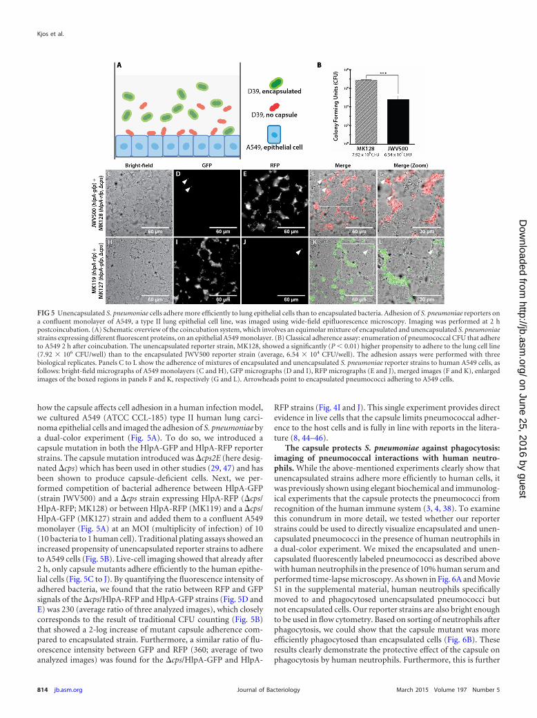
how the capsule affects cell adhesion in a human infection model,we cultured A549 (ATCC CCL-185) type II human lung carci-noma epithelial cells and imaged the adhesion of S. pneumoniae bya dual-color experiment (Fig. 5A). To do so, we introduced acapsule mutation in both the HlpA-GFP and HlpA-RFP reporterstrains. The capsule mutation introduced was �cps2E (here desig-nated �cps) which has been used in other studies (29, 47) and hasbeen shown to produce capsule-deficient cells. Next, we per-formed competition of bacterial adherence between HlpA-GFP(strain JWV500) and a �cps strain expressing HlpA-RFP (�cps/HlpA-RFP; MK128) or between HlpA-RFP (MK119) and a �cps/HlpA-GFP (MK127) strain and added them to a confluent A549monolayer (Fig. 5A) at an MOI (multiplicity of infection) of 10(10 bacteria to 1 human cell). Traditional plating assays showed anincreased propensity of unencapsulated reporter strains to adhereto A549 cells (Fig. 5B). Live-cell imaging showed that already after2 h, only capsule mutants adhere efficiently to the human epithe-lial cells (Fig. 5C to J). By quantifying the fluorescence intensity ofadhered bacteria, we found that the ratio between RFP and GFPsignals of the �cps/HlpA-RFP and HlpA-GFP strains (Fig. 5D andE) was 230 (average ratio of three analyzed images), which closelycorresponds to the result of traditional CFU counting (Fig. 5B)that showed a 2-log increase of mutant capsule adherence com-pared to encapsulated strain. Furthermore, a similar ratio of flu-orescence intensity between GFP and RFP (360; average of twoanalyzed images) was found for the �cps/HlpA-GFP and HlpA-
RFP strains (Fig. 4I and J). This single experiment provides directevidence in live cells that the capsule limits pneumococcal adher-ence to the host cells and is fully in line with reports in the litera-ture (8, 44–46).
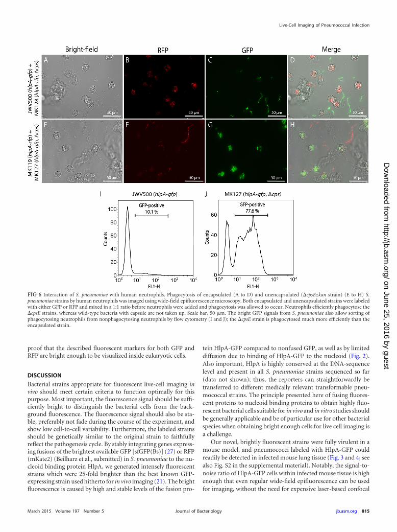
The capsule protects S. pneumoniae against phagocytosis:imaging of pneumococcal interactions with human neutro-phils. While the above-mentioned experiments clearly show thatunencapsulated strains adhere more efficiently to human cells, itwas previously shown using elegant biochemical and immunolog-ical experiments that the capsule protects the pneumococci fromrecognition of the human immune system (3, 4, 38). To examinethis conundrum in more detail, we tested whether our reporterstrains could be used to directly visualize encapsulated and unen-capsulated pneumococci in the presence of human neutrophils ina dual-color experiment. We mixed the encapsulated and unen-capsulated fluorescently labeled pneumococci as described abovewith human neutrophils in the presence of 10% human serum andperformed time-lapse microscopy. As shown in Fig. 6A and MovieS1 in the supplemental material, human neutrophils specificallymoved to and phagocytosed unencapsulated pneumococci butnot encapsulated cells. Our reporter strains are also bright enoughto be used in flow cytometry. Based on sorting of neutrophils afterphagocytosis, we could show that the capsule mutant was moreefficiently phagocytosed than encapsulated cells (Fig. 6B). Theseresults clearly demonstrate the protective effect of the capsule onphagocytosis by human neutrophils. Furthermore, this is further
FIG 5 Unencapsulated S. pneumoniae cells adhere more efficiently to lung epithelial cells than to encapsulated bacteria. Adhesion of S. pneumoniae reporters ona confluent monolayer of A549, a type II lung epithelial cell line, was imaged using wide-field epifluorescence microscopy. Imaging was performed at 2 hpostcoincubation. (A) Schematic overview of the coincubation system, which involves an equimolar mixture of encapsulated and unencapsulated S. pneumoniaestrains expressing different fluorescent proteins, on an epithelial A549 monolayer. (B) Classical adherence assay: enumeration of pneumococcal CFU that adhereto A549 2 h after coincubation. The unencapsulated reporter strain, MK128, showed a significantly (P � 0.01) higher propensity to adhere to the lung cell line(7.92 � 106 CFU/well) than to the encapsulated JWV500 reporter strain (average, 6.54 � 104 CFU/well). The adhesion assays were performed with threebiological replicates. Panels C to L show the adherence of mixtures of encapsulated and unencapsulated S. pneumoniae reporter strains to human A549 cells, asfollows: bright-field micrographs of A549 monolayers (C and H), GFP micrographs (D and I), RFP micrographs (E and J), merged images (F and K), enlargedimages of the boxed regions in panels F and K, respectively (G and L). Arrowheads point to encapsulated pneumococci adhering to A549 cells.
Kjos et al.
814 jb.asm.org March 2015 Volume 197 Number 5Journal of Bacteriology
on June 25, 2016 by guesthttp://jb.asm
.org/D
ownloaded from
proof that the described fluorescent markers for both GFP andRFP are bright enough to be visualized inside eukaryotic cells.
DISCUSSION
Bacterial strains appropriate for fluorescent live-cell imaging invivo should meet certain criteria to function optimally for thispurpose. Most important, the fluorescence signal should be suffi-ciently bright to distinguish the bacterial cells from the back-ground fluorescence. The fluorescence signal should also be sta-ble, preferably not fade during the course of the experiment, andshow low cell-to-cell variability. Furthermore, the labeled strainsshould be genetically similar to the original strain to faithfullyreflect the pathogenesis cycle. By stably integrating genes express-ing fusions of the brightest available GFP [sfGFP(Bs)] (27) or RFP(mKate2) (Beilharz et al., submitted) in S. pneumoniae to the nu-cleoid binding protein HlpA, we generated intensely fluorescentstrains which were 25-fold brighter than the best known GFP-expressing strain used hitherto for in vivo imaging (21). The brightfluorescence is caused by high and stable levels of the fusion pro-
tein HlpA-GFP compared to nonfused GFP, as well as by limiteddiffusion due to binding of HlpA-GFP to the nucleoid (Fig. 2).Also important, HlpA is highly conserved at the DNA-sequencelevel and present in all S. pneumoniae strains sequenced so far(data not shown); thus, the reporters can straightforwardly betransferred to different medically relevant transformable pneu-mococcal strains. The principle presented here of fusing fluores-cent proteins to nucleoid binding proteins to obtain highly fluo-rescent bacterial cells suitable for in vivo and in vitro studies shouldbe generally applicable and be of particular use for other bacterialspecies when obtaining bright enough cells for live cell imaging isa challenge.
Our novel, brightly fluorescent strains were fully virulent in amouse model, and pneumococci labeled with HlpA-GFP couldreadily be detected in infected mouse lung tissue (Fig. 3 and 4; seealso Fig. S2 in the supplemental material). Notably, the signal-to-noise ratio of HlpA-GFP cells within infected mouse tissue is highenough that even regular wide-field epifluorescence can be usedfor imaging, without the need for expensive laser-based confocal
FIG 6 Interaction of S. pneumoniae with human neutrophils. Phagocytosis of encapsulated (A to D) and unencapsulated (�cpsE::kan strain) (E to H) S.pneumoniae strains by human neutrophils was imaged using wide-field epifluorescence microscopy. Both encapsulated and unencapsulated strains were labeledwith either GFP or RFP and mixed in a 1:1 ratio before neutrophils were added and phagocytosis was allowed to occur. Neutrophils efficiently phagocytose the�cpsE strains, whereas wild-type bacteria with capsule are not taken up. Scale bar, 50 �m. The bright GFP signals from S. pneumoniae also allow sorting ofphagocytosing neutrophils from nonphagocytosing neutrophils by flow cytometry (I and J); the �cpsE strain is phagocytosed much more efficiently than theencapsulated strain.
Live-Cell Imaging of Pneumococcal Infection
March 2015 Volume 197 Number 5 jb.asm.org 815Journal of Bacteriology
on June 25, 2016 by guesthttp://jb.asm
.org/D
ownloaded from
fluorescence microscopy. Use of the HlpA-GFP-labeled strain willthus simplify the detection of S. pneumoniae during mouse infec-tion experiments.
We used our fluorescently labeled pneumococci to directly im-age different stages of pneumococcal infection using live cells. Tostudy adhesion to the epithelial layer, which is important for theinitial stages of infection, we used A549 (ATCC CCL-185) type IIhuman lung carcinoma epithelial cells. A549 has been used exten-sively to elucidate bacterial adherence and invasion to the host(48), and the pneumococcal coincubation model has been suc-cessfully used to identify essential host-pathogen virulence fac-tors, including PsaA (49), PavA (50), and PspC (51). We couldconfirm and demonstrate that the exopolysaccharide capsule in-hibits adhesion of S. pneumoniae to human epithelial cells andthus also the infection process (Fig. 5). This result is in line with astudy of Hammerschmidt et al. (52), where fixed samples of pneu-mococci and the A549 cell line were used for electron microscopyto show that pneumococci adhering to the human host have re-duced levels of capsule. Possibly, the capsule covers crucial pneu-mococcal adhesion-mediating surface proteins that are otherwiseexposed on the bacterial cell surface (45, 52), and this might alsopartly explain the success of colonization of nontypeable strainswithin human populations (9).
While the capsule is disadvantageous for adhesion to host cells,it is highly advantageous and necessary for pneumococcal evasionof the host immune system. For example, the capsule protectspneumococci against mucus-mediated clearing in the very earlystages of infection since unencapsulated bacteria agglutinatewithin mucus (44). Using dual-color strains, we were able to im-age in real time the protective function of the capsule againstphagocytosis by human neutrophils (Fig. 6). This protection maybe mediated by sterically hindering the neutrophils or by blockingof complement binding proteins (4, 7, 45, 53).
By the use of relatively straightforward imaging approaches,this work, for the first time, uses live-cell imaging to demonstratethe opposing attributes of the pneumococcal capsule at differentstages of infection. These results confirm that regulation of cap-sule production is critical for colonization of S. pneumoniaewithin the human host. The presence of such regulation is re-flected by the observation of phase variation of pneumococci (8,54), and studies suggest that little capsule is produced when pneu-mococci are in contact with the epithelial cell layer (52). However,the mode of regulation remains to be further unraveled, and anumber of genes and environmental factors seem to be important(45, 54–57). The imaging tools used in this work will pave the wayfor new types of experiments which will help further our knowl-edge on the pathogenesis of the pneumococcus.
ACKNOWLEDGMENTS
M.K. was supported by a long-term fellowship from the EuropeanBiochemical Societies. Work in the lab of J.-W.V. is supported by theEMBO Young Investigator Programme, a VIDI fellowship from theNetherlands Organization for Scientific Research, Earth and Life Sci-ences (864.12.001), and a European Research Council starting grant(337399-PneumoCell).
We thank W. J. Quax and R. Setroikromo from the Department ofPharmaceutical Biology, Groningen Research Institute of Pharmacy, Uni-versity of Groningen, the Netherlands, for their kind gift of human cellline A549.
REFERENCES1. O’Brien KL, Wolfson LJ, Watt JP, Henkle E, Deloria-Knoll M, McCall
N, Lee E, Mulholland K, Levine OS, Cherian T. 2009. Burden of diseasecaused by Streptococcus pneumoniae in children younger than 5 years:global estimates. Lancet 374:893–902. http://dx.doi.org/10.1016/S0140-6736(09)61204-6.
2. Griffith F. 1928. The significance of pneumococcal types. J Hyg (Lond)27:113–159. http://dx.doi.org/10.1017/S0022172400031879.
3. Hyams C, Camberlein E, Cohen JM, Bax K, Brown JS. 2010. TheStreptococcus pneumoniae capsule inhibits complement activity and neu-trophil phagocytosis by multiple mechanisms. Infect Immun 78:704 –715.http://dx.doi.org/10.1128/IAI.00881-09.
4. Abeyta M, Hardy GG, Yother J. 2003. Genetic alteration of capsule typebut not PspA type affects accessibility of surface-bound complement andsurface antigens of Streptococcus pneumoniae. Infect Immun 71:218 –225.http://dx.doi.org/10.1128/IAI.71.1.218-225.2003.
5. Kim JO, Romero-Steiner S, Sørensen UB, Blom J, Carvalho M, BarnardS, Carlone G, Weiser JN. 1999. Relationship between cell surface carbo-hydrates and intrastrain variation on opsonophagocytosis of Streptococcuspneumoniae. Infect Immun 67:2327–2333.
6. Weinberger DM, Trzcinski K, Lu Y-J, Bogaert D, Brandes A, Galagan J,Anderson PW, Malley R, Lipsitch M. 2009. Pneumococcal capsularpolysaccharide structure predicts serotype prevalence. PLoS Pathog5:e1000476. http://dx.doi.org/10.1371/journal.ppat.1000476.
7. Henriques-Normark B, Tuomanen EI. 2013. The pneumococcus: epide-miology, microbiology, and pathogenesis. Cold Spring Harb PerspectMed 3:a010215. http://dx.doi.org/10.1101/cshperspect.a010215.
8. Weiser JN, Austrian R, Sreenivasan PK, Masure HR. 1994. Phase vari-ation in pneumococcal opacity: relationship between colonial morphol-ogy and nasopharyngeal colonization. Infect Immun 62:2582–2589.
9. Chewapreecha C, Harris SR, Croucher NJ, Turner C, Marttinen P,Cheng L, Pessia A, Aanensen DM, Mather AE, Page AJ, Salter SJ, HarrisD, Nosten F, Goldblatt D, Corander J, Parkhill J, Turner P, Bentley SD.2014. Dense genomic sampling identifies highways of pneumococcal re-combination. Nat Genet 46:305–309. http://dx.doi.org/10.1038/ng.2895.
10. Zhang Z, Clarke TB, Weiser JN. 2009. Cellular effectors mediatingTh17-dependent clearance of pneumococcal colonization in mice. J ClinInvest 119:1899 –1909. http://dx.doi.org/10.1172/JCI36731.
11. Sanchez CJ, Shivshankar P, Stol K, Trakhtenbroit S, Sullam PM, SauerK, Hermans PWM, Orihuela CJ. 2010. The pneumococcal serine-richrepeat protein is an intra-species bacterial adhesin that promotes bacterialaggregation in vivo and in biofilms. PLoS Pathog 6:e1001044. http://dx.doi.org/10.1371/journal.ppat.1001044.
12. Parker D, Soong G, Planet P, Brower J, Ratner AJ, Prince A. 2009. TheNanA neuraminidase of Streptococcus pneumoniae is involved in biofilmformation. Infect Immun 77:3722–3730. http://dx.doi.org/10.1128/IAI.00228-09.
13. Elhaik-Goldman S, Kafka D, Yossef R, Hadad U, Elkabets M, Vallon-Eberhard A, Hulihel L, Jung S, Ghadially H, Braiman A, Apte RN,Mandelboim O, Dagan R, Mizrachi-Nebenzahl Y, Porgador A. 2011.The natural cytotoxicity receptor 1 contribution to early clearance ofStreptococcus pneumoniae and to natural killer-macrophage cross talk.PLoS One 6:e23472. http://dx.doi.org/10.1371/journal.pone.0023472.
14. Srivastava A, Casey H, Johnson N, Levy O, Malley R. 2007. Recombi-nant bactericidal/permeability-increasing protein rBPI21 protects againstpneumococcal disease. Infect Immun 75:342–349. http://dx.doi.org/10.1128/IAI.01089-06.
15. Colino J, Shen Y, Snapper CM. 2002. Dendritic cells pulsed with intactStreptococcus pneumoniae elicit both protein- and polysaccharide-specificimmunoglobulin isotype responses in vivo through distinct mechanisms.J Exp Med 195:1–14. http://dx.doi.org/10.1084/jem.20011432.
16. Mook-Kanamori BB, Rouse MS, Kang C-I, van de Beek D, Steckel-berg JM, Patel R. 2009. Daptomycin in experimental murine pneu-mococcal meningitis. BMC Infect Dis 9:50. http://dx.doi.org/10.1186/1471-2334-9-50.
17. Francis KP, Yu J, Bellinger-Kawahara C, Joh D, Hawkinson MJ, Xiao G,Purchio TF, Caparon MG, Lipsitch M, Contag PR. 2001. Visualizingpneumococcal infections in the lungs of live mice using bioluminescentStreptococcus pneumoniae transformed with a novel Gram-positive luxtransposon. Infect Immun 69:3350 –3358. http://dx.doi.org/10.1128/IAI.69.5.3350-3358.2001.
18. Orihuela CJ, Gao G, Francis KP, Yu J, Tuomanen EI. 2004. Tissue-
Kjos et al.
816 jb.asm.org March 2015 Volume 197 Number 5Journal of Bacteriology
on June 25, 2016 by guesthttp://jb.asm
.org/D
ownloaded from
specific contributions of pneumococcal virulence factors to pathogenesis.J Infect Dis 190:1661–1669. http://dx.doi.org/10.1086/424596.
19. Wang J, Barke RA, Charboneau R, Schwendener R, Roy S. 2008.Morphine induces defects in early response of alveolar macrophages toStreptococcus pneumoniae by modulating TLR9-NF-�B signaling. J Immu-nol 180:3594 –3600. http://dx.doi.org/10.4049/jimmunol.180.5.3594.
20. Johnson AW, Sidman JD, Lin J. 2013. Bioluminescent imaging of pneu-mococcal otitis media in chinchillas. Ann Otol Rhinol Laryngol 122:344 –352. http://dx.doi.org/10.1177/000348941312200510.
21. Kadioglu A, Sharpe JA, Lazou I, Svanborg C, Ockleford C, MitchellTJ, Andrew PW. 2001. Use of green fluorescent protein in visualisa-tion of pneumococcal invasion of broncho-epithelial cells in vivo.FEMS Microbiol Lett 194:105–110. http://dx.doi.org/10.1111/j.1574-6968.2001.tb09454.x.
22. Ribes S, Ebert S, Regen T, Agarwal A, Tauber SC, Czesnik D, Spreer A,Bunkowski S, Eiffert H, Hanisch U-K, Hammerschmidt S, Nau R. 2010.Toll-like receptor stimulation enhances phagocytosis and intracellularkilling of nonencapsulated and encapsulated Streptococcus pneumoniae bymurine microglia. Infect Immun 78:865– 871. http://dx.doi.org/10.1128/IAI.01110-09.
23. Martin B, Garcia P, Castanié M-P, Claverys J-P. 1995. The recA gene ofStreptococcus pneumoniae is part of a competence-induced operon andcontrols lysogenic induction. Mol Microbiol 15:367–379. http://dx.doi.org/10.1111/j.1365-2958.1995.tb02250.x.
24. Sorg RA, Kuipers OP, Veening J-W. Gene expression platform for syn-thetic biology in the human pathogen Streptococcus pneumoniae. ACSSynth Biol, in press. http://dx.doi.org/10.1021/sb500229s.
25. Arai R, Ueda H, Kitayama A, Kamiya N, Nagamune T. 2001. Design ofthe linkers which effectively separate domains of a bifunctional fusionprotein. Protein Eng 14:529 –532. http://dx.doi.org/10.1093/protein/14.8.529.
26. Yuzenkova Y, Gamba P, Herber M, Attaiech L, Shafeeq S, Kuipers OP,Klumpp S, Zenkin N, Veening J-W. 2014. Control of transcriptionelongation by GreA determines rate of gene expression in Streptococcuspneumoniae. Nucleic Acids Res 42:10987–10999. http://dx.doi.org/10.1093/nar/gku790.
27. Overkamp W, Beilharz K, Detert Oude Weme R, Solopova A, KarsensH, Kovács ÁT, Kok J, Kuipers OP, Veening J-W. 2013. Benchmarkingvarious green fluorescent protein variants in Bacillus subtilis, Streptococcuspneumoniae, and Lactococcus lactis for live cell imaging. Appl EnvironMicrobiol 79:6481– 6490. http://dx.doi.org/10.1128/AEM.02033-13.
28. Kjos M, Veening J-W. 2014. Tracking of chromosome dynamics in liveStreptococcus pneumoniae reveals that transcription promotes chromo-some segregation. Mol Microbiol 91:1088 –1105. http://dx.doi.org/10.1111/mmi.12517.
29. Ramos-Montañez S, Kazmierczak KM, Hentchel KL, Winkler ME.2010. Instability of ackA (acetate kinase) mutations and their effects onacetyl phosphate and ATP amounts in Streptococcus pneumoniae D39. JBacteriol 192:6390 – 6400. http://dx.doi.org/10.1128/JB.00995-10.
30. Jørgensen MG, van Raaphorst R, Veening J-W. 2013. Noise and sto-chasticity in gene expression: a pathogenic fate determinant, p 157–176. InHarwood C, Wipat A (ed), Microbial synthetic biology Academic Press,Oxford, United Kingdom.
31. De Jong IG, Beilharz K, Kuipers OP, Veening J-W. 2011. Live cellimaging of Bacillus subtilis and Streptococcus pneumoniae using auto-mated time-lapse microscopy. J Vis Exp 53:3145. http://dx.doi.org/10.3791/3145.
32. Shoemaker NB, Guild WR. 1972. Kinetics of integration of transformingDNA in pneumococcus. Proc Natl Acad Sci U S A 69:3331–3335. http://dx.doi.org/10.1073/pnas.69.11.3331.
33. Morton DB, Griffiths PH. 1985. Guidelines on the recognition of pain,distress and discomfort in experimental animals and an hypothesis forassessment. Vet Rec 116:431– 436. http://dx.doi.org/10.1136/vr.116.16.431.
34. Kadioglu A, Gingles NA, Grattan K, Kerr A, Mitchell TJ, Andrew PW.2000. Host cellular immune response to pneumococcal lung infection inmice. Infect Immun 68:492–501. http://dx.doi.org/10.1128/IAI.68.2.492-501.2000.
35. Mlacha SZK, Romero-Steiner S, Hotopp JCD, Kumar N, Ishmael N,Riley DR, Farooq U, Creasy TH, Tallon LJ, Liu X, Goldsmith CS,Sampson J, Carlone GM, Hollingshead SK, Scott JAG, Tettelin H. 2013.Phenotypic, genomic, and transcriptional characterization of Streptococ-
cus pneumoniae interacting with human pharyngeal cells. BMC Genomics14:383. http://dx.doi.org/10.1186/1471-2164-14-383.
36. Surewaard BGJ, van Strijp JAG, Nijland R. 2013. Studying interactionsof Staphylococcus aureus with neutrophils by flow cytometry and timelapse microscopy. J Vis Exp 77:e50788. http://dx.doi.org/10.3791/50788.
37. Yu J, Xiao J, Ren X, Lao K, Xie XS. 2006. Probing gene expression in livecells, one protein molecule at a time. Science 311:1600 –1603. http://dx.doi.org/10.1126/science.1119623.
38. Davis RW, Timlin JA, Kaiser JN, Sinclair MB, Jones HDT, Lane TW.2010. Accurate detection of low levels of fluorescence emission in auto-fluorescent background: Francisella-infected macrophage cells. MicroscMicroanal 16:478 – 487. http://dx.doi.org/10.1017/S1431927610000322.
39. Liu D, Yumoto H, Murakami K, Hirota K, Ono T, Nagamune H,Kayama S, Matsuo T, Miyake Y. 2008. The essentiality and involvementof Streptococcus intermedius histone-like DNA-binding protein in bacte-rial viability and normal growth. Mol Microbiol 68:1268 –1282. http://dx.doi.org/10.1111/j.1365-2958.2008.06232.x.
40. Slager J, Kjos M, Attaiech L, Veening J-W. 2014. Antibiotic-induced repli-cation stress triggers bacterial competence by increasing gene dosage near theorigin. Cell 157:395–406. http://dx.doi.org/10.1016/j.cell.2014.01.068.
41. Avery OT, Macleod CM, McCarty M. 1944. Studies on the chemicalnature of the substance inducing transformation of pneumococcal types:induction of transformation by a deoxyribonucleic acid fraction isolatedfrom pneumococcus type III. J Exp Med 79:137–158. http://dx.doi.org/10.1084/jem.79.2.137.
42. Nieto C, Espinosa M. 2003. Construction of the mobilizable plasmidpMV158GFP, a derivative of pMV158 that carries the gene encoding thegreen fluorescent protein. Plasmid 49:281–285. http://dx.doi.org/10.1016/S0147-619X(03)00020-9.
43. Cormack BP, Valdivia RH, Falkow S. 1996. FACS-optimized mutants ofthe green fluorescent protein (GFP). Gene 173:33–38. http://dx.doi.org/10.1016/0378-1119(95)00685-0.
44. Nelson AL, Roche AM, Gould JM, Chim K, Ratner AJ, Weiser JN. 2007.Capsule enhances pneumococcal colonization by limiting mucus-mediated clearance. Infect Immun 75:83–90. http://dx.doi.org/10.1128/IAI.01475-06.
45. Kadioglu A, Weiser JN, Paton JC, Andrew PW. 2008. The role ofStreptococcus pneumoniae virulence factors in host respiratory coloniza-tion and disease. Nat Rev Microbiol 6:288 –301. http://dx.doi.org/10.1038/nrmicro1871.
46. Weiser JN. 2010. The pneumococcus: why a commensal misbehaves. JMol Med 88:97–102. http://dx.doi.org/10.1007/s00109-009-0557-x.
47. Cartee RT, Forsee WT, Bender MH, Ambrose KD, Yother J. 2005. CpsEfrom type 2 Streptococcus pneumoniae catalyzes the reversible addition ofglucose-1-phosphate to a polyprenyl phosphate acceptor, initiating type 2capsule repeat unit formation. J Bacteriol 187:7425–7433. http://dx.doi.org/10.1128/JB.187.21.7425-7433.2005.
48. Talbot UM, Paton AW, Paton JC. 1996. Uptake of Streptococcus pneu-moniae by respiratory epithelial cells. Infect Immun 64:3772–3777.
49. Berry AM, Paton JC. 1996. Sequence heterogeneity of PsaA, a 37-kilodalton putative adhesin essential for virulence of Streptococcus pneu-moniae. Infect Immun 64:5255–5262.
50. Pracht D, Elm C, Gerber J, Bergmann S, Rohde M, Seiler M, Kim KS,Jenkinson HF, Nau R, Hammerschmidt S. 2005. PavA of Streptococcuspneumoniae modulates adherence, invasion, and meningeal inflamma-tion. Infect Immun 73:2680 –2689. http://dx.doi.org/10.1128/IAI.73.5.2680-2689.2005.
51. Hammerschmidt S, Agarwal V, Kunert A, Haelbich S, Skerka C, ZipfelPF. 2007. The host immune regulator factor H interacts via two contactsites with the PspC protein of Streptococcus pneumoniae and mediatesadhesion to host epithelial cells. J Immunol 178:5848 –5858. http://dx.doi.org/10.4049/jimmunol.178.9.5848.
52. Hammerschmidt S, Wolff S, Hocke A, Rosseau S, Müller E, Rohde M.2005. Illustration of pneumococcal polysaccharide capsule during adher-ence and invasion of epithelial cells. Infect Immun 73:4653– 4667. http://dx.doi.org/10.1128/IAI.73.8.4653-4667.2005.
53. Wartha F, Beiter K, Albiger B, Fernebro J, Zychlinsky A, Normark S,Henriques-Normark B. 2007. Capsule and D-alanylated lipoteichoic acidsprotect Streptococcus pneumoniae against neutrophil extracellular traps.Cell Microbiol 9:1162–1171. http://dx.doi.org/10.1111/j.1462-5822.2006.00857.x.
54. Manso AS, Chai MH, Atack JM, Furi L, De Ste Croix M, Haigh R,Trappetti C, Ogunniyi AD, Shewell LK, Boitano M, Clark TA, Korlach
Live-Cell Imaging of Pneumococcal Infection
March 2015 Volume 197 Number 5 jb.asm.org 817Journal of Bacteriology
on June 25, 2016 by guesthttp://jb.asm
.org/D
ownloaded from

J, Blades M, Mirkes E, Gorban AN, Paton JC, Jennings MP, OggioniMR. 2014. A random six-phase switch regulates pneumococcal virulencevia global epigenetic changes. Nat Commun 5:5055. http://dx.doi.org/10.1038/ncomms6055.
55. Shainheit MG, Mulé M, Camilli A. 2014. The core promoter of thecapsule operon of Streptococcus pneumoniae is necessary for colonizationand invasive disease. Infect Immun 82:694 –705. http://dx.doi.org/10.1128/IAI.01289-13.
56. Geno KA, Hauser JR, Gupta K, Yother J. 2014. Streptococcus pneumoniaephosphotyrosine phosphatase CpsB and alterations in capsule productionresulting from changes in oxygen availability. J Bacteriol 196:1992–2003.http://dx.doi.org/10.1128/JB.01545-14.
57. Yother J. 2011. Capsules of Streptococcus pneumoniae and other bacteria:paradigms for polysaccharide biosynthesis and regulation. Annu Rev Mi-crobiol 65:563–581. http://dx.doi.org/10.1146/annurev.micro.62.081307.162944.
Kjos et al.
818 jb.asm.org March 2015 Volume 197 Number 5Journal of Bacteriology
on June 25, 2016 by guesthttp://jb.asm
.org/D
ownloaded from
Related Documents