■ ■ • ■ WWW.JENONLINE.ORG 1 Master Proof ymen1986.pdf 3 TECHNIQUE—WHEN IN DOUBT , DON’T 4 NECESSARILY T AKE IT OUT! 5 Authors: Scott DeBoer, MSN, RN, CEN, CPEN, CCRN, CFRN, EMT-P, 6 Michael Seaver, RN, David Vidra, CLPN, WCC, MA, Bill Robinson, and Jennifer Klepacki, 7 Chicago and Vernon Hills, IL, Dyer, IN, Cleveland, OH, Las Vegas, NV, Southington, CT 30 9 body piercing, or implants, have been practiced in become increasingly common to remove body jewelry for 31 10 societies across the world for thousands of years. In diagnostic imaging (computed tomography/magnetic reso- 32 11 recent years, these practices have become more common nance imaging) or surgical procedures. 2 However, the ques- 33 12 and widespread, moving beyond what we might have pre- tion of whether removal of the jewelry is indeed necessary, 34 13 viously considered a curious subculture of practitioners. especially for elective or non-emergency health care, con- 35 14 The implications on emergency nursing care have also tinues to be debated in the medical literature. For example, 36 15 become more evident as we strive to understand and pro- does tongue jewelry need to be removed for emergent intu- 37 16 vide optimal care for this ever-increasing segment of our bation? (No; intubate first, then worry about removing the 38 17 population. This article reviews the transfer technique of jewelry later.) Do nipple piercings need to be removed 39 18 exchanging body piercing jewelry with an intravenous prior to defibrillation? (No.) Does a piercing need to be 40 19 catheter to maintain piercing patency. removed if the site is infected? (No, or an abscess can 41 20 In a recent study of U.S. undergraduate college stu- form.) Does a penile piercing need to be removed prior 42 21 Q1 dents, 51% had something pierced besides their ears. 1 That to placement of a Foley urinary catheter? (Sometimes, 43 22 means that in this study, 1 odds were better than a “coin depending on whether the piercing crosses the urethra.) 3,4 44 23 flip” as to whether a young adult had body piercings For many medical procedures and types of body jewelry, 45 24 (and 22% had tattoos as well). Although no formal statis- the jury is still out and the verdict has yet to be decided. 46 25 tics exist regarding the number of body piercings per- Removal of body jewelry for procedures has raised con- 47 26 formed each year, it is estimated that at least several cerns among patients about maintaining patency of the pier- 48 27 hundred thousand body piercings are performed annually, cing tract. When experienced body piercers are asked how 49 28 and that number is not declining (E. Angel, oral commu- long a piercing “hole” stays open once the jewelry has been 50 29 Q2 nication, December 2010). removed, the most common answer is simply, “We just don’t 51 know.” Certainly, piercing tracts that have been in place for 52 many years have a better chance of staying open for a longer 53 Scott DeBoer is Flight Nurse, University of Chicago Hospitals, Chicago, IL, period when the jewelry is removed, but the truth is we just 54 and Founder, Peds-R-Us Medical Education, Dyer, IN. don’t know how long any tract will stay open. According to 55 Michael Seaver is Senior Healthcare Informatics Consultant, Vernon Hills, IL. professional body piercers, the only reliable way to ensure that 56 David Vidra is a Body Piercer and President, Health Educators, Inc, the piercing tract remains open is to keep something in it. Cleveland, OH. 57 C L I N I C A L 1 BREASTS, BELLIES, BELOW , AND BEYOND: 2 BODY PIERCING JEWELRY AND THE TRANSFER 8 B ody modifications or body art, whether tattooing, With the recent “epidemic” of body piercings, it has Bill Robinson is a Body Piercer, Starlight Tattoo, Las Vegas, NV. Jennifer Klepacki is an Artist and Illustrator, Southington, CT. For correspondence, write: Scott DeBoer, MSN, RN, CEN, CPEN, CCRN, CFRN, EMT-P, 9052 Beall St, Dyer, IN 46311; E-mail: [email protected]. J Emerg Nurs ■. 0099-1767/$36.00 Copyright © 2011 Emergency Nurses Association. Published by Elsevier Inc. All rights reserved. doi: 10.1016/j.jen.2011.03.001 In the past, several anesthesia and surgical journal arti- 58 cles have described the use of various sorts of retainers 59 (ranging from epidural catheters to intravenous extension 60 tubing) to attempt to keep the hole open. 2,5-9 Although 61 some retainers for body jewelry are commercially available, 62 the experience of several body piercers is that these devices 63 can come apart more easily than conventional metal jewelry, 64 making them potentially less desirable. These retainers have 65 a shape that is similar body jewelry, but they generally are 66 non-metallic and either clear or flesh colored. It is important 67

Welcome message from author
This document is posted to help you gain knowledge. Please leave a comment to let me know what you think about it! Share it to your friends and learn new things together.
Transcript
■ ■ • ■ WWW.JENONLINE.ORG 1
Master Proof ymen1986.pdf
3 TECHNIQUE—WHEN IN DOUBT, DON’T
4 NECESSARILY TAKE IT OUT!
5
Authors: Scott DeBoer, MSN, RN, CEN, CPEN, CCRN, CFRN, EMT-P,
6 Michael Seaver, RN, David Vidra, CLPN, WCC, MA, Bill Robinson, and Jennifer Klepacki,
7 Chicago and Vernon Hills, IL, Dyer, IN, Cleveland, OH, Las Vegas, NV, Southington, CT
30
9 body piercing, or implants, have been practiced in become increasingly common to remove body jewelry for 31
10 societies across the world for thousands of years. In diagnostic imaging (computed tomography/magnetic reso- 32
11 recent years, these practices have become more common nance imaging) or surgical procedures.2 However, the ques- 33
12 and widespread, moving beyond what we might have pre- tion of whether removal of the jewelry is indeed necessary, 34
13 viously considered a curious subculture of practitioners. especially for elective or non-emergency health care, con- 35
14 The implications on emergency nursing care have also tinues to be debated in the medical literature. For example, 36
15 become more evident as we strive to understand and pro- does tongue jewelry need to be removed for emergent intu- 37
16 vide optimal care for this ever-increasing segment of our bation? (No; intubate first, then worry about removing the 38
17 population. This article reviews the transfer technique of jewelry later.) Do nipple piercings need to be removed 39
18 exchanging body piercing jewelry with an intravenous prior to defibrillation? (No.) Does a piercing need to be 40
19 catheter to maintain piercing patency. removed if the site is infected? (No, or an abscess can 41
20 In a recent study of U.S. undergraduate college stu- form.) Does a penile piercing need to be removed prior 42
21Q1 dents, 51% had something pierced besides their ears.1 That to placement of a Foley urinary catheter? (Sometimes, 43
22 means that in this study,1 odds were better than a “coin depending on whether the piercing crosses the urethra.)3,4 44
23 flip” as to whether a young adult had body piercings For many medical procedures and types of body jewelry, 45
24 (and 22% had tattoos as well). Although no formal statis- the jury is still out and the verdict has yet to be decided. 46
25 tics exist regarding the number of body piercings per- Removal of body jewelry for procedures has raised con- 47
26 formed each year, it is estimated that at least several cerns among patients about maintaining patency of the pier- 48
27 hundred thousand body piercings are performed annually, cing tract. When experienced body piercers are asked how 49
28 and that number is not declining (E. Angel, oral commu- long a piercing “hole” stays open once the jewelry has been 50
29Q2 nication, December 2010). removed, the most common answer is simply, “We just don’t 51
know.” Certainly, piercing tracts that have been in place for 52
many years have a better chance of staying open for a longer 53
Scott DeBoer is Flight Nurse, University of Chicago Hospitals, Chicago, IL, period when the jewelry is removed, but the truth is we just 54
and Founder, Peds-R-Us Medical Education, Dyer, IN.
don’t know how long any tract will stay open. According to 55
Michael Seaver is Senior Healthcare Informatics Consultant, Vernon Hills, IL. professional body piercers, the only reliable way to ensure that
56
David Vidra is a Body Piercer and President, Health Educators, Inc, the piercing tract remains open is to keep something in it.
Cleveland, OH. 57
C L I N I C A L
1 BREASTS, BELLIES, BELOW, AND BEYOND:
2 BODY PIERCING JEWELRY AND THE TRANSFER
8
Body modifications or body art, whether tattooing,
With the recent “epidemic” of body piercings, it has
Bill Robinson is a Body Piercer, Starlight Tattoo, Las Vegas, NV.
Jennifer Klepacki is an Artist and Illustrator, Southington, CT.
For correspondence, write: Scott DeBoer, MSN, RN, CEN, CPEN, CCRN,
CFRN, EMT-P, 9052 Beall St, Dyer, IN 46311; E-mail: [email protected].
J Emerg Nurs ■.
0099-1767/$36.00
Copyright © 2011 Emergency Nurses Association. Published by Elsevier Inc.
All rights reserved.
doi: 10.1016/j.jen.2011.03.001
In the past, several anesthesia and surgical journal arti- 58
cles have described the use of various sorts of retainers 59
(ranging from epidural catheters to intravenous extension 60
tubing) to attempt to keep the hole open.2,5-9 Although 61
some retainers for body jewelry are commercially available, 62
the experience of several body piercers is that these devices 63
can come apart more easily than conventional metal jewelry, 64
making them potentially less desirable. These retainers have 65
a shape that is similar body jewelry, but they generally are 66
non-metallic and either clear or flesh colored. It is important 67

CLINICAL/DeBoer et al
■ • ■ ■ 2 JOURNAL OF EMERGENCY NURSING
Master Proof ymen1986.pdf
FIGURE 1
Various types of barbell body jewelry: A, closed; B, open. Remove one end of the barbell by twisting counterclockwise and maintaining a grasp on the other end of Q3 the jewelry. (Photos courtesy of Anatometal, www.anatometal.com.) This figure can be viewed in color and as a full-page document at www.jenonline.org.
FIGURE 2
Various types of ring body jewelry: A, closed; B, open. Removal is best accomplished with the use of ring-opening pliers. (Photos courtesy of Anatometal,
www.anatometal.com.) This figure can be viewed in color and as a full-page document at www.jenonline.org.
68 to note that many retainer materials have not been specifi-
69 cally approved for medical use.7-10
70 In the emergency department, we suggest using a very 71 cost-effective device—intravenous catheters. Whereas argu-
72 ments have been published both for and against the routine
73 removal of body jewelry for diagnostic imaging and surgical 74 procedures, the focus of this article is to demonstrate step-
75 by-step transfer techniques that can be used to maintain
76 piercing patency. These techniques work not only in the
navel but for many other areas above, below, and beyond 77
the navel (Figures 1-20, Tables 1 and 2).
78
In summary, some medical practitioners routinely 79
recommend that all jewelry come out, whereas others
80
believe that a selective approach to the removal of body
81
jewelry is appropriate. If the removal of facial or oral jew- 82
elry is to be undertaken, the utmost care should be taken to 83
minimize the risk of jewelry aspiration, which can be done 84
through the use of upright or lateral positioning, gauze 85

■ ■ • ■ WWW.JENONLINE.ORG 3
Master Proof ymen1986.pdf
DeBoer et al/CLINICAL
FIGURE 3
Various types of labret body jewelry: A, closed; B, open. Remove one end of the labret by twisting counterclockwise and maintaining a grasp on the other end of the
jewelry. (Photos courtesy of Anatometal, www.anatometal.com.) This figure can be viewed in color and as a full-page document at www.jenonline.org.
FIGURE 4 Q4 Commercially available navel (A) and tongue (B) body piercing jewelry retainers. (Jewelry retainer photos courtesy of Tribalectic, www.tribalectic.com.) This figure
can be viewed in color and as a full-page document at www.jenonline.org.
86 packing, and possibly hemostats.7 Certainly, for patients in
87 critical condition, life-saving procedures such as defibrilla- 88 tion or airway management should be performed first and
89 jewelry removal issues addressed later. If time and the
90 patient condition permits, consideration should be given 91 to the use of a transfer technique to maintain piercing
patency. Maintaining piercing patency will increase patient 92
satisfaction and possibly save the patient from the pain and 93
expense of undergoing replacement body piercing proce- 94
dures. Cultural sensitivity, which continues to be a focus 95
in emergency health care, should involve not only race or 96
religion but body art and body modifications as well. 97

■ • ■ ■ 4 JOURNAL OF EMERGENCY NURSING
Master Proof ymen1986.pdf
CLINICAL/DeBoer et al
FIGURE 5
Ring-opening pliers, which are needed for safe removal of ring type body jewelry. (Photo courtesy of Peds-R-Us Medical Education [www.Peds-R-Us.com], with ring-
opening pliers provided by Industrial Strength Needles [www.isneedles.com].) This figure can be viewed in color and as a full-page document at www.jenonline.org.
FIGURE 6
Transfer technique—navel jewelry. Step I: While utilizing universal precautions, cleanse the site with antimicrobial agents per hospital policy. (Photo from the
Body Piercing Removal Photo Guide courtesy of Peds-R-Us Medical Education [www.Peds-R-Us.com]. Medical illustration courtesy of Jennifer Klepacki [www.
theconjured.com].) This figure can be viewed in color and as a full-page document at www.jenonline.org.
Acknowledgment 98 99 We thank Elayne Angel from Rings of Desire, New Orleans, LA (www.ring-
100 sofdesire.com) and Troy Amundson from Apocalypse Piercing, Seattle, WA
101 (www.apocalypsetattoo.com) for their invaluable help in creating Table 2.
102 REFERENCES
103 1. Mayers L, Chiffriller SH. Body art (body piercing and tattooing) among
104 undergraduate university students: “then and now”. J Adolesc Health.
105 2008;42(2):201-3.
2. DeBoer S, Fishman D, Chwals W, Straus C, Amundson T. Body pier-
cing/tattooing and trauma diagnostic imaging: medical myths vs. reali-
ties. J Trauma Nurs. 2007;14(1):35-8.
3. DeBoer S, Seaver M, Angel E, Amunson T. Body piercing jewelry
removal: the professional piercer’s perspective. J Emerg Nurs. 2005;
32(2):159-64.
4. DeBoer S, McNeil M, Amundson T. Tubing & tongues: body piercing
and airway management. Pediatr Emerg Care. 2006;2(10):755-8.
5. Brown D. Anesthetic considerations of a patient with a tongue piercing,
and a safe solution. Anesthesiology. 2000;93:307-8.
106 107 108
109 110 111
112 113
114 115
■ ■ • ■ WWW.JENONLINE.ORG 5
Master Proof ymen1986.pdf
DeBoer et al/CLINICAL
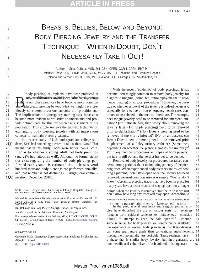
FIGURE 7
Transfer technique—navel jewelry. Step II: Remove one end of the navel jewelry by twisting counterclockwise while maintaining a grasp on the other end of the jewelry.
(Photo from the Body Piercing Removal Photo Guide courtesy of Peds-R-Us Medical Education [www.Peds-R-Us.com]. Medical illustration courtesy of Jennifer Klepacki
[www.theconjured.com].) This figure can be viewed in color and as a full-page document at www.jenonline.org.
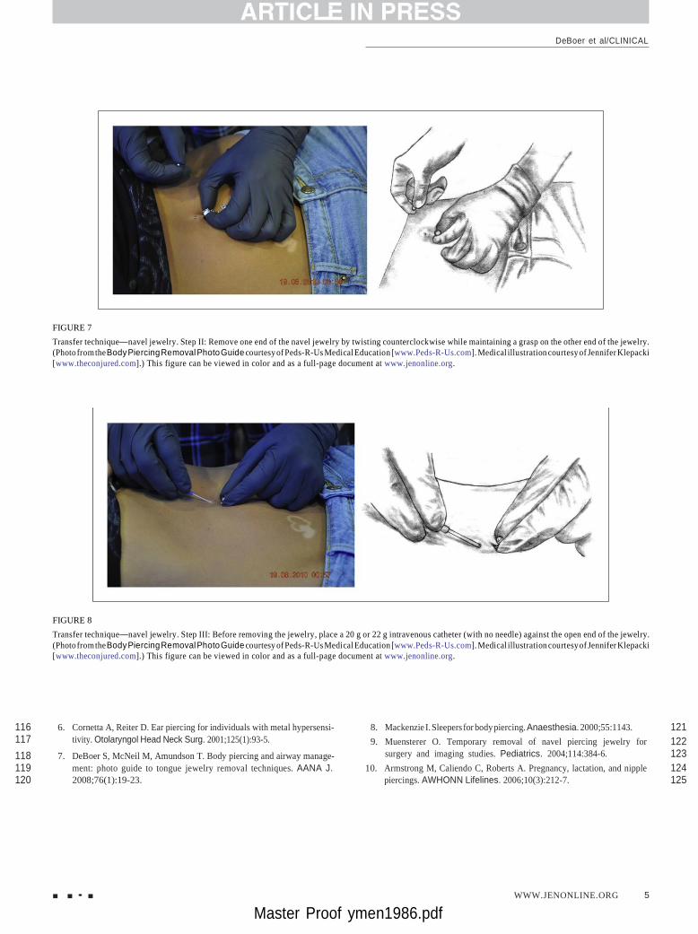
FIGURE 8
Transfer technique—navel jewelry. Step III: Before removing the jewelry, place a 20 g or 22 g intravenous catheter (with no needle) against the open end of the jewelry.
(Photo from the Body Piercing Removal Photo Guide courtesy of Peds-R-Us Medical Education [www.Peds-R-Us.com]. Medical illustration courtesy of Jennifer Klepacki
[www.theconjured.com].) This figure can be viewed in color and as a full-page document at www.jenonline.org.
116 6. Cornetta A, Reiter D. Ear piercing for individuals with metal hypersensi- 8. Mackenzie I. Sleepers for body piercing. Anaesthesia. 2000;55:1143. 121 117 tivity. Otolaryngol Head Neck Surg. 2001;125(1):93-5. 9. Muensterer O. Temporary removal of navel piercing jewelry for 122
118 7. DeBoer S, McNeil M, Amundson T. Body piercing and airway manage- surgery and imaging studies. Pediatrics. 2004;114:384-6. 123
119 ment: photo guide to tongue jewelry removal techniques. AANA J. 10. Armstrong M, Caliendo C, Roberts A. Pregnancy, lactation, and nipple 124 120 2008;76(1):19-23. piercings. AWHONN Lifelines. 2006;10(3):212-7. 125

■ • ■ ■ 6 JOURNAL OF EMERGENCY NURSING
Master Proof ymen1986.pdf
CLINICAL/DeBoer et al
Q6 FIGURE 9
Transfer technique—navel jewelry. Step IV: Slowly withdraw the navel jewelry while introducing the intravenous catheter into the piercing tract. Once the
intravenous catheter has been completely inserted, fully remove the body jewelry. (Photo from the Body Piercing Removal Photo Guide courtesy of Peds-R-Us
Medical Education [www.Peds-R-Us.com]. Medical illustration courtesy of Jennifer Klepacki [www.theconjured.com].) This figure can be viewed in color and
as a full-page document at www.jenonline.org.
FIGURE 10
Transfer technique—navel jewelry. Step V: Successful removal of jewelry and replacement with catheter. (Photo from the Body Piercing Removal Photo Guide courtesy
of Peds-R-Us Medical Education [www.Peds-R-Us.com]. Medical illustration courtesy of Jennifer Klepacki [www.theconjured.com].) This figure can be viewed in
color and as a full-page document at www.jenonline.org.

■ ■ • ■ WWW.JENONLINE.ORG 7
Master Proof ymen1986.pdf
DeBoer et al/CLINICAL
FIGURE 11
Transfer technique—navel jewelry. Step VI: After the successful transfer, cover the site with a clear, bio-occlusive dressing such as a Tegaderm or Opsite.
(Photo from the Body Piercing Removal Photo Guide courtesy of Peds-R-Us Medical Education [www.Peds-R-Us.com]. Medical illustration courtesy of Jennifer
Klepacki [www.theconjured.com].) This figure can be viewed in color and as a full-page document at www.jenonline.org.
FIGURE 12
Transfer technique—tongue jewelry. Step I: While utilizing universal precautions, remove one end of the jewelry by twisting counterclockwise and maintaining a grasp
on the other end of the jewelry. Then place a 20 g or 22 g intravenous catheter (with no needle) against the open end of the jewelry. (Photo from the Body Piercing
Removal Photo Guide courtesy of Peds-R-Us Medical Education [www.Peds-R-Us.com]. Medical illustration courtesy of Jennifer Klepacki [www.theconjured.com].)
This figure can be viewed in color and as a full-page document at www.jenonline.org.

■ • ■ ■ 8 JOURNAL OF EMERGENCY NURSING
Master Proof ymen1986.pdf
CLINICAL/DeBoer et al
FIGURE 13
Transfer technique—tongue jewelry. Steps IIa (A) and IIb (B): Slowly withdraw the tongue jewelry while introducing the IV catheter into the piercing tract. (Photo
from the Body Piercing Removal Photo Guide courtesy of Peds-R-Us Medical Education [www.Peds-R-Us.com]. Medical illustration courtesy of Jennifer Klepacki
[www.theconjured.com].) This figure can be viewed in color and as a full-page document at www.jenonline.org.

■ ■ • ■ WWW.JENONLINE.ORG 9
Master Proof ymen1986.pdf
DeBoer et al/CLINICAL
FIGURE 14
Transfer technique—tongue jewelry. Step III: Once the intravenous catheter has been completely inserted, fully remove the body jewelry. (Photo from the
Body Piercing Removal Photo Guide courtesy of Peds-R-Us Medical Education [www.Peds-R-Us.com]. Medical illustration courtesy of Jennifer Klepacki [www.
theconjured.com].) This figure can be viewed in color and as a full-page document at www.jenonline.org.
FIGURE 15
Transfer technique—tongue jewelry. Step IV: 20 g intravenous catheter secured with size 0 suture. (Photo from the Body Piercing Removal Photo Guide courtesy of
Peds-R-Us Medical Education [www.Peds-R-Us.com]. Medical illustration courtesy of Jennifer Klepacki [www.theconjured.com].) This figure can be viewed in color
and as a full-page document at www.jenonline.org.

CLINICAL/DeBoer et al
■ • ■ ■ 10 JOURNAL OF EMERGENCY NURSING
Master Proof ymen1986.pdf
FIGURE 16
Transfer technique for ear piercing with 22 g intravenous catheter. (Photo
from the Body Piercing Removal Photo Guide courtesy of Peds-R-Us Medical
Education [www.Peds-R-Us.com].) This figure can be viewed in color and
as a full-page document at www.jenonline.org.
FIGURE 18
Transfer technique for lip piercing with 22 g intravenous catheter. (Photo
from the Body Piercing Removal Photo Guide courtesy of Peds-R-Us Medical
Education [www.Peds-R-Us.com].) This figure can be viewed in color and
as a full-page document at www.jenonline.org.
Q5 FIGURE 17
Transfer technique for nostril piercing with 22 g intravenous catheter. (Photo
from the Body Piercing Removal Photo Guide courtesy of Peds-R-Us Medical
Education [www.Peds-R-Us.com].) This figure can be viewed in color and as
a full-page document at www.jenonline.org.
FIGURE 19
Transfer technique for nasal septal piercing with 22 g intravenous catheter.
(Photo from the Body Piercing Removal Photo Guide courtesy of Peds-R-Us
Medical Education [www.Peds-R-Us.com].) This figure can be viewed in color
and as a full-page document at www.jenonline.org.

DeBoer et al/CLINICAL
■ ■ • ■ WWW.JENONLINE.ORG 111
Master Proof ymen1986.pdf
FIGURE 20
A piercing transfer technique model demonstrating septum, cheek, and lip
intravenous catheter retainers. This figure can be viewed in color and as a
full-page document at www.jenonline.org.
TABLE 1
Body parts for which transfer techniques have been
used successfully
· Ear
· Eyebrow
· Nostril
· Septum
· Cheek
· Tongue
· Lip
· Nipples
· Navel
· Male and female intimate areas
t1.1
t0.2
t0.3
t0.4
t0.5
t0.6
t0.7
t0.8
t0.9
t0.10
t0.11
t0.12
t0.13

CLINICAL/DeBoer et al
■ • ■ ■ 12 JOURNAL OF EMERGENCY NURSING
Master Proof ymen1986.pdf
Qt2.71
t0.2
TABLE 2
Type of piercing
Anatomical location of the jewelry
Common types of jewelry placed
t0.3 Ampallang Horizontally through the glans (spongy head of Barbell
the penis) and through the urethra t0.4 Apadravya Vertically through the glans (spongy head of the penis) Barbell
and urethra and out on the head in front of the coronal ridge (you get a free Prince Albert with every apadravya) t0.5 Branding You name it, it's been branded N/A
t0.6 Cheek Through the buccinator muscle, between the facial Barbell with a disc back on the inside
artery and vein t0.7 Chest (surface) Usually between the jugular notch and superior Surface barbell
to the xiphoid process (front of the chest) t0.8 Christina Vertically just below the mons pubis, superior to the Barbell
anterior commissure of labia majora (on the pubic mound and above the vagina) t0.9 Clitoral hood Vertically through the prepuce (thin bit of tissue) Bent barbell or ring
t0.10 (vertical) above the clitoris (not through the clitoris itself) t0.11 Clitoris Self explanatory; very rare Ring
t0.12 Cutting Through the epidermis and dermis into the N/A
subcutaneous tissue t0.13 Dermal anchors Just about anywhere on the skin Dermal anchors
t0.14 (also known as t0.15 microdermals) t0.16 Dydoe Top and sides rim of glans (spongy head of penis) Curved barbell
through the coronal ridge (commonly done in pairs) t0.17 Ear (daith) Through the crux helix or innermost ridge of cartilage Ring
above the tragus (cartilage in front of the ear canal opening) t0.18 Ear (cartilage) Commonly refers to the helix of the ear; conch, daith, Barbell, ring, or circular barbell
rook, and tragus are all cartilage piercings t0.19 Ear (conch) The concha (shell of the ear) may be pierced in many Barbell, ring, circular barbell, plug,
different directions or eyelet
t0.20 Ear (head) Juncture of the ear and head in the cartilage Ring, barbell, or mini-barbell
t0.21 Ear (lobe) Lobule (traditional) or transverse lobe piercing Transverse barbell or ring
t0.22 Ear (rook) Crura of antihelix, opposite of the crus of the
helix—through the antihelix (upper ridge of cartilage
Barbell or ring
in the ear) t0.23 Ear (tragus) Through the prominence of cartilage in front of the Barbell or ring
opening of the ear canal t0.24 Earl (mid brow) Below the glabella and above the nasal bone Barbell
(middle of an eyebrow) t0.25 Etching Anywhere on the skin N/A
t0.26 Eyebrow Through the soft tissue, behind the eyebrow ridge Barbell or ring
t0.27 Foreskin Self-explanatory Barbell or ring
t0.28 Fourchette Vertical perineum piercing from the vestibular fossa Bent barbell
to the posterior commissure of the labia majora (female Guichet)

DeBoer et al/CLINICAL
■ ■ • ■ WWW.JENONLINE.ORG 113
Master Proof ymen1986.pdf
Table 2
Continued
t0.29 Type of piercing Anatomical location of the jewelry Common types of jewelry placed
t0.30 Frenum Loose piece of flesh between the head and shaft of the
penis—not through the shaft itself—anywhere on the
shaft where it is possible to pinch up the tissue—your
imagination and “pinchability factor” of the tissue are
the main limitations
t0.31 Guichet In the urogenital triangle, behind the pubic symphysis
near the prostate (inseam of flesh between the
scrotum and anus)
t0.32 Hafada Anywhere on the scrotum, typically near the top and
in pairs
Barbell, rows of jewelry, or a large ring
that fits snugly around the erect head
of the penis (once fully healed)
Ring or curved barbell
Barbell or ring
t0.33 Hand (web) Between the fingers Barbell or ring
t0.34 Implants Facial, sternal, arms, and genitals Titanium, Teflon, silicone, pearl,
stainless steel implants (eg, shapes
and horns)
t0.35 Labia (inner) Labia minora Rings
t0.36 Labia (outer) In the pudendal cleft, behind the labia majora Captive rings, bead rings,
or circular barbells
t0.37 Labret Through the inferior part of the orbicularis oris muscle
(below the lower lip and above the chin)
Disc back barbell or fishtail
t0.38 Lingual frenulum Laterally, through the frenulum (web under the tongue) Barbell or ring
t0.39 Lip (side) Just above or below the tubercle of the lip (side of the lip) Ring or circular barbell
t0.40 Lorum Like a frenum, but lower-um (juncture of penile shaft and
scrotum in the center)
t0.41 Madison (mid neck) Superior to the jugular notch (front of the neck) near the
insertion of the sternocleidomastoid muscles
t0.42 Nape (back of neck) Between the external occipital protuberance and the spinous
process of the vertebrae (back of the neck)
t0.43 Nasal septum Between the major alar cartilage and the nasal septal
cartilage (in the middle of the nose)
Ring or barbell
Surface barbell or Tygon
Surface barbell
Barbell, circular barbell, ring,
plug, septum spike or tusk
t0.44 Navel Usually above but not through the umbilicus (belly button) Curved barbell, circular barbell, or ring
t0.45 Nipple Between the nipple and the areola (vertically or horizontally) Barbell, circular barbell, or ring
t0.46 Nostril Between the greater and lesser alar cartilage
(outside of the nose)
t0.47 Prince Albert In the underside of the glans (spongy head of penis)
and out through urethra
Nostril screw, nose bone, or ring
Captive ring, circular barbell, or
curved barbell
t0.48 Princess Albertina
t0.49 (female Prince Albert)
Through the lower portion of external urethral orifice,
resting within the vagina
Barbell or ring
t0.50 Pubic (male) Placed in the natural juncture where the pubic mound
and the shaft of the penis meet
t0.51 Reverse Prince Albert In the top of the glans (spongy head of penis) near the
center and out through the urethra
Ring
Ring or circular barbell
t0.52 Surface You name it, something can go under it; also see implants Surface barbell or Tygon
t0.53 Tattoos You name it, it's been inked N/A
t0.54 Tongue Vertically through the midline groove and lateral fold of
the tongue, although some get venoms or snake bites
through the sides of the tongue
Barbell
(continued on next page )

CLINICAL/DeBoer et al
■ • ■ ■ 14 JOURNAL OF EMERGENCY NURSING
Master Proof ymen1986.pdf
Table 2
Continued
t0.55 Type of piercing Anatomical location of the jewelry Common types of jewelry placed
t0.56 Tongue (tip) Vertically through the apex (tip) of the tongue Ring or barbell
t0.57 Triangle Horizontal piercing behind the nerve bundle of the
clitoris at the base of the hood tissue where it forms
from the body
Circular barbell, barbells, bent bars,
or rings
t0.58 Uvula Self-explanatory Barbell or rings
Journal : YMEN Article No : 1986
Dear Author,
Author Query Form
During the preparation of your manuscript for typesetting some questions have arisen. These are listed below. Please
check your typeset proof carefully and mark any corrections in the margin of the proof or compile them as a separate
list*. This form should then be returned with your marked proof/list of corrections to Dartmouth Journal Services.
Disk use In some instances we may be unable to process the electronic file of your article and/or artwork. In that case we have, for
efficiency reasons, proceeded by using the hard copy of your manuscript. If this is the case the reasons are indicated below:
□ Disk damaged □ Incompatible file format □ LaTeX file for non-LaTeX journal
□ Virus infected □ Discrepancies between electronic file and (peer-reviewed, therefore definitive) hard copy.
□ Other:
We have proceeded as follows:
□ Manuscript scanned □ Manuscript keyed in □ Artwork scanned
□ Files only partly used (parts processed differently: )
Bibliography If discrepancies were noted between the literature list and the text references, the following may apply:
❑ The references listed below were noted in the text but appear to be missing from your literature list. Please complete the list or remove the references from the text.
❑ Uncited references: This section comprises references which occur in the reference list but not in the body of the text. Please position each reference in the text or, alternatively, delete it. Any reference not dealt with will be retained in this section.
Queries and/or remarks
Query
markers
Details required Author’s response
Q1 Please note Journal style for references: References should be
numbered in consecutive order as they appear in the text, and mention in the text is by number, not by last name of author.
Q2 Please note that per Journal style, personal communications
are not listed as references but are mentioned in the text.
Q3 Please check all figures to ensure that legends correspond to the correct images.
Q4 Figure 4: Please verify accuracy of figure parts and legend.
The .jpg image provided for B, tongue retainer, did not exactly match image shown in PDF
Q5 Figure 17 was taken from the Word file (there was no
original .tif or .jpeg). Please provide .tif or .jpg, if possible, as quality will be better.
Q6 Most of the figures and tables were set after References section. Please confirm layout if accepted.
Page 1 of 2

Q7 Please provide caption for Table 2.
ELSEVIER Many thanks for your assistance
*In case artwork needs revision, guidance can be found at http://authors.elsevier.com/artwork .
Page 2 of 2
Related Documents