BRCA1 is a novel target to improve endothelial dysfunction and retard atherosclerosis Krishna K. Singh, PhD, a,c Praphulla C. Shukla, PhD, a,c Adrian Quan, MPhil, a,c Mohammed Al-Omran, MD, MSc, FRCSC, c,f Fina Lovren, PhD, a,c Yi Pan, MD, a,c Christine Brezden-Masley, MD, PhD, FRCPC, b,c,d Alistair J. Ingram, MD, g William L. Stanford, PhD, h,i Hwee Teoh, PhD, a,c and Subodh Verma, MD, PhD, FRCSC a,c,e Objective: BRCA1, a tumor suppressor gene implicated in breast and ovarian cancers, exerts multiple effects on DNA repair and affords resistance against cellular stress responses. We hypothesized that BRCA1 limits endo- thelial cell apoptosis and dysfunction, and via this mechanism attenuates atherosclerosis. Methods: Loss and gain of function were achieved in cultured endothelial cells by silencing and overexpressing BRCA1, respectively. In vivo loss and gain of function were performed by generating endothelial cell-specific knockout (EC-BRCA1 / ) mice and administering a BRCA1 adenovirus. Well-established cell and animal models of angiogenesis and atherosclerosis were used. Results: BRCA1 is basally expressed in endothelial cells. BRCA1 overexpression protected and BRCA1 silenc- ing exaggerated inflammation- and doxorubicin-induced endothelial cell apoptosis. Key indices of endothelial function were modulated in a manner consistent with an effect of BRCA1 to limit endothelial cell apoptosis and improve endothelial function. BRCA1 overexpression strongly attenuated the production of reactive oxygen species and upregulated endothelial nitric oxide synthase, phosphorylated endothelial nitric oxide synthase, phosphorylated Akt, and vascular endothelial growth factor-a expression. BRCA1 overexpression also improved capillary density and promoted blood flow restoration in mice subjected to hind-limb ischemia. BRCA1- overexpressing ApoE / mice fed a Western diet developed significantly less aortic plaque lesions, exhibited reduced macrophage infiltration, and generated less reactive oxygen species. Lung sections and aortic segments from EC-BRCA1 / mice demonstrated greater inflammation-associated apoptosis and impaired endothelial function, respectively. BRCA1 expression was attenuated in the plaque region of human atherosclerotic carotid artery samples compared with the adjacent plaque-free area. Conclusions: These data collectively highlight a previously unrecognized role of BRCA1 as a gatekeeper of inflammation-induced endothelial cell function and a target to limit atherosclerosis. Translational studies eval- uating endothelial function and atherosclerosis in individuals with BRCA1 mutations are suggested. (J Thorac Cardiovasc Surg 2013;146:949-60) Supplemental material is available online. Germline mutations in the tumor suppressor BRCA1 (BReast CAncer susceptibility gene 1) are highly pene- trant for the increased risk of breast and ovarian cancer. 1 Mutations in the BRCA1 gene occur in up to 50% of in- herited breast cancers, and individuals who carry a defec- tive copy of the gene have a 50% to 85% cumulative lifetime risk of developing breast cancer and a 12% to 60% chance of developing ovarian cancer. 1 BRCA1 func- tions as a caretaker of genome stability and exerts multiple effects on key cellular processes involved in DNA repair, From the Divisions of Cardiac Surgery a and Medicine & Hematology-Oncology, b Keenan Research Centre c in the Li Ka Shing Knowledge Institute at St Michael’s Hospital, Toronto, Ontario, Canada; Departments of Medicine d and Surgery, e Uni- versity of Toronto, Toronto, Ontario, Canada; Division of Vascular Surgery, f Col- lege of Medicine and Peripheral Vascular Disease Research Chair, King Saud University, Riyadh, Kingdom of Saudi Arabia; Department of Medicine, g McMas- ter University, Hamilton, Ontario, Canada; Sprott Centre for Stem Cell Research, h Regenerative Medicine Program, Ottawa Hospital Research Institute, Ottawa, On- tario, Canada; and Department of Cellular and Molecular Medicine, i Faculty of Medicine, University of Ottawa, Ottawa, Ontario, Canada. Funding: This work was supported by grants from the Heart and Stroke Foundation of Canada and the Canadian Institutes of Health Research (to S.V.), an unrestricted Cardiology-Oncology Education Program grant from Sanofi-Aventis Canada Inc and Roche Canada (to C.B.-M. and S.V.), and an unrestricted grant from Abbott Can- ada. K.K.S. was the recipient of an Ontario Ministry of Research and Innovation’s Post-Doctoral Fellowship and received the 2009 American Heart Association Vivien Thomas Young Investigator Award for this work. P.C.S. was the recipient of a Fellow- ship from the Heart and Stroke/Richard Lewar Centre of Excellence in Cardiovascu- lar Research. H.T. was the St Michael’s Hospital Sanofi-Aventis Cardiometabolic Risk Initiative Research Fellow. W.L.S. is the Canada Research Chair in Integrative Stem Cell Biology at the University of Ottawa. S.V. is the Canada Research Chair in Atherosclerosis at the University of Toronto. Disclosures: Authors have nothing to disclose with regard to commercial support. K.K.S. and P.C.S. share the position of first author. Received for publication Oct 22, 2012; revisions received Dec 12, 2012; accepted for publication Dec 18, 2012; available ahead of print Feb 18, 2013. Address for reprints: Subodh Verma, MD, PhD, FRCSC, Division of Cardiac Surgery, St Michael’s Hospital, Suite 8-003, Bond Wing, 30 Bond St, Toronto, Ontario, M5B 1W8 (E-mail: [email protected]). 0022-5223/$36.00 Copyright Ó 2013 by The American Association for Thoracic Surgery http://dx.doi.org/10.1016/j.jtcvs.2012.12.064 The Journal of Thoracic and Cardiovascular Surgery c Volume 146, Number 4 949 Singh et al Evolving Technology/Basic Science ET/BS

Welcome message from author
This document is posted to help you gain knowledge. Please leave a comment to let me know what you think about it! Share it to your friends and learn new things together.
Transcript
Singh et al Evolving Technology/Basic Science
BRCA1 is a novel target to improve endothelial dysfunction andretard atherosclerosis
Krishna K. Singh, PhD,a,c Praphulla C. Shukla, PhD,a,c Adrian Quan, MPhil,a,c
Mohammed Al-Omran, MD, MSc, FRCSC,c,f Fina Lovren, PhD,a,c Yi Pan, MD,a,c
Christine Brezden-Masley, MD, PhD, FRCPC,b,c,d Alistair J. Ingram, MD,g
William L. Stanford, PhD,h,i Hwee Teoh, PhD,a,c and Subodh Verma, MD, PhD, FRCSCa,c,e
From th
Keen
Hosp
versit
lege
Univ
ter U
Rege
tario,
Medi
Funding
Cana
Cardi
andR
ada. K
Post-
Objective: BRCA1, a tumor suppressor gene implicated in breast and ovarian cancers, exerts multiple effects onDNA repair and affords resistance against cellular stress responses. We hypothesized that BRCA1 limits endo-thelial cell apoptosis and dysfunction, and via this mechanism attenuates atherosclerosis.
Methods: Loss and gain of function were achieved in cultured endothelial cells by silencing and overexpressingBRCA1, respectively. In vivo loss and gain of function were performed by generating endothelial cell-specificknockout (EC-BRCA1�/�) mice and administering a BRCA1 adenovirus. Well-established cell and animalmodels of angiogenesis and atherosclerosis were used.
Results: BRCA1 is basally expressed in endothelial cells. BRCA1 overexpression protected and BRCA1 silenc-ing exaggerated inflammation- and doxorubicin-induced endothelial cell apoptosis. Key indices of endothelialfunction were modulated in a manner consistent with an effect of BRCA1 to limit endothelial cell apoptosis andimprove endothelial function. BRCA1 overexpression strongly attenuated the production of reactive oxygenspecies and upregulated endothelial nitric oxide synthase, phosphorylated endothelial nitric oxide synthase,phosphorylated Akt, and vascular endothelial growth factor-a expression. BRCA1 overexpression also improvedcapillary density and promoted blood flow restoration in mice subjected to hind-limb ischemia. BRCA1-overexpressing ApoE�/� mice fed a Western diet developed significantly less aortic plaque lesions, exhibitedreduced macrophage infiltration, and generated less reactive oxygen species. Lung sections and aortic segmentsfrom EC-BRCA1�/� mice demonstrated greater inflammation-associated apoptosis and impaired endothelialfunction, respectively. BRCA1 expression was attenuated in the plaque region of human atherosclerotic carotidartery samples compared with the adjacent plaque-free area.
Conclusions: These data collectively highlight a previously unrecognized role of BRCA1 as a gatekeeper ofinflammation-induced endothelial cell function and a target to limit atherosclerosis. Translational studies eval-uating endothelial function and atherosclerosis in individuals with BRCA1 mutations are suggested. (J ThoracCardiovasc Surg 2013;146:949-60)
Supplemental material is available online.
Germline mutations in the tumor suppressor BRCA1(BReast CAncer susceptibility gene 1) are highly pene-trant for the increased risk of breast and ovarian cancer.1
e Divisions of Cardiac Surgerya and Medicine & Hematology-Oncology,b
an Research Centrec in the Li Ka Shing Knowledge Institute at St Michael’s
ital, Toronto, Ontario, Canada; Departments of Medicined and Surgery,e Uni-
y of Toronto, Toronto, Ontario, Canada; Division of Vascular Surgery,f Col-
of Medicine and Peripheral Vascular Disease Research Chair, King Saud
ersity, Riyadh, Kingdom of Saudi Arabia; Department of Medicine,g McMas-
niversity, Hamilton, Ontario, Canada; Sprott Centre for Stem Cell Research,h
nerative Medicine Program, Ottawa Hospital Research Institute, Ottawa, On-
Canada; and Department of Cellular and Molecular Medicine,i Faculty of
cine, University of Ottawa, Ottawa, Ontario, Canada.
: This work was supported by grants from the Heart and Stroke Foundation of
da and the Canadian Institutes of Health Research (to S.V.), an unrestricted
ology-Oncology Education Program grant from Sanofi-Aventis Canada Inc
ocheCanada (toC.B.-M. andS.V.), and an unrestricted grant fromAbbottCan-
.K.S. was the recipient of an Ontario Ministry of Research and Innovation’s
Doctoral Fellowship and received the 2009AmericanHeartAssociationVivien
The Journal of Thoracic and Ca
Mutations in the BRCA1 gene occur in up to 50% of in-herited breast cancers, and individuals who carry a defec-tive copy of the gene have a 50% to 85% cumulativelifetime risk of developing breast cancer and a 12% to60% chance of developing ovarian cancer.1 BRCA1 func-tions as a caretaker of genome stability and exerts multipleeffects on key cellular processes involved in DNA repair,
ThomasYoung InvestigatorAward for thiswork.P.C.S.was the recipient of aFellow-
ship from theHeart and Stroke/Richard Lewar Centre of Excellence in Cardiovascu-
lar Research. H.T. was the St Michael’s Hospital Sanofi-Aventis Cardiometabolic
Risk Initiative Research Fellow. W.L.S. is the Canada Research Chair in Integrative
StemCell Biology at the University of Ottawa. S.V. is the Canada Research Chair in
Atherosclerosis at the University of Toronto.
Disclosures: Authors have nothing to disclose with regard to commercial support.
K.K.S. and P.C.S. share the position of first author.
Received for publication Oct 22, 2012; revisions received Dec 12, 2012; accepted for
publication Dec 18, 2012; available ahead of print Feb 18, 2013.
Address for reprints: Subodh Verma, MD, PhD, FRCSC, Division of Cardiac Surgery,
St Michael’s Hospital, Suite 8-003, Bond Wing, 30 Bond St, Toronto, Ontario,
M5B 1W8 (E-mail: [email protected]).
0022-5223/$36.00
Copyright � 2013 by The American Association for Thoracic Surgery
http://dx.doi.org/10.1016/j.jtcvs.2012.12.064
rdiovascular Surgery c Volume 146, Number 4 949
ET/BS
Abbreviations and Acronymsad ¼ adenovirusCMV ¼ cytomegalovirusDox ¼ doxorubicinEC ¼ endothelial cellENOS ¼ endothelial nitric oxide synthaseFITC ¼ fluorescein isothiocyanateGAPDH ¼ glyceraldehyde 3-phosphate
dehydrogenaseGFP ¼ green fluorescent proteinHUVEC ¼ human umbilical vein endothelial cellICAM-1 ¼ intercellular adhesion molecule 1MOI ¼ multiplicity of infectionoxLDL ¼ oxidized low-density lipoproteinPCR ¼ polymerase chain reactionPFU ¼ plaque-forming unitROS ¼ reactive oxygen speciessiRNA ¼ small interfering RNASNP ¼ single nucleotide polymorphismTNFa ¼ tumor necrosis factor-alphaVCAM-1 ¼ vascular cell adhesion molecule 1VEGF ¼ vascular endothelial growth factor
Evolving Technology/Basic Science Singh et al
ET/BS
transcriptional regulation, ubiquitination, and cell-cyclecontrol.2
DNA damage not only is important in cancer syndromesbut also represents a common pathophysiologic basis for di-verse diseases characterized by increased cellular and oxi-dative stress.3,4 Atherosclerosis, a leading cause of deathworldwide, is a chronic inflammatory condition, andendothelial dysfunction plays an early and permissive rolein the development and progression of the disease.5 A com-mon mechanism through which cardiovascular risk factorslead to endothelial dysfunction is via increased generationof reactive oxygen species (ROS), thus elevating oxidativestress that in turn promotes DNA damage and endothelialcell (EC) apoptosis.4
We recently demonstrated in mice a cardioprotective andanti-inflammatory role for BRCA1.6,7 We have alsoreported on the cardioprotective nature of BRCA2(BReast CAncer susceptibility gene 2).8 Because BRCA1plays an integral role in promoting DNA repair, we hypoth-esized that BRCA1 also may be involved in regulating ECsurvival and function, and thus may retard the progressionof atherosclerosis. We report for the first time thatBRCA1 limits EC apoptosis, restores endothelial function,and attenuates atherosclerotic lesion development, therebyhighlighting a novel role for BRCA1 as a regulator of vas-cular health. In addition to identifying a potentially new tar-get for vascular diseases, these data may point towarda heightened susceptibility of BRCA1 mutation carrierstoward atherothrombotic vascular diseases.
950 The Journal of Thoracic and Cardiovascular Surg
MATERIALS AND METHODSA detailed, expanded Materials and Methods section is available in the
Online Supplement.
Cell Culture, Adenoviral Gene Delivery, and BRCA1Gene Silencing
Human umbilical vein endothelial cells (HUVECs) were cultured in
MCDB-131 complete medium and used at passages 3 to 5. We used a cus-
tom-designed BRCA1 adenovirus (ad-BRCA1) that contained the com-
plete coding sequence of human BRCA1 (accession no. NM_007294.2)
and was under the control of the cytomegalovirus (CMV) promoter. Where
appropriate, ad-null (ad-CMV-null) or ad-green fluorescent protein (GFP)
served as controls for ad-BRCA1. BRCA1 gene silencing was achieved
with the small interfering RNA (siRNA) against human BRCA1, whereas
a combination of 4 scrambled siRNAs served as the negative control.
Animal StudiesAll animal-associated procedures were performed in accordance with
the guidelines of the Canadian Council on Animal Care and approved by
the St Michael’s Hospital Animal Care Committee.
Male BALB/c mice from Charles River Laboratories International, Inc
(Wilmington, Mass) were used for hind-limb ischemia studies.
ApoE�/� mice were obtained from The Jackson Laboratory (Bar Har-
bor, Me) and divided into 3 study groups. The first and second groups
were fed the Western diet for 4 and 16 weeks, respectively, while concur-
rently administered ad-GFP, ad-null, or ad-BRCA1. These mice were
subsequently euthanized, and their aortas were isolated for immunohisto-
chemical staining. The third group was fed the Western diet for 2 weeks,
after which plasma samples were collected for lipid analyses.
EC-specific BRCA1 knockout mice were generated by crossing Tie2
Cretg/ mice (The Jackson Laboratory) with BRCA1fl/fl mice (MMHCC, Na-
tional Cancer Institute, Frederick, Md) to generate Tie2-Cretg/; BRCA1fl/þ
(EC-BRCA1þ/�) mice. EC-BRCA1þ/� mice were crossed with BRCA1fl/þ
mice to generate Tie2-Cretg/ BRCA1fl/fl (EC-BRCA1�/�) mice, which dem-
onstrate inactivation of the BRCA1 gene specifically in ECs.
Human Carotid Artery SamplesStudy protocols were reviewed and approved by the Research Ethics
Boards of St Michael’s Hospital and Hamilton Health Sciences. Participa-
tion was voluntary, and all study subjects provided informed consent. Ca-
rotid artery segments were collected en bloc at the time of endarterectomy
from the intima to the external elastic lamina. Two sections were taken
from below (‘‘normal’’) and at (plaque containing) the carotid bifurcation.
Apoptosis AssaysApoptosis in HUVECs was induced with 20 ng/mL human recombinant
tumor necrosis factor-alpha (TNFa), 10 mg/mL oxidized low-density lipo-
protein (oxLDL), or 2 mmol/L doxorubicin (Dox). The extent of apoptosis
was measured via flow cytometry by enumerating HUVECs positive for an-
nexin V–fluorescein isothiocyanate (FITC) and propidium iodide. Total
HUVEC genomic DNA samples were electrophoresed on 1% agarose
gels and subsequently stained with ethidium bromide for assessment of
DNA fragmentation. Lipopolysaccharide-induced lung injury as deter-
mined by the terminal deoxynucleotidyl transferase assay was examined
in EC-BRCA1þ/� mice.
Western Blot Analyses, RNA Extraction, and Real-Time Polymerase Chain Reaction
Protein lysates were processed for Western blot analysis. Blots
were probed for BRCA1, endothelial nitric oxide synthase (eNOS),
Ser1177-phospho-eNOS, Akt, Ser473-phospho-Akt, cleaved caspase-3, vas-
cular cell adhesion molecule 1 (VCAM)-1, intercellular adhesion molecule
ery c October 2013
Singh et al Evolving Technology/Basic Science
1 (ICAM-1), E-selectin, and glyceraldehyde 3-phosphate dehydrogenase
(GAPDH). Total RNA was prepared for polymerase chain reaction
(PCR) or real-time PCR reactions. Transcript levels of BRCA1,
VCAM-1, ICAM-1, E-selectin, vascular endothelial growth factor
(VEGF)-a, and GAPDH were quantified.
Quantification of Reactive Oxygen Species andVascular Endothelial Growth Factor Levels
ROS and VEGFa levels were determined with the OxiSelect ROSAssay
Kit (Cell Biolabs, Inc, San Diego, Calif) and a VEGF ELISA Kit (Thermo
Scientific, Burlington, Ontario, Canada) respectively.
In Vitro Angiogenesis and MigrationThe angiogenic and migratory properties of HUVECs were examined
with the In Vitro Angiogenesis Assay Kit (Chemicon, Temecula, Calif)
and the CytoSelect 24-well Cell Migration Fluorometric Assay system
(Cell Biolabs, Inc), respectively.
Immunohistochemical and ImmunofluorescentStaining
Murine aortic root sections, human carotid artery sections, and en face
aorta samples were subjected to routine immunohistochemical staining
with BRCA1, Oil Red O, F4/80, or dihydroethidium. Sections of gastroc-
nemius muscles from the ischemic limbs of BALB/cmicewere stained with
rhodamine-conjugated isolectin-B4 and FITC-conjugated a-smooth mus-
cle actin.
Vascular FunctionEndothelial function of the aortic segments from EC-BRCA1�/� mice
was assessed via wire myography. Unilateral hind-limb ischemia was in-
duced in 8-week-old male BALB/c mice. Limb perfusion was monitored
by laser Doppler flow imaging for up to 28 days post-surgery.
Statistical AnalysisUnless otherwise stated, data are presented as mean � standard devia-
tion for the number of samples. The Student t test was applied when the
means of 2 groups were being compared. Differences between multiple
means were evaluated by analysis of variance, and when overall differences
were detected, individual means were compared post hoc with the Bonfer-
roni’s test.
ET/BS
RESULTSBRCA1 Is Basally Expressed in Human UmbilicalVein Endothelial Cells and Limits Human UmbilicalVein Endothelial Cell Apoptosis After Inflammatoryand Genotoxic Stress
BRCA1 is basally expressed inHUVECs (FigureE1,A andB). To evaluate the functional relevance of BRCA1 in HU-VECs, we used gain- and loss-of-function strategies.Optimal efficacy of adenoviral delivery was determinedwith ad-GFP to be 20 multiplicity of infection (MOI). GFPwas detected 24 hours post-transfection in approximately80% of HUVECs (Figure E1, C). Ad-BRCA1–mediatedBRCA1overexpressionwas confirmed byPCRand immuno-blotting (Figure E1, A and B). The reader should note thatalthough all of the subsequent studies were performed in 3groups of HUVECs—na€ıve (nontransfected) HUVECs, ad-null–transfected HUVECs, and ad-BRCA1–transfected
The Journal of Thoracic and Ca
HUVECs—only data from the ad-null– and ad-BRCA1–treated groups are shown because findings from the na€ıveand ad-null–transfected HUVECs were indistinguishable.TNFa evoked significant apoptosis in ad-null–transfectedHUVECs but not in BRCA1-overexpressing HUVECs(Figure 1, A). This observation was confirmed via immuno-blots for cleaved caspase-3 and DNA fragmentation assays,where BRCA1 overexpression dampened TNFa-associatedincreases in cleaved caspase-3 protein levels (Figure 1, B)and DNA fragmentation (Figure 1, C). That oxLDL notonly elevated the percentage of apoptotic HUVECs(Figure 1, D) but also enhanced the levels of cleavedcaspase-3 (Figure 1, E) was not unexpected because ECapoptosis induced by oxLDL is integral to the atherogenicpathway. Of more interest was the observation that oxLDL-triggered apoptosis also was significantly reduced inBRCA1-overexpressing HUVECs (Figure 1,D and E). Gen-otoxic stress caused by chemotherapeutic drugs, such asDox,has been shown to activate apoptosis in ECs. We reportmarked apoptosis in Dox-treated ad-null–transfectedHUVECs, an effect that was significantly reduced in ad-BRCA1–transfected HUVECs (Figure 1, F and G). Loss offunction was performed with silenced-BRCA1 at an optimalconcentration of 10 nM (Figure 1, H). Apoptosis in TNFa-treated BRCA1-silenced HUVEC cultures was significantlygreater compared with that in TNFa-treated controlHUVECs (Figure 1, I). These data suggest that BRCA1 pro-tects HUVECs against apoptosis induced by inflammatoryand genotoxic stresses.To gain critical insights into the role of BRCA1 in
endothelial regulation and to circumvent the embryoniclethality associatedwith systemic BRCA1 loss,9 we generatedEC-BRCA1þ/� andEC-BRCA1�/�mice. Thesemicewere vi-able, fertile, and born in the expected Mendelian ratios (datanot shown). Terminal deoxynucleotidyl transferase stainingrevealed that, in comparison with the wild-type littermatecontrol group, lung sections from EC-BRCA1þ/� mice ex-hibited more extensive lipopolysaccharide-associatedendothelial apoptosis (Figure 1, J).
BRCA1 Impairs Tumor Necrosis Factor-a–InducedReactive Oxygen Species Generation in HumanUmbilical Vein Endothelial CellsBRCA1 reduces the generation of ROS,10,11 a critical
mediator of EC apoptosis elicited by TNFa,12 oxLDL,13
and Dox.14 We observed significantly lower levels of ROSin ad-BRCA1–transfected HUVECs compared with ad-null–treated HUVECs after TNFa stimulation (Figure 1,K). Furthermore, we detected significantly higher levels ofROS in TNFa-treated BRCA1-silenced HUVECs relativeto HUVECs that had been treated with the control siRNA(Figure 1, L). These data demonstrate the importance ofBRCA1 in controlling ROS generation in HUVECs and ar-guably HUVEC apoptosis.
rdiovascular Surgery c Volume 146, Number 4 951
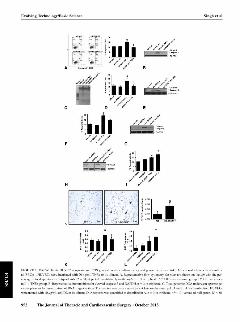
FIGURE 1. BRCA1 limits HUVEC apoptosis and ROS generation after inflammatory and genotoxic stress. A-C, After transfection with ad-null or
ad-BRCA1, HUVECs were incubated with 20 ng/mL TNFa or its diluent. A, Representative flow cytometry dot plots are shown on the left with the per-
centage of total apoptotic cells (quadrants S2þ S4) depicted quantitatively on the right. n¼ 3 in triplicate. *P<.01 versus ad-null group. yP<.01 versus ad-
null þ TNFa group. B, Representative immunoblots for cleaved caspase-3 and GAPDH. n ¼ 3 in triplicate. C, Total genomic DNA underwent agarose gel
electrophoresis for visualization of DNA fragmentation. The marker was from a nonadjacent lane on the same gel. D and E, After transfection, HUVECs
were treated with 10 mg/mL oxLDL or its diluent. D, Apoptosis was quantified as described in A. n¼ 3 in triplicate. *P<.01 versus ad-null group. yP<.01
Evolving Technology/Basic Science Singh et al
952 The Journal of Thoracic and Cardiovascular Surgery c October 2013
ET/BS
=
Singh et al Evolving Technology/Basic Science
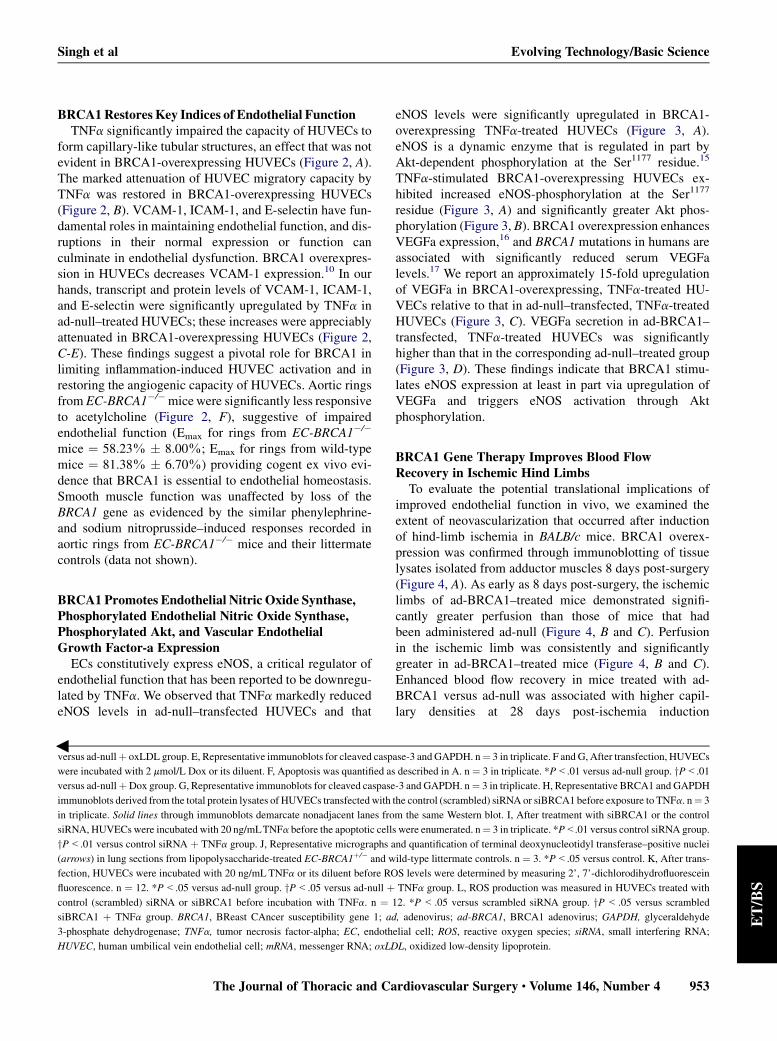
BRCA1Restores Key Indices of Endothelial FunctionTNFa significantly impaired the capacity of HUVECs to
form capillary-like tubular structures, an effect that was notevident in BRCA1-overexpressing HUVECs (Figure 2, A).The marked attenuation of HUVEC migratory capacity byTNFa was restored in BRCA1-overexpressing HUVECs(Figure 2, B). VCAM-1, ICAM-1, and E-selectin have fun-damental roles in maintaining endothelial function, and dis-ruptions in their normal expression or function canculminate in endothelial dysfunction. BRCA1 overexpres-sion in HUVECs decreases VCAM-1 expression.10 In ourhands, transcript and protein levels of VCAM-1, ICAM-1,and E-selectin were significantly upregulated by TNFa inad-null–treated HUVECs; these increases were appreciablyattenuated in BRCA1-overexpressing HUVECs (Figure 2,C-E). These findings suggest a pivotal role for BRCA1 inlimiting inflammation-induced HUVEC activation and inrestoring the angiogenic capacity of HUVECs. Aortic ringsfrom EC-BRCA1�/�micewere significantly less responsiveto acetylcholine (Figure 2, F), suggestive of impairedendothelial function (Emax for rings from EC-BRCA1�/�
mice ¼ 58.23% � 8.00%; Emax for rings from wild-typemice ¼ 81.38% � 6.70%) providing cogent ex vivo evi-dence that BRCA1 is essential to endothelial homeostasis.Smooth muscle function was unaffected by loss of theBRCA1 gene as evidenced by the similar phenylephrine-and sodium nitroprusside–induced responses recorded inaortic rings from EC-BRCA1�/� mice and their littermatecontrols (data not shown).
BRCA1 Promotes Endothelial Nitric Oxide Synthase,Phosphorylated Endothelial Nitric Oxide Synthase,Phosphorylated Akt, and Vascular EndothelialGrowth Factor-a Expression
ECs constitutively express eNOS, a critical regulator ofendothelial function that has been reported to be downregu-lated by TNFa. We observed that TNFa markedly reducedeNOS levels in ad-null–transfected HUVECs and that
versus ad-nullþ oxLDL group. E, Representative immunoblots for cleaved casp
were incubated with 2 mmol/L Dox or its diluent. F, Apoptosis was quantified as
versus ad-nullþDox group. G, Representative immunoblots for cleaved caspase
immunoblots derived from the total protein lysates of HUVECs transfected with
in triplicate. Solid lines through immunoblots demarcate nonadjacent lanes fro
siRNA, HUVECswere incubated with 20 ng/mLTNFa before the apoptotic cells
yP<.01 versus control siRNA þ TNFa group. J, Representative micrographs a
(arrows) in lung sections from lipopolysaccharide-treated EC-BRCA1þ/� and w
fection, HUVECs were incubated with 20 ng/mLTNFa or its diluent before RO
fluorescence. n ¼ 12. *P<.05 versus ad-null group. yP<.05 versus ad-null þcontrol (scrambled) siRNA or siBRCA1 before incubation with TNFa. n ¼ 1
siBRCA1 þ TNFa group. BRCA1, BReast CAncer susceptibility gene 1; ad
3-phosphate dehydrogenase; TNFa, tumor necrosis factor-alpha; EC, endoth
HUVEC, human umbilical vein endothelial cell; mRNA, messenger RNA; oxLD
The Journal of Thoracic and Ca
eNOS levels were significantly upregulated in BRCA1-overexpressing TNFa-treated HUVECs (Figure 3, A).eNOS is a dynamic enzyme that is regulated in part byAkt-dependent phosphorylation at the Ser1177 residue.15
TNFa-stimulated BRCA1-overexpressing HUVECs ex-hibited increased eNOS-phosphorylation at the Ser1177
residue (Figure 3, A) and significantly greater Akt phos-phorylation (Figure 3, B). BRCA1 overexpression enhancesVEGFa expression,16 and BRCA1 mutations in humans areassociated with significantly reduced serum VEGFalevels.17 We report an approximately 15-fold upregulationof VEGFa in BRCA1-overexpressing, TNFa-treated HU-VECs relative to that in ad-null–transfected, TNFa-treatedHUVECs (Figure 3, C). VEGFa secretion in ad-BRCA1–transfected, TNFa-treated HUVECs was significantlyhigher than that in the corresponding ad-null–treated group(Figure 3, D). These findings indicate that BRCA1 stimu-lates eNOS expression at least in part via upregulation ofVEGFa and triggers eNOS activation through Aktphosphorylation.
BRCA1 Gene Therapy Improves Blood FlowRecovery in Ischemic Hind LimbsTo evaluate the potential translational implications of
improved endothelial function in vivo, we examined theextent of neovascularization that occurred after inductionof hind-limb ischemia in BALB/c mice. BRCA1 overex-pression was confirmed through immunoblotting of tissuelysates isolated from adductor muscles 8 days post-surgery(Figure 4, A). As early as 8 days post-surgery, the ischemiclimbs of ad-BRCA1–treated mice demonstrated signifi-cantly greater perfusion than those of mice that hadbeen administered ad-null (Figure 4, B and C). Perfusionin the ischemic limb was consistently and significantlygreater in ad-BRCA1–treated mice (Figure 4, B and C).Enhanced blood flow recovery in mice treated with ad-BRCA1 versus ad-null was associated with higher capil-lary densities at 28 days post-ischemia induction
ase-3 and GAPDH. n¼ 3 in triplicate. F and G, After transfection, HUVECs
described in A. n ¼ 3 in triplicate. *P<.01 versus ad-null group. yP<.01
-3 and GAPDH. n¼ 3 in triplicate. H, Representative BRCA1 and GAPDH
the control (scrambled) siRNA or siBRCA1 before exposure to TNFa. n¼ 3
m the same Western blot. I, After treatment with siBRCA1 or the control
were enumerated. n¼ 3 in triplicate. *P<.01 versus control siRNA group.
nd quantification of terminal deoxynucleotidyl transferase–positive nuclei
ild-type littermate controls. n ¼ 3. *P<.05 versus control. K, After trans-
S levels were determined by measuring 2’, 7’-dichlorodihydrofluorescein
TNFa group. L, ROS production was measured in HUVECs treated with
2. *P< .05 versus scrambled siRNA group. yP< .05 versus scrambled
, adenovirus; ad-BRCA1, BRCA1 adenovirus; GAPDH, glyceraldehyde
elial cell; ROS, reactive oxygen species; siRNA, small interfering RNA;
L, oxidized low-density lipoprotein.
rdiovascular Surgery c Volume 146, Number 4 953
ET/BS
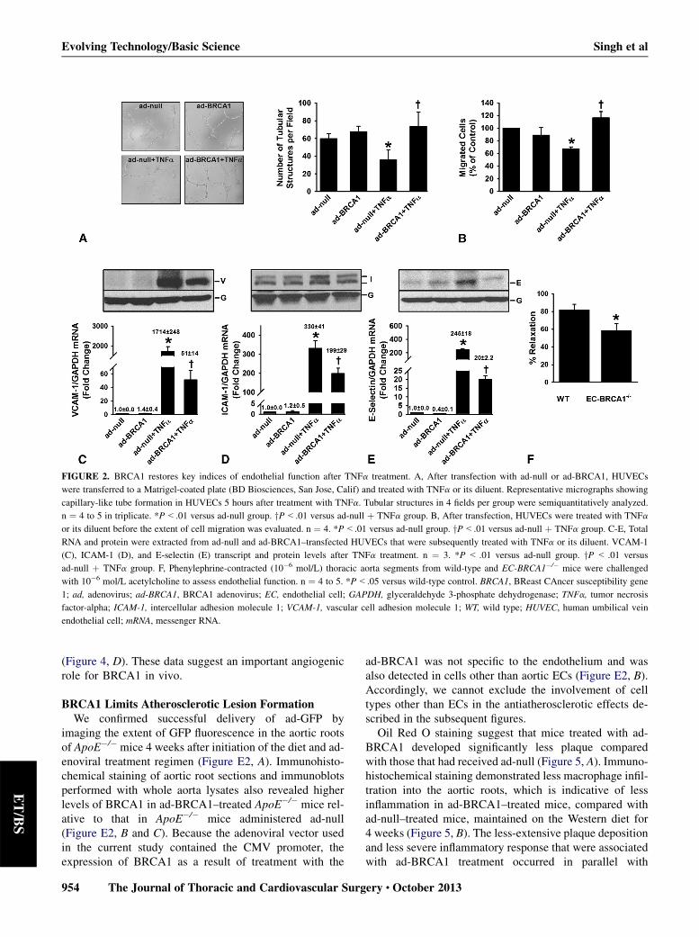
FIGURE 2. BRCA1 restores key indices of endothelial function after TNFa treatment. A, After transfection with ad-null or ad-BRCA1, HUVECs
were transferred to a Matrigel-coated plate (BD Biosciences, San Jose, Calif) and treated with TNFa or its diluent. Representative micrographs showing
capillary-like tube formation in HUVECs 5 hours after treatment with TNFa. Tubular structures in 4 fields per group were semiquantitatively analyzed.
n ¼ 4 to 5 in triplicate. *P<.01 versus ad-null group. yP<.01 versus ad-null þ TNFa group. B, After transfection, HUVECs were treated with TNFa
or its diluent before the extent of cell migration was evaluated. n ¼ 4. *P<.01 versus ad-null group. yP<.01 versus ad-null þ TNFa group. C-E, Total
RNA and protein were extracted from ad-null and ad-BRCA1–transfected HUVECs that were subsequently treated with TNFa or its diluent. VCAM-1
(C), ICAM-1 (D), and E-selectin (E) transcript and protein levels after TNFa treatment. n ¼ 3. *P < .01 versus ad-null group. yP < .01 versus
ad-null þ TNFa group. F, Phenylephrine-contracted (10�6 mol/L) thoracic aorta segments from wild-type and EC-BRCA1�/� mice were challenged
with 10�6 mol/L acetylcholine to assess endothelial function. n ¼ 4 to 5. *P<.05 versus wild-type control. BRCA1, BReast CAncer susceptibility gene
1; ad, adenovirus; ad-BRCA1, BRCA1 adenovirus; EC, endothelial cell; GAPDH, glyceraldehyde 3-phosphate dehydrogenase; TNFa, tumor necrosis
factor-alpha; ICAM-1, intercellular adhesion molecule 1; VCAM-1, vascular cell adhesion molecule 1; WT, wild type; HUVEC, human umbilical vein
endothelial cell; mRNA, messenger RNA.
Evolving Technology/Basic Science Singh et al
ET/BS
(Figure 4, D). These data suggest an important angiogenicrole for BRCA1 in vivo.
BRCA1 Limits Atherosclerotic Lesion FormationWe confirmed successful delivery of ad-GFP by
imaging the extent of GFP fluorescence in the aortic rootsof ApoE�/� mice 4 weeks after initiation of the diet and ad-enoviral treatment regimen (Figure E2, A). Immunohisto-chemical staining of aortic root sections and immunoblotsperformed with whole aorta lysates also revealed higherlevels of BRCA1 in ad-BRCA1–treated ApoE�/� mice rel-ative to that in ApoE�/� mice administered ad-null(Figure E2, B and C). Because the adenoviral vector usedin the current study contained the CMV promoter, theexpression of BRCA1 as a result of treatment with the
954 The Journal of Thoracic and Cardiovascular Surg
ad-BRCA1 was not specific to the endothelium and wasalso detected in cells other than aortic ECs (Figure E2, B).Accordingly, we cannot exclude the involvement of celltypes other than ECs in the antiatherosclerotic effects de-scribed in the subsequent figures.
Oil Red O staining suggest that mice treated with ad-BRCA1 developed significantly less plaque comparedwith those that had received ad-null (Figure 5, A). Immuno-histochemical staining demonstrated less macrophage infil-tration into the aortic roots, which is indicative of lessinflammation in ad-BRCA1–treated mice, compared withad-null–treated mice, maintained on the Western diet for4 weeks (Figure 5, B). The less-extensive plaque depositionand less severe inflammatory response that were associatedwith ad-BRCA1 treatment occurred in parallel with
ery c October 2013
FIGURE 3. BRCA1 enhances eNOS, phosphorylated eNOS, phosphorylated Akt, and VEGFa expression. After transfection with ad-null or ad-BRCA1,
HUVECs were incubated with 20 ng/mLTNFa or its diluent. Representative immunoblots and semiquantitative data for (A) eNOS, phosphorylated eNOS,
and GAPDH, and (B) Akt, phosphorylated Akt, and GAPDH. C, Real-time PCR analysis of VEGFa transcript levels. D, VEGFa levels as determined
by enzyme-linked immunosorbent assay. n ¼ 12. *P<.01 versus ad-null group. yP<.01 versus ad-null þ TNFa group. BRCA1, BReast CAncer suscep-
tibility gene 1; ad, Adenovirus; ad-BRCA1, BRCA1 adenovirus; eNOS, endothelial nitric oxide synthase; p-eNOS, phosphorylated endothelial nitric
oxide synthase; GAPDH, glyceraldehyde 3-phosphate dehydrogenase; TNFa, tumor necrosis factor-alpha; VEGF, vascular endothelial growth factor;
mRNA, messenger RNA.
Singh et al Evolving Technology/Basic Science
ET/BS
lower ROS production (Figure 5, C). Plaque deposition inad-BRCA1–treated ApoE�/� mice (vs ad-null–treatedApoE�/� mice) was continuously and significantly lowerup to week 16 of the diet-adenoviral treatment (Figure 5,D). The liver is often the main target for injected ad vectors.Considering the role of the liver in the regulation of lipid
The Journal of Thoracic and Ca
metabolism and the implications of dysfunctional lipid biol-ogy in the development and progression of atherosclerosis,we collected plasma for lipid measurements from ApoE�/�
mice that had been on the Western diet for 2 weeks. Lipidprofiles of these mice were unaffected by the diet-adenoviral treatment strategy (Table E1).
rdiovascular Surgery c Volume 146, Number 4 955
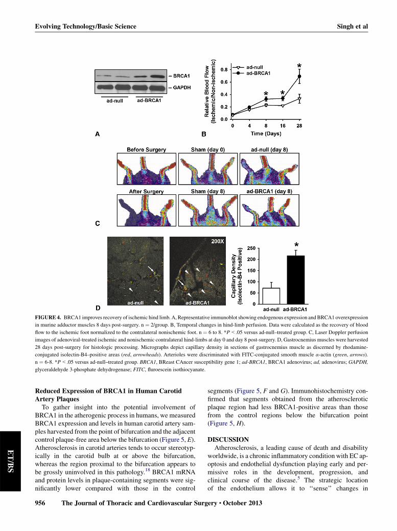
FIGURE 4. BRCA1 improves recovery of ischemic hind limb. A, Representative immunoblot showing endogenous expression and BRCA1 overexpression
in murine adductor muscles 8 days post-surgery. n ¼ 2/group. B, Temporal changes in hind-limb perfusion. Data were calculated as the recovery of blood
flow to the ischemic foot normalized to the contralateral nonischemic foot. n ¼ 6 to 8. *P<.05 versus ad-null–treated group. C, Laser Doppler perfusion
images of adenoviral-treated ischemic and nonischemic contralateral hind-limbs at day 0 and day 8 post-surgery. D, Gastrocnemius muscles were harvested
28 days post-surgery for histologic processing. Micrographs depict capillary density in sections of gastrocnemius muscle as discerned by rhodamine-
conjugated isolectin-B4–positive areas (red, arrowheads). Arterioles were discriminated with FITC-conjugated smooth muscle a-actin (green, arrows).
n ¼ 6-8. *P<.05 versus ad-null–treated group. BRCA1, BReast CAncer susceptibility gene 1; ad-BRCA1, BRCA1 adenovirus; ad, adenovirus; GAPDH,
glyceraldehyde 3-phosphate dehydrogenase; FITC, fluroescein isothiocyanate.
Evolving Technology/Basic Science Singh et al
ET/BS
Reduced Expression of BRCA1 in Human CarotidArtery Plaques
To gather insight into the potential involvement ofBRCA1 in the atherogenic process in humans, we measuredBRCA1 expression and levels in human carotid artery sam-ples harvested from the point of bifurcation and the adjacentcontrol plaque-free area below the bifurcation (Figure 5, E).Atherosclerosis in carotid arteries tends to occur stereotyp-ically in the carotid bulb at or above the bifurcation,whereas the region proximal to the bifurcation appears tobe grossly uninvolved in this pathology.18 BRCA1 mRNAand protein levels in plaque-containing segments were sig-nificantly lower compared with those in the control
956 The Journal of Thoracic and Cardiovascular Surg
segments (Figure 5, F and G). Immunohistochemistry con-firmed that segments obtained from the atheroscleroticplaque region had less BRCA1-positive areas than thosefrom the control regions below the bifurcation point(Figure 5, H).
DISCUSSIONAtherosclerosis, a leading cause of death and disability
worldwide, is a chronic inflammatory condition with EC ap-optosis and endothelial dysfunction playing early and per-missive roles in the development, progression, andclinical course of the disease.5 The strategic locationof the endothelium allows it to ‘‘sense’’ changes in
ery c October 2013
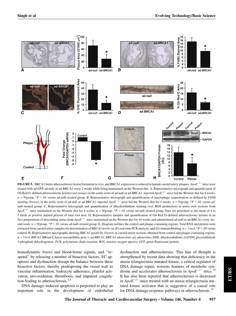
FIGURE 5. BRCA1 limits atherosclerosis lesion formation in vivo, and BRCA1 expression is reduced in human carotid artery plaques.ApoE�/�micewere
treated with ad-GFP, ad-null, or ad-BRCA1 every 2 weeks while being maintained on the Western diet. A, Representative micrograph and quantification of
Oil Red O–defined atherosclerotic lesions (red orange) in the aortic roots of ad-null or ad-BRCA1–injected ApoE�/�mice fed the Western diet for 4 weeks.
n ¼ 9/group. *P<.01 versus ad-null–treated group. B, Representative micrograph and quantification of macrophage sequestration, as defined by F4/80
staining (brown), in the aortic roots of ad-null or ad-BRCA1–injected ApoE�/� mice fed the Western diet for 4 weeks. n ¼ 9/group. *P<.01 versus ad-
null–treated group. C, Representative micrograph and quantification of dihydroethidium staining (red, ROS production) in aortic root sections from
ApoE�/� mice maintained on the Western diet for 4 weeks. n ¼ 9/group. *P<.01 versus ad-null–treated group. Data are presented as the mean of 4 to
5 fields as positive stained percent of total root area. D, Representative samples and quantification of Oil Red O–defined atherosclerotic lesions in en
face preparations of descending aortas from ApoE�/� mice maintained on the Western diet for 16 weeks and administered ad-null or ad-BRCA1 every sec-
ond week. n ¼ 9/group. *P<.01 versus ad-null–treated group. E, Diagram outlines the control and plaque containing regions. Total RNA and protein were
extracted from carotid artery samples for determination of BRCA1 levels via (F) real-time PCR analysis, and (G) immunoblotting. n¼ 3 to 4. *P<.05 versus
control. H, Representative micrographs showing BRCA1 positivity (brown) in carotid artery sections obtained from control and plaque-containing regions.
n¼ 3 to 4. BRCA1, BReast CAncer susceptibility gene 1; ad-BRCA1, BRCA1 adenovirus; ad, adenovirus;DHE, dihydroethidium;GAPDH, glyceraldehyde
3-phosphate dehydrogenase; PCR, polymerase chain reaction; ROS, reactive oxygen species; GFP, green flourescent protein.
Singh et al Evolving Technology/Basic Science
ET/BS
hemodynamic forces and blood-borne signals, and ‘‘re-spond’’ by releasing a number of bioactive factors. EC ap-optosis and dysfunction disrupt the balance between thesebioactive factors, thereby predisposing the vessel wall tovascular inflammation, leukocyte adherence, platelet acti-vation, pro-oxidation, thrombosis, and impaired coagula-tion leading to atherosclerosis.3,4
DNA damage–induced apoptosis is purported to play animportant role in the development of endothelial
The Journal of Thoracic and Ca
dysfunction and atherosclerosis. This line of thought isstrengthened by recent data showing that deficiency in theataxia telangiectasia mutated kinase, a critical regulator ofDNA damage repair, worsens features of metabolic syn-drome and accelerates atherosclerosis in ApoE�/� mice.19
It has also been reported that atherosclerosis is decreasedin ApoE�/� mice treated with an ataxia telangiectasia mu-tated kinase activator that is suggestive of a causal rolefor DNA damage-response pathways in atherosclerosis.19
rdiovascular Surgery c Volume 146, Number 4 957
Evolving Technology/Basic Science Singh et al
ET/BS
Although the role of BRCA1 in DNA damage repair re-sponses has been largely studied in cancer, this work sup-ports the contention that BRCA1 is a critical modulator ofEC apoptosis, endothelial dysfunction, and atherosclerosis,all of which are characterized by aberrant DNA damage andcellular stress. We provide evidence that BRCA1 is ex-pressed in HUVECs and that it is capable of protectingHUVECs from undergoing apoptosis induced by variousstressors that include TNFa, oxLDL, and Dox. We alsodemonstrate that BRCA1 silencing exaggerates TNFa-in-duced HUVEC apoptosis.
Both inflammation and genotoxic stress induce apoptosisvia ROS generation,12-14 and ROS-induced DNA damagemay play an important role in the development of cancerand inflammatory diseases. Evidence to date suggests thatseveral proatherogenic stimuli trigger ROS production inECs and that the resultant oxidative stress plays a key rolein mediating the pathologic manifestations of EC apoptosisand endothelial dysfunction that are associated with athero-sclerosis. Because BRCA1 not only affords protectionagainst DNA damage but also reduces ROS generation,10,11
we measured ROS levels in HUVECs after TNFa treatmentand found that BRCA1 overexpression reduces and BRCA1silencing enhances TNFa-induced ROS production,thereby confirming that BRCA1 may protect ECs againstapoptosis by negatively regulating stress-induced ROSgeneration.
In the milieu of cardiovascular risk factors that disturbvascular homeostasis, the endothelium becomes dysfunc-tional resulting in perturbed angiogenesis and enhanced ex-pression of cellular adhesion molecules by the endothelium.Our current data indicate that key indices of endothelialfunction, such as the ability of ECs to form capillary-liketubes, migrate, and express surface markers of endothelialinjury (VCAM-1, ICAM-1, and E-selectin), and the reducedresponse of aortic rings from EC-BRCA1�/� mice to acetyl-choline ex vivo, are modulated in a manner consistent withan effect of BRCA1 to limit EC apoptosis and improve en-dothelial function. Indeed, our results demonstrating signif-icant upregulation of eNOS and VEGFa expression, as wellas elevatedAkt activation in BRCA1-overexpressing TNFa-stimulated HUVECs, suggest that the restorative nature ofBRCA1 on endothelial function occurs at least in partthrough enhanced eNOS expression via VEGFa upregula-tion and eNOS activation by means of Akt phosphorylation,both of which are recognized survival mechanisms in ECs.
There seems to be no consensus as yet on the associativerelationship between BRCA1 and VEGFa expression andactivity. Kawai and colleagues20 demonstrated that wild-type BRCA1 inhibited VEGFa promoter activity andsecretion in breast cancer cells by modulating the estrogenreceptor in a dose-dependent manner and primarily in thepresence of estrogen. In contrast to the study by Kawaiand colleagues but in agreement with ours, Kang and
958 The Journal of Thoracic and Cardiovascular Surg
colleagues16 found that elevation of BRCA1 levels in breastcancer cells enhanced VEGF promoter activity after expo-sure to conditions of hypoxia and increased VEGF mRNAlevels. The same authors showed that silencing of endoge-nous BRCA1 resulted in decreased hypoxia-stimulatedVEGF promoter activity and secretion. In an article byWelcsh and King,21 overexpression of wild-type BRCA1in prostrate and breast cancer cell lines was associatedwith an approximately 2.5-fold increase in VEGFa expres-sion in the setting of oxidative stress.21 At the clinical level,significantly lower levels of VEGFwere detected in patientswith mutation of the BRCA1 gene when compared with pa-tients with breast cancer without this mutation.11 The bal-ance of current data suggests that how BRCA1 modulatesVEGFa expression and activity may be particularly depen-dent on the cell type, the presence of necessary cofactors,and whether the cells being studied are primary cells ora transformed cancer cell line.
By using a mouse model of neoangiogenesis after induc-tion of hind-limb ischemia, we successfully translated ourin vitro data to the in vivo level. Treatment with ad-BRCA1 not only improved capillary density but alsorestored blood flow to the ischemic limb. BRCA1 gene ther-apy also limited the formation of atherosclerotic lesions andattenuated inflammation, macrophage infiltration, and ROSgeneration but had no effect on lipid metabolism. Thesedata are critical because they represent an accepted transla-tional end point of progressive endothelial dysfunction. Ofnote, the benefits of BRCA1 on endothelial dysfunction andROS may be more applicable to the chronic experiments onendothelial dysfunction and atherosclerosis, as opposed tothe setting of acute reperfusion injury, which shares manyof the contributory factors. We also provide preliminaryclinical confirmation in human atherosclerotic plaqueswherein reduced BRCA1 levels are observed relative toplaque-free areas of the same vessel.
Our work collectively highlights a previously unrecog-nized role of BRCA1 as a gatekeeper of EC survival andfunction, and through this mechanism a novel target to im-prove ischemic neovascularization and attenuate athero-sclerosis. Our data may also provide clues regarding thepotential susceptibility of patients with mutations of theBRCA1 gene to atherosclerotic vascular disease, in additionto cancer. Reports have suggested unknown effects ofBRCA1 mutations on non-neoplastic deaths, particularlyat older ages.22 Individuals with BRCA1 mutations seemto face a 2-fold increased risk of developing diabetes, a dis-ease characterized by increased endothelial dysfunction, in-flammation, and atherosclerosis.23 Single nucleotidepolymorphisms (SNPs) in the BRCA1-associated proteinhave been linked to risk of myocardial infarction in Asianpopulations.24 Zbuk and colleagues25 recently reported anassociation between 2 BRCA2 SNPs and cardiovasculardisease in a multiethnic population,25 and Asselbergs and
ery c October 2013
Singh et al Evolving Technology/Basic Science
colleagues26 found a strong association between 2 otherBRCA2 SNPs and low-density lipoprotein and total choles-terol levels in a large-scale, gene-centric meta-analysis.Collectively, these studies support the contention for clini-cal and population-based research.
Ad- or lentivirus-mediated tissue-specific (myocardial)and systemic gene therapy have successfully beendemonstrated.7,27-29 Our data suggest that BRCA1 cell orgene therapy approaches that target DNA damage repairmechanisms may represent a novel approach to treat nativeand vein graft atherosclerosis—diseases characterized byendothelial dysfunction. This therapy may have additionalbenefits in surgical patients who develop severe sepsis,a disease also characterized by pronounced endothelialinjury. Indeed, we have recently demonstrated that BRCA1gene delivery before induction of experimental sepsisreduces systemic inflammatory response and multipleorgan failure and improves survival in mice.7 The benefitsof BRCA1 on cardiovascular protection seem to involveboth thevascular endothelium and themyocardium.Our pre-vious work6 coupled with the current article suggests thatBRCA1-based gene therapy may have the potential to limitboth ischemic and genotoxic cardiac failure, while also lim-iting the process of endothelial injury and atherogenicity.
Study LimitationsThis study has a few limitations that should be men-
tioned. Because the ad vectors that we used were underthe control of the CMV promoter, the results that we reportcannot be specifically associated with ECs. Accordingly,we are unable to rule out any role that may be played byother cell types as a result of the ensuing BRCA1 overex-pression. Although our data from the human carotid sam-ples suggest a potential association between reducedBRCA1 expression and plaque formation, they are prelim-inary and speculative, and do not provide any evidence ofcausality.
S
CONCLUSIONSSeveral lines of investigation arewarranted.Definitive stud-
ies evaluating plaque burden in EC-specific BRCA1 knockouton the apolipoprotein-E background (EC-BRCA1�/�;ApoE�/� double knockout) mice should be performed.Studies in larger animal models of coronary endothelial dys-function (eg, the porcine model) also should be performed toconfirm these observations. Finally, genetic studies evaluatingwhether SNPs in BRCA1/2 are linked to cardiovascular dis-ease, such as those by Zbuk and colleagues25 and Asselbergsand colleagues,26 should be performed.
ET/B
References1. Antoniou A, Pharoah PD, Narod S, Risch HA, Eyfjord JE, Hopper JL, et al.
Average risks of breast and ovarian cancer associated with BRCA1 or BRCA2
The Journal of Thoracic and Ca
mutations detected in case Series unselected for family history: a combined anal-
ysis of 22 studies. Am J Hum Genet. 2003;72:1117-30.
2. Greenberg RA. Recognition of DNA double strand breaks by the BRCA1 tumor
suppressor network. Chromosoma. 2008;117:305-17.
3. Paravicini TM, Touyz RM. NADPH oxidases, reactive oxygen species, and hy-
pertension: clinical implications and therapeutic possibilities. Diabetes Care.
2008;31(Suppl 2):S170-80.
4. Singh U, Jialal I. Oxidative stress and atherosclerosis. Pathophysiology. 2006;13:
129-42.
5. Hansson GK. Inflammation, atherosclerosis, and coronary artery disease. N Engl
J Med. 2005;352:1685-95.
6. Shukla PC, Singh KK, Quan A, Al-Omran M, Teoh H, Lovren F, et al. BRCA1 is
an essential regulator of heart function and survival following myocardial infarc-
tion. Nat Commun. 2011;2:593.
7. Teoh H, Quan A, Creighton AK, Bang AKW, Singh KK, Shukla PC, et al.
BRCA1 gene therapy reduces systemic inflammatory response and multiple or-
gan failure and improves survival in experimental sepsis. Gene Ther. 2013;20:
51-61.
8. Singh KK, Shukla PC, Quan A, Desjardins JF, Lovren F, Pan Y, et al. BRCA2
protein deficiency exaggerates doxorubicin-induced cardiomyocyte apoptosis
and cardiac failure. J Biol Chem. 2012;287:6604-14.
9. Hakem R, de la Pompa JL, Mak TW. Developmental studies of Brca1 and Brca2
knock-out mice. J Mammary Gland Biol Neoplasia. 1998;3:431-45.
10. Huang Z, Zuo L, Zhang Z, Liu J, Chen J, Dong L, et al. 3,3’-Diindolylmethane
decreases VCAM-1 expression and alleviates experimental colitis via a BRCA1-
dependent antioxidant pathway. Free Radic Biol Med. 2011;50:228-36.
11. Saha T, Rih JK, Rosen EM. BRCA1 down-regulates cellular levels of reactive ox-
ygen species. FEBS Lett. 2009;583:1535-43.
12. Larrick JW, Wright SC. Cytotoxic mechanism of tumor necrosis factor-alpha.
FASEB J. 1990;4:3215-23.
13. Hsieh CC, Yen MH, Yen CH, Lau YT. Oxidized low density lipoprotein induces
apoptosis via generation of reactive oxygen species in vascular smooth muscle
cells. Cardiovasc Res. 2001;49:135-45.
14. TsangWP, Chau SP, Kong SK, Fung KP, Kwok TT. Reactive oxygen species me-
diate doxorubicin induced p53-independent apoptosis. Life Sci. 2003;73:
2047-58.
15. Sessa WC. eNOS at a glance. J Cell Sci. 2004;117:2427-9.
16. Kang HJ, Kim HJ, Rih JK, Mattson TL, Kim KW, Cho CH, et al. BRCA1 plays
a role in the hypoxic response by regulating HIF-1alpha stability and by modu-
lating vascular endothelial growth factor expression. J Biol Chem. 2006;281:
13047-56.
17. Tarnowski B, Chudecka-Glaz A, Gorski B, Rzepka-Gorska I. Vascular endothe-
lial growth factor (VEGF) levels and mutation of the BRCA1 gene in breast can-
cer patients. Breast Cancer Res Treat. 2004;88:287-8.
18. Zarins CK, Giddens DP, Bharadvaj BK, Sottiurai VS, Mabon RF, Glagov S. Ca-
rotid bifurcation atherosclerosis. Quantitative correlation of plaque localization
with flow velocity profiles and wall shear stress. Circ Res. 1983;53:502-14.
19. Schneider JG, Finck BN, Ren J, Standley KN, Takagi M, Maclean KH, et al.
ATM-dependent suppression of stress signaling reduces vascular disease in met-
abolic syndrome. Cell Metab. 2006;4:377-89.
20. Kawai H, Li H, Chun P, Avraham S, Avraham HK. Direct interaction between
BRCA1 and the estrogen receptor regulates vascular endothelial growth factor
(VEGF) transcription and secretion in breast cancer cells. Oncogene. 2002;21:
7730-9.
21. Welcsh PL, KingMC. BRCA1 and BRCA2 and the genetics of breast and ovarian
cancer. Hum Mol Genet. 2001;10:705-13.
22. Mai PL, Chatterjee N, Hartge P, Tucker M, Brody L, Struewing JP, et al. Potential
excess mortality in BRCA1/2 mutation carriers beyond breast, ovarian, prostate,
and pancreatic cancers, and melanoma. PLoS One. 2009;4:e4812.
23. Bordeleau L, Lipscombe L, Lubinski J, Ghadirian P, Foulkes WD, Neuhausen S,
et al. Diabetes and breast cancer among women with BRCA1 and BRCA2 mu-
tations. Cancer. 2011;117:1812-8.
24. Ozaki K, Sato H, Inoue K, Tsunoda T, Sakata Y, Mizuno H, et al. SNPs in BRAP
associated with risk of myocardial infarction in Asian populations. Nat Genet.
2009;41:329-33.
25. Zbuk K, Xie C, Young R, Heydarpour M, Pare G, Davis AD, et al. BRCA2 var-
iants and cardiovascular disease in a multi-ethnic study. BMC Med Genet. 2012;
13:56.
26. Asselbergs FW, Guo Y, van Iperen EP, Sivapalaratnam S, Tragante V,
Lanktree MB, et al. Large-scale gene-centric meta-analysis across 32 studies
identifies multiple lipid loci. Am J Hum Genet. 2012;91:823-38.
rdiovascular Surgery c Volume 146, Number 4 959

Evolving Technology/Basic Science Singh et al
ET/BS
27. Leotta E, Patejunas G, Murphy G, Szokol J, McGregor L, Carbray J, et al.
Gene therapy with adenovirus-mediated myocardial transfer of vascular en-
dothelial growth factor 121 improves cardiac performance in a pacing
model of congestive heart failure. J Thorac Cardiovasc Surg. 2002;123:
1101-13.
28. Jessup M, Greenberg B, Mancini D, Cappola T, Pauly DF, Jaski B, et al. Calcium
Upregulation by Percutaneous Administration of Gene Therapy in Cardiac
960 The Journal of Thoracic and Cardiovascular Surg
Disease (CUPID): a phase 2 trial of intracoronary gene therapy of sarcoplasmic
reticulum Ca2þ-ATPase in patients with advanced heart failure. Circulation.
2011;124:304-13.
29. Niwano K, Arai M, Koitabashi N, Watanabe A, Ikeda Y, Miyoshi H, et al. Lenti-
viral vector-mediated SERCA2 gene transfer protects against heart failure and
left ventricular remodeling after myocardial infarction in rats. Mol Ther. 2008;
16:1026-32.
ery c October 2013
Singh et al Evolving Technology/Basic Science
EXPANDED MATERIALS AND METHODSCell Culture, Adenoviral Gene Delivery, and BRCA1Gene Silencing
HUVECs and MCDB-131 complete medium were obtained from Clo-
netics (VEC Technologies, Inc, Rensselaer, NY). Studies were performed
on cultured HUVECs at passages 3 to 5.
We used a custom-designed BRCA1 adenovirus (ad-BRCA1; Vector
BioLabs, Philadelphia, Pa) that contained the complete coding sequence
of human BRCA1 (accession no. NM_007294.2) and was under the control
of the CMV promoter. Ad-null (ad-CMV-null) or ad-GFP (both from Vec-
tor BioLabs), both also under the control of the CMV promoter, served as
controls for ad-BRCA1. The entire transfection process took 24 hours, after
which HUVECswere processed as required for various analyses. As shown
in Figure E1, optimal adenoviral transfection efficacy was achieved at 20
MOI.
Before study initiation, the siRNA against human BRCA1 (BRCA1
siRNA SMARTpool) or the negative control (consisting of 4 scrambled
siRNAs) were incubated with OptiMEM (Invitrogen, Burlington, Ontario,
Canada) and the siPORT NeoFX Transfection Agent (Ambion, Burling-
ton, Ontario, Canada) per the manufacturer’s instructions. HUVECs
were plated in 6-well plates at a density of 1.5 3 105/well for the
24-hour transfection process. Optimal siBRCA1 concentration was deter-
mined in preliminary studies to be 10 nM. After transfection, HUVECs
were washed in PBS and maintained for an additional 24 hours in
MCDB-131 complete medium before incubation with TNFa for 24 hours.
HUVECs were then processed for flow cytometry studies or protein ex-
pression analysis.
Animal StudiesAll of the procedures that were performed on animals were in accor-
dance with the guidelines of the Canadian Council on Animal Care and
were approved by the St Michael’s Hospital Animal Care Committee.
Male BALB/cmice were from Charles River Laboratories International,
Inc (Wilmington, Mass), and ApoE�/� mice were from The Jackson Lab-
oratory (Bar Harbor, Me).
EC-specific BRCA1 knockout mice were generated by crossing Tie2
Cretg/ mice (The Jackson Laboratory), wherein the Tie2 promoter drives
Cre recombinase expression, with BRCA1fl/fl (where exon 11 of the
BRCA1 gene is flanked by 2 loxP sites) mice (MMHCC, National
Cancer Institute, Frederick, Md) to generate Tie2-Cretg/; BRCA1fl/þ
(EC-BRCA1þ/�) mice. EC-BRCA1þ/� mice were crossed with BRCA1fl/þ
mice to generate Tie2-Cretg/; BRCA1fl/fl (EC-BRCA1�/�) mice, which
demonstrate inactivation of the BRCA1 gene specifically in ECs.
Human Carotid Artery SamplesStudy protocols were reviewed and approved by the Research Ethics
Boards of St Michael’s Hospital and Hamilton Health Sciences before
study commencement. Participation was voluntary, and all study subjects
provided informed consent. Carotid artery segments were collected en
bloc at the time of endarterectomy from the intima to the external elastic
lamina. Two sections were taken from below (‘‘normal’’) and at (plaque
containing) the carotid bifurcation. Onewas snap-frozen in liquid nitrogen,
and the other was fixed and embedded in Tissue-Tek optimum cutting tem-
perature (Sakura Finetek, Torrance, Calif).
ET/BS
Apoptosis AssaysHuman recombinant TNFa (R&D, Burlington, Ontario, Canada),
oxLDL (Biomedical Technologies Inc, Stoughton,Mass), and Dox (Sigma,
Oakville, Ontario, Canada) were purchased. Optimal TNFa, oxLDL, and
Dox concentrations for inducing significant apoptosis after 24 hours of
treatment were determined in preliminary experiments to be 20 ng/mL,
10 mg/mL, and 2 mmol/L, respectively. Where appropriate, the control
The Journal of Thoracic and Card
groups were treated with the respective diluents: PBS for TNFa and
oxLDL, and water for Dox.
HUVECs, at 50% to 60% confluency, were transfected with ad-null
or ad-BRCA1. At the end of the transfection process, HUVECs were
treated with TNFa, Dox, or oxLDL for an additional 24 hours before
the extent of apoptosis was measured by enumerating HUVECs posi-
tive for Annexin V–FITC and propidium iodide (both from BD Biosci-
ences, Mississauga, Ontario, Canada) via flow cytometry. Fluoresceins
were excited at 488 nm, and emissions were detected at 512 nm and
543 nm on a Cytomics FC500 system (Beckman Coulter Inc, Carlsbad,
Calif). The dots in quadrants S2 and S4 represent late and early apo-
ptotic events, respectively. The percentage of total apoptotic cells
(S2 þ S4) is presented in the accompanying graphs as percent apopto-
tic cells.
HUVECs were cultured in 100-mm dishes and transfected with ad-null
or ad-BRCA1 on reaching 50% to 60% confluency. At the end of the trans-
fection protocol, HUVECs were treated with TNFa or its diluent for an ad-
ditional 24 hours before total genomic DNAwas extracted using standard
protocols and 20 mg of genomic DNA electrophoresed on 1% agarose gels
that were subsequently stained with ethidium bromide for assessment of
DNA fragmentation.
Lipopolysaccharide-induced lung injury as determined by the terminal
deoxynucleotidyl transferase assay was examined in EC-BRCA1þ/� mice.
Western Blot AnalysesProtein lysates from HUVECs, mouse, and human tissues were ex-
tracted in RIPA buffer (Sigma) supplemented with a protease inhibitor
cocktail (Roche, Mississauga, Ontario, Canada). Protein concentrations
were determined with the Bradford Dye Binding Assay (Bio-Rad, Mis-
sissauga, Ontario, Canada), and 40- to 80-mg sample loads were resolved
on sodium dodecyl sulfate-polyacrylamide gels, electrotransferred onto
nitrocellulose membranes, and probed with antibodies against BRCA1
(1:200), eNOS (1:1000), Ser1177-phospho-eNOS (1:1000), Akt
(1:1000), Ser473-phospho-Akt (1:1000), cleaved caspase-3 (1:1000),
VCAM-1 (1:1000), ICAM-1 (1:1000), E-selectin (1:1000), or GAPDH
(1:2000). After incubation with the appropriate horseradish peroxi-
dase–conjugated secondary antibodies, immunocomplexes were visual-
ized by enhanced chemiluminescence (GE Healthcare, Mississauga,
Ontario, Canada), and band intensity was quantified by densitometry us-
ing ImageJ software. BRCA1 and secondary antibodies were from Santa
Cruz Biotechnology (Santa Cruz, Calif). The GAPDH antibody was
from Chemicon (Ontario, Canada). The eNOS antibody was from BD
Biosciences (Temecula, Calif). VCAM-1 and E-selectin antibodies
were from Abcam (Cambridge, Mass). All other antibodies were from
Cell Signaling Technology, Inc (Danvers, Mass).
RNA Extraction and Real-Time Polymerase ChainReaction
Total RNAwere extracted with the RNeasy Plus Mini Kit (Qiagen, Tor-
onto, Ontario, Canada) and reverse-transcribed with the Quantitect Reverse
Transcription Kit (Qiagen) before undergoing PCR or real-time PCR on
the ABI 7900HT Fast Real-Time PCR System (Applied Biosystems,
Carlsbad, Calif). Transcripts were quantified using the following primer
pairs: 50-TCAAGGAACCTGTCTCCACA-30 and 50-CTTTCTTGTAGGCTCCTTTTGG-30 for BRCA1, 50-TGGGAAAAACAGAAAAGAGGTG-30 and 50-GTCTCCAATCTGAGCAGCAA-30 for VCAM-1, 50-TGATGGGCAGTCAACAGCTA-30 and 50-AGGGTAAGGTTCTTGCCCAC-30 for ICAM-1, 50-AAGCCTTGAATCAGACGGAA-30 and 50-TCCCTCTAGTTCCCCAGATG-30 for E-selectin, 50-CTACCTCCACCATGCCAAGT-30 and 50-AGCTGCGCTGATAGACATCC-30 for VEGFa,
and 50-CACCAGGGCTGCTTTTAACTCTGGTA-30 and 50-CCTTGACGGTGCCATGGAATTTGC-30 for GAPDH.
iovascular Surgery c Volume 146, Number 4 960.e1
Evolving Technology/Basic Science Singh et al
ET/BS
Quantification of Reactive Oxygen Species andVascular Endothelial Growth Factor Levels
HUVECs underwent transfection or silencing before they were treated
for 12 hours with TNFa. ROS production was determined by the fluores-
cent intensity of 20, 70-dichlorodihydrofluorescein (ex/em: 480 nm/530
nm) using the OxiSelect ROS Assay Kit (Cell Biolabs, Inc, San Diego,
Calif). VEGFa levels in HUVECs that had been transfected with ad-null
or ad-BRCA1 before treatment with TNFa for 24 hours were quantified
with a Human VEGF ELISA Kit (Thermo Scientific, Burlington, Ontario,
Canada).
In Vitro AngiogenesisThe InVitroAngiogenesisAssayKit (Chemicon)was used to examine the
angiogenic properties of ad-null– and ad-BRCA1–transfected HUVECs that
had been incubated at 37�C in the absence or presence of TNFa. HUVECs
were seeded onto ECMatrix Gel–coated 96-well plates (Millipore, Billerica,
Mass) at a density of 9 3 103/well. The extent of angiogenesis was deter-
mined with the aid of a phase-contrast microscope (Nikon, Tokyo, Japan)
5 hours post-plating. Each experiment was performed thrice in triplicates.
Migration AssayAd-null– and ad-BRCA1–transfected HUVECs were suspended in
serum-free MCDB-131 medium and plated at a density of 0.25 3 106/
well, in the absence or presence of TNFa, in the upper chambers of a Cyto-
Select 24-well Cell Migration Fluorometric Assay system (Cell Biolabs,
Inc). Ten percent fetal bovine serum complete MCDB-131 medium was
added to the lower chambers and acted as the chemoattractant. After incu-
bation for 12 hours at 37�C, the fluorescence emitted by the cells that had
migrated to the lower chambers was quantified with the Thermo Scientific
Fluoroskan Ascent FL Luminometer (ex/em: 480 nm/520 nm). Each exper-
iment was performed thrice in triplicates.
ImmunohistochemistryCryosections (5 mm) of OCT-embedded murine aortic root and human
carotid artery samples were subjected to immunohistochemical staining us-
ing standard methods and visualized with a DAB substrate (Vector Labora-
tories, Burlington, Ontario, Canada), and the degree of staining was
quantified with the NIH ImageJ analysis software.
Endothelial Function and Hind-Limb IschemiaEndothelial function in aortic segments from EC-BRCA1�/� mice was
studied via wire myography.
Unilateral hind-limb ischemiawas induced in 8-week-oldmale BALB/c
mice. The left femoral artery was ligated and excised distal to the origin of
the deep femoral artery and proximal to the popliteal artery. Either ad-
BRCA1 or ad-null (20 mL of 1010 PFU/mL, intramuscularly) was adminis-
tered on days 0 (immediately after surgery) and 14 at 5 different points in
960.e2 The Journal of Thoracic and Cardiovascular S
the ischemic adductor muscle. Perfusion in the ischemic limb and the con-
tralateral control limb was monitored by laser Doppler flow imaging before
and immediately after surgery, and on postoperative days 4, 8, 16, and 28.
On day 28, mice were euthanized, and the gastrocnemius muscles from the
ischemic limbs isolated, embedded, and frozen in OCT cryosections (5 mm)
were stained with rhodamine-conjugated isolectin-B4 to aid in the identi-
fication of capillaries and with FITC-conjugated a-smooth muscle actin
to discriminate arterioles. Capillary density, as defined by the number of
isolectin-B4–positive areas over 5 to 7 microscopic fields, was determined
using the NIH ImageJ software by an investigator blinded to the treatment
groups.
Atherosclerotic Lesion and In Situ Reactive OxygenSpecies Production
Eight-week-old male ApoE�/� mice were fed the Western diet for 4
weeks and concurrently administered ad-GFP, ad-null, or ad-BRCA1
(20 m of 1010 PFU/m) via the tail vein on days 0 and 14 of the study pe-
riod. At the end of week 4, mice were euthanized and their aortic roots
were isolated and embedded in OCT for cryosectioning (5 mm). Sections
were stained with BRCA1, Oil Red O, F4/80, or dihydroethidium (Molec-
ular Probes, Burlington, Ontario, Canada) to determine the extent of
BRCA1 expression, areas of atherosclerotic lesions, macrophage infiltra-
tion, and ROS production, respectively. Atherosclerotic lesions were iden-
tified by manually tracing the entire Oil Red O–positive areas and
quantified with the NIH ImageJ analysis software. Macrophage infiltration
was determined by coupling the F4/80 antibody to a DAB substrate. To
measure ROS production, dihydroethidium (0.30 mmol/L) was applied
topically to each section. After 30 minutes of incubation at 37�C, sectionswere washed in PBS before they were overlaid with a coverslip. Fluores-
cent images of the sections were captured with a Nikon Eclipse E800 fluo-
rescent microscope (Nikon, Tokyo, Japan) and quantified with the NIH
ImageJ analysis software. A second group of ApoE�/� mice were main-
tained on the Western diet for 16 weeks and concomitantly treated on
day 0 and every second week thereafter with ad-null or ad-BRCA1 (20
mL of 1010 PFU/mL). At the end of week 16, en face preparations of
the whole aortas were stained with Oil Red O, and the stained areas
were quantified with the NIH Image J analysis software. A third group
of ApoE�/� mice were fed the Western diet for 2 weeks, after which
plasma samples were collected for evaluation of lipid levels through the
in-house hospital laboratory services.
Statistical AnalysisUnless otherwise stated, data are presented as mean � standard devia-
tion for the number of samples. The Student t test was applied when the
means of 2 groups were being compared. Differences between multiple
means were evaluated by analysis of variance, and when overall differences
were detected, individual means were compared post hoc with the Bonfer-
roni’s test.
urgery c October 2013
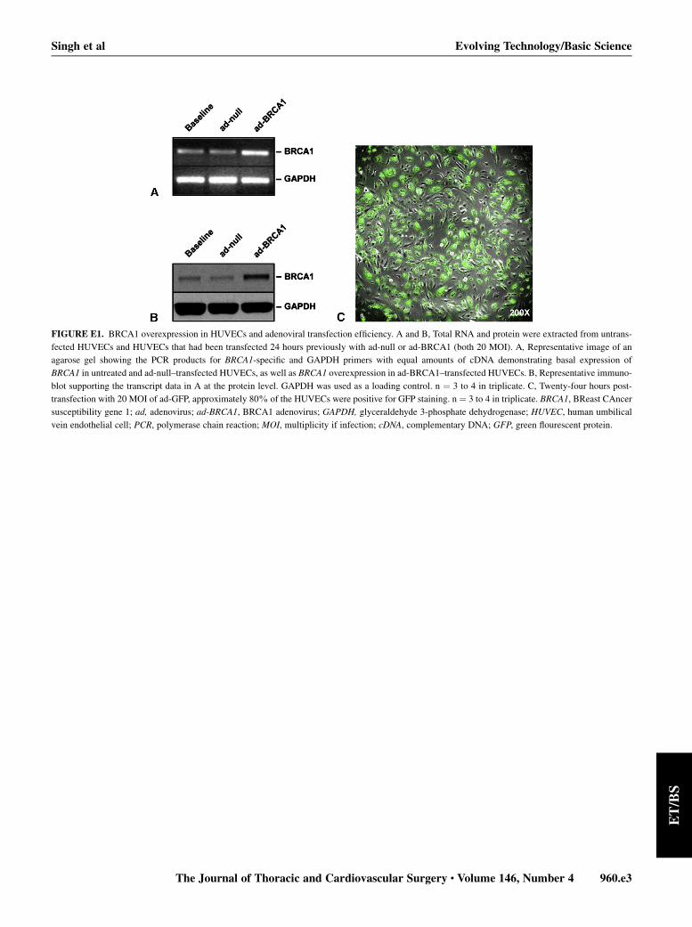
FIGURE E1. BRCA1 overexpression in HUVECs and adenoviral transfection efficiency. A and B, Total RNA and protein were extracted from untrans-
fected HUVECs and HUVECs that had been transfected 24 hours previously with ad-null or ad-BRCA1 (both 20 MOI). A, Representative image of an
agarose gel showing the PCR products for BRCA1-specific and GAPDH primers with equal amounts of cDNA demonstrating basal expression of
BRCA1 in untreated and ad-null–transfected HUVECs, as well as BRCA1 overexpression in ad-BRCA1–transfected HUVECs. B, Representative immuno-
blot supporting the transcript data in A at the protein level. GAPDH was used as a loading control. n ¼ 3 to 4 in triplicate. C, Twenty-four hours post-
transfection with 20 MOI of ad-GFP, approximately 80% of the HUVECs were positive for GFP staining. n ¼ 3 to 4 in triplicate. BRCA1, BReast CAncer
susceptibility gene 1; ad, adenovirus; ad-BRCA1, BRCA1 adenovirus; GAPDH, glyceraldehyde 3-phosphate dehydrogenase; HUVEC, human umbilical
vein endothelial cell; PCR, polymerase chain reaction; MOI, multiplicity if infection; cDNA, complementary DNA; GFP, green flourescent protein.
Singh et al Evolving Technology/Basic Science
The Journal of Thoracic and Cardiovascular Surgery c Volume 146, Number 4 960.e3
ET/BS
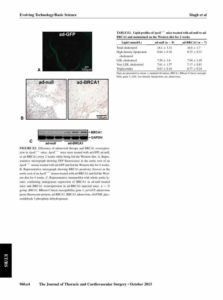
TABLE E1. Lipid profiles of ApoE�/� mice treated with ad-null or ad-
BRCA1 and maintained on the Western diet for 2 weeks
Lipid (mmol/L) ad-null (n ¼ 8) ad-BRCA1 (n ¼ 7)
Total cholesterol 18.1 � 3.31 16.6 � 1.7
High-density lipoprotein
cholesterol
0.64 � 0.16 0.72 � 0.21
LDL cholesterol 7.54 � 1.6 7.94 � 1.45
Very LDL cholesterol 7.61 � 1.57 7.17 � 0.81
Triglycerides 0.67 � 0.16 0.77 � 0.24
Data are presented as mean � standard deviation. BRCA1, BReast CAncer suscepti-
bility gene 1; LDL, low-density lipoprotein; ad, adenovirus.
FIGURE E2. Efficiency of adenoviral therapy and BRCA1 overexpres-
sion in ApoE�/� mice. ApoE�/� mice were treated with ad-GFP, ad-null,
or ad-BRCA1 every 2 weeks while being fed the Western diet. A, Repre-
sentative micrograph showing GFP fluorescence in the aortic root of an
ApoE�/� mouse treated with ad-GFP and fed the Western diet for 4 weeks.
B, Representative micrograph showing BRCA1 positivity (brown) in the
aortic root of an ApoE�/�mouse treated with ad-BRCA1 and fed the West-
ern diet for 4 weeks. C, Representative immunoblot with whole aortic ly-
sates confirming endogenous expression of BRCA1 in ad-null–treated
mice and BRCA1 overexpression in ad-BRCA1–injected mice. n ¼ 3/
group. BRCA1, BReast CAncer susceptibility gene 1; ad-GFP, adenovirus
green fluorescent protein; ad-BRCA1, BRCA1 adenovirus; GAPDH, glyc-
eraldehyde 3-phosphate dehydrogenase.
960.e4 The Journal of Thoracic and Cardiovascular Surgery c October 2013
Evolving Technology/Basic Science Singh et al
ET/BS
Related Documents