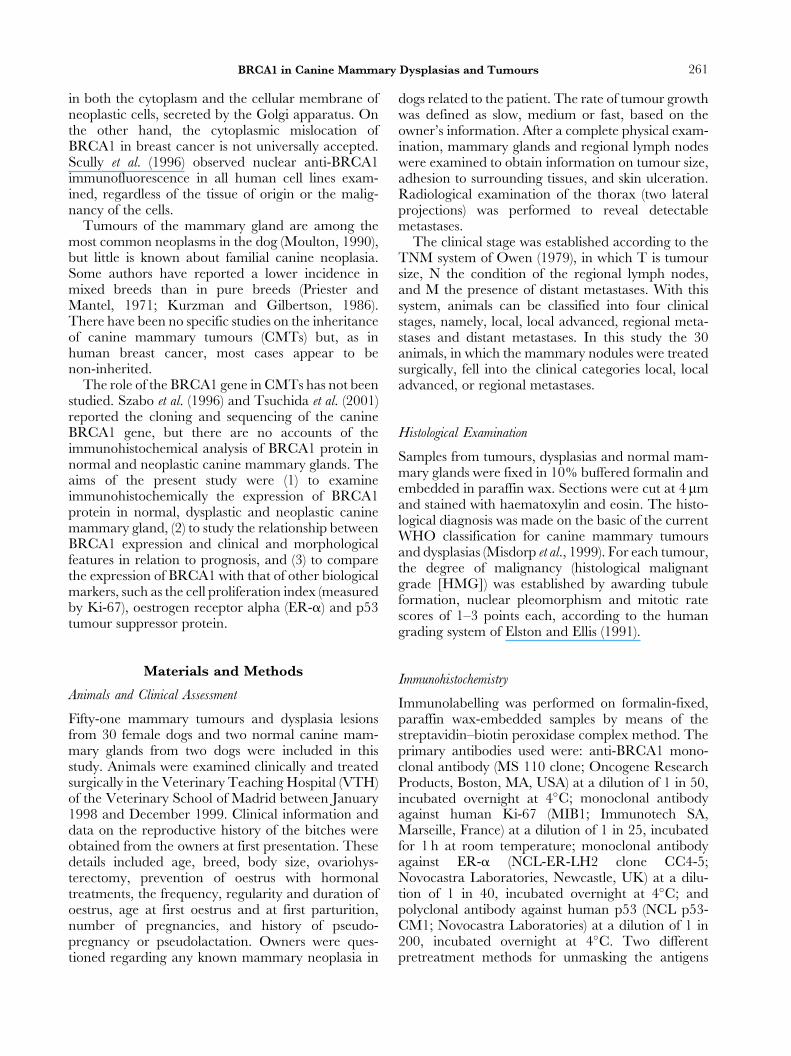
J. Comp. Path. 2003, Vol. 128, 260–268 doi:10.1053/jcpa.2002.0631 BRCA1 Expression in Canine Mammary Dysplasias and Tumours: Relationship with Prognostic Variables A. Nieto, M. D. Pe ´rez-Alenza, N. Del Castillo, E. Tabanera, M. Castan ˜o and L. Pen ˜a Departamento de Patologı ´a Animal II, Hospital Clı ´nico Veterinario, Facultad de Veterinaria, Universidad Complutense de Madrid, 28040, Madrid, Spain Summary BRCA1 is a nuclear phosphoprotein that participates in the regulation of the cell cycle. The role of the BRCA1 gene in canine mammary tissue and mammary tumours has not been studied. The present study examined immunohistochemically the expression and intracellular distribution of BRCA1 protein in two normal, seven dysplastic and 44 neoplastic canine mammary glands and its relationship with clinical and pathological variables and other prognostic parameters. Strong nuclear immunolabelling of BRCA1 protein was observed in the epithelial cells of the normal mammary glands and mammary dysplasias. The majority of benign tumours, and more especially of malignant tumours, showed a significant reduction in the nuclear expression of BRCA1 protein and an increase in cytoplasmic expression. Loss of BRCA1 expression was associated with high proliferation marker Ki-67 and ER-a negative tumours. The reduction and aberrant distribution of BRCA1 in canine mammary tumours were significantly associated with malignant characteristics. The results may indicate that BRCA1 has a role in the malignant behaviour of these tumours. # 2003 Elsevier Science Ltd. All rights reserved. Introduction The BRCA1 gene, located on human chromosome 17q21, is conserved and expressed in a wide range of mammalian tissues (Yang and Lipmann, 1999). This gene, which was cloned in 1994 (Miki et al., 1994), encodes a 220 kDa nuclear phosphoprotein (BRCA1) that is associated with functional differentiation in numerous tissues and participates in the negative regulation of the cell cycle in mammary epithelial cells (Hall et al., 1990; Thompson et al., 1995; Lee et al., 1999). Loss of BRCA1 results in defective DNA repair, abnormal cell cycle arrest, increased apoptosis, genetic instability and tumorigenesis (Deng and Scott, 2000). In addition, BRCA1 mRNA levels are mark- edly reduced in the progression of carcinoma in situ as compared with invasive breast cancer (Thompson et al., 1995). These findings support the role of BRCA1 as a tumour suppressor gene, decreased expression of which represents a critical step in the development of breast cancer. Mutation of the human BRCA1 gene is associated with a large number of hereditary breast cancers, representing 5–10% of all breast cancers (Lynch et al., 1984). However, the great majority of breast cancers are non-inherited, and in such cases different authors have demonstrated a decreased nuclear expression or aberrant distribution of BRCA1 protein, attributed to acquired somatic mutations of the gene (Jarvis et al., 1998; Taylor et al., 1998; Lee et al., 1999; Yang and Lipmann, 1999). The intracellular distribution of BRCA1 protein is controversial. Chen et al. (1995, 1996) reported that BRCA1 protein was located in the nucleus of normal mammary epithelial cells, but was aberrantly located in the cytoplasm of most breast and ovarian cancer cell lines. Similarly, Jensen et al. (1996) found BRCA1 0021–9975/03/$ – see front matter # 2003 Elsevier Science Ltd. All rights reserved.

Welcome message from author
This document is posted to help you gain knowledge. Please leave a comment to let me know what you think about it! Share it to your friends and learn new things together.
Transcript
J. Comp. Path. 2003, Vol. 128, 260±268doi:10.1053/jcpa.2002.0631
BRCA1 Expression in Canine MammaryDysplasias and Tumours: Relationship with
Prognostic Variables
A. Nieto, M. D. PeÂrez-Alenza, N. Del Castillo, E. Tabanera,M. CastanÄo and L. PenÄa
Departamento de PatologõÂa Animal II, Hospital ClõÂnico Veterinario, Facultad de Veterinaria,
Universidad Complutense de Madrid, 28040, Madrid, Spain
Summary
BRCA1 is a nuclear phosphoprotein that participates in the regulation of the cell cycle. The role of theBRCA1 gene in canine mammary tissue and mammary tumours has not been studied. The present studyexamined immunohistochemically the expression and intracellular distribution of BRCA1 protein in twonormal, seven dysplastic and 44 neoplastic canine mammary glands and its relationship with clinical andpathological variables and other prognostic parameters. Strong nuclear immunolabelling of BRCA1 proteinwas observed in the epithelial cells of the normal mammary glands and mammary dysplasias. The majority ofbenign tumours, and more especially of malignant tumours, showed a significant reduction in the nuclearexpression of BRCA1 protein and an increase in cytoplasmic expression. Loss of BRCA1 expression wasassociated with high proliferation marker Ki-67 and ER-a negative tumours. The reduction and aberrantdistribution of BRCA1 in canine mammary tumours were significantly associated with malignantcharacteristics. The results may indicate that BRCA1 has a role in the malignant behaviour of these tumours.
# 2003 Elsevier Science Ltd. All rights reserved.
Introduction
The BRCA1 gene, located on human chromosome17q21, is conserved and expressed in a wide range ofmammalian tissues (Yang and Lipmann, 1999). Thisgene, which was cloned in 1994 (Miki et al., 1994),encodes a 220 kDa nuclear phosphoprotein (BRCA1)that is associated with functional differentiation innumerous tissues and participates in the negativeregulation of the cell cycle in mammary epithelial cells(Hall et al., 1990; Thompson et al., 1995; Lee et al.,1999). Loss of BRCA1 results in defective DNArepair, abnormal cell cycle arrest, increased apoptosis,genetic instability and tumorigenesis (Deng and Scott,2000). In addition, BRCA1 mRNA levels are mark-edly reduced in the progression of carcinoma in situ ascompared with invasive breast cancer (Thompsonet al., 1995). These findings support the role of BRCA1as a tumour suppressor gene, decreased expression of
0021±9975/03/$ ± see front matter
which represents a critical step in the development ofbreast cancer.
Mutation of the human BRCA1 gene is associatedwith a large number of hereditary breast cancers,representing 5±10% of all breast cancers (Lynch et al.,1984). However, the great majority of breast cancersare non-inherited, and in such cases different authorshave demonstrated a decreased nuclear expression oraberrant distribution of BRCA1 protein, attributed toacquired somatic mutations of the gene (Jarvis et al.,1998; Taylor et al., 1998; Lee et al., 1999; Yang andLipmann, 1999).
The intracellular distribution of BRCA1 protein iscontroversial. Chen et al. (1995, 1996) reported thatBRCA1 protein was located in the nucleus of normalmammary epithelial cells, but was aberrantly locatedin the cytoplasm of most breast and ovarian cancercell lines. Similarly, Jensen et al. (1996) found BRCA1
# 2003 Elsevier Science Ltd. All rights reserved.
261BRCA1 in Canine Mammary Dysplasias and Tumours
in both the cytoplasm and the cellular membrane ofneoplastic cells, secreted by the Golgi apparatus. Onthe other hand, the cytoplasmic mislocation ofBRCA1 in breast cancer is not universally accepted.Scully et al. (1996) observed nuclear anti-BRCA1immunofluorescence in all human cell lines exam-ined, regardless of the tissue of origin or the malig-nancy of the cells.
Tumours of the mammary gland are among themost common neoplasms in the dog (Moulton, 1990),but little is known about familial canine neoplasia.Some authors have reported a lower incidence inmixed breeds than in pure breeds (Priester andMantel, 1971; Kurzman and Gilbertson, 1986).There have been no specific studies on the inheritanceof canine mammary tumours (CMTs) but, as inhuman breast cancer, most cases appear to benon-inherited.
The role of the BRCA1 gene in CMTs has not beenstudied. Szabo et al. (1996) and Tsuchida et al. (2001)reported the cloning and sequencing of the canineBRCA1 gene, but there are no accounts of theimmunohistochemical analysis of BRCA1 protein innormal and neoplastic canine mammary glands. Theaims of the present study were (1) to examineimmunohistochemically the expression of BRCA1protein in normal, dysplastic and neoplastic caninemammary gland, (2) to study the relationship betweenBRCA1 expression and clinical and morphologicalfeatures in relation to prognosis, and (3) to comparethe expression of BRCA1 with that of other biologicalmarkers, such as the cell proliferation index (measuredby Ki-67), oestrogen receptor alpha (ER-a) and p53tumour suppressor protein.
Materials and Methods
Animals and Clinical Assessment
Fifty-one mammary tumours and dysplasia lesionsfrom 30 female dogs and two normal canine mam-mary glands from two dogs were included in thisstudy. Animals were examined clinically and treatedsurgically in the Veterinary Teaching Hospital (VTH)of the Veterinary School of Madrid between January1998 and December 1999. Clinical information anddata on the reproductive history of the bitches wereobtained from the owners at first presentation. Thesedetails included age, breed, body size, ovariohys-terectomy, prevention of oestrus with hormonaltreatments, the frequency, regularity and duration ofoestrus, age at first oestrus and at first parturition,number of pregnancies, and history of pseudo-pregnancy or pseudolactation. Owners were ques-tioned regarding any known mammary neoplasia in
dogs related to the patient. The rate of tumour growthwas defined as slow, medium or fast, based on theowner's information. After a complete physical exam-ination, mammary glands and regional lymph nodeswere examined to obtain information on tumour size,adhesion to surrounding tissues, and skin ulceration.Radiological examination of the thorax (two lateralprojections) was performed to reveal detectablemetastases.
The clinical stage was established according to theTNM system of Owen (1979), in which T is tumoursize, N the condition of the regional lymph nodes,and M the presence of distant metastases. With thissystem, animals can be classified into four clinicalstages, namely, local, local advanced, regional meta-stases and distant metastases. In this study the 30animals, in which the mammary nodules were treatedsurgically, fell into the clinical categories local, localadvanced, or regional metastases.
Histological Examination
Samples from tumours, dysplasias and normal mam-mary glands were fixed in 10% buffered formalin andembedded in paraffin wax. Sections were cut at 4 mmand stained with haematoxylin and eosin. The histo-logical diagnosis was made on the basic of the currentWHO classification for canine mammary tumoursand dysplasias (Misdorp et al., 1999). For each tumour,the degree of malignancy (histological malignantgrade [HMG]) was established by awarding tubuleformation, nuclear pleomorphism and mitotic ratescores of 1±3 points each, according to the humangrading system of Elston and Ellis (1991).
Immunohistochemistry
Immunolabelling was performed on formalin-fixed,paraffin wax-embedded samples by means of thestreptavidin±biotin peroxidase complex method. Theprimary antibodies used were: anti-BRCA1 mono-clonal antibody (MS 110 clone; Oncogene ResearchProducts, Boston, MA, USA) at a dilution of 1 in 50,incubated overnight at 4�C; monoclonal antibodyagainst human Ki-67 (MIB1; Immunotech SA,Marseille, France) at a dilution of 1 in 25, incubatedfor 1 h at room temperature; monoclonal antibodyagainst ER-a (NCL-ER-LH2 clone CC4-5;Novocastra Laboratories, Newcastle, UK) at a dilu-tion of 1 in 40, incubated overnight at 4�C; andpolyclonal antibody against human p53 (NCL p53-CM1; Novocastra Laboratories) at a dilution of 1 in200, incubated overnight at 4�C. Two differentpretreatment methods for unmasking the antigens
262 A. Nieto et al.
were used. For BRCA1, ER-a and p-53, slides wereboiled for 2 min in a pressure cooker containing 2 litresof 10 nM sodium citrate buffer (pH 6�0). For Ki-67detection, the slides were placed in plastic jarsand boiled in sodium citrate (10 mM) three times(5 min each) at maximum power in a microwave oven.
After pretreatment, endogenous peroxidase activitywas blocked by incubation for 15 min in methanolcontaining H2O2 1�0%. The sections were thenwashed in water and Tris-buffered saline (TBS)(0�1 M Tris base, 0�9% NaCl, pH 7�4) and incubatedin a humidity chamber with the primary antibodies(times and temperatures as described above). Slideswere then incubated with anti-mouse biotinylatedsecondary antibody (EO 4233; Dako A/S, Glostrup,Denmark) diluted 1 in 200, for 45 min at roomtemperature, for monoclonal antibodies; or withgoat anti-rabbit biotinylated secondary antibody(BA-1000; Vector Laboratories, Burlingame, CA,USA) diluted 1 in 400, for 45 min at room tempera-ture, for the polyclonal antibody anti-p-53. The slideswere next treated with streptavidin±biotin peroxidasecomplex (P50242; Zymed Laboratories, SanFrancisco, USA) diluted 1 in 400, for 45 min at roomtemperature. The slides were washed three times inTBS between each incubation step. Immunoreactivitywas `̀ visualized'' with 0�01% H2O2 and 3-30-diaminobenzidine tetrachloride (D5059; SigmaChemical Co., St Louis, MO, USA) in TBS. Afterwashing in distilled water for 10 min, slides werecounterstained for 2 min with Harris's haematoxylin,washed in tap water, dehydrated and mounted inDPx. For negative control purposes, the primaryantibody was replaced by TBS, or normal mouse orrabbit serum. Normal canine uterus and normalcanine mammary tissues were used as positive con-trols for detection of ER-a and BRCA1, respectively.Normal mammary gland or hyperplastic tissue adja-cent to the neoplasms was used as an internal positivecontrol in many slides. Tumours of canine mammarygland, known to be positive for p53 and Ki-67,were used as positive controls for p53 and Ki-67immunolabelling.
Assessment of labelling for BRCA1 was qualitative,immunoreactivity being graded as follows: 0/�,negative (two tumours only) or ,25% of cells weaklylabelled; ��, 25±50% of cells with labelling ofmoderate intensity; ���, .50% of cells intenselypositive. The immunoreactivity was recorded asnuclear, cytoplasmic, or both.
The indices of Ki-67, ER-a and p53 were deter-mined with a computer-assisted image analyzer(Olympus MicroimageTM image analysis [softwareversion 4.0. for Windows], Barcelona, Spain) as thepercentage of positive brown nuclei in the tumour
cells in 10 representative high-power fields (minimumof 1000 cells). For ER-a and p53, tumours in whichmore than 10% of nuclei were immunolabelled wereconsidered positive.
Follow-up Study
Dogs with at least one malignant tumour weremonitored every 3±4 months for 24 months aftersurgical treatment. On each occasion, the examina-tion included a radiological evaluation of the thorax.Tumour recurrences, metastases and time and causeof death were registered. Disease-free survival (DFS)and overall survival (OS) were calculated for eachdog (low, 0±6 months; medium, 7±17 months; high,.18 months).
Statistical Analysis
The association between BRCA1 protein expressionand clinical and pathological variables was studiedwith Biomedical Data Processing Software (Dixon,1993). Linear regression was calculated to establishthe correlation coefficient of continuous expression ofvariables (age, Ki-67 and p53). Analysis of variance(ANOVA) was used to study the differences ofBRCA1 in the groups of categorical variables (F test,pooled t-test if variances are assumed to be equal, andWelch test or separate t-test if variances are not equal).To analyse the dog-related variables (age, breed,reproductive status, etc) in animals with more thanone mammary nodule, the most representative valueof BRCA1 was assigned to each dog. P� 0�05 wasconsidered significant.
Results
Two normal mammary glands, seven dysplasias, 21benign tumours and 23 malignant tumours wereexamined immunohistochemically for BRCA1.Parenchymal cells of dysplasias and tumours showedvariable immunolabelling, but stromal cells wereinvariably negative. Only two neoplasms (one mixedbenign mammary tumour and one simple tubulo-papillary carcinoma) were completely negative toBRCA1 labelling.
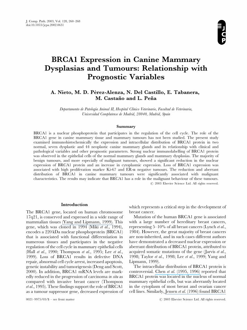
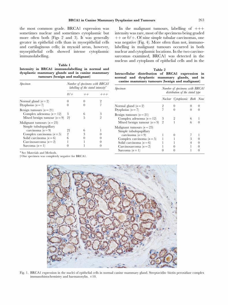
The intensity of intracellular expression of BRCA1in the various samples is shown in Table 1, and theintracellular distribution in Table 2. Strongly positive,homogeneous nuclear labelling was observed in theepithelial cells of the two normal mammary glandsand all seven dysplasias, but there was little or nocytoplasmic labelling (Fig. 1).
In the benign tumours, all three grades of intensity(0/�, �� and ���) were represented, but �� was
263BRCA1 in Canine Mammary Dysplasias and Tumours
the most common grade. BRCA1 expression wassometimes nuclear and sometimes cytoplasmic butmore often both (Figs 2 and 3). It was generallygreater in epithelial cells than in myoepithelial cellsand cartilaginous cells; in myxoid areas, however,myoepithelial cells showed intense cytoplasmicimmunolabelling.
Table 1Intensity in BRCA1 immunolabelling in normal anddysplastic mammary glands and in canine mammary
tumours (benign and malignant)
Specimen Number of specimens with BRCA1labelling of the stated intensity�
0/� �� ���Normal gland (n� 2) 0 0 2Dysplasias (n� 7) 0 0 7
Benign tumours (n� 21)Complex adenoma (n� 12) 3 6 3Mixed benign tumour (n� 9) 2y 5 2
Malignant tumours (n� 23)Simple tubulopapillary
carcinoma (n� 9) 2y 6 1Complex carcinoma (n� 5) 2 3 0Solid carcinoma (n� 6) 6 0 0Carcinosarcoma (n� 2) 1 1 0Sarcoma (n� 1) 0 1 0
�See Materials and Methods.yOne specimen was completely negative for BRCA1.
Fig. 1. BRCA1 expression in the nuclei of epithelial cells in normalimmunohistochemistry and haematoxylin. �10.
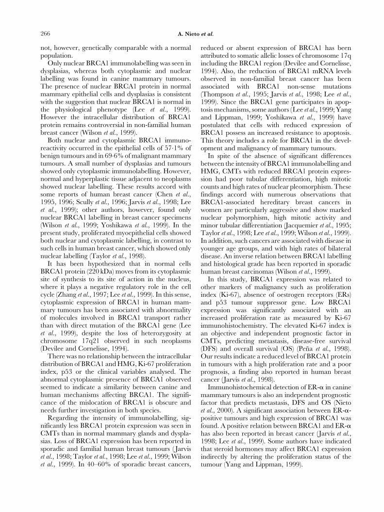
In the malignant tumours, labelling of ���intensity was rare, most of the specimens being graded�� or 0/�. Of nine simple tubular carcinomas, onewas negative (Fig. 4). More often than not, immuno-labelling in malignant tumours occurred in bothnuclear and cytoplasmic locations. In the two carcino-sarcomas examined, BRCA1 was detected in thenucleus and cytoplasm of epithelial cells and in the
Table 2Intracellular distribution of BRCA1 expression innormal and dysplastic mammary glands, and in
canine mammary tumours (benign and malignant)
Specimen Number of specimens with BRCA1distribution of the stated type
Nuclear Cytoplasmic Both None
Normal gland (n� 2) 2 0 0 0Dysplasias (n� 7) 7 0 0 0
Benign tumours (n� 21)Complex adenoma (n� 12) 3 2 6 1Mixed benign tumour (n� 9) 2 1 6 0
Malignant tumours (n� 23)Simple tubulopapillary
carcinoma (n� 9) 1 0 7 1Complex carcinoma (n� 5) 1 1 3 0Solid carcinoma (n� 6) 1 1 4 0Carcinosarcoma (n� 2) 1 0 1 0Sarcoma (n� 1) 0 0 1 0
canine mammary gland. Streptavidin±biotin peroxidase complex
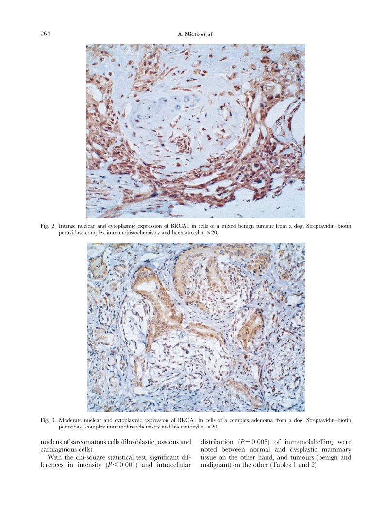
Fig. 2. Intense nuclear and cytoplasmic expression of BRCA1 in cells of a mixed benign tumour from a dog. Streptavidin±biotinperoxidase complex immunohistochemistry and haematoxylin. �20.
Fig. 3. Moderate nuclear and cytoplasmic expression of BRCA1 in cells of a complex adenoma from a dog. Streptavidin±biotinperoxidase complex immunohistochemistry and haematoxylin. �20.
264 A. Nieto et al.
nucleus of sarcomatous cells (fibroblastic, osseous andcartilaginous cells).
With the chi-square statistical test, significant dif-ferences in intensity (P , 0�001) and intracellular
distribution (P� 0�008) of immunolabelling werenoted between normal and dysplastic mammarytissue on the other hand, and tumours (benign andmalignant) on the other (Tables 1 and 2).
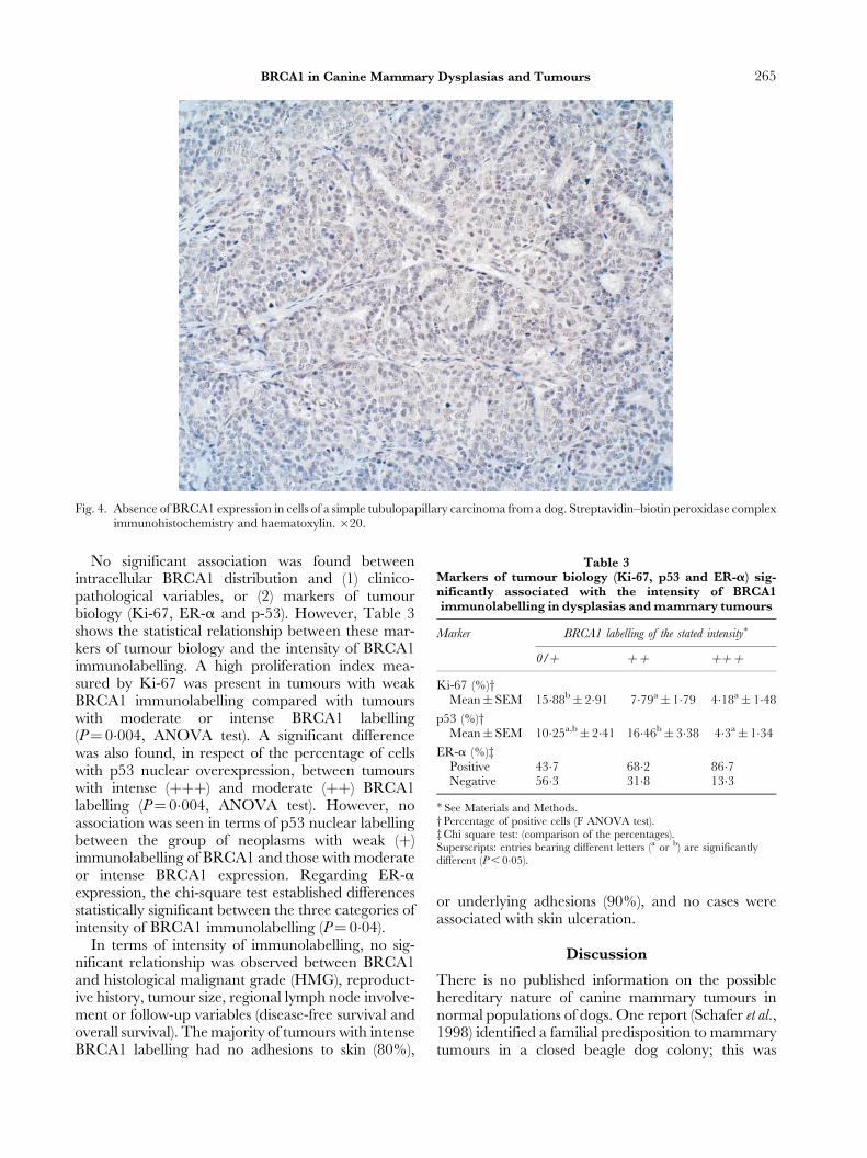
Fig. 4. Absence of BRCA1 expression in cells of a simple tubulopapillary carcinoma from a dog. Streptavidin±biotin peroxidase compleximmunohistochemistry and haematoxylin. �20.
Table 3Markers of tumour biology (Ki-67, p53 and ER-a) sig-nificantly associated with the intensity of BRCA1immunolabelling in dysplasias and mammary tumours
Marker BRCA1 labelling of the stated intensity�
0/� �� ���Ki-67 (%)y
Mean� SEM 15�88b� 2�91 7�79a� 1�79 4�18a� 1�48
p53 (%)yMean� SEM 10�25a,b� 2�41 16�46b� 3�38 4�3a� 1�34
ER-a (%)zPositive 43�7 68�2 86�7Negative 56�3 31�8 13�3
� See Materials and Methods.yPercentage of positive cells (F ANOVA test).zChi square test: (comparison of the percentages).Superscripts: entries bearing different letters (a or b) are significantlydifferent (P , 0�05).
265BRCA1 in Canine Mammary Dysplasias and Tumours
No significant association was found betweenintracellular BRCA1 distribution and (1) clinico-pathological variables, or (2) markers of tumourbiology (Ki-67, ER-a and p-53). However, Table 3shows the statistical relationship between these mar-kers of tumour biology and the intensity of BRCA1immunolabelling. A high proliferation index mea-sured by Ki-67 was present in tumours with weakBRCA1 immunolabelling compared with tumourswith moderate or intense BRCA1 labelling(P� 0�004, ANOVA test). A significant differencewas also found, in respect of the percentage of cellswith p53 nuclear overexpression, between tumourswith intense (���) and moderate (��) BRCA1labelling (P� 0�004, ANOVA test). However, noassociation was seen in terms of p53 nuclear labellingbetween the group of neoplasms with weak (�)immunolabelling of BRCA1 and those with moderateor intense BRCA1 expression. Regarding ER-aexpression, the chi-square test established differencesstatistically significant between the three categories ofintensity of BRCA1 immunolabelling (P� 0�04).
In terms of intensity of immunolabelling, no sig-nificant relationship was observed between BRCA1and histological malignant grade (HMG), reproduct-ive history, tumour size, regional lymph node involve-ment or follow-up variables (disease-free survival andoverall survival). The majority of tumours with intenseBRCA1 labelling had no adhesions to skin (80%),
or underlying adhesions (90%), and no cases wereassociated with skin ulceration.
Discussion
There is no published information on the possiblehereditary nature of canine mammary tumours innormal populations of dogs. One report (Schafer et al.,1998) identified a familial predisposition to mammarytumours in a closed beagle dog colony; this was
266 A. Nieto et al.
not, however, genetically comparable with a normalpopulation.
Only nuclear BRCA1 immunolabelling was seen indysplasias, whereas both cytoplasmic and nuclearlabelling was found in canine mammary tumours.The presence of nuclear BRCA1 protein in normalmammary epithelial cells and dysplasias is consistentwith the suggestion that nuclear BRCA1 is normal inthe physiological phenotype (Lee et al., 1999).However the intracellular distribution of BRCA1protein remains controversial in non-familial humanbreast cancer (Wilson et al., 1999).
Both nuclear and cytoplasmic BRCA1 immuno-reactivity occurred in the epithelial cells of 57�1% ofbenign tumours and in 69�6% of malignant mammarytumours. A small number of dysplasias and tumoursshowed only cytoplasmic immunolabelling. However,normal and hyperplastic tissue adjacent to neoplasmsshowed nuclear labelling. These results accord withsome reports of human breast cancer (Chen et al.,1995, 1996; Scully et al., 1996; Jarvis et al., 1998; Leeet al., 1999); other authors, however, found onlynuclear BRCA1 labelling in breast cancer specimens(Wilson et al., 1999; Yoshikawa et al., 1999). In thepresent study, proliferated myoepithelial cells showedboth nuclear and cytoplasmic labelling, in contrast tosuch cells in human breast cancer, which showed onlynuclear labelling (Taylor et al., 1998).
It has been hypothesized that in normal cellsBRCA1 protein (220 kDa) moves from its cytoplasmicsite of synthesis to its site of action in the nucleus,where it plays a negative regulatory role in the cellcycle (Zhang et al., 1997; Lee et al., 1999). In this sense,cytoplasmic expression of BRCA1 in human mam-mary tumours has been associated with abnormalityof molecules involved in BRCA1 transport ratherthan with direct mutation of the BRCA1 gene (Leeet al., 1999), despite the loss of heterozygosity atchromosome 17q21 observed in such neoplasms(Devilee and Cornelisse, 1994).
There was no relationship between the intracellulardistribution of BRCA1 and HMG, Ki-67 proliferationindex, p53 or the clinical variables analysed. Theabnormal cytoplasmic presence of BRCA1 observedseemed to indicate a similarity between canine andhuman mechanisms affecting BRCA1. The signifi-cance of the mislocation of BRCA1 is obscure andneeds further investigation in both species.
Regarding the intensity of immunolabelling, sig-nificantly less BRCA1 protein expression was seen inCMTs than in normal mammary glands and dyspla-sias. Loss of BRCA1 expression has been reported insporadic and familial human breast tumours ( Jarviset al., 1998; Taylor et al., 1998; Lee et al., 1999; Wilsonet al., 1999). In 40±60% of sporadic breast cancers,
reduced or absent expression of BRCA1 has beenattributed to somatic allelic losses of chromosome 17qincluding the BRCA1 region (Devilee and Cornelisse,1994). Also, the reduction of BRCA1 mRNA levelsobserved in non-familial breast cancer has beenassociated with BRCA1 non-sense mutations(Thompson et al., 1995; Jarvis et al., 1998; Lee et al.,1999). Since the BRCA1 gene participates in apop-tosis mechanisms, some authors (Lee et al., 1999; Yangand Lippman, 1999; Yoshikawa et al., 1999) havepostulated that cells with reduced expression ofBRCA1 possess an increased resistance to apoptosis.This theory includes a role for BRCA1 in the devel-opment and malignancy of mammary tumours.
In spite of the absence of significant differencesbetween the intensity of BRCA1 immunolabelling andHMG, CMTs with reduced BRCA1 protein expres-sion had poor tubular differentiation, high mitoticcounts and high rates of nuclear pleomorphism. Thesefindings accord with numerous observations thatBRCA1-associated hereditary breast cancers inwomen are particularly aggressive and show markednuclear polymorphism, high mitotic activity andminor tubular differentiation (Jacquemier et al., 1995;Taylor et al., 1998; Lee et al., 1999; Wilson et al., 1999).In addition, such cancers are associated with disease inyounger age groups, and with high rates of bilateraldisease. An inverse relation between BRCA1 labellingand histological grade has been reported in sporadichuman breast carcinomas (Wilson et al., 1999).
In this study, BRCA1 expression was related toother markers of malignancy such as proliferationindex (Ki-67), absence of oestrogen receptors (ERs)and p53 tumour suppressor gene. Low BRCA1expression was significantly associated with anincreased proliferation rate as measured by Ki-67immunohistochemistry. The elevated Ki-67 index isan objective and independent prognostic factor inCMTs, predicting metastasis, disease-free survival(DFS) and overall survival (OS) (PenÄa et al., 1998).Our results indicate a reduced level of BRCA1 proteinin tumours with a high proliferation rate and a poorprognosis, a finding also reported in human breastcancer (Jarvis et al., 1998).
Immunohistochemical detection of ER-a in caninemammary tumours is also an independent prognosticfactor that predicts metastasis, DFS and OS (Nietoet al., 2000). A significant association between ER-a-positive tumours and high expression of BRCA1 wasfound. A positive relation between BRCA1 and ER-ahas also been reported in breast cancer (Jarvis et al.,1998; Lee et al., 1999). Some authors have indicatedthat steroid hormones may affect BRCA1 expressionindirectly by altering the proliferation status of thetumour (Yang and Lippman, 1999).
267BRCA1 in Canine Mammary Dysplasias and Tumours
Mutations in the p53 gene have been found inbenign and malignant canine tumours, indicatingmalignancy and poor prognosis (Muto et al., 2000).Recent reports demonstrate that BRCA1 and p53genes interact and that BRCA1 requires the presenceof p53 for the enhancement of transcriptional acti-vation (Zhang et al., 1997). In our study, increased p53immunolabelling was found in tumours with a mod-erate BRCA1 expression. The p53 overexpressionfound in some human (Armes et al., 1998) and canine(Muto et al., 2000) benign mammary tumours hasbeen regarded as an early event in mammary carci-nogenesis, related to preinvasive stages of the disease.
No statistically significant relationship betweenBRCA1 expression and epidemiological or clinicalvariables such as age, breed, hormonal status, preg-nancies or rate of tumour growth was found in thisstudy. Some clinical features of prognostic value(PeÂrez-Alenza et al., 2000), such as adhesion to under-lying tissues or skin, and skin ulceration, wereobserved with increased frequency in weakly labelledmalignant tumours; this apparent association, how-ever, did not reach the accepted level (P , 0�05) ofsignificance. Lymph node involvement, DFS and OSwere not related to intensity or distribution of BRCA1expression, but further cases of malignant CMTs needto be studied to confirm this observation.
In conclusion, the study demonstrated the expres-sion of BRCA1 in the nuclei of normal mammaryepithelial cells and in the nuclei and cytoplasm ofneoplastic cells (epithelial, myoepithelial and cartila-ginous cells). Reduced BRCA1 in CMTs was charac-teristic of malignant tumours and significantlyassociated with the histological grade of malignancyand with high levels of the proliferation marker Ki-67.These data indicate a potential role of BRCA1 in themalignant behaviour of CMTs. Additional studies,including those of hereditary and sporadic caninemammary tumours, would help to elucidate theimplication of BRCA1 in mammary carcinogenesisand its possible use as a prognostic marker, as well asto establish new diagnostic and therapeutic strategies.
Acknowledgments
This study was supported by the Research Project ofthe Complutense University (PR 269/98-8178/98).The authors thank Pedro Cuesta for the statisticalwork, and Pedro Aranda, Sandra Contreras andMario Hernando for technical assistance.
References
Armes, J. E., Egan, A. J., Southey, M. C., Dite, G. S.,McCredie, M. R., Giles, G. G., Hopper, J. L. and
Venter, D. J. (1998). The histologic phenotypes ofbreast carcinoma occurring before age 40 years inwomen with and without BRCA1 or BRCA2germline mutations: a population-based study. Cancer,83, 2335±2345.
Chen, Y., Chen, C. F., Riley, D. J., Allred, D. C.,Chen, P. L., Van Hoff, D., Osborne, C. K. and Lee, W. H.(1995). Aberrant subcellular localization of BRCA1 inbreast cancer. Science, 270, 789±791.
Chen, Y., Chen, P. L., Riley, D., Lee, W. H., Allred, D. C.and Osborne, C. K. (1996). Localization of BRCA1 inhuman breast and ovarian cancer cells. Science, 272,125±126.
Deng, C. X. and Scott, F. (2000). Role of the tumorsuppressor Brca1 in genetic stability and mammarygland tumor formation. Oncogene, 19, 1059±1064.
Devilee, P. and Cornelisse, C. J. (1994). Somatic geneticchanges in human breast cancer. Biochemical andBiophysical Acta, 1198, 113±130.
Dixon, W. (1993). BMDP Statistical Software, release 7.0.University of California Press, Los Angeles, CA.
Elston, C. W. and Ellis, I. O. (1991). Pathologicalprognostic factors in breast cancer. I. The value ofhistological grade in breast cancer: experience from alarge study with long-term follow-up. Histopathology, 19,403±410.
Hall, J. M., Lee, M. K., Newman, B., Morrow, J. E.,Anderson, L. A., Huey, B. and King, M. C. (1990).Linkage of early-onset familial breast cancer tochromosome 17q21. Science, 250, 1684±1689.
Jacquemier, J., Eisinger, F., Birnbaum, D. and Sobol, H.(1995). Histoprognostic grade in BRCA1-associatedbreast cancer. Lancet, 345, 1503.
Jarvis, E. M., Kirk, J. A. and Clarke, C. L. (1998). Loss ofnuclear BRCA1 expression in breast cancers isassociated with a highly proliferative tumor phenotype.Cancer Genetics and Cytogenetics, 101, 109±115.
Jensen, R. A., Thompson, M. E., Jetton, T. L., Szabo, C. I.,Van der Meer, R., Helou, B., Tronick, S. R., Page, D. L.,King, M. C. and Holt, J. T. (1996). BRCA1 is secretedand exhibits properties of a granin. Nature Genetics, 12,303±308.
Kurzman, I. D. and Gilbertson, S. R. (1986). Prognosticfactors in canine mammary tumors. Seminars in VeterinaryMedicine and Surgery (Small Animal), 1, 25±32.
Lee, W. Y., Jin, Y. T., Chang, T. W., Lin, P. W. and Su, I. J.(1999). Immunolocalization of BRCA1 protein innormal breast tissue and sporadic invasive ductalcarcinomas: a correlation with other biological param-eters. Histopathology, 34, 106±112.
Lynch, H. T., Albano, W. A., Banes, B. S., Layton, M. A.,Kimberling, W. J. and Lynch, J. F. (1984). Geneticpredisposition to breast cancer. Cancer, 53, 612±622.
Miki, Y., Swensen, J., Shattuck-Eidens, D., Futreal, P. A.,Harshman, K., Tavtigian, S., Liu, Q., Cochran, C.,Bennett, L. M., Ding, W., Bell, R., Rosenthal, J.,Hussey, C., Tran, T., McClure, M., Frye, C., Hattier, T.,Phelps,R.,Haugen-Strano,A.,Katcher,H.,Yahumo,K.,Cholami, Z., Schaffer, D., Stone, S., Bayer, S., Wray, C.,Bogden, R., Dayananth, P., Ward, J., Tonin, P.,Narod, P., Bristow, P. K., Norris, F. H., Helvering, L.,Morrison, P., Rosteck, P., Lai, M., Barret, J. C.,
268 A. Nieto et al.
Lewis, C., Neuhausen, S., Cannon-Albright, L.,Goldgar, D., Wiseman, R., Kamb, A. and Skolnick, M. H.(1994). A strong candidate for the breast andovarian cancer susceptibility gene BRCA1. Science, 266,66±71.
Misdorp, W., Else, R. W., HellmeÂn, E. and Lipscomb, T. P.(1999). Histological Classification of Mammary Tumors of theDog and the Cat. Armed Forces Institute of Pathology,WHO and Worldwide Reference on ComparativeOncology, Washington DC.
Moulton, J.E. (1990). Tumors of the mammary gland.In: Tumors in Domestic Animals, 3rd Edit., J.E. Moulton,Ed., University of California Press, Berkeley, pp.518±552.
Muto, T., Wakui, S., Takahashi, H., Maekawa, S.,Masaoka, T., Ushigome, S. and Furusato, M. (2000).p53 Gene mutations occurring in spontaneous benignand malignant mammary tumors of the dog. VeterinaryPathology, 37, 248±253.
Nieto, A., PenÄa, L., PeÂrez-Alenza, M. D., S�anchez, M. A.,Flores, J. M. and CastanÄo, M. (2000). Immunohistologicdetection of estrogen receptor alpha in caninemammary tumors: clinical and pathologic associationsand prognostic significance. Veterinary Pathology, 37,239±247.
Owen, L. N. (1979). A comparative study of canine andhuman breast cancer. Investigation in Cell Pathology, 2,257±275.
PenÄa, L. L., Nieto, A. I., PeÂrez-Alenza, D., Cuesta, P. andCastanÄo, M. (1998). Immunohistochemical detection ofKi-67 and PCNA in canine mammary tumors: relation-ship to clinical and pathologic variables. Journal ofVeterinary Diagnostic Investigation, 10, 237±246.
PeÂrez-Alenza, M. D., PenÄa, L., Del Castillo, N. andNieto, A. I. (2000). Factors influencing the incidenceand prognosis of canine mammary tumours. Journal ofSmall Animal Practice, 41, 287±291.
Priester, W. A. and Mantel, N. (1971). Occurrence oftumors in domestic animals. Data from 12 UnitedStates and Canadian colleges of veterinary medicine.Journal of the National Cancer Institute, 47,1333±1334.
Schafer, K. A., Kelly, G., Schrader, R., Griffith, W. C.,Muggenburg, B. A., Tierney, L. A., Lechner, J. F.,Janovitz, E. B. and Hann, F. F. (1998). A canine modelof familial mammary gland neoplasia. Veterinary Pathol-ogy, 35, 168±177.
Scully, R., Ganesan, S., Brown, M., De Caprio, J. A.,Cannistra, S. A., Feunteun, J., Schnitt, S. and
Livingston, D. M. (1996). Location of BRCA1 in humanbreast and ovarian cell lines. Science, 272, 123±125.
Szabo, C. I., Wagner, L. A., Francisco, L. V., Roach, J.C., Argonza, R., King, M. C. and Ostrander, E. A.(1996). Human, canine and murine BRCA1 genes:sequence comparison among species. Human MolecularGenetics, 5, 1289±1298.
Taylor, J., Lymboura, M., Pace, P. E., A'Hern,R. P., Desai, A. J., Shousha, S., Coombes, R. C. andAli, S. (1998). An important role for BRCA1 in breastcancer progression is indicated by its loss in a largeproportion of non-familial breast cancers. InternationalJournal of Cancer, 79, 334±342.
Thompson, M. E., Jensen, R. A., Obermiller, P. S.,Page, D. L. and Holt, J. T. (1995). Decreasedexpression of BRCA1 accelerates growth and is oftenpresent during sporadic breast cancer progression.Nature Genetics, 9, 445±450.
Tsuchida, S., Ikemoto, S. and Tagawa, M. (2001).Microsatellite polymorphism in intron 14 of the canineBRCA1 gene. Journal of Veterinary Medical Science, 63,479±481.
Wilson, C. A., Ramos, L., Villasenor, M. R., Anders, K. H.,Press, M. F., Clarke, K., Karlan, B., Chen, J. J.,Scully, R., Livingston, D., Zuch, R. H., Kanter, M. H.,Cohen, S., Calzone, F. J. and Slamon, D. J. (1999).Localization of human BRCA1 and its loss in high-grade non-inherited breast carcinomas. Nature Genetics,21, 236±240.
Yang, X. and Lippman, M. E. (1999). BRCA1 andBRCA2 in breast cancer. Breast Cancer Research andTreatment, 54, 1±10.
Yoshikawa, K., Honda, K., Inamoto, T., Shinohara, H.,Yamauki, A., Suga, K., Okuyama, T., Shimada, T.,Kodama, H., Noguchi, S., Gazdar, A.F., Yamaoka, Y.and Takahashi, R. (1999). Reduction of BRCA1protein expression in Japanese sporadic breast carcino-mas and its frequent loss in BRCA1 associated cases.Clinical Cancer Research, 5, 1249±1261.
Zhang, H. T., Zhang, X., Zhao, H. Z., Kajino, Y.,Weber, B. L., Davis, J. G., Wang, Q., O'Rourke, D. M.,Zhang, H. B., Kajino, K. and Greene, M. I. (1997).Relationship of p215BRCA1 to tyrosine kinase signal-ing pathways and the cell cycle in normal andtransformed cells. Oncogene, 14, 2863±2869.
�Received , May 14th, 2002
Accepted , November 29th, 2002
�
Related Documents