BRAIN TUMORS Dr.Abdulrahman Al-Shudifat Neurosurgery Department

BRAIN TUMORS Dr.Abdulrahman Al-Shudifat Neurosurgery Department.
Jan 16, 2016
Welcome message from author
This document is posted to help you gain knowledge. Please leave a comment to let me know what you think about it! Share it to your friends and learn new things together.
Transcript
BRAIN TUMORS
Dr.Abdulrahman Al-ShudifatNeurosurgery Department

INTRODUCTION
Intracranial tumors can be divided into primary brain tumors Metastasis
Percentage of each will differ according to the age ,
can be divided into benign malignant
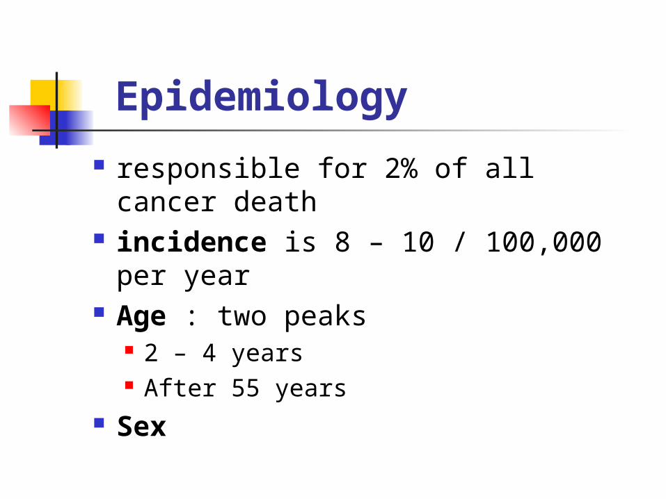
Epidemiology
responsible for 2% of all cancer death
incidence is 8 – 10 / 100,000 per year
Age : two peaks 2 – 4 years After 55 years
Sex

Etiology and pathogenesis
As any neoplastic process in the body . there must be : Induction , promotion and progression
Carcinogenesis process on molecular level oncogene tumor suppressor gene

Risk Factors
1. no genetic predisposition except in certain inherited syndromes
1. NF1 : optic nerve glioma , peripheral neurofibroma
2. NF2 :bilateral acoustic neuroma , multiple meningioma
3. Tuberous sclerosis : subependymal glioma
4. Li-fraumeni disease glioma , ependymoma and medulloblastoma
5. Von hippel lindau disease: hemiangioma and hemiangioblastoma

Risk Factors
2. radiation of head 3. immunosuppresion 4. viral infection 5. Chemicals as anthracen and
nitrosurea 6. Head trauma

Classification
WHO classification depend on cell of origin

neuroepithelia tumors glial cells
astrocytoma oligodendroglioma ependymoma choroids plexus tumors
neurons ganglioglioma gangliocytoma neuroblastoma
pineal tumors medulloblastoma
nerve sheath tumors : shwanomma , neurofibroma meningial tumors : meningioma microglial cells : primary CNS lymphoma pituitary tumors germ cell tumors :
germinoma teratoma
TUMOR LIKE MALFORMATUION Craniopharyngioma Dermoid and epidermoid tumors Colloid cyst
Metastasis and extension from regional tumors .

Classification
In general adults : supratentorial tumors form
85% of all intracranial tumors , most common are astrocytoma , meningioma and mets
children : infratentorial are most common specially medulloblastoma and cerebellar astrocytoma
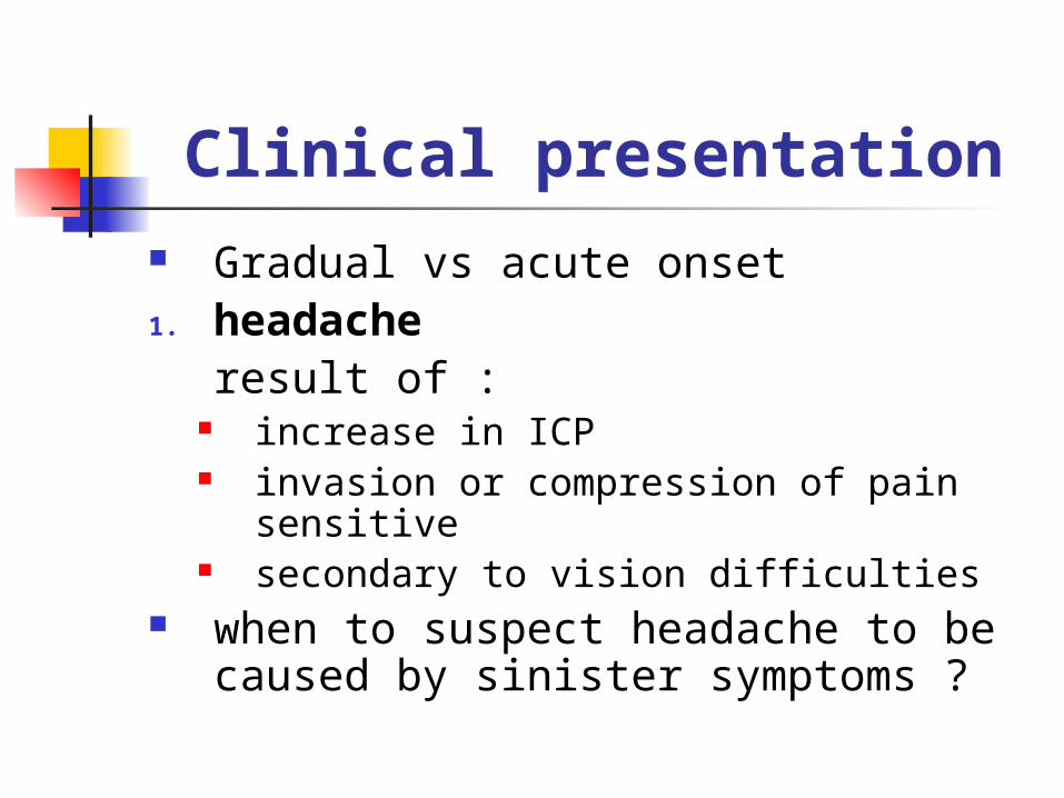
Clinical presentation
Gradual vs acute onset1. headache
result of : increase in ICP invasion or compression of pain
sensitive secondary to vision difficulties
when to suspect headache to be caused by sinister symptoms ?

Clinical presentation
2. other features of increased ICP 3. lateralizing features of brain shift
and herniation 4. epilepsy
new onset epilepsy in adult specially above age of 30 should warn the physician for possibility of tumor . because this occur in 30% of patients with tumors

Clinical presentation
5. subtle changes in personality and behavior
6. progressive neurological deficit depend on site
Clinical presentation
signs and symptoms are divided according to tentorium cerebelli
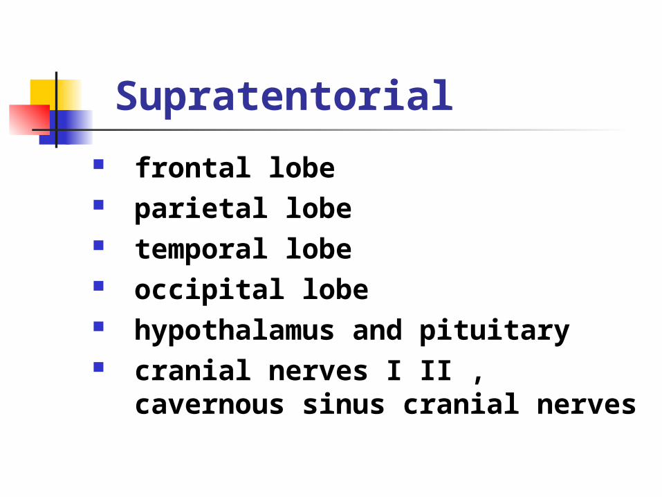
Supratentorial
frontal lobe parietal lobe temporal lobe occipital lobe hypothalamus and pituitary cranial nerves I II , cavernous
sinus cranial nerves
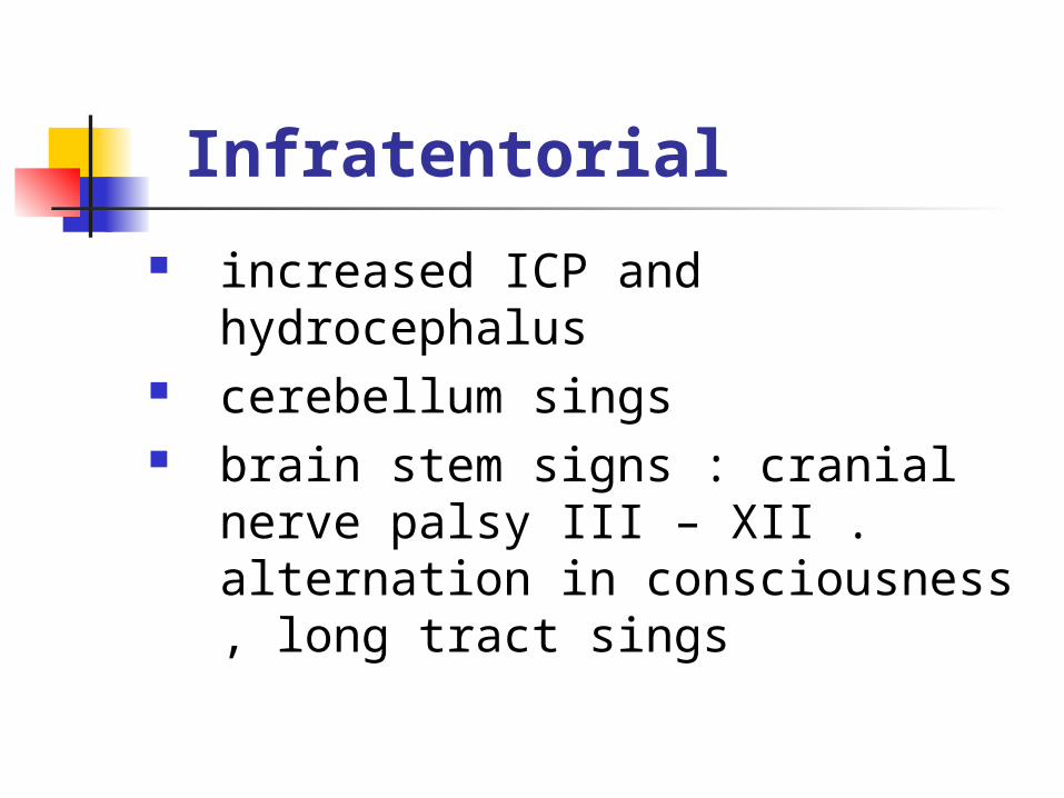
Infratentorial
increased ICP and hydrocephalus cerebellum sings brain stem signs : cranial nerve
palsy III – XII . alternation in consciousness , long tract sings

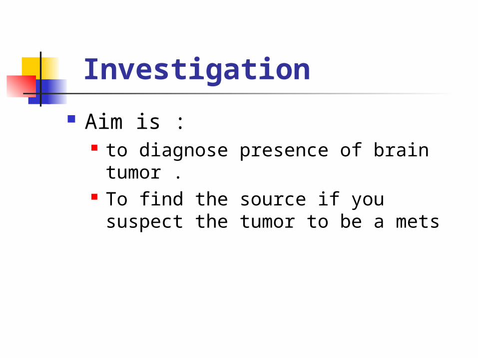
Investigation
Aim is : to diagnose presence of brain tumor . To find the source if you suspect the
tumor to be a mets

Investigation Skull X-RAY
calcification : Oligodendroglioma , meningioma craniopharyngioma and ependymoma
hyperostosis of skull bone destruction : mets , chordoma ,
craniopharyngioma erosion of sella tursica sings of ICP midline shift of pineal gland if calcified

Investigation brain CT
site , mass effect , bone destruction , enhancement , multiplicity
enhanced tumors high grade gliomas meningioma mets acoustic neuroma large pituitary tumors

Investigation MRI Better than CT for
posterior fossa tumors skull base tumors
Angiography or MRA PET scan CSF cytology : remember the
contraindications
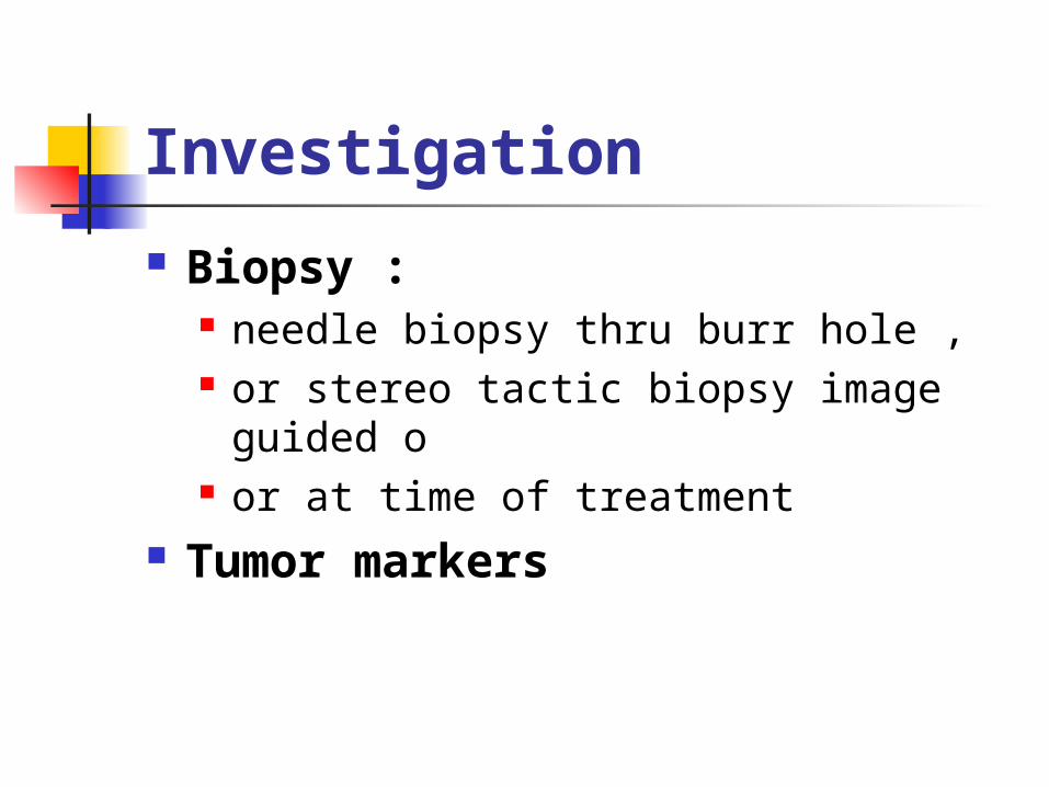
Investigation
Biopsy : needle biopsy thru burr hole , or stereo tactic biopsy image guided
o or at time of treatment
Tumor markers

Differential diagnosis
vascular : hematoma , aneurysm AVM
infection : abscess , tubercloma , hydatid cyst
arachnoid cyst , dermoid and epidermoid cyst

Treatment
medical therapy medical treatment doesn’t affect
tumor it self this used only to reduce edema
surrounding the tumor steroid are used specially with mets ,
meningioma and GBM

Surgical Treatment
aim of surgery to take a biopsy removal of tumor either completely or
partially (cytoreduction) to treat complication as hydrocephalus
Surgical removal is recommended for most types of brain tumors

Surgical Treatment craniotomy cranioctomy tras-sphenoidal trans-oral
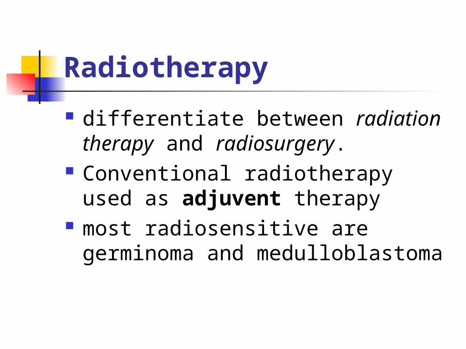
Radiotherapy
differentiate between radiation therapy and radiosurgery.
Conventional radiotherapy used as adjuvent therapy
most radiosensitive are germinoma and medulloblastoma

Radiotherapy
complication : increase edema demylenation radionecrosis affect cognitive functions may induce other kind of tumors as
meningioma
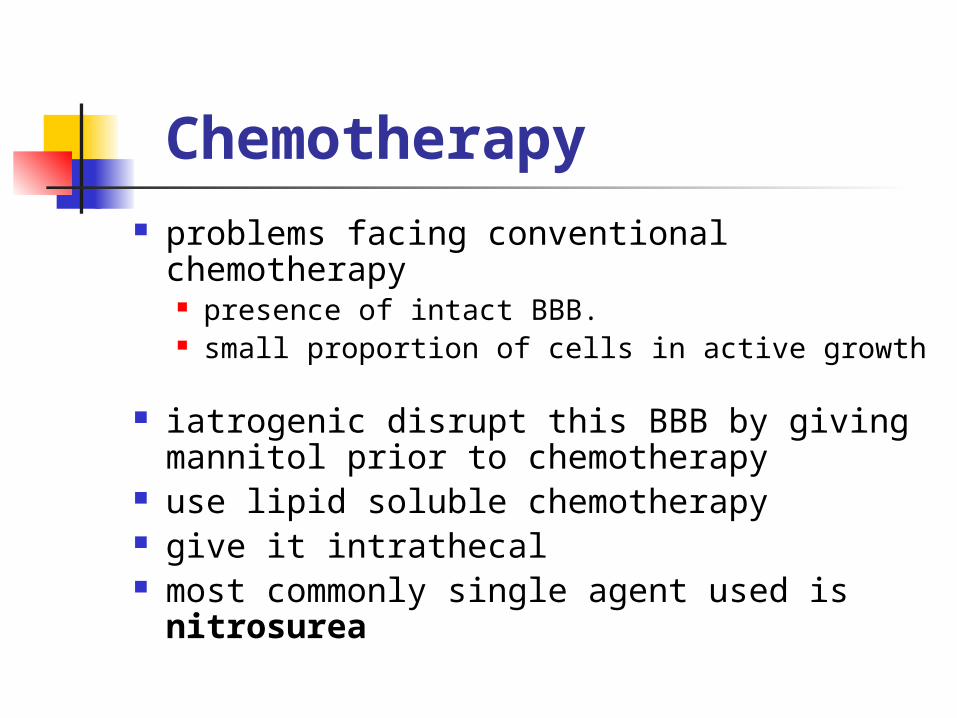
Chemotherapy problems facing conventional
chemotherapy presence of intact BBB. small proportion of cells in active growth
iatrogenic disrupt this BBB by giving
mannitol prior to chemotherapy use lipid soluble chemotherapy give it intrathecal most commonly single agent used is
nitrosurea

New Treatment
hyperthermia treatment immunotherapy : LAK gene therapy
Posterior Fossa Tumors
May need shunting or EVD prior to definitive surgery .
risk are : possible peritoneal seeding prolonged hospitalization risk of shunt complications
GLIOMA
Tumors that arise from cells derived from neuroectoderm , the glial cells
Most common brain tumors 52% Four different types
Astrocytoma
tumor that arise from astrocyte function in
support neurons absorb neurotransmitter release neuroactive molecules aid in formation of BBB
Astrocytoma
most common primary tumors of brain , 45%
peak age : 40 – 60 years astrocytoma ranges in
aggressiveness site : equal incidence in frontal ,
temporal parietal and thalamic . less common in occipital

Astrocytoma
multiple classification systems WHO :
Graed 1 : pilocytic astrocytoma Grade 2 : low grade astrocytoma
Variants : fibrillar protoplasmic Grade 3 : anaplastic astocytoma Grade 4 : glioblastoma multiforme
Variants : giant cell gliosarcoma

Low grade Site :
In adults usually in cerebral hemispheres
In children : in cerebellum
Macroscopic features : Not capsulated , no distinct margins Relatively Avascular Firm fibrous consistency 15% show fine calcium deposit Occasionally may invade diffusely
Microscopically


High grade Site :
cerebral hemisphere Macroscopic features:
Highly vascular margin ,necrosis Butterfly glioma :
Microscopic features Grade 3 Grade 4
Rapidly growing and widely infiltrating

Clinical features
Duration and progression of symptoms will depend on the grade
1. epilepsy 2. feature if increase ICP3. focal neurological deficit
investigations
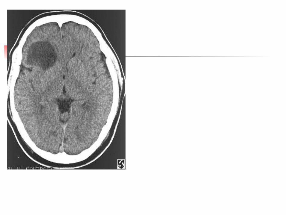
CT Low grade :
small hypodense mass little surrounding edema no enhancement calcification may present
high grade large mass marked edema enhance in non uniform manner ,


investigations MRI More sensitive than CT specially :
posterior fossa , brain stem and skull base tumor and for small tumor mass
usually both low and high appear decrease t1 signal increase t2 signal
Angiograph Skull X-RAY

Astrocytoma
spread : systemic : rare CSF seeding : 10 -25% of high grades tracing thru white matter
Management surgical :
aim is to take biopsy decrease tumor size reduce tumor mass prior to adjuvant therapy
radiotherapy
as adjuvant therapy
other therapy : chemotherapy , immunotherapy , hyperthermia

Prognosis
at present there is no satisfactory treatment for grade 3 and 4 surgery alone is 17 weeks adjuvant radiotherapy is 37 weeks
low grades is approximately 8 years .

Oligodendroglioma
Origin 5% of all gliomas peak age : maximal incidence in 5th
decade site : supratentorial Presented as range most are well differentiated 40 % are mixed glioma with astrocytoma
or ependymoma

Clinical features
as astrocytoma
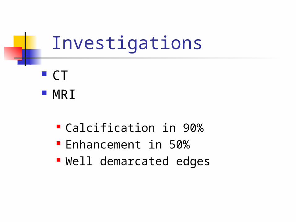
Investigations
CT MRI
Calcification in 90% Enhancement in 50% Well demarcated edges

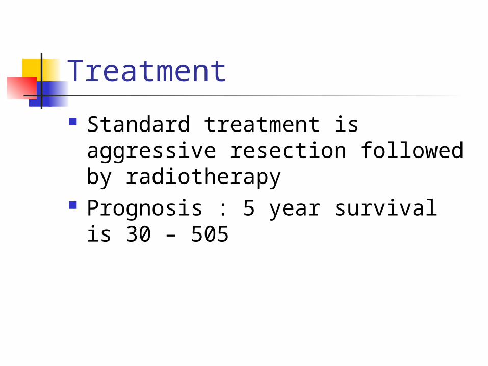
Treatment
Standard treatment is aggressive resection followed by radiotherapy
Prognosis : 5 year survival is 30 – 505

Ependymoma
Origin 5% of all glioma Age : most are in children and
adolescents Site :
30% of cases are supratentorial , mainly in adults
70% are infratentorial , mainly in children

classification non-anaplastic tumors :
papillary : occur in 2 patterns ( rosette and psudorosette
myxopapillary subependymoma : usually heavily
calcified, may be found incidentally at autopsy or present clinically
anaplastic anaplastic and pappilary are most
common symptomatic ependymoma

clinically
supratentorial : presented with increased ICP focal neurological deficit
infratentorial : increased ICP due to hydrocephalus ataxia due to cerebellum involvement

Investigation
CT MRI
Tumor arise in ventricle and enhance calcification in 90% specially
supratentorial
Spread by: seeding thru CSF systemic spread is rare

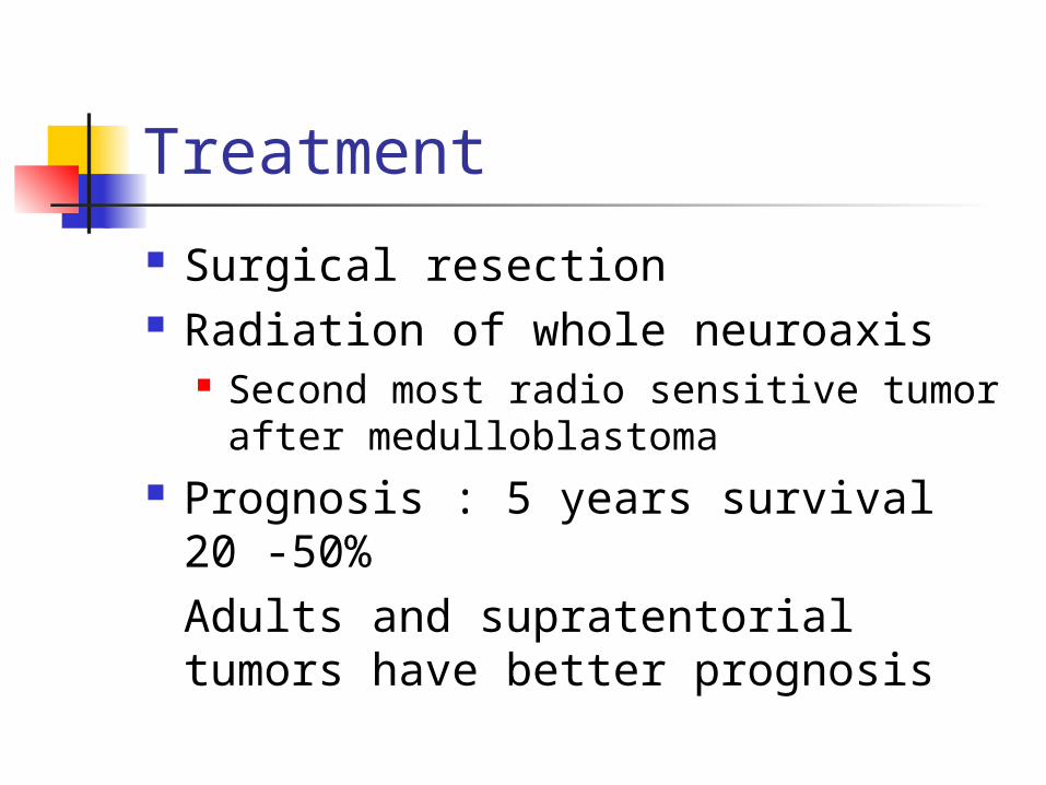
Treatment
Surgical resection Radiation of whole neuroaxis
Second most radio sensitive tumor after medulloblastoma
Prognosis : 5 years survival 20 -50% Adults and supratentorial tumors have better prognosis

Medulloblastoma
Peak age is 5 years It is most common midline
posterior fossa tumor All are highly malignant Spread by
CSF seeding hematogenous spread

Medulloblastoma
CT Isodense midline lesion compressing
4th ventricle , with strong enhancement
MRI

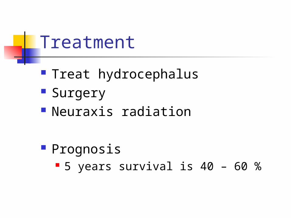
Treatment
Treat hydrocephalus Surgery Neuraxis radiation
Prognosis 5 years survival is 40 – 60 %

Meningioma
Tumor arise from arachnoids layer of meninges
Most common benign brain tumors ,
15% of all tumors Occur at any age , peak in middle
age More in females
Etiology
Possible risk factors head trauma Low levels of radiation Nf2 Sex hormones are important

Meningioma Site :
Most common is parasagital region Less frequently from convexity sphenoidal wing Olfactory groove suprasellar
Classification Depend on position of origin rather than histology

Histological types
syncytial or meningiotheliomatous
transitional type fibroblastic angiomatous malignant infrequent
Clinically parasagital tumors
patient present with epilepsy , contalateral lower limb paresis
may present with ICP in bilateral tumors urinary incontinence especially if bilateral if arise from posterior falx : hemianopia
convexity tumors ICP
Sphenoid ridge May compress optic nerve May cause ICP foster kennedy syndrome : contraleteral
papilledema and optic atrophy in the other

Clinically Olfactory groove
Anosmia initially unilateral Increased ICP Foster kennedy
Suprasellar Bitemporal hemianopia but without
endocrine disturbances Ventricular tumors
Increased ICP
Investigations
CT Hyperdense Enhance uniformly Hyperostosis of cranial vault
MRI Isointense in t1


Treatment
Total surgical excision Radiation may be used to treat
residual tumors
Risk of recurrence Most common source tumor invaded
dural sinus and not removed by surgery
And in malignant variant
Related Documents