International Journal of Scientific & Engineering Research Volume 10, Issue 12, December-2019 187 ISSN 2229-5518 IJSER © 2019 http://www.ijser.org Brain Tumor Detection Using KNN Miss. Priyanka Aiwale, E&TC Department, D.Y.P.S.O.E Pune, Maharashtra, India E-mail - [email protected] Dr. Saniya Ansari E&TC Department, D.Y.P.S.O.E Pune, Maharashtra, India E-mail- [email protected] ABSTRACT Abstract- Detection of Brain Tumor is actually a difficult task and the correct analysis of the Tumor structure is also difficult as a result an automatic method for the detection of Tumor is in usage nowadays. Undoubtedly, this saves the time as well as it gives more accurate results as in comparison to manual detection. The proposed method is a novel approach for detection Tumor along with the ability to calculate the area (%age) occupied by the Tumor in the overall brain cells. Firstly, Tumor regions from an MR image are segmented using an OSTU Algorithm. KNN& LLOYED are used for detecting as well as distinguishing Tumor affected tissues from the not affected tissues. 12 features are extracted like correlation, contrast, energy, homogeneity etc. by performing “wavelet transform on the converted gray scale image”. For feature extraction DB5 wavelet transform is used. Keywords- KNN& Lloyd, wavelet transform, tumour, MRI image 1. INTRODUCTION The development of additional phones frequently shapes a mass of tissue called a development or tumour. Cerebrum Tumor is one of the real reasons for death among individuals. The manifestations of a mind Tumor rely upon Tumor size, sort and area. Indications might be caused when a Tumor pushes on a nerve or damages a piece of a cerebrum. Additionally, they might be caused when a Tumor obstructs the liquid that moves through and around the or when the mind swells since develop of liquid. Cerebral pains, queasiness and heaving, Changes in discourse, vision or hearing, issue adjusting or strolling, changes in temperament, identity or capacity to focus, issues with memory, muscle snapping or tingling, deadness or shivering in the arms or legs. Accurate identification of the type of mind 1variation among the majority is extremely basic for treatment 1 arranging which could restrict the deadly outcomes. [2] Detection of mind Tumor manually is a recurring activity which consumes a lot of time and also the results are not accurate, shifts 1starting with one specialist then onto the next. PC supported robotized frameworks provides the appropriate outcomes. Not only being exactly same, these procedures must 1scope at a brick pace with a mind set that the final target for their implemation on continuous applications. MRI helps in analyzation of brain Tumor along with CT images as well as ultrasonic or X-Rays. MRI (Magnrtic Resonance Imaging) is an essential 1instrument utilize in a great many fields of recommendation which is outfitted for producing a explicit image of any part of the body of human. X-ray remains for MRI. A Magnetic Resonance Imaging scanner make use of magnets for the objective of enrapturing as well as for energizing hydrogen cores (single proton) in tissue of humans, “that produces a flag that can be distinguished and it’s encoded spatially, bringing about images of the body. The MRI machine produces radio recurrence IJSER

Welcome message from author
This document is posted to help you gain knowledge. Please leave a comment to let me know what you think about it! Share it to your friends and learn new things together.
Transcript
International Journal of Scientific & Engineering Research Volume 10, Issue 12, December-2019 187 ISSN 2229-5518
IJSER © 2019
http://www.ijser.org
Brain Tumor Detection Using KNN
Miss. Priyanka Aiwale,
E&TC Department, D.Y.P.S.O.E Pune, Maharashtra,
India
E-mail - [email protected]
Dr. Saniya Ansari
E&TC Department, D.Y.P.S.O.E Pune, Maharashtra,
India
E-mail- [email protected]
ABSTRACT
Abstract- Detection of Brain Tumor is actually a difficult task and the correct analysis of the Tumor structure is also
difficult as a result an automatic method for the detection of Tumor is in usage nowadays. Undoubtedly, this saves the
time as well as it gives more accurate results as in comparison to manual detection. The proposed method is a novel
approach for detection Tumor along with the ability to calculate the area (%age) occupied by the Tumor in the overall
brain cells. Firstly, Tumor regions from an MR image are segmented using an OSTU Algorithm. KNN& LLOYED are
used for detecting as well as distinguishing Tumor affected tissues from the not affected tissues. 12 features are extracted
like correlation, contrast, energy, homogeneity etc. by performing “wavelet transform on the converted gray scale image”.
For feature extraction DB5 wavelet transform is used.
Keywords- KNN& Lloyd, wavelet transform, tumour, MRI image
1. INTRODUCTION
The development of additional phones frequently
shapes a mass of tissue called a development or
tumour. Cerebrum Tumor is one of the real reasons for
death among individuals. The manifestations of a mind
Tumor rely upon Tumor size, sort and area. Indications
might be caused when a Tumor pushes on a nerve or
damages a piece of a cerebrum. Additionally, they
might be caused when a Tumor obstructs the liquid
that moves through and around the or when the mind
swells since develop of liquid. Cerebral pains,
queasiness and heaving, Changes in discourse, vision
or hearing, issue adjusting or strolling, changes in
temperament, identity or capacity to focus, issues with
memory, muscle snapping or tingling, deadness or
shivering in the arms or legs. Accurate identification of
the type of mind 1variation among the majority is
extremely basic for treatment 1 arranging which could
restrict the deadly outcomes. [2]
Detection of mind Tumor manually is a recurring
activity which consumes a lot of time and also the
results are not accurate, shifts 1starting with one
specialist then onto the next. PC supported robotized
frameworks provides the appropriate outcomes. Not
only being exactly same, these procedures must 1scope
at a brick pace with a mind set that the final target for
their implemation on continuous applications. MRI
helps in analyzation of brain Tumor along with CT
images as well as ultrasonic or X-Rays. MRI (Magnrtic
Resonance Imaging) is an essential 1instrument utilize
in a great many fields of recommendation which is
outfitted for producing a explicit image of any part of
the body of human. X-ray remains for MRI. A
Magnetic Resonance Imaging scanner make use of
magnets for the objective of enrapturing as well as for
energizing hydrogen cores (single proton) in tissue of
humans, “that produces a flag that can be distinguished
and it’s encoded spatially, bringing about images of the
body. The MRI machine produces radio recurrence
IJSER
International Journal of Scientific & Engineering Research Volume 10, Issue 12, December-2019 188 ISSN 2229-5518
IJSER © 2019
http://www.ijser.org
(RF) beat that” particularly ties just “to hydrogen. The
framework sends the beat to that particular territory of
the body that should be inspected. Because of the RF
beat, protons here retain the vitality expected to
influence them to turn in an alternate heading. This is
implied by the reverberation of MRI. The RF beat
influences the protons to turn at the larmour
recurrence, in a particular bearing. This recurrence is
discovered in light of the specific tissue being” imaged
and the quality of the principle attractive field. [5]
Grouping of the mind Tumor is likewise a vital
undertaking for treatment arranging. There are two
sorts of Tumor which are-benevolent (non-destructive)
and threatening (carcinogenic) tumours. Ordinary
strategies include intrusive systems, for example,
biopsy, lumbar cut and flag tap technique, to identify
and group cerebrum Tumor into benevolent and
harmful which are exceptionally agonizing and tedious.
Wavelet investigation is a practicable strategy suitable
to unveil various sections of information which other
flag as procedures for examination. Segmented the
images at a great many levels, this method can
eliminate much better reason of interest from itself as
well as thusly inflates the behaviour of the image.
What is more, for the process of compacting or de-
noising a flag, equipment of it is done with no
extensive debasement. It is actually of from almost all
importance when there ought to develop an event of
flimsy details, for instance, when there to be an event
of therapeutic 1imaging [7]
2. RELATED WORK
In below section, various techniques are utilized in
literature by various authors who summarized
grounded on primary categories such as segmentation,
feature extraction as well as classification method
used.
Different methods Used in previous research work.
Jin Liu, Min Li, Jianxin Wang et al, studies the
MRI based brain Tumor segmentation which is more
and more attractive because of good soft tissue contrast
and non-invasive imaging of Magnetic Resonance
Imaging images. They purposed to make an extensive
introduction for MRI-based brain Tumor segmentation
strategies. Then, the pre processing activities as well as
the state-of-the-art methods of MRI based Tumor
segmentation are actually introduced. [1]
Pavel Dvorak and Bjoern Menze et al, Indeed,
even under treatment, patients don't make due all
things considered over fourteen weeks after conclusion
[3]. Present day medicines incorporate surgery,
radiotherapy, chemotherapy or all of them. X-ray is
very beneficial to make use of gliomas in various
clinical practices, as it is conceivable to procure MRI
arrangements giving corresponding information. An
actual division of glioma’s as well as its intra-tumoural
structures is essential for treatment arranging, and also
for the regular follow-up schedules. Be that as it may,
manual division is laborious and subjected to between
along with intra-rater blunders hard to summarize. In
this manner, doctors more often than not utilize harsh
measures for assessment. Hence, accurate self-loader
or perhaps programmed techniques are needed. [4]
V. Karthikeyan, B. Menze and K. Sreedhar et al,
the Tumor mass impact alter the couse of action of the
encompassing typical tissues. Along these lines, the
emphasis is on planning structures as opposed to
creating handmade elements, which may require
particular learning. CNNs have been utilized to win a
few question acknowledgment [6], [12] as well as
challenges of natural picture division [5]. Since a CNN
operates over patches utilizing pieces, it has the benefit
of considering as well as being used with crude
information. In the arena of mind Tumor division, late
proposition additionally examine the utilization of
CNNs [11].
J. Selvakumar, A. Lakshami & T. Arivoli et al,
analyzes the methodologies carried out by the image
IJSER
International Journal of Scientific & Engineering Research Volume 10, Issue 12, December-2019 189 ISSN 2229-5518
IJSER © 2019
http://www.ijser.org
Intensification “used in Mathematical Morphological
[MM] theory on the dark images. Some Morphological
Transformation have been processed through Block
Analysis, Morphological Operation and Opening by
Reconstruction on the Images with poor lighting.
Analysis of the methods which is mentioned above
illustrated through the processing of images with
various filtering techniques along with” various
background images of less intensity of light. [7]
Raunaq Rewari, with the utilization of pan
morphological methods for the purpose of detection of
various background features of the images with poor
lighting has implemented the improvement in the
digital images. The intial operator works with the
information reterived from the block analysis while the
next tranformation make use of the reconstruction
opening employed to state various backgrounds.
Lastly, through the images with different backgrounds,
most of them light backgrounds, the performances of
the proposed operators are processed. [8]
Stefan Bauer, Roland Wiest et al, are the creators
decided on 2D filters despite the fact that 3D filters can
exploit the 3D way of the pictures; however, it builds
computational load. The vast spatial and basic
fluctuation in mind tumours is additionally an essential
worry that we think about utilizing information growth.
[9]
K. Sreedhar and B. Panlal, taken automation of
brain Tumor segmentation continues to be a
challenging task because of significant variations in its
structure. In this paper, an automated brain Tumor
segmentation algorithm using deep convolutional
neural network (DCNN) is presented. [12]
Nikesh T. Gadare, Dr. S. A. Ladhake, et al,
implemented few of the transformations which were
morphological in nature and these were processed
through block analysis, morphological operations
followed by reconstruction opening of images with less
intensity of light. Through Weber’s Law Operator,
Background detection and Image enhancement are
illustrated. In Mathematical Morphology it has
transformation that enables filtering of the Image with
new contour leads to closing by reconstruction and
opening by reconstruction. [13]
Bjoern Menze and Pavel Dvorak worked on the
medical images includes an excessive similarity in the
intensities of close by pixels and a powerful correlation
of various image modalities. All the images deal with
correlation used by local image patches. As well as,
there is a high correlation between close labels in the
image; this feature is utilized in “local structure
prediction” of the “local label patches. For 3D
segmentation tasks and for systematically evaluating
different parameters that are appropriate for the dense
annotation of anatomical” structures, local framework
prediction approach is used by them. [14]
Vaishnavi S. Mehekare, Dr.S.R., Ganorkar,
from all among cerebrum tumours, Glioma are the
most widely recognized, forceful, prompting a brief
long term in their most lofty evaluation. There are
different proposes of automatic division strategy in
light of Convolutional Neural Networks), investigating
little kernel. The use of kernel permits outlining a far
more deep design, apart from not having a destructive
outcome against over fitting, provided the less number
of weights in the system.. [15]
3. PROPOSED METHODOLOGY
Image processing techniques are being used to detect
the bain tumour. For the purpose of detecting Tumor in
the MRI images we are using MATLAB software here.
The figure shown below is the block diagram of the
proposed system.
IJSER
International Journal of Scientific & Engineering Research Volume 10, Issue 12, December-2019 190 ISSN 2229-5518
IJSER © 2019
http://www.ijser.org
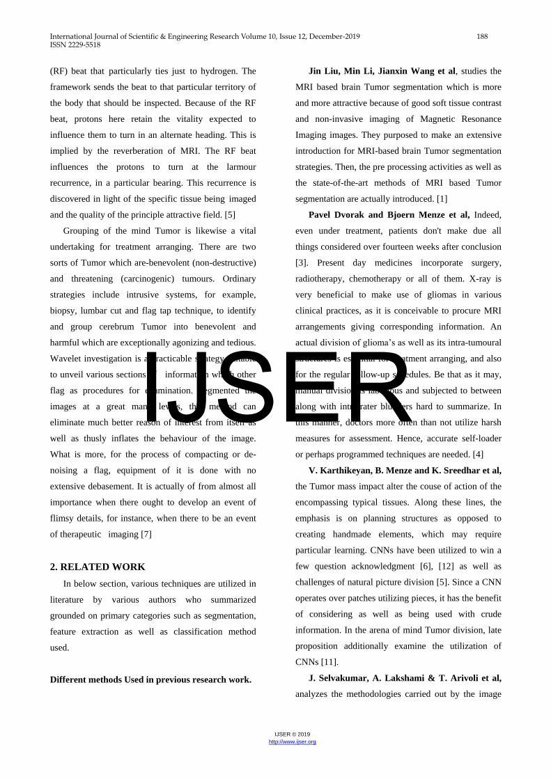
Figure 1: 1Block Diagram of proposed system
The detail description of system proposed is as
follows:
Pre-processing: It generally entails removal of
background noise having frequency low,
1normalizing 1the 1intensity 1of the 1individual
1particles’ images, masking 1of some portions of the
images and removing reflections. Image pre-
processing is the method to improve data images
prior to computational processing.
Image conversion: In greyscale image or RGB
image is that image the value of each pixel is only a
single sample which carries information related to
the intensity of light or in other words which
reprensents only the amount of light. This sort of
images is composed of various shades of gray
colour. The range of the contrats from black
colour at the weakest intensity to the white colour
at the strongest. Keeping this in mind, the
conversion of the image in black and white is done.
As we understand Tumor is actually big enough to
not deemed as tiny bound, therefore we are going
to detach little pixel bound.
Wavelets transform: “The Daubechies wavelets,
based on each wavelet type of this class, there is a
scaling function (called the father wavelet) which
generates an orthogonal multi resolution analysis.
the scaling filter associated with the Daubechies
wavelet specified by wname. Where f is a real-
valued vector.”
Feature extraction: For the purpose of extracting
features from input image different operations are
needed to perform like entropy, contrast,
correlation, energy, root mean square, standard
deviation etc.
Classification: KNN & LLOYED for the purpose
of classifying the tissue 1into normal or cancerous.
If the tissue is normal or not-infectious, no Tumor
detected displays on MATLAB output window. If
in case the tissue is infectious or in simple words
we can say that if Tumor is detected then the
following steps are taken.
Step 1: For smoothing the Tumor MRI Image low
pass and high pass filter are applied.
Step 2: For encircling the areas which are affected
OSTU Thresholding is used. Draw a circle of
maximum possible size covering maximum
affected area and next then other circle of
small size are drawn.
Step 3: One circle having exact center as that of
maximum radius circle from above step with
60% large radius is chosen so that it can cover
complete affected areas called region of
interest.
Step 4: For calculating the area of Tumor cells
thresholding is performed. Thresholding can
be approximated as follow:
% 𝐴𝑟𝑒𝑎
=𝑛𝑜. 𝑜𝑓 𝑡𝑢𝑚𝑜𝑢𝑟 𝑝𝑖𝑥𝑒𝑙𝑠
𝑛𝑜. 𝑜𝑓 𝑡𝑜𝑡𝑎𝑙 𝑏𝑟𝑎𝑖𝑛 𝑝𝑖𝑥𝑒𝑙𝑠𝑋 100
Step 5: Segment the tumour
Step 6: Classify the tumour
Step 7: Display the resulting Image
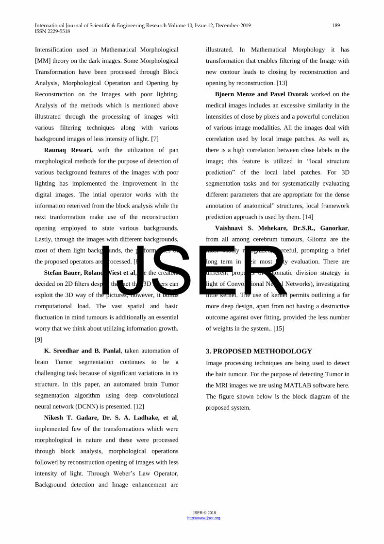
4. FLOW CHART
Below figure shows the flow diagram.
IJSER
International Journal of Scientific & Engineering Research Volume 10, Issue 12, December-2019 191 ISSN 2229-5518
IJSER © 2019
http://www.ijser.org
Figure3. Flow chart
5. ALGORITHM
1. Start
2. Take input original MRI brain image
3. Convert it into gray scale
4. Filter the image using LPF & HPF
5. Morphological operations on image
6. Take OSTU Segmentation
7. LLOYD clustering to segment Tumor
8. Use KNN to find Equlidian distance
9. Hybrid feature extraction using 2 stage Discrete
Wavelet Transform
10. Calculate contrast, colleration, Energy, Mean,
RMS, Standard Deviation, Smoothness
11. Tran image using PNN & RBF
12. Classify the tumour
13. Find the percentage of Tumor
14. Stop
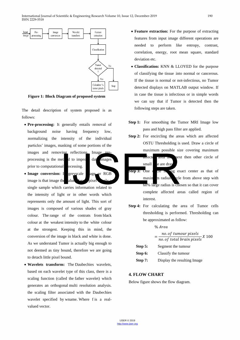
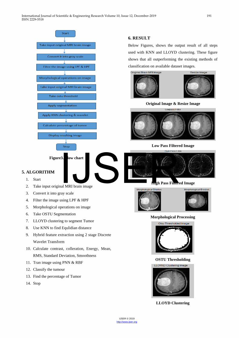
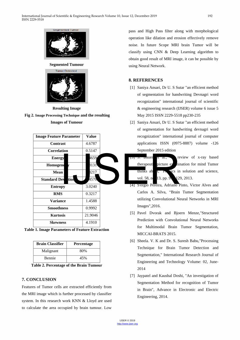
6. RESULT
Below Figures, shows the output result of all steps
used with KNN and LLOYD clustering. These figure
shows that all outperforming the existing methods of
classification on available dataset images.
Original Image & Resize Image
Low Pass Filtered Image
High Pass Filtered Image
Morphological Processing
OSTU Thresholding
LLOYD Clustering
IJSER
International Journal of Scientific & Engineering Research Volume 10, Issue 12, December-2019 192 ISSN 2229-5518
IJSER © 2019
http://www.ijser.org
Segmented Tumour
Resulting Image
Fig 2. Image Processing Technique and the resulting
Images of Tumour
Image Feature Parameter Value
Contrast 4.6787
Correlation 0.5147
Energy 0.4659
Homogeneity 0.8131
Mean 0.3217
Standard Deviation 1.4570
Entropy 3.0240
RMS 0.3217
Variance 1.4588
Smoothness 0.9992
Kurtosis 21.9046
Skewness 4.1910
Table 1. Image Parameters of Feature Extraction
Brain Classifier Percentage
Malignant 80%
Bennie 45%
Table 2. Percentage of the Brain Tumour
7. CONCLUSION
Features of Tumor cells are extracted efficiently from
the MRI image which is further processed by classifier
system. In this research work KNN & Lloyd are used
to calculate the area occupied by brain tumour. Low
pass and High Pass filter along with morphological
operation like dilation and erosion effectively remove
noise. In future Scope MRI brain Tumor will be
classify using CNN & Deep Learning algorithm to
obtain good result of MRI image, it can be possible by
using Neural Network.
8. REFERENCES
[1] Saniya Ansari, Dr U. S Sutar "an efficient method
of segmentation for handwriting Devnagri word
recognization" international journal of scientific
& engineering research (IJSER) volume 6 issue 5
May 2015 ISSN 2229-5518 pp230-235
[2] Saniya Ansari, Dr U. S Sutar "an efficient method
of segmentation for handwriting devnagri word
recognization" international journal of computer
applications ISSN (0975-8887) volume -126
September 2015 edition
[3] S. Bauer et al., "A review of x-ray based
therapeutic picture examination for mind Tumor
thinks about," Physics in solution and science,
vol. 58, no. 13, pp. 97– 129, 2013.
[4] S'ergio Pereira, Adriano Pinto, Victor Alves and
Carlos A. Silva, “Brain Tumor Segmentation
utilizing Convolutional Neural Networks in MRI
Images",2016.
[5] Pavel Dvorak and Bjoern Menze,"Structured
Prediction with Convolutional Neural Networks
for Multimodal Brain Tumor Segmentation,
MICCAI-BRATS 2015.
[6] Sheela. V. K and Dr. S. Suresh Babu,"Processing
Technique for Brain Tumor Detection and
Segmentation," International Research Journal of
Engineering and Technology Volume: 02, June-
2014
[7] Jaypatel and Kaushal Doshi, "An investigation of
Segmentation Method for recognition of Tumor
in Brain", Advance in Electronic and Electric
Engineering, 2014.
IJSER
International Journal of Scientific & Engineering Research Volume 10, Issue 12, December-2019 193 ISSN 2229-5518
IJSER © 2019
http://www.ijser.org
[8] B. Menze et al., "The multimodal mind Tumor
picture division benchmark (whelps)," IEEE
Transactions on Medical Imaging, vol. 34, no. 10,
pp. 1993–2024, 2015.
[9] J. Selvakumar, A. Lakshami & T. Arivoli,"Brain
Tumor Segmentation and Its Area Calculation
utilizing K-mean Clustering and Fuzzy C-Mean
Algorithm", IEEE-International Conference On
Advances In Engineering, March30,2012.
[10] Raunaq Rewari, "Programmed Tumor
Segmentation Using Convolutional Neural
Network."
[11] Stefan Bauer, Roland Wiest and Lutz-P Nolte, "A
Survey Of MRI-based restorative picture
examination for Brain Tumor Studies".
[12] Vaishnavi Dr. P. Eswaran "Enhanced Color
Image Enhancement Scheme utilizing
Mathematical Morphology ", Volume 3, Issue 4,
April 2013 IJARCSSE.
[13] V. Karthikeyan1, V. J. Vijayalakshmi, P.
Jeyakumar, “A Novel Approach For The
Enrichment Of Digital Images Using
Morphological Operators”, 2013.
[14] K.Sreedhar and B.Panlal, Enhancement of images
using morphological transformation, 2012.
[15] Nikesh T. Gadare, Dr. S. A. Ladhake, Prof. P. D.
Gawande, “Mathematical Morphology based
Image Enhancement and Background Detection”
2014.
[16] Pavel Dvoˇr´ak1,2 and Bjoern Menze3 Structured
Prediction with Convolutional Neural Networks
for Multimodal Brain Tumor Segmentation, 2015.
[17] Vaishnavi S. Mehekare, Dr.S.R.Ganorkar, “A
Survey on Brain Tumor Detection Using Neural
Network 2017.Samjith Raj C.P. and Shreeja R,
Automatic brain Tumor tissue detection in T-1
weighted MRI 2017.
[18] Manisha, Radhakrishnan.B and Dr. L.Padma
Suresh, “Tumor Region Extraction using Edge
Detection Method in Brain MRI Images” 2017
[19] V. Zeljkovic1, C. Druzgalski2, Y. Zhang1, Z.
Zhu1, Z. Xu1, D. Zhang1, P. Mayorga3,
“Automatic Brain Tumor Detection and
Segmentation in MR Images” 2014.
[20] Anatoly Sorokin, Evgeny Zhvansky, Konstantin
Bocharov, and Vsevolod Shurkhay, Alexander
Potapov, “Multi-label classification of brain
Tumor mass spectrometry data” 2017.
[21] Alexis Arnaud, Florence Forbes, Nicolas
Coquery, Nora Collomb, Benjamin Lemasson,
and Emmanuel L. Barbier, ”Fully Automatic
Lesion Localization and Characterization:
Application to Brain Tumours using Multi
parametric Quantitative MRI Data”2018.
[22] Swathi P S, “Brain Tumor Detection and
Classification Using Histogram Thresholding and
ANN”2015
[23] Ms. Priya Patil, Ms. Seema Pawar, Ms. Sunayna
Patil, Prof. Arjun Nichal, “A Review Paper on
Brain Tumor Segmentation and Detection”2017.
[24] Moitra D and Mandal R” Review of Brain Tumor
Detection using Pattern Recognition
Techniques”2017
[25] Neha Rani” Brain Tumor Detection and
Classification with Feed Forward Back-Prop
Neural Network”2016.
[26] M. Avula, and Lakkhkula, et al., ``Bone Cancer
from MRI Scan Imagery using Mean pixel
intensity”, The International Conference of
Electronic Computer Technology, pp, 112-116,
2014.
IJSER
Related Documents