Brain Structural Alterations in Obsessive-Compulsive Disorder Patients with Autogenous and Reactive Obsessions Marta Subira ` 1,2 , Pino Alonso 1,2,3 , Cinto Segala `s 1,2 , Eva Real 1,2 , Clara Lo ´ pez-Sola ` 1,2,3 , Jesu ´ s Pujol 4 , IgnacioMartı´nez-Zalacaı´n 1 , Ben J. Harrison 5 , Jose ´ M. Mencho ´n 1,2,3 , Narcı´s Cardoner 1,2,3 , Carles Soriano- Mas 1,2 * 1 Psychiatry Department, Bellvitge University Hospital, Bellvitge Biomedical Research Institute (IDIBELL), Barcelona, Spain, 2 Carlos III Health Institute, Centro de Investigacio ´ n Biome ´dica en Red de Salud Mental (CIBERSAM), Barcelona, Spain, 3 Department of Clinical Sciences, School of Medicine, University of Barcelona, Barcelona, Spain, 4 Magnetic Resonance Unit, CRC-Hospital del Mar, Barcelona, Spain, 5 Melbourne Neuropsychiatry Centre, Department of Psychiatry, The University of Melbourne, Melbourne, Australia Abstract Obsessive-compulsive disorder (OCD) is a clinically heterogeneous condition. Although structural brain alterations have been consistently reported in OCD, their interaction with particular clinical subtypes deserves further examination. Among other approaches, a two-group classification in patients with autogenous and reactive obsessions has been proposed. The purpose of the present study was to assess, by means of a voxel-based morphometry analysis, the putative brain structural correlates of this classification scheme in OCD patients. Ninety-five OCD patients and 95 healthy controls were recruited. Patients were divided into autogenous (n = 30) and reactive (n = 65) sub-groups. A structural magnetic resonance image was acquired for each participant and pre-processed with SPM8 software to obtain a volume-modulated gray matter map. Whole-brain and voxel-wise comparisons between the study groups were then performed. In comparison to the autogenous group, reactive patients showed larger gray matter volumes in the right Rolandic operculum. When compared to healthy controls, reactive patients showed larger volumes in the putamen (bilaterally), while autogenous patients showed a smaller left anterior temporal lobe. Also in comparison to healthy controls, the right middle temporal gyrus was smaller in both patient subgroups. Our results suggest that autogenous and reactive obsessions depend on partially dissimilar neural substrates. Our findings provide some neurobiological support for this classification scheme and contribute to unraveling the neurobiological basis of clinical heterogeneity in OCD. Citation: Subira ` M, Alonso P, Segala ` s C, Real E, Lo ´ pez-Sola ` C, et al. (2013) Brain Structural Alterations in Obsessive-Compulsive Disorder Patients with Autogenous and Reactive Obsessions. PLoS ONE 8(9): e75273. doi:10.1371/journal.pone.0075273 Editor: Noam Harel, University of Minnesota, United States of America Received May 15, 2013; Accepted August 15, 2013; Published September 30, 2013 Copyright: ß 2013 Subira ` et al. This is an open-access article distributed under the terms of the Creative Commons Attribution License, which permits unrestricted use, distribution, and reproduction in any medium, provided the original author and source are credited. Funding: This study was supported in part by the Carlos III Health Institute (PI09/01331 PI10/01753, PI10/01003, CP10/00604, CIBER-CB06/03/0034) and by the Agencia de Gestio ´ d’Ajuts Universitaris i de Recerca (AGAUR; 2009SGR1554). M.S. is funded by the Bellvitge Biomedical Research Institute (IDIBELL). E.R. is supported by a ‘Rio Hortega’ contract from the Carlos III Health Institute (I.D. CM11/00077). C. L-S. is supported by the Spanish Ministry of Education, Culture and Sport (FPU12/01636). B.H. is supported by a National Health and Medical Research Council of Australia (NHMRC) Clinical Career Development Award (I.D. 628509). C.S-M. is funded by a ‘Miguel Servet’ contract from the Carlos III Health Institute (CP10/00604). The funders had no role in study design, data collection and analysis, decision to publish, or preparation of the manuscript. Competing Interests: Please note that co-authors Charles Soriano-Mas and Ben J Harrison are PLOS ONE Editorial Board members, although this does not alter the authors’ adherence to all the PLOS ONE policies on sharing data and materials. * E-mail: [email protected] Introduction In recent years there has been an increasing interest in studying the clinical heterogeneity of obsessive-compulsive disorder (OCD) [1–3]. In particular, the so-called multidimensional model [4,5] has attempted to summarize OCD in a few temporally stable dimensions that may co-exist within individual patients. Reports comparing patients with different symptom profiles have identified differences in their sociodemographical and clinical features, as well as neurobiological correlates. For instance, in symptom provocation studies, relatively distinctive patterns of brain activity have been associated with the presentation of stimuli representing discrete OCD symptom dimensions, such as aggression/checking and contamination/cleaning symptoms [6–8]. Although studies of brain structural alterations in OCD have resulted in a mostly consistent pattern of findings [9–11], the assessment of such alterations in relation to specific symptoms or illness subtypes has provided mixed results. Specifically, while in some reports aggressive/checking symptoms were associated with volume alterations in temporolimbic regions, including the amygdala [12,13], in others this dimension was associated to volume changes in the insula and putamen, among other areas [14]. Similarly, while in some assessments contamination/cleaning and symmetry/ordering symptoms were associated with volume reductions in the dorsal caudate and the sensorimotor cortex, respectively [13], in others such symptoms did not show any significant correlation with brain anatomy [14]. Despite such discrepant findings may be partially explained by the use of different scales to assess symptom dimensions (e.g., the PLOS ONE | www.plosone.org 1 September 2013 | Volume 8 | Issue 9 | e75273

Welcome message from author
This document is posted to help you gain knowledge. Please leave a comment to let me know what you think about it! Share it to your friends and learn new things together.
Transcript
Brain Structural Alterations in Obsessive-CompulsiveDisorder Patients with Autogenous and ReactiveObsessionsMarta Subira1,2, Pino Alonso1,2,3, Cinto Segalas1,2, Eva Real1,2, Clara Lopez-Sola1,2,3, Jesus Pujol4,
Ignacio Martınez-Zalacaın1, Ben J. Harrison5, Jose M. Menchon1,2,3, Narcıs Cardoner1,2,3, Carles Soriano-
Mas1,2*
1 Psychiatry Department, Bellvitge University Hospital, Bellvitge Biomedical Research Institute (IDIBELL), Barcelona, Spain, 2Carlos III Health Institute, Centro de
Investigacion Biomedica en Red de Salud Mental (CIBERSAM), Barcelona, Spain, 3Department of Clinical Sciences, School of Medicine, University of Barcelona, Barcelona,
Spain, 4Magnetic Resonance Unit, CRC-Hospital del Mar, Barcelona, Spain, 5Melbourne Neuropsychiatry Centre, Department of Psychiatry, The University of Melbourne,
Melbourne, Australia
Abstract
Obsessive-compulsive disorder (OCD) is a clinically heterogeneous condition. Although structural brain alterations havebeen consistently reported in OCD, their interaction with particular clinical subtypes deserves further examination. Amongother approaches, a two-group classification in patients with autogenous and reactive obsessions has been proposed. Thepurpose of the present study was to assess, by means of a voxel-based morphometry analysis, the putative brain structuralcorrelates of this classification scheme in OCD patients. Ninety-five OCD patients and 95 healthy controls were recruited.Patients were divided into autogenous (n = 30) and reactive (n = 65) sub-groups. A structural magnetic resonance image wasacquired for each participant and pre-processed with SPM8 software to obtain a volume-modulated gray matter map.Whole-brain and voxel-wise comparisons between the study groups were then performed. In comparison to theautogenous group, reactive patients showed larger gray matter volumes in the right Rolandic operculum. When comparedto healthy controls, reactive patients showed larger volumes in the putamen (bilaterally), while autogenous patientsshowed a smaller left anterior temporal lobe. Also in comparison to healthy controls, the right middle temporal gyrus wassmaller in both patient subgroups. Our results suggest that autogenous and reactive obsessions depend on partiallydissimilar neural substrates. Our findings provide some neurobiological support for this classification scheme and contributeto unraveling the neurobiological basis of clinical heterogeneity in OCD.
Citation: Subira M, Alonso P, Segalas C, Real E, Lopez-Sola C, et al. (2013) Brain Structural Alterations in Obsessive-Compulsive Disorder Patients with Autogenousand Reactive Obsessions. PLoS ONE 8(9): e75273. doi:10.1371/journal.pone.0075273
Editor: Noam Harel, University of Minnesota, United States of America
Received May 15, 2013; Accepted August 15, 2013; Published September 30, 2013
Copyright: � 2013 Subira et al. This is an open-access article distributed under the terms of the Creative Commons Attribution License, which permitsunrestricted use, distribution, and reproduction in any medium, provided the original author and source are credited.
Funding: This study was supported in part by the Carlos III Health Institute (PI09/01331 PI10/01753, PI10/01003, CP10/00604, CIBER-CB06/03/0034) and by theAgencia de Gestio d’Ajuts Universitaris i de Recerca (AGAUR; 2009SGR1554). M.S. is funded by the Bellvitge Biomedical Research Institute (IDIBELL). E.R. issupported by a ‘Rio Hortega’ contract from the Carlos III Health Institute (I.D. CM11/00077). C. L-S. is supported by the Spanish Ministry of Education, Culture andSport (FPU12/01636). B.H. is supported by a National Health and Medical Research Council of Australia (NHMRC) Clinical Career Development Award (I.D. 628509).C.S-M. is funded by a ‘Miguel Servet’ contract from the Carlos III Health Institute (CP10/00604). The funders had no role in study design, data collection andanalysis, decision to publish, or preparation of the manuscript.
Competing Interests: Please note that co-authors Charles Soriano-Mas and Ben J Harrison are PLOS ONE Editorial Board members, although this does not alterthe authors’ adherence to all the PLOS ONE policies on sharing data and materials.
* E-mail: [email protected]
Introduction
In recent years there has been an increasing interest in studying
the clinical heterogeneity of obsessive-compulsive disorder (OCD)
[1–3]. In particular, the so-called multidimensional model [4,5]
has attempted to summarize OCD in a few temporally stable
dimensions that may co-exist within individual patients. Reports
comparing patients with different symptom profiles have identified
differences in their sociodemographical and clinical features, as
well as neurobiological correlates. For instance, in symptom
provocation studies, relatively distinctive patterns of brain activity
have been associated with the presentation of stimuli representing
discrete OCD symptom dimensions, such as aggression/checking
and contamination/cleaning symptoms [6–8].
Although studies of brain structural alterations in OCD have
resulted in a mostly consistent pattern of findings [9–11], the
assessment of such alterations in relation to specific symptoms or
illness subtypes has provided mixed results. Specifically, while in
some reports aggressive/checking symptoms were associated with
volume alterations in temporolimbic regions, including the
amygdala [12,13], in others this dimension was associated to
volume changes in the insula and putamen, among other areas
[14]. Similarly, while in some assessments contamination/cleaning
and symmetry/ordering symptoms were associated with volume
reductions in the dorsal caudate and the sensorimotor cortex,
respectively [13], in others such symptoms did not show any
significant correlation with brain anatomy [14].
Despite such discrepant findings may be partially explained by
the use of different scales to assess symptom dimensions (e.g., the
PLOS ONE | www.plosone.org 1 September 2013 | Volume 8 | Issue 9 | e75273
Padua Inventory [15] vs. the Dimensional Yale-Brown Obsessive-
Compulsive Scale [16]), it is also true that although the
multidimensional approach appropriately accounts for the hetero-
geneity of OCD symptoms, it may be probably less adequate to
describe clear-cut anatomical patterns associated with each clinical
subtype, mainly because patients typically score in more than one
dimension. In this sense, there have been some attempts to classify
OCD symptoms into less overlapping categories, which may well
allow us to achieve a less ambiguous identification of the
neurobiological underpinnings of clinical heterogeneity in OCD.
Nevertheless, the neuroanatomical correlates of such approaches
have not been extensively investigated so far.
One such categorical classification was proposed by Lee and
Kwon [17,18], who separated OCD subjects in patients with
autogenous and reactive obsessions. While this taxonomy also
included a third group of patients experiencing a mixture of
obsessions, most patients were classified into one of the two main
categories. Significantly, the categorization was not exclusively
based on the content of the obsessions, since other clinical features
were also taken into account, including the egodystonicity of
intrusive thoughts, the induced emotional response or the
cognitive appraisals and control strategies evoked by such
obsessions. The proposal basically classified subjects with aggres-
sive, sexual or religious thoughts as patients with autogenous
obsessions, whereas obsessions concerning contamination, mis-
takes, accidents or asymmetry and disarray were deemed to be of a
reactive nature. Autogenous obsessions were described as being
more egodystonic, suddenly appearing without clearly perceived
triggers. They were typically observed in patients exhibiting
avoidance strategies and high ratings on the ‘‘control over
thoughts’’ and ‘‘over importance of thoughts’’ dimensions of the
Revised Obsessional Intrusion Inventory [19,20]. By contrast,
reactive obsessions were considered to be less egodystonic and
evoked by identifiable stimuli. Such patients normally used
confrontational strategies and presented a high level of responsi-
bility in their cognitive appraisals.
This classification was reported as stable in a 3-year follow-up
study [21], and has been successfully applied to classify the nature
of worries in non-clinical samples [22,23]. Moreover, studies in
clinical samples have substantiated the distinction of both these
OCD-patient subtypes through the description of between-group
differences in sociodemographical and clinical variables and
neurocognitive performance. A higher frequency of males and a
higher onset age have been reported in patients with autogenous
obsessions [24]. Likewise, autogenous patients presented lower
comorbidity with dissociative and OCD spectrum disorders [24]
and a better response to pharmacotherapy [21] and cognitive
therapy [25], albeit they also showed impaired response inhibition
[26,27]. Neurobiological evidence supporting this classification
scheme has also been reported, although further evidence is
warranted. Specifically, decreased N-acetyl-aspartate (NAA) levels
in the limbic medial temporal lobe have been described in
autogenous patients, a finding that was partially reversed after a
16-week fluoxetine treatment [28].
The aim of the present study was to assess differences in brain
anatomy between patients with primary autogenous and reactive
obsessions by means of an exploratory voxel-wise, whole-brain
analysis of structural magnetic resonance images (MRI). Addi-
tionally, to obtain a reference measurement, both groups were
compared with a sample of healthy volunteers of similar age and
gender distribution. In addition to substantiating the classification
proposed by Lee and Kwon, the identification of anatomical
patterns of alteration specifically associated with each category
may further expand our knowledge about the neurobiological
basis of clinical heterogeneity in OCD.
Materials and Methods
Ethics StatementWritten informed consent was obtained from all subjects after a
complete description of the study, which was performed in
accordance with the Declaration of Helsinki and approved by
Bellvitge Hospital’s ethical committee.
SubjectsA total of 192 subjects were scanned, although two patients were
excluded from the final sample as they reported obsessions of both
an autogenous and reactive nature. The study sample was thus
made up of 95 OCD outpatients (46 females, mean age 6
SD = 33.8569.33 years) and 95 healthy controls (40 females,
mean age 6 SD = 33.92610.53). Subjects with OCD were
recruited from the OCD Clinic and Research Unit of Bellvitge
University Hospital (Barcelona, Spain). Patients were interviewed
by two psychiatrists with extensive experience in OCD (P.A and
C.S), and the Structured Clinical Interview for DSM-IV Axis I
Disorders-Clinician Version (SCID-IV) was used to confirm the
diagnosis. All patients met DSM-IV criteria for OCD for at least
one year, and had been stably medicated for at least a 3-month
period prior to the MRI. Exclusion criteria included: 1) age under
18 or over 65, 2) presence or past history (in the previous six
months) of psychoactive substance abuse or dependence, 3) mental
retardation, 4) neurological disease comorbidity except tic
disorder, 5) present or past history of psychotic disorders, 6)
presence or past history of any other severe medical condition, and
7) any contraindication to MRI scanning. Comorbidity with other
Axis I disorders was not considered an exclusion criterion provided
that OCD was the main diagnosis and the reason for seeking
medical assistance.
Healthy controls were recruited from the same sociodemo-
graphic environment. Prior to inclusion, each control participant
underwent the Structured Clinical Interview for DSM-IV (non-
patient version) to exclude presence or past history of any
psychiatric disorder. The other exclusion criteria were the same
used for OCD patients’ selection.
Clinical AssessmentSociodemographic and clinical information was obtained from a
semi-structured interview. OCD severity was assessed using the
clinician-administered version of the Yale-Brown Obsessive-
Compulsive Scale (Y-BOCS) [29] (the Spanish version was used
[30]), and depressive symptoms were assessed by means of the 17-
item Hamilton Depression Rating Scale (HDRS) [31] (the Spanish
version was used [32]). OCD patients were classified into the
autogenous and reactive groups based on the primary obsession
referred to by the patients following two independent clinical
interviews conducted by two psychiatrists with a wide experience
in OCD (P.A. and C.S.), who subsequently reached a consensus on
each patient’s final classification. Specifically, different clinical
features were assessed to obtain a complete depiction of the
symptom profile of each participant, such as obsession content, as
assessed by the Y-BOCS Symptom Checklist [29,30], the
egodystonicity and perceived rationality of the obsessions, the
presence of trigger stimuli, and the cognitive appraisals and
avoidance strategies displayed by the patients. To ensure the
reliability of the classification process, we developed a classification
template to contribute to patient categorization (see Figure S1),
and intra- and inter-rater reliabilities were estimated.
Autogenous and Reactive Obsessions: A VBM Study
PLOS ONE | www.plosone.org 2 September 2013 | Volume 8 | Issue 9 | e75273
The autogenous group was made up of patients with primary
aggressive, sexual and moral/religious obsessions that were
perceived as highly egodystonic and unrealistic with either no
clear triggers or triggers only symbolically related to their
obsessions. Such patients typically display avoidance strategies
and refrain from confronting their obsessions. By contrast, the
reactive group included patients with primary obsessions concern-
ing contamination, mistakes, accidents, asymmetry or disarray.
Such obsessions were perceived as more realistic and less
egodystonic and were associated to clear trigger stimuli. Patients
with reactive obsessions typically display confrontational strategies
in response to their obsessions (e.g. washing, checking or counting).
MRI AcquisitionAll images were acquired with a 1.5 T scanner (Signa Excite
system, General Electric, Milwaukee, WI, USA) equipped with an
eight-channel phased-array head coil. A high resolution T1-
weighted anatomical image was obtained for each subject using a
3-dimensional fast spoiled gradient inversion-recovery prepared
sequence with 130 contiguous slices in the axial plane (repetition
time = 11.8 ms, echo time = 4.2 ms and flip angle = 90u, within a
field of view of 30 cm, with a 2566256 pixel matrix and a slice
thickness of 1.2 mm). Imaging data were transferred and
processed on a Microsoft Windows platform using MATLAB
version 7.8 (The Mathworks Inc, Natick, Massachusetts) and
Statistical Parametric Mapping software (SPM8; Wellcome
Department of Imaging Neuroscience, London, United Kingdom).
Data PreprocessingAfter inspection for the presence of artifacts, images were pre-
processed in accordance with a standard protocol involving tissue
segmentation, normalization and smoothing. Image segmentation
was performed by means of the ‘new segment’ algorithm, as
implemented in SPM8. Specifically, after an initial normalization
to the Montreal Neurological Institute (MNI) standard space, for
each subject we obtained a gray matter image segment, although
we discarded final output images from this pre-processing step and
reserved the rigidly transformed versions to be used for DARTEL
normalization [33]. Thus, with the ‘Create Templates’ function,
such images were iteratively matched to a template generated by
averaging all individual images to create a series of templates with
increasing resolution. Subsequently, native space gray matter
images were registered to the highest resolution gray matter
template within a high-dimensional diffeomorphic framework.
Spatially normalized tissue maps were then modulated by the
Jacobian determinants derived from the corresponding flow-fields
to restore volumetric information. Finally, images were smoothed
with an 8 mm full-width at half-maximum isotropic Gaussian
kernel.
Statistical AnalysesSociodemographic variables were compared between autoge-
nous and reactive patients and healthy controls by means of one-
way ANOVA and X2 tests. Furthermore, autogenous and reactive
groups were also compared in terms of clinical features using
Student’s t and X2 tests. Significance threshold was set at p,0.05.
The analyses were conducted in SPSS v.20 (SPSS Inc., Chicago,
IL).
Voxel-wise regional volumes were compared between groups
using SPM8 within the framework of the general linear model.
Specifically, we used a one-way ANOVA model to compare
autogenous and reactive OCD patients both mutually and with a
healthy control group. Age, gender and total gray matter volume
were entered as nuisance covariates. In the analyses, statistical
significance was established by combining voxel-level and cluster-
level significance thresholds. The cluster extent threshold was
determined, using the AlphaSim function implemented in the
SPM-REST toolbox [34], by means of 5000 Monte Carlo
simulations, with a voxel-level significance of p,0.001, a cluster
connection radius of 5 mm (SPM default), a gray-matter whole
brain mask of 303,754 voxels and the actual smoothing of the data
after model estimation. This resulted in a minimum spatial cluster
extent (KE) of 300 voxels to satisfy a family-wise error (FWE) rate
of p,0.05. However, the resulting cluster extent was further
adjusted to account for the non-isotropic smoothness of VBM
images in accordance with Hayasaka et al. [35].
SPM findings were further characterized in post-hoc analyses
conducted in SPSS. We extracted the voxel-values from the peak
coordinates of the above analyses to assess for differences in all
pair-wise comparisons. Such analyses allowed us to characterize
regions of gray matter volume difference between autogenous and
reactive patients in relation to healthy controls and differences
between control subjects and one subgroup of patients in relation
to the other group of OCD subjects. Likewise, such voxel-values
were correlated, within the OCD groups, against clinical variables
such as age at onset, symptom severity or depressive symptoms.
Age, gender and total gray matter volume were entered as
nuisance covariates and a significance threshold of p,0.05 was
used.
Results
Sample CharacteristicsSociodemographic and clinical characteristics of patient groups
and healthy controls are summarized in Table 1. No differences
between patients and healthy controls were observed in age,
gender or handedness. Regarding patient distribution into the two
OCD groups, 30 patients (31.6%) were classified as having
autogenous obsessions and 65 (68.4%) as suffering from reactive
obsessions. Two patients, who reported obsessions of both an
autogenous and reactive nature, were consequently excluded from
any further analysis.
We estimated the intra- and inter-rater reliability of such a
classification, on the one hand, by instructing both the classifying
psychiatrists to independently re-assess a random sub-sample of 47
patients, and, on the other hand, by instructing two psychiatrists
from outside the OCD unit to classify all the study participants on
the basis of the same classification template used for the original
classification (see Figure S1). We obtained full agreement in both
cases.
Importantly, the two groups of patients only differed in terms of
the disorder onset age (with autogenous obsession patients showing
an earlier age of onset, see Table 1).
Imaging AnalysesIn the direct comparison between the two groups of patients,
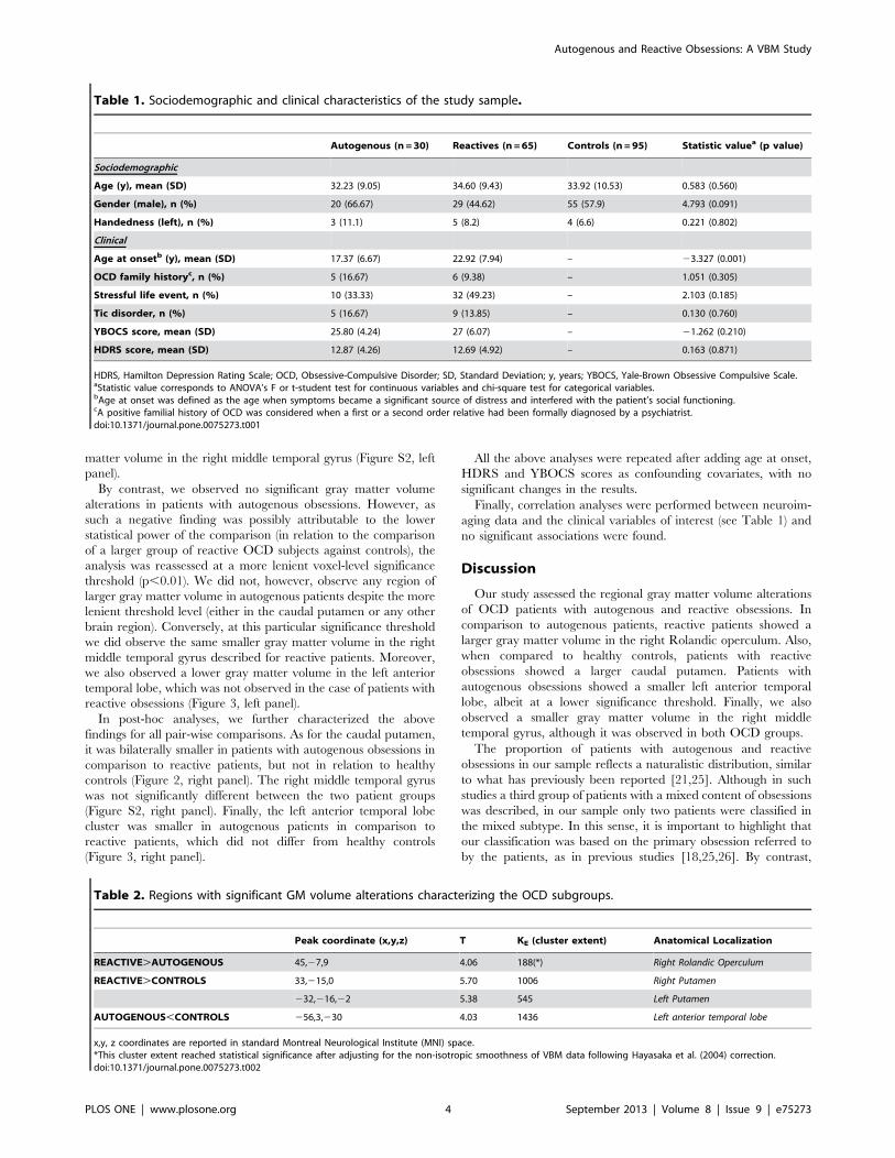
reactive subjects showed a significantly greater gray matter volume
in the right Rolandic operculum/posterior insula region (Table 2
and Figure 1, left panel). In a post-hoc analysis, we compared the
volume of this region between both OCD groups and healthy
controls. While autogenous patients showed a lower gray matter
volume, reactive patients presented a significantly higher volume
(Figure 1, right panel).
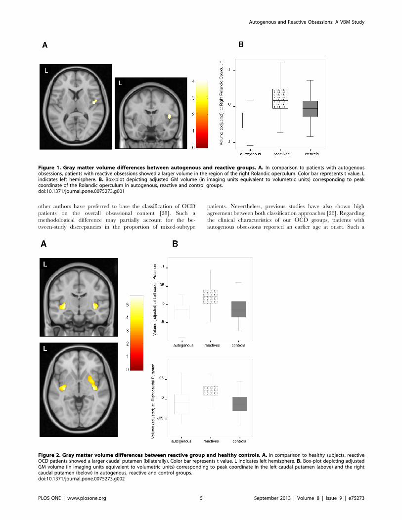
We further explored for potential volume differences between
the OCD groups by assessing their respective volume changes in
relation to healthy controls. Patients with reactive obsessions
showed a significantly larger volume of the caudal putamen
(bilaterally) (Figure 2, left panel) and a significantly lower gray
Autogenous and Reactive Obsessions: A VBM Study
PLOS ONE | www.plosone.org 3 September 2013 | Volume 8 | Issue 9 | e75273
matter volume in the right middle temporal gyrus (Figure S2, left
panel).
By contrast, we observed no significant gray matter volume
alterations in patients with autogenous obsessions. However, as
such a negative finding was possibly attributable to the lower
statistical power of the comparison (in relation to the comparison
of a larger group of reactive OCD subjects against controls), the
analysis was reassessed at a more lenient voxel-level significance
threshold (p,0.01). We did not, however, observe any region of
larger gray matter volume in autogenous patients despite the more
lenient threshold level (either in the caudal putamen or any other
brain region). Conversely, at this particular significance threshold
we did observe the same smaller gray matter volume in the right
middle temporal gyrus described for reactive patients. Moreover,
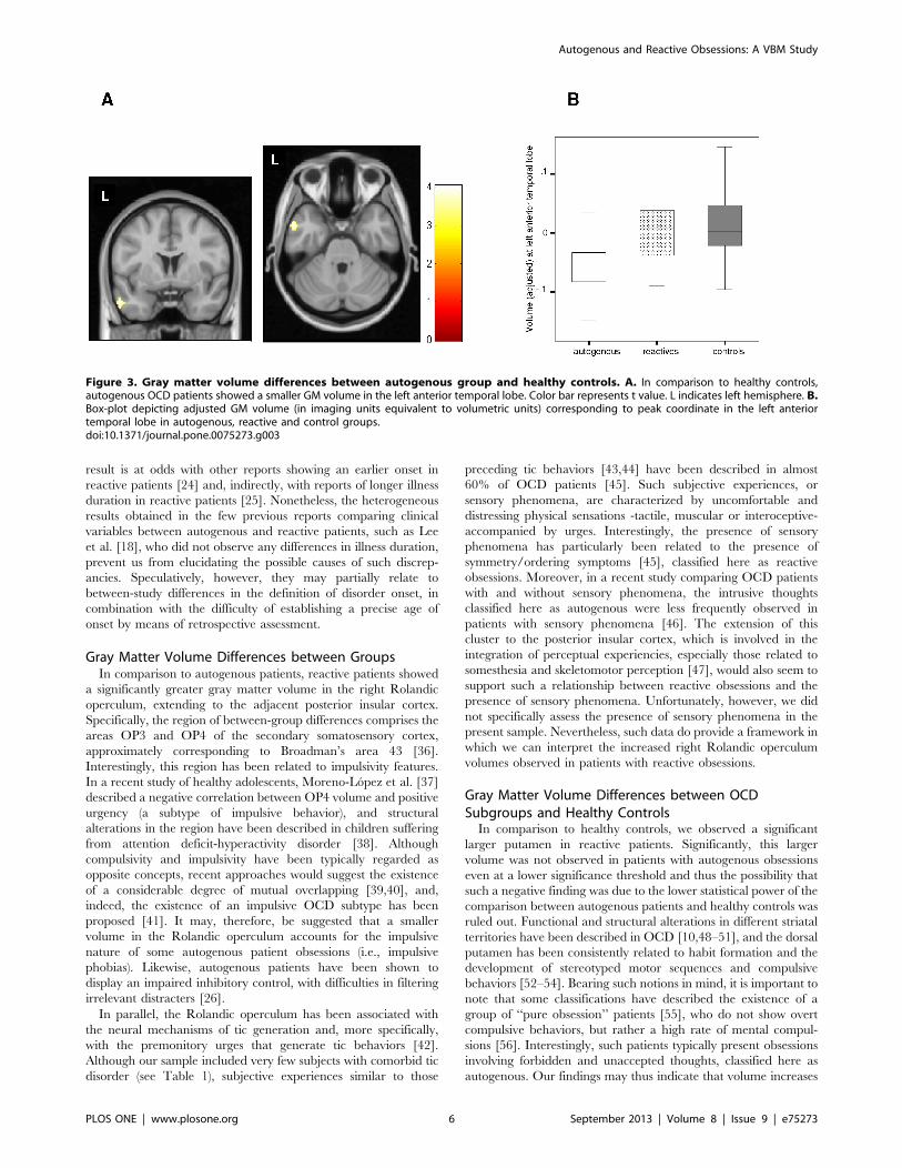
we also observed a lower gray matter volume in the left anterior
temporal lobe, which was not observed in the case of patients with
reactive obsessions (Figure 3, left panel).
In post-hoc analyses, we further characterized the above
findings for all pair-wise comparisons. As for the caudal putamen,
it was bilaterally smaller in patients with autogenous obsessions in
comparison to reactive patients, but not in relation to healthy
controls (Figure 2, right panel). The right middle temporal gyrus
was not significantly different between the two patient groups
(Figure S2, right panel). Finally, the left anterior temporal lobe
cluster was smaller in autogenous patients in comparison to
reactive patients, which did not differ from healthy controls
(Figure 3, right panel).
All the above analyses were repeated after adding age at onset,
HDRS and YBOCS scores as confounding covariates, with no
significant changes in the results.
Finally, correlation analyses were performed between neuroim-
aging data and the clinical variables of interest (see Table 1) and
no significant associations were found.
Discussion
Our study assessed the regional gray matter volume alterations
of OCD patients with autogenous and reactive obsessions. In
comparison to autogenous patients, reactive patients showed a
larger gray matter volume in the right Rolandic operculum. Also,
when compared to healthy controls, patients with reactive
obsessions showed a larger caudal putamen. Patients with
autogenous obsessions showed a smaller left anterior temporal
lobe, albeit at a lower significance threshold. Finally, we also
observed a smaller gray matter volume in the right middle
temporal gyrus, although it was observed in both OCD groups.
The proportion of patients with autogenous and reactive
obsessions in our sample reflects a naturalistic distribution, similar
to what has previously been reported [21,25]. Although in such
studies a third group of patients with a mixed content of obsessions
was described, in our sample only two patients were classified in
the mixed subtype. In this sense, it is important to highlight that
our classification was based on the primary obsession referred to
by the patients, as in previous studies [18,25,26]. By contrast,
Table 1. Sociodemographic and clinical characteristics of the study sample.
Autogenous (n = 30) Reactives (n = 65) Controls (n = 95) Statistic valuea (p value)
Sociodemographic
Age (y), mean (SD) 32.23 (9.05) 34.60 (9.43) 33.92 (10.53) 0.583 (0.560)
Gender (male), n (%) 20 (66.67) 29 (44.62) 55 (57.9) 4.793 (0.091)
Handedness (left), n (%) 3 (11.1) 5 (8.2) 4 (6.6) 0.221 (0.802)
Clinical
Age at onsetb (y), mean (SD) 17.37 (6.67) 22.92 (7.94) – 23.327 (0.001)
OCD family historyc, n (%) 5 (16.67) 6 (9.38) – 1.051 (0.305)
Stressful life event, n (%) 10 (33.33) 32 (49.23) – 2.103 (0.185)
Tic disorder, n (%) 5 (16.67) 9 (13.85) – 0.130 (0.760)
YBOCS score, mean (SD) 25.80 (4.24) 27 (6.07) – 21.262 (0.210)
HDRS score, mean (SD) 12.87 (4.26) 12.69 (4.92) – 0.163 (0.871)
HDRS, Hamilton Depression Rating Scale; OCD, Obsessive-Compulsive Disorder; SD, Standard Deviation; y, years; YBOCS, Yale-Brown Obsessive Compulsive Scale.aStatistic value corresponds to ANOVA’s F or t-student test for continuous variables and chi-square test for categorical variables.bAge at onset was defined as the age when symptoms became a significant source of distress and interfered with the patient’s social functioning.cA positive familial history of OCD was considered when a first or a second order relative had been formally diagnosed by a psychiatrist.doi:10.1371/journal.pone.0075273.t001
Table 2. Regions with significant GM volume alterations characterizing the OCD subgroups.
Peak coordinate (x,y,z) T KE (cluster extent) Anatomical Localization
REACTIVE.AUTOGENOUS 45,27,9 4.06 188(*) Right Rolandic Operculum
REACTIVE.CONTROLS 33,215,0 5.70 1006 Right Putamen
232,216,22 5.38 545 Left Putamen
AUTOGENOUS,CONTROLS 256,3,230 4.03 1436 Left anterior temporal lobe
x,y, z coordinates are reported in standard Montreal Neurological Institute (MNI) space.*This cluster extent reached statistical significance after adjusting for the non-isotropic smoothness of VBM data following Hayasaka et al. (2004) correction.doi:10.1371/journal.pone.0075273.t002
Autogenous and Reactive Obsessions: A VBM Study
PLOS ONE | www.plosone.org 4 September 2013 | Volume 8 | Issue 9 | e75273
other authors have preferred to base the classification of OCD
patients on the overall obsessional content [28]. Such a
methodological difference may partially account for the be-
tween-study discrepancies in the proportion of mixed-subtype
patients. Nevertheless, previous studies have also shown high
agreement between both classification approaches [26]. Regarding
the clinical characteristics of our OCD groups, patients with
autogenous obsessions reported an earlier age at onset. Such a
Figure 1. Gray matter volume differences between autogenous and reactive groups. A. In comparison to patients with autogenousobsessions, patients with reactive obsessions showed a larger volume in the region of the right Rolandic operculum. Color bar represents t value. Lindicates left hemisphere. B. Box-plot depicting adjusted GM volume (in imaging units equivalent to volumetric units) corresponding to peakcoordinate of the Rolandic operculum in autogenous, reactive and control groups.doi:10.1371/journal.pone.0075273.g001
Figure 2. Gray matter volume differences between reactive group and healthy controls. A. In comparison to healthy subjects, reactiveOCD patients showed a larger caudal putamen (bilaterally). Color bar represents t value. L indicates left hemisphere. B. Box-plot depicting adjustedGM volume (in imaging units equivalent to volumetric units) corresponding to peak coordinate in the left caudal putamen (above) and the rightcaudal putamen (below) in autogenous, reactive and control groups.doi:10.1371/journal.pone.0075273.g002
Autogenous and Reactive Obsessions: A VBM Study
PLOS ONE | www.plosone.org 5 September 2013 | Volume 8 | Issue 9 | e75273
result is at odds with other reports showing an earlier onset in
reactive patients [24] and, indirectly, with reports of longer illness
duration in reactive patients [25]. Nonetheless, the heterogeneous
results obtained in the few previous reports comparing clinical
variables between autogenous and reactive patients, such as Lee
et al. [18], who did not observe any differences in illness duration,
prevent us from elucidating the possible causes of such discrep-
ancies. Speculatively, however, they may partially relate to
between-study differences in the definition of disorder onset, in
combination with the difficulty of establishing a precise age of
onset by means of retrospective assessment.
Gray Matter Volume Differences between GroupsIn comparison to autogenous patients, reactive patients showed
a significantly greater gray matter volume in the right Rolandic
operculum, extending to the adjacent posterior insular cortex.
Specifically, the region of between-group differences comprises the
areas OP3 and OP4 of the secondary somatosensory cortex,
approximately corresponding to Broadman’s area 43 [36].
Interestingly, this region has been related to impulsivity features.
In a recent study of healthy adolescents, Moreno-Lopez et al. [37]
described a negative correlation between OP4 volume and positive
urgency (a subtype of impulsive behavior), and structural
alterations in the region have been described in children suffering
from attention deficit-hyperactivity disorder [38]. Although
compulsivity and impulsivity have been typically regarded as
opposite concepts, recent approaches would suggest the existence
of a considerable degree of mutual overlapping [39,40], and,
indeed, the existence of an impulsive OCD subtype has been
proposed [41]. It may, therefore, be suggested that a smaller
volume in the Rolandic operculum accounts for the impulsive
nature of some autogenous patient obsessions (i.e., impulsive
phobias). Likewise, autogenous patients have been shown to
display an impaired inhibitory control, with difficulties in filtering
irrelevant distracters [26].
In parallel, the Rolandic operculum has been associated with
the neural mechanisms of tic generation and, more specifically,
with the premonitory urges that generate tic behaviors [42].
Although our sample included very few subjects with comorbid tic
disorder (see Table 1), subjective experiences similar to those
preceding tic behaviors [43,44] have been described in almost
60% of OCD patients [45]. Such subjective experiences, or
sensory phenomena, are characterized by uncomfortable and
distressing physical sensations -tactile, muscular or interoceptive-
accompanied by urges. Interestingly, the presence of sensory
phenomena has particularly been related to the presence of
symmetry/ordering symptoms [45], classified here as reactive
obsessions. Moreover, in a recent study comparing OCD patients
with and without sensory phenomena, the intrusive thoughts
classified here as autogenous were less frequently observed in
patients with sensory phenomena [46]. The extension of this
cluster to the posterior insular cortex, which is involved in the
integration of perceptual experiencies, especially those related to
somesthesia and skeletomotor perception [47], would also seem to
support such a relationship between reactive obsessions and the
presence of sensory phenomena. Unfortunately, however, we did
not specifically assess the presence of sensory phenomena in the
present sample. Nevertheless, such data do provide a framework in
which we can interpret the increased right Rolandic operculum
volumes observed in patients with reactive obsessions.
Gray Matter Volume Differences between OCDSubgroups and Healthy Controls
In comparison to healthy controls, we observed a significant
larger putamen in reactive patients. Significantly, this larger
volume was not observed in patients with autogenous obsessions
even at a lower significance threshold and thus the possibility that
such a negative finding was due to the lower statistical power of the
comparison between autogenous patients and healthy controls was
ruled out. Functional and structural alterations in different striatal
territories have been described in OCD [10,48–51], and the dorsal
putamen has been consistently related to habit formation and the
development of stereotyped motor sequences and compulsive
behaviors [52–54]. Bearing such notions in mind, it is important to
note that some classifications have described the existence of a
group of ‘‘pure obsession’’ patients [55], who do not show overt
compulsive behaviors, but rather a high rate of mental compul-
sions [56]. Interestingly, such patients typically present obsessions
involving forbidden and unaccepted thoughts, classified here as
autogenous. Our findings may thus indicate that volume increases
Figure 3. Gray matter volume differences between autogenous group and healthy controls. A. In comparison to healthy controls,autogenous OCD patients showed a smaller GM volume in the left anterior temporal lobe. Color bar represents t value. L indicates left hemisphere. B.Box-plot depicting adjusted GM volume (in imaging units equivalent to volumetric units) corresponding to peak coordinate in the left anteriortemporal lobe in autogenous, reactive and control groups.doi:10.1371/journal.pone.0075273.g003
Autogenous and Reactive Obsessions: A VBM Study
PLOS ONE | www.plosone.org 6 September 2013 | Volume 8 | Issue 9 | e75273

in the putamen characterize the group of patients with reactive
obsessions, who display a higher incidence of overt compulsive
behavior.
Patients with reactive obsessions also exhibited less gray matter
in the right middle temporal region, although such a finding was
equally observed in patients with autogenous obsessions at a lower
significance threshold. Conversely, a smaller volume of the left
anterior temporal lobe was specifically observed in patients with
autogenous obsessions. While previous studies reported the
development of obsessive-compulsive symptoms after temporal
pole lesions [57], Van den Heuvel et al. [13] showed a specific
association of anterior temporal lobe volumes and harm/checking
symptoms that partially overlap with the symptoms classified here
as autogenous. Moreover, patients with autogenous obsessions
typically display an exacerbated distress when dealing with
questions of a moral nature. Anterior temporal lobe alterations
may account for such dysfunctional cognitions, as this region has
been related to complex cognitive processes such as moral
cognition [58], and the interaction between anterior temporal
lobe and fronto-mesolimbic activity has been hypothesized to
underpin the experience of moral sentiments [59].
According to our data, general OCD populations should
normally include a larger proportion of patients with reactive
obsessions. It is, therefore, not surprising that putamen alterations
have been previously reported in general OCD samples [9,60,61].
At the same time, however, it may seem surprising that volume
increases in the Rolandic operculum have not previously been
documented. Nevertheless, it should be pointed out that, in our
post-hoc analysis, autogenous patients presented a significantly
smaller volume of this particular brain region, which may well
partially compensate the larger volumes of reactive patients when
assessing general OCD populations. Likewise, it is noteworthy that
we have not described alterations involving other brain regions
such as the dorsal-medial prefrontal and the medial and lateral
orbitofrontal cortices, which have typically presented alterations in
studies assessing general OCD samples [9,10]. Smaller volumes in
these regions may not, therefore, depend on the classification
scheme used here and are most probably related to other
symptoms. By way of example, the presence of comorbid
depression would seem to be particularly important in relation
to orbitofrontal alterations [62] and our study groups did not differ
in terms of this variable.
Certain limitations apply to the current findings. Firstly, as
recruitment was conducted in a specialized OCD clinical unit, the
mean severity of our sample was somewhat higher in comparison
to other reports. Secondly, patients were undergoing pharmaco-
logical treatment during the study period. Nevertheless, no
significant effects of antidepressant treatment on brain morphol-
ogy were detected in a voxel-wise meta-analysis of structural
studies in OCD [9]. Finally, given the lack of an objective
measurement to identify autogenous and reactive patients, the
possibility of a classification bias cannot be ruled out. Be that as it
may, the classification was carried out by two expert psychiatrists
who reached a consensus as to the nature of each patient’s
symptoms.
In summary, the existence of specific structural alterations in
patients with autogenous and reactive obsessions provides some
neurobiological support for this classification scheme, as proposed
by Lee and Kwon [18]. These findings add to emerging evidence
from neuroimaging studies that the clinical heterogeneity in OCD
can be differentiated in terms of discrete brain systems. Future
studies should expand our results by relating the anatomical
abnormalities of patients with autogenous and reactive obsessions
with specific clinical features, such as impulsivity, sensory
phenomena, overt compulsions, or exacerbated moral distress.
Supporting Information
Figure S1 Classification template used for OCD pa-tients’ characterization. This template was intended to assist
the psychiatrists in the classification of OCD patients according to
their primary obsessions.
(TIF)
Figure S2 Gray matter volume differences betweenOCD subgroups and healthy controls. A. In comparison
to healthy controls, reactive OCD patients showed a smaller GM
volume in the right middle temporal gyrus. Color bar represents t
value. L indicates left hemisphere. B. Box-plot depicting adjusted
GM volume (in imaging units equivalent to volumetric units)
corresponding to peak coordinate in the right middle temporal
gyrus in autogenous, reactive and control groups.
(TIF)
Acknowledgments
We thank Drs. Rosa Hernandez-Ribas and Ester Cerrillo for their
assistance in the inter-rater reliability assessments. We also thank Mr.
Gerald Fannon for revising the manuscript.
Author Contributions
Conceived and designed the experiments: MS PA NC CS-M. Performed
the experiments: MS PA CS ER CL-S JMM NC. Analyzed the data: MS
IM-Z CS-M. Contributed reagents/materials/analysis tools: JP IM-Z.
Wrote the paper: MS CS-M PA BJH JMM NC.
References
1. Leckman JF, Denys D, Simpson HB, Mataix-Cols D, Hollander E, et al. (2010)
Obsessive-compulsive disorder: a review of the diagnostic criteria and possible
subtypes and dimensional specifiers for DSM-V. Depress Anxiety 27: 507–27.
2. De Mathis MA, Diniz JB, do Rosario MC, Torres AR, Hoexter M, et al. (2006)
What is the optimal way to subdivide obsessive-compulsive disorder? CNS
Spectr 11: 762–79.
3. Miguel EC, Leckman JF, Rauch S, do Rosario-Campos, Hounie AG, et al.
(2005) Obsessive-compulsive disorder phenotypes: implications for genetic
studies. Mol Psychiatry 10: 258–75.
4. Mataix-Cols D, Rosario-Campos MC, Leckman JF (2005) A multidimensional
model of obsessive-compulsive disorder. Am J Psychiatry 162: 228–38.
5. Mataix-Cols D (2006) Deconstructing obsessive-compulsive disorder: a multidi-
mensional perspective. Curr Opin Psychiatry 19: 84–9.
6. Mataix-Cols D, Wooderson S, Lawrence N, Brammer MJ, Speckens A, et al.
(2004) Distinct neural correlates of washing, checking, and hoarding symptom
dimensions in obsessive-compulsive disorder. Arch Gen Psychiatry 61: 564–76.
7. Gilbert AR, Mataix-Cols D, Almeida JR, Lawrence N, Nutche J, et al. (2008)
Brain structure and symptom dimensions relationships in obsessive-compulsive
disorder: a voxel-based morphometry study. J Affect Disord 109: 117–26.
8. Murayama K, Nakao T, Sanematsu H, Okada K, Yoshiura T, et al. (2013)
Differential neural network of checking versus washing symptoms in obsessive-
compulsive disorder. Prog Neuropsychopharmacol Biol Psychiatry 10: 160–6.
9. Radua J, Mataix-Cols D (2009) Voxel-wise meta-analysis of grey matter changes
in obsessive-compulsive disorder. Br J Psychiatry 195: 393–402.
10. Rotge JY, Guehl D, Dilharreguy B, Tignol J, Bioulac B, et al. (2009) Meta-
analysis of brain volume changes in obsessive-compulsive disorder. Biol
Psychiatry 65: 75–83.
11. Soriano-Mas C, Pujol J, Alonso P, Cardoner N, Menchon JM, et al. (2007)
Identifying patients with obsessive-compulsive disorder using whole-brain
anatomy. Neuroimage 35: 1028–37.
12. Pujol J, Soriano-Mas C, Alonso P, Cardoner N, Menchon JM, et al. (2004)
Mapping structural brain alterations in obsessive-compulsive disorder. Arch Gen
Psychiatry 61: 720–30.
13. Van den Heuvel OA, Remijnse PL, Mataix-Cols D, Vrenken H, Groenewegen
HJ, et al. (2009) The major symptom dimensions of obsessive-compulsive
disorder are mediated by partially distinct neural systems. Brain 132: 853–68.
Autogenous and Reactive Obsessions: A VBM Study
PLOS ONE | www.plosone.org 7 September 2013 | Volume 8 | Issue 9 | e75273
14. Alvarenga PG, do Rosario MC, Batistuzzo MC, Diniz JB, Shavitt RG, et al.
(2012) Obsessive-compulsive symptom dimensions correlate to specific graymatter volumes in treatment-naıve patients. J Psychiatr Res 46: 1635–42.
15. Sanavio E (1998) Obsessions and compulsions: The Padua Inventory. Behav Res
Ther 26: 169–177.16. Rosario-Campos MC, Miguel EC, Quatrano S, Chacon P, Ferrao Y, et al.
(2006) The Dimensional Yale-Brown Obsessive-Compulsive Scale (DY-BOCS):an instrument for assessing obsessive-compulsive symptom dimensions. Mol
Psychiatry 11: 495–504.
17. Lee HJ, Kwon SM (2003) Two different types of obsession: autogenousobsessions and reactive obsessions. Behav Res Ther 41: 11–29.
18. Lee HJ, Kwon SM, Kwon JS, Telch MJ, et al. (2005) Testing the autogenous-reactive model of obsessions. Depress Anxiety 21: 118–29.
19. Purdon C, Clark DA (1993) Obsessive intrusive thoughts in nonclinical subjects:Part I. Content and relation with depressive, anxious and obsessional symptoms.
Behav Res Ther 31: 713–720.
20. Purdon C, Clark DA (1994) Obsessive intrusive thoughts in non clinical subjects:Part II. Cognitive appraisal, emotional response and thought control strategies.
Behav Res Ther 32: 403–410.21. Besiroglu L, Uguz F, Ozbebit O, Guler O, Cilli AS, et al. (2007) Longitudinal
assessment of symptom and subtype categories in obsessive-compulsive disorder.
Depress Anxiety 24: 461–6.22. Lee HJ, Lee SH, Kim HS, Kwon SM, Telch MJ (2005) A comparison of
autogenous/reactive obsessions and worry in a nonclinical population: a test ofthe continuum hypothesis. Behav Res Ther 43: 999–1010.
23. Moulding R, Kyrios M, Doron G, Nedeljkovic M (2007) Autogenous andreactive obsessions: further evidence for a two-factor model of obsessions.
J Anxiety Disord 21: 677–90.
24. Besiroglu L, Agargun MY, Ozbebit O, Aydin A (2006) A discrimination basedon autogenous versus reactive obsessions in obsessive-compulsive disorder and
related clinical manifestations. CNS Spectr 11: 179–86.25. Belloch A, Cabedo E, Carrio C, Larsson C (2010) Cognitive therapy for
autogenous and reactive obsessions: clinical and cognitive outcomes at post-
treatment and 1-year follow-up. J Anxiety Disord 24: 573–80.26. Lee HJ, Telch MJ (2010) Differences in latent inhibition as a function of the
autogenous-reactive OCD subtype. Behav Res Ther 48: 571–9.27. Lee HJ, Yost BP, Telch MJ (2009) Differential performance on the go/no-go
task as a function of the autogenous-reactive taxonomy of obsessions: findingsfrom a no-treatment seeking sample. Behav Res Ther 47: 294–300.
28. Besiroglu L, Sozen M, Ozbebit O, Avcu S, Selvi Y, et al. (2011) The
involvement of distinct neural systems in patients with obsessive-compulsivedisorder with autogenous and reactive obsessions. Acta Psychiatr Scand 124:
141–51.29. Goodman WK, Price LH, Rasmussen SA, Mazure C, Fleischmann RL, et al.
(1989) The Yale-Brown Obsessive-Compulsive Scale. I. Devolopment, use, and
reliability. Arch Gen Psychiatry 46: 1006–11.30. Vega-Dienstmaier JM, Sal Y Rosas HJ, Mazzotti Suarez G, Vidal H, Guimas B,
et al. (2002) Validation of a version in Spanish of the Yale-Brown Obsessive-Compulsive Scale. Actas Esp Psiquiatr 30: 30–5.
31. Hamilton M (1960) A rating scale for depression. J Neurol Neurosurg Psychiatry23: 56–62.
32. Ramos-Brieva JA, Cordero Villafafila A (1986) Validation of the Spanish version
of the Hamilton Rating Scale for Depression. Actas Luso Esp Neurol PsiquiatrCienc Afines 14: 324–34.
33. Ashburner J (2007) A fast diffeomorphic image registration algorithm. Neuro-image 38: 95–113.
34. Song XW, Dong ZY, Long XY, Li SF, Zuo XN, et al. (2011) REST: a toolkit for
resting-state functional magnetic resonance imaging data processing. PLoS One6: e25031.
35. Hayasaka S, Phan KL, Liberzon I, Worsley KJ, Nichols TE (2004)Nonstationary cluster-size inference with random field and permutation
methods. Neuroimage 22: 676–87.
36. Eickhoff SB, Amunts K, Mohlberg H, Zilles K (2006) The human parietaloperculum II. Stereotaxic maps and correlation with functional imaging results.
Cereb Cortex 16: 268–79.37. Moreno-Lopez L, Soriano-Mas C, Delgado-Rico E, Rıo-Valle JS, Verdejo-
Garcıa A (2012) Brain structural correlates of reward sensitivity and impulsivityin adolescents with normal and excess weight. PLoS One 7: e49185.
38. Carmona S, Vilarroya O, Bielsa A, Tremols V, Soliva JC, et al. (2005) Global
and regional gray matter reductions in ADHD: a voxel-based morphometricstudy. Neurosci Lett 389: 88–93.
39. Fineberg NA, Potenza MN, Chamberlain SR, Berlin HA, Menzies L, et al.
(2010) Probing compulsive and impulsive behaviors, from animal models to
endophenotypes: a narrative review. Neuropsychopharmacology 35: 591–604.
40. Lochner C, Stein DJ (2010) Obsessive-compulsive spectrum disorders in
obsessive-compulsive disorder and other anxiety disorders. Psychopathology
43: 389–96.
41. Kashyap H, Fontenelle LF, Miguel EC, Ferrao YA, Torres AR, et al. (2012)
‘Impulsive compulsivity’ in obsessive-compulsive disorder: a phenotypic marker
of patients with poor clinical outcome. J Psychiatr Res 46: 1146–52.
42. Wang Z, Maia TV, Marsh R, Colibazzi T, Gerber A, et al. (2011) The neural
circuits that generate tics in Tourette’s syndrome. Am J Psychiatry 168: 1326–
37.
43. Cohen AJ, Leckman JF (1992) Sensory phenomena associated with Gilles de la
Tourette’s syndrome. J Clin Psychiatry 53: 319–23.
44. Leckman JF (2003) Phenomenology of tics and natural history of tic disorders.
Brain Dev 25 S24–S28.
45. Rosario MC, Prado HS, Borcato S, Diniz JB, Shavitt RG, et al. (2009)
Validation of the University of Sao Paulo Sensory Phenomena Scale: initial
psychometric properties. CNS Spectr 14: 315–23.
46. Ferrao YA, Shavitt RG, Prado H, Fontenelle LF, Malavazzi DM, et al. (2012)
Sensory phenomena associated with repetitive behaviors in obsessive-compulsive
disorder: an exploratory study of 1001 patients. Psychiatry Res 197: 253–8.
47. Augustine JR (1996) Circuitry and functional aspects of the insular lobe in
primates including humans. Brain Research Reviews 22: 229–244.
48. Harrison BJ, Soriano-Mas C, Pujol J, Ortiz H, Lopez-Sola M, et al. (2009)
Altered corticostriatal functional connectivity in obsessive-compulsive disorder.
Arch Gen Psychiatry 66: 1189–200.
49. Harrison BJ, Pujol J, Cardoner N, Deus J, Alonso P, et al. (2013) Brain
corticostriatal systems and the major clinical symptom dimensions of obsessive-
compulsive disorder. Biol Psychiatry 73: 321–8.
50. Szeszko PR, Christian C, Macmaster F, Lencz T, Mirza Y, et al. (2008) Gray
matter structural alterations in psychotropic drug-naıve pediatric obsessive-
compulsive disorder: an optimized voxel-based morphometry study.
Am J Psychiatry 165: 1299–307.
51. Whiteside SP, Port JD, Abramowitz JS (2004) A meta-analysis of functional
neuroimaging in obsessive-compulsive disorder. Psychiatry Res 132: 69–79.
52. Ruge H, Wolfensteller U (2013) Functional integration processes underlying the
instruction-based learning of novel goal-directed behaviors. Neuroimage 68:
162–72.
53. Balleine BW, O’Doherty JP (2010) Human and rodent homologies in action
control: corticostriatal determinants of goal-directed and habitual action.
Neuropsychopharmacology 35: 48–69.
54. Hollander E, Anagnostou E, Chaplin W, Esposito K, Haznedar MM, et al.
(2005) Striatal volume on magnetic resonance imaging and repetitive behaviors
in autism. Biol Psychiatry 58: 226–32.
55. Baer L (1994) Factor analysis of symptom subtypes of obsessive-compulsive
disorder and their relation to personality and tic disorders. J Clin Psychiatry 55:
18–23.
56. Williams MT, Farris SG, Turkheimer E, Pinto A, Ozanick K, et al. (2011) Myth
of the pure obsessional type in obsessive-compulsive disorder. Depress Anxiety
28: 495–500.
57. Roth RM, Jobst BC, Thadani VM, Gilbert KL, Roberts DW (2009) New-onset
obsessive-compulsive disorder following neurosurgery for medication-refractory
seizure disorder. Epilepsy Behav 14: 677–80.
58. Zahn R, Moll J, Paiva M, Garrido G, Krueger F, et al. (2009) The neural basis
of human social values: evidence from functional MRI. Cereb Cortex 19: 276–
83.
59. Moll J, Zahn R, de Oliveira-Souza R, Krueger F, Grafman J (2005) Opinion:
the neural basis of human moral cognition. Nat Rev Neurosci 6: 799–809.
60. Zarei M, Mataix-Cols D, Heyman I, Hough M, Doherty J, et al. (2011) Changes
in gray matter volume and white matter microstructure in adolescents with
obsessive-compulsive disorder. Biol Psychiatry 70: 1083–1090.
61. Yoo SY, Roh MS, Choi JS, Kang DH, Ha TH, et al. (2008) Voxel-based
morphometry study of gray matter abnormalities in obsessive-compulsive
disorder. J Korean Med Sci 23: 24–30.
62. Cardoner N, Soriano-Mas C, Pujol J, Alonso P, Harrison BJ, et al. (2007) Brain
structural correlates of depressive comorbidity in obsessive-compulsive disorder.
Neuroimage 38: 413–21.
Autogenous and Reactive Obsessions: A VBM Study
PLOS ONE | www.plosone.org 8 September 2013 | Volume 8 | Issue 9 | e75273
Related Documents