Brain Neuroplasticity in Healthy, Hyperactive and Psychotic Children: Insights from Neuroimaging Judith L Rapoport 1 and Nitin Gogtay* ,1 1 Child Psychiatry Branch, NIMH, Bethesda, MD, USA Noninvasive brain imaging permits longitudinal studies of anatomic brain development in healthy and psychiatrically ill children. The time course for gray matter maturation varies by region and parallels earlier histological studies, indicating dynamic patterns of overproduction and regression. Developmental trajectories vary in relation to gender, intelligence, and overall functioning. Twin studies show high heritability for brain volumes, which varies with region and with age. Diagnostically specific, illness-related changes as well as outcome-associated plastic response are observed as illustrated for two pediatric populations, childhood-onset schizophrenia and attention-deficit/hyperactivity disorder, conditions which may be, in part, disorders of brain plasticity. Neuropsychopharmacology Reviews (2008) 33, 181–197; doi:10.1038/sj.npp.1301553; published online 12 September 2007 Keywords: brain; neuroplasticity; neuroimaging; healthy development; childhood schizophrenia; ADHD INTRODUCTION Neuroplasticity is the change in neural structure and function in response to experience or environmental stimuli. The question of whether and how much the environment influences brain development is of broad general interest, with particular attention from the fields of developmental neurobiology, child development, education, and child psychopathology. The availability of brain imaging, particularly suitable for pediatric studies due to its safety, has enabled human developmental studies. Structural MRI studies in adults have shown activity- dependent plastic responses in the brain structure, such as the increased hippocampal volume (which is involved in spatial navigation) in London taxi drivers that correlated with the amount of time spent in navigating the streets of London (Maguire et al, 2000). However, individual trajectories for these changes can only be addressed by longitudinal studies. Functional imaging studies have the advantage of performance measures that can be obtained simultaneously with imaging that let us visualize the plastic reorganization of various brain regions in response to stimuli. For example, subjects learning sequential finger movements show altera- tions in the motor cortex, cerebellum, and basal ganglia (Elbert et al, 1995; Ungerleider et al, 2002) or healthy adult subjects trained in juggling show transient activity-depen- dent structural changes in visual motor cortex (Draganski et al, 2004). Here, we discuss brain plasticity, particularly, as it relates to childhood psychopathology. Early cross-sectional and more recent longitudinal data show dynamic age-related changes that can vary with intelligence, clinical status, and/ or treatment and long-term outcome. A wealth of normal and clinical data has been acquired in recent years which we discuss for normal brain development, and that for two childhood disorders: childhood-onset schizophrenia (COS) and attention-deficit/hyperactivity disorder (ADHD). Although some functional studies are mentioned, closer attention is paid to structural brain morphology. Similarly, while Diffusion Tensor Imaging (DTI) has begun to be studied in pediatric populations (Cascio et al, 2007), this important area is not addressed, as no DTI studies have yet focused on plastic change. Throughout this review are speculations about possible mechanisms underlying anatomic brain MRI changes seen in healthy and clinical psychiatric populations. It needs to be clear at the outset that the MRI gives no information about the cellular correlates of the anatomic brain changes discussed in this review. The wealth of information about synaptic development in relation to age and to function has led to common ‘shorthand’ of interpreting many of these changes in terms of synaptic production and pruning. The weakness in this approach is not only that we do not have sufficient documentation of the underlying actual brain changes, but the field itself remains in flux as to what actual cellular components are involved at various time points (Sur and Rubenstein, 2005). Received 7 May 2007; revised 11 July 2007; accepted 30 July 2007 *Correspondence: Dr N Gogtay, Child Psychiatry Branch, NIMH, Bldg 10, Room 3N202, MSC-1600, Bethesda, MD 20892, USA, Tel: +1 301 435 4494, E-mail: [email protected] Neuropsychopharmacology REVIEWS (2008) 33, 181–197 & 2008 Nature Publishing Group All rights reserved 0893-133X/08 $30.00 ............................................................................................................................................................... www.neuropsychopharmacology.org 181 REVIEW .............................................................................................................................................. Neuropsychopharmacology REVIEWS

Welcome message from author
This document is posted to help you gain knowledge. Please leave a comment to let me know what you think about it! Share it to your friends and learn new things together.
Transcript

Brain Neuroplasticity in Healthy, Hyperactive andPsychotic Children: Insights from Neuroimaging
Judith L Rapoport1 and Nitin Gogtay*,1
1Child Psychiatry Branch, NIMH, Bethesda, MD, USA
Noninvasive brain imaging permits longitudinal studies of anatomic brain development in healthy and psychiatrically ill
children. The time course for gray matter maturation varies by region and parallels earlier histological studies, indicating
dynamic patterns of overproduction and regression. Developmental trajectories vary in relation to gender, intelligence, and
overall functioning. Twin studies show high heritability for brain volumes, which varies with region and with age. Diagnostically
specific, illness-related changes as well as outcome-associated plastic response are observed as illustrated for two pediatric
populations, childhood-onset schizophrenia and attention-deficit/hyperactivity disorder, conditions which may be, in part,
disorders of brain plasticity.
Neuropsychopharmacology Reviews (2008) 33, 181–197; doi:10.1038/sj.npp.1301553; published online 12 September 2007
Keywords: brain; neuroplasticity; neuroimaging; healthy development; childhood schizophrenia; ADHD
���������������������������������
INTRODUCTION
Neuroplasticity is the change in neural structure andfunction in response to experience or environmentalstimuli. The question of whether and how much theenvironment influences brain development is of broadgeneral interest, with particular attention from the fields ofdevelopmental neurobiology, child development, education,and child psychopathology. The availability of brainimaging, particularly suitable for pediatric studies due toits safety, has enabled human developmental studies.
Structural MRI studies in adults have shown activity-dependent plastic responses in the brain structure, such as theincreased hippocampal volume (which is involved in spatialnavigation) in London taxi drivers that correlated with theamount of time spent in navigating the streets of London(Maguire et al, 2000). However, individual trajectories forthese changes can only be addressed by longitudinal studies.
Functional imaging studies have the advantage ofperformance measures that can be obtained simultaneouslywith imaging that let us visualize the plastic reorganizationof various brain regions in response to stimuli. For example,subjects learning sequential finger movements show altera-tions in the motor cortex, cerebellum, and basal ganglia(Elbert et al, 1995; Ungerleider et al, 2002) or healthy adultsubjects trained in juggling show transient activity-depen-
dent structural changes in visual motor cortex (Draganskiet al, 2004).
Here, we discuss brain plasticity, particularly, as it relatesto childhood psychopathology. Early cross-sectional andmore recent longitudinal data show dynamic age-relatedchanges that can vary with intelligence, clinical status, and/or treatment and long-term outcome. A wealth of normaland clinical data has been acquired in recent years whichwe discuss for normal brain development, and that fortwo childhood disorders: childhood-onset schizophrenia(COS) and attention-deficit/hyperactivity disorder (ADHD).Although some functional studies are mentioned, closerattention is paid to structural brain morphology. Similarly,while Diffusion Tensor Imaging (DTI) has begun to bestudied in pediatric populations (Cascio et al, 2007), thisimportant area is not addressed, as no DTI studies have yetfocused on plastic change.
Throughout this review are speculations about possiblemechanisms underlying anatomic brain MRI changes seenin healthy and clinical psychiatric populations. It needs tobe clear at the outset that the MRI gives no informationabout the cellular correlates of the anatomic brain changesdiscussed in this review. The wealth of information aboutsynaptic development in relation to age and to function hasled to common ‘shorthand’ of interpreting many of thesechanges in terms of synaptic production and pruning. Theweakness in this approach is not only that we do not havesufficient documentation of the underlying actual brainchanges, but the field itself remains in flux as to what actualcellular components are involved at various time points(Sur and Rubenstein, 2005).Received 7 May 2007; revised 11 July 2007; accepted 30 July 2007
*Correspondence: Dr N Gogtay, Child Psychiatry Branch, NIMH, Bldg 10,Room 3N202, MSC-1600, Bethesda, MD 20892, USA, Tel: + 1 301 4354494, E-mail: [email protected]
Neuropsychopharmacology REVIEWS (2008) 33, 181–197& 2008 Nature Publishing Group All rights reserved 0893-133X/08 $30.00...............................................................................................................................................................
www.neuropsychopharmacology.org 181
REVIEW
..............................................................................................................................................
Neuropsychopharmacology REVIEWS

PLASTICITY DURING BRAIN DEVELOPMENT
Childhood is a time of considerable changes in brainanatomical structure and connectivity (Huttenlocher, 1979;Huttenlocher and de Courten, 1987; Casey et al, 2000) andthere is more striking evidence for plastic brain changes inchildhood. As mechanisms of neural circuitry developmentare increasingly understood, phenomena surroundingbehavioral development formerly referred to as ‘sensitiveperiods’ are being reframed in terms of brain plasticity withthe recognition that some are only attributable to theimmature brain (Knudsen, 2004).
During fetal development, neurons proliferate from earlyprecursor cells that migrate radially outward from theventricular zone to establish the normal cellular architec-ture of the cerebral cortex (Caviness and Takahashi, 1995).This leads to a human brain with about 100 billion neuronsat birth. Following the neuronal migration, dendrites andaxons arborize forming the intercellular communicationsand synapses. In humans, the initial overproduction ofneurons is followed by selective apoptosis later in gestationresulting in the loss of up to 50% of cortical neurons(Rabinowicz et al, 1996). Similarly, during early postnatalbrain development, there is a marked increase in synapticdensity reaching substantially above adult levels (Bourgeoisand Rakic, 1993; Bourgeois et al, 1994) which aresubsequently ‘pruned’ during childhood and adolescenceto adult levels with primary sensory and motor regionsmaturing early relative to regions subserving more complexfunctions (Huttenlocher and Dabholkar, 1997). Recentevidence indicates that further functional parcellation ofcerebral cortical areas into discrete processing centersinvolves a rich array of signals, with complex interaction,between mechanisms both intrinsic to cortical progenitorsand neurons and environmental stimuli extrinsic to thecortex requiring neural activity such as light stimulation tothe developing eye (Sur and Rubenstein, 2005).
ADAPTIVE/COMPENSATORY PLASTICITY
Unique childhood plasticity has been demonstrated parti-cularly in the areas of vision, audition, motor, and languageabilities (Huttenlocher et al, 2002; Neville and Bavelier,2002; Spessot et al, 2004). In both animals and humans,early lesions undergo more efficient repair. It is clear, forexample, that lesions such as therapeutic hemispherectomyor stroke are handled better in childhood than at later ages.Perhaps the best known example of compensatory plasticitycould be seen in the observation that the functional effectsof strabismus (squint) in the infant are different from thatin the adults. In infancy, strabismus causes suppression ofthe image from the squinting eye, whereas in adults squintcauses persistent double vision, a disabling symptom.Surgical correction with preservation of vision is possibleup to the age of 7 years (VonNoorden and Crawford, 1979).A similar period for maximal plasticity for auditory cortexhas been defined within the first 7 years of life in childrenfitted with cochlear implants for early deafness (Sharmaet al, 2002). These observations have led to the hope thattreatment of cognitive or behavior disturbance during thechildhood years might be more effective in reshaping
trajectories of early development either to normalize orcompensate for earlier abnormalities.
Age-related adaptive plasticity has been particularly wellstudied in relation to language functions. Unilateral braindamage with focal brain injury to the left cerebralhemisphere causes aphasia in adults but not in infantsand young children. Although inconsistencies remain withthese observations, as lesions in Wernicke’s area in infantsare still associated with some delay in language production(Bates, 1993) and subtle language deficits in children remainwith early left hemisphere damage (Vargha-Khadem et al,1994; Isaacs et al, 1996), there is general agreement aboutstrikingly less impairment to language function whenlesions are in early childhood. Similarly, recent functionalstudies indicate that training in the auditory domains (usingmusical or auditory working memory tasks) leads toimproved reading skills in reading disabled populations(Gaab et al, 2005; Tallal and Gaab, 2006) raising thepossibility as to whether auditory/musical training couldalso influence normal language development.
Another area of adaptive plasticity has been the adverseeffects of drugs on the developing brain (Andersen andNavalta, 2004). There is a general concern that the use ofpsychotropic drugs in children will lead to drug-inducedplasticity introducing harmful changes in the circuits ofdeveloping brains. Stimulant drugs in particular have beenthe subject of concern, because stimulant drugs exposurecan produce sensitization of certain behaviors in animalmodels and addiction in humans. Some representativestudies (Bolanos et al, 2003; Carlezon et al, 2003)administered methylphenidate during pre-adolescent peri-ods of development in rats and observed the effect onsubsequent adult behaviors in response to emotionalstimuli. Bolanos et al, found methylphenidate decreasedresponsiveness to rewarding stimuli. Both groups alsofound the exposed animals more responsive to aversivestimuli as adults. The interest in these reports is basedprimarily on their possible relevance for clinical researchand practice. However, any implications at this point wouldseem premature. There is no evidence that stimulant drugtreatment for ADHD increases risk for substance abuse and,in fact, some evidence to suggest that the treatmentdecreases the risk (Biederman et al, 1999). Nonetheless,these studies are instructive as to how ultimately clinicalresearch will likely reveal the most useful information ondrug-induced brain changes in relatively subtle disorderssuch as ADHD.
A more interesting possibility based on preclinical orpharmacological studies is that early GABAergic drugsmight alter critical period plasticity. Experience-dependentplasticity shapes the early postnatal brain, as exemplified bythe loss of responsiveness to an eye briefly deprived ofvision during a critical period, which results in severeamblyopia (poor visual acuity) (Fagiolini et al, 2004).Critical period onset can be delayed indefinitely if release ofinhibitory neurotransmitter gamma aminobutyric acid(GABA) is kept low by gene-tarted disruption of itssynthetic enzyme glutamic acid decarboxylase 65 (GAD65) (Fagiolini and Hensch, 2000). The specificity of theGABA circuitry underlying visual cortical plasticity wasstudied using benzodiazepine agonists concurrently withmonocular deprivation to prematurely trigger ocular
Brain neuroplasticity in childrenJL Rapoport and N Gogtay
...............................................................................................................................................................
182
..............................................................................................................................................
Neuropsychopharmacology REVIEWS
REVIEW
dominance plasticity (Fagiolini et al, 2004). This furthershowed that only GABA alpha-1-containing circuits drovecortical plasticity, whereas alpha-2 did not. This hasimplications for the safe design of benzodiazepines for usein children.
MECHANISMS IN PLASTICITY
The molecular mechanisms in developmental as well asadaptive/compensatory plasticity are still largely unknown.Much of the brain development is assumed to requiremodification of gene expression and protein production(Lamprcht and Ledoux, 2004). However, the role ofenvironment in the development of neural circuits isincreasingly being understood with the abundant evidencefor the influence of experience-dependent activity inshaping the ultimate synaptic patterns (Hebb, 1949;Changeux and Danchin, 1976; Kandel et al, 2000). Thereis also evidence for plasticity in functional connectivity asseen in primates, where opposing wrist muscle movementscould be reversed using an electronic neural implant(Jackson et al, 2006) and thus plasticity across modalitieswould imply more extensive connectivity within theinvolved regions (Neville and Bavelier, 2002).
Development of cortical areas involves a rich interplay ofsignals intrinsic to cortical progenitors and neurons withthose extrinsic to the cortex requiring neural activity(Knudsen, 2004; Sur and Rubenstein, 2005). For example,repetitive activation (in response to external stimuli) ofexcitatory synapses increases the synaptic strength througha phenomenon called long-term potentiation (LTP) in manybrain regions that are crucial in learning and memory(Malenka and Nicoll, 1999). This process in which synapticconnections are refined requires localized increase inintracellular calcium in dendritic spines, which is typicallyachieved via the activation of the NMDA subtype ofglutamate receptor (McDonald and Johnston, 1990; Pennand Shatz, 1999) and is modulated further by activation ofAMPA subtype of glutamate receptors (Malenka and Nicoll,1999; Barry and Ziff, 2002). Persistent coincident firing ofneurons leads to calcium entry through NMDA receptorsinto post-synaptic neurons and release of trophic factorsthat support synaptic connections (Kovalchuk et al, 2002).We propose that enhanced activation of the immatureNMDA receptors would lead to enhanced plasticity (Crairand Malenka, 1995; Crair, 1999). The activity of theexcitatory glutamatergic neurons is also modulated byinhibitory interneurons which use the major neurotrans-mitter GABA and also provide an important inhibitorysignal for the development of neural networks (Zhang,2006). Cortical GABA interneurons are a heterogeneouspopulation with functionally and morphologically distinctsubtypes, and thus consequences of GABAergic deficitsdepend on which subpopulation is affected. For example, inschizophrenia, only the calcium binding protein ‘parvalbu-min’ expressing interneurons (chandelier cells) are affected(Lewis and Moghaddam, 2006). Recent studies suggest thatthe neurotrophin BDNF influences the GABA release bypromoting the mobilization of presynaptic calcium chan-nels near the vesicle release site (Woo and Lu, 2006). Thus,either the up- or downregulation of calcium channels on
both glutamatergic (NMDA) as well as GABAergic neurons(with resultant changes in the Ca + + traffic in and out ofthe neurons) appears central in the mechanisms ofneuroplasticity. This is of interest in light of the exaggeratedgray matter loss seen in very early-onset schizophrenia asdiscussed below.
CEREBELLAR DEVELOPMENT
Although only 10% of total brain volume, the maturecerebellum contains more than half of the CNS neurons. Itis the most sexually dimorphic part of the brain (the onlyregional volume remaining significantly larger in malesafter controlling for total cerebral volume). The cerebellumis critical for motor learning in vertebrates and evolutiona-rily conserved. Recently developed semiautomated mea-sures allow for developmental studies of cerebellar regions(Pierson et al, 2002) and a number of recent imagingfindings lead to its prominence in this review. Although it isone of the first brain structures to differentiate, it is one ofthe last to achieve maturity as cellular organization of thecerebellum continues to change for many months afterbirth. This protracted developmental process not onlycreates a special susceptibility to disruptions (Wang andZoghbi, 2001), but also suggests its relevance to cognitivefunctions that develop during adolescence. Recent animalstudies on simple behaviors such as vestibule-ocular reflexshow that multiple plasticity mechanisms contribute to thecerebellum-dependent learning (Boyden et al, 2004), andfunctional and lesion studies have also documentedimportant cognitive roles for the cerebellum (Desmondand Fiez, 1998; Desmond et al, 2005).
BRAIN MRI AND NORMAL BRAINDEVELOPMENT
Developmental Trajectories
Considerable effort has gone into the use of non-invasivebrain MRI for studies in pediatric populations (Cavinesset al, 1999; Toga et al, 2006). Differences in cognition,behavior, and emotion between children and adolescentsare self evident. Animal and postmortem studies have beeninformative, but have not been able to address howindividuals change over time, what is the extent ofvariability between individuals and what factors impactthese changes. Magnetic resonance imaging (MRI) haspermitted serial observations of human development, andallowed us to address some of these questions (Toga et al,2006).
Earlier longitudinal studies by Giedd et al (1999), usingwhole-lobe gray matter volumes confirmed, for the firsttime, the nonlinear nature of human gray matter develop-ment (Sowell et al, 2003). Gray matter increases duringchildhood and adolescence and then decreases with peakages differing between the lobes, whereas the white mattermyelination process appears to continue well into adult-hood. In addition to the lobar GM heterogeneity, this studyalso showed a pronounced effect of gender with femalesreaching peak GM volumes typically 1–2 years earlier than
Brain neuroplasticity in childrenJL Rapoport and N Gogtay...............................................................................................................................................................
183
..............................................................................................................................................
Neuropsychopharmacology REVIEWS
REVIEW

males, particularly for frontal, parietal, and temporalregions (see Figure 1).
The region specific variation in GM maturation washighlighted further in a recent study where, using a novelsurface pattern matching method, GM development wasmapped in spatio-temporal detail from age 4 to 21 years(Gogtay et al, 2004). Developmentally essential areas such asprimary motor and sensory cortices, or primary visual fieldsmature at the earlier ages, whereas brain regions whichsubserve more complex functions (and hence require moreintrinsic and extrinsic inputs) such as the associationcortices (eg, the dorsolateral prefrontal cortex or superiortemporal sulcus) mature at much later ages. Within theprefrontal cortex, however, the orbitofrontal (followed by
ventrolateral prefrontal) cortex appears to mature at theearliest, showing functional maturation at the of age3 years (Crone et al, 2006; Bunge and Wright, 2007). Theseobservations show that structural brain developmentparallels functional milestones in the brain representing acomplex interplay between programmed development andenvironmental inputs.
The parallels between the GM trajectories and those forregional synaptic counts in postmortem brains have led tothe speculation that these trajectories are driven by synapticover production and pruning (Sowell et al, 2003), as hadbeen suggested earlier by the parallel age-related changes insleep physiology and EEG coherence studies (Feinberg,1982; Feinberg et al, 2006; Whitford et al, 2007). During
Occipital Gray Matter (cc)
50
55
60
65
70
75
Age 205
Frontal Gray Matter (cc)
190
205
220
235
250
Parietal Gray Matter (cc)
90
105
120
135
Temporal Gray Matter (cc)
160
175
190
205
Gra
y m
atte
r am
ount
1.00.90.80.70.60.50.40.30.20.10.0
7 11 13 15 17 199 7 11 13 15 17 199
7 11 13 15 17 199 7 11 13 15 17 199
Figure 1 Cortical GM development in healthy children between ages 4 and 22. Right lateral and top views of the dynamic sequences of cortical GMmaturation in healthy children ages 4–22 (n¼ 13; 54 scans; upper panel) rescanned every two years. Scale bar shows GM amount at each of the 65,536cortical points across the entire cortex represented using a color scale (red to pink: more GM; blue: GM Loss). Cortical GM maturation appears to progressin a ‘back-to-front’ (parietotemporal) manner (Gogtay et al, 2004). The graphs show total lobar volumes of frontal, parietal, temporal, and occipital lobes inmen (blue) and women (red) healthy children between ages 7 and 20. Arrows indicate peak GM volume for each curve and dotted lines representconfidence intervals. Adapted from Giedd et al (1999).
Brain neuroplasticity in childrenJL Rapoport and N Gogtay
...............................................................................................................................................................
184
..............................................................................................................................................
Neuropsychopharmacology REVIEWS
REVIEW

adolescence, the amount of deep sleep and the rate of brainmetabolism fall sharply. Feinberg speculated that thereported fall in synaptic density seen in the postmortemstudies might have relationship with the sleep patterns.Whether the best correlates will prove to be neuronalsize, dendritic or axonal arborization, or vascular or glialchanges will have to be answered by parallel primateimaging and cellular studies. Such studies should have highpriority given their likely relevance to clinical outcome andresilience as discussed further in the section on schizo-phrenia below.
More focused studies dealing with changes within arelatively short time period, and examining nonlinearchanges have extended the earlier studies of Giedd et al(1999) and documented regional brain maturation inrelation to cognitive development. In an important study,Sowell and co-workers examined the relationship betweenGM development in the left inferior frontal gyrus (Brod-mann’s areas (BA) 6, 44, and 45) with maturation of alinguistic skill (Sowell et al, 2004; Lu et al, 2007). Forty-fivechildren between ages of 5 and 11 years were scanned twiceacross a 2 year interval. Two tests of auditory processingwere used; in addition, motor ability was assessed with thePurdue Pegboard Test of fine motor dexterity. As predicted,gray matter thickening in the left inferior frontal cortex(areas 6 and 44) was associated with improving phonolo-gical processing scores, but not with improving hand motorskills. In contrast, motor skill improvement was associatedwith thinning in the hand region of the left motor cortex(BA 4; an area that showed age-related thinning in theprevious longitudinal study) and cortical change in thisregion was not associated with phonological processing.This specific correspondence between regional gray materthickness change and language skill acquisition could onlybe identified with prospective studies because of the largeindividual variability in anatomy and maturational rate.Moreover, area 45 did not correlate significantly with thephonological processing measures, perhaps because thisregion is associated more selectively with syntactic proces-sing (Bookheimer, 2002). As with all imaging studies, it isnot known whether these regional GM changes are drivenby synaptic/dendritic loss or proliferation, or other vascularor glial changes. The double dissociation found within thisstudy, however, is particularly impressive and the regionalcortical change (both thinning and thickening) was seenacross only a 2-year time period.
A related study used a novel strategy to look at patterns ofcorrelated age-related cortical thickness for 292 normalchildren and adolescents (Lerch et al, 2006). The questionwas whether thickness of one area of the cortex changed in astatistically correlated fashion with changes in thickness ofother cortical regions. Taking BA44, a language processingarea that has shown age-dependent connectivity patterns, asa ‘seed region’, the correlation maps were strikingly similarto tractography maps from DTI, with association areasshowing the highest correlational strength and the correla-tions for BA44 changing with age with older subjectsfeaturing tighter correlations with BA44 in the anteriorportions of the superior temporal gyri. Furthermore, steepercorrelations between BA44 and multiple frontal and parietalregions and anterior cingulate (highest) were found forhigher IQ group, within this healthy population (Lerch et al,
2006). This association of the ACC, parietal, and lateralfrontal cortex may reflect the role of these regions, as theseregions have been implicated in the allocation of attentionresources, planning, and cognitive control which arecore facets of intelligence (Haier et al, 2004; Toga andThompson, 2005).
GENETICS OF NORMAL BRAINDEVELOPMENT
Imaging studies of adult twins have found a strong geneticinfluence on total cerebral volume and lobar gray and whitematter volumes (Baare et al, 2001). An ongoing study ofhealthy MZ (n¼ 90) and DZ (n¼ 38) same sex twinsconfirmed high heritability (0.77–0.88) for nearly all brainregions (Wallace et al, 2006), but a surprising yet notableexception was the cerebellum with additive genetic effect of0.49 (Dr Jay Giedd, unpublished data). This observation fitswith several preclinical studies suggesting particular plas-ticity for the cerebellum (Floeter and Greenough, 1979;Boyden et al, 2004; Dong and Greenough, 2004) and addsinterest to the findings of abnormal cerebellar developmentin ADHD described below.
Most developmental genes are expressed differentiallyacross brain regions and age (Redmond et al, 2003). Anadoption study showed that adoptee cognitive patterns arerelated to that of their biological, rather than adoptive parentsacross childhood and adolescence (Plomin et al, 1997).
Cross-sectional analyses from recent singleton, sibling,and twin data (n¼ 600 subjects, ages 5–19) also indicatesome evidence of age-related alterations in regionalanatomic brain heritability. Significant age by heritabilityinteractions were observed with gray matter volumesshowing a reduction in heritability with increasing age,whereas white matter volume heritability increased withgreater age (Wallace et al, 2006). Subregional analyses ofthese data indicate a pattern in which cortical thickness ofthe regions involved in complex cognitive processes such aslanguage, tool use, and executive functions shows strongenvironmental influences in childhood, but become in-creasingly genetically determined during adolescence. Thiswas most marked for the dorsolateral prefrontal cortex,superior parietal cortex, and temporal cortex. Language-related regions in the left hemisphere including Brocas andWernicke’s areas showed greater increases in heritabilitythan the corresponding regions on the right side. Con-versely, heritability of cortical thickness within primarymotor and somatosensory cortices decreases with age, beinghigher in subjects 5–11 years than in 12–19 years (Lenrootet al, submitted). Thus, the genetic and environmentalfactors seem to affect cortical development in both regionaland age-specific manner. Longitudinal pediatric twinstudies are underway by this group to further refine theseregional findings.
COGNITIVE FUNCTIONS AND BRAINDEVELOPMENT
Localization of complex cognitive functions (eg, executivefunctions) is relatively less precise and may be represented
Brain neuroplasticity in childrenJL Rapoport and N Gogtay...............................................................................................................................................................
185
..............................................................................................................................................
Neuropsychopharmacology REVIEWS
REVIEW

in multiple brain regions with additional individualdifferences in cortical processing and notably large areasof activation are different across subjects doing the sametask, suggesting that different cortical regions may beenlisted for the processing of the same task (Derbyshireet al, 1998). For example, the existence of multiple circuitsinvolved in attentional tasks is supported by evidence fromfMRI studies (Posner and Petersen, 1990; Posner andDehaene, 1994). For example, executive functions such asplanning, problem solving, working memory are thought tobe represented in the prefrontal cortex based on recentfMRI and PET studies (Smith and Jonides, 1999), whereasposterior attentional system is subserved by the rightparietal lobe which may not develop fully until adult years(Konrad et al, 2005). Thus, a developmental perspective onbrain-cognition relationships is particularly important asage-specific patterns would be anticipated.
IQ AND NORMAL CORTICAL DEVELOPMENT
The existence of a general factor of intelligence suggests anoverarching principle of brain development associated withintellectual level that extends beyond the specific connectivityof any particular cognitive ability and is consistent with amodel of neural plasticity (Garlick, 2002). Garlick (2002)
argued that the connectionist model for neural systemsimplies that the development of differentiated neuronalconnections would have to be characterized by some abilityto respond rapidly and appropriately to any phenomenonwith which it (the nervous system) is presented.
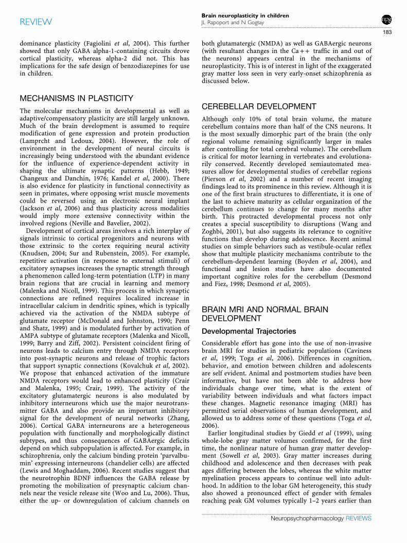
The availability of a large cohort of children studiedlongitudinally across childhood and adolescence enabledthe examination of regional cortical trajectories in relationto IQ. Recently, Shaw et al (2006a) examined the NIMHpediatric longitudinal data set for anatomic brain develop-ment in relation to IQ. Developmental trajectories for totaland regional brain cortical thickness were examined inrelation to IQ (measured at time of initial scan) for 307children (the majority of whom has prospective repeatedneuroanatomic scans) ages 4–26 years as shown in Figure 2.At younger ages, the relationship between cortical thicknessand IQ was relatively diffuse, but approached the morecircumscribed regional frontal lobe ‘hot spots’ in lateradolescence. This established the development of increas-ingly specific neural connectivity with age. The contrastin trajectories across IQ groups, however, was parti-cularly interesting. As speculated by Garlick (2002), moreintelligent children demonstrated a particularly plasticcortex with an initial accelerated and prolonged phase ofcortical increase followed by a particularly vigorous phaseof cortical thinning.
4.00
4.25
4.50
4.75
5.00
3.75
4.00
4.25
4.50
4.75
Age
4.00
4.25
4.50
4.75
5.00
4.00
4.25
4.50
4.75
5.00
Cor
tical
thic
knes
s (m
m)
Cor
tical
thic
knes
s (m
m)
Cor
tical
thic
knes
s (m
m)
Cor
tical
thic
knes
s (m
m)
T statistic
2.65
Trajectories at indicated cortical point(MNI coord. x=10, y=44, z=48)
Right superior/medial prefrontal gyral cluster
Left superior/medial prefrontal gyral cluster Left middle temporal gyral cluster
Superior intelligence (S)
High intelligence (H)
Average intellience (A)
S vsH, S vsA p<0.0001
H vsA p=0.56
S vsA p<0.001, S vsH p=0.0005
H vsA p=0.56
S vsH, S vsA p<0.0001
H vsA p=0.99
S vsA p=0.0005, S vsH p=0.003
H vsA p=0.14
Age7 11 13 15 17 199
Age7 11 13 15 17 199
Age7 11 13 15 17 1997 11 13 15 17 199
Figure 2 Trajectories of cortical change in children with superior (n¼ 91), high (n¼ 101), and average (n¼ 115) intelligence (total 629 scans). The brainmaps (center panel) show prominent clusters where the superior and average intelligence groups differ significantly in the trajectories of corticaldevelopment (t-statistic maps show areas of significant interaction between these IQ groups and the cubic age term). (a) Graph showing the trajectories atthe cortical point of maximum trajectory difference in the right superior frontal gyrus (point indicated in upper brain map). (b–d) Graphs showing thetrajectories of the mean thickness of all cortical points in the other clusters. The graph in (d) relates to the area indicated in the lower brain map. The age ofpeak cortical thickness is arrowed and significance values of differences in shapes of trajectories are given on the graphs. Adapted from Shaw et al (2006a).
Brain neuroplasticity in childrenJL Rapoport and N Gogtay
...............................................................................................................................................................
186
..............................................................................................................................................
Neuropsychopharmacology REVIEWS
REVIEW
The implications of these findings are for a general modelof neural plasticity underlying IQ. It would seem that the‘sensitive period’ is shifted to a somewhat later period for thehighest IQ population. This, somewhat counterintuitivefinding is not only compatible with both different geneticdetermination, but also allowing for somewhat more complexexperiential input during a period of great synaptic and otherchange (Knudsen, 2004). In addition, the steeper slope of bothincrease and decrease in cortical thickness for the highest IQgroup suggests that these individuals share a more dynamicpattern of plasticity. Future functional studies should pursueother temporal response patterns in very high IQ individuals.
CHILDHOOD-ONSET SCHIZOPHRENIA
It is now accepted that schizophrenia is associated withstructural brain abnormalities, with the most consistentfindings being enlarged lateral ventricles and reducedtemporal (medial and superior) and prefrontal GM volumes(Lawrie and Abukmeil, 1998). Early studies includinganimal models (Lipska et al, 1993) and cohort studiessuggested an early, fixed ‘lesion’ and thus suggesting the‘neurodevelopmental’ model for schizophrenia (Weinber-ger, 1987; Rapoport et al, 2005). This model, however, hascome under increasing scrutiny as more recent series oflongitudinal studies, both in adults and early-onset popula-tions have demonstrated progressive gray matter lossstarting with the earliest phase of the illness (Bridle et al,2002; Mathalon et al, 2003; Pantelis et al, 2005). Although itis still possible that an early fixed lesion could manifest lateron in life, the temporally dynamic pattern of the illnessalong with progression of structural brain abnormalitieswould also support a model of schizophrenia as aprogressive neurodevelopmental disorder (Woods, 1998).Similarly, many of the schizophrenia genes associated withthe illness appear to have functional roles throughout thelifespan and thus for a complex genetic disorder likeschizophrenia, it is possible to imagine an ongoing interplayof genetic, epistatic, and the environmental factors(Harrison and Weinberger, 2005; Rapoport et al, 2005).
Postmortem studies show no widespread neuronal loss inschizophrenia, or a glial response to a potential neuronalinjury. The cortical GM loss observed in schizophrenia hasbeen shown to be due to the loss of ‘neuropil’ which consistsof glia, synaptic, and dendritic arbors and vasculature(Selemon and Goldman-Rakic, 1999). These observationshave lead investigators to revisit Feinberg’s (1982) ‘ex-cessive pruning hypothesis’ where a (genetically orenvironmentally based) fault in programmed synapticelimination during adolescence could lead to neuropathol-ogy of schizophrenia (Feinberg, 1982). Neurophysiologicand functional neuroimaging studies also suggest abnormalsynaptic activity and disrupted functional connectivity inschizophrenia (Volkow et al, 1988; Weinberger et al, 1992;Friston, 1999, 2005), supporting a general model in whichstructural or functional synaptic abnormalities could be thecore deficit of schizophrenia and highlighting the role ofsynaptic plasticity in this disease (Frost et al, 2004; Friston,2005). A greater understanding of the cellular correlates ofthe gray matter change remains crucial for the advocates ofthe ‘excessive pruning hypothesis’.
Although the studies of COS below represent a rareopportunity to examine the interaction of the illness withearly brain development, there is abundant evidence ofmuch earlier prepsychotic brain developmental abnormal-ities than our age 8 onwards imaging data can reveal. Forexample, delayed or impaired development across a widesphere of behaviors and abilities is seen in the childhood ofadult patients with schizophrenia (Cannon et al, 2003; Makiet al, 2005; Ridler et al, 2006), and our very early-onset COSpatients also show clinical evidence of early abnormalitiesin brain development with more frequent language (43%)and learning problems (65%), years before onset ofpsychosis (Alaghband-Rad et al, 1995). Forty-three percentof the COS had an anxiety disorder at initial screening,perhaps indicating a phenotype with prominent GABAergicabnormalities (Hensch, 2004; Mohler et al, 2004).
PATTERN AND SPECIFICITY OF GMFINDINGS
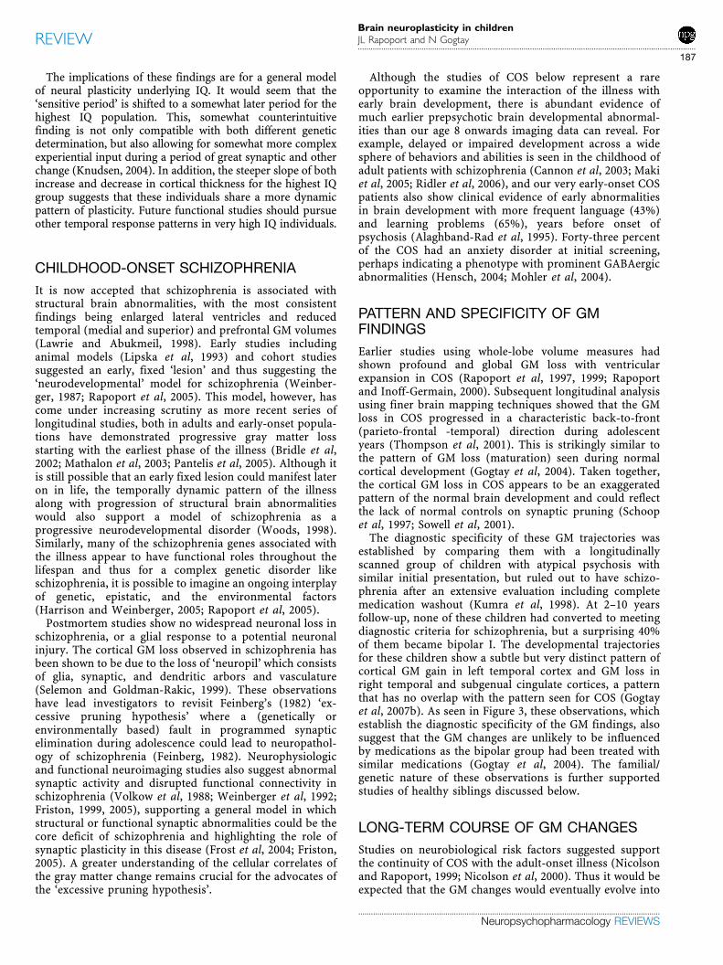
Earlier studies using whole-lobe volume measures hadshown profound and global GM loss with ventricularexpansion in COS (Rapoport et al, 1997, 1999; Rapoportand Inoff-Germain, 2000). Subsequent longitudinal analysisusing finer brain mapping techniques showed that the GMloss in COS progressed in a characteristic back-to-front(parieto-frontal -temporal) direction during adolescentyears (Thompson et al, 2001). This is strikingly similar tothe pattern of GM loss (maturation) seen during normalcortical development (Gogtay et al, 2004). Taken together,the cortical GM loss in COS appears to be an exaggeratedpattern of the normal brain development and could reflectthe lack of normal controls on synaptic pruning (Schoopet al, 1997; Sowell et al, 2001).
The diagnostic specificity of these GM trajectories wasestablished by comparing them with a longitudinallyscanned group of children with atypical psychosis withsimilar initial presentation, but ruled out to have schizo-phrenia after an extensive evaluation including completemedication washout (Kumra et al, 1998). At 2–10 yearsfollow-up, none of these children had converted to meetingdiagnostic criteria for schizophrenia, but a surprising 40%of them became bipolar I. The developmental trajectoriesfor these children show a subtle but very distinct pattern ofcortical GM gain in left temporal cortex and GM loss inright temporal and subgenual cingulate cortices, a patternthat has no overlap with the pattern seen for COS (Gogtayet al, 2007b). As seen in Figure 3, these observations, whichestablish the diagnostic specificity of the GM findings, alsosuggest that the GM changes are unlikely to be influencedby medications as the bipolar group had been treated withsimilar medications (Gogtay et al, 2004). The familial/genetic nature of these observations is further supportedstudies of healthy siblings discussed below.
LONG-TERM COURSE OF GM CHANGES
Studies on neurobiological risk factors suggested supportthe continuity of COS with the adult-onset illness (Nicolsonand Rapoport, 1999; Nicolson et al, 2000). Thus it would beexpected that the GM changes would eventually evolve into
Brain neuroplasticity in childrenJL Rapoport and N Gogtay...............................................................................................................................................................
187
..............................................................................................................................................
Neuropsychopharmacology REVIEWS
REVIEW

a pattern similar to that seen in the adult-onset illness.Adult-onset studies using VBM or cortical thicknessmapping methods, show predominant cortical GM loss inprefrontal and superior temporal cortices (Kuperberg et al,2003; White et al, 2003; Wiegand et al, 2004; Narr et al,2005). In a recent analyses on a group of 70 COS childrenfollowed from ages 8 to 28 years, the pattern of GM lossindeed became more circumscribed to the prefrontal andsuperior temporal cortices by age 26 years, furthersupporting the continuity with the adult-onset disorder(Greenstein et al, 2006).
GM LOSS OR WM ENCROACHMENT?
The brain MRI studies showing GM loss have raised thequestion whether the cortical GM loss is just a perceivedloss resulting from the encroachment by continued whitematter myelination, a process which normally is extendedthrough the 4th decade (Benes, 1993; Benes et al, 1994;Sowell et al, 1999). A recent analysis using whole-lobe WM
volumes on 70 COS children (ages 7–26 years) withprospective rescans (n¼ 162 scans) showed that the slopefor WM growth was significantly lower in COS childrencompared to matched healthy controls in all lobes(Po0.05), indicating that the GM reduction in COS is notsecondary to WM encroachment (Dr N Gogtay, unpublisheddata). We are currently applying the novel tensor-basedmapping technique to create dynamic maps of WM growthfor these subjects.
CLINICAL REMISSION AND CORTICALTHICKNESS
The cortical GM thickness of COS children who met thecriteria for clinical remission (n¼ 16) at the time of theirdischarge from the NIMH typically 3–4 months after theinitial scan (mean duration 104.8±25.4 days), was thickerin prefrontal, temporal as well as parietal cortices for theremitted children than for those COS children who did notmeet the remission criteria (n¼ 40) (Greenstein et al,
Age5 22
Age
Age 227
1612
OnsetPost onset Post onset
–0.070
–0.060
–0.050
–0.040
–0.030
–0.020
–0.010
0.000
0.010
Per
cent
cha
nge
COS(n=23) Psychosis NOS(n=19) Healthy controls (n=38).
Total GM
p=0.007
p=0.015
P values with one way ANOVA with Tukey post hoc test
Normal GM Development
Bipolar I GM Development
COS GM Development
COS vs Healthy P maps
Bipolar vs Healthy Ratio maps
2.22.01.81.61.41.21.00.80.60.4
Rat
io B
P/?
?
1.00.90.80.70.60.50.40.30.20.10.0
Gra
y m
atte
r am
ount
0.00002
0.00010.00050.0010.0050.01
0.05
P-V
alue
Temporal GMFrontal GM Parietal GM
Figure 3 Comparison and specificity of GM developmental patterns in healthy, COS, and Bipolar children. Top panel shows right lateral views of dynamicsequences of cortical GM maturation in 13 healthy children between ages 4 and 22 as was displayed in Figure 1. The GM maturation (blue color) proceeds inparietofrontal-temporal direction (Gogtay et al, 2004). The middle panel shows dynamic sequence of significant GM loss (p-maps) in 12 prospectivelyscanned COS children compared to matched healthy controls between ages 12 and 16. The pattern of GM loss in COS appears to be an exaggeration ofthe normal GM maturation (Thompson et al, 2001). The bottom panel shows dynamic sequence of comparison of the GM amount (ratio maps) betweennine psychotic bipolar I children and their 18 matched controls before and after onset of mania. There is no overlap of GM developmental pattern betweenCOS and bipolar GM development establishing the diagnostic specificity of the findings (Gogtay et al, 2007b). The graph shows three way comparison ofchange in GM amount over 2-year period in COS (red bars), psychosis NOS (blue bars), and healthy controls (yellow bars). Psychosis NOS children hadsimilar medications and treatment history as COS children at the time of initial scans. Thus GM changes seen COS are unlikely to be related to drugexposure (Gogtay et al, 2004).
Brain neuroplasticity in childrenJL Rapoport and N Gogtay
...............................................................................................................................................................
188
..............................................................................................................................................
Neuropsychopharmacology REVIEWS
REVIEW

submitted). Thicker cortex could reflect a general protectiveprocess seen in both probands and healthy COS siblings asdescribed below.
HEALTHY COS SIBLINGS
GM abnormalities in schizophrenia are at least in partfamilial/trait markers (Weinberger and McClure, 2002;Cannon et al, 2003; Gilbert et al, 2003; Yucel et al, 2003).Most studies of high-risk populations, typically closerelatives of schizophrenia patients, show smaller total andregional GM volumes (Cannon et al, 2002a, b; Job et al,2005; McIntosh et al, 2006), total white matter volumes(Narr et al, 2002; Hulshoff Pol et al, 2004), reduced GMvolumes of thalamus (Staal et al, 1998) and/or hippocam-pal-amygdalar complex (Lawrie et al, 2001; Cannon et al,2002; Keshavan et al, 2002; Seidman et al, 2002; Van Erpet al, 2002), although inconsistencies remain across thestudies (Boos et al, in Press; Marcelis et al, 2003; McDonaldet al, 2006).
Longitudinal brain MRI studies on healthy COS siblingsprovide further insights into the phenomenon of corticalGM changes. Because of the striking progression of GMdeficits in COS probands, their full siblings (age 8 to 28years) were also followed prospectively (Gogtay et al,2007a). For this analysis, siblings were chosen with minorpsychopathology and any ‘schizophrenia spectrum’ dis-order either on Axis I or Axis II was exclusionary. Long-itudinal GM development of 52 healthy COS full siblingsshowed these siblings to have initial GM loss which, for thisgroup, did not progress during adolescence (unlike their
COS probands), but in fact normalized by age 20. The earlyGM loss in siblings was most prominent in the prefrontaland temporal cortices (see Figure 4) as has been seen forCOS probands, but unlike that seen in probands, siblingsdid not show parietal GM loss in younger ages (Gogtay et al,2007a). The age-related amelioration of deficits did follow asomewhat similar pattern in both probands and siblings.This apparent ‘plastic’ response (inhibition of corticalthinning) in healthy siblings is intriguing and warrantsreplication. Within the healthy sibling group, regionalcortical thickness increased with increase in overall ‘lifefunctioning’ as measured by the Global Assessment Scale(GAS), which suggests a direct relationship between corticalthickness and restitutive normalization process with generalcompetence (GAS scores). Furthermore, mean corticalthickness (MCT) at initial scan positively correlated withthe most current GAS scores and was not significantlyrelated to the IQ. In other words, better social and cognitive‘competence’ was predicted by initial cortical thickness.Whether this relationship between GM thickness and GASreflects cause, or is secondary to both having a commonantecedent remains unknown.
GENETICS AND COS BRAIN DEVELOPMENT
Schizophrenia is a complex genetic disorder with manygenes of small effect thought to be etiologic for mostpatients and likely modified by other genetic, epigenetic,and environmental factors (Harrison and Weinberger,2005). Recent advances in neuroimaging may allow strongerand more biologically meaningful associations between
10
2.2
Age8 yrs 17 yrs 20 yrs
Prefrontal cortex ROIB Inf. Parietal cortex ROI
T s
tatis
tics
11 yrs 14 yrs
Temporal cortex ROI5.5
5.0
4.5
4.0
3.5
Ver
tex
Agescan10 20 30
5.5
5.0
4.5
4.0
3.5
3.0
Ver
tex
Agescan10 20 30
Agescan10 20 30
5.0
4.5
4.0
3.5
3.0
Ver
tex
Figure 4 Early Cortical GM loss in healthy COS siblings normalizes by age 20. Cortical GM thickness in healthy COS siblings (n¼ 52; 110 scans) comparedto age, sex, and scan interval matched healthy controls (n¼ 52; 108 scans) between ages 8 and 28. Healthy COS siblings show significant GM deficits leftprefrontal and bilateral temporal cortices and smaller deficits in right prefrontal and inferior parietal cortices. These deficits in healthy siblings normalize withage with no abnormalities remaining by age 20. Side bar shows t-statistic with threshold to control for multiple comparisons using the false discovery rate(FDR) procedure with q¼ 0.05; significant at t¼ 2.2. The graphs (a–c) show trajectories of GM development in healthy COS siblings (Blue) and those formatched healthy controls (red) in select regions of interest. Adapted from Gogtay et al (2007a).
Brain neuroplasticity in childrenJL Rapoport and N Gogtay...............................................................................................................................................................
189
..............................................................................................................................................
Neuropsychopharmacology REVIEWS
REVIEW

potential risk alleles and intermediate phenotypes such asmorphological brain abnormalities (Meyer-Lindenberg andWeinberger, 2006). The assumption is that the effect size ofrisk allele association will be greater for a brain imagingmeasure (or other intermediate phenotype) than that withthe diagnosis (Flint and Munafo, 2007). Many of thesusceptibility genes reported for schizophrenia appear tohave an impact on the synaptic molecular biology (Harrisonand Owen, 2003; Harrison and Weinberger, 2005), thusassociations between specific risk alleles and brain mor-phological changes could provide further insights intogenetic influences on synaptic plasticity. For example, ourrecent analyses using cortical thickness measures showthat both COS probands and healthy siblings with GAD riskallele, a modulator enzyme that converts glutamate toGABA which has an inhibitory influence on the downstreamprefrontal pyramidal neurons (Coyle, 2004), have steeperslopes of cortical GM loss in prefrontal cortex. Thus, GADrisk status may influence synaptic/dendritic plasticity inprefrontal cortex (Dr N Gogtay unpublished data); possiblythrough various mechanisms such as, glutamatergic over-drive resulting in excitotoxicity due to reduced GABAinhibition, reduced synaptic protein synthesis due tosimultaneous reduction of reelin expression (Fatemi et al,2000), or calcium channel abnormalities in GABA inter-neuron described earlier. Although it is difficult to calculatethe effect size for the brain morphological findings obtainedusing mixed model regression analyses, it is hoped thatthese observations will enhance the understanding of geneeffects on brain morphology. It would be worth exploringthe association of other risk genes involved either inglutamate/NMDA (eg, dysbindin, mGlu3) or GABA circui-tries (eg, Reelin, Parvalbumin) (Fatemi et al, 2005; Gisabellaet al, 2005; Stephan et al, 2006) and their relationship todevelopmental trajectories.
ADHD
ADHD is a common childhood disorder characterized bydevelopmentally inappropriate levels of inattention, im-pulsivity, and motor restlessness. Although these are usuallyseen as the core deficits, other factors such as responsepreparation and control and reward processes are alsoaltered. Approximately half of most ADHD populationshave a benign outcome, although controversy remains as torate of complete remission (Klein, 1987).
Brain anatomic studies of ADHD have confirmed long-standing indirect evidence that suggested subtle abnormal-ities of subcortical and regional cortical volumes inregions related to executive functions. Most prominentlycortical thinning is seen in medial and superior prefrontaland precentral regions (Casey and Durston, 2006; Mackieet al, 2007; Shaw et al, 2006b). These findings of corticalthinning were also recently replicated in a study of 24Adult ADHD subjects who had continuous disorder intoadult years compared to 18 healthy controls (Makris et al,2007). Unfortunately, this study did not include adultswhose childhood ADHD had remitted, which may haveprovided replication of the Shaw et al provocativeoutcome finding.
STIMULANT DRUGS AND BRAINDEVELOPMENT
In understanding the brain abnormalities of ADHD, thestudies of the effects of stimulant drug exposure cited aboveon rat brain development might suggest that any brainabnormalities found for ADHD might be stimulant induced.This does not appear to be the case. Castellanos et al (2002)found no significant effects of stimulant drug for a largecohort of ADHD children and in Shaw’s study the good andpoor outcome subjects had virtually equivalent stimulantdrug exposure. As observed by Hyman (Hyman, 2003), it isnot only the pharmacologic agents that regulate geneexpression in neurons and alter neural circuits; but alsomental illness and experience do so as well and preclinicalstudies so far have not proved useful for informing humanstudies.
BRAIN IMAGING AND PLASTICITY IN ADHD
A unique recent study examined data from prospectiveanatomic brain images using a measure of total and regionalcortical thickness for 163 children with ADHD, 97 of whohad two or more images along with clinical evaluations ateach scan point (Lerch et al, 2006; Shaw et al, 2006b). Theseprospective data enabled regional thickness trajectories tobe examined, using mixed model regression analyses, inrelation to both diagnosis and (within diagnostic group) toclinical outcome. Trajectories of cortical thickness differedsignificantly for the ADHD group in the right parietalcortex, an area known be important for attentional control.In the ADHD group, the age-related thinning in this regionappeared to be slowed resulting in a relatively sparedregional thickness (See Figure 5).
A second analysis addressed the question of whetherregional cortical development differed between good andpure outcome ADHD groups (defined as both a continuousand dichotomous measure). The ‘normalization’ of rightposterior parietal cortical thickness was driven by the betteroutcome children and this finding could not be ascribed tomedication exposure which was virtually identical for goodand poor outcome groups. Further analyses showed that thegood outcome cases exhibited compensatory right parietal‘slowing’ of the normal decline, most plausibly activitydriven. This interpretation is also supported by functionalstudies indicating that ADHD children have excess activa-tion in this posterior parietal region (Rubia et al, 2000;Durston et al, 2003). Moreover, activation of the rightparietal cortex during tasks of alerting and reorienting ofattention is not fully mature until adulthood, and thiscomponent of the attentional network may develop duringadolescence (Konrad et al, 2005), and there is also evidenceof training-induced plasticity for activation at this site(Olesen et al, 2004).
Recent studies show a uniquely delayed pattern of corticaldevelopment for ADHD children with good outcome.Utilizing a program which maps the age of maximal corticalthickness across childhood and adolescence, quadraticcurves of cortical development were established using alarge number of subjects with multiple scans and mixedmodel regression analyses. A back to front ‘wave’ of cortical
Brain neuroplasticity in childrenJL Rapoport and N Gogtay
...............................................................................................................................................................
190
..............................................................................................................................................
Neuropsychopharmacology REVIEWS
REVIEW

development is seen for both populations, but the GM peaksin ADHD subjects were about 3 years later than in thehealthy controls (Dr P Shaw, submitted).
The cerebellum has been of increasing interest in thepathogenesis of ADHD. Dopaminergic innervation appearsearly in cerebellar development and there is widespreadimmunoreactivity for various dopamine receptors on thedendritic arbors of the Purkinje as well as granule cells (Yewet al, 1995; Khan et al, 1998, 2000; Ishibashi et al, 2002).Structural abnormalities are one of the most consistentlyreported features with reductions in overall volumeparticularly in the cerebellar vermis which has the highestconcentration of dopaminergic innervation (Melchitzkyand Lewis, 2000; Hurley et al, 2003). Although traditionallyconsidered a site of motor control; lesion studies andfunctional imaging studies involving the cerebellum havedemonstrated deficits in a wide range of cognitive andaffective functioning (Schmahmann, 2004). A longitudinalcase–control study of ADHD patients and matched controlsincluded cerebellar measures for six lobes of the cerebellarhemispheres, and three vermal subdivisions both acquiredat baseline and two or more follow-up time points at 2 yearsintervals. The results showed a loss of superior cerebellarvermis volume in the ADHD group which persisted at 5-year follow-up regardless of clinical outcome. However, inthe cerebellar hemispheres, ADHD subjects with a worseclinical outcome displayed a downward trajectory in theinferior posterior lobes which became progressively smallduring adolescence relative to both healthy controls andADHD subjects with a better outcome (Mackie et al, 2007).
Thus, the cerebellar hemispheres, like the right parietalregion may show a plastic, state-specific response andpossibly be targets for clinical intervention in ADHD. Giventhe relatively higher rate of environmental effects on thesecerebellar volumes discussed above (Wallace et al, 2006), ifthese findings are replicated, compensatory training shouldbe considered, although to date such approaches have notbeen encouraging (Klingberg et al, 2005). Alternately,genetic causes of subtle progressive decline in cerebellarvolumes should be explored. Finally, the specificity of thesefindings needs to be documented. To date, we know thatthese patterns do not resemble those for age matchedpsychotic adolescents; differentiation from other neurode-
velopmental disorders including specific learning disabil-ities remains to be demonstrated.
RISK GENES AND ADHD BRAINDEVELOPMENT
ADHD is one of the most heritable neuropsychiatricdisorders (Levy et al, 1997; Faraone, 2000). Limited twin-brain imaging data suggest that discordant MZ twins oftenrepresent lesion based phenocopies in the ill twin (Levyet al, 1997; Castellanos et al, 2003). Several candidate riskgenes have been replicated for ADHD among them, the7-repeat polymorphism within the D4 receptor gene. In spiteof small diagnostic effect size, there have been some reportsof risk gene association with brain volume. Using thelongitudinal cohort study, Shaw et al found that the DRD47-repeat allele was associated with a thinner prefrontal andposterior parietal cortex overlapping with the generallythinner regions in ADHD subjects studied prospectively asdescribed above. However, the 7-repeat group of subjectsshowed greater normalization of the right parietal corticalregion, a pattern previously linked with better clinicaloutcome (Shaw et al, 2007). Thus, the neuroanatomiccorrelates of the DRD4 7-repeat were limited to subjects atyounger ages (there were no significant effects of DRD1 orDAT1 polymorphisms on clinical outcome or corticaldevelopment) (Shaw et al, 2007). This finding remainscontroversial as some find good outcome with this gene,whereas others do not (Barkley et al, 2006). Possession ofthe 7 repeat allele had the same effect on cortical thicknessin ADHD and controls (Shaw et al, 2007). There is someevidence for the specificity of the gene effect as Durston andher group have also shown a different effect (on the caudatevolume) for DAT1, a gene expressed in the basal gangliarather than in the cortex (Durston et al, 2005). The findingof gene effect on brain volume or trajectory is impressivefor several reasons. ADHD is a complex genetic disorderwith many risk genes involved each of relatively small effect;a recent meta-analysis shows the pooled Odds Ratio for theD4VNTR 7-repeat to be 1.4 (in family based) and 1.24 (incase–control) studies, and 1.13 (CC) for DAT1 (Faraoneet al, 2005). The similar relationship of the D4 risk allele to
t=-4
t=-2.6
3.00
3.25
3.50
3.75
4.00
Age
Cor
tical
thic
knes
s (m
m) Better outcome
Healthy
Worse outcome
Difference in shapes:Better outcome v NV p=0.001,better v poorer outcome p=0.03;poorer outcome v NV p=0.6 7 11 13 15 179
Figure 5 Normalization of Right parietal cortex and better outcome in ADHD. The t map indicates vertices where there was a significant interaction inthe regional trajectory slopes between the better outcome and healthy control groups. The graph illustrates group trajectories in this region (difference ingradients: better outcome group vs controls, P¼ 0.001; better vs worse outcome groups, P¼ 0.03; and worse outcome group vs controls, P¼ 0.60).Adapted from Shaw et al (2006b).
Brain neuroplasticity in childrenJL Rapoport and N Gogtay...............................................................................................................................................................
191
..............................................................................................................................................
Neuropsychopharmacology REVIEWS
REVIEW

cortical thickness in both healthy and ADHD brainsencourages the use of regional volumes and trajectories asendophenotypes, and is consistent with the dimensionalnature of ADHD.
We can only speculate on the mechanisms through whichthe DRD4 allele may exert structural effects. Corticalchanges were concentrated partly in the frontal regionwhich have the highest levels of dopamine D4 receptors(Meador-Woodruff et al, 1996). Variants of the receptorhave subtle differences in pharmacological binding proper-ties and the 7-repeat allele inhibits cyclic adenosinemonophosphate less efficiently, which may denote sensitiv-ity to endogenous dopamine. Studies with antipsychotics(which are D2 receptor family antagonists) may havetrophic effects on the cortex through increasing metabolismand ultrastructural change in the dendritic tree and synapsemorphology (Konradi and Heckers, 2001; Miller et al, 2001).It is thus possible that changes in D4 receptor levels andactivity associated with the 7-repeat allele could havetrophic effects in part through altering endogenousdopamine levels.
These findings of outcome-related brain developmentaltrajectories are paralleled by studies on Tourette’s syn-drome, a disorder, closely related to ADHD; approximately80% of Tourette’s patients greatly improve or remit by theend of adolescence. In recent years, Peterson and co-workers have carried out a series of imaging studies onadolescent and adult patients with Tourette’s syndrome.These cross-sectional studies show that both adult and childpatient have decreased caudate volume, which mayrepresent a trait marker for this disorder. Functionalstudies had indicated prefrontal, parietal, and temporalactivation during tic suppression for this group. It isintriguing therefore, that prefrontal cortical volumes wereuniquely low in the non-remitted adults with TS incomparison with the wide range of volumes seen in thechildhood sampleFa group ultimately expected to have alarge remission rate. The implication from this cross-sectional data is that prefrontal hypertrophy seen in theyounger subjects mediates their ultimate remission reflect-ing a plastic compensatory process. A longitudinal studyand one that includes remitted adults is waiting to beundertaken. It is anticipated that remitted subjected willshow a greater cortical plastic response.
DISCUSSION AND FUTURE DIRECTIONS
The availability of normative human brain developmentaldata has provided new developmental and clinical insights.Longitudinal data for human studies counter balance thelarge individual differences which can obscure the devel-opmental changes. These have also shown the importance oftiming of trajectory to the level of functioning withinhealthy individuals (as shown in the Shaw et at IQ study)and the exaggerated cortical loss in psychotic patients(Thompson et al, 2001). Clinical outcome is also related tothe mean gray matter thickness in the childhood schizo-phrenia cohort illustrating the relevance of such measuresto core features of the illness (Greenstein et al, submitted).
Twin studies indicate high heritability with respect todevelopmental trajectories (Lenroot et al, submitted), and
change in regional heritability which may vary acrossdevelopmental period. The probable plasticity and latematuration of the human cerebellar lobes should beexplored in relation to clinical and cognitive status for avariety of disorders following on the data from the ADHDstudies, indicating possible plastic brain response in thecerebellar lobes and posterior parietal cortex. It is possiblethat some children with developmental lag have slowercortical development and longitudinal studies shouldcompare timing of peak volumes in relation to the clinicalamelioration.
A major limitation to date has been the lack of parallelinformation about the cellular changes underlying thedevelopmental anatomic MRI changes discussed through-out this review. The observed changes are almost certainlyglial and vascular in nature rather than purely neuronal.This could (and should) be addressed by a paralleldevelopmental primate study in which young monkeys arescanned at regular intervals as appropriate with sacrificeand postmortem brain cellular characteristics obtained.
The bridging between the anatomic measures of com-pensatory plasticity and regional molecular events remainsunknown. The degree to which new utilization of unspeci-fied (labile) synapses and competition for synaptic sitesaccounts for these changes is unknown. Some hints howevermay be found in the seminal studies of Hensch (2004),demonstrating the ability to control the timing, duration,and closure of critical periods of brain development inanimal models such as by modulating receptors pharma-cologically.
The probable heterogeneity and lack of major geneinfluence in these disorders severely limits understanding todate of signaling pathways underlying these changes. Incontrast, for a few developmental cognitive disorders forwhich major genes have been identified, there have beenrapid advances in the identification and characterization ofcellular, molecular, and genetic mechanisms important inneuroplasticity and the brain abnormalities of these condi-tions. Recent work on Neurofibromatosis, Fragile X, Rettsyndrome, informally classified as disorders of brainplasticity, and other genetic syndromes with impairedcognitive phenotypes shows how one may move from geneto signaling pathways to putative treatments (Johnston, 2006).Johnston and others provide schema of signaling pathways asthey affect transcription and lead to unique treatment trials.For example, based on the anticholesterol drug Lovastatin’sability to inhibit brain MAP kinase, this drug was successfullyused to reverse learning deficits in a mouse model ofneurofibromatosis (Li et al, 2005). New information onmolecular contributions to patterning plasticity of braindevelopment is overwhelming (Sur and Rubenstein, 2005;Kriegstein and Parnavelas, 2006), but the combining of thesemouse/cellular data with human studies remains critical. Thisis being tested in the relatively rare neurodevelopmentaldisorders for which one or more major genes have beenidentified. This review has also shown the surprising successachieved so far in linking behavioral changes with brainanatomic change. The next need will be to link the grossanatomic developmental trajectories to their underlyingcellular/molecular processes. In parallel with clinical studiesoutlined here, it will be crucial to carry out investigations ofmolecular changes in synapses during normal development to
Brain neuroplasticity in childrenJL Rapoport and N Gogtay
...............................................................................................................................................................
192
..............................................................................................................................................
Neuropsychopharmacology REVIEWS
REVIEW

gain insight into the mechanism governing structural changesunderlying functional plasticity.
DISCLOSURE OF CONFLICT OF INTEREST
Both authors declare that, except for income received fromtheir primary employer, no financial support or compensa-tion has been received from any individual or corporateentity over the past three years for research or professionalservices and there are no personal financial holdings thatcould be perceived as constituting a potential conflict ofinterest.
REFERENCES
Alaghband-Rad J, McKenna K, Gordon CT, Albus KE, HamburgerSD, Rumsey JM et al (1995). Childhood-onset schizophrenia: theseverity of premorbid course. J Am Acad Child AdolescPsychiatry 34: 1273–1283.
Andersen SL, Navalta CP (2004). Altering the course of neurode-velopment: a framework for understanding the enduring effectsof psychotropic drugs. Int J Dev Neurosci 22: 423–440.
Baare WF, Hulshoff Pol HE, Boomsma DI, Posthuma D, de GeusEJ, Schnack HG et al (2001). Quantitative genetic modeling ofvariation in human brain morphology. Cereb Cortex 11: 816–824.
Barkley RA, Smith KM, Fischer M, Navia B (2006). An examinationof the behavioral and neuropsychological correlates of threeADHD candidate gene polymorphisms (DRD4 7+, DBH TaqI A2,and DAT1 40 bp VNTR) in hyperactive and normal childrenfollowed to adulthood. Am J Med Genet B Neuropsychiatr Genet141: 487–498.
Barry MF, Ziff EB (2002). Receptor trafficking and the plasticity ofexcitatory synapses. Curr Opin Neurobiol 12: 279–286.
Bates E (1993). Comprehension and production in early languagedevelopment (see comment). Monogr Soc Res Child Dev 58:222–242; discussion 243–252.
Benes FM (1993). The relationship between structural brainimaging and histopathologic findings in schizophrenia research.Harv Rev Psychiatry 1: 100–109.
Benes FM, Turtle M, Khan Y, Farol P (1994). Myelination of a keyrelay zone in the hippocampal formation occurs in the humanbrain during childhood, adolescence, and adulthood. Arch GenPsychiatry 51: 477–484.
Biederman J, Wilens T, Mick E, Spencer T, Faraone SV (1999).Pharmacotherapy of attention-deficit/hyperactivity disorderreduces risk for substance use disorder. Pediatrics 104: e20.
Bolanos CA, Barrot M, Berton O, Wallace-Black D, Nestler EJ(2003). Methylphenidate treatment during pre- and periadoles-cence alters behavioral responses to emotional stimuli atadulthood. Biol Psychiatry 54: 1317–1329.
Bookheimer S (2002). Functional MRI of language: new ap-proaches to understanding the cortical organization of semanticprocessing. Ann Rev Neurosci 25: 151–188.
Boos BM, Aleman A, Cahn W, Hulshoff Pol H, Kahn RS (in Press).Brain volumes in relatives of patients with schizophrenia: ameta-analysis. Arch Gen Psychiatry 64: 297–304.
Bourgeois JP, Goldman-Rakic PS, Rakic P (1994). Synaptogenesis inthe prefrontal cortex of rhesus monkeys. Cereb Cortex 4: 78–96.
Bourgeois JP, Rakic P (1993). Changes of synaptic density in theprimary visual cortex of the macaque monkey from fetal to adultstage. J Neurosci 13: 2801–2820.
Boyden ES, Katoh A, Raymond JL (2004). Cerebellum-dependentlearning: the role of multiple plasticity mechanisms. Annu RevNeurosci 27: 581–609.
Bridle N, Pantelis C, Wood SJ, Coppola R, Velakoulis D,McStephen M et al (2002). Thalamic and caudate volumes inmonozygotic twins discordant for schizophrenia. Aust N Z JPsychiatry 36: 347–354.
Bunge SA, Wright SB (2007). Neurodevelopmental changes inworking memory and cognitive control. Curr Opin Neurobiol 17:243–250.
Cannon TD, Thompson PM, van Erp TG, Toga AW, Poutanen VP,Huttunen M et al (2002a). Cortex mapping reveals regionallyspecific patterns of genetic and disease-specific gray-matterdeficits in twins discordant for schizophrenia. Proc Natl Acad SciUSA 99: 3228–3233.
Cannon TD, van Erp TG, Bearden CE, Loewy R, Thompson P, TogaAW et al (2003). Early and late neurodevelopmental influencesin the prodrome to schizophrenia: contributions of genes,environment, and their interactions. Schizophr Bull 29: 653–669.
Cannon TD, van Erp TG, Rosso IM, Huttunen M, Lonnqvist J,Pirkola T et al (2002b). Fetal hypoxia and structural brainabnormalities in schizophrenic patients, their siblings, andcontrols. Arch Gen Psychiatry 59: 35–41.
Carlezon Jr WA, Mague SD, Andersen SL (2003). Enduringbehavioral effects of early exposure to methylphenidate in rats.Biol Psychiatry 54: 1330–1337.
Cascio CJ, Gerig G, Piven J (2007). Diffusion tensor imaging:application to the study of the developing brain. J Am AcadChild Adolesc Psychiatry 46: 213–223.
Casey BJ, Durston S (2006). From behavior to cognition to thebrain and back: what have we learned from functional imagingstudies of attention deficit hyperactivity disorder? Am JPsychiatry 163: 957–960.
Casey BJ, Giedd JN, Thomas KM (2000). Structural and functionalbrain development and its relation to cognitive development.Biol Psychol 54: 241–257.
Castellanos FX, Lee PP, Sharp W, Jeffries NO, Greenstein DK,Clasen LS et al (2002). Developmental trajectories of brainvolume abnormalities in children and adolescents with atten-tion-deficit/hyperactivity disorder. JAMA 288: 1740–1748.
Castellanos FX, Sharp WS, Gottesman RF, Greenstein DK, GieddJN, Rapoport JL (2003). Anatomic brain abnormalities inmonozygotic twins discordant for attention deficit hyperactivitydisorder. Am J Psychiatry 160: 1693–1696.
Caviness VSJ, Lange NT, Makris N, Herbert MR, Kennedy DN(1999). MRI-based brain volumetrics: emergence of a develop-mental brain science. Brain Dev 21: 289–295.
Caviness Jr VS, Takahashi T (1995). Proliferative events in thecerebral ventricular zone. Brain Dev 17: 159–163.
Changeux JP, Danchin A (1976). Selective stabilization ofdeveloping synapses as a mechanism for the specification ofneuronal networks. Nature 264: 705–712.
Coyle JT (2004). The GABA-glutamate connection in schizophre-nia: which is the proximate cause? Biochem Pharmacol 68:1507–1514.
Crair MC (1999). Neuronal activity during development: permis-sive or instructive? Curr Opin Neurobiol 9: 88–93.
Crair MC, Malenka RC (1995). A critical period for long-termpotentiation at thalamocortical synapses. Nature 375: 325–328.
Crone EA, Donohue SE, Honomichl R, Wendelken C, Bunge SA(2006). Brain regions mediating flexible rule use duringdevelopment. J Neurosci 26: 11239–11247.
Derbyshire SW, Vogt BA, Jones AK (1998). Pain and Stroopinterference tasks activate separate processing modules inanterior cingulate cortex. Exp Brain Res 118: 52–60.
Desmond JE, Chen SH, Shieh PB (2005). Cerebellar transcranialmagnetic stimulation impairs verbal working memory. AnnNeurol 58: 553–560.
Desmond J, Fiez JA (1998). Neuroimaging studies of thecerebellum: language, learning and memory. Trends Cogn Sci2: 355–363.
Brain neuroplasticity in childrenJL Rapoport and N Gogtay...............................................................................................................................................................
193
..............................................................................................................................................
Neuropsychopharmacology REVIEWS
REVIEW

Dong WK, Greenough WT (2004). Plasticity of nonneuronal braintissue: roles in developmental disorders. Ment Retard DevDisabil Res Rev 10: 85–90.
Draganski B, Gaser C, Busch V, Schuierer G, Bogdahn U, May A(2004). Neuroplasticity: changes in grey matter induced bytraining. Nature 427: 311–312.
Durston S, Fossella JA, Casey BJ, Hulshoff Pol HE, Galvan A,Schnack HG et al (2005). Differential effects of DRD4 and DAT1genotype on fronto-striatal gray matter volumes in a sample ofsubjects with attention deficit hyperactivity disorder, theirunaffected siblings, and controls. Mol Psychiatry 10: 678–685.
Durston S, Tottenham NT, Thomas KM, Davidson MC, Eigsti IM,Yang Y et al (2003). Differential patterns of striatal activation inyoung children with and without ADHD. Biol Psychiatry 53: 871–878.
Elbert T, Pantev C, Wienbruch C, Rockstroh B, Taub E (1995).Increased cortical representation of the fingers of the left hand instring players. Science 270: 305–307.
Fagiolini M, Fritschy JM, Low K, Mohler H, Rudolph U, Hensch TK(2004). Specific GABAA circuits for visual cortical plasticity.Science 303: 1681–1683.
Fagiolini M, Hensch TK (2000). Inhibitory threshold for critical-period activation in primary visual cortex. Nature 404: 183–186.
Faraone SV (2000). Genetics of childhood disorders: XX. ADHD,Part 4: is ADHD genetically heterogeneous? J Am Acad ChildAdolesc Psychiatry 39: 1455–1457.
Faraone SV, Perlis RH, Doyle AE, Smoller JW, Goralnick JJ,Holmgren MA et al (2005). Molecular Genetics of Attention-Deficit/Hyperactivity Disorder. Biol Psychiatry 57: 1313–1323.
Fatemi SH, Earle JA, McMenomy T (2000). Reduction in Reelinimmunoreactivity in hippocampus of subjects with schizophre-nia, bipolar disorder and major depression. Mol Psychiatry 5:571, 654–663.
Fatemi SH, Stary JM, Earle JA, Araghi-Niknam M, Eagan E (2005).GABAergic dysfunction in schizophrenia and mood disorders asreflected by decreased levels of glutamic acid decarboxylase 65and 67 kDa and Reelin proteins in cerebellum. Schizophr Res 72:109–122.
Feinberg I (1982). Schizophrenia: caused by a fault in programmedsynaptic elimination during adolescence? J Psychiatr Res 17:319–334.
Feinberg I, Higgins LM, Khaw WY, Campbell IG (2006). Theadolescent decline of NREM delta, an indicator of brainmaturation, is linked to age and sex but not to pubertal stage.Am J Physiol Regul Integr Comp Physiol 291: R1724–R1729.
Flint J, Munafo MR (2007). The endophenotype concept inpsychiatric genetics. Psychol Med 37: 163–180.
Floeter MK, Greenough WT (1979). Cerebellar plasticity: mod-ification of Purkinje cell structure by differential rearing inmonkeys. Science 206: 227–229.
Friston K (2005). Disconnection and cognitive dysmetria inschizophrenia. Am J Psychiatry 162: 429–432.
Friston KJ (1999). Schizophrenia and the disconnection hypoth-esis. Acta Psychiatr Scand 395(Suppl): 68–79.
Frost DO, Tamminga CA, Medoff DR, Caviness V, Innocenti G,Carpenter WT (2004). Neuroplasticity and schizophrenia. BiolPsychiatry 56: 540–543.
Gaab N, Tallal P, Kim H, Lakshminarayanan K, Archie JJ, GloverGH et al (2005). Neural correlates of rapid spectrotemporalprocessing in musicians and nonmusicians. Ann New York AcadSci 1060: 82–88.
Garlick D (2002). Understanding the nature of the general factor ofintelligence: the role of individual differences in neural plasticityas an explanatory mechanism. Psychol Rev 109: 116–136.
Giedd JN, Blumenthal J, Jeffries NO, Castellanos FX, Liu H,Zijdenbos A et al (1999). Brain development during childhoodand adolescence: a longitudinal MRI study. Nat Neurosci 2:861–863.
Gilbert AR, Montros DM, Sahni SD, Diwadkar VA, Keshavan MS(2003). Obstetric complications correlate with neurobehavioraland brain structural alterations in young relatives at risk forschizophrenia. Ann NY Acad Sci 1008: 269–275.
Gisabella B, Bolshakov VY, Benes FM (2005). Regulation ofsynaptic plasticity in a schizophrenia model. Proc Natl Acad SciUSA 102: 13301–13306.
Gogtay N, Giedd JN, Lusk L, Hayashi KM, Greenstein D, VaituzisAC et al (2004). Dynamic mapping of human cortical develop-ment during childhood through early adulthood. Proc Natl AcadSci USA 101: 8174–8179.
Gogtay N, Greenstein D, Lenane M, Clasen L, Sharp W, Gochman Pet al (2007a). Cortical Brain Development in Non-PsychoticSiblings of Patients with Childhood Onset Schizophrenia. ArchGen Psychiatry 64: 772–780.
Gogtay N, Ordonez A, Herman D, Hayashi KM, Greenstein D,Vaituzis AC et al (2007b). Dynamic Mapping of CorticalDevelopment Before and After the Onset of Pediatric BipolarIllness. J Child Psycol Psychiatry 48: 852–862.
Gogtay N, Sporn A, Clasen LS, Nugent III TF, Greenstein D,Nicolson R et al (2004). Comparison of progressive cortical graymatter loss in childhood-onset schizophrenia with that inchildhood-onset atypical psychoses. Arch Gen Psychiatry 61:17–22.
Greenstein D, Lerch J, Shaw P, Clasen L, Giedd J, Gochman Pet al (2006). Childhood onset schizophrenia: cortical brainabnormalities as young adults. J Child Psychol Psychiatry 47:1003–1012.
Haier RJ, Jung RE, Yeo RA, Head K, Alkire MT (2004). Structuralbrain variation and general intelligence. NeuroImage 23:425–433.
Harrison PJ, Owen MJ (2003). Genes for schizophrenia? Recentfindings and their pathophysiological implications. Lancet 361:417–419.
Harrison PJ, Weinberger DR (2005). Schizophrenia genes, geneexpression, and neuropathology: on the matter of theirconvergence. Mol Psychiatry 10: 40–68; image 5.
Hebb DO (1949). The Organization of Behavior: A Neuropsycho-logical Theory. Wiley: New York.
Hensch TK (2004). Critical period regulation. Annu Rev Neurosci27: 549–579.
Hulshoff Pol HE, Brans RG, van Haren NE, Schnack HG, LangenM, Baare WF et al (2004). Gray and white matter volumeabnormalities in monozygotic and same-gender dizygotic twinsdiscordant for schizophrenia. Biol Psychiatry 55: 126–130.
Hurley MJ, Mash DC, Jenner P (2003). Markers for dopaminergicneurotransmission in the cerebellum in normal individuals andpatients with Parkinson’s disease examined by RT-PCR. Eur JNeurosci 18: 2668–2672.
Huttenlocher J, Duffy S, Levine S (2002). Infants and toddlersdiscriminate amount: are they measuring? Psychol Sci 13:244–249.
Huttenlocher PR (1979). Synaptic density in human frontalcortexFdevelopmental changes and effects of aging. Brain Res163: 195–205.
Huttenlocher PR, Dabholkar AS (1997). Regional differences insynaptogenesis in human cerebral cortex. J Comp Neurol 387:167–178.
Huttenlocher PR, de Courten C (1987). The development ofsynapses in striate cortex of man. Hum Neurobiol 6: 1–9.
Hyman SE (2003). Methylphenidate-induced plasticity: whatshould we be looking for? Biol Psychiatry 54: 1310–1311.
Isaacs E, Christie D, Vargha-Khadem F, Mishkin M (1996). Effectsof hemispheric side of injury, age at injury, and presence ofseizure disorder on functional ear and hand asymmetries inhemiplegic children. Neuropsychologia 34: 127–137.
Ishibashi T, Wakabayashi J, Ohno Y (2002). 7-Hydroxy-N,N0-di-n-propyl-2-aminotetraline, a preferential dopamine D3 agonist,
Brain neuroplasticity in childrenJL Rapoport and N Gogtay
...............................................................................................................................................................
194
..............................................................................................................................................
Neuropsychopharmacology REVIEWS
REVIEW

induces c-fos mRNA expression in the rat cerebellum. Jpn JPharmacol 89: 309–315.
Jackson A, Mavoori J, Fetz EE (2006). Long-term motor cortexplasticity induced by an electronic neural implant. Nature 444:56–60.
Job DE, Whalley HC, Johnstone EC, Lawrie SM (2005). Grey matterchanges over time in high risk subjects developing schizo-phrenia. NeuroImage 25: 1023–1030.
Johnston MV (2006). Fresh ideas for treating developmentalcognitive disorders. Curr Opin Neurol 19: 115–118.
Kandel ER, Schwartz JH, Jessell TM (2000). Principles ofNeuroscience. Elsevier: New York.
Keshavan MS, Dick E, Mankowski I, Harenski K, Montrose DM,Diwadkar V et al (2002). Decreased left amygdala andhippocampal volumes in young offspring at risk for schizo-phrenia. Schizophr Res 58: 173–183.
Khan ZU, Gutierrez A, Martin R, Penafiel A, Rivera A, De La CalleA (1998). Differential regional and cellular distribution ofdopamine D2-like receptors: an immunocytochemical study ofsubtype-specific antibodies in rat and human brain. J CompNeurol 402: 353–371.
Khan ZU, Gutierrez A, Martin R, Penafiel A, Rivera A, de la Calle A(2000). Dopamine D5 receptors of rat and human brain.Neuroscience 100: 689–699.
Klein RG (1987). Prognosis of attention deficit disorder and itsmanagement of adolescence. Pediatr Rev 8: 216–222.
Klingberg T, Fernell E, Olesen PJ, Johnson M, Gustafsson P,Dahlstrom K et al (2005). Computerized training of workingmemory in children with ADHDFa randomized, controlledtrial. J Am Acad Child Adolesc Psychiatry 44: 177–186.
Knudsen EI (2004). Sensitive periods in the development of thebrain and behavior. J Cogn Neurosci 16: 1412–1425.
Konrad K, Neufang S, Thiel CM, Specht K, Hanisch C, Fan J et al(2005). Development of attentional networks: an fMRI studywith children and adults. NeuroImage 28: 429–439.
Konradi C, Heckers S (2001). Antipsychotic drugs and neuroplas-ticity: insights into the treatment and neurobiology of schizo-phrenia. Biol Psychiatry 50: 729–742.
Kovalchuk Y, Hanse E, Kafitz K, Konnerth A (2002). Postsynapticinduction of BDNF-mediated long-term potentiation. Science295: 1729–1734.
Kriegstein A, Parnavelas JG (2006). Progress in corticogenesis.Cereb Cortex (New York, NY) 16(Suppl 1): i1–i2.
Kumra S, Jacobsen LK, Lenane M, Zahn TP, Wiggs E, Alaghband-Rad J et al (1998). ‘Multidimensionally impaired disorder’: is it avariant of very early-onset schizophrenia? J Am Acad ChildAdolesc Psychiatry 37: 91–99.
Kuperberg GR, Broome MR, McGuire PK, David AS, Eddy M,Ozawa F et al (2003). Regionally localized thinning of thecerebral cortex in schizophrenia. Arch Gen Psychiatry 60:878–888.
Lamprcht A, Ledoux J (2004). Structural plasticity and memory.Nat Neurosci 5: 45–54.
Lawrie SM, Abukmeil SS (1998). Brain abnormality in schizo-phrenia. A systematic and quantitative review of volumetricmagnetic resonance imaging studies. Br J Psychiatry 172:110–120.
Lawrie SM, Whalley HC, Abukmeil SS, Kestelman JN, Donnelly L,Miller P et al (2001). Brain structure, genetic liability, andpsychotic symptoms in subjects at high risk of developingschizophrenia. Biol Psychiatry 49: 811–823.
Lenroot R, Schmitt E, Ordaz S, Wallace G, Neale M, Lerch J et al(submitted). Developmental changes in postnatal genetic andenvironmentla influences on the human cerebral cortex.
Lerch JP, Worsley K, Shaw WP, Greenstein DK, Lenroot RK, GieddJ et al (2006). Mapping anatomical correlations across cerebralcortex (MACACC) using cortical thickness from MRI. Neuro-Image 31: 993–1003.
Levy F, Hay DA, McStephen M, Wood C, Waldman I (1997).Attention-deficit hyperactivity disorder: a category or a con-tinuum? Genetic analysis of a large-scale twin study. J Am AcadChild Adolesc Psychiatry 36: 737–744.
Lewis DA, Moghaddam B (2006). Cognitive dysfunction inschizophrenia: convergence of gamma-aminobutyric acid andglutamate alterations. Arch Neurol 63: 1372–1376.
Li W, Cui Y, Kushner SA, Brown RA, Jentsch JD, Frankland PWet al (2005). The HMG-CoA reductase inhibitor lovastatinreverses the learning and attention deficits in a mouse modelof neurofibromatosis type 1. Curr Biol 15: 1961–1967.
Lipska BK, Jaskiw GE, Weinberger DR (1993). Postpubertalemergence of hyperresponsiveness to stress and to amphetamineafter neonatal excitotoxic hippocampal damage: a potentialanimal model of schizophrenia. Neuropsychopharmacology 9:67–75.
Lu L, Leonard C, Thompson P, Kan E, Jolley J, Welcome SEet al (2007). Normal developmental changes in inferior frontalgray matter are associated with improvement in phonologicalprocessing: a longitudinal MRI Analysis. Cereb Cortex (in press).
Mackie S, Shaw P, Lenroot R, Pierson R, Sharp W, Giedd J et al(2007). Cerebellar Development and Clinical Outcome inAttention Deficit/Hyperactivity Disorder. Am J Psychiatry 164:647–655.
Maguire EA, Gadian DG, Johnsrude IS, Good CD, Ashburner J,Frackowiak RS et al (2000). Navigation-related structural changein the hippocampi of taxi drivers. Proc Natl Acad Sci USA 97:4398–4403.
Maki P, Veijola J, Jones PB, Murray GK, Koponen H, Tienari Pet al (2005). Predictors of schizophreniaFa review. Br Med Bull73–74: 1–15.
Makris N, Biederman J, Valera EM, Bush G, Kaiser J, Kennedy DNet al (2007). Cortical thinning of the attention and executivefunction networks in adults with attention-deficit/hyperactivitydisorder. Cereb Cortex 17: 1364–1375.
Malenka RC, Nicoll RA (1999). Long-term potentiationFa decadeof progress? Science 285: 1870–1874.
Marcelis M, Suckling J, Woodruff P, Hofman P, Bullmore E, van OsJ (2003). Searching for a structural endophenotype in psychosisusing computational morphometry. Psychiatry Res 122: 153–167.
Mathalon DH, Rapoport JL, Davis KL, Krystal JH (2003).Neurotoxicity, neuroplasticity, and magnetic resonance imagingmorphometry. Arch Gen Psychiatry 60: 846–848; author reply848–849.
McDonald JW, Johnston MV (1990). Physiological and pathophy-siological roles of excitatory amino acids during central nervoussystem development. Brain Res 15: 41–70.
McDonald C, Marshall N, Sham PC, Bullmore ET, Schulze K,Chapple B et al (2006). Regional brain morphometry in patientswith schizophrenia or bipolar disorder and their unaffectedrelatives. Am J Psychiatry 163: 478–487.
McIntosh AM, Job DE, Moorhead WJ, Harrison LK, Whalley HC,Johnstone EC et al (2006). Genetic liability to schizophrenia orbipolar disorder and its relationship to brain structure. Am JMed Genet B Neuropsychiatr Genet 141: 76–83.
Meador-Woodruff JH, Damask SP, Wang J, Haroutunian V, DavisKL, Watson SJ (1996). Dopamine receptor mRNA expression inhuman striatum and neocortex. Neuropsychopharmacology 15:17–29.
Melchitzky DS, Lewis DA (2000). Tyrosine hydroxylase- anddopamine transporter-immunoreactive axons in the primatecerebellum. Evidence for a lobular- and laminar-specificdopamine innervation. Neuropsychopharmacology 22: 466–472.
Meyer-Lindenberg A, Weinberger DR (2006). Intermediate phe-notypes and genetic mechanisms of psychiatric disorders. NatRev Neurosci 7: 818–827.
Miller DD, Andreasen NC, O’Leary DS, Watkins GL, Boles PontoLL, Hichwa RD (2001). Comparison of the effects of risperidone
Brain neuroplasticity in childrenJL Rapoport and N Gogtay...............................................................................................................................................................
195
..............................................................................................................................................
Neuropsychopharmacology REVIEWS
REVIEW

and haloperidol on regional cerebral blood flow in schizo-phrenia. Biol Psychiatry 49: 704–715.
Mohler H, Fritschy JM, Crestani F, Hensch T, Rudolph U (2004).Specific GABA(A) circuits in brain development and therapy.Biochem Pharmacol 68: 1685–1690.
Narr KL, Bilder RM, Toga AW, Woods RP, Rex DE, Szeszko PRet al (2005). Mapping cortical thickness and gray matterconcentration in first episode schizophrenia. Cereb Cortex 15:708–719.
Narr KL, Cannon TD, Woods RP, Thompson PM, Kim S,Asunction D et al (2002). Genetic contributions to alteredcallosal morphology in schizophrenia. J Neurosci 22: 3720–3729.
Neville H, Bavelier D (2002). Human brain plasticity: evidencefrom sensory deprivation and altered language experience. ProgBrain Res 138: 177–188.
Nicolson R, Lenane M, Hamburger SD, Fernandez T, Bedwell J,Rapoport JL (2000). Lessons from childhood-onset schizophre-nia. Brain Res Brain Res Rev 31: 147–156.
Nicolson R, Rapoport JL (1999). Childhood-onset schizophrenia:rare but worth studying. Biol Psychiatry 46: 1418–1428.
Olesen PJ, Westerberg H, Klingberg T (2004). Increased prefrontaland parietal activity after training of working memory. NatNeurosci 7: 75–79.
Pantelis C, Yucel M, Wood SJ, Velakoulis D, Sun D, Berger Get al (2005). Structural brain imaging evidence for multiplepathological processes at different stages of brain developmentin schizophrenia. Schizophr Bull 31: 672–696.
Penn AA, Shatz CJ (1999). Brain waves and brain wiring: the role ofendogenous and sensory-driven neural activity in development.Pediatr Res 45(4 Pt 1): 447–458.
Pierson R, Corson PW, Sears LL, Alicata D, Magnotta V, Oleary Det al (2002). Manual and semiautomated measurement ofcerebellar subregions on MR images. NeuroImage 17: 61–76.
Plomin R, Fulker D, Corley R, DeFries JC (1997). Nature, nurtureand cognitive development from 1 to 16 years: a parent–offspring adoption study. Psychol Sci 8: 442–447.
Posner MI, Dehaene S (1994). Attentional networks. TrendsNeurosci 17: 75–79.
Posner MI, Petersen SE (1990). The attention system of the humanbrain. Ann Rev Neurosci 13: 25–42.
Rabinowicz T, de Courten-Myers GM, Petetot JM, Xi G, de losReyes E (1996). Human cortex development: estimates ofneuronal numbers indicate major loss late during gestation.J Neuropathol Exp 55: 320–328.
Rapoport JL, Addington AM, Frangou S, Psych MR (2005). Theneurodevelopmental model of schizophrenia: update 2005. MolPsychiatry 10: 434–449.
Rapoport JL, Giedd J, Kumra S, Jacobsen L, Smith A, Lee P et al(1997). Childhood-onset schizophrenia. Progressive ventricularchange during adolescence. Arch Gen Psychiatry 54: 897–903.
Rapoport JL, Giedd JN, Blumenthal J, Hamburger S, Jeffries N,Fernandez T et al (1999). Progressive cortical change duringadolescence in childhood-onset schizophrenia. A longitudinalmagnetic resonance imaging study. Arch Gen Psychiatry 56:649–654.
Rapoport JL, Inoff-Germain G (2000). Update on childhood-onsetschizophrenia. Curr Psychiatry Rep 2: 410–415.
Redmond Jr DE, Zhao JL, Randall JD, Eklund AC, Eusebi LO,Roth RH et al (2003). Spatiotemporal patterns of geneexpression during fetal monkey brain development. Brain Res146: 99–106.
Ridler K, Veijola JM, Tanskanen P, Miettunen J, Chitnis X,Suckling J et al (2006). Fronto-cerebellar systems are associatedwith infant motor and adult executive functions in healthy adultsbut not in schizophrenia. Proc Natl Acad Sci USA 103: 15651–15656.
Rubia K, Overmeyer S, Taylor E, Brammer M, Williams SC,Simmons A et al (2000). Functional frontalisation with age:
mapping neurodevelopmental trajectories with fMRI. NeurosciBiobehav Rev 24: 13–19.
Schmahmann JD (2004). Disorders of the cerebellum: ataxia,dysmetria of thought, and the cerebellar cognitive affectivesyndrome. J Neuropsychiatry Clin Neurosci 16: 367–378.
Schoop VM, Gardziella S, Muller CM (1997). Critical period-dependent reduction of the permissiveness of cat visual cortextissue for neuronal adhesion and neurite growth. Eur J Neurosci9: 1911–1922.
Seidman LJ, Faraone SV, Goldstein JM, Kremen WS, Horton NJ,Makris N et al (2002). Left hippocampal volume as avulnerability indicator for schizophrenia: a magnetic resonanceimaging morphometric study of nonpsychotic first-degreerelatives. Arch Gen Psychiatry 59: 839–849.
Selemon LD, Goldman-Rakic PS (1999). The reduced neuropilhypothesis: a circuit based model of schizophrenia. BiolPsychiatry 45: 17–25.
Sharma A, Dorman MF, Spahr AJ (2002). A sensitive period for thedevelopment of the central auditory system in children withcochlear implants: implications for age of implantation. Ear andhearing 23: 532–539.
Shaw P, Gornick M, Lerch J, Addington A, Seal J, Greenstein D et al(2007). Polymorphisms of the dopamine D4 receptors, clinicaloutcome and cortical structure in attention0deficit/hyperactivitydisorder. Arch Gen Psychiatry 64: 921–931.
Shaw P, Greenstein D, Lerch J, Clasen L, Lenroot R, Gogtay N et al(2006a). Intellectual ability and cortical development in childrenand adolescents. Nature 440: 676–679.
Shaw P, Lerch J, Greenstein D, Sharp W, Clasen L, Evans A et al(2006b). Longitudinal mapping of cortical thickness and clinicaloutcome in children and adolescents with attention-deficit/hyperactivity disorder. Arch Gen Psychiatry 63: 540–549.
Smith EE, Jonides J (1999). Storage and executive processes in thefrontal lobes. Science 283: 1657–1661.
Sowell ER, Peterson BS, Thompson PM, Welcome SE, HenkeniusAL, Toga AW (2003). Mapping cortical change across the humanlife span. Nat Neurosci 6: 309–315.
Sowell ER, Thompson PM, Holmes CJ, Jernigan TL, Toga AW(1999). In vivo evidence for post-adolescent brain maturation infrontal and striatal regions. Nat Neurosci 2: 859–861.
Sowell ER, Thompson PM, Leonard CM, Welcome SE, Kan E, TogaAW (2004). Longitudinal mapping of cortical thickness andbrain growth in normal children. J Neurosci 24: 8223–8231.
Sowell ER, Thompson PM, Tessner KD, Toga AW (2001). Mappingcontinued brain growth and gray matter density reduction indorsal frontal cortex: Inverse relationships during postadoles-cent brain maturation. J Neurosci 21: 8819–8829.
Spessot AL, Plessen KJ, Peterson BS (2004). Neuroimaging ofdevelopmental psychopathologies: the importance of self-reg-ulatory and neuroplastic processes in adolescence. Ann N YAcad Sci 1021: 86–104.
Staal WG, Hulshoff Pol HE, Schnack H, van der Schot AC, Kahn RS(1998). Partial volume decrease of the thalamus in relatives ofpatients with schizophrenia. Am J Psychiatry 155: 1784–1786.
Stephan KE, Baldeweg T, Friston KJ (2006). Synaptic plasticity anddysconnection in schizophrenia. Biol Psychiatry 59: 929–939.
Sur M, Rubenstein JL (2005). Patterning and plasticity of thecerebral cortex. Science 310: 805–810.
Tallal P, Gaab N (2006). Dynamic auditory processing, musicalexperience and language development. Trends Neurosci 29: 382–390.
Thompson PM, Vidal C, Giedd JN, Gochman P, Blumenthal J,Nicolson R et al (2001). Mapping adolescent brain changereveals dynamic wave of accelerated gray matter loss invery early-onset schizophrenia. Proc Natl Acad Sci USA 98:11650–11655.
Toga AW, Thompson PM (2005). Genetics of brain structure andintelligence. Ann Rev Neurosci 28: 1–23.
Brain neuroplasticity in childrenJL Rapoport and N Gogtay
...............................................................................................................................................................
196
..............................................................................................................................................
Neuropsychopharmacology REVIEWS
REVIEW

Toga AW, Thompson PM, Sowell ER (2006). Mapping brainmaturation. Trends Neurosci 29: 148–159.
Ungerleider LG, Doyon J, Karni A (2002). Imaging brainplasticity during motor skill learning. Neurobiol Learn Mem78: 553–564.
Van Erp TG, Saleh PA, Rosso IM, Huttunen M, Lonnqvist J,Pirkola T et al (2002). Contributions of genetic risk andfetal hypoxia to hippocampal volume in patients withschizophrenia or schizoaffective disorder, their unaffectedsiblings, and healthy unrelated volunteers. Am J Psychiatry159: 1514–1520.
Vargha-Khadem F, Isaacs E, Muter V (1994). A review of cognitiveoutcome after unilateral lesions sustained during childhood.J Child Neurol 9(Suppl 2): 67–73.
Volkow ND, Wolf AP, Brodie JD, Cancro R, Overall JE, Rhoades Het al (1988). Brain interactions in chronic schizophrenics underresting and activation conditions. Schizophr Res 1: 47–53.
VonNoorden GK, Crawford ML (1979). The sensitive period. TransOpthal soc UK 99: 442–446.
Wallace GL, Eric Schmitt J, Lenroot R, Viding E, Ordaz S,Rosenthal MA et al (2006). A pediatric twin study of brainmorphometry. J Child Psychol Psychiatry 47: 987–993.
Wang VY, Zoghbi HY (2001). Genetic regulation of cerebellardevelopment. Nat Rev 2: 484–491.
Weinberger DR (1987). Implications of normal brain developmentfor the pathogenesis of schizophrenia. Arch Gen Psychiatry 44:660–669.
Weinberger DR, Berman KF, Suddath R, Torrey EF (1992).Evidence of dysfunction of a prefrontal-limbic network inschizophrenia: a magnetic resonance imaging and regionalcerebral blood flow study of discordant monozygotic twins.Am J Psychiatry 149: 890–897.
Weinberger DR, McClure RK (2002). Neurotoxicity, neuroplasti-city, and magnetic resonance imaging morphometry: what ishappening in the schizophrenic brain? Arch Gen Psychiatry 59:553–558.
White T, Andreasen NC, Nopoulos P, Magnotta V (2003).Gyrification abnormalities in childhood- and adolescent-onsetschizophrenia. Biol Psychiatry 54: 418–426.
Whitford TJ, Rennie CJ, Grieve SM, Clark CR, Gordon E, WilliamsLM (2007). Brain maturation in adolescence: concurrent changesin neuroanatomy and neurophysiology. Hum Brain Mapp 28:228–237.
Wiegand LC, Warfield SK, Levitt JJ, Hirayasu Y, Salisbury DF,Heckers S et al (2004). Prefrontal cortical thickness in first-episode psychosis: a magnetic resonance imaging study. BiolPsychiatry 55: 131–140.
Woo NH, Lu B (2006). Regulation of cortical interneurons byneurotrophins: from development to cognitive disorders.Neuroscientist 12: 43–56.
Woods BT (1998). Is schizophrenia a progressive neurodevelop-mental disorder? Toward a unitary pathogenetic mechanism. AmJ Psychiatry 155: 1661–1670.
Yew DT, Luo CB, Shen WZ, Chow PH, Zheng DR, Yu MC (1995).Tyrosine hydroxylase- and dopamine-beta-hydroxylase-positiveneurons and fibres in the developing human cerebellumFanimmunohistochemical study. Neuroscience 65: 453–461.
Yucel M, Wood SJ, Phillips LJ, Stuart GW, Smith DJ, Yung A et al(2003). Morphology of the anterior cingulate cortex in youngmen at ultra-high risk of developing a psychotic illness. Br JPsychiatry 182: 518–524.
Zhang ZW (2006). Canadian Association of Neurosciences review:postnatal development of the mammalian neocortex: role ofactivity revisited. Can J Neurol Sci 33: 158–169.
Brain neuroplasticity in childrenJL Rapoport and N Gogtay...............................................................................................................................................................
197
..............................................................................................................................................
Neuropsychopharmacology REVIEWS
REVIEW
Related Documents