Brain Neuronal CB2 Cannabinoid Receptors in Drug Abuse and Depression: From Mice to Human Subjects Emmanuel S. Onaivi 1,2 *, Hiroki Ishiguro 2,3 , Jian-Ping Gong 2 , Sejal Patel 1 , Paul A. Meozzi 1 , Lester Myers 1 , Alex Perchuk 1 , Zoila Mora 1 , Patricia A. Tagliaferro 4 , Eileen Gardner 1 , Alicia Brusco 4 , B. Emmanuel Akinshola 5 , Bruce Hope 6 , Javier Lujilde 4 , Toshiya Inada 7 , Shinya Iwasaki 2 , David Macharia 1 , Lindsey Teasenfitz 1 , Tadao Arinami 2 , George R. Uhl 2 1 Department of Biology, William Paterson University, Wayne, New Jersey, United States of America, 2 Molecular Neurobiology Branch, Intramural Research Program, National Institute on Drug Abuse (NID)-National Institutes of Health (NIH), Bethesda, Maryland, United States of America, 3 Department of Medical Genetics, Institute of Basic Medical Sciences, University of Tsukuba, Japan, 4 Facultad de Medicina, Universidad de Buenos Aires, Buenos Aires, Argentina, 5 Department of Pharmacology, Howard University, Washington, D. C., United States of America, 6 Behavioral Neuroscience Branch, Intramural Research Program, National Institute on Drug Abuse (NID)- National Institutes of Health (NIH), Bethesda, Maryland, United States of America, 7 Chiba Medical Center, Teikyo University, Ichihara, Chiba, Japan Abstract Background: Addiction and major depression are mental health problems associated with stressful events in life with high relapse and reoccurrence even after treatment. Many laboratories were not able to detect the presence of cannabinoid CB2 receptors (CB2-Rs) in healthy brains, but there has been demonstration of CB2-R expression in rat microglial cells and other brain associated cells during inflammation. Therefore, neuronal expression of CB2-Rs had been ambiguous and controversial and its role in depression and substance abuse is unknown. Methodology/Principal Findings: In this study we tested the hypothesis that genetic variants of CB2 gene might be associated with depression in a human population and that alteration in CB2 gene expression may be involved in the effects of abused substances including opiates, cocaine and ethanol in rodents. Here we demonstrate that a high incidence of (Q63R) but not (H316Y) polymorphism in the CB2 gene was found in Japanese depressed subjects. CB2-Rs and their gene transcripts are expressed in the brains of naı ¨ve mice and are modulated following exposure to stressors and administration of abused drugs. Mice that developed alcohol preference had reduced CB2 gene expression and chronic treatment with JWH015 a putative CB2-R agonist, enhanced alcohol consumption in stressed but not in control mice. The direct intracerebroventricular microinjection of CB2 anti-sense oligonucleotide into the mouse brain reduced mouse aversions in the plus-maze test, indicating the functional presence of CB2-Rs in the brain that modifies behavior. We report for the using electron microscopy the sub cellular localization of CB2-Rs that are mainly on post-synaptic elements in rodent brain. Conclusions/Significance: Our data demonstrate the functional expression of CB2-Rs in brain that may provide novel targets for the effects of cannabinoids in depression and substance abuse disorders beyond neuro-immunocannabinoid activity. Citation: Onaivi ES, Ishiguro H, Gong J-P, Patel S, Meozzi PA, et al (2008) Brain Neuronal CB2 Cannabinoid Receptors in Drug Abuse and Depression: From Mice to Human Subjects. PLoS ONE 3(2): e1640. doi:10.1371/journal.pone.0001640 Editor: Justin Harris, University of Sydney, Australia Received October 10, 2007; Accepted January 29, 2008; Published February 20, 2008 Copyright: ß 2008 Onaivi et al. This is an open-access article distributed under the terms of the Creative Commons Attribution License, which permits unrestricted use, distribution, and reproduction in any medium, provided the original author and source are credited. Funding: This work was supported in part by NIDA/IRP and ESO acknowledges financial support from William Paterson University center for research and the Dean, Dr. DeYoung, student worker fund. HI and TA acknowledge Public interest trust, Research aid fund for stress related diseases with commemoration of Imaikimi. The sponsors or funders had no role in the design and conduct of the study, in the collection, analysis, and interpretation of the data, and in the preparation, review, or approval of the manuscript. Competing Interests: The authors have declared that no competing interests exist. *E-mail: [email protected] Introduction Drug addiction and major depression are mental health problems associated with stressful events in life with high relapse and reoccurrence even after treatment [1]. Major depression is characterized by mood changes and anhedonia. Anhedonia is a lack of interest in pleasurable things of life and can be studied using the chronic mild stress (CMS) model of depression in rodents [2]. Like depression, it is recognized that drug addiction is a brain disease [3]. Significant effort has been made to uncover genetic markers for substance abuse and depression [4,5]. One rationale for use of abused substances including marijuana is the self- medication hypothesis. Evidence for an association between cannabis use and depression has grown [1]. Comorbid presenta- tion of cannabis abuse and depression is common [4]. Studies suggest that cannabis abuse in adults increases depressive symptoms, but depression does not predict later cannabis abuse [6,7]. The discovery of an endocannabinoid physiological control system (EPCS) [8], has led to the examination of this system in CNS and its role in mental disorders [4]. Thus a role of the EPCS in a number of neuropsychiatric disorders has been described [5]. Two receptors are activated by cannabinoids or marijuana use [8]. PLoS ONE | www.plosone.org 1 February 2008 | Volume 3 | Issue 2 | e1640

Welcome message from author
This document is posted to help you gain knowledge. Please leave a comment to let me know what you think about it! Share it to your friends and learn new things together.
Transcript
Brain Neuronal CB2 Cannabinoid Receptors in DrugAbuse and Depression: From Mice to Human SubjectsEmmanuel S. Onaivi1,2*, Hiroki Ishiguro2,3, Jian-Ping Gong2, Sejal Patel1, Paul A. Meozzi1, Lester Myers1,
Alex Perchuk1, Zoila Mora1, Patricia A. Tagliaferro4, Eileen Gardner1, Alicia Brusco4, B. Emmanuel
Akinshola5, Bruce Hope6, Javier Lujilde4, Toshiya Inada7, Shinya Iwasaki2, David Macharia1, Lindsey
Teasenfitz1, Tadao Arinami2, George R. Uhl2
1 Department of Biology, William Paterson University, Wayne, New Jersey, United States of America, 2 Molecular Neurobiology Branch, Intramural Research Program,
National Institute on Drug Abuse (NID)-National Institutes of Health (NIH), Bethesda, Maryland, United States of America, 3 Department of Medical Genetics, Institute of
Basic Medical Sciences, University of Tsukuba, Japan, 4 Facultad de Medicina, Universidad de Buenos Aires, Buenos Aires, Argentina, 5 Department of Pharmacology,
Howard University, Washington, D. C., United States of America, 6 Behavioral Neuroscience Branch, Intramural Research Program, National Institute on Drug Abuse (NID)-
National Institutes of Health (NIH), Bethesda, Maryland, United States of America, 7 Chiba Medical Center, Teikyo University, Ichihara, Chiba, Japan
Abstract
Background: Addiction and major depression are mental health problems associated with stressful events in life with highrelapse and reoccurrence even after treatment. Many laboratories were not able to detect the presence of cannabinoid CB2receptors (CB2-Rs) in healthy brains, but there has been demonstration of CB2-R expression in rat microglial cells and otherbrain associated cells during inflammation. Therefore, neuronal expression of CB2-Rs had been ambiguous and controversialand its role in depression and substance abuse is unknown.
Methodology/Principal Findings: In this study we tested the hypothesis that genetic variants of CB2 gene might beassociated with depression in a human population and that alteration in CB2 gene expression may be involved in the effectsof abused substances including opiates, cocaine and ethanol in rodents. Here we demonstrate that a high incidence of(Q63R) but not (H316Y) polymorphism in the CB2 gene was found in Japanese depressed subjects. CB2-Rs and their genetranscripts are expressed in the brains of naı̈ve mice and are modulated following exposure to stressors and administrationof abused drugs. Mice that developed alcohol preference had reduced CB2 gene expression and chronic treatment withJWH015 a putative CB2-R agonist, enhanced alcohol consumption in stressed but not in control mice. The directintracerebroventricular microinjection of CB2 anti-sense oligonucleotide into the mouse brain reduced mouse aversions inthe plus-maze test, indicating the functional presence of CB2-Rs in the brain that modifies behavior. We report for the usingelectron microscopy the sub cellular localization of CB2-Rs that are mainly on post-synaptic elements in rodent brain.
Conclusions/Significance: Our data demonstrate the functional expression of CB2-Rs in brain that may provide noveltargets for the effects of cannabinoids in depression and substance abuse disorders beyond neuro-immunocannabinoidactivity.
Citation: Onaivi ES, Ishiguro H, Gong J-P, Patel S, Meozzi PA, et al (2008) Brain Neuronal CB2 Cannabinoid Receptors in Drug Abuse and Depression: From Mice toHuman Subjects. PLoS ONE 3(2): e1640. doi:10.1371/journal.pone.0001640
Editor: Justin Harris, University of Sydney, Australia
Received October 10, 2007; Accepted January 29, 2008; Published February 20, 2008
Copyright: � 2008 Onaivi et al. This is an open-access article distributed under the terms of the Creative Commons Attribution License, which permitsunrestricted use, distribution, and reproduction in any medium, provided the original author and source are credited.
Funding: This work was supported in part by NIDA/IRP and ESO acknowledges financial support from William Paterson University center for research and theDean, Dr. DeYoung, student worker fund. HI and TA acknowledge Public interest trust, Research aid fund for stress related diseases with commemoration ofImaikimi. The sponsors or funders had no role in the design and conduct of the study, in the collection, analysis, and interpretation of the data, and in thepreparation, review, or approval of the manuscript.
Competing Interests: The authors have declared that no competing interests exist.
*E-mail: [email protected]
Introduction
Drug addiction and major depression are mental health
problems associated with stressful events in life with high relapse
and reoccurrence even after treatment [1]. Major depression is
characterized by mood changes and anhedonia. Anhedonia is a
lack of interest in pleasurable things of life and can be studied
using the chronic mild stress (CMS) model of depression in rodents
[2]. Like depression, it is recognized that drug addiction is a brain
disease [3]. Significant effort has been made to uncover genetic
markers for substance abuse and depression [4,5]. One rationale
for use of abused substances including marijuana is the self-
medication hypothesis. Evidence for an association between
cannabis use and depression has grown [1]. Comorbid presenta-
tion of cannabis abuse and depression is common [4]. Studies
suggest that cannabis abuse in adults increases depressive
symptoms, but depression does not predict later cannabis abuse
[6,7]. The discovery of an endocannabinoid physiological control
system (EPCS) [8], has led to the examination of this system in
CNS and its role in mental disorders [4]. Thus a role of the EPCS
in a number of neuropsychiatric disorders has been described [5].
Two receptors are activated by cannabinoids or marijuana use [8].
PLoS ONE | www.plosone.org 1 February 2008 | Volume 3 | Issue 2 | e1640
CB1-Rs are expressed in brain and periphery, while CB2-Rs were
thought to be expressed in immune cells and were referred to as
peripheral CB2-Rs. However, the neuronal expression of CB2-Rs
in the brain and its role in depression and substance abuse is
unknown. While a number of laboratories were not able to detect
the presence of CB2-Rs in healthy brains [9–11], there has been
demonstration of CB2-R expression in rat microglial cells and
other brain associated cells during inflammation [12–17].
Preliminary report of some of the data have been presented as
abstracts at scientific conferences and described in summary form
in a recent general review paper [18]. We have also reported the
involvement of cannabinoid CB2-Rs in alcohol preference in mice
and alcoholism in humans [19], which supports the functional
presence of neuronal CB2-Rs in the mammalian CNS.
With novel and precise cannabinoid probes, our results indicate
the expression of brain CB2-Rs in mouse model of depression and
in the effects of abused substances [20]. We and others have now
identified and reported the presence of CB2-Rs in brain neuronal
and glial process [20–24]. To further improve understanding of
the role of CB2-Rs in the brain, we hypothesized that genetic
variants of CB2 gene might be associated with depression in a
human population and that alteration in CB2 gene expression may
be involved in the effects of abused substances in rodents. Our data
reveals that CB2-Rs are expressed in brain and plays a role in
depression and substance abuse.
Results
Involvement of CB2 gene expression in depression and inthe effects of abused drugs
We first determined CB2 gene expression in mice brains
exposed to chronic mild stressors or those treated chronically with
abused substances like heroin, cocaine, or those on the alcohol
consumption preference test [22]. CB2 gene was expressed in
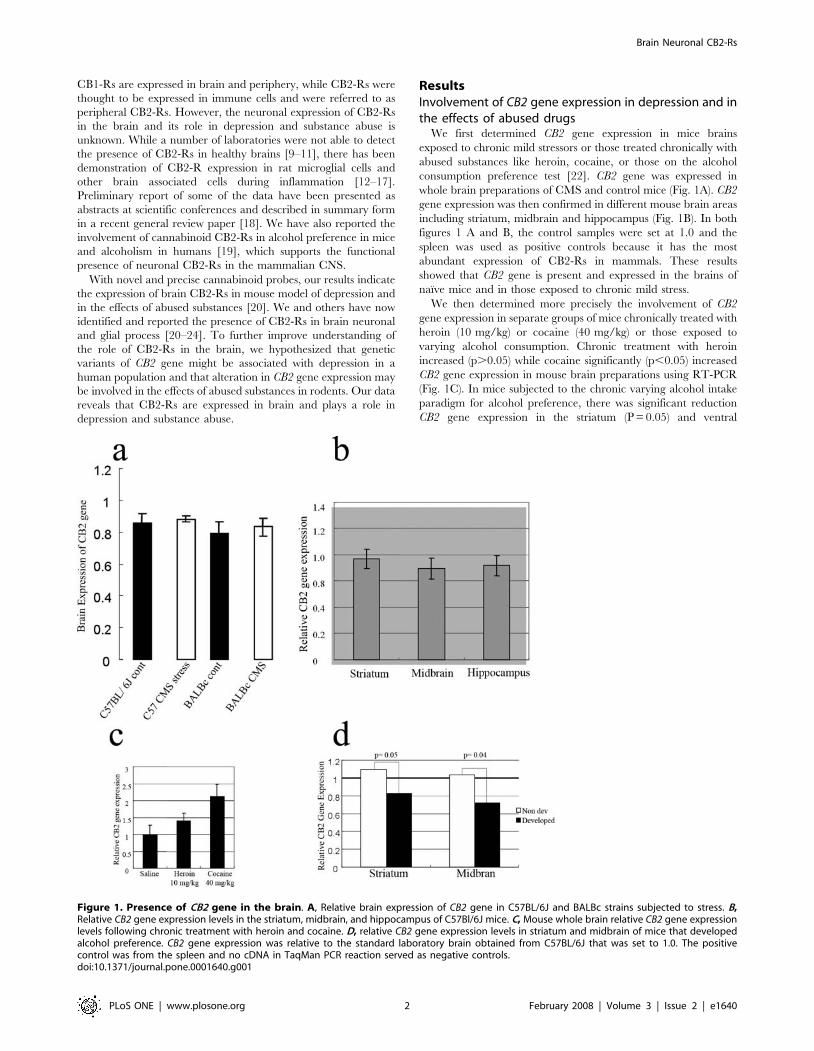
whole brain preparations of CMS and control mice (Fig. 1A). CB2
gene expression was then confirmed in different mouse brain areas
including striatum, midbrain and hippocampus (Fig. 1B). In both
figures 1 A and B, the control samples were set at 1.0 and the
spleen was used as positive controls because it has the most
abundant expression of CB2-Rs in mammals. These results
showed that CB2 gene is present and expressed in the brains of
naı̈ve mice and in those exposed to chronic mild stress.
We then determined more precisely the involvement of CB2
gene expression in separate groups of mice chronically treated with
heroin (10 mg/kg) or cocaine (40 mg/kg) or those exposed to
varying alcohol consumption. Chronic treatment with heroin
increased (p.0.05) while cocaine significantly (p,0.05) increased
CB2 gene expression in mouse brain preparations using RT-PCR
(Fig. 1C). In mice subjected to the chronic varying alcohol intake
paradigm for alcohol preference, there was significant reduction
CB2 gene expression in the striatum (P = 0.05) and ventral
Figure 1. Presence of CB2 gene in the brain. A, Relative brain expression of CB2 gene in C57BL/6J and BALBc strains subjected to stress. B,Relative CB2 gene expression levels in the striatum, midbrain, and hippocampus of C57Bl/6J mice. C, Mouse whole brain relative CB2 gene expressionlevels following chronic treatment with heroin and cocaine. D, relative CB2 gene expression levels in striatum and midbrain of mice that developedalcohol preference. CB2 gene expression was relative to the standard laboratory brain obtained from C57BL/6J that was set to 1.0. The positivecontrol was from the spleen and no cDNA in TaqMan PCR reaction served as negative controls.doi:10.1371/journal.pone.0001640.g001
Brain Neuronal CB2-Rs
PLoS ONE | www.plosone.org 2 February 2008 | Volume 3 | Issue 2 | e1640

midbrain (p = 0.04), whereas in mice with little preference for
drinking alcohol, there were no changes in CB2 gene expression in
these brain regions (Fig. 1D). The alcohol data support our
previous studies [19] that CB2-R agonist JWH015 administration
enhances alcohol intake in stressed but not in non-stressed control
mice. In contrast the administration of the CB2-R antagonist
AM630 reduced alcohol intake (P = 0.08) in stressed but had no
effect in the alcohol consumption in non-stressed naive mice. The
presence of CB2-Rs in the brain was further investigated in CB2-R
deficient mice and their wild type litter mates. In-situ hybridization
data show that CB2 gene is expressed in the cerebellum of wild
type and not in the cerebellum of the CB2-R deficient mice and
also in sense controls in the wild type mice (Fig. 2C). Altogether,
these results revealed the functional presence of brain CB2-Rs that
plays a role in the effects of abused substances.
We then examined the association between CB2 gene polymor-
phism and depression in a human population to test the hypothesis
that genetic variants of CB2 gene might be associated with depression
and substance abuse in Japanese population. A significant
association was found between the CB2 Q63R polymorphism and
Japanese depressed subjects (p = 0.007, odds ratio 1.42, 95%
confidence interval: 1.09–1.831), (Fig. 2B). Furthermore, because a
previous study showed a significant functional difference of RR
genotype in lymphocyte, we compared the distribution of subjects
without or with Q allele. The RR genotype was significantly
associated with depression [p = 0.01, odds ratio 1.95; 95%
confidence interval, (1.11–3.4402.28)], (Table 1).
Analysis of CB2-Rs in the rodent brain with or withoutexposure to stressors
To determine the localization of CB2-Rs in mouse and rat
brains, we used a combination of Western blotting, immunohis-
tochemistry and in-situ hybridization. The CB2-R knockout mice
and their wild type litter mates were included as controls for the in-
situ hybridization. We then analyzed CB2-Rs in the brains of mice
subjected to chronic mild stressors, including adult mice that had
been prenatally exposed to capsaicin. Western blotting analyses
from mice brains revealed a major CB2-R band of approximately
53 kDa (Fig. 2A), with other visible bands around 37 kDa and
75 kDa, similar to those reported [21]. CB2 gene was expressed in
mouse whole brain preparations and the CB2-R protein was also
present in the CMS and prenatal capsaicin exposure (Fig. 2A).
The specificity of three commercial CB2-R antibodies had been
examined in our previous studies to map CB2-R immunoreactivity
in the rat brain [24]. In this study the specificity of the CB2-R
antibody used was further confirmed as the CB2-R immunoreac-
tivity detected in the cerebellum were undetectable when the CB2-
R antibody was pre-adsorbed with the immunizing peptide
(Fig. 3A and B) using 8.3 mg/ml of the CB2 sequence peptide
used to produce the antiserum. It is important to note that we
previously demonstrated and reported that CB2-R immunoreac-
tivity was present in the CA2 region of the hippocampus, spleen
and interpolar part of spinal 5th nucleus of wild type brain and the
CB2-R immunoreactivity was absent in these structures in the
global CB2-R knockout mouse [24].
We then performed immunohistochemical analysis in the naı̈ve
mouse and rat brain sections (Fig. 4). Apical dendrites and cell bodies
of pyramidal neurons of rat cerebral cortex were moderately to
heavily immunolabeled for CB2-R. Scattered fibers in the rat
cerebral cortex showed CB2-R-IR (Fig. 4A). CB2-R immunoreac-
tivity (IR) was also observed in the mouse cerebral cortex (Fig. 4B).
CB2-R-IR was also observed in rat corpus callosum (Fig. 4C). A
moderate to dense CB2 immunostaing was observed in pyramidal
neuron of mouse hippocampal allocortex and some interneurons in
the stratum oriens and stratum radiatum (Fig. 4D). Some glial cells
were also immunolabeled for CB2-Rs in the hippocampus (Fig. 4D).
This localization pattern is in agreement with the perfect overlay
when double labeling of CB2-Rs and neuron specific enolase (NSE)
in hippocampal neuronal cultures was visualized by confocal
immunofluorescence imaging [24]. Thus, in the brain areas analyzed
CB2-R immunoreactivity was detected in mice and rat brains, and
this is supported by reports of identification of neuronal CB2-Rs in
the brain stem involved in emesis [21].
Figure 2. Brain CB2-Rs: Immunoblots, genotyping and in-situhybridization. A, In-situ hybridization indicating CB2 gene isexpressed in the cerebellum of wild type and not in the cerebellumof the CB2-R deficient mice and also in sense controls in the wild typemice. B, RFLP genotyping discrimination on agarose gel for CB2 Q63Rpolymorphism in depressed subjects (Ba) and, Resequences of CB2Q63R polymorphism (Bb). C, Western blotting of CB2-Rs in CMS andcontrol mice (left panel) and in right panel in mice exposed to 4 mg/kgcapsaicin in utero.doi:10.1371/journal.pone.0001640.g002
Brain Neuronal CB2-Rs
PLoS ONE | www.plosone.org 3 February 2008 | Volume 3 | Issue 2 | e1640
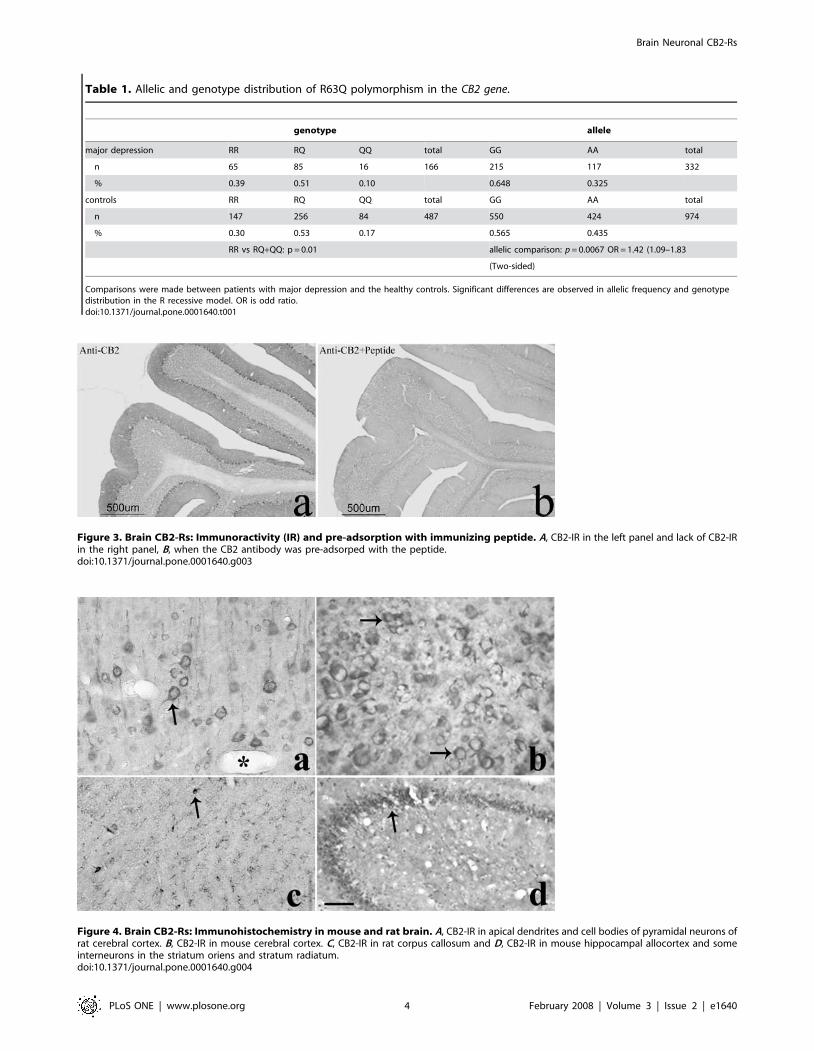
Table 1. Allelic and genotype distribution of R63Q polymorphism in the CB2 gene.
genotype allele
major depression RR RQ QQ total GG AA total
n 65 85 16 166 215 117 332
% 0.39 0.51 0.10 0.648 0.325
controls RR RQ QQ total GG AA total
n 147 256 84 487 550 424 974
% 0.30 0.53 0.17 0.565 0.435
RR vs RQ+QQ: p = 0.01 allelic comparison: p = 0.0067 OR = 1.42 (1.09–1.83
(Two-sided)
Comparisons were made between patients with major depression and the healthy controls. Significant differences are observed in allelic frequency and genotypedistribution in the R recessive model. OR is odd ratio.doi:10.1371/journal.pone.0001640.t001
Figure 3. Brain CB2-Rs: Immunoractivity (IR) and pre-adsorption with immunizing peptide. A, CB2-IR in the left panel and lack of CB2-IRin the right panel, B, when the CB2 antibody was pre-adsorped with the peptide.doi:10.1371/journal.pone.0001640.g003
Figure 4. Brain CB2-Rs: Immunohistochemistry in mouse and rat brain. A, CB2-IR in apical dendrites and cell bodies of pyramidal neurons ofrat cerebral cortex. B, CB2-IR in mouse cerebral cortex. C, CB2-IR in rat corpus callosum and D, CB2-IR in mouse hippocampal allocortex and someinterneurons in the striatum oriens and stratum radiatum.doi:10.1371/journal.pone.0001640.g004
Brain Neuronal CB2-Rs
PLoS ONE | www.plosone.org 4 February 2008 | Volume 3 | Issue 2 | e1640
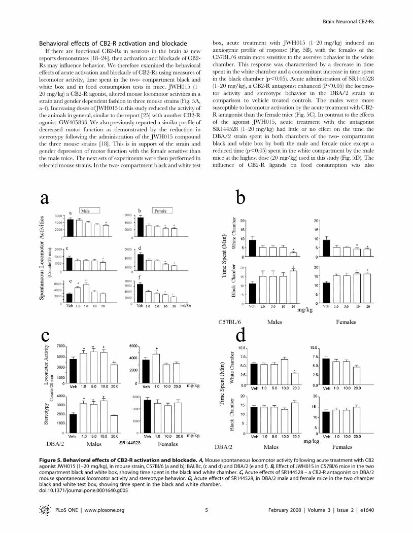
Behavioral effects of CB2-R activation and blockadeIf there are functional CB2-Rs in neurons in the brain as new
reports demonstrates [18–24], then activation and blockade of CB2-
Rs may influence behavior. We therefore examined the behavioral
effects of acute activation and blockade of CB2-Rs using measures of
locomotor activity, time spent in the two- compartment black and
white box and in food consumption tests in mice. JWH015 (1–
20 mg/kg) a CB2-R agonist, altered mouse locomotor activities in a
strain and gender dependent fashion in three mouse strains (Fig. 5A,
a–f). Increasing doses of JWH015 in this study reduced the activity of
the animals in general, similar to the report [25] with another CB2-R
agonist, GW405833. We also previously reported a similar profile of
decreased motor function as demonstrated by the reduction in
stereotypy following the administration of the JWH015 compound
the three mouse strains [18]. This is in support of the strain and
gender depression of motor function with the female sensitive than
the male mice. The next sets of experiments were then performed in
selected mouse strains. In the two- compartment black and white test
box, acute treatment with JWH015 (1–20 mg/kg) induced an
anxiogenic profile of response (Fig. 5B), with the females of the
C57BL/6 strain more sensitive to the aversive behavior in the white
chamber. This response was characterized by a decrease in time
spent in the white chamber and a concomitant increase in time spent
in the black chamber (p,0.05). Acute administration of SR144528
(1–20 mg/kg), a CB2-R antagonist enhanced (P,0.05) the locomo-
tor activity and stereotype behavior in the DBA/2 strain in
comparison to vehicle treated controls. The males were more
susceptible to locomotor activation by the acute treatment with CB2-
R antagonist than the female mice (Fig. 5C). In contrast to the effects
of the agonist JWH015, acute treatment with the antagonist
SR144528 (1–20 mg/kg) had little or no effect on the time the
DBA/2 strain spent in both chambers of the two- compartment
black and white box by both the male and female mice except a
reduced time (p,0.05) spent in the white compartment by the male
mice at the highest dose (20 mg/kg) used in this study (Fig. 5D). The
influence of CB2-R ligands on food consumption was also
Figure 5. Behavioral effects of CB2-R activation and blockade. A, Mouse spontaneous locomotor activity following acute treatment with CB2agonist JWH015 (1–20 mg/kg), in mouse strain, C57Bl/6 (a and b); BALBc, (c and d) and DBA/2 (e and f). B, Effect of JWH015 in C57Bl/6 mice in the twocompartment black and white box, showing time spent in the black and white chamber. C, Acute effects of SR144528 – a CB2-R antagonist on DBA/2mouse spontaneous locomotor activity and stereotype behavior. D, Acute effects of SR144528, in DBA/2 male and female mice in the two chamberblack and white test box, showing time spent in the black and white chamber.doi:10.1371/journal.pone.0001640.g005
Brain Neuronal CB2-Rs
PLoS ONE | www.plosone.org 5 February 2008 | Volume 3 | Issue 2 | e1640
investigated. The enhancement and suppressant effects of CB2-R
ligands were strain and time dependent (data are not shown). Thus,
there was a clear strain and gender dependent effects following CB2-
R activation or inhibition on behavioral responses as measured by
locomotor activity, emotionality and food consumption tests.
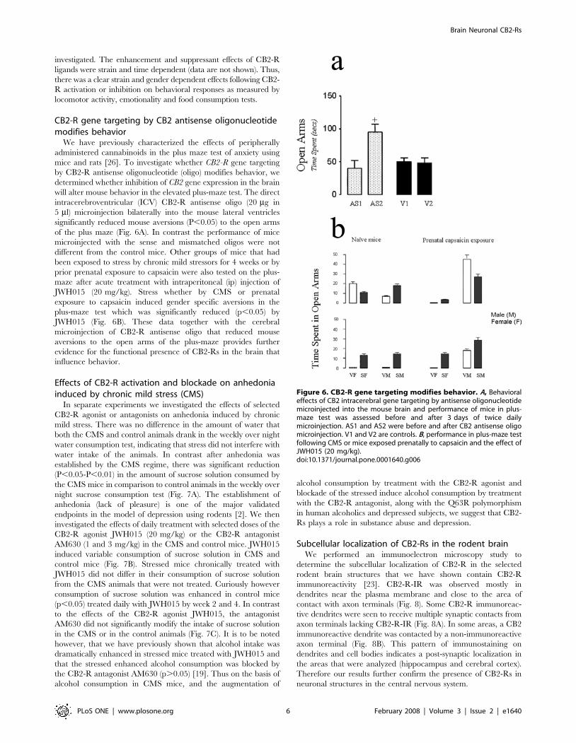
CB2-R gene targeting by CB2 antisense oligonucleotidemodifies behavior
We have previously characterized the effects of peripherally
administered cannabinoids in the plus maze test of anxiety using
mice and rats [26]. To investigate whether CB2-R gene targeting
by CB2-R antisense oligonucleotide (oligo) modifies behavior, we
determined whether inhibition of CB2 gene expression in the brain
will alter mouse behavior in the elevated plus-maze test. The direct
intracerebroventricular (ICV) CB2-R antisense oligo (20 mg in
5 ml) microinjection bilaterally into the mouse lateral ventricles
significantly reduced mouse aversions (P,0.05) to the open arms
of the plus maze (Fig. 6A). In contrast the performance of mice
microinjected with the sense and mismatched oligos were not
different from the control mice. Other groups of mice that had
been exposed to stress by chronic mild stressors for 4 weeks or by
prior prenatal exposure to capsaicin were also tested on the plus-
maze after acute treatment with intraperitoneal (ip) injection of
JWH015 (20 mg/kg). Stress whether by CMS or prenatal
exposure to capsaicin induced gender specific aversions in the
plus-maze test which was significantly reduced (p,0.05) by
JWH015 (Fig. 6B). These data together with the cerebral
microinjection of CB2-R antisense oligo that reduced mouse
aversions to the open arms of the plus-maze provides further
evidence for the functional presence of CB2-Rs in the brain that
influence behavior.
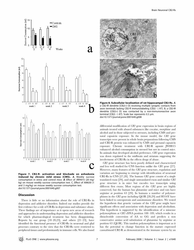
Effects of CB2-R activation and blockade on anhedoniainduced by chronic mild stress (CMS)
In separate experiments we investigated the effects of selected
CB2-R agonist or antagonists on anhedonia induced by chronic
mild stress. There was no difference in the amount of water that
both the CMS and control animals drank in the weekly over night
water consumption test, indicating that stress did not interfere with
water intake of the animals. In contrast after anhedonia was
established by the CMS regime, there was significant reduction
(P,0.05-P,0.01) in the amount of sucrose solution consumed by
the CMS mice in comparison to control animals in the weekly over
night sucrose consumption test (Fig. 7A). The establishment of
anhedonia (lack of pleasure) is one of the major validated
endpoints in the model of depression using rodents [2]. We then
investigated the effects of daily treatment with selected doses of the
CB2-R agonist JWH015 (20 mg/kg) or the CB2-R antagonist
AM630 (1 and 3 mg/kg) in the CMS and control mice. JWH015
induced variable consumption of sucrose solution in CMS and
control mice (Fig. 7B). Stressed mice chronically treated with
JWH015 did not differ in their consumption of sucrose solution
from the CMS animals that were not treated. Curiously however
consumption of sucrose solution was enhanced in control mice
(p,0.05) treated daily with JWH015 by week 2 and 4. In contrast
to the effects of the CB2-R agonist JWH015, the antagonist
AM630 did not significantly modify the intake of sucrose solution
in the CMS or in the control animals (Fig. 7C). It is to be noted
however, that we have previously shown that alcohol intake was
dramatically enhanced in stressed mice treated with JWH015 and
that the stressed enhanced alcohol consumption was blocked by
the CB2-R antagonist AM630 (p.0.05) [19]. Thus on the basis of
alcohol consumption in CMS mice, and the augmentation of
alcohol consumption by treatment with the CB2-R agonist and
blockade of the stressed induce alcohol consumption by treatment
with the CB2-R antagonist, along with the Q63R polymorphism
in human alcoholics and depressed subjects, we suggest that CB2-
Rs plays a role in substance abuse and depression.
Subcellular localization of CB2-Rs in the rodent brainWe performed an immunoelectron microscopy study to
determine the subcellular localization of CB2-R in the selected
rodent brain structures that we have shown contain CB2-R
immunoreactivity [23]. CB2-R-IR was observed mostly in
dendrites near the plasma membrane and close to the area of
contact with axon terminals (Fig. 8). Some CB2-R immunoreac-
tive dendrites were seen to receive multiple synaptic contacts from
axon terminals lacking CB2-R-IR (Fig. 8A). In some areas, a CB2
immunoreactive dendrite was contacted by a non-immunoreactive
axon terminal (Fig. 8B). This pattern of immunostaining on
dendrites and cell bodies indicates a post-synaptic localization in
the areas that were analyzed (hippocampus and cerebral cortex).
Therefore our results further confirm the presence of CB2-Rs in
neuronal structures in the central nervous system.
Figure 6. CB2-R gene targeting modifies behavior. A, Behavioraleffects of CB2 intracerebral gene targeting by antisense oligonucleotidemicroinjected into the mouse brain and performance of mice in plus-maze test was assessed before and after 3 days of twice dailymicroinjection. AS1 and AS2 were before and after CB2 antisense oligomicroinjection. V1 and V2 are controls. B, performance in plus-maze testfollowing CMS or mice exposed prenatally to capsaicin and the effect ofJWH015 (20 mg/kg).doi:10.1371/journal.pone.0001640.g006
Brain Neuronal CB2-Rs
PLoS ONE | www.plosone.org 6 February 2008 | Volume 3 | Issue 2 | e1640
Discussion
There is little or no information about the role of CB2-Rs in
depression and addictive disorders. Indeed our studies provide the
first evidence for a role of CB-Rs in depression and substance abuse.
These findings are of importance as it opens new areas of research
and approaches in understanding depression and addictive disorders
for which pharmacological treatment has been disappointing.
Reports by our group [18–20,23], and others [21–23], have
identified the functional presence of CB2-Rs in neuronal and glial
processes contrary to the view that the CB2-Rs were restricted to
peripheral tissues and predominantly in immune cells. We also found
differential modification of CB2 gene expression in brain regions of
animals treated with abused substances like cocaine, morphine and
alcohol and in those subjected to stressors, including CMS and pre-
natal capsaicin exposure. In the mouse model, the CB2 gene
transcripts were present in whole brain preparations following CMS
and CB2-R protein was enhanced by CMS and prenatal capsaicin
exposure. Chronic treatment with CB2-R agonist JWH015
enhanced alcohol consumption in stressed but not in control mice.
In animals that developed alcohol preference, CB2 gene expression
was down regulated in the midbrain and striatum suggesting the
involvement of CB2-Rs in the effects drugs of abuse.
CB2 gene structure has been poorly defined and characterized
and less well studied for CNS function unlike the CB1 gene [27].
However, many features of the CB2 gene structure, regulation and
variation are beginning to emerge with identification of neuronal
CB2-Rs in CNS [27,28]. The human CB2 gene consist of a single
translated exon [29], and single untranslated exon and similar CB2
gene structure is in mice but encodes two transcripts using
different first exons. Most regions of the CB2 gene are highly
conserved, but the human has glutamine and mice and rats have
arginine at position 63 [29]. In humans a number of polymor-
phisms in the CB2 gene including Q63R [28,29] and H316Y have
been linked to osteoporosis and autoimmune disorders. We tested
the hypothesis that genetic variants of the CB2 gene might have
significant effects and association with depression and alcoholism.
This hypothesis is supported by the identification of a missense
polymorphism at CB2 cDNA position 188–189, which results in a
dinucleotide conversion of AA to GG and predicts a non
conservative amino acid substitution of glutamine by arginine at
position 63 (Q63R). This tandem polymorphism is important as it
has the potential to change function in the mature expressed
cannabinoid CB2-R as demonstrated in the immune system by an
Figure 7. CB2-R- activation and blockade on anhedoniainduced by chronic mild stress (CMS). A, Weekly sucroseconsumption in stress and control mice. B, Effect of JWH015 (20 mg/kg) on mouse weekly sucrose consumption test. C, Effect of AM630 (1and 3 mg/kg) on mouse weekly sucrose consumption test.doi:10.1371/journal.pone.0001640.g007
Figure 8. Subcellular localization of rat hippocampal CB2-Rs. A,a CB2-IR dendrite [CB2(+) D] receiving multiple synaptic contacts fromaxon terminals lacking CB2-R immunolabeling [CB2(2) AT]. B, a CB2-IRdendrite [CB2(+) D] was contacted by a non-immunoreactive axonterminal [CB2(2) AT]. Scale bar represents 0.3 mm.doi:10.1371/journal.pone.0001640.g008
Brain Neuronal CB2-Rs
PLoS ONE | www.plosone.org 7 February 2008 | Volume 3 | Issue 2 | e1640
in vitro assay [29]. The association of CB2 gene variation was
probed in Japanese subjects to examine the non-synonymous
polymorphism, Q63R, in the CB2 gene for association with
depression or alcoholism. There was significant difference in allelic
frequency between cases and controls at Q63R polymorphism in
the CB2 gene in depression in this study and alcoholism [19]. As
many genetic variants play various roles in depression and/or
substance abuse, variation in CB2 gene, (Q63R) polymorphism
may be a previously unknown risk factor in depression and/or
alcoholism at least in the Japanese population. If this can be
generalized to other ethnicities, then the results support the
possibility of targeting the cannabinoid system using CB2-R
ligands in depression and drug abuse and perhaps in their co-
morbidity. It is therefore tempting to speculate that the reported
effects of alcohol may be associated with changes in the
cannabinoid system, with CB2-Rs playing a regulatory role.
We then hypothesized that if CB2-Rs are present in the brain,
then antisense oligonucleotides complementary to CB2 mRNA
transcript will block translation of or stimulate degradation of CB2
mRNA. It is therefore of importance to determine whether
inhibition of CB2 gene expression in the brain will alter behavior
as observed with the exogenous administration of CB2-R ligands.
Direct intracerebroventricular CB2 oligonucleotide microinjection
into the mouse brain reduced mouse aversion, further indicating
the functional presence of CB2-Rs in the brain that influence
behavior. This behavioral evidence for the functional presence of
CB2-Rs in brain was further investigated by the exogenous
administration of CB2-R agonists and antagonist. The behavioral
effects of acute treatment with JWH015, a CB2-R agonist and
SR144528, a CB2-R antagonist, in mouse spontaneous locomotor
activities, and in the two- compartment black and white box lends
further support that CB2-Rs in the brain modifies behavior.
Similar observations have been reported for the effects of a CB2-R
agonist, GW405833 [25]. Curiously, the observation that CB2-R
agonists induces sedation and catalepsy only at higher doses has
been interpreted by this group and others in rodent models of
pain, to have a potential to treat pain without eliciting the
centrally-mediated side effects without the psychoactivity associ-
ated with CB1-R [13,25]. With the recent definitive demonstra-
tion of neuronal CB2-Rs in the brain, one possible explanation
may be that CB2 and CB1 cannabinoid receptors work
independently and/or cooperatively in different neuronal popu-
lations to regulate a number of physiological activities influenced
by cannabinoids. These effects of CB2-R ligands in in vivo
behavioral tests are provided as functional evidence of CB2-R in
the brain that plays a role in motor function and emotionality tests.
The antagonism of the behavioral effects of CB2-R agonist,
JWH015 by SR144528 or AM630 was not determined in this
study. However, other studies have demonstrated the selectivity of
JWH015 on mediating its effects via CB2-Rs [30,31] and the effect
of JWH015 was completely blocked by the CB2 specific
antagonist, SR144528 [32].
Abundant CB2-R immunoreactivity in neuronal and glial
processes was detected but at a much lower level than CB1
receptors as reported [23]. This is supported by reports of the
presence of CB2-Rs in brain stem, cortex, cerebellum, dorsal root
ganglion and spinal cord [21–23]. There is still some controversy
on the specificity of CB2-R staining because most of the antibodies
are capable of producing non-specific staining. Therefore, very
rigorous controls have been utilized in our study including 1) the
pre-adsorption and co-incubation of the CB2-R antibody with the
immunizing peptide resulting in blocking CB2 staining in the rat
cerebellum, 2) in situ hybridization data show that CB2 gene is
expressed in the cerebellum of wild type and not in the CB2
knockout mice, with CB2 gene being absent in the sense control in
the wild type mice. The absence of CB2 mRNA in CB2-R
deficient mice and presence in wild type controls has also been
demonstrated by others [21]. Moreover, in previous control
experiments we had demonstrated that using two types of CB2
antibodies, similar staining patterns in both the rat spleen and
cerebellum [24] were reported. Western blot analyses revealed
specific bands that were identified using CB2 antibodies and were
absent when the CB2 antibodies were pre-adsorbed with the
immunizing peptide [24]. Furthermore, the expression levels of the
CB1 gene using RT-PCR analysis was 100 times that of the CB2
gene expression levels with reference to the brain stem. We have
also confirmed that the spleen has the most abundant CB2 gene
transcripts when compared to other regions [24].
As definitive electron microscopic evidence is needed to
precisely determine the subcellular localization of CB2-Rs, our
transmission electron micrograph data using immunoelectron
microscopy approach shows a high-resolution definition of
hippocampal CB2-R localization at the ultrastructural level.
Electron micrographs from hippocampal areas show dendrites
with immunostaining for CB2-Rs with diffuse black deposits and
mitochondria clearly visible. In some areas axon terminals were
not immunoreactive for CB2-Rs and small rounded synaptic
vesicles were seen. An axon terminal making contact with a
dendrite but without immunostaining for CB2-Rs was apparent.
The pattern of staining in most hippocampal areas appears to be
mainly post-synaptic localization of CB2-Rs. For example at the
area of the synaptic contacts seen the synapse appear to be
excitatory and possibly glutamatergic. We cannot exclude that
some of the CB2-Rs may be presynaptic, just like CB1-Rs are not
exclusively presynaptic in the brain [33]. CB1-Rs are known to be
mainly presynaptic in the CNS where cannabinoids act at
presynaptic CB1-Rs and endocannabinoids have emerged as one
of the classes of retrograde messengers involved in the regulation
of synaptic transmission. The functional implication of pre- and/
or post-synaptic localization of CB2-Rs awaits further electro-
physiological investigation and image analysis of this interesting
component of the EPCS. The current understanding of CNS
CB2-Rs was the subject of our review [18] and future studies will
continue to characterize the specificity of CB2-R mediated
behavioral effects and their physiological roles. Thus, our data
demonstrate the functional expression of CB2-Rs in brain that
may provide novel targets for the effects of cannabinoids in
depression and substance abuse disorders beyond neuro-immuno-
cannabinoid activity.
Materials and MethodsHuman subjects
166 patients with Major depression (excluded bipolar disorders)
diagnosed as depressed by DSM-IIIR criteria without other
psychiatric diagnoses, recruited under informed consent. 487 age-
and gender-matched controls were research volunteers. They were
recruited from north-mid main island area in Japan and provided
written informed consent. The genetics study using the DNA of
subjects, who provided written consent, was approved by ethics
committee of University of Tsukuba.
Animal subjectsThree strains (DBA/2, C57BL/6 and BALBc), male and female
mice and Sprague Dawley rats were used. CB2 knockout mice
(CB22/2) and their wild type littermates used in this study
CB22/2 was developed by Buckley et al, 2000, [34] and obtained
from the National Institutes of Health through Dr. Kunos of
NIAAA-NIH, USA. Animals were housed according to National
Brain Neuronal CB2-Rs
PLoS ONE | www.plosone.org 8 February 2008 | Volume 3 | Issue 2 | e1640
Institutes of Health and institutional guidelines for laboratory
animals. All procedures were approved by the local Animal Care
and Use Committees in all the institutions involved with the
project.
DrugsJWH015 (a putative CB2 agonist) and AM60, a CB2-R antagonist
were obtained from Sigma-Aldrich (MO, USA) and Cayman
Chemicals (MI, USA) while SR144528 (a CB2 antagonist) was
donated by Sanofi, (France). Primary CB2 antibodies and their
blocking peptide were obtained from Santa Cruz (Ca, USA). For the
in vivo experiments, JWH015, AM630 and SR144528 were made up
in ethanol: emulphur: water in a ratio of 1:1:18.
Behavioral AnalysesBALB/c male and female mice were housed 12 hrs in light and
12 hrs in dark. Experimental mice (N = 10 per group) were
exposed to CMS everyday for four weeks to achieve anhedonia
(CMS test). These experimental animals were subjected to the
weekly CMS regime consisting of three 10 hr periods of 45u cage
tilt; 3 periods of overnight stroboscopic illumination, two 10 hr
periods of empty water bottle; two periods of overnight food or
water deprivation; two 10 hr periods of damp bedding. The CMS
treated and non-stressed groups consisted of 30 mice each and
were split into three subgroups, respectively. All non-stressed groups
were given food and water at all times, as well as comfortable cage
surroundings, while the experimental group was housed in a
different room. In the first set of studies animals in both the stress
and control groups of 10 animals per group were treated daily with
the CB2 agonist JWH015 (20 mg/kg) and the control groups with
the vehicle for 4 weeks. In the second round of CMS study animals
in both the stress and control groups of 10 animals per group were
treated daily with the CB2 antagonist AM630 ( 1 and 3 mg/kg) and
the control groups with the vehicle for 4 weeks. Once every week
sucrose consumption was measured as a test of anhedonia. At the
end of the stress regime, locomotor activities stereotype behavior was
measured in activity monitors in all groups.
The acute effects of JWH015, a CB2-R agonist and SR144528
a CB2-R antagonist on mouse locomotor activity and stereotypy
using activity monitors and in the two- compartment black and
white box were assessed. The pretreatment times were 10 min- for
the agonist and 30 min for the antagonist. Animals were placed in
activity monitors or in the two- compartment black and white box.
Spontaneous locomotor activities and stereotype behavior in the
activity monitors and time spent and locomotor activities in the
box were obtained from the automated system. The doses of the
agonist and antagonist were 1–20 mg/kg except as indicated in
specific experiments as described for the CMS experiments. The
performance of mice in the plus-maze test of anxiety following
intracerebroventricular (ICV) administration of CB2 antisense
oligonucleotide (oligos) (20 mg in 5 ml) was assessed before and
after 3 days of twice daily microinjection and compared to mice
injected with sense and mismatched oligos.
Western BlottingEqual amount of protein 20 mg obtained from the brains of
stressed and control mice were loaded and separated by 10% SDS-
PAGE and then transferred onto nitrocellulose membrane. The
nitrocellulose was washed and blocked in PBS containing 2% non-
fat milk and incubated with the CB2 antibody overnight. The
membranes were washed and incubated with a conjugated goat
anti-rabbit secondary antibody and processed for immunoreactiv-
ities with and without pre-incubation of the primary antibodies
with CB2 peptide.
CB2 gene expression and regulation by drug and alcoholtreatment
CB2-R gene expression was determined in whole mouse brains
subjected to stressors and those treated chronically with heroin
(10 mg/kg) and cocaine (40 mg/kg) and then precisely in brain
regions of naı̈ve mice and those exposed acutely or chronically to
escalating doses of alcohol. CB2 gene expression was then
determined in animals that developed or did not develop alcohol
preference. Mice were sacrificed and whole brains were taken or
dissected out for extraction of RNA. Control group of mice (n = 6)
did not have access to ethanol but only to water in that
experiment, and RNA was also extracted in a same way for
comparison to the mice that developed ethanol preference. Where
indicated brains were dissected into striatum, hippocampus and
midbrain. RNA was extracted using RNeasy kit (QIAGEN, K.K.,
Tokyo, Japan) and cDNA was synthesized by Revertra Ace
(TOYOBO, Japan) and oligo dT primer. The expression of CB2
gene was compared by TaqMan real-time PCR with an ABI
PRISM 7900 HT Sequence Detection System (Applied Biosys-
tems, Foster City, CA, USA), using the TaqMan gene expression
assay for CB2 (Mm0043826_m1).
Association study between the Q63R polymorphism anddepression
CB2 gene has two non-synonymous polymorphisms, Q63R and
H316Y according to public database NCBI (http://www.ncbi.
nlm.nih.gov/). However, analysis of secondary structure of CB2
gene with Chou-Fasman, Robson and hydrophilic/ hydrophobic
structure extimation methods using computer program GENE-
TYX (Genetyx corporation, Tokyo, Japan) revealed a potential
structural change in CB2 gene only by the Q63R but not by the
H316Y polymorphism of the gene. Therefore, we focused on the
Q63R polymorphism and the genotype was determined by
restricted fragment length polymorphism (RFLP) method as
described in our previous report [19].
Real Time-PCRTotal RNA was extracted from brain tissues using RNAzol B
(Tel-Test, Friendswood, TX). Single strand cDNA was synthesized
from total RNA using SuperScriptTM first-strand synthesis system
for RT-PCR (GIBCO/BRL, Rockville, MD). For quantitative real
time PCR assays, the exon-specific primers and fluorescent Fam-
labeled probes across different exon regions were designed using
Primer Express program (Applied Biosystems, Foster City, CA).
Beta-actin Fam-labeled probe was used for normalization. Two-
step PCR program was used as the default of ABI 7900 HT PCR
instrument (Applied Biosystems, Foster City, CA). In the assay, we
used spleen as positive control because of its high expression of
CB2 gene and no cDNA in the TaqMan PCR reaction as negative
control. The control brain samples were set at 1.0 with
glyceraldehydes-3-phosphate dehydrogenase (GAPDH) pre-devel-
oped TaqMan assay reagent as endogenous control (FAMTM
Dye/MGB probe). Calculation of real time PCR was carried out
according to User Bulletin #2 for ABI Prism 7700 Sequence
Detection System.
Immunohistochemistry and electron microscopyMice and male Sprague Dawley (180–200 g) rats were
anesthetized with choral hydrate (300 mg/kg) pentobarbital,
perfused transcardially with saline and then with 100 ml of 4%
paraformaldehyde in phosphate buffer (PB; 0.1 M, pH 7.4) for
mice and 500 ml of the same fixative solution for rats. Brains were
dissected, postfixed in the same fixative solution for 2 hours at
Brain Neuronal CB2-Rs
PLoS ONE | www.plosone.org 9 February 2008 | Volume 3 | Issue 2 | e1640
room temperature, equilibrated with 30% sucrose in PB at 4uC,
frozen and cut into saggital 20–40 mm sections using a cryostat.
Sections were processed for immunohistochemistry as follows.
Floating sections were incubated with 1% hydrogen peroxide in
phosphate-buffered saline (PBS) for 10 min at room temperature
to inhibit endogenous peroxidase, washed three times with PBS,
incubated for 1 h in 3% normal goat serum (NGS) in Tris-
buffered saline (TBS), pH 7.4 at 22uC, incubated in primary CB2
antibody obtained from (Santa Cruz, Ca, USA), diluted 1: 300 in
TBS containing 3% NGS for 24 h at 4uC, rinsed, incubated for
1 h at 22uC in 1:200 dilution of biotinylated goat anti-rabbit
secondary antibody (Vector, Burlingame, CA, USA) for 1 h,
rinsed, incubated with avidin-biotin peroxidase complex (ABC)
reagent for 1 h (Vector), rinsed, and then incubated in a solution
containing 22 mg/ml diaminobenzidine (DAB) (Electron Micros-
copy Sciences, Fort Washington, PA) and 0.003% hydrogen
peroxide (H2O2) for color deposition. Sections were mounted on
coated slides, dehydrated, cover slipped, viewed and photographed
using Zeiss and Leitz microscope and a Nikon digital camera,
immunoreactive elements plotted onto the atlas depictions [35],
and images edited using photoshop (vCS; Adobe systems). As
additional control, iCB2 of brain sections from CB2-R deficient
mice and wild type controls were also analyzed. For electron
microscopy, rats were perfused with 500 ml of 4% paraformalde-
hyde, 0.1% purified glutaraldehyde fixative in PB, brains were
removed, postfixed in the same fixative solution for two hours and
saggital sections (50 mm) were obtained using a vibrotome. Then,
sections were processed for inmmunohistochemistry following the
same immunoperoxidase protocol. After that, sections were fixed
with 1% osmium tetroxide in 0.1 M PB for 1 h, dehydrated through
a series of graded alcohols (including 60 min in 70% alcohol
containing 1% uranyl acetate), and then with propylene oxide.
Afterwards, they were flat-embedded in Durcupan ACM epoxy resin
(Electron Microscopy Sciences, Fort Washington, PA). Embedded
sections were polymerized at 60uC for 2 days. Ultrathin sections of
70 nm were cut from the outer surface of the tissue with an
ultramicrotome (Leica, Microsystems, Wetzlar, Germany) using a
diamond knife (Diatome, fort Washington, PA). The sections were
collected onto 300 mesh cooper grids and counterstained with
Reynolds lead citrate [36]. Sections were examined and photo-
graphed using a Zeiss 109 transmission electron microscope and
35 mm Kodak technical Pan professional 2415 films.
In situ hybridization and probesBiotin labeled RNA probes were used for in situ hybridization.
The full length of human CB2 gene was subcloned from
pcDNA3.1/CB2 (UMR cDNA resource center, Rolla, MO) into
pBluescript II at the restriction sites of EcoR I and Xho I. The
pcBluescript II/CB2 was linearized with Xho I (Anti-sense probe)
or Eco RI (sense probe). The CB2 riboprobes were synthesized by
incubating for 60 min at 37uC. 1 mg linearized plasmid in 2 ml
10X transcription buffer, 1 ml RNase inhibitor, 2 ml Biotin RNA
labeling Mix containing 1 mM ATP/GTP/CTP, 650 mM UTP,
350 mM biotin–UTP (Roche Applied Science, Germany), 40 U
T7 (anti-sense probe), or T3 RNA polymerase (sense probe) in a
final volume of 20 ml. The reaction mixture was subsequently
incubated for 15 min at 37uC with 1 U RNase-free DNase I. The
riboprobes were precipitated using LiCl and ethanol. The CB2
probes were diluted in 100 ml TE. Coronal cerebellum sections
(20 mm) of wild type and CB2 knock-out mouse were cut in a
cryostat microtome. All solutions were prepared in deionized H2O
treated with 0.1% (V/V) diethylpyrocarbonate and autoclaved.
Sections were incubated with 1% hydrogen peroxide in phosphate
buffered saline (PBS) for 10 minutes at room temperature to
inhibit endogenous peroxidase, washed three times with PBS.
Sections were fixed by immersion in 4% paraformaldehyde in
PBS, pH 7.4, and then briefly rinsed twice with PBS. After
treatment with Proteinase K, sections were refixed in 4%
paraformaldehyde. The sections then were acetylated by immer-
sion in 0.1 M triethanolamine containing 0.25% acetic anhydride,
permeabilized by 1% Triton X-100, and rinsed twice with PBS.
Prehybridization was carried out at 4uC overnight with prehy-
bridization solution (50% formamide, 46SSC, 0.56 Denhardt’s
solution, 100 mM DTT, 250 mg/ml yeast tRNA, and 250 mg/ml
salmon sperm DNA). For hybridization, the sections were
incubated in a prehybridization solution containing 1 mg/ml of
cRNA probe, incubated at room temperature overnight on a
shaker. Sections were immersed sequentially in 0.26 SSC twice
and buffer 1(0.1 M tris pH 7.5, 0.15 M NaCl) twice. The sections
were incubated with ABC reagent for 1 hour (Vector), rinsed, and
then incubated with diaminobenzidine for color deposition.
Statistical analysisData for motor function tests and emotionality tests were
analyzed by analysis of variance for multiple comparisons followed
by Turkey’s test where appropriate. The accepted level of
significance is p,0.05. For CB2 gene expression analysis, unpaired
t test (GraphPad software) was used and p,0.05 is the accepted
level of significant difference. Deviations of the observed allele and
genotype distributions from Hardy-Weinberg equilibrium (HWE),
were calculated by HWE computer program, and differences in
allele frequencies between case-control groups were tested for
significance using Fisher’s exact tests on 262 contingency tables.
Acknowledgments
The CB2 knockout and their wild-type control mice were developed by
Buckley et al., 2000 [34], and obtained from NIAAA through Dr. Kunos.
Author Contributions
Conceived and designed the experiments: EO HI PT. Performed the
experiments: EO HI JG SP PM LM AP ZM JL TI SI DM LT. Analyzed
the data: EO HI JG TI. Contributed reagents/materials/analysis tools:
GU AB EO HI JG EG BA BH TI TA. Wrote the paper: EO HI.
References
1. Degenhardt L, Hall W, Lynskey M (2001) Alcohol, cannabis and tobacco use
among Australians: a comparison of their associations with other drug use and
use disorders, affective and anxiety disorders, and psychosis. Addiction 96:
1603–1614.
2. Willner P (2005) Chronic mild stress (CMS) revisited: consistency and
behavioural-neurobiological concordance in the effects of CMS. Neuropsycho-
biology 52: 90–110.
3. Volkow ND, Li TK (2005) Drugs and alcohol: treating and preventing abuse,
addiction and their medical consequences. Pharmacol Ther 108: 3–17.
4. Manzanares J, Uriguen L, Rubio G, Palomo T (2004) Role of endocannabinoid
system in mental diseases. Neurotox Res 6: 213–224.
5. Vinod KY, Hungund BL (2006) Role of the endocannabinoid system in
depression and suicide. Trends Pharmacol Sci. 27: 539–545.
6. Bovasso GB (2001) Cannabis abuse as a risk factor for depressive symptoms.
Am J Psychiatry 158: 2033–2037.
7. Patton GC, Coffey C, Carlin JB, Degenhardt L, Lynskey M, Hall W (2002)
Cannabis use and mental health in young people: cohort study. Bmj 325:
1195–1198.
8. Onaivi ES, Di Marzo V, Sugiura T (2005) Endocannabinoids : the brain and
body’s marijuana and beyond. Boca Raton, Fla.: London: Crc. 563 p.
9. Munro S, Thomas KL, Abu-Shaar M (1993) Molecular characterization of a
peripheral receptor for cannabinoids. Nature 365: 61–65.
Brain Neuronal CB2-Rs
PLoS ONE | www.plosone.org 10 February 2008 | Volume 3 | Issue 2 | e1640

10. Galiegue S, Mary S, Marchand J, Dussossoy D, Carriere D, et al. (1995)
Expression of central and peripheral cannabinoid receptors in human immunetissues and leukocyte subpopulations. Eur J Biochem 232: 54–61.
11. Griffin G, Wray EJ, Tao Q, McAllister SD, Rorrer WK, et al. (1999) Evaluation
of the cannabinoid CB2 receptor-selective antagonist, SR144528: furtherevidence for cannabinoid CB2 receptor absence in the rat central nervous
system. Eur J Pharmacol 377: 117–125.12. Benito C, Nunez E, Tolon RM, Carrier EJ, Rabano A, et al. (2003)
Cannabinoid CB2 receptors and fatty acid amide hydrolase are selectively
overexpressed in neuritic plaque-associated glia in Alzheimer’s disease brains.J Neurosci 23: 11136–11141.
13. Ibrahim MM, Deng H, Zvonok A, Cockayne DA, Kwan J, et al. (2003)Activation of CB2 cannabinoid receptors by AM1241 inhibits experimental
neuropathic pain: pain inhibition by receptors not present in the CNS. Proc NatlAcad Sci U S A 100: 10529–10533.
14. Golech SA, McCarron RM, Chen Y, Bembry J, Lenz F, et al. (2004) Human
brain endothelium: coexpression and function of vanilloid and endocannabinoidreceptors. Brain Res Mol Brain Res 132: 87–92.
15. Nunez E, Benito C, Pazos MR, Barbachano A, Fajardo O, et al. (2004)Cannabinoid CB2 receptors are expressed by perivascular microglial cells in the
human brain: an immunohistochemical study. Synapse 53: 208–213.
16. Benito C, Kim WK, Chavarria I, Hillard CJ, Mackie K, et al. (2005) A glialendogenous cannabinoid system is upregulated in the brains of macaques with
simian immunodeficiency virus-induced encephalitis. J Neurosci 25: 2530–2536.17. Sheng WS, Hu S, Min X, Cabral GA, Lokensgard JR, et al. (2005) Synthetic
cannabinoid WIN55,212-2 inhibits generation of inflammatory mediators by IL-1beta-stimulated human astrocytes. Glia 49: 211–219.
18. Onaivi ES, Ishiguro H, Gong J-P, Sejal P, Perchuk A, et al. (2006) Discovery of
the presence and functional expression of cannabinoid CB2 receptors in brain.Ann. N.Y. Acad. Sci. 1074: 514–536.
19. Ishiguro H, Iwasaki S, Teasenfitz L, Higuchi S, Horiuchi Y, et al. (2006)Involvement of cannabinoid CB2 receptor in alcohol preference in mice and
alcoholism in humans. Pharmacogenomics J; (ahead of print).
20. Onaivi ES, Ishiguro H, Sejal P, Meozzi PA, Myers L, et al. (2006) Methods tostudy the behavioral effects and expression of CB2 cannabinoid receptor and its
gene transcripts in the chronic mild stress model of depression. Methods MolMed 123: 291–298.
21. Van Sickle MD, Duncan M, Kingsley PJ, Mouihate A, Urbani P, et al. (2005)Identification and functional characterization of brainstem cannabinoid CB2
receptors. Science 310: 329–332.
22. Ashton JC, Friberg D, Darlington CL, Smith PF (2006) Expression of thecannabinoid CB2 receptor in the rat cerebellum: an immunohistochemical
study. Neurosci Lett 396: 113–116.
23. Beltramo M, Bernardini N, Bertorelli R, Campanella M, Nicolussi E (2006) CB2
receptor-mediated antihyperalgesia: possible direct involvement of neural
mechanisms. Eur J Neurosci 23: 1530–1538.
24. Gong JP, Onaivi ES, Ishiguro H, Liu QR, Tagliaferro PA, et al. (2006)
Cannabinoid CB2 receptors: immunohistochemical localization in rat brain.
Brain Res 1071: 10–23.
25. Valenzano KJ, Tafesse L, Lee G, Harrison JE, Boulet JM, et al. (2005)
Pharmacological and pharmacokinetic characterization of the cannabinoid
receptor 2 agonist, GW405833, utilizing rodent models of acute and chronic
pain, anxiety, ataxia and catalepsy. Neuropharmacology 48: 658–672.
26. Onaivi ES, Green MR, Martin BR (1990) Pharmacological characterization of
cannabinoids in the elevated plus maze. J Pharmacol Exp Ther 253: 1002–1009.
27. Zhang PW, Ishiguro H, Ohtsuki T, Hess J, Carillo F, et al. (2004) Human
cannabinoid receptor 1: 59 exons, candidate regulatory regions, polymorphisms,
haplotypes and association with polysubstance abuse. Mol Psychiatry 9:
916–931.
28. Karsak M, Cohen-Solal M, Freudenberg J, Ostertag A, Morieux C, et al. (2005)
Cannabinoid receptor type 2 gene is associated with human osteoporosis. Hum
Mol Genet 14: 3389–3396.
29. Sipe JC, Arbour N, Gerber A, Beutler E (2005) Reduced endocannabinoid
immune modulation by a common cannabinoid 2 (CB2) receptor gene
polymorphism: possible risk for autoimmune disorders. J Leukoc Biol 78:
231–238.
30. Nieri P, Greco R, Adinolfi B, Breschi MC, Martinotti E, et al. (2003) CB1- and
CB2-cannabinoid receptor-independent lipolysis induced by WIN 55,212-2 in
male rat adipocytes. Naunyn Schmiedebergs Arch Pharmacol 368: 352–359.
31. Ehrhart J, Obregon D, Mori T, Hou H, Sun N, et al. (2005) Stimulation of
cannabinoid receptor 2 (CB2) suppresses microglial activation. J Neuroin-
flammation 2: 29.
32. Zhong L, Geng L, Njie Y, Feng W, Song ZH (2005) CB2 cannabinoid receptors
in trabecular meshwork cells mediate JWH015-induced enhancement of
aqueous humor outflow facility. Invest Ophthalmol Vis Sci 46: 1988–1992.
33. Pickel VM, Chan J, Kearn CS, Mackie K (2006) Targeting dopamine D2 and
cannabinoid-1 (CB1) receptors in rat nucleus accumbens. J Comp Neurol 495:
299–313.
34. Buckley NE, McCoy KL, Mezey E, Bonner T, Zimmer A, et al. (2000)
Immunomodulation by cannabinoids is absent in mice deficient for the
cannabinoid CB(2) receptor. Eur J Pharmacol 396: 141–149.
35. Paxinos G, Watson C (2005) The rat brain in stereotaxic coordinates.
Amsterdam; Boston: Elsevier Academic Press. pp xliii, [166].
36. Reynolds ES (1963) The use of lead citrate at high pH as an electron-opaque
stain in electron microscopy. J Cell Biol 17: 208–212.
Brain Neuronal CB2-Rs
PLoS ONE | www.plosone.org 11 February 2008 | Volume 3 | Issue 2 | e1640
Related Documents