Chapter 5 Brain Mapping of Language Processing Using Functional MRI Connectivity and Diffusion Tensor Imaging Todd L. Richards and Virginia W. Berninger Additional information is available at the end of the chapter http://dx.doi.org/10.5772/56501 1. Introduction Because the brain’s language systems have no end organs for interacting directly with the external world, language systems work with sensory (ears or eyes) and motor (mouth and hands) systems, which are the only brain systems with direct links to external environment. Liberman contributed to understanding of how the language by ear (listening) and language by mouth (reading) systems work together at the behavioral level and also become integrated to support acquisition of language by eye (reading) [1]. Berninger and colleagues extended the work of Liberman and colleagues at the Haskins Laboratory to language by hand (writing), which is not just a motor skill as many assume [2]. This University of Washington research team also showed that Language by Ear, Language by Mouth, Language by Eye, and Language by Hand are separate, but interacting functional language systems, which draw on common as well as unique processes at the behavioral [3] and brain levels of analysis [4]. Moreover, each of the functional language systems has different levels of organization, ranging from subword, to word, to syntax, to text, and has connections with other brain systems such as working memory, attention and executive functions, and cognitive. The emerging work on the complex functional language systems that connect with other brain systems illustrates the need for brain imaging methods that not only assess localized brain areas or functions but also their structural and functional connections. First, we discuss how the modern imaging techniques have confirmed knowledge of localized structures and functions first acquired in autopsy studies with patients. Second, we discuss how advances in imaging techniques are adding knowledge about the structural and functional connections among specific functional language systems. © 2013 Richards and Berninger; licensee InTech. This is an open access article distributed under the terms of the Creative Commons Attribution License (http://creativecommons.org/licenses/by/3.0), which permits unrestricted use, distribution, and reproduction in any medium, provided the original work is properly cited.

Welcome message from author
This document is posted to help you gain knowledge. Please leave a comment to let me know what you think about it! Share it to your friends and learn new things together.
Transcript

Chapter 5
Brain Mapping of Language Processing Using FunctionalMRI Connectivity and Diffusion Tensor Imaging
Todd L. Richards and Virginia W. Berninger
Additional information is available at the end of the chapter
http://dx.doi.org/10.5772/56501
1. Introduction
Because the brain’s language systems have no end organs for interacting directly with theexternal world, language systems work with sensory (ears or eyes) and motor (mouth andhands) systems, which are the only brain systems with direct links to external environment.Liberman contributed to understanding of how the language by ear (listening) and languageby mouth (reading) systems work together at the behavioral level and also become integratedto support acquisition of language by eye (reading) [1]. Berninger and colleagues extended thework of Liberman and colleagues at the Haskins Laboratory to language by hand (writing),which is not just a motor skill as many assume [2]. This University of Washington researchteam also showed that Language by Ear, Language by Mouth, Language by Eye, and Languageby Hand are separate, but interacting functional language systems, which draw on commonas well as unique processes at the behavioral [3] and brain levels of analysis [4]. Moreover,each of the functional language systems has different levels of organization, ranging fromsubword, to word, to syntax, to text, and has connections with other brain systems such asworking memory, attention and executive functions, and cognitive.
The emerging work on the complex functional language systems that connect with other brainsystems illustrates the need for brain imaging methods that not only assess localized brainareas or functions but also their structural and functional connections. First, we discuss howthe modern imaging techniques have confirmed knowledge of localized structures andfunctions first acquired in autopsy studies with patients. Second, we discuss how advances inimaging techniques are adding knowledge about the structural and functional connectionsamong specific functional language systems.
© 2013 Richards and Berninger; licensee InTech. This is an open access article distributed under the terms ofthe Creative Commons Attribution License (http://creativecommons.org/licenses/by/3.0), which permitsunrestricted use, distribution, and reproduction in any medium, provided the original work is properly cited.

1.1. Localized structures and functions
In early work in neurolinguistics researchers studied people with brain lesions and discoveredrelationships between the patient’s specific language deficit and the location of the lesion. Inthis way, they discovered that two areas in the brain are involved in language processing:Wernicke's area located in the posterior section of the superior temporal gyrus in the dominantcerebral hemisphere. People with a lesion in this area of the brain develop receptive aphasia,a condition in which there is a major language comprehension impairment, but the capabilityfor speech production remains intact. The other area is Broca's area located in the posteriorinferior frontal gyrus of the dominant hemisphere. Patients with a lesion to this area developexpressive aphasia and are unable to produce speech even though they are able to understandother’s that they hear [4].
Neurolinguist researchers have adopted non-invasive brain imaging techniques such asfunctional magnetic resonance imaging and electrophysiology to study language processingin individuals without impairments [5]. For example, in the study of phonological processing,the receptive processing of phonemes in heard words has been localized to Wernicke's area(posterior Brodmann's Area [BA] 22) and BA 40 [6] [7-11], and expressive production ofphonemes during speech has been localized to the posterior Broca's area (BAs 44 and 6) [11-15].Thus, research using these newly developed brain imaging techniques has confirmed whatwas was classically thought based on patient studies for right-handed individuals: The twomajor language areas are Broca’s area for production of language by mouth [16] and Wer‐nicke’s area for comprehension of language by ear [17], which receives input from the earthrough the auditory cortex. The arcuate fasciculus, a fiber pathway that originates in thetemporal lobe and curves in an anterior/posterior direction to project to the frontal lobe [18],was thought to connect these 2 areas.
Figure 1 that follows shows these important language processing areas of the brain superim‐posed on a side/surface view of the brain based on more recent non-invasive brain imagingmethods. These areas may also play a role in production of language by hand (writing) andcomprehension of language by eye (reading), via related processing in angular gyrus andsupramarginal gyrus [4].
2. Brain’s structural and functional connectivity
In 2010 the US National Institute of Health (NIH) announced the Human Connectome Project:
“Knowledge of human brain connectivity will transform human neuroscience by providingnot only a qualitatively novel class of data, but also by providing the basic frameworknecessary to synthesize diverse data and, ultimately, elucidate how our brains work in health,illness, youth, and old age.” Included in this connectome is the study of language-relatedneural connections which enable the brain to perform written and oral language.
Mullen [19] has on online manual that defines several important terms used in research aboutstructural and functional networks.
Functional Brain Mapping and the Endeavor to Understand the Working Brain78

The study of human brain connectivity generally falls under one or more of three categories:structural, functional, and effective [20].
2.1. DTI structural connectivity studies of brain
Structural connectivity denotes networks of anatomical (e.g., axonal) links) for which theprimary goal is to understand what brain structures are capable of influencing each other viadirect or indirect axonal connections. Structural connectivity might be studied in vivo usinginvasive axonal labeling techniques or noninvasive MRI-based diffusion weighted imaging(DWI/DTI) methods. These methods cannot measure individual axons but can measure thewater diffusion signal from a group of axons that have parallel geometric properties within afiber bundle. DTI connectivity is influenced by the number of axons and the amount ofmyelination within the fiber bundle.
Diffusion Tensor Imaging (DTI) tractography is a neuroimaging technique that allows for thevirtual dissection of fiber tracts in the living brain based on the directionally biased diffusionof water in white matter [21]. DTI analysis provides several parameters that quantify theproperties of the fiber bundle: fractional anisotropy ( a measure of the amount of anisotropyof water diffusion between the primary fiber direction and the perpendicular to the primaryfiber direction); axial water diffusion diffusivity ( the amount of water diffusion along theprimary direction of the fiber bundle); radial diffusivity ( the amount the water diffusionperpendicular to the primary direction of the fiber bundle); mean diffusivity (characterizes the
Figure 1. Brain regions important for language. Broca’s area (blue), auditory cortex (pink), Wernicke’s area (green),Supramarginal gyrus (yellow), angular gyus (orange). (Figure from the wikipedia website http://en.wikipedia.org/wiki/File:Brain_Surface_Gyri.SVG).
Brain Mapping of Language Processing Using Functional MRI…http://dx.doi.org/10.5772/56501
79

overall mean-squared displacement of water molecules); relative anisotropy; and volumeratio. These parameters can be calculated on a voxel by voxel basis within the DTI image Theexact equations used to calculate these DTI parameters have been published by LeBihan et al[22]. Other important parameters that characterize the fiber bundle are the tractographyanalysis which is a procedure to demonstrate the neural tracts[23]. These neural tracts haveproperties such as mean fiber length, fiber volume, and mean FA within the fiber tract. Thistractography analysis can be used to measure connectivity between specific regions of the brainsuch as between Broca’s area and Wernicke’s area or other language-related brain regions. Thefigures that follow (Figures 2 A, 2B, and 2C) show an example of fibers tract4s connected toBroca’s area in the left hemisphere.
DTI [24-27] has been used to study language connections. For example, DTI studies haveidentified association between variation in white matter microstructure and differences inreading skill [28] [29] [30]. Klingberg et al [30] found that white matter diffusion anisotropy inthe temporo-parietal region of the left hemisphere was significantly correlated with readingscores within the reading-impaired adults and within the control group. Nioqi et al [28] foundstrong correlation between fractional anisotropy (FA) values in a left temporo-parietal whitematter region and standardized reading scores of typically developing children. Deutsch et al[29] found that white matter structure (as measured by fractional anisotropy) and coherenceindex (CI) significantly correlated with behavioral measurements of reading, spelling, andrapid naming performance in children. Glasser et al used Diffusion Tensor Imaging (DTI)tractography to detect leftward asymmetries in the arcuate fasciculus [31]. The arcuate fasciclusis a pathway that links temporal and inferior frontal language cortices and is divided into 2segments with different hypothesized functions, one terminating in the posterior superiortemporal gyrus (STG) and another terminating in the middle temporal gyrus (MTG). STGterminations were strongly left lateralized and overlapped with phonological activations inthe left but not the right hemisphere, suggesting that only the left hemisphere phonologicalcortex is directly connected with the frontal lobe via the arcuate fasciculus. MTG terminationswere also strongly left lateralized, overlapping with left lateralized lexical--semantic activa‐tions. Smaller right hemisphere MTG terminations overlapped with right lateralized prosodicactivations. They used a recent model of brain language processing to explain 6 aphasiasyndromes [31].These studies demonstrate the potential for using DTI to measure white matterstructural changes in dyslexia.
2.2. Brain studies of functional and effective connectivity
Functional connectivity denotes symmetrical correlations in activity between brain regionsduring information processing. Here the primary goal is to understand which regions arefunctionally related through correlations in their activity, as measured by some imagingtechnique. Functional connectivity is a powerful noninvasive technique used to investigate thedistribution of neural networks in healthy participants and affected subjects, which can becharacterized by low-frequency fluctuations in the BOLD signal when the subject is perform‐ing a task [32, 33]. The BOLD response of a continuous task leads to coherent signal changesin anatomically different, but functionally connected, brain structures and thus implies the
Functional Brain Mapping and the Endeavor to Understand the Working Brain80

(A)
(B)
(C)
Figure 2. DTI fiber tracts connected to Broca’s area. Sagittal view (part A), axial view (part B), and coronal view (part C)showing fibers in the frontal and temporal lobe. The color coding of the fibers is related to the amplitude of the frac‐tional anisotropy within the fiber. A color scale bar is shown at the bottom.
Brain Mapping of Language Processing Using Functional MRI…http://dx.doi.org/10.5772/56501
81

existence of neuronal connections between these regions. Coherent signal changes in anatom‐ically different brain structures imply the existence of neuronal connections between theseregions. Exploratory data analysis methods have the attractive feature of being model free andthus allowing unbiased studies of brain signal responses.
Examples in fMRI/PET include principal component analysis (PCA), independent componentanalysis (ICA), and cluster analysis. There are also model-free analyses of interregionalconnectivity [34-41]. A popular form of functional connectivity analysis using functionalmagnetic resonance imaging (fMRI) has been to compute the pairwise correlation (or partialcorrelation) in BOLD activity for a large number of voxels or regions of interest within thebrain volume. The figure 3 below shows an example pair of BOLD signals that have a highdegree of correlation. For example functional MRI connectivity can be used to study thefunctional signal correlations between Broca’s area and Wernicke’s area.
Figure 3. Example of the time course of fMRI signals from two different brain regions which are functionally connect‐ed. Notice that the two signals (black and red lines) are closely correlated but not exactly the same.
In contrast to the symmetric nature of functional connectivity, effective connectivity denotesasymmetric or causal dependencies between brain regions. Here the primary goal is to identifywhich brain structures in a functional network are causally influencing other elements of thenetwork during some stage or form of information processing. Often the term “informationflow” is used to indicate directionally specific (although not necessarily causal) effectiveconnectivity between neuronal structures. Popular effective connectivity methods, applied tofMRI and/or electrophysiological (EEG, iEEG, MEG) imaging data, include dynamic causalmodeling, structural equation modeling, transfer entropy, and Granger-causal methods. Anexample of fMRI connectivity using Broca’s area as a seed region is shown below in Figure 4.
3. Connectivity imaging studies of specific learning disabilities
3.1. Functional connectivity studies
Currently, imaging research studies of dyslexia are moving away from simply localizing task-related activation to regions of interest (ROI) to analyzing functional connectivity amongdifferent brain regions in specific task environments [42] or resting states [43]. Previousfunctional connectivity studies of dyslexia were mostly focused on the angular gyrus. Asyn‐
Functional Brain Mapping and the Endeavor to Understand the Working Brain82

chrony of regional cerebral blood flow changes in the angular gyrus and extrastriate occipital/temporal lobe regions suggested functional disconnection during single word reading [44].Pugh et al [45] showed functional disconnections between the angular gyrus and temporal andoccipital areas (namely, lateral extrastriate, medial extrastriate, and primary visual cortex) inthe left hemisphere specific to the phonological processing. Shaywitz et al. [46] found func‐tional connections between the occipitotemporal region and inferior frontal gyrus in the lefthemisphere in normal readers under a real-word reading condition. Poor readers, in contrast,exhibited more functional connections between the left occipitotemporal region and rightmiddle and inferior frontal gyri [46].
Shaywitz et al documented that the important difference between compensated young adultswith a history of dyslexia and young adults who are good readers without a history of dyslexialies in connectivity among regions rather than in regions of activation per se [46]. Milne et al.[47] reported that an individual with developmental dyslexia showed increased activation, asthe phonological processing demands increased, in the left inferior frontal gyrus, right parietalcortex, right occipital cortex, and cerebellum. Both the Shaywitz et al. [46] and Milne et al. [47]studies had shown the importance of connectivity between posterior and anterior languagesystems in supporting the reading process. Betan et al, [48] have recently used fMRI connec‐tivity to examine task-specific modulations of effective connectivity within a left-hemisphere
Figure 4. FMRI connectivity analysis related to left-sided Broca’s area using FSL’s Independent Component Analysissoftware Melodic combined with UW software. The red plot shows the time course of this ICA component and theplot in blue shows the frequency spectrum. Notice that there are several anatomical regions of the brain that are in‐volved in this component including the left frontal lobe (which includes Broca’s area), left and right parietal lobe, leftand right temporal lobe.
Brain Mapping of Language Processing Using Functional MRI…http://dx.doi.org/10.5772/56501
83
language network during spelling and rhyming judgments on visually presented words. Theyused dynamic causal modeling to show that each task preferentially strengthened modulatoryinfluences converging on its task-specific site (LTC for rhyming, IPS for spelling). Theirfindings also showed that switching tasks led to changes in the target area influenced by theIFG, suggesting that the IFG may play a pivotal role in setting the cognitive context for eachtask [48].
3.2. Converging fMRI and DTI Imaging findings
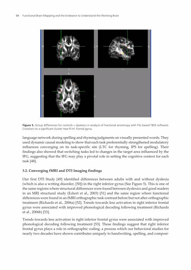
Our first DTI Study [49] identified differences between adults with and without dyslexia(which is also a writing disorder, [50]) in the right inferior gyrus (See Figure 5). This is one ofthe same regions where structural differences were found between dyslexics and good readersin an MRI structural study (Eckert et al., 2003) [51] and the same region where functionaldifferences were found in an fMRI orthographic task contrast before but not after orthographictreatment (Richards et al., 2006a) [52]. Trends towards less activation in right inferior frontalgyrus were associated with improved phonological decoding following treatment (Richardset al., 2006b) [53].
Trends towards less activation in right inferior frontal gyrus were associated with improvedphonological decoding following treatment [53]. These findings suggest that right inferiorfrontal gyrus plays a role in orthographic coding, a process which our behavioral studies fornearly two decades have shown contributes uniquely to handwriting, spelling, and composi‐
Figure 5. Group differences for controls > dyslexics in analysis of fractional anisotropy with FSL-based TBSS software.Crosshair on a significant cluster near R inf. frontal gyrus.
Functional Brain Mapping and the Endeavor to Understand the Working Brain84

tion[54]. Thus, we predict that in studies in progress children with handwriting disabilitieswill differ from good writers in the right inferior frontal gyrus.
Differences in functional connectivity were also found between children with and withoutdyslexia before but not after treatment on a phonological spelling task (phoneme mapping—deciding whether letter(s) in pair of pronounceable nonwords could stand for the samesound[55]. These data were analyzed with a seed point correlational method for functionalconnectivity from four seed points based on prior studies: inferior frontal gyrus, middle frontalgyrus, the occipital region, and cerebellum. Before treatment, there was a significant differencein fMRI connectivity between children with dyslexia and normal reading controls in the degreeof connectivity between left inferior frontal gyrus and the following regions: right and leftmiddle frontal gyrus, right and left supplemental motor area, left precentral gyrus, and rightsuperior frontal gyrus. There were no significant differences when seed regions were placedin the middle frontal gyrus, occipital gyrus or cerebellum. Children with dyslexia had greaterfunctional connectivity from the left inferior frontal gyrus seed point to the right inferior frontalgyrus than did the children without dyslexia as shown in Figure 6.
The children with dyslexia then participated in a 3-week instructional program that providedexplicit instruction in linguistic awareness, alphabetic principle (taught in a way to maximizetemporal contiguity of grapheme–phoneme associations and to train both phonological andorthographic loops), decoding and spelling. At Time2, the treated children with did not differ
Figure 6. Group difference map for dyslexics greater than controls. The individual maps used in this analysis were cor‐relation maps created when the seed ROI in the left inferior frontal gyrus was compared to the rest of the brain voxels.
Brain Mapping of Language Processing Using Functional MRI…http://dx.doi.org/10.5772/56501
85

from the children without dyslexia in any of the clusters in the group. The main result wasthat children with dyslexia had greater functional connectivity from the left inferior frontalgyrus seed point to the right inferior frontal gyrus than did the children without dyslexia beforebut not after treatment [55]. Thus, the structural and functional connectivity studies providedconverging evidence for abnormalities related to inferior frontal gyrus (on right or left) inchildren with dyslexia.
3.3. Stanberry model of fMRI connectivity in dyslexia
Stanberry et al [35] developed a model of fMRI connectivity based on earlier results thatpredicts that for normal readers there will be functional connectivity among 5 major reading-related brain regions: (a) frontal lobe (including the inferior frontal gyrus and middle frontalgyrus); (b) parietal lobe (including the angular gyrus); (c) visual processing areas (includingoccipitotemporal region); (d) fusiform/lingual word form region; and (e) the cerebellum. Thismodel is generally consistent with that reported by other research groups for normal reading[46]; it is also consistent with phonological loop in verbal working memory as a deficit indyslexia [56, 57]. We predicted that individual dyslexics may have impaired connectivity inany one or a combination of these major circuits. In our first fMRI connectivity study, weinvestigated differences in cortical networks used by adult controls compared to adultdyslexics during the previously described Phoneme Mapping. By definition, functionalconnectivity refers to a correlation or synchronization between the time courses of activationof two brain regions. We hypothesized that two brain regions that work together have similartemporal response profiles [58]. A model-independent method was used to analyze the time-synchronized activations induced by the phoneme mapping paradigm (adapted from [59])presented during a continuous task presentation. A standard fMRI acquisition and analysis ofthe on-off block design was also performed using Phoneme Mapping. Native English speakersranging in age from 30 to 45 years participated in the connectivity study: 10 healthy right-handed control males (fathers from the family genetics study who did not meet researchcriteria for dyslexia on tests and also did not have a history of reading problems) and 13 right-handed, otherwise healthy, adult males who did meet the research criteria for dyslexia andhad a history of reading and writing problems. The two groups did not differ significantly inmean Verbal IQ [dyslexics, M=113.8 (SD = 10.3); controls, M=107.7 (SD=11.1), but the dyslexicswere significantly lower than the control fathers on each of the reading, spelling, and RANmeasures.
Structural and functional MR images were collected in accordance with institutional regula‐tions (IRB approval) on a commercial 1.5T MR scanner (General Electric, Waukesha, WI)equipped with echo-speed gradients and a standard birdcage head coil. Functional imageswere acquired using an echo-planar sequence with imaging parameters set as follows:“On-Off” task: 20 axial slices, FOV 24cm x 24cm, BW +/- 62.5 kHz, TR 2000ms, TE 40ms, Flip82 deg, slice thickness 6mm, gap 1mm, resolution 64x64, 162 time points; Continuous task:20 axial slices, FOV 24cm x 24cm, BW +/- 62.5 kHz, TR 2000ms, TE 40 ms, Flip 82 deg, slicethickness 6mm, gap 1mm, resolution 64x64, 483 time points.
Functional Brain Mapping and the Endeavor to Understand the Working Brain86

Cardiac and respiratory rates were digitally recorded with a pulse oximeter and a flexible belt,respectively, using a sampling frequency of 100Hz. Three different seed regions were used forconnectivity analysis – right and left inferior frontal gyrus and cerebellum.
For the standard block fMRI acquisition and analysis of controls, fMRI brain activation wasdetected in the following brain regions: for the right side - inferior frontal gyrus, middle frontalgyrus, cerebellum crus I, cerebellum crus II, occipital gyrus, superior parietal gyrus, inferiorparietal gyrus, angular gyrus, lingual gyrus, and fusiform gyrus; for the left side – superiorparietal gyrus, angular, occipital gyrus, cerebellum crus I, cerebellum crus II, lingual.
For the fMRI connectivity analysis of the continuous phoneme mapping paradigm, wenarrowed the five region model above to a focus on three regions based on structural MRIdifferences in dyslexics from a family genetics study [51]. Results showed that (a) when theright IFG was chosen as the seed region, significant differences (p<.05) were found betweendyslexics and controls in right inferior frontal triangularis, bilateral fusiform, bilateral middleand inferior occipital gyri, right angular gyrus, bilateral ITG and cerebellum; (b) when the leftIFG was chosen as the seed region, significant differences (p<.05) were found between dyslexicsand controls in the following brain regions: right inferior frontal triangularis, right middleoccipital gyrus, right inferior occipital gyrus, and right cerebellum (VI); and (c) when thecerebellum was chosen as the seed region, significant differences (p<.05) were found betweendyslexics and controls in the following brain regions: bilateral superior frontal gyrus, leftmiddle frontal gyrus, right angular gyrus, and right middle occipital gyrus. Adult dyslexics,when compared to controls, had impaired cortical connections in brain regions important forphonological processing. The abnormality in functional connectivity from cerebellum indyslexics may be related to Klingberg et al.’s [30] finding, based on DTI, that white matterdiffusion anisotropy in the temporo-parietal region of the left hemisphere is significantlycorrelated with reading in normal and dyslexic readers. Insufficient myelination of the axonalpathways is one possible explanation for the low anisotropy index values observed in poorreaders [60]. Structural abnormalities in white matter pathways could interfere with neuronaltransmission, which will directly affect the synchrony of the BOLD signal. Of most importance,functional disconnections were also observed when seed regions were set in bilateral IFG.Bilateral IFG and right cerebellum were found to be abnormal in child dyslexics compared tonormal controls ascertained using the same research criteria in our structural MRI studies [51].Also see Berninger, Raskind, Richards et al. [50].
4. Future perspectives
One of the great potential techniques in this area of language connectivity analysis is theintegration of both functional and structural connectivity as shown by Morgan et al [61]. Theymeasured connections between Wernicke's (WA), Broca's (BA) and supplementary motor area(SMA). Along the path between BA and SMA, they showed that fibers tracked measured fromDTI generally formed a single bundle and the mean radius of the bundle was positivelycorrelated with functional connectivity. They concluded that the insights gained from this
Brain Mapping of Language Processing Using Functional MRI…http://dx.doi.org/10.5772/56501
87
work offers a useful guidance for non-invasive means to evaluate brain network integrity invivo for use in diagnosing and determining disease progression and recovery [61]. The conceptof integrating information across brain imaging modalities will allow the study of humanlanguage network as a systems approach. Another futuristic concept has been described byRota et al [62] where they discuss the mechanisms of cortical reorganization underlying theenhancement of speech. They were able to measure changes in functional and effectiveconnectivity induced in subjects who learned to deliberately increase activation in the rightinferior frontal gyrus [62]. Also, see [63] for a model of the four multi-leveled functionallanguage systems, which provides the conceptual framework for testing a model that differ‐entiates among typical oral and written language learners (OWLs), dysgraphia, dyslexia, andOWL LD at the behavioral (phenotype and response to instruction) and brain levels of analysis.
5. Conclusions
The language connectivity findings discussed in this chapter suggest that structural andfunctional connectivity are adding and will continue to add to our understanding of languageand language learning. There are specific language pathways and connections that are crucialfor language acquisition and function. The integrity of these connections can be tested usingstructural DTI and functional MRI connectivity imaging. Individuals with learning andlanguage disabilities have been reported to have different fMRI and DTI measurable connec‐tions than those with normal language functions. Once the techniques have been fully testedand developed, the application of language connectivity techniques to the individual assess‐ment, treatment design, and response to treatment would also have enormous practicalapplications in the clinic and schools.
Acknowledgements
This project received support from the NIH/NICHD Grant 1P50HD071764 (overall PI VirginiaBerninger, PI of project 3 Todd Richards).
Author details
Todd L. Richards1* and Virginia W. Berninger2
*Address all correspondence to: [email protected]
1 Department of Radiology, University of Washington Medical Center, Seattle, WA, USA
2 Department of Educational Psychology, University of Washington, Seattle, WA, USA
Functional Brain Mapping and the Endeavor to Understand the Working Brain88

References
[1] Liberman A. The reading researcher and the reading teacher need the right theory ofspeech. Scientific Studies of Reading 1999;3:95-111.
[2] Berninger V, Graham S. Language by hand: A synthesis of a decade of research onhandwriting. Handwriting Review 1998;12:11-25.
[3] Berninger V. Development of language by hand and its connections to language byear, mouth, and eye. Topics in Language Disorders, 20, 65-84 2000;20:65-84.
[4] Trask RL. Language: The Basics. New York: Routledge; 1999.
[5] Lesser R. Language in the Brain: Neurolinguistics, An Encyclopedia of Language.New York: Routledge; 1989.
[6] Binder JR, Frost JA, Hammeke TA, Cox RW, Rao SM, Prieto T. Human brain lan‐guage areas identified by functional magnetic resonance imaging. J Neurosci1997;17:353-362.
[7] Binder JR, Frost JA, Hammeke TA, Bellgowan PS, Springer JA, Kaufman JN, et al.Human temporal lobe activation by speech and nonspeech sounds. Cereb Cortex2000;10(5):512-28.
[8] Cannestra AF, Bookheimer SY, Pouratian N, O'Farrell A, Sicotte N, Martin NA, et al.Temporal and topographical characterization of language cortices using intraopera‐tive optical intrinsic signals. Neuroimage 2000;12(1):41-54.
[9] Castillo EM, Simos PG, Davis RN, Breier J, Fitzgerald ME, Papanicolaou AC. Levelsof word processing and incidental memory: dissociable mechanisms in the temporallobe. Neuroreport 2001;12(16):3561-6.
[10] Jancke L, Wustenberg T, Scheich H, Heinze HJ. Phonetic perception and the temporalcortex. Neuroimage 2002;15(4):733-46.
[11] McDermott KB, Petersen SE, Watson JM, Ojemann JG. A procedure for identifyingregions preferentially activated by attention to semantic and phonological relationsusing functional magnetic resonance imaging. Neuropsychologia 2003;41(3):293-303.
[12] Bookheimer S. Functional MRI of language: new approaches to understanding thecortical organization of semantic processing. Annu Rev Neurosci 2002;25:151-88.
[13] Hickok G, Poeppel D. Dorsal and ventral streams: a framework for understandingaspects of the functional anatomy of language. Cognition 2004;92(1-2):67-99.
[14] Price CJ, Mummery CJ, Moore CJ, Frakowiak RS, Friston KJ. Delineating necessaryand sufficient neural systems with functional imaging studies of neuropsychologicalpatients. J Cogn Neurosci 1999;11(4):371-82.
Brain Mapping of Language Processing Using Functional MRI…http://dx.doi.org/10.5772/56501
89
[15] Paulesu E, Goldacre B, Scifo P, Cappa SF, Gilardi MC, Castiglioni I, et al. Functionalheterogeneity of left inferior frontal cortex as revealed by fMRI. Neuroreport1997;8(8):2011-7.
[16] Broca P. Nouvelle observation d'aphemie produite par une lesion de la troisieme cir‐convolution frontale. Bulletins de la Societe anatomie (Paris), 2e serie 1861;6:398-407.
[17] Wernicke C. The symptom complex of aphasia (1874). Reprinted in English in Proc.Boston Colloq. Philos. Sci. 1874;4:34-97.
[18] Dejerine J. Anatomy of central nervous system. Paris: Masson; 1895.
[19] Mullen T. SIFT Online Handbook and User Manual. http://sccn.ucsd.edu/wiki/SIFT2010.
[20] Bullmore E, Sporns O. Complex brain networks: graph theoretical analysis of struc‐tural and functional systems. Nat Rev Neurosci 2009;10(3):186-98.
[21] Beaulieu C. The basis of anisotropic water diffusion in the nervous system - a techni‐cal review. NMR Biomed 2002;15(7-8):435-55.
[22] Le Bihan D, Mangin JF, Poupon C, Clark CA, Pappata S, Molko N, et al. Diffusiontensor imaging: concepts and applications. Journal of magnetic resonance imaging:JMRI 2001;13(4):534-546.
[23] Filler AI. The History, Development and Impact of Computed Imaging in Neurologi‐cal Diagnosis and Neurosurgery: CT, MRI, and DTI. Internet Journal of Neurosur‐gery 2010;7(1).
[24] Catani M, Mesulam M. The arcuate fasciculus and the disconnection theme in lan‐guage and aphasia: history and current state. Cortex 2008;44(8):953-61.
[25] Nucifora PG, Verma R, Melhem ER, Gur RE, Gur RC. Leftward asymmetry in rela‐tive fiber density of the arcuate fasciculus. Neuroreport 2005;16(8):791-4.
[26] Parker GJ, Luzzi S, Alexander DC, Wheeler-Kingshott CA, Ciccarelli O, LambonRalph MA. Lateralization of ventral and dorsal auditory-language pathways in thehuman brain. Neuroimage 2005;24(3):656-66.
[27] Powell HW, Parker GJ, Alexander DC, Symms MR, Boulby PA, Wheeler-KingshottCA, et al. Hemispheric asymmetries in language-related pathways: a combined func‐tional MRI and tractography study. Neuroimage 2006;32(1):388-99.
[28] Niogi SN, McCandliss BD. Left lateralized white matter microstructure accounts forindividual differences in reading ability and disability. Neuropsychologia 2006;44:2178-2188.
[29] Deutsch GK, Dougherty RF, Bammer R, Siok WT, Gabrieli JD, Wandell B. Children'sreading performance is correlated with white matter structure measured by diffusiontensor imaging. Cortex 2005;41:354-363.
Functional Brain Mapping and the Endeavor to Understand the Working Brain90

[30] Klingberg T, Hedehus M, Temple E, Salz T, Gabrieli JD, Moseley ME, et al. Micro‐structure of temporo-parietal white matter as a basis for reading ability: evidencefrom diffusion tensor magnetic resonance imaging [see comments]. Neuron2000;25(2):493-500.
[31] Glasser MF, Rilling JK. DTI tractography of the human brain's language pathways.Cereb Cortex 2008;18(11):2471-82.
[32] Hampson M, Peterson B, Skudlarski P., Gatenby J, Gore J. Detection of functionalconnectivity using temporal correlations in MR images. Human Brain Mapping2002;15:247-262.
[33] Lowe M, Mock B, Sorenson J. Functional connectivity in single and multislice echo‐planar imaging using resting-state fluctuations. Neuroimage 1998;7:119-132.
[34] Stanberry L, Nandy R, Cordes D. Cluster analysis of fMRI Data using DendrogramSharpening. Human Brain Mapping 2003;20:201-219.
[35] Stanberry LI, Richards T, Berninger VW, Stock P, Nandy RR, Aylward E, et al. LowFrequency Signal Changes Reflect Differences in Functional Connectivity betweenGood Readers and Dyslexics during Continuous Phoneme Mapping. Magnetic Reso‐nance Imaging 2006;24:217-229.
[36] Nandy R, Cordes D. Improving the spatial specificity of canonical correlation analy‐sis in fMRI. Magn Reson Med 2004;52(4):947-52.
[37] Nandy RR, Cordes D. Novel nonparametric approach to canonical correlation analy‐sis with applications to low CNR functional MRI data. Magn Reson Med 2003;50(2):354-65.
[38] Nandy RR, Cordes D. Novel ROC-type method for testing the efficiency of multivari‐ate statistical methods in fMRI. Magn Reson Med 2003;49(6):1152-62.
[39] Cordes D, Haughton V, Carew JD, Arfanakis K, Maravilla K. Hierarchical clusteringto measure connectivity in fMRI resting-state data. Magn Reson Imaging 2002;20(4):305-17.
[40] Cordes D, Haughton VM, Arfanakis K, Carew JD, Turski PA, Moritz CH, et al. Fre‐quencies contributing to functional connectivity in the cerebral cortex in "resting-state" data. AJNR Am J Neuroradiol 2001;22(7):1326-33.
[41] Cordes D, Haughton VM, Arfanakis K, Wendt GJ, Turski PA, Moritz CH, et al. Map‐ping functionally related regions of brain with functional connectivity MR imaging[In Process Citation]. AJNR Am J Neuroradiol 2000;21(9):1636-44.
[42] Büchel C, Coull, J., Friston, K. The predictive value of changes in effective connectivi‐ty for human learning. Science 1999;283:1538-1540.
Brain Mapping of Language Processing Using Functional MRI…http://dx.doi.org/10.5772/56501
91
[43] Cordes D, Haughton VM, Arfanakis K, Wendt GJ, Turski PA, Moritz CH, et al. Map‐ping functionally related regions of brain with functional connectivity MR imaging.AJNR Am J Neuroradiol 2000;21(9):1636-44.
[44] Horwitz B, Rumsey JM, Donohue BC. Functional connectivity of the angular gyrus innormal reading and dyslexia. Proc Natl Acad Sci U S A 1998;95(15):8939-44.
[45] Pugh K, Mencl, W., Shaywitz, B., Shaywitz, S., Fulbright, R., Constable, R., Skudlar‐ski, P., Marchione, K., Jenner, A., Fletcher, J., Liberman, A., Shankweiler, D., Katz, L.,Lacadie, C., Gore,J. The angular gyrus in developmental dyslexia: Task-specific dif‐ferences in functional connectivity within posterior cortex. Psychological Science2000;11:51-56.
[46] Shaywitz SE, Shaywitz BA, Fulbright RK, Skudlarski P, Mencl WE, Constable RT, etal. Neural systems for compensation and persistence: young adult outcome of child‐hood reading disability. Biol Psychiatry 2003;54(1):25-33.
[47] Milne D, Syngeniotis A, Jackson G, Corballis M. Mixed lateralization of phonologicalassembly in developmental dyslexia. Neurocase 2002;8:205-209.
[48] Bitan T, Booth JR, Choy J, Burman DD, Gitelman DR, Mesulam MM. Shifts of effec‐tive connectivity within a language network during rhyming and spelling. J Neurosci2005;25(22):5397-403.
[49] Richards T, Stevenson J, Crouch J, Johnson LC, Maravilla K, Stock P, et al. Tract-based spatial statistics of diffusion tensor imaging in adults with dyslexia. AJNR AmJ Neuroradiol 2008;29(6):1134-9.
[50] Berninger VW, Raskind W, Richards T, Abbott R, Stock P. A multidisciplinary ap‐proach to understanding developmental dyslexia within working-memory architec‐ture: genotypes, phenotypes, brain, and instruction. Dev Neuropsychol 2008;33(6):707-44.
[51] Eckert MA, Leonard CM, Richards TL, Aylward EH, Thomson J, Berninger VW. Ana‐tomical correlates of dyslexia: frontal and cerebellar findings. Brain 2003;126(Pt 2):482-494.
[52] Richards T, Aylward E, Berninger V, Field K, Parsons A, Richards A, et al. IndividualfMRI activation in orthographic mapping and morpheme mapping after orthograph‐ic or morphological spelling treatment in child dyslexics. Journal of Neurolinguistics2006;19:56-86.
[53] Richards T, Aylward E, Raskind W, Abbott R, Field K, Parsons A, et al. Convergingevidence for triple word form theory in child dyslexia. Special Issue on Brain Imag‐ing in Developmental Neuropsychology 2006;in press.
[54] Berninger V, Richards T. Inter-relationships among behavioral markers, genes, brainand treatment in dyslexia and dysgraphia. Future Neurol 2011;5(4):597-617.
Functional Brain Mapping and the Endeavor to Understand the Working Brain92

[55] Richards TL, Berninger VW. Abnormal fMRI Connectivity in Children with DyslexiaDuring a Phoneme Task: Before But Not After Treatment. J Neurolinguistics2008;21(4):294-304.
[56] Heilman KM, Voeller K, Alexander AW. Developmental dyslexia: a motor-articulato‐ry feedback hypothesis. Ann Neurol 1996;39(3):407-12.
[57] Chen SH, Desmond JE. Cerebrocerebellar networks during articulatory rehearsal andverbal working memory tasks. Neuroimage 2005;24:332-338.
[58] Koshino H, Carpenter PA, Minshew NJ, Cherkassky VL, Keller TA, Just MA. Func‐tional connectivity in an fMRI working memory task in high-functioning autism.Neuroimage 2005;24(3):810-21.
[59] Aylward EH, Richards TL, Berninger VW, Nagy WE, Field KM, Grimme AC, et al.Instructional treatment associated with changes in brain activation in children withdyslexia. Neurology 2003;61(2):212-9.
[60] Wimberger DM, Roberts TP, Barkovich AJ, Prayer LM, Moseley ME, Kucharczyk J.Identification of 'premyelination" by diffusion-weighted MRI. J Comput Assist To‐mogr 1995;19:28-33.
[61] Morgan VL, Mishra A, Newton AT, Gore JC, Ding Z. Integrating functional and dif‐fusion magnetic resonance imaging for analysis of structure-function relationship inthe human language network. PLoS One 2009;4(8):e6660.
[62] Rota G, Handjaras G, Sitaram R, Birbaumer N, Dogil G. Reorganization of functionaland effective connectivity during real-time fMRI-BCI modulation of prosody proc‐essing. Brain Lang 2011;117(3):123-32.
[63] Berninger V, Niedo J. Individualizing instruction for students with oral and writtenlanguage difficulties. In: Mascolo J, Flanagan D, Alfonso V, editors. Essentials ofplanning, selecting and tailoring intervention: Addressing the needs of unique learn‐ers. New York: Wiley; 2013. In Press
Brain Mapping of Language Processing Using Functional MRI…http://dx.doi.org/10.5772/56501
93

Related Documents