13-1 Brain and Cranial Nerves Chapter 13

Brain Gross Anatomy
Nov 28, 2015
gross anatomy of the brain kin 1y03
Welcome message from author
This document is posted to help you gain knowledge. Please leave a comment to let me know what you think about it! Share it to your friends and learn new things together.
Transcript

13-1
Brain and Cranial NervesChapter 13

Brain-2
BrainAnterior
Posterior Inferior view
Tanweer Hoosen
cranial nerves are part of the peripheral nervous sytem

Brain-3
Brain: Gross Anatomy• Brain
– Part of CNS contained in cranial cavity
• Brainstem: connects spinal cord to brain; integration of reflexes necessary for survival
• Cerebellum: involved in control of locomotion, balance, posture
• Diencephalon: thalamus, subthalamus, epithalamus, hypothalamus
• Cerebrum: conscious thought, control
Tanweer Hoosen
4 main components to the brain
Tanweer Hoosen
Brain stem - most inferior portion of the brain, connection between spinal cord and rest of structures of the brain, Foramen magnum - superior is the brain stem, below is the spinal cord, functions that are really important to keep you alive, but are unconsciously controlled, ex respiratory centre, cardiovascular control centre
Tanweer Hoosen
Tanweer Hoosen
Tanweer Hoosen
Tanweer Hoosen
hormones can also control the heart rate
Tanweer Hoosen
Tanweer Hoosen
posterior to the brain stem, but on the same level as the brain stem
Tanweer Hoosen
Tanweer Hoosen
coordinate skeletal muscle movement (how to smooth them out), but doesnt initiate them
Tanweer Hoosen
Tanweer Hoosen
proprioception - contraction of skeletal muscle compared to joints, cerebellum
Tanweer Hoosen
Tanweer Hoosen
small set of structures that are deep in the brain, sit on top of the brain stem,
Tanweer Hoosen
diencephalon functions for regulation of thirst, emotions, moods, body temperature, controlling functions that are vital but not respiratory or cardiovascular, main relay center for sensory information in the brain
Tanweer Hoosen
Tanweer Hoosen
Tanweer Hoosen
cerebrum - higher level brain function ,conscious thought, control of the body, reasoning, accessing and storing memories, planning, interpreting sensory information and creating an appropriate motor response
Tanweer Hoosen
Tanweer Hoosen
Tanweer Hoosen

Brain-4
Sagittal Section of Brain
Cerebrum
Cerebellum
Thalamus
Hypothalamus
Midbrain
Pons
Medulla Oblongata
Brainstem
Diencephalon
Tanweer Hoosen
cerebellum - 10% of mass of brain, but contains 50% of the neurons

Brain-5
Brainstem
Pyramid
(a) Anterior view
(b) Posterolateral view
Midbrain
MidbrainPons
Brainstem
Olive
Superior colliculusInferior colliculus
Olive
Pons
Brainstem
Superior cerebellarpeduncleMiddle cerebellarpeduncleInferior cerebellarpeduncle
Medulla oblongata
Medullaoblongata
Pyramidaldecussation
Tanweer Hoosen
light pink are the brain stem, dark pink is the diencephalon, yellow are cranial nerves
Tanweer Hoosen
Tanweer Hoosen
peduncles are the white matter that connect the cerebellum to stem

Brainstem
Medulla oblongata Pons Midbrain

Brain-7
BrainstemMedulla Oblongata
• transmits ascending and descending impulses between the brain and spinal cord
• pyramids – nerve tracts – conscious control of skeletal muscle
• center for vital reflexes – heart rate, BP, respiration, swallowing, coughing, sneezing
• olives - nuclei involved in balance, coordination, modulation of sound
• nuclei of cranial nerves V, VII, IX, X, XI, XII
Tanweer Hoosen
bundle of myelinated fibres, white looking
Tanweer Hoosen
pyramidal decussation - the axons on the left side cross over to the right side, and the axons on right side cross over to left side
Tanweer Hoosen
Tanweer Hoosen
relay information to the cerebelum about positions of skeletal muscle and joints, relayed through the olives, are bilateral
Tanweer Hoosen
Tanweer Hoosen
only motor information through the pyramids, descending tracts
Tanweer Hoosen
Tanweer Hoosen
vomitting
Tanweer Hoosen
Tanweer Hoosen
receive information from inner ear, helping with relay of sounds
Tanweer Hoosen
Tanweer Hoosen
located in the medulla oblongata
Tanweer Hoosen
bundles of myelinated axons in the peripheral system are called nerves, if in central nervous system are called tracts

Brain-8
BrainstemPons
• ascending and descending nerve tracts
• nuclei
• sleep centre, respiratory centre
Anterior• Pontine nuclei – communication between
cerebrum and cerebellum
Posterior• nuclei of cranial nerves V, VI, VII, VIII
Tanweer Hoosen
Rapid eye movement sleep,
Tanweer Hoosen
works with the medulla oblongata to regulate the respiratory system
Tanweer Hoosen
Tanweer Hoosen
pons means bridge
Tanweer Hoosen
a bridge between the two areas

BrainstemMidbrain• most superior portion of the brainstem
• nuclei of cranial nerves III, IV, V
• Tectum: 4 nuclei - form mounds on dorsal surface of midbrain
- each separate part is a colliculus
- two superior colliculi involved in visual reflexes
- two inferior colliculi involved in hearing
Tanweer Hoosen

Reticular Formation
• Group of nuclei scattered throughout brainstem
• Cloud throughout the brainstem
• Controls cyclic activities such as sleep-wake cycle
Brainstem
Brain-10
Tanweer Hoosen
nuclei connected by web like white matter
Tanweer Hoosen
wont have to label, but know the function and name
Tanweer Hoosen
aka reticular cloud

Brain-11
Purkinje cell
Golgicell
Granulecell
Mossy fiberAxon of Purkinje cell
(b) Inferior view
Cerebellum
Lateral hemisphere
FoliaVermis
Anteriorlobe
Posteriorlobe
Vermis
Anterior lobe
FoliaPons
Flocculonodularlobe
Lateralhemisphere
Medullaoblongata
Posteriorlobe
Arborvitae
(a) Medial view
Vermis
Flocculonodular lobe
Tanweer Hoosen
10 percent of brain mass, but has 50 percent of neurons

Cerebellum

Brain-13
CerebellumCommunicates with other regions of the CNS:
�
superior cerebellar peduncle – midbrain
�
middle cerebellar peduncle – pons
�
inferior cerebellar peduncle – medulla oblongata
3 Regions:
1. Flocculonodular lobe – balance2. Vermis – gross motor coordination (Anterior), fine
motor coordination (Posterior)3. Lateral hemisphere – fine motor coordination

Brain-14
Diencephalon• between brainstem and cerebrum• 4 main components:
• Thalamus• Subthalamus• Epithalamus• Hypothalamus
Diencephalon, medial view
ThalamusInterthalamicadhesion
Hypothalamus
Pituitary gland
HabenulaPineal gland Epithalamus
Subthalamus

Diencephalon

Brain-16
DiencephalonThalamus• largest part of the diencephalon
• lateral portions connected by interthalamic adhesion - surrounded by third ventricle
• receives major portion of sensory input – projections to cerebral cortex
• Auditory impulses – medial geniculate nucleus
• Visual impulses – lateral geniculate nucleus
Interthalamicadhesion
Lateral geniculatenucleus
Tanweer Hoosen
"bent knee"
Tanweer Hoosen
Tanweer Hoosen

Thalamus
14-17

Brain-18
DiencephalonSubthalamus
• inferior to the thalamus
• ascending and descending nerve tracts
• subthalamic nuclei – controlling motor function
Thalamus
Hypothalamus Subthalamus

Brain-19
DiencephalonEpithalamus
• posterior and superior to the thalamus
• Habenula – emotional and visceral responses to odor
• Pineal gland – may influence the onset of puberty
Thalamus
Hypothalamus
HabenulaPineal gland EpithalamusSubthalamus
Tanweer Hoosen
melatonin releaed from pineal gland, light degrades melatoninmelatonin helps maintian sleep
Tanweer Hoosen

Pineal Gland
Brain-21
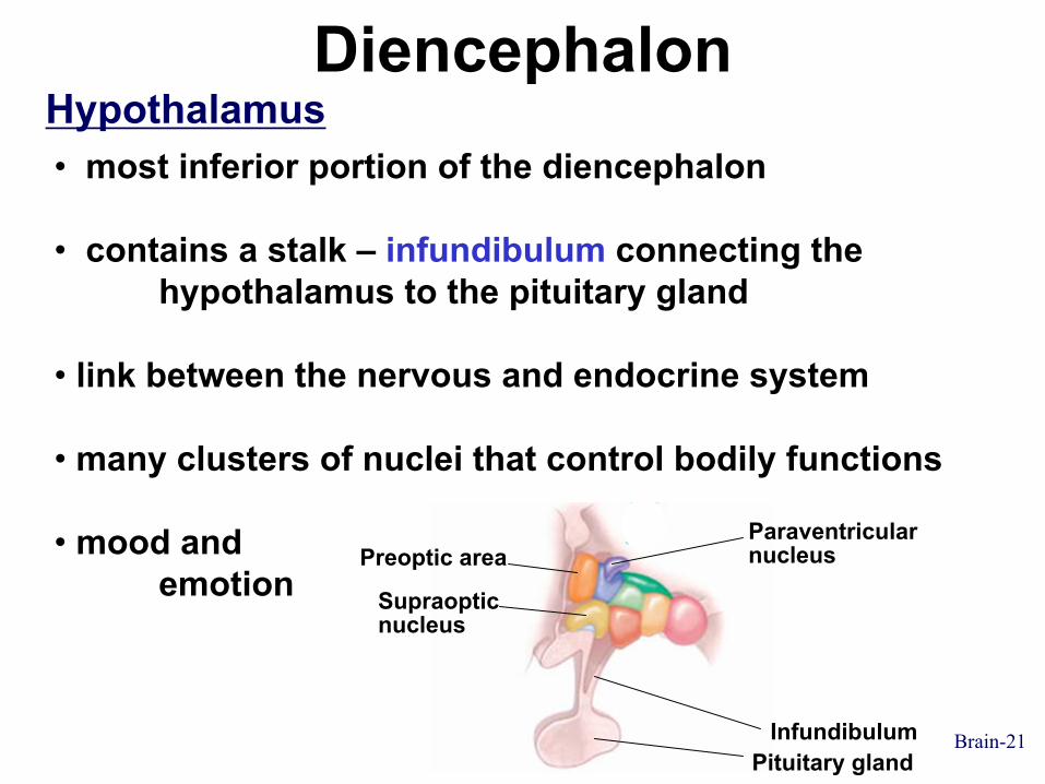
DiencephalonHypothalamus• most inferior portion of the diencephalon
• contains a stalk – infundibulum connecting the hypothalamus to the pituitary gland
• link between the nervous and endocrine system
• many clusters of nuclei that control bodily functions
• mood and emotion
Paraventricularnucleus
Infundibulum
Supraopticnucleus
Preoptic area
Pituitary gland

Hypothalamus

Brain-23
Cerebrum• outer layer – cerebral cortex (gray matter)
• deep clusters of nuclei (gray matter)
• in between – cerebral medulla (white matter)
Gray matter
White matter
Basal nuclei

Brain-24
Cerebrum
Anterior
Frontal lobe-Motor function-Aggression, mood
Parietal lobe-touch, taste, pressure-blood pH-not smell, vision, hearing
Occipital lobe-reception and integration of visual input
Temporal lobe-olfactory, auditory input-memory
Central sulcus
Lateral fissure
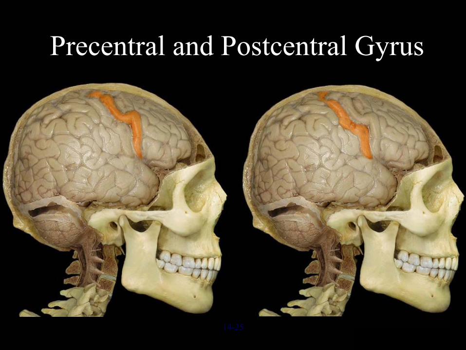
Precentral and Postcentral Gyrus
14-25

Brain-26
CerebrumCerebral Medulla
• nerve tracts connect the cortex to other areas of cortex or other parts of the CNS
• Association fibers – connect areas of cerebral cortex within the same hemisphere
• Commissural fibers – connect the cerebral hemispheres (Corpus Callosum)
• Projection fibers – between the cerebrum and other parts of the brain and spinal cord (Internal capsule)

Brain-27
CerebrumCerebral Medulla
Longitudinalassociationfibers
Commissural fibers(corpus callosum)
NucleiCortex
Shortassociationfibers
Projection fibersInternalcapsule Cerebral
medulla
Association fibersCommissural fibersProjection fibers
(a) Anterior view
Projectionfibers inthe internalcapsule
AnteriorCerebrum
Brainstem
(b) Lateral view Cerebellum
Posterior

Brain-28
Meninges
(a) Anterosuperior view
Skull
Subdural space
Arachnoid mater
Subarachnoid space
Pia mater
Cerebrum
Periosteal duraMeningeal dura
Dura mater
Dural venous sinus(superior sagittal sinus)

Brain-29
Ventricles
Lateral ventricle
Cerebral aqueduct
Fourth ventricle
Third ventricle
Lateral view
Posterior horn oflateral ventricle
Central canalof spinal cord
Anterior horn oflateral ventricleInterventricularforamen
Inferior horn oflateral ventricle
Brain-30
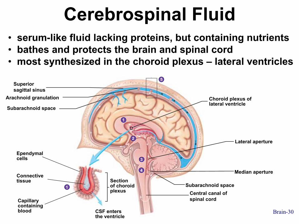
Cerebrospinal Fluid
Ependymalcells
Connectivetissue
Capillarycontainingblood CSF enters
the ventricle
Section of choroidplexus
Choroid plexus oflateral ventricle
1
2
3
4
5
1
• serum-like fluid lacking proteins, but containing nutrients• bathes and protects the brain and spinal cord• most synthesized in the choroid plexus – lateral ventricles
Superiorsagittal sinus
Arachnoid granulation
Subarachnoid space
Median aperture
Lateral aperture
Subarachnoid spaceCentral canal ofspinal cord

i Clicker QuestionThe pyramids are __________ nerve tracts
that are involved in the conscious control of __________ .
A. ascending, smooth muscle B. ascending, skeletal muscle C. descending, smooth muscle D. descending, cardiac muscle E. descending, skeletal muscle
Tanweer Hoosen

The medulla oblongata does not include the
A. nuclei of some cranial nervesB. OlivesC. PyramidsD. centers for some vital reflexesE. thalamus
i Clicker Question
Tanweer Hoosen

iClicker Question
The pineal gland is found in the
A. subthalamus. B. thalamus. C. epithalamus. D. hypothalamus. E. pons.
Tanweer Hoosen

i Clicker Question
The infundibulum connects
A. the hypothalamus and pituitary gland. B. the thalamus and hypothalamus. C. the thalamus and epithalamus. D. the thalamus and subthalamus. E. the medulla and the pons.
Tanweer Hoosen

i Clicker Question
The primary motor cortex is located in the
A. frontal lobe. B. occipital lobe. C. postcentral gyrus. D. precentral gyrus. E. temporal lobe.
Tanweer Hoosen
Tanweer Hoosen
i Clicker Question
Cerebrospinal fluid passes into the blood by way of the
A. arachnoid granulations. B. cerebral aqueduct. C. lateral ventricles. D. choroid plexus. E. brachial plexus.
Tanweer Hoosen
Related Documents