University of Vermont University of Vermont UVM ScholarWorks UVM ScholarWorks Graduate College Dissertations and Theses Dissertations and Theses 2021 Brain-Behavior Connections Underlying Emotion and Theory of Brain-Behavior Connections Underlying Emotion and Theory of Mind In Autism Spectrum Disorder Mind In Autism Spectrum Disorder Yu Han University of Vermont Follow this and additional works at: https://scholarworks.uvm.edu/graddis Part of the Neuroscience and Neurobiology Commons Recommended Citation Recommended Citation Han, Yu, "Brain-Behavior Connections Underlying Emotion and Theory of Mind In Autism Spectrum Disorder" (2021). Graduate College Dissertations and Theses. 1421. https://scholarworks.uvm.edu/graddis/1421 This Dissertation is brought to you for free and open access by the Dissertations and Theses at UVM ScholarWorks. It has been accepted for inclusion in Graduate College Dissertations and Theses by an authorized administrator of UVM ScholarWorks. For more information, please contact [email protected].

Welcome message from author
This document is posted to help you gain knowledge. Please leave a comment to let me know what you think about it! Share it to your friends and learn new things together.
Transcript
University of Vermont University of Vermont
UVM ScholarWorks UVM ScholarWorks
Graduate College Dissertations and Theses Dissertations and Theses
2021
Brain-Behavior Connections Underlying Emotion and Theory of Brain-Behavior Connections Underlying Emotion and Theory of
Mind In Autism Spectrum Disorder Mind In Autism Spectrum Disorder
Yu Han University of Vermont
Follow this and additional works at: https://scholarworks.uvm.edu/graddis
Part of the Neuroscience and Neurobiology Commons
Recommended Citation Recommended Citation Han, Yu, "Brain-Behavior Connections Underlying Emotion and Theory of Mind In Autism Spectrum Disorder" (2021). Graduate College Dissertations and Theses. 1421. https://scholarworks.uvm.edu/graddis/1421
This Dissertation is brought to you for free and open access by the Dissertations and Theses at UVM ScholarWorks. It has been accepted for inclusion in Graduate College Dissertations and Theses by an authorized administrator of UVM ScholarWorks. For more information, please contact [email protected].

Brain-Behavior Connections UnderlyingEmotion and Theory of Mind In Autism
Spectrum Disorder
A Dissertation Presented
by
Yu Han
to
The Faculty of the Graduate College
of
The University of Vermont
In Partial Fulfillment of the Requirementsfor the Degree of Doctor of Philosophy
Specializing in Neuroscience
August, 2021
Defense Date: May 14th, 2021Dissertation Examination Committee:
Patricia A. Prelock, Ph.D., AdvisorDonna M. Rizzo, Ph.D., Chairperson
Emily L. Coderre, Ph.D.Rodney C. Scott, Ph.D.
Tiffany L. Hutchins, Ph.D.Joseph M. Orr, Ph.D.
Cynthia J. Forehand, Ph.D., Dean of the Graduate College
Abstract
Autism Spectrum Disorder (ASD) is a complex neurodevelopmental disorder that af-fects nearly 1 in 54 children. Children with ASD struggle with social, communication,and behavioral challenges due to deficits in theory of mind (ToM). In addition, diag-nosis of ASD is complicated and there is an urgent need to identify ASD-associatedbiomarkers and features to help automate diagnostics and develop predictive ASDmodels. In this study, we conducted two experiments collecting behavioral and neu-roimaging data from 9 children with ASD and 19 neurotypical children (NT) betweenthe age of 7 and 14 years.
The first experiment examined specific elements of emotion recognition to bet-ter understand those skills needed for meaningful social interaction among childrenwith ASD. Two previously tested measures of ToM, the Theory of Mind Inventory-2(ToMI-2) and the Theory of Mind Task Battery (ToMTB), were used to evaluateearly developing, basic, and advanced theory of mind skills impacting children’s so-cial skills. We also created and implemented two novel fMRI paradigms to probe theneural mechanisms underlying ToM related desire-based emotion and more complexemotions (i.e., surprise and embarrassment), as well as two early-developing emotions(i.e., happy and sad). Results suggested impaired abilities in multiple ToM metricsand brain deficits associated with ToM-related emotion recognition and processingamong children with ASD. Findings from this study established connections betweenbehavior and brain activities surrounding ToM in ASD, which may assist the devel-opment of neuroanatomical diagnostic criteria and may provide new pathways formeasuring intervention outcomes in special populations such as those with ASD.
The second experiment adopted a novel evolutionary algorithm, the conjunctiveclause evolutionary algorithm (CCEA), to select features most significant for distin-guishing individuals with and without ASD, accommodating datasets having a smallnumber of samples with a large number of feature measurements. Potential biomarkercandidates identified included brain volume, area, cortical thickness, and mean cur-vature in specific regions around the cingulate cortex, the frontal cortex, and thetemporal-parietal junction, as well as behavioral features associated with theory ofmind. A separate machine learning classifier (i.e., k-nearest neighbors algorithm) wasused to validate the CCEA feature selection and then used for ASD prediction. Studyfindings demonstrated how machine learning tools might help to facilitate diagnosticand predictive models of ASD.

Citations
Material from this dissertation has been submitted for publication to Journal ofAutism and Developmental Disorders on April 9, 2021 in the following form:
Han, Y., Prelock, P.A., Coderre, E.L., & Orr, J.M.. (2021). A pilot study usingtwo novel fmri tasks: Understanding theory of mind and emotion recognition amongchildren with ASD (under review). Journal of Autism and Developmental Disorders.
Material from this dissertation has been submitted for publication to PLOS ONEon March 25th, 2021 in the following form:
Han, Y., Rizzo, D.M., Hanley, J.P., Coderre, E.L., & Prelock, P.A.. (2021).Identifying neuroanatomical and behavioral features for autism spectrum disorder di-agnosis in children using machine learning (under review). PLOS ONE.
ii
I dedicate my dissertation work to my amazing family, friends and many kindacquaintances who have shared their life and career wisdom with me. A special
feeling of gratitude to my loving parents. It is hard for me to put it into words howmuch they have sacrificed to help me become who I am and to get me where I amtoday. They have given me unconditional love, generous support, patience, and
encouragement. They also raised me to be courageous, adventurous and fearless andto step out of my comfort-zone to achieve my goals. Most importantly, they alwaysmake sure I know no matter how difficult things can be, they are always behind me.They are my biggest cheerleaders and greatest motivation to keep moving forward!
I also dedicate this dissertation to my beloved dog, whom I adopted onSeptember,14, 2016 from the Chittenden County Humane Society, Burlington,Vermont. Ever since, she has never left my side, brought me countless joy and
warmth, and kept me sane throughout this journey.
iii
Acknowledgements
I wish to thank my academic advisor, Dr. Patricia A. Prelock, for always going above
and beyond to be there for me and providing me with a tremendous amount of sup-
port, training, and resources to help accomplish my scientific goals. I faced so many
obstacles in the beginning, my dissertation project would not have been launched suc-
cessfully if it wasn’t her pushing me, encouraging me and guiding me. In addition to
her outstanding scientific mentorship, she has served as my mental health counselor
and life coach. She looked after me and guided me through every step of the way.
Her knowledge, intelligence, wisdom, resilience and attitudes towards life inspire me
every single day to be a better scientist, a colleague, and a mentor to others, to be
strong and brave, and to give back.
A special thanks to my committee chair, Dr. Donna M. Rizzo. She is one of the
most intelligent people I have met so far who seems to know everything. She has
always made me one of her priorities and provided consistent encouragement. Her
mentorship gave me so much more confidence and a competitive edge in the computer
science field. She exposed me to an incredibly fun world of artificial intelligence, along
with animals, plants, and other weird and nerdy things.
I also wish to thank my co-mentor, Dr. Emily L. Coderre, for joining this jour-
ney with me when I was in a desperate search for a faculty carrying neuroimaging
expertise. She has been committed to helping me achieve my scientific milestones.
She always had me as her priority and was generous with her time to address my
countless questions. She provided me with great mentorship in MRI/fMRI.
iv
I am very grateful for these incredibly intelligent, strong and kind women to guide
me and keep me company in this journey.
I thank Dr. Joseph M. Orr from Texas A&M University for agreeing to serve
on my committee even though he lives halfway across the United States, and consis-
tently sharing his substantial knowledge in neuroimaging. He guided and taught me
to problem solve through all kinds of neuroimaging issues and questions since day
one. I thank Dr. Tiffany L. Hutchins and Dr. Rodney C. Scott for their precious
time and support while being on my committee, especially to challenge me to think
outside the box.
I thank Dr. John P. Hanley for introducing me to the CCEA algorithm and es-
tablishing a great collaboration. I thank Jay V. Gonyea, Administrative Director,
and Scott Hipko, Senior Research Technologist, in the MRI Research Unit at the
University of Vermont. They were so knowledgeable and patient while supporting me
in acquiring the MRI scans with ASD children. I thank Dr. Richard Watts, Director
of the FAS Brain Imaging Center at Yale University, for sharing his knowledge in
MRI data pre-processing.
I am particularly thankful for a private donor committed to advancing research in
ASD who funded my graduate studies. Most importantly, I am truly grateful for all
the families and incredible children who dedicated their time and trust in my study.
I learned so much through the very special and magical minds of these children.
v

Table of ContentsDedication . . . . . . . . . . . . . . . . . . . . . . . . . . . . . . . . . . . . iiiAcknowledgements . . . . . . . . . . . . . . . . . . . . . . . . . . . . . . . ivList of Figures . . . . . . . . . . . . . . . . . . . . . . . . . . . . . . . . . . viiiList of Tables . . . . . . . . . . . . . . . . . . . . . . . . . . . . . . . . . . ix
1 Comprehensive Literature Review 11.1 Definition and Prevalence . . . . . . . . . . . . . . . . . . . . . . . . 11.2 Current Challenges . . . . . . . . . . . . . . . . . . . . . . . . . . . . 4
1.2.1 Cause . . . . . . . . . . . . . . . . . . . . . . . . . . . . . . . 51.2.2 Assessment and Diagnosis . . . . . . . . . . . . . . . . . . . . 91.2.3 Intervention . . . . . . . . . . . . . . . . . . . . . . . . . . . . 10
1.3 Theory of Mind . . . . . . . . . . . . . . . . . . . . . . . . . . . . . . 121.3.1 Behavioral Measurements of ToM in ASD . . . . . . . . . . . 151.3.2 Examining Specific Aspects of ToM: Emotion Recognition . . 18
1.4 Imaging Studies of ToM in ASD . . . . . . . . . . . . . . . . . . . . . 221.4.1 Imaging Studies of Emotion Recognition in ASD . . . . . . . . 241.4.2 Introduction of Chapter Two . . . . . . . . . . . . . . . . . . . 25
1.5 Machine Learning Approach . . . . . . . . . . . . . . . . . . . . . . . 261.5.1 Introduction of Chapter Three . . . . . . . . . . . . . . . . . . 29
2 A Pilot Study Using Two Novel fMRI Tasks: Understanding Theoryof Mind and Emotion Recognition Among Children With ASD 302.1 Introduction . . . . . . . . . . . . . . . . . . . . . . . . . . . . . . . . 31
2.1.1 Emotion Recognition in Neurotypical Development . . . . . . 332.1.2 Emotion Recognition in ASD . . . . . . . . . . . . . . . . . . 342.1.3 Purpose of the Study . . . . . . . . . . . . . . . . . . . . . . . 37
2.2 Methods . . . . . . . . . . . . . . . . . . . . . . . . . . . . . . . . . . 392.2.1 Participants . . . . . . . . . . . . . . . . . . . . . . . . . . . . 392.2.2 Behavioral Measures of ToM . . . . . . . . . . . . . . . . . . . 412.2.3 MRI Acquisition . . . . . . . . . . . . . . . . . . . . . . . . . 422.2.4 Task fMRI Parameters and Preprocessing . . . . . . . . . . . 432.2.5 fMRI Emotion Recognition (fER) Task . . . . . . . . . . . . . 432.2.6 fMRI Theory of Mind (fToM) Task . . . . . . . . . . . . . . . 45
2.3 Statistical Analyses . . . . . . . . . . . . . . . . . . . . . . . . . . . . 462.4 Results . . . . . . . . . . . . . . . . . . . . . . . . . . . . . . . . . . . 48
2.4.1 Behavioral Results . . . . . . . . . . . . . . . . . . . . . . . . 482.4.2 Brain Activity Patterns . . . . . . . . . . . . . . . . . . . . . . 49
2.5 Discussion . . . . . . . . . . . . . . . . . . . . . . . . . . . . . . . . . 52
vi

2.6 Limitations . . . . . . . . . . . . . . . . . . . . . . . . . . . . . . . . 562.7 Conclusions and Implications . . . . . . . . . . . . . . . . . . . . . . 572.8 Supplemental Materials . . . . . . . . . . . . . . . . . . . . . . . . . . 58
3 Identifying Neuroanatomical and Behavioral Features for AutismSpectrum Disorder Diagnosis in Children using Machine Learning 603.1 Introduction . . . . . . . . . . . . . . . . . . . . . . . . . . . . . . . . 613.2 Materials and Methods . . . . . . . . . . . . . . . . . . . . . . . . . . 66
3.2.1 Participants . . . . . . . . . . . . . . . . . . . . . . . . . . . . 663.2.2 Behavioral Measurements . . . . . . . . . . . . . . . . . . . . 673.2.3 MRI Acquisition and Preprocessing . . . . . . . . . . . . . . . 703.2.4 Conjunctive Clause Evolutionary Algorithm . . . . . . . . . . 713.2.5 K-nearest Neighbors Algorithm and Leave-One-Out Cross Val-
idation . . . . . . . . . . . . . . . . . . . . . . . . . . . . . . . 723.3 Results . . . . . . . . . . . . . . . . . . . . . . . . . . . . . . . . . . . 74
3.3.1 CCEA Feature Selection: 14 NT and 7 ASD . . . . . . . . . . 743.3.2 KNN Leave-One-Out Cross Validation . . . . . . . . . . . . . 803.3.3 Classification of ASD and NT subjects using the KNN model . 81
3.4 Discussion . . . . . . . . . . . . . . . . . . . . . . . . . . . . . . . . . 82
4 Conclusion and Future Direction 86
vii

List of Figures
1.1 Chapter One Advanced Organizer . . . . . . . . . . . . . . . . . . . . 21.2 DSM-5 ASD Diagnostic Criteria and Specifiers. [207] . . . . . . . . . 41.3 Examples from the Theory of Mind Task Battery. [131,132] . . . . . 171.4 The Social Brain: mPFC (green), TPJ (orange), pSTS (pink). [187] . 231.5 Simple Illustration of Machine Learning. [1] . . . . . . . . . . . . . . 29
2.1 Illustration of the fMRI Emotion Recognition (fER) Task . . . . . . . 452.2 Illustration of the fMRI Theory of Mind (fToM) Task . . . . . . . . . 462.3 fMRI Emotion Recognition (fER) Task Response Time and Accuracy 492.4 fMRI Theory of Mind (fToM) Task Response Time and Accuracy . . 502.5 fMRI Emotion Recognition (fER) Task Brain Activation CohenDMaps,
thresholded at d=0.5 . . . . . . . . . . . . . . . . . . . . . . . . . . . 512.6 fMRI Theory of Mind (fToM) Task Brain Activation CohenD Maps,
thresholded at d=0.5 . . . . . . . . . . . . . . . . . . . . . . . . . . . 522.7 fMRI Emotion Recognition (fER) Task Brain Activation Beta Maps.
Beta maps corresponding to voxel-wise mean beta values calculatedfrom the GLM model for each group show the general activation pat-terns for each contrast. ASD group shows apparent decreased brainactivities in the frontal pole region for all conditions and increasedbrain activities in the angular gyrus and the mPFC when recognizinghappy and sad faces. . . . . . . . . . . . . . . . . . . . . . . . . . . . 58
2.8 fMRI Theory of Mind (fToM) Task Brain Activation Beta Maps. Betamaps corresponding to voxel-wise mean beta values calculated fromthe GLM model for each group show the general activation patterns.Both groups show apparent increased brain activities in the visual cortex. 59
3.1 2D visualization of second-order CC models. . . . . . . . . . . . . . . 763.2 3D visualization of third-order CC models. . . . . . . . . . . . . . . . 79
viii
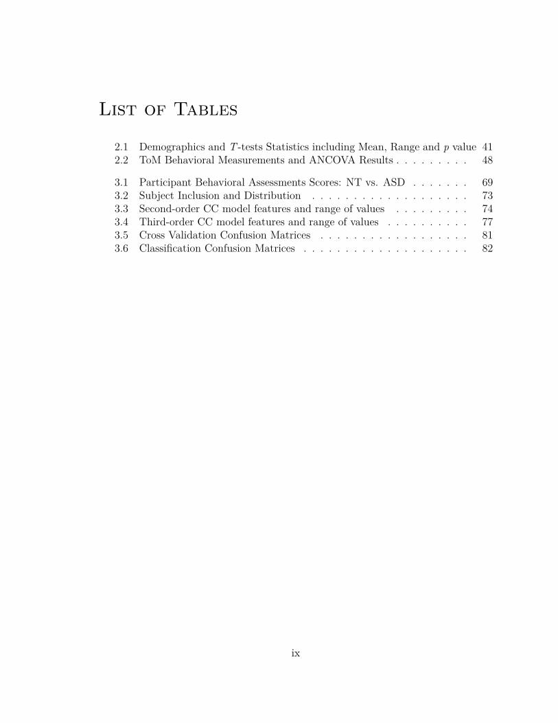
List of Tables
2.1 Demographics and T -tests Statistics including Mean, Range and p value 412.2 ToM Behavioral Measurements and ANCOVA Results . . . . . . . . . 48
3.1 Participant Behavioral Assessments Scores: NT vs. ASD . . . . . . . 693.2 Subject Inclusion and Distribution . . . . . . . . . . . . . . . . . . . 733.3 Second-order CC model features and range of values . . . . . . . . . 743.4 Third-order CC model features and range of values . . . . . . . . . . 773.5 Cross Validation Confusion Matrices . . . . . . . . . . . . . . . . . . 813.6 Classification Confusion Matrices . . . . . . . . . . . . . . . . . . . . 82
ix

Chapter 1
Comprehensive Literature Review
Chapter one provides an overview of Autism Spectrum Disorder (ASD) including
a definition of the disorder, and a discussion of prevalence and possible causes. It
then addresses theory of mind (ToM) as a core deficit in the ASD population. The
relevant behavioral and brain literature related to ToM emotions in ASD are also
discussed. Finally, machine learning is explored as an automatic diagnostic system
and predictive model for ASD. See Figure 1.1.
1.1 Definition and Prevalence
ASD is a lifelong neurodevelopmental disorder in which the symptoms of a child
can vary from mild to severe. According to the 2016 report from the US Centers for
Disease Control and Prevention (CDC), about 1 in every 54 individuals has ASD [58].
Individuals with ASD have difficulties in communicating and interacting with others;
they may also exhibit impairments in language and intellectual abilities. As a lifelong
neurodevelopmental disorder, independence and quality of life are often impaired [92].
1

Figure 1.1: Chapter One Advanced Organizer
In the last several years, the prevalence and diagnostic criteria for ASD have changed
in both epidemiological and clinical settings.
ASD was first described by Leo Kanner in 1943 [144] who found a new form of
emotional disorder presented by 11 children. These children were able to engage in
intellectual activities but had a strong desire to be left alone and rarely showed af-
fection while interactions with others. In 1978, Rutter [233] described autism as a
distinct syndrome that could be differentiated from other developmental disorders
and outlined four criteria for diagnosis: onset before 30 months of age; impaired
social development; delayed and aberrant language development; and insistence on
uniformity, as shown by stereotyped play patterns, abnormal preoccupations, or re-
sistance to change. The World Health Organization (WHO) included autism in the
International Classification of Diseases (ICD-9) in 1975 [284]. The American Psychi-
2

atric Association included autism in the Diagnostic and Statistical Manual of Mental
Disorders-III (DSM-III) in 1980 [8].
It was not until the 1980s that a less severe form of autism was found and identified
as Asperger’s syndrome and was eventually included in official nosographies in the
1990s, however, with no clear validity. People with Asperger’s disorder tend to bear
mild signs and symptoms of autism without language delays. Children with autism are
often seen as aloof and uninterested in others. However, individuals with Asperger’s
disorder do not carry the same character as individuals with more severe autism
[17]. Individuals with Asperger’s disorder want to fit in a social group and engage
in interaction with others in most scenarios, although they struggle with finding
the appropriate approach. They can be perceived as being socially awkward while
breaking conventional social rules or showing certain levels of apathy. Their interests
in a particular subject tend to be obsessive. Individuals with Asperger’s disorder
typically do not have deficits in their language skills, however, they use language in
ways that are different from others. Specifically, their speech patterns can appear to
be unusual with a lack of inflection and excessive formality. They also have difficulty
understanding the subtleties of language including irony, sarcasm and humor, or the
give-and-take nature of a conversation [17]. Cognitively, a person with Asperger’s
disorder has an average to above-average intelligence [17]. Notably, the DSM-5 no
longer has Asperger disorder as an independent disorder but instead considers it as
part of ASD. It is an effort to eliminate distinctions that were made idiosyncratically
and unreliably across different diagnostic centers and clinicians.
With several advances in science over the past 10 years, attention to the clinical,
financial and social needs of those with ASD has increased. Significant challenges
3
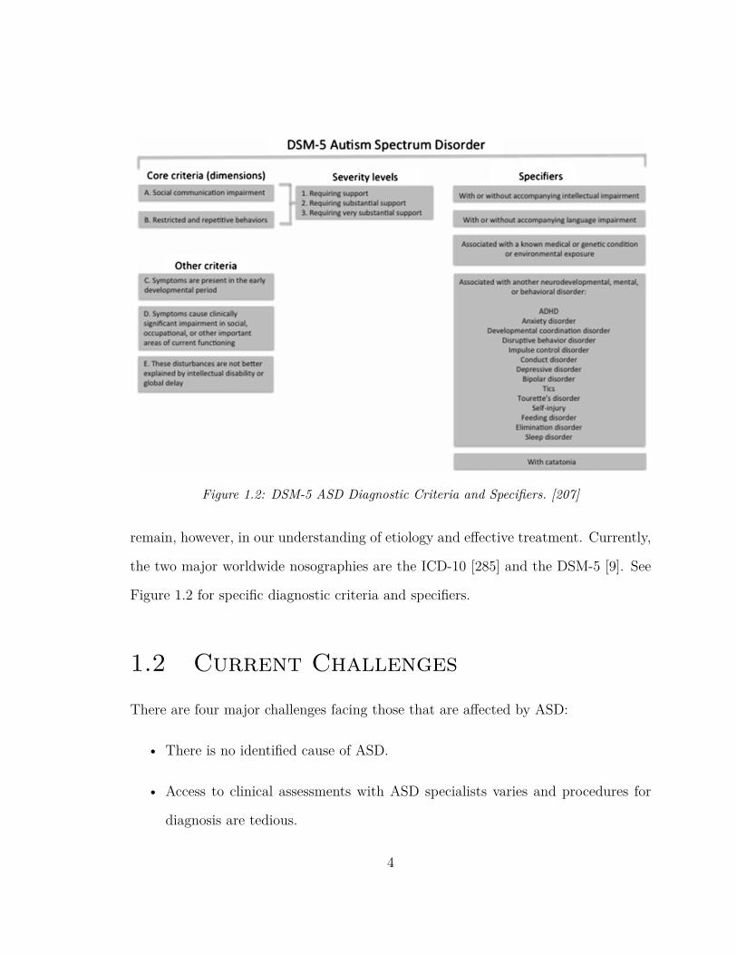
Figure 1.2: DSM-5 ASD Diagnostic Criteria and Specifiers. [207]
remain, however, in our understanding of etiology and effective treatment. Currently,
the two major worldwide nosographies are the ICD-10 [285] and the DSM-5 [9]. See
Figure 1.2 for specific diagnostic criteria and specifiers.
1.2 Current Challenges
There are four major challenges facing those that are affected by ASD:
• There is no identified cause of ASD.
• Access to clinical assessments with ASD specialists varies and procedures for
diagnosis are tedious.
4
• There are high social and economic burdens for family and society.
• There are either limited resources or a lack of information to guide patients to
choose appropriate intervention methods.
1.2.1 Cause
There is no definitive answer to what causes ASD or the range of severity that charac-
terizes the disorder. Most clinical researchers would agree there are both environmen-
tal and genetic factors to consider. In fact, more than 100 genes are known to be risk
factors [230] and about 1,000 genes (e.g., Pten gene [51,52,220]) have been associated
with ASD [113]. Debate between whether genetic effects outweigh the environmental
factors is ongoing. For example, one twin study suggested that shared environmental
factors contribute to about more than 50% of the etiology for autism, with 37% is
potentially led by genetic heritability [264]. However, other twin studies suggest that
strong genetic effects play a significant role in the development of ASD such that
concordance for monozygotic twins is roughly 45% while concordance for dizygotic
twins is 16% [42]. Given the inconclusive and inconsistent results across twin studies,
the exact role of genetic and environmental factors in ASD is ambiguous. It is possi-
ble that genes and the environment both influence the occurrence of ASD, such that
certain environmental exposures combined with particular genetic predisposition can
potentially lead to ASD [71,226].
A few specific environmental factors have been identified to interact with genes
in ASD. Maternal infection is a primary concern that can often be linked to ASD, in
which naturally occurring pathogen exposure often provides the strongest evidence
5
for environmental etiology [82,172]. Although the maternal rubella (German measles)
epidemic no longer exists, it was widely spread globally before the dissemination of
effective vaccines. Between 1963 to 1965, 10,000 to 30,000 infants whose mothers were
exposed to rubella were born with moderate to severe neurodevelopmental disorders,
in which 741 per 10,000 were found to have autism [82, 172]. Another common and
most prevalent infection is influenza. A series of animal research studies has associated
prenatal exposure with influenza during fetal life with an increased risk of autism
[209,245]. Patterson and Shi [209, 245] suggest that the influenza virus activates the
immune system of the pregnant woman, which is potentially harmful to fetal brain
development. However, there is a lack of evidence suggesting the use of antibiotics
and vaccines of influenza can cause ASD. Noticeably, nearly 64% of US women have
had an infection during their pregnancies, and the newborns of these mothers did not
develop ASD in most cases [64]. In summary, mild infection during pregnancy can
increase the risk of a fetus for developing autism, but little evidence suggests that
viruses are directly associated with ASD [287].
Autoimmune diseases is a disease in which immune cells attack other cells that
are mistaken as "foreign". This process is mediated by circulating antibodies. Au-
toimmune disease currently affects as much as 9% of the world’s population [6, 65].
Twelve percent of mothers of children with ASD carry unusual antibodies directed at
fetal brain proteins. It indicates that circulating antibodies may lead to some forms
of autism [46]. It is suggested that Maternal Antibody-Related (MAR) causes can be
associated with to as many as 22% of autism cases according to the specific assays
for these antibodies. It demonstrated a strong possibility that such form of ASD can
potentially be prevented [101]. These studies have led a new direction to discover
6

potential therapeutic targets for ASD. There are still many challenges remaining to
answer questions such as how antibodies enter the fetal brain and how that could alter
the neurodevelopmental processes. However, there is no evidence arguing against the
possibility of the circulating antibodies being a prenatal environmental risk factors
for ASD.
Another important environmental factor is drug use during pregnancy. In the
1960s, there was evidence showing the association between the use of thalidomide,
a sedative drug that was prescribed for relief of nausea during pregnancy, and the
increased risk of autism in newborns [142]. More recently, there have been increased
concerns surrounding the use of valproic acid and serotonin reuptake inhibitors (SS-
RIs) which are prescribed to treat epilepsy, migraine headaches, bipolar disorder,
and depression during pregnancy [151]. To date, the largest epidemiological study
included 415 children, among which 201 were born to mothers who took antiepileptic
medication during their pregnancies. Nearly 7.5% of the children of those mother who
took the medications developed a neurodevelopmental disorder, especially autism,
comparing to 1.9% in the non-epileptic women [49]. Serotonin is an important brain
neurotransmitter that plays a significant role regulating sleep, mood and appetite.
Dysregulation of serotonin during early fetal life can lead to serious negative con-
sequences for brain development [6]. The name, SSRIS, have been used since late
1980s. It delays the reuptake of serotonin from the synaptic cleft into the presynaptic
terminal to enhance its effect on the postsynaptic receptors [6, 167]. A recent review
and meta-analysis of six case-controlled studies and four cohort studies have found
that SSRI use during pregnancy can be greatly linked to an increased risk of ASD in
offspring, especially during the first and second trimesters of pregnancy [289]. Inter-
7

estingly, other studies linked the preconceptual exposure to SSRIs to increased ASD
risks, which was the same case as to use of non-SSRI antidepressants. Specifically,
although there was significantly more ASD cases in the SSRI-exposed (experimental)
group comparing to the control group, if a mother already had a existing unmedi-
cated psychiatric disorder or had discontinued the medication, the chances of their
newborns developing ASD were as much as those mothers who had been exposed to
SSRIs [145, 167]. It is noted that it is nearly impossible to remove the SSRIs if they
are the needed drugs for a maternal condition. To summarize, a brief review of the
literature has shown that the intake of some drugs during pregnancy [145] increases
the risk of ASD. Thus, use of drug during pregnancy and fetal development needs to
be evaluated carefully. Mothers need to consider all potential risky outcomes before
taking a drug for widespread medical purposes.
Environmental toxins, such as air pollution produced by automobiles and cigarettes,
heavy metals, and pesticides are also considered potential risk factors for autism
[182, 206], although with little evidence for this. One historical concern is vaccines,
such as the measles, mumps, and rubella (MMR) vaccine. These vaccines are typi-
cally administered initially when the child is 12 to 18 months old, which can become
high risk factors of developing ASD for a healthy child [222]. This fear was sus-
tained by regressive onset in some cases. Specifically, a child may start to show social
and language deficits after the first year and slowly develop autistic characteristics.
However, research suggests that even in children who display this regressive form of
autism, brain changes happen long before behavior changes, typically around four
to six months. Furthermore, there is no evidence showing a relationship between
MMR administration and the development of ASD. The author who published data
8

suggesting increased risk of ASD with MMR administration faced significant public
shaming. These findings from many large-scale epidemiologic studies are consistent
with the conclusion that the US National Academy of Sciences reached in a thorough
review carried out in 2011 [76,200].
Overall, many theories exist about how one develops autism, but these possible
risk factors still remain mysterious as there is no direct and established conclusion
for a single causal factor.
1.2.2 Assessment and Diagnosis
Currently, the diagnosis of autism is solely based on behavioral symptoms. A typi-
cal diagnostic appointment includes a multi-hour behavioral evaluation by a team of
clinicians. Diagnostic appointment usually happens in a specialized diagnostic clinic
or developmental medicine center. In many cases, such appointment can only be
made after a referral from the child’s general pediatrician. During diagnostic ap-
pointments, clinicians and interventionist deliver a series of behavioral assessments
with different rating scales. There are standardized schemes regarding the evaluations
derived from the rating scales for clinicians to follow in order to reach a best-estimate
diagnosis [168]. There are two gold standard behavioral assessment tools guiding
the diagnostic process, The Autism Diagnostic Observation Schedule-second edition
(ADOS-2) and The Autism Diagnostic Interview-revised (ADI-R) [173, 174]. The
ADOS-2 is considered the gold standard for assessment of ASD. It is an observation-
based clinical assessment that is broken into five modules based on age and language
level: "the toddler module is for children between 12 and 30 months of age who do
not consistently use phrase speech, module 1 is intended for young children with no
9
or single-word speech, module 2 is intended for individuals with phrase speech, mod-
ule 3 is intended for verbally fluent children, module 4 is intended for verbally fluent
adolescents and adults" (Levy et al., 2017, p.4) [168]. During the ADOS assessment,
the administrator engages in series of standardized activities with the child and an-
swers a set of questions based on his/her observations of the child’s behavior. The
total time for administration and scoring of the ADOS is approximately 60 min. The
process of ASD diagnostic examinations is time-consuming due to its rigorous nature.
Because of that, many diagnostic centers have a long waiting list as the capacity of
available clinicians is extremely limited. In addition, those using the ADOS-2 must
complete a multi-day training to administer the assessment. This bottleneck directly
leads to delays in diagnosis of 13 months and longer for minority and lower socio-
economic status groups. These delays can also delay insurance coverage and access
to behavioral therapies [20,180,181,225].
1.2.3 Intervention
Both biological and social cognitive intervention methods are available to help man-
age ASD-related symptoms and improve an individual’s social communication skills.
Psychosocial therapies such as applied behavior analysis (ABA), pivotal response
treatment (PRT), and cognitive behavior therapy (CBT) have been commonly used
to treat ASD and elicit positive effects to improve learning and verbal communication
and ease ASD-associated symptoms such as anxiety [75].
In 2009 and then again in 2015, The National Autism Center (NAC) reviewed
hundreds of interventions used to address the symptoms of autism described in peer-
reviewed scientific journals, and described 11 established interventions (NAC, 2009)
10
which expanded to 14 (NAC, 2015) based on the available research for children, ado-
lescents, and young adults (under 22 years of age) with ASD [16]. Four factors were
adopted to help select appropriate and effective intervention methods: evidence of in-
tervention effectiveness, professional judgment, data-based clinical decision making,
values and preferences of families including the individual on the autism spectrum,
and capacity to accurately implement an intervention. The 14 established interven-
tions identified in 2015 include behavioral interventions, cognitive behavioral inter-
vention package, comprehensive behavioral treatment for young children, language
training and production, modeling, natural teaching strategies, parent training, peer
training package, pivotal response training, schedules, scripting, self-management,
social skills package, and story-based intervention. For individuals who are 22 years
and older, behavioral interventions is the only recommended intervention [16]. Most
of these interventions come from the behavioral literature, including ABA, behav-
ioral psychology, and positive behavior supports. It is important to know that most
of the intervention methods benefited from a broad range of expertise and knowledge
in fields such as developmental psychology, special education, and speech-language
pathology.
Caregivers of individuals with ASD often face more stress than those who deal with
other disabilities, which contributes to challenges in their own relationships and men-
tal and physical health conditions. Caregivers are required to commit a tremendous
amount of time, effort and patience to meet the high care demands of individuals with
ASD. Moreover, many parents of children with ASD suffer with financial challenges,
especially with the high out-of-pocket health care expenses, underemployment, or em-
ployment loss [114,150,157,158]. Not surprisingly, these parents often feel the strain
11
of caregiving and are at risk for mental health challenges such as anxiety and depres-
sion [114, 150]. At a societal level, Leigh and Du [165] revealed that the economic
burden of the ASD population in 2015 was around $268.3 billion and in 2025 will
be $460.8 billion, representing 1.5 and 1.6 %, respectively, of GDP. These estimates
range from $161.6 billion (0.9 % of GDP) to $367.3 billion (2.0 % of GDP) in 2015
and from $275.6 billion (1.0 % of GDP) to $1,010.6 billion (3.6 % of GDP) in 2025.
These estimates are based on the increased number of ASD individuals, the expen-
ditures on medical care and non-medical care, and the lost productivity for parents
and their children with ASD [165]. Given the hardship at both family and society
levels, it is essential to find appropriate and efficient methods to diagnose ASD and
manage symptoms, particularly the significant social impairment which differentiates
ASD from other neurodevelopmental disorders.
1.3 Theory of Mind
Among all of the deficits identified for children with ASD, their social impairment is
primary and interferes with many aspects of their development. Many believe that at
the core of this social impairment is a deficit in theory of mind (ToM) [21, 23]. ToM
is the ability to reason about the thoughts and feelings of self and others, including
the ability to predict what others will do or how they will feel in a given situation on
the basis of their inferred beliefs [21, 23]. ASD individuals have trouble interpreting
or reading the verbal and non-verbal social communication of other individuals in a
way that accords with normative expectations [9]. It has been argued that individ-
uals often encounter difficulties interacting with others appropriately within a social
12

context when their abilities to interpret the beliefs, intentions, and emotions of others
are impaired [48,126].
There are three major components of ToM [53]. The first one is shared world
knowledge, such that ToM is always situated in the context of the surrounding world.
For example, an individual must be able to infer their partners’ thoughts, beliefs,
emotions, and goals during a typical conversation to respond properly. The individ-
ual also needs to be able to integrate cues from their surroundings during interactions
with conversational partners, "such as prior world knowledge (e.g., amount of per-
sonal space the conversational partner needs to feel comfortable), knowledge about
the relationship between individuals (e.g., how much is an appropriate amount of dis-
closure with a close friend vs. a co-worker), the goal of the conversation (e.g., what
information is required to exchange between the two individuals), and the condition
where the conversation occurs (e.g., in a group setting or a private room)" (Byom and
Mutlu, 2013, p.2) [53, 153, 238]. The second component of ToM is the perception of
various social cues, such as gaze, facial expressions, and vocal cues. Gaze is a major
cue of the direction of one’s attention and people often follow one’s gaze to deter-
mine the partner’s intention. Gaze also helps an individual track the understanding
of one’s message as well as to send feedback [29, 30, 103, 149]. Like gaze, emotion
recognition is a crucial ability to infer mental states. The ability to discriminate
between different facial expressions is typically generated in childhood and continues
to develop into adulthood with both children and adults more accurately identifying
positive emotions (e.g., happy) than negative emotions (e.g., sad) [185,249].
The last component of ToM is interpretation of actions, such that humans be-
lieve that others act in ways that are consistent with their beliefs and goals. People
13
are able to understand other’s intentions and beliefs by passively observing their
actions [4, 282]. Many studies have demonstrated correlations between ToM and
circumscribed aspects of NT children’s everyday behavior including social pretend
playing and secret-keeping, aggression and bullying, and reciprocated friendship [95,
137, 212, 224, 253, 257]. More importantly, however, such relations are limited and
have not emerged as clearly in the domain of generalized social skills. Instead, stud-
ies have either found no significant associations at all between social skills and false
belief understanding in NT children, or mixed results indicating no relationship be-
tween social behaviors and ToM development [79]. A recent study, however, showed
a strong correlation between peer interaction surrounding leadership and group entry
and ToM understanding among NT and deaf children, but such a correlation did not
exist in the ASD group. The apparent link of ToM to peer competence in ASD was
instead greatly mediated by language ability [213].
ToM deficits supposedly underlie social communication impairments in ASD [25,
119, 254]. ToM has also demonstrated potential as a severity index in ASD. That
means that better ToM is associated with improved behavior towards social rules
[262], better social interaction skills [41, 100] and increased language use [60, 116].
ToM is particularly useful in discriminating the level of support needed in "high-
functioning" ASD children. Besides levels of intelligence quotient (IQ), cognitive
modifiability, executive functioning, and central coherence, studies that examine po-
tential cognitive indicators in terms of level of special support needed have found that
ToM is the only cognitive indicator to predict school placement. ToM successfully dif-
ferentiated between children who need support and those who do not [5]. Behavioral
and social competencies strongly predict children’s ability to successfully integrate
14
into public education system [141,177,286].
1.3.1 Behavioral Measurements of ToM in ASD
Many instruments have been developed to measure ToM in ASD [48]; however, there
has not been any universally accepted operationalization of ToM. Research in the old
days were mostly shaped by studies examining ToM in young NT children. These
studies often used a variety of different false belief tasks in primary developmental
research [19,43,282]. Findings from these older studies showed that many older chil-
dren and adolescents with ASD could pass such common tests regardless of their
pronounced social impairments associated with ToM deficits. Thus, researchers de-
veloped more age-appropriate tests that can accurately measure the social-cognitive
deficits among older individuals. For example, The Reading the Mind in the Eyes
Test examines the person’s ability to link a specific mental state descriptor (e.g., flir-
tatious, hostile) to the expression demonstrated by an image of a pair of eyes [26].
Another test, the Strange Stories [117] task includes a number of scenarios or stories
that are presented on paper. In this task, the examinee is required to explain the
purpose of the behavior of the key characters within the scenarios. In these scenar-
ios, the characters use expressions that have meanings that are different from what
a literal interpretation of the expression might indicate (e.g., metaphors, sarcasm,
white lies). There were mental or social stories (i.e., stories requiring a reading of the
social intent of the characters) and control stories (i.e., stories not requiring any social
inferences) in Happe’s original instrument. When comparing to the IQ-matched con-
trols, individuals with ASD were expected to perform worse on the mental or social,
but not the control (i.e., physical) stories. Subsets of items from the Strange Stories
15

test [99, 118] have provided the stimuli for many other examinations of ToM deficits
in both children and adults with ASD.
Nearly all behavioral tests of ToM currently available only examine one or a few
aspects of ToM; however, two measurement tools The Theory of Mind Inventory-2
(ToMI-2) [131] and The Theory of Mind Task Battery (ToMTB) [134] are multi-
faceted tools that cover several aspects of ToM (e.g., emotion recognition, false belief,
perspective taking etc.), including information from both parent and child. The
ToMI-2 measures a parent’s perception of their child’s ToM understanding of 60 items
using a 20-unit rating scale from "Definitely Not" to "Definitely". Primary caregivers
use a vertical hash mark to indicate where on the continuous scale best represents their
perceptions. Item, subscale, and composite scores range from 0-20. A higher number
indicates a parent’s greater confidence in their child’s understanding of a particular
ToM skill. The ToMI-2 items represent typical social interactions to ensure it is
a socially and ecologically valid ToM index. The tool demonstrates excellent test-
retest reliability, internal consistency, and criterion-related validity for neurotypical
children and children with ASD as well as contrasting-groups validity and statistical
evidence of construct validity (i.e., factor analysis) [131, 133, 166]. The ToMTB is a
direct measure of a child’s understanding of ToM, see Figure 1.3. It consists of nine
ToM tasks presented as short vignettes in a story-book format arranged in ascending
difficulty. For each of the nine tasks, children are provided with one correct response
option and three possible distracters. There are 15 total questions asked, including
memory control questions that must be answered correctly to get credit for ToM
understanding. The ToMTB has strong test-retest reliability [131,134].
16
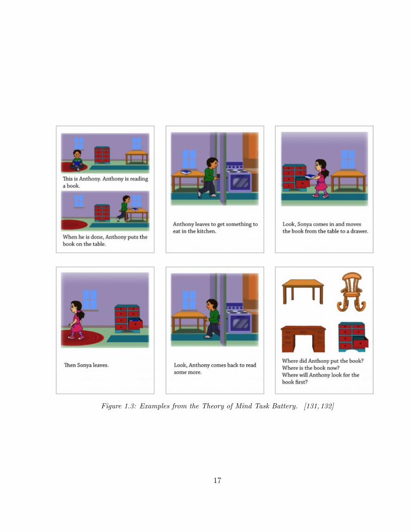
Figure 1.3: Examples from the Theory of Mind Task Battery. [131,132]
17
1.3.2 Examining Specific Aspects of ToM: Emo-
tion Recognition
Although ToM has been studied for decades, it still remains a challenging research
area due to its multi-faceted composition. Further, few studies have investigated
the neural mechanisms underlying ToM, especially among children. Thus, a greater
understanding of the brain-behavior connections associated with ToM will provide
researchers with a potential link between the biological mechanisms of ToM associated
with the behavioral characteristics, leading to more efficient diagnostic processes and
prognostic indicators for special populations like children with ASD. To facilitate
increased understanding of this linkage, this study emphasized emotion recognition
as one aspect of ToM with a specific focus on less well-studied and more complex
emotions (i.e., surprise, embarrassment and desire-based emotion).
Emotion recognition is one particular aspect of ToM that has a critical role in
an individual’s ability to meaningfully engage in social communication and social
interaction. It is the ability to discriminate between different facial expressions and
is key to understanding empathy or the feelings of others. Children with ASD have
impairments in social interaction often due to a lack of understanding of emotions
and the minds of others, as well as difficulty attending to social cues (e.g., gaze, facial
expressions, body postures etc.). Some studies have found that children with ASD use
the lower part of the face to determine one’s facial expression and often ignore or have
difficulty identifying negative facial affect (e.g., distress, fear) evident near the eyes
as early as the age of three [160]. However, other studies suggest that children with
ASD have trouble recognizing emotions from the lower part of the face comparing to
18

NT children [160]. There is a wealth of behavioral evidence showing that recognition
of even more "basic" emotions like happy and sad are impaired in individuals with
ASD [10,66,72], with some emotions requiring greater ToM development than others
(e.g., surprise, embarrassment, desire-based emotion).
Happy
Happiness is one of the first emotions neurotypical (NT) infants discriminate [40,132].
Mastery might occur because it is easily visible, biologically influenced or frequently
observed [91, 251]. With the early awareness of happiness, it is described as an early
developing ToM skill [132]. Further, it is the most frequently recognized emotion
not only for NT children but for those with developmental disabilities [183]. In fact,
research shows the recognition of happy expressions may be intact for those children
with ASD who have higher cognitive and linguistic abilities [11, 132] and appears to
be more intact than recognition of other emotions [70, 162, 232, 265, 271]. Notably,
however, individuals with ASD may not be attuned to the social value of happiness
which could impact their desire to engage with others [132,161,240].
Sad
Similar to the early recognition of "happy" or "happiness" in children, negative emo-
tions such as "sad" also emerge early in development and can be distinguished by
infants [94, 132, 268]. In contrast to the ease in the development of recognizing hap-
piness in children with ASD, recognizing "sadness" is more challenging. There seems
to be a disconnection for those with ASD in their ability to process and visually scan
atypical faces, and since understanding "sadness" requires the ability to make sense of
19
the eye region of the face, their responsiveness is reduced [67,90,132,218,269]. Dimin-
ished ability to recognize sadness also appears to be related to more severe autistic
symptoms and greater difficulty independently managing day to day activities such
as personal care [121,132].
Surprise
As a "cognitive" versus "basic" or early developing emotion, surprise requires an indi-
vidual to understand the ways in which emotions are influenced by one’s expectations
or beliefs about something [132]. Understanding the emotion of surprise requires chil-
dren "to understand that a person approaches a situation with a specific expectation
in mind, and if the situation does not match that expectation, then the person will be
surprised" (Lacroix et al., 2014, p. 1147) [162]. Surprise emerges later in development
and not before preschool [24,83,108,112,132]. Knowing that understanding one’s own
and others’ desires, beliefs and values is a particular area of deficit for children with
ASD, it is not unexpected that they would experience difficulty making sense of "sur-
prise". Difficulty in the recognition of surprise among children with ASD also appears
to fall behind understanding false belief [178,231]. It is an emotion induced by what
someone thinks is the case, even if the reality does not match with what is actually
on one’s mind.
Embarrassment
Embarrassment is often described as a "self-conscious" emotion that is associated with
a feeling of shame or awkwardness around some action or statement [56, 124, 125].
Experiencing "embarrassment" does suggest some level of self-awareness that an "ex-
20

pected" behavior in a social context was unmet [33, 86–88]. Children with ASD may
show an emotional response of embarrassment, although to a lesser degree than their
NT peers. Further, they seldom recognize embarrassment or express their experiences
in situations of embarrassment [132]. Often, they miss social gaffes where what is said
is perceived as an inappropriate comment in a social context. In addition, conditions
that bring out embarrassment among children with ASD are often different from those
described by NT children. For example, children with ASD often respond to embar-
rassment more strongly when they are embarrassed vs. when something they have
done is embarrassing to others. Having a better understanding of embarrassment will
ultimately allow children with ASD to better navigate the social world and interact
with other people in more appropriate ways, following the expected social norms and
expectations [77, 84,132,176,239].
Desire-Based Emotion
Desire-based emotion recognizes the relationship between getting what you want and
feeling happy and not getting what you want and feeling sad or disappointed [132,227,
228]. Thus, desire-based emotion can lead to positive emotions or negative emotions,
depending on a fulfilled or unfulfilled desire. Importantly, research suggests that
children with ASD are better able to navigate emotions at a basic level in support
of previous research [14, 15, 55, 80, 98, 120, 132, 215]. Challenges remain, however, in
understanding the vulnerability of desire-based emotions in children with ASD as they
often are unable to generalize their understanding without explicit instruction and
support in social contexts [132]. Desire-based emotion plays an important role when
understanding and empathizing with others’thoughts and feelings [216,229,278].
21
1.4 Imaging Studies of ToM in ASD
Most studies have examined ToM through behavioral approaches, especially among
the ASD population due to challenges associated with procedures such as staying still
in a magnetic resonance imaging (MRI) scanner or wearing an electroencephalogra-
phy (EEG) cap. Studies have established important roles of the medial prefrontal
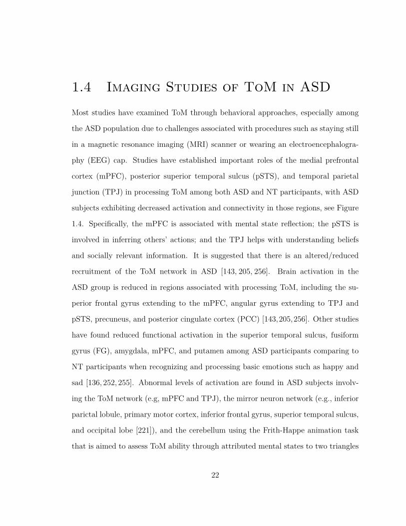
cortex (mPFC), posterior superior temporal sulcus (pSTS), and temporal parietal
junction (TPJ) in processing ToM among both ASD and NT participants, with ASD
subjects exhibiting decreased activation and connectivity in those regions, see Figure
1.4. Specifically, the mPFC is associated with mental state reflection; the pSTS is
involved in inferring others’ actions; and the TPJ helps with understanding beliefs
and socially relevant information. It is suggested that there is an altered/reduced
recruitment of the ToM network in ASD [143, 205, 256]. Brain activation in the
ASD group is reduced in regions associated with processing ToM, including the su-
perior frontal gyrus extending to the mPFC, angular gyrus extending to TPJ and
pSTS, precuneus, and posterior cingulate cortex (PCC) [143,205,256]. Other studies
have found reduced functional activation in the superior temporal sulcus, fusiform
gyrus (FG), amygdala, mPFC, and putamen among ASD participants comparing to
NT participants when recognizing and processing basic emotions such as happy and
sad [136, 252, 255]. Abnormal levels of activation are found in ASD subjects involv-
ing the ToM network (e.g, mPFC and TPJ), the mirror neuron network (e.g., inferior
parictal lobule, primary motor cortex, inferior frontal gyrus, superior temporal sulcus,
and occipital lobe [221]), and the cerebellum using the Frith-Happe animation task
that is aimed to assess ToM ability through attributed mental states to two triangles
22

Figure 1.4: The Social Brain: mPFC (green), TPJ (orange), pSTS (pink). [187]
interacting with each other [2,116]. Further, the anterior cingulate cortex, mPFC, and
left superior temporal gyrus show decreased activities when regulating ToM-related
self-conscious emotions (e.g., embarrassment and guilt) in ASD subjects [136,252,255].
Although there have been studies examining less well-understood (i.e., desire-based
emotions, surprise) and more complex (i.e., embarrassment) emotions, they are either
conducted from a behavioral perspective or with adult populations. Studies involving
the adult ASD population, however, have given us some direction regarding the neu-
ral mechanisms underlying emotions in ASD, yet we remain unclear whether children
exhibit similar or different brain activity patterns or no.
23

1.4.1 Imaging Studies of Emotion Recognition in
ASD
The neural mechanisms underlying interpretation of happy faces in ASD are well
studied, with a few important brain regions identified. A particular region of the
cortex, the FG, is known to be the special area for processing of facial features and
emotions. One study recorded the face-sensitive ERP to neutral and emotional faces
with a high-density EEG system. The study indicated impaired activity patterns
in the area of FG among ASD subjects when processing happy faces [10]. fMRI
studies have suggested that there are decreased brain activities in areas of the left
medial frontal gyrus including the left superior and medial frontal gyri, right superior
and medial frontal gyri, and the anterior cingulate gyri, the right and left temporal
poles, left TPJ, left pSTS, dorsomedial prefrontal cortex, and right and left middle
superior temporal sulcus when processing facial emotions [72]. Due to high variability
across fMRI studies, other brain regions are also identified, but overall reduced brain
activities when processing happy faces are observed in ASD groups [189, 250, 279].
Research also suggests the ASD population is more sensitive to sad faces [279]. When
processing sad faces, the ASD group tends to show greater activation relative to
the control group in the amygdala, ventromedial PFC, putamen, and striatum, and
younger adolescents show greater activation than older adolescents [72,279]. However,
research found decreased activities in mPFC among ASD groups when processing sad
faces [72].
Embarrassment has been studied at a neural level only among adults with ASD,
suggesting altered circuitry underlying mPFC, anterior cingulate cortex (ACC), in-
24
ferior frontal gyrus (IFG), TPJ/pSTS, PCC, and amygdala [87, 124, 136, 169, 255];
surprise and desire-based emotions have only been examined in ASD subjects at a
behavioral level and little is known about their neural correlates. Thus, examining the
neural correlates of these emotions requiring ToM and establishing the connections be-
tween behavior and brain activities in children with ASD is important. As mentioned
earlier, there are no current identified biomarkers that are considered to be necessary
and sufficient to indicate ASD. However, research suggests that there is a strong link
between biological (e.g., genes and hormones) and neurological factors (e.g., abnormal
brain connectivity and structures) and the development of ASD [36,202,211,223].
Building upon findings from previous studies, this study aimed to provide a deeper
and more systematic understanding of the brain-behavior connections associated with
ToM, leading to increased understanding of the brain regions associated with ToM.
It allowed us to identify those brain structures associated with deficits in specific
aspects of ToM. With this knowledge, intervention research can then be developed
that supports brain behavior connections leading to normalized social performance.
Such brain behavior research might also help predict those brain behavior profiles of
children most likely to benefit from specific ToM or social cognitive based interven-
tions. This is important as causal modeling in ASD suggests that interventions need
to be delivered at the cognitive level to bridge behavior with brain function [128].
1.4.2 Introduction of Chapter Two
Chapter 2 introduces a study in which behavioral and neuroimaging data for children
with and without ASD were collected, specifically in areas of emotion recognition
to understand which key skills are required for meaningful social interaction. The
25

study included 9 children with ASD and 19 neurotypical children (NT) between the
age of 7 to 14 years old. The ToMI-2 and the ToMTB were adopted to evaluate
children’s ToM understanding important to their development of social skills. Two
novel functional magnetic resonance imaging (fMRI) paradigms were implemented to
probe the neural mechanisms underlying ToM related to desire-based emotion, and
more advanced and complex emotions (i.e., surprise and embarrassment), as well as
two early developing emotions (i.e., happy and sad). The results suggest impaired
abilities in multiple ToM metrics and brain deficits associated with ToM related
emotion recognition and processing among children with ASD. The study findings
suggest future research directions in the field when working with special populations
such as those with ASD.
1.5 Machine Learning Approach
With the ongoing challenges discussed earlier and growing awareness of ASD, there
is a high demand for immediate access to diagnostic services. An automated ASD
diagnostic approach might allow for early diagnosis of ASD and help to provide a
map of high-risk populations [208]. Building an automatic diagnostic and predictive
model of ASD is timely, with many studies adopting machine learning approaches
to identify sets of significant biomarkers including both behavioral and biological as-
pects. [81]. Duda and colleagues (2016) applied machine learning to distinguish ASD
from attention deficit hyperactivity disorder (ADHD) using a 65-item Social Respon-
siveness Scale. Bone et al. [39] trained their models to discriminate ASD subjects
from healthy controls using the same Social Responsiveness Scale and the Autism
26

Diagnostic Interview-Revised score. Other studies aggregated items from the ADOS
and scores from the Autism Quotient to accurately classify an ASD group. As a re-
sult of the wide variation in ASD behavioral measures, many studies have searched
for brain-based biological markers to identify a common etiology across individuals
with ASD. These brain-based biological markers are less subjective than behavioral
measures and may represent potential targets for treatments. Currently, markers that
are measurable via magnetic resonance imaging (MRI) are highly desirable because
they can represent potential targets for both assessment and intervention [93]. Inde-
pendent structural MRI studies have found differences in whole brain volume and the
developmental trajectories between individuals with ASD and those who do not have
ASD [7, 45, 63]. Other structural brain abnormalities associated with ASD include
cortical folding signatures, showing in brain regions of the TPJ, anterior insula, pos-
terior cingulate, lateral and medial prefrontal, corpus callosum, intra-parietal sulcus,
and occipital cortex [123, 163, 201, 246]. Evidence also shows that an accelerated ex-
pansion of the cortical surface area, but not cortical thickness, can lead to the early
overgrowth of the ASD brain [110], while other studies suggest that individuals with
ASD tend to have thinner cortices and reduced surface area as an effect of aging [85].
Machine learning (ML) has been introduced to the neuroimaging field to identify
the abnormal brain regions in individuals with ASD, see Figure 1.5. Support vector
machines (SVM) is an algorithm that generates high classification accuracy without
requiring large sample sizes to avoid over-fitting [170]. The SVM algorithm is able
to classify ASD from corresponding controls using extracted features from functional
connections and grey matter volume [59, 62, 107, 140, 204]. Other algorithm-based
classifications of ASD include the random forests (RF) algorithm, which uses random
27
ensembles of independently grown decision trees, and deep neural networks [61,146].
Although these studies have proven accurate for classifying ASD, they have failed
to identify precise neuroimaging-based biomarkers. The majority of studies have
adopted data from the Autism Brain Imaging Data Exchange (ABIDE) dataset col-
lected from 24 international brain imaging laboratories. The ABIDE dataset includes
1112 existing resting-state functional magnetic resonance (rs-fMRI) imaging datasets.
It also includes the corresponding structural MRI and phenotypic information from
539 individuals with ASD and 573 age-matched NT controls [68, 127]. Classification
across a heterogeneous population is extremely challenging. There is a huge amount
of considerable variation in demographic and phenotypic profiles of participants. Such
variation becomes more apparent and problematic especially when neuroimaging data
are collected from multiple acquisition sites, such as ABIDE [68, 127]. Many factors
can lead to such variances in datasets such as scanner hardware, imaging proto-
cols, operator characteristics, demographics of the regions, acquisition site-specific
problems, greatly affecting the classification performance. This problem is especially
relevant for ASD given its notable heterogeneity. It is often difficult to collect neu-
roimaging data from individuals with autism given the loudness of the scanner and
the challenges to remain still. In fact, most individual site datasets have small sample
sizes, which can lead to overfitting and classification inaccuracies. Moreover, many
traditional ML algorithms are designed to classify large amount of data (e.g., ABIDE)
rather than optimize the selection of features, while the ultimate goal for machine
learning based diagnostic classification in neuroimaging is to identify discriminative
features to provide insight into abnormal structure and dysfunctional connectivity
patterns in the affected population [164].
28

Figure 1.5: Simple Illustration of Machine Learning. [1]
1.5.1 Introduction of Chapter Three
Although some ML-based methods have been applied to ASD, the suitability of ma-
chine learning and the choice of algorithms with regard to the specific behavior exam-
ined, as well as the quality and quantity of the data obtained from individual studies,
requires further investigation. Chapter 3 introduces a study that adopts a novel evo-
lutionary algorithm, the conjunctive clause evolutionary algorithm (CCEA), to select
features most significant for distinguishing individuals with and without ASD, and is
able to accommodate datasets having a small number of samples with a large num-
ber of feature measurements. The dataset is unique and comprises both behavioral
and neuroimaging measurements from a total of 28 children from 7 to 14 years old.
Potential biomarker candidates including volume, area, cortical thickness and mean
curvature in specific regions in the cingulate cortex, frontal cortex and temporal-
parietal junction areas were identified. Behavioral features associated with theory of
mind were selected. Additional classification models were developed to validate the
selected features by CCEA using the k-nearest neighbors algorithm. Study findings
demonstrate how machine learning tools can advance ASD research in the genre of
big data to benefit this special population in the future.
29
Chapter 2
A Pilot Study Using Two Novel
fMRI Tasks: Understanding The-
ory of Mind and Emotion Recog-
nition Among Children With ASD
Yu Han1,2*, Patricia A. Prelock1,2, Emily L. Coderre1,2, Joseph M. Orr3,4
1 Department of Communication Sciences and Disorders, University of Vermont.
2 Neuroscience Graduate Program, University of Vermont.
3 Department of Psychological and Brain Sciences, Texas A&M University.
4 Texas A&M Institute for Neuroscience, Texas A&M University.
30

2.1 Introduction
Autism spectrum disorder (ASD) is a lifelong neurodevelopmental disorder in which
an individual’s symptoms can vary from mild to severe. According to the most recent
prevalence rates from the US Centers for Disease Control and Prevention (CDC),
about 1 in every 54 individuals has ASD [58]. Although many theories exist about
the pathology and causes of autism, such as genetic and environmental factors, ASD
is a heterogeneous disorder without a specific known cause or cure. Language and
intellectual impairments may or may not be characteristic of children with ASD, but
the most significant challenges they face are difficulties communicating and interacting
with others in social situations [92]. Early diagnosis and intervention (e.g., speech and
language therapy, social cognitive behavioral intervention, etc.) targeting these social
difficulties are especially critical if we wish to improve the social communication skills
of children with autism, as well as help them build relationships, engage in activities
with others, and be successful in school.
An important component of social communication and social interaction in chil-
dren with ASD is theory of mind (ToM). ToM is the ability to reason about the
thoughts and feelings of self and others, including the ability to predict what oth-
ers will do or how they will feel in a given situation on the basis of their inferred
beliefs [22, 23]. Difficulties with ToM are thought to lead to impairments in social
interactions among individuals with ASD. It has been argued that individuals often
encounter difficulties interacting with others appropriately within a social context
when their abilities to interpret the beliefs, intentions, and emotions of others are im-
paired, [48,126]. Individuals with ASD often have trouble interpreting or reading the
31
verbal and non-verbal communications of others, specifically in social interactions [9].
ToM abilities have been adopted as proxies to functioning level in ASD for several
reasons: (1) the developmentally sequenced acquisition of ToM skills in childhood is
well documented [215,277]; (2) ToM tests have been used in a variety of populations
and cultures [18, 28, 122]; and (3) ToM deficits ostensibly underlie social communi-
cation impairments in ASD [119, 254, 267]. Additionally, general ToM assessment is
internationally applicable in that ToM skills develop in roughly the same manner
across the world [247, 275, 276]. ToM abilities have also been proposed as a poten-
tial severity index in ASD: better ToM is associated with improved behavior towards
social rules [263], better social interaction skills [41, 100], and increased language
use [60,116].
At a neural level, studies have established a ToM network involving the medial
prefrontal cortex (mPFC), the posterior superior temporal sulcus (pSTS), the tempo-
ral parietal junction (TPJ), the precuneus, and the posterior cingulate cortex (PCC).
More specifically, the mPFC is associated with mental state reflection; the pSTS
is involved in inferring to other’s actions; and the TPJ with understanding beliefs
and socially relevant information [143, 205, 256]. Individuals with ASD exhibit de-
creased activation and connectivity among these identified ToM regions, as well as
decreased connectivity in the frontal-medial, frontal-parietal and medial cerebellum
anatomical networks [143, 205, 256]. The purpose of this study is to examine behav-
ioral and neurobiological measures of emotions involving ToM, contributing to what
is known about ToM markers at the brain and behavior levels that can distinguish
those with and without ASD. In the review of the literature that follows, we discuss
the development of emotion recognition as one aspect of ToM in neurotypical (NT)
32
and ASD populations surrounding happiness, sadness, surprise, embarrassment and
desire-based emotion. This includes a description of how emotion recognition has
been tested and measured at both a behavioral and neural level in individuals with
ASD.
2.1.1 Emotion Recognition in Neurotypical De-
velopment
One particular aspect of ToM, emotion recognition, plays a critical role in an individ-
ual’s ability to meaningfully engage in social communication and social interaction.
Emotion recognition is the ability to discriminate between different facial expressions
and is key to understanding empathy or the feelings of others. The present study fo-
cuses on three specific emotions (i.e., surprise, embarrassment, desire-based emotion)
as they are critical aspects of ToM.
Happiness is considered to be the easiest recognized emotion while sadness is as-
sociated with the most negative affective reactions among the NT population [179].
Meta-analyses have found that the processing of emotional faces is associated with
increased activation in a number of visual, limbic, TPJ and prefrontal areas, where
happy and sad faces specifically also activate the amygdala [104]. Surprise conveys a
sense of novelty or unexpectedness and most research indicates that accurate recog-
nition of surprise will happen around the preschool years or even later among the
NT population [132,274]. One functional magnetic resonance imaging (fMRI) study
suggests that rapid recognition of surprised faces is associated with greater brain ac-
tivities in the right postcentral gyrus and left posterior insula [147]. Embarrassment is
33
often described as a "self-conscious" emotion that is associated with a feeling of shame
or awkwardness around some action or statement [56,124,125,132]. Experiencing "em-
barrassment" does suggest some level of self-awareness that an "expected" behavior in
a social context was unmet [31, 33, 86–88,132, 169]. Embarrassment is evoked during
negative evaluation following norm violations and supported by a fronto-temporo-
posterior network. It often recruits greater anterior temporal regions, representing
conceptual social knowledge [136]. Desire-based emotion recognizes the relationship
between getting what you want and feeling happy and not getting what you want and
feeling sad or disappointed. Thus, desire-based emotion can lead to positive emotions
or negative emotions, depending on a fulfilled or unfulfilled desire [132,227,228]. There
is abundant evidence that around the age of two, NT children understand desire-based
emotion and can accurately predict emotional consequences when another’s desire and
the situational outcome are known (i.e., others are judged as "happy" if the outcome
was wanted and "sad" if it was not) [291].
2.1.2 Emotion Recognition in ASD
Children with ASD have impairments in social interaction often due to a lack of
understanding of emotions and the minds of others, as well as difficulty attending
to social cues (e.g., gaze, facial expressions, body postures, etc.) [160]. Some studies
have found that children with ASD use the lower part of the face to determine one’s
facial expression and often ignore or have difficulty identifying negative facial affects
evident near the eyes (e.g., distress, fear) as early as the age of three [160]. However,
other studies suggest that children with ASD have trouble recognizing emotions from
the lower part of the face compared to NT children [160]. There is a wealth of
34

behavioral evidence showing that recognition of even more early developing emotions
like happiness and sadness are impaired in individuals with ASD [10,66,194]. On the
other hand, there is evidence of intact recognition of happiness in some individuals
with ASD( [11] as well as a "happy advantage" as recognition of happiness within ASD
groups tends to be better than recognition of other emotions [70, 132, 162, 265, 271].
Better recognition of happiness is also associated with greater social competence
[70,132]. The recognition of negative emotions including sadness is generally found to
be impaired in ASD [67,132,160,218,271]. Poor accuracy during sadness recognition
tasks is associated with higher symptom severity and poorer adaptive functioning in
individuals with ASD [121].
Research has also demonstrated that during face recognition tasks, individuals
with ASD show activity in brain areas typically related to the object perception
pathway in NT individuals [156, 237], suggesting that individuals with ASD may be
compensating for a lack of functionality in the core and extended face perception
pathways by recruiting regions comprising more general object perception networks.
This may explain why ASD individuals perform reasonably well on some behavioral
tasks involving emotional face processing [199], perhaps by adopting a compensatory
strategy.
The fusiform gyrus (FG), the superior temporal sulcus (STS), and the amygdala
have been implicated in the aberrant neuropathology of ASD during face processing.
In general, there is evidence for atypical patterns of brain activity in the form of
hypoactivation of the FG, STS, amygdala and the occipital lobes, alongside hypocon-
nectivity of the FG in individuals with ASD. In addition, individuals with ASD
demonstrate hypoactivation and hypoconnectivity in areas of the face perception
35
network, including the inferior frontal gyrus (IFG) [111], the inferior temporal gyrus
(ITG) [111], and the middle frontal gyrus (MFG) [156]. These results demonstrate
that atypical brain activation during emotional face perception is not restricted to
the core face perception pathway, but also extends to other cortical areas related to
executive functions such as attentional control and inhibition. Taken together, these
findings suggest that atypical face perception in ASD is mediated by other factors in
addition to pure visual perception.
The neural mechanisms underlying the interpretation of basic emotions such as
happy and sad faces in ASD are well studied. Although most studies have reported de-
creased amygdala activation during emotional face processing (e.g., angry and fearful),
one study has found greater right amygdala activation in the ASD group compared
to the NT group when processing happy and sad faces [12, 69, 73, 109, 111, 210, 217].
Specifically, there was a greater positive functional connectivity between the right
amygdala and ventromedial prefrontal cortex to happy faces but less positive func-
tional connectivity between the right amygdala superior/medial temporal gyri [189].
Other studies also found that the ASD group showed greater bilateral activation in
the amygdala, vPFC and striatum comparing to the NT group [72]. Due to high
variability across fMRI studies, other brain regions have also been identified, but
overall reduced brain activities when processing happy faces are observed in ASD
groups [189, 250, 279]. The literature has also found consistent results that the ASD
population is more sensitive to sad faces. When processing sad faces, ASD groups
tend to show greater activation relative to control groups in the amygdala, vPFC,
putamen, and striatum, and younger adolescents show greater activation than older
adolescents [72,279]. However, one study found decreased activities in mPFC among
36
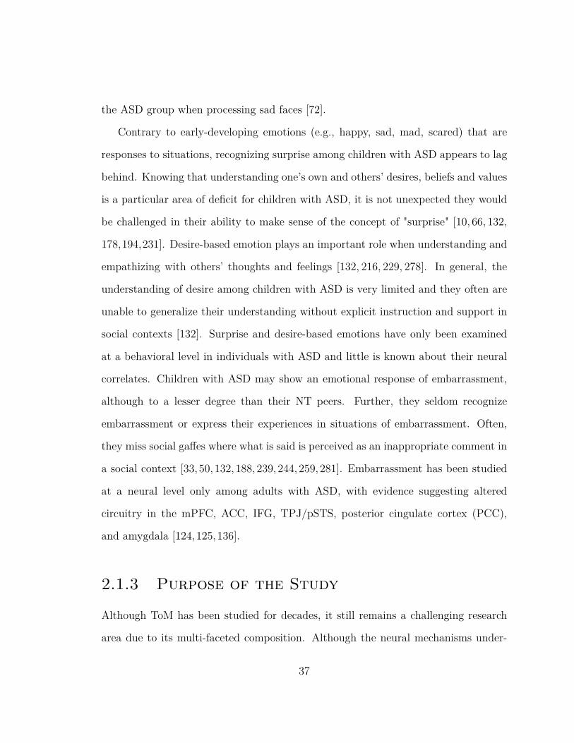
the ASD group when processing sad faces [72].
Contrary to early-developing emotions (e.g., happy, sad, mad, scared) that are
responses to situations, recognizing surprise among children with ASD appears to lag
behind. Knowing that understanding one’s own and others’ desires, beliefs and values
is a particular area of deficit for children with ASD, it is not unexpected they would
be challenged in their ability to make sense of the concept of "surprise" [10, 66, 132,
178,194,231]. Desire-based emotion plays an important role when understanding and
empathizing with others’ thoughts and feelings [132, 216, 229, 278]. In general, the
understanding of desire among children with ASD is very limited and they often are
unable to generalize their understanding without explicit instruction and support in
social contexts [132]. Surprise and desire-based emotions have only been examined
at a behavioral level in individuals with ASD and little is known about their neural
correlates. Children with ASD may show an emotional response of embarrassment,
although to a lesser degree than their NT peers. Further, they seldom recognize
embarrassment or express their experiences in situations of embarrassment. Often,
they miss social gaffes where what is said is perceived as an inappropriate comment in
a social context [33, 50, 132, 188, 239, 244, 259, 281]. Embarrassment has been studied
at a neural level only among adults with ASD, with evidence suggesting altered
circuitry in the mPFC, ACC, IFG, TPJ/pSTS, posterior cingulate cortex (PCC),
and amygdala [124,125,136].
2.1.3 Purpose of the Study
Although ToM has been studied for decades, it still remains a challenging research
area due to its multi-faceted composition. Although the neural mechanisms under-
37
lying ToM have been examined, few brain-based studies include children with ASD.
Thus, a greater understanding of the brain-behavior connections associated with ToM
in children will provide researchers a potential link between the biological mechanisms
of ToM and behavioral characteristics. It will help lead to more efficient diagnostic
processes and prognostic indicators for special populations like children with ASD.
To facilitate increased understanding of this linkage, the current study will emphasize
emotion recognition ToM with a specific focus on less well-studied and more complex
emotions (i.e., surprise, embarrassment, and desire-based emotion). Surprise and em-
barrassment are particularly difficult for children with ASD to recognize [121, 265].
While desire-based emotions are easier for children with ASD to recognize, their un-
derstanding of these emotions is delicate and often requires explicit descriptions.
The current study is the first to examine the neural correlates of selected ToM
constructs including desire-based emotions and more complex emotions (surprise,
embarrassment) to establish connections between behavior and brain activities in
children with ASD (i.e., 7 to 14 years old). We used The Theory of Mind Inventory-2
(ToMI-2) [131] and The Theory of Mind Task Battery (ToMTB) [134] to establish
the behavioral patterns and identify differences between ASD and NT groups in their
understanding of these less well-studied and complex emotions. We developed two
novel fMRI tasks to identify brain regions associated with the recognition and pro-
cessing of these emotions. Although our primary interest was in the neural response
to recognition of more complex emotions requiring ToM (i.e., embarrassment and sur-
prise), happy and sad faces were also included to provide a comparison with previous
literature investigating recognition of basic emotions. We established brain activa-
tion patterns in both groups to further probe brain deficits and neural compensation
38

mechanisms being adopted by the ASD group. Specifically, we expected to see al-
tered brain activity patterns among the ASD group in brain regions involved in the
ToM neural network (e.g., mPFC, pSTS, cingulate cortex, and TPJ). Building upon
findings from previous studies, the present study provides a deeper and more sys-
tematic understanding of the brain-behavior connections associated with ToM. This
knowledge may lead to both behavioral and neural pathways for examining the im-
pact of intervention research to achieve more normalized social performance. Such
research might also help predict those brain-behavior profiles of children most likely
to benefit from specific ToM or social cognitive based interventions, emphasizing the
importance of interventions delivered at the cognitive level to bridge behavior with
brain function [128].
2.2 Methods
2.2.1 Participants
Eleven children with ASD (1 female) and 22 NT children (7 females) participated
in the study, in which 9 ASD and 19 NT subjects were included as they completed
all of the the behavioral testing, magnetic resonance imaging (MRI) scans and fMRI
tasks. The remaining subjects either completed only the behavioral testing, or with-
drew from the study due to dental appliances precluding MRI scanning or because
of study interruption due to COVID-19. The full study included 2-3 hours of base-
line behavioral assessments along with a 1-hour brain scan including T1 imaging, T2
imaging, and two fMRI tasks. Since the understanding of surprise and embarrass-
39

ment has been shown to develop substantially between the ages of 5 and 8 years in
NT populations [32–34], we set the minimum cut off age as 7 years old. We expanded
the upper age limit to 14 years old to improve recruitment efforts given the challenges
of recruiting subjects with ASD. All children were native English speakers.
We administered the Autism Diagnostic Observation Schedule-2 (ADOS-2) [175]
and the Social Communication Questionnaire-Lifetime version (SCQ) [234] to con-
firm the clinical diagnosis for participants with ASD. Non-verbal intelligence and
language levels were tested for all participants using the Universal Nonverbal Intelli-
gence Test (UNIT-2) [44,184] and the Comprehensive Assessment of Spoken Language
(CASL) [54], respectively, to ensure participants could demonstrate understanding of
the instructions given in the behavioral and fMRI tasks. The UNIT-2 is a multidi-
mensional assessment of intelligence for individuals with speech, language, or hearing
impairments. It consists of nonverbal tasks that test symbolic memory, non-symbolic
quantity, analogic reasoning, spatial memory, numerical series, and cube design. The
CASL is an orally administered language assessment consisting of 15 subtests mea-
suring language for individuals ranging from 3 to 21 years of age. For the present
study, only those basic subsets that establish the CASL language core were used:
Antonyms, Sentence Completion, Syntax Construction, Paragraph Comprehension,
and Pragmatic Judgment.
Full demographic statistics are presented in Table 2.1. The groups differed on
CASL and UNIT-2 scores, with the ASD children obtaining lower scores on both
measures compared to the NT children. Because of these group differences in language
and intellectual abilities, CASL and UNIT-2 scores were included as covariates in
statistical analyses of ToM metrics.
40

Table 2.1: Demographics and T-tests Statistics including Mean, Range and p value
2.2.2 Behavioral Measures of ToM
Two norm-referenced tools were used as behavioral outcome measures to assess ToM.
The ToMI-2 [131] measures a parent’s perception of their child’s ToM understanding
of 60 items using a 20-unit rating scale from "Definitely Not" to "Definitely". Primary
caregivers use a vertical hash mark to indicate where on the continuous scale best
represents their perceptions. Item, subscale, and composite scores range from 0-20. A
higher number indicates a parent’s greater confidence in their child’s understanding
of a particular ToM skill. The ToMI-2 items represent typical social interactions
to ensure it is a socially and ecologically valid ToM index. The tool demonstrates
excellent test-retest reliability, internal consistency, and criterion-related validity for
neurotypical children and children with ASD as well as contrasting-groups validity
and statistical evidence of construct validity (i.e., factor analysis) [131,133,166].
The ToMTB [134] is a direct measure of a child’s understanding of ToM. It consists
of nine ToM tasks presented as short vignettes in a story-book format arranged in
ascending difficulty. For each of the nine tasks, children are provided with one correct
response option and three possible distracters. There are 15 total questions asked,
including memory control questions that must be answered correctly to get credit for
41

ToM understanding. The ToMTB has strong test-retest reliability [131,134].
To examine subjects’ ToM abilities, we used the total score of the ToMTB, total
composite mean of the ToMI-2 (i.e., assessing overall ToM ability), early subscale
mean of the ToMI-2 (i.e., assessing early developing ToMI ability such as regulat-
ing desire-based emotion and recognition of happy and sad), basic subscale mean
of the ToMI-2 (i.e., assessing basic ToM ability such as recognition of surprise), and
advanced subscale mean of the ToMI-2 (assessing advanced ToM ability such as recog-
nition of embarrassment). We also included scores from single items assessing recog-
nition of simple emotions such as happy and sad, as well as more complex emotions
such as surprise and embarrassment.
2.2.3 MRI Acquisition
All neuroimaging data was acquired using the University of Vermont MRI Center
for Biomedical Imaging 3T Philips Achieva dStream scanner and 32-channel head
coil. The imaging protocol is based on that developed for the multicenter NIH-
funded Adolescent Brain Cognitive Development (ABCD) study, which is derived
from large studies such as the Human Connectome Project (HCP) and the Lifespan
Connectome Project. The protocols make extensive use of simultaneous multislice
imaging [47,241,242] (multiband SENSE) to accelerate functional and diffusion MRI
acquisitions.
42

2.2.4 Task fMRI Parameters and Preprocessing
Task fMRI parameters were: TR 800ms, TE 30ms, flip angle 52 degrees, 2.4mm
isotropic imaging resolution with a 216x216x144mm3 field of view using a multiband
acceleration factor of 6 (60 slices, no gap). For fMRI acquisitions, corresponding field
maps were generated using pairs of reference acquisitions with opposite phase-encode
directions. fMRI preprocessing used the pipelines developed as parts of the HCP
[241]. The HCP functional pipeline corrects for EPI spatial distortions using magnetic
field maps and realigns volumes to account for subject motion. Specifically, the
fMRI surface pipeline was included in the preprocessing analysis while independent
component analysis (ICA) denoising was not. Participants were trained to remain
still during the scan in a mock scanner supplemented by video model (a short video
demonstrating expected behavior in the scan) prior to each assessment. The HCP
task fMRI pipelines were used for first- and second-level analysis of task fMRI data.
These pipelines incorporate high-pass filtering and application of general linear models
(GLMs) to model task parameters and nuisance regressors (e.g., motion parameters)
[138,139].
2.2.5 fMRI Emotion Recognition (fER) Task
This task was designed to assess emotion recognition, which was also a behavioral item
tested on both ToMI-2 and ToMTB [131, 166]. The fMRI task required participants
to either identify the emotions expressed in cartoon faces (emotion recognition) or
judge the gender of the same emotional faces (perceptual control). Faces depicting
happiness, sadness, surprise, and embarrassment were included. Since we did not
43

find existing stimulus/picture sets depicting embarrassment or of facial expressions
of children in general, we hired a professional artist to create digital drawings of the
cartoon face stimuli that matched the age of the participants and the style of pictures
used in the ToMTB and ToMI-2. An independent sample of 163 NT participants from
Amazon Mturk validated and selected the expressions that were recognized with 80%
to 90% accuracy and were matched for valence. The final selection included eight
different characters and two versions of each expression for each character, leading to
16 unique stimuli for each expression (see Figure 2.1 panel b for examples).
The fER task utilized a mixed design, with a block design for task (emotion
recognition of surprise/embarrassment, emotion recognition of happy/sad, and per-
ceptual control) and an event-related design for facial expression within each block
(surprise/embarrassment or happy/sad). Across two runs, we presented 8 blocks of
emotion recognition of surprise and embarrassment, 8 blocks of emotion recognition
of happy and sad, and 8 blocks of perceptual control. Each block presented 8 faces in
an event-related fashion for 2 seconds each with a jittered ISI (i.e., multiples of the
TR, optimized using the optseq tool [102]. Participants pressed one of two buttons to
indicate the emotional expression (surprise/embarrassment or happy/sad, in emotion
recognition blocks) or the gender of the face (boy/girl, in perceptual control blocks);
see Figure 2.1, panel a. An instructional cue (i.e., Label Emotion, Label Gender) was
provided before each block. Between blocks there was an 8 second interval. The mixed
design allowed us to not only create different contrasts between each emotion, but also
between advanced emotion processing (i.e., recognition of combined embarrassment
and surprise) with basic emotion processing (i.e., combined happy and sad).
44

Figure 2.1: Illustration of the fMRI Emotion Recognition (fER) Task
2.2.6 fMRI Theory of Mind (fToM) Task
We developed this fMRI task to directly model the desire-based emotion task in the
ToMTB [134]. Participants were required to infer the reaction of a cartoon character
to a gift using the knowledge provided about the preferences of that character. Each
trial was broken into three images: encoding, probe, and decision-making (see Figure
2.2). In the encoding image (2.4s), a child was seen holding a gift box. Two items
were presented in thought bubbles displaying the preferences for that character. One
item was presented with hearts to indicate the character likes the item, the other with
an X over it to indicate the character dislikes that item. In the probe image (2s),
a top view of the unwrapped gift box showed the desired/undesired item. Finally,
in the decision-making image (2s), participants saw two images of the character’s
face expressing either happiness or sadness and respond by pressing a button to
indicate which face best captured the reaction of the character to the gift. In control
trials, participants saw an empty thought bubble and gift box and were asked at
the decision-making step to indicate the gender of the cartoon face. There were 10
different characters and 30 unique items ranging from items commonly recognized as
45
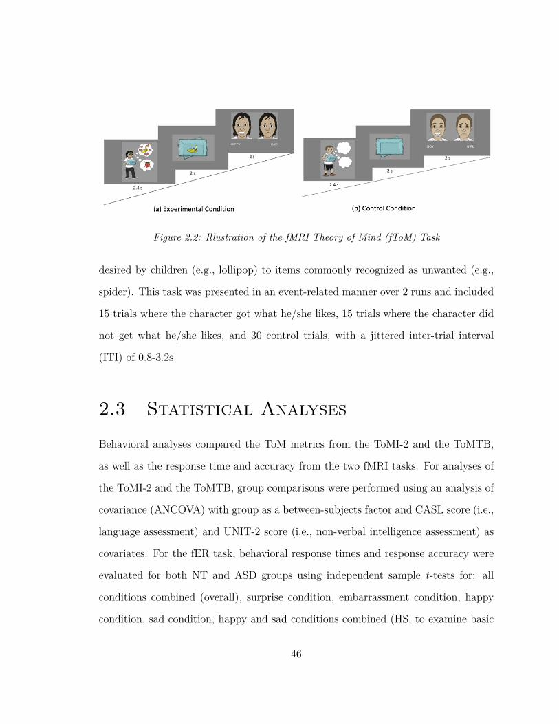
Figure 2.2: Illustration of the fMRI Theory of Mind (fToM) Task
desired by children (e.g., lollipop) to items commonly recognized as unwanted (e.g.,
spider). This task was presented in an event-related manner over 2 runs and included
15 trials where the character got what he/she likes, 15 trials where the character did
not get what he/she likes, and 30 control trials, with a jittered inter-trial interval
(ITI) of 0.8-3.2s.
2.3 Statistical Analyses
Behavioral analyses compared the ToM metrics from the ToMI-2 and the ToMTB,
as well as the response time and accuracy from the two fMRI tasks. For analyses of
the ToMI-2 and the ToMTB, group comparisons were performed using an analysis of
covariance (ANCOVA) with group as a between-subjects factor and CASL score (i.e.,
language assessment) and UNIT-2 score (i.e., non-verbal intelligence assessment) as
covariates. For the fER task, behavioral response times and response accuracy were
evaluated for both NT and ASD groups using independent sample t-tests for: all
conditions combined (overall), surprise condition, embarrassment condition, happy
condition, sad condition, happy and sad conditions combined (HS, to examine basic
46
emotion processing), embarrassment and surprise condition combined (ES, to ex-
amine advanced emotion processing), and the control (gender) condition. For the
fToM task, response times and response accuracy were evaluated for both NT and
ASD groups using independent sample t-tests for: all conditions combined (overall),
control (gender) condition, and experimental conditions (dislike and like conditions
combined).
In order to detect neural activation related to the two fMRI tasks, first-level neu-
roimaging analyses were performed first. Vectors of stimuli onsets using the stimulus
duration were created for each trial type and were convolved with a canonical double
gamma hemodynamic response function to produce a regressor for each condition.
Six motion regressors were included as covariates. For second-level neuroimaging
group analyses, whole-brain between-group changes in activation patterns over time
were assessed for each experimental condition using FSL’s permutation-based non-
parametric testing and threshold-free cluster enhancement to control for multiple
comparisons [248]. Analyses were restricted to gray matter. Different group GLMs
were constructed for each hypothesis separately. For the fER task, GLM models
were constructed for contrasts between happy vs. gender, sad vs. gender, surprise
vs. gender, embarrassment vs. gender, happy & sad vs. gender (HS, to assess ba-
sic emotion processing), and embarrassment & surprise vs. gender (ES, to assess
advanced emotion processing). For fToM task, GLM models were constructed for
contrasts between dislike & like (experimental) vs. control. CASL scores (i.e., lan-
guage assessment) and UNIT-2 scores (i.e., non-verbal intelligence assessment) were
also included as covariates in the permutation analysis of linear models (PALM) for
group comparisons [283].
47

Table 2.2: ToM Behavioral Measurements and ANCOVA Results
2.4 Results
2.4.1 Behavioral Results
When adjusted for covariates of the CASL (i.e., language assessment) and UNIT-
2 scores (i.e., non-verbal intelligence assessment), the NT group had a significantly
better performance on the ToMI-2 total score (F(1,24)=4.48, p<0.05) and the ToMI-2
early subscale score (F(1,24)=7.76, p<0.01). See Table 2.2.
On the fER task, the NT group responded significantly faster than the ASD group
when considering all conditions together (t(54)=2.56, p=0.01), as well as in condi-
tions of surprise (t(54)=2.58, p=0.01), embarrassment (t(54)=2.64, p=0.01), sadness
(t(54)=2.03, p<0.05), control (t(54)=2.73, p<0.01), and ES (embarrassed + sur-
prise) (t(54)=2.77, p<0.01); see Figure 2.3. There was no significant difference found
for the happy condition (t(54)=1.75, p=0.09) or the HS (happy + sad) condition
(t(54)=1.95, p<0.06). On this task the NT group also had a significantly higher re-
sponse accuracy compared to the ASD group when considering all conditions together
(t(54)=2.55, p=0.01), as well as in conditions of ES (t(54)=2.29, p=0.03) and control
48

Figure 2.3: fMRI Emotion Recognition (fER) Task Response Time and Accuracy
(t(54)=2.52, p=0.02). There was no significant difference in terms of response accu-
racy between the two groups in conditions of surprise (t(54)=1.92, p=0.06), embar-
rassment (t(54)=1.87, p=0.07), happiness (t(54)=0.77, p=0.44), sadness (t(54)=1.89,
p=0.06), or HS (t(54)=1.77, p=0.08).
On the fToM task, there was no significant difference in response time between the
NT and ASD groups for any of the comparisons: all conditions combined (t(54)=1.27,
p=0.21), experimental condition (t(54)=1.14, p=0.26), and control condition (t(54)=1.33,
p=0.19); see Figure 2.4. The NT group had a significantly higher response accuracy
compared to the ASD group when considering all conditions combined (t(54)=2.21,
p=0.03) and for the experimental condition only (t(54)=2.23, p=0.03). There was
no significant difference in response accuracy for the control condition (t(54)=1.92,
p=0.06).
2.4.2 Brain Activity Patterns
Cohen’s D effect size maps were generated for each contrast of both fMRI tasks.
The present study adopted a threshold of d=0.5 (medium) to show the effect size of
49

Figure 2.4: fMRI Theory of Mind (fToM) Task Response Time and Accuracy
the difference in brain activities between the ASD and NT groups. The contrast was
conducted as ASD minus NT. Positive d-values (hot colors) indicate greater activation
in the ASD group compared to the NT groups, while negative d-values (cool colors)
indicate less activation in the ASD group compared to the NT groups.
In the fER task, when recognizing happy faces the ASD group showed greater
brain activation in the mPFC and the angular gyrus (AG) compared to the NT
group (see Figure 2.5), but less brain activation in most areas of the frontal cortex,
the temporal lobe especially around the inferior temporal sulcus (ITS), and the TPJ.
When recognizing sad faces, the ASD group showed greater brain activation in the left
temporal lobe, the left AG, the anterior and posterior cingulate cortex, the occipital
lobe, and the perirhinal area, but less brain activation in the right temporal lobe, the
mPFC, and the inferior part of the post central gyrus. When combining happy and
sad faces (i.e., HS), the brain activation pattern was similar to recognizing sad faces
alone.
When recognizing surprised faces in the fER task, the ASD group showed greater
brain activation in the left AG, the left temporal pole and STS, the left anterior and
50
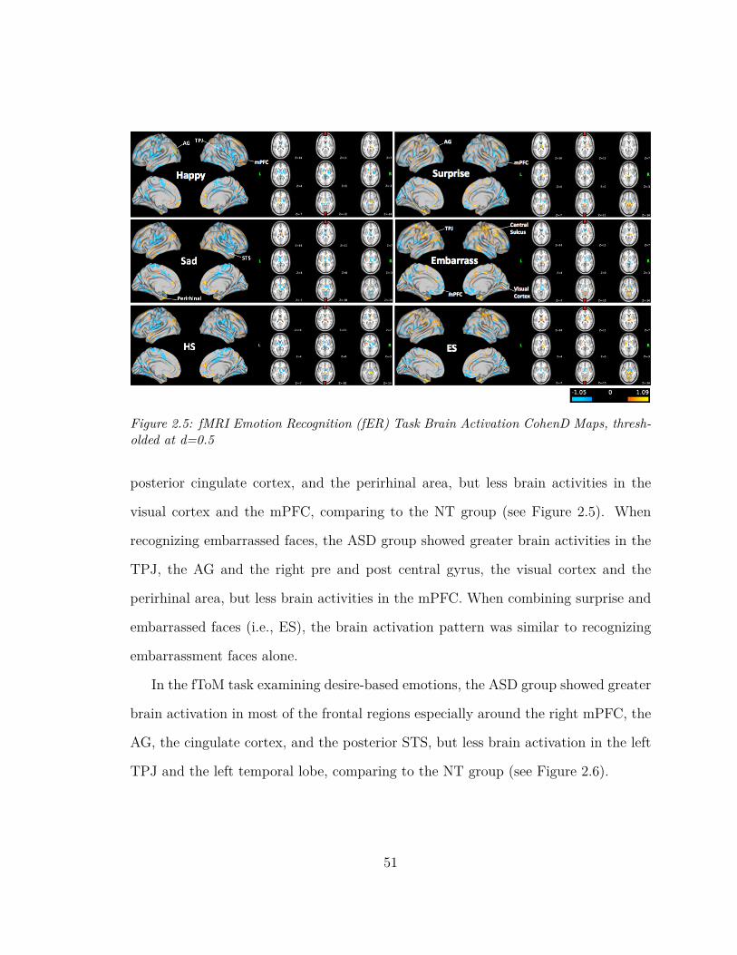
Figure 2.5: fMRI Emotion Recognition (fER) Task Brain Activation CohenD Maps, thresh-olded at d=0.5
posterior cingulate cortex, and the perirhinal area, but less brain activities in the
visual cortex and the mPFC, comparing to the NT group (see Figure 2.5). When
recognizing embarrassed faces, the ASD group showed greater brain activities in the
TPJ, the AG and the right pre and post central gyrus, the visual cortex and the
perirhinal area, but less brain activities in the mPFC. When combining surprise and
embarrassed faces (i.e., ES), the brain activation pattern was similar to recognizing
embarrassment faces alone.
In the fToM task examining desire-based emotions, the ASD group showed greater
brain activation in most of the frontal regions especially around the right mPFC, the
AG, the cingulate cortex, and the posterior STS, but less brain activation in the left
TPJ and the left temporal lobe, comparing to the NT group (see Figure 2.6).
51
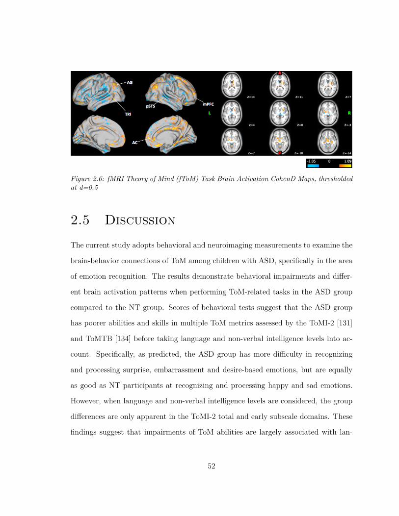
Figure 2.6: fMRI Theory of Mind (fToM) Task Brain Activation CohenD Maps, thresholdedat d=0.5
2.5 Discussion
The current study adopts behavioral and neuroimaging measurements to examine the
brain-behavior connections of ToM among children with ASD, specifically in the area
of emotion recognition. The results demonstrate behavioral impairments and differ-
ent brain activation patterns when performing ToM-related tasks in the ASD group
compared to the NT group. Scores of behavioral tests suggest that the ASD group
has poorer abilities and skills in multiple ToM metrics assessed by the ToMI-2 [131]
and ToMTB [134] before taking language and non-verbal intelligence levels into ac-
count. Specifically, as predicted, the ASD group has more difficulty in recognizing
and processing surprise, embarrassment and desire-based emotions, but are equally
as good as NT participants at recognizing and processing happy and sad emotions.
However, when language and non-verbal intelligence levels are considered, the group
differences are only apparent in the ToMI-2 total and early subscale domains. These
findings suggest that impairments of ToM abilities are largely associated with lan-
52
guage and intellectual levels, especially during more complex emotion recognition.
In addition, these findings are consistent with the diagnostic criteria and other com-
mon challenges in ASD, including language and intellectual impairments and social
challenges requiring advanced ToM abilities.
According to the results from the fER task, the ASD group takes longer to recog-
nize facial expressions of surprise, embarrassment and sadness. This phenomenon is
especially apparent when combining conditions of surprise and embarrassment (ES).
This is consistent with previous studies suggesting intact ability for recognizing happi-
ness in children with ASD [11] but impairments in recognizing sadness [11,67,218,271].
There are more severe impairments among children with ASD for recognizing surprise
and embarrassment compared to happiness and sadness, as these advanced emotions
require more cognitive processes [121,265]. Thus, it makes sense that the ASD group
needs more time to recognize embarrassment and surprise faces. In addition, the ASD
group does not recognize surprise and embarrassment faces as accurately as the NT
group. Results from the fToM task also indicate poorer performance (i.e., response
accuracy) in processing desire-based emotion in the ASD group than the NT group,
providing evidence that children with ASD have impaired skills in understanding
ToM-associated desire-based emotion.
The brain activation pattern generated by the novel fMRI tasks demonstrate some
consistent findings from previous literature. Specifically, when recognizing basic emo-
tions such as happy and sad, the ASD group displays unusual brain activation pat-
terns in the mPFC, the temporal sulcus, and the AG, compared to the NT group. In
addition, compared to happiness, recognition of sadness in the ASD group seems to
engage a more diverse range of brain regions including the occipital lobe, the left tem-
53
poral lobe and the perirhinal area associated with the amygdala. This implies that
the ASD population has more difficulty recognizing sadness compared to happiness
as there is greater brain effort as well as different brain areas that are recruited as
compensatory mechanisms [11,67,70,121,162,218,232,265,271]. On the other hand,
when recognizing happiness, there is less brain activation in the TPJ but more brain
activation in the AG among the ASD group as compared to the NT group. Multi-
ple studies have established the collaborative relationship between the TPJ and AG
during ToM-related tasks among children with ASD, where the AG would display sig-
nificantly increased activation if a particular ToM task required TPJ activation [135].
When recognizing more complex emotions such as surprise and embarrassment, the
ASD group displayed more brain activation in the left AG but less brain activity in
the mPFC area compared to the NT group. This suggests that advanced emotion
recognition requires more ToM related abilities than abilities related to executive
functions among the ASD group. It also seems that ToM requires more effort for the
ASD group to recognize embarrassment compared to surprise as the ASD group en-
gages in more brain activation in regions of the TPJ, the pre and post central gyrus,
the visual cortex and the perirhinal area. This implies that the recognition of em-
barrassment requires more visual information processing and advanced ToM abilities,
and may trigger the brain’s fear center [27, 105, 193]. When processing desire-based
emotion, the ASD group displays more brain activity around the right mPFC, the AG,
the anterior cingulate cortex, and the posterior STS, but less brain activity around
the left TPJ and the left temporal lobe, compared to the NT group. It is obvious
that desire-based emotion processing is associated with traditional ToM related brain
network including the TPJ, the STS, and the cingulate cortex. With decreased brain
54
activation in the TPJ area, the ASD group seems to engage the AG as a compen-
satory mechanism. In addition, the ASD group showed increased brain activity in the
mPFC area compared to the NT group suggesting the possibility of recruiting more
executive control regions to process the task to compensate for poorer ToM abilities.
As a pilot study, one major contribution of the present study is the successful
implementation of two novel fMRI tasks targeting emotion recognition and process-
ing associated with ToM. To our knowledge, this is the first time a set of facial
stimuli were able to be adapted to an fMRI task to include expressions of surprise
and embarrassment using the faces of children. It is also the first time that neural
mechanisms underlying desire-based emotion processing are examined through fMRI.
Further, these tasks are directly adapted from the ToMTB and ToMI-2 which allows
comparison between behavioral and neuroimaging measurements. In addition, nearly
all behavioral tests of ToM currently available only examine one or a few aspects of
ToM; however, the ToMI-2 and ToMTB used in the present study are multi-faceted
tools that cover several aspects of ToM (e.g., emotion recognition, false belief, per-
spective taking etc.), including information from both a parent and the child. These
tools have helped to identify specific domains in ToM that are a struggle for ASD
children. The present study has also identified brain activation maps associated with
such domains by using two novel fMRI tasks. These maps provide evidence of how
different brain regions are involved in the ability to reflect on mental states (i.e.,
mPFC), understand other’s actions (i.e., pSTS), integrate relevant social information
(i.e., TPJ), create and process emotions (i.e., cingulate cortex), and regulate negative
emotions (i.e., amygdala) [72,143,205,256,258].
55
2.6 Limitations
Studies using MRI/fMRI technology face the typical challenges for any population
with the requirements for tolerating loud noises, remaining still during the assessment
and managing feelings of claustrophobia. For an ASD population these challenges are
exacerbated, particularly in the recruitment phase of the study and in obtaining usable
data. These challenges along with children being fitted with dental pieces precluded
our ability to complete the MRI/fMRI images and COVID restrictions further limited
access to an ASD population.
To address the challenges for using MRI/fMRI, all participants participated in a
mock scanner experience. They viewed two videos explaining the process including
one video specifically made to rehearse the experience. These strategies supported
the successful participation of many of the participants with ASD but not all, which
led to missing data.
As a part of our recruitment efforts, we expanded the age limit to 14 years old.
The age of the participants in this study ranged from early childhood to the early
adolescent period, typically an ideal age range to measure brain size while considering
other possible neuroanatomical variables. Importantly, as the brain continues to grow
and change into adulthood with rapid and significant development among adolescence
[155,195], the interpretation of the results provides insights for children in general, but
does not provide age-specific information. Further, language abilities varied, so the
possibility exists that participants may not have fully comprehended the instructions
of the fMRI tasks and hence given wrong or negligent responses. To address these
concerns, all participants were provided with in-person introductions for both tasks
56

which were also practiced on a computer with the same response device outside the
scanner to ensure correct understanding and performance. During the scan, a research
team member monitored the response signal box in the monitor room at all times
to reassure that participants were paying attention to the task and following task
instructions.
2.7 Conclusions and Implications
Currently, the diagnosis of ASD is based on behavioral symptoms alone. Delays in
diagnosis of ASD can be 13 months and longer for minority and lower socioeconomic
status groups [35, 57, 180, 181, 190, 225, 280]. It is also believed that a substantial
number of individuals on the spectrum remain undetected [260]. Children with ASD
are a challenging population to engage in experimental procedures that have specific
task requirements and involve neuroimaging measurements. However, it is crucial
to gather both behavioral and neuroimaging data to increase our understanding of
the nature of the social deficits characteristic in ASD. This is important if we wish
to advance the diagnostic process and implement intervention methods for which we
are able to show positive behavioral and neural outcomes. Insights derived from this
study are expected to help scientists and physicians understand how social deficits
surrounding ToM in ASD are associated with the brain. By taking advantage of such
knowledge, we might be able to offer insights into the development of neuroanatom-
ical diagnostic criteria to allow for more efficient diagnosis and early identification
of high-risk populations. It might also help provide a vehicle for examining neural
and behavioral outcomes following individualized treatment methods. Although the
57
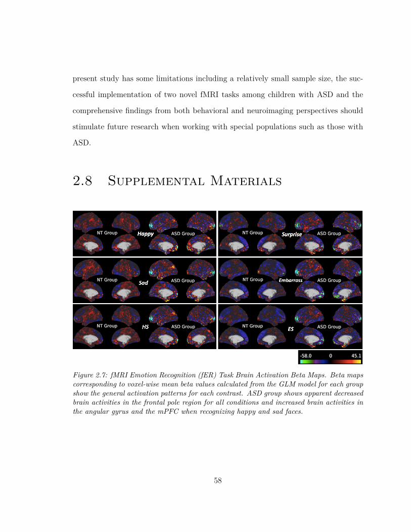
present study has some limitations including a relatively small sample size, the suc-
cessful implementation of two novel fMRI tasks among children with ASD and the
comprehensive findings from both behavioral and neuroimaging perspectives should
stimulate future research when working with special populations such as those with
ASD.
2.8 Supplemental Materials
Figure 2.7: fMRI Emotion Recognition (fER) Task Brain Activation Beta Maps. Beta mapscorresponding to voxel-wise mean beta values calculated from the GLM model for each groupshow the general activation patterns for each contrast. ASD group shows apparent decreasedbrain activities in the frontal pole region for all conditions and increased brain activities inthe angular gyrus and the mPFC when recognizing happy and sad faces.
58
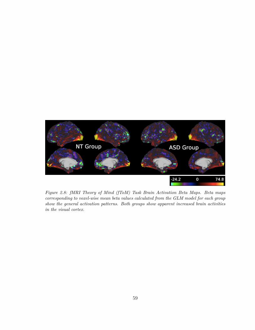
Figure 2.8: fMRI Theory of Mind (fToM) Task Brain Activation Beta Maps. Beta mapscorresponding to voxel-wise mean beta values calculated from the GLM model for each groupshow the general activation patterns. Both groups show apparent increased brain activitiesin the visual cortex.
59
Chapter 3
Identifying Neuroanatomical and
Behavioral Features for Autism Spec-
trum Disorder Diagnosis in Chil-
dren using Machine Learning
Yu Han1,2*, Donna M. Rizzo3, John P. Hanley4, Emily L. Coderre1,2, Patricia A.
Prelock1,2
1 Department of Communication Sciences and Disorders, University of Vermont.
2 Neuroscience Graduate Program, University of Vermont.
3 Department of Civil and Environmental Engineering, University of Vermont.
4 Department of Microbiology and Molecular Genetics, University of Vermont.
60

3.1 Introduction
Autism spectrum disorder (ASD) is a lifelong neurodevelopmental disorder in which
an individual’s symptoms can vary from mild to severe. It is characterized by signif-
icant social, communication, and behavioral challenges [171]. According to the most
recent report from the Center for Disease Control and Prevention (CDC), the number
of children in the U.S. diagnosed with ASD is about 1 in every 54 in year 2016 [58].
While genetic and environmental factors have been linked to the development of ASD,
at present there is no identified cause or cure for ASD.
ASD is characterized by impairments in social interaction and the presence of
restricted and repetitive behaviors, interests, or activities [9, 37, 97, 168]. Children
with ASD may not show any symptoms until age two or later [38, 152]. Currently,
the diagnosis of autism is based on behavioral symptoms alone: 1) impairments in
social communication and interaction; and 2) the presence of restricted and repetitive
behaviors, interests, or activities [9, 37, 97, 168]. There are two common behavioral
assessment tools guiding the diagnostic process: The Autism Diagnostic Observa-
tion Schedule-Second edition (ADOS-2) and The Autism Diagnostic Interview-revised
(ADI-R) [173,174]. However, a typical diagnostic appointment consists of evaluations
lasting several hours at a designated clinical office. Due to the rigorous and time-
consuming nature of ASD diagnostic examinations, the demand exceeds the capacity
to see patients. As a result, many diagnostic centers have expanding wait lists for
appointments. This bottleneck can translate to delays in diagnosis of 13 months and
longer [35, 38, 57, 180, 181, 190, 225]. It is also believed that a substantial number of
individuals on the spectrum remain undetected [260]. With growing awareness of
61

ASD, there is a high demand for a faster and automated ASD diagnostic approach
that might allow for more efficient diagnosis and early identification of high-risk pop-
ulations [208].
Building an automated diagnostic and predictive model of ASD is timely as many
studies have adopted machine learning approaches to identify significant biomarkers
that include both behavioral and biological features. Duda and colleagues (2016)
applied machine learning to distinguish ASD from attention deficit hyperactivity dis-
order (ADHD) using the Social Responsiveness Scale for children between 5 to 13
years old [81]. Bone et al. (2015) trained their models to diagnose autism against
healthy controls using the same Social Responsiveness Scale and the Autism Diagnos-
tic Interview-Revised score for children between 5 to 17 years old [39]. Other studies
aggregated items from the ADOS and scores from the Autism Quotient (AQ) to accu-
rately classify an ASD group [13]. However, one limitation of using these behavioral
outcome measures to classify participants is that they can be interpreted as being
subjective. Furthermore, these studies identify a wide range of features depending on
which models and tests are used. Consequently, the ability to identify more consis-
tent ASD markers using neuroimaging measures to supplement behavioral measures
becomes important.
As a result of the wide range and subjective nature of behavioral measures used
in diagnosing ASD, many studies are exploring brain-based biological markers (e.g.,
measurable via magnetic resonance imaging (MRI)) to identify a common etiology
across individuals with ASD. Currently, these less subjective markers are attrac-
tive not only for diagnostic purposes, but as possible targets for interventions [93].
Independent structural MRI studies have found differences in whole brain volume
62
and the developmental trajectories between individuals with ASD and those without
ASD [7,45,78,123,163,201,246,266,270]. Other structural brain abnormalities associ-
ated with ASD include cortical folding signatures that appear in the following regions
of the brain: temporal-parietal junction, anterior insula, posterior cingulate, lateral
and medial prefrontal cortices, corpus callosum, intra-parietal sulcus, and occipital
cortex [7, 45, 78, 123, 163, 201, 246, 270]. Evidence also shows that an accelerated ex-
pansion of cortical surface area, but not cortical thickness, causes an early overgrowth
of the brain in children with ASD [110], while other studies suggest that individuals
with ASD tend to have thinner cortices and reduced surface area as an effect of ag-
ing [85]. With these clear brain differences among those with and without ASD, it
therefore is informative and critical to look for brain-based ASD biomarkers.
Machine learning (ML) has been introduced to the neuroimaging field to identify
the abnormal brain regions in individuals with ASD. The support vector machine
(SVM) is an algorithm that avoids overfitting and is known for high classification
accuracy without requiring large sample sizes. It has been used to classify ASD from
corresponding control participants using extracted features from functional connec-
tivity metrics and grey matter volume [59, 62, 107, 140]. Other ASD applications of
ML classifiers include deep neural networks [204] and the random forest (RF) algo-
rithm; the latter uses random ensembles of independently grown decision trees [61].
Although these methods have demonstrated high accuracy for classifying ASD, to our
knowledge, they have not been used to identify input variables most closely associated
with ASD (i.e., feature selection). In addition, the majority of studies use data from
the Autism Brain Imaging Data Exchange (ABIDE) dataset, which includes 1112 ex-
isting resting-state functional MRI (rs-fMRI) datasets with corresponding structural
63
MRI and phenotypic information from 539 individuals with ASD and 573 age-matched
typical controls between the ages of 7 and 64 collected from 24 international brain
imaging laboratories [68].
Classification across a heterogeneous population is challenging [127, 148], partic-
ularly when neuroimaging data are pooled from multiple acquisition sites, such as
the ABIDE dataset, which has considerable variation in demographic and phenotypic
profiles. Data variance introduced via scanner hardware, imaging protocols, operator
characteristics, regional demographics, and other site-specific acquisition factors can
affect the classification performance. This problem is especially relevant for ASD given
the inherent heterogeneity of the population. It is often difficult to collect neuroimag-
ing data from individuals with ASD given the loudness of the scanner and difficulties
participants have remaining still. In fact, most individual site datasets have small
sample sizes that can lead to overfitting and classification inaccuracies using tradi-
tional ML algorithms. Moreover, while the ultimate goal for ML-based diagnostic
classification in neuroimaging is to identify discriminative features that provide in-
sight into abnormal structure and dysfunctional connectivity patterns in the affected
population [164], many of the ML algorithms applied to ASD were designed to clas-
sify large amounts of data (e.g., ABIDE) rather than optimize the selection of input
features.
The drivers or markers of ASD are likely the result of a complex interaction of
factors with no single factor (i.e., main effect or univariate model) driving the system.
As such, traditional statistical tools (e.g., logistic regression) that search for univariate
drivers of ASD are unlikely to find consistent patterns. Thus, ML techniques that
explore large search spaces for multivariate interactions are both needed and becoming
64

popular in helping to elucidate the complex interactions in systems such as ASD.
Our study employs one such ML tool: an evolutionary algorithm [186] called the
conjunctive clause evolutionary algorithm (CCEA) [115]. The CCEA was specifically
designed to efficiently explore large search spaces for complex interactions between
features and some associated nominal outcome (e.g., ASD or neurotypical (NT)).
In addition, the CCEA has built-in tools to prevent overfitting to produce easily
interpretable parsimonious models.
This study examines the validity of using the CCEA for feature selection in ASD,
particularly to address traditional statistical challenges associated with datasets hav-
ing small sample sizes and a large number of feature measurements. Additionally,
the selected features are validated and used for diagnostic classification by applying
a separate and more traditional ML classifier (i.e., the k-nearest neighbors (KNN)
algorithm). The dataset in this present study has a relatively large number of fea-
tures, consisting of both behavioral and neuroimaging measurements. The behavioral
measurements include scores of language ability, intellectual ability, and theory of
mind (ToM). The neuroimaging measurements include brain volume, brain surface
area, cortical thickness, and cortical curvature extracted from MRI whole-brain T1-
weighted scans. These features were collected from a total of 28 children ages 7 to
14, of which 9 children had been diagnosed with ASD. Only a subset of these 28 chil-
dren were used for feature selection in the CCEA (7 children with ASD and 14 NT
children), as another subset (2 children with ASD and 5 NT children) were enrolled
at a later time (i.e., after the CCEA was trained). While this later cohort was not
included in the CCEA feature selection analysis, it was included in the subsequent
validation and predictive KNN modeling.
65
Using the CCEA, we aim to identify discriminative biomarkers and behavioral
features to help develop an automatic diagnostic and predictive system for ASD. We
believe this is the first study to:
• Select discriminative biomarkers among children from 7 to 14 years old and
classify ASD.
• Include both behavioral and neuroimaging measurements in the feature selection
model.
• Identify models (sets of features) that most strongly correlate to children with
ASD given a dataset with a relatively small sample size (i.e., N=28) and large
number of features (i.e., 247 neuroimaging features and 13 behavioral features).
• Develop a predictive ML model using input features selected by the CCEA.
3.2 Materials and Methods
3.2.1 Participants
A total of 9 children with ASD (1 female) and 19 NT children (7 female), ages 7-
14, were enrolled in the study. In addition to the behavioral assessments described
below, the ASD group also completed the ADOS-2 and the Social Communication
Questionnaire-Lifetime version (SCQ) [234] to confirm their ASD diagnosis. Although
diagnosis of ASD is typically done at an early age, the characteristics of ASD are
long-term, and classification with additional neurobiological information at any age
66
recognizes the potential for brain-behavior comparisons with neurotypical popula-
tions. Potential changes behaviorally and neurobiologically at any age may also in-
form types, duration, and intensity of intervention that may influence these changes.
Therefore although we test older children, this study is still informative for increasing
the understanding of likely biomarkers of ASD.
3.2.2 Behavioral Measurements
All children participated in 2-3 hours of baseline behavioral assessments that in-
cluded the Comprehensive Assessment of Spoken Language (CASL) [54], the Univer-
sal Nonverbal Intelligence Test-2 (UNIT-2) [44,184], the Theory of Mind Task Battery
(ToMTB) [134] and the Theory of Mind Inventory-2 (ToMI-2) [131,133]. Measures of
language and cognition are typical in the assessment of ASD, as the Diagnostic and
Statistical Manual of Mental Disorders (DSM-5) requires an assessment of language
and intellectual functioning beyond the diagnosis of ASD.
The CASL is an orally administered research-based assessment consisting of 15
subtests measuring language for individuals ranging from 3 to 21 years of age. For
the present study, only those basic subsets that establish the CASL language core
are used: Antonyms, Sentence Completion, Syntax Construction, Paragraph Com-
prehension, and Pragmatic Judgment. The UNIT-2 is a multidimensional assessment
of intelligence for individuals with speech, language, or hearing impairments. It con-
sists of nonverbal tasks that test symbolic memory, non-symbolic quantity, analogic
reasoning, spatial memory, numerical series, and cube design.
ToM is a core deficit in ASD that is often used to explain the social impairment
characteristic of the disorder. ToM is the ability to reason about the thoughts and
67

feelings of self and others, including the ability to predict what others will do or how
they will feel in a given situation on the basis of their inferred beliefs [21,23]. Scores
from both ToMTB and ToMI-2 were included to provide representative measures of a
child’s social cognition level. The ToMTB and ToMI-2 are two norm-referenced tools
and behavioral tasks used as outcome measures to assess ToM [21, 23]. Scores from
both ToMTB and ToMI-2 provide valid representations of a child’s social cognition
level. The ToMI-2 measures a parent’s perception of their child’s ToM understanding
of 60 items using a 20-unit rating scale from "Definitely Not" to "Definitely". Primary
caregivers use a vertical hash mark to indicate where on the continuous scale best
represents their perceptions. Item, subscale, and composite scores range from 0-20. A
higher number indicates a parent’s greater confidence in their child’s understanding
of a particular ToM skill. The ToMI-2 items represent typical social interactions
to ensure it is a socially and ecologically valid ToM index. The tool demonstrates
excellent test-retest reliability, internal consistency, and criterion-related validity for
neurotypical children and children with ASD as well as contrasting-groups validity
and statistical evidence of construct validity (i.e., factor analysis) [131,133,166]. The
ToMTB is a direct measure of a child’s understanding of ToM. It consists of nine
ToM tasks presented as short vignettes in a story-book format arranged in ascending
difficulty. For each of the nine tasks, children are provided with one correct response
option and three possible distracters. There are 15 total questions asked, including
memory control questions that must be answered correctly to get credit for ToM
understanding. The ToMTB has strong test-retest reliability [131,134]. .
In selecting potential features for the CCEA, we included 13 behavioral features
in total. These included the total score of the CASL, full scale score of the UNIT-2,
68
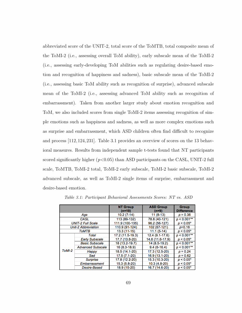
abbreviated score of the UNIT-2, total score of the ToMTB, total composite mean of
the ToMI-2 (i.e., assessing overall ToM ability), early subscale mean of the ToMI-2
(i.e., assessing early-developing ToM abilities such as regulating desire-based emo-
tion and recognition of happiness and sadness), basic subscale mean of the ToMI-2
(i.e., assessing basic ToM ability such as recognition of surprise), advanced subscale
mean of the ToMI-2 (i.e., assessing advanced ToM ability such as recognition of
embarrassment). Taken from another larger study about emotion recognition and
ToM, we also included scores from single ToMI-2 items assessing recognition of sim-
ple emotions such as happiness and sadness, as well as more complex emotions such
as surprise and embarrassment, which ASD children often find difficult to recognize
and process [112,124,231]. Table 3.1 provides an overview of scores on the 13 behav-
ioral measures. Results from independent sample t-tests found that NT participants
scored significantly higher (p<0.05) than ASD participants on the CASL, UNIT-2 full
scale, ToMTB, ToMI-2 total, ToMI-2 early subscale, ToMI-2 basic subscale, ToMI-2
advanced subscale, as well as ToMI-2 single items of surprise, embarrassment and
desire-based emotion.
Table 3.1: Participant Behavioral Assessments Scores: NT vs. ASD
69

3.2.3 MRI Acquisition and Preprocessing
All data were acquired using the MRI Center for Biomedical Imaging 3T Philips
Achieva dStream scanner and 32-channel head coil at the University of Vermont
(UVM). Parameters for T1 acquisition are TR 6.4s, TE 2.9s, flip angle 8 degree,
1mm isotropic imaging resolution with a 256x240mm2 field of view and 225 slices.
Participants watched three videos at home before coming to the MRI center. The first
was a cartoon video explaining what an MRI is, and what one might experience while
lying in an MRI scanner [261]. The second video, recorded at the UVM MRI mock
scanner room, helped visualize the real setting and procedures a child would experi-
ence. The third video explained the procedures of wearing earplugs. All participants
practiced laying still and became familiar with the scanner noise in the mock scanner
room. The T1 structural scan was preprocessed using the Human Connectome Project
(HCP) minimal preprocessing pipelines, including spatial artifact/distortion removal,
surface generation, cross-modal registration, and alignment to standard space. These
pipelines are specially designed to capitalize on the high-quality data offered by the
HCP. The final standard space makes use of a recently introduced CIFTI file format
and the associated grayordinates spatial coordinate system. This allows for com-
bined cortical surface and subcortical volume analyses while reducing the storage and
processing requirements for high spatial and temporal resolution data [106]. Brain
anatomical features were extracted using FreeSurfer aparcstats2tabl script [102], in-
cluding volume, cortical thickness, mean curvature, and area of all ROIs for each
subject. These ROIs were defined using the automatic segmentation procedures that
assign one of 37 labels to each brain voxel, including left and right caudate, putamen,
70
pallidum, thalamus, lateral ventricles, hippocampus, and amygdala [96]. There were
276 brain features included in total.
3.2.4 Conjunctive Clause Evolutionary Algorithm
We used an evolutionary algorithm to identify the features associated with ASD. The
CCEA is a machine learning tool that searches for both the combinations of features
associated with a given category (e.g., ASD) as well as their corresponding range of
feature values [115]. The CCEA can find feature interactions even in the absence of
main-effects, and can, therefore, identify feature combinations that would be difficult
to discover using traditional statistics. The CCEA selects for the best conjunctive
clauses (CCs) of the form:
CCk = Fi ∈ ai ∧ Fj ∈ aj..., (3.1)
where Fi represents a risk factor i whose value lies in the range ai; and the symbol
∧ represents a conjunction (i.e., logical AND). One benefit of the CCEA is that it
produces parsimonious models that are correlated with a select category (e.g., ASD).
The models generated by the evolutionary algorithm can be described by their order
or total number of features in the conjunctive clause. One example of a parsimonious
second order conjunctive clause is: a person with a right hemisphere isthmus cingulate
volume of 3,300 - 4,100 mm3 AND a right hemisphere posterior cingluate volume of
4,100 - 6,200 mm3 is more likely to have ASD than someone who does not meet these
criteria.
71
The fitness of each conjunctive clause (CC) is evaluated using the hypergeometric
probability mass function (PMF), and only the most-fit conjunctive clauses are saved.
The hypergeometric PMF is not a p-value and thus, is not constrained by issues
associated with what threshold is "significant" [203, 272, 273]. To prevent overfitting,
the CCEA performs feature sensitivity on each conjunctive clause to ensure each
feature contributes to the overall fitness. The sensitivity of each feature is calculated
by taking the difference between the conjunctive clause fitness and the fitness when
that feature is removed. Thus, a feature’s sensitivity may be viewed as the amount of
fitness that it contributes to the conjunctive clause. To visualize the fitness landscape,
both positive predictive value and coverage are calculated. Positive predictive value
(PPV) is the number of true positives divided by the sum of true and false positives;
and class coverage is the number of true positives divided by the sum of true positives
and false negatives (i.e., the percent of ASD individuals that match the conjunctive
clause). In this work, the CCEA was run five times using the training set to ensure
a more thorough search of the fitness landscape.
3.2.5 K-nearest Neighbors Algorithm and Leave-
One-Out Cross Validation
In order to further validate the CCEA’s selection of features capable of discriminating
between children with and without ASD, we built a separate KNN classification model
and used leave-one-out cross validation on all 28 subjects. The KNN is a classification
algorithm that assumes that things that exist in close proximity (i.e., nearer to each
other) are more similar. In this study, each subject was classified into one of two
72
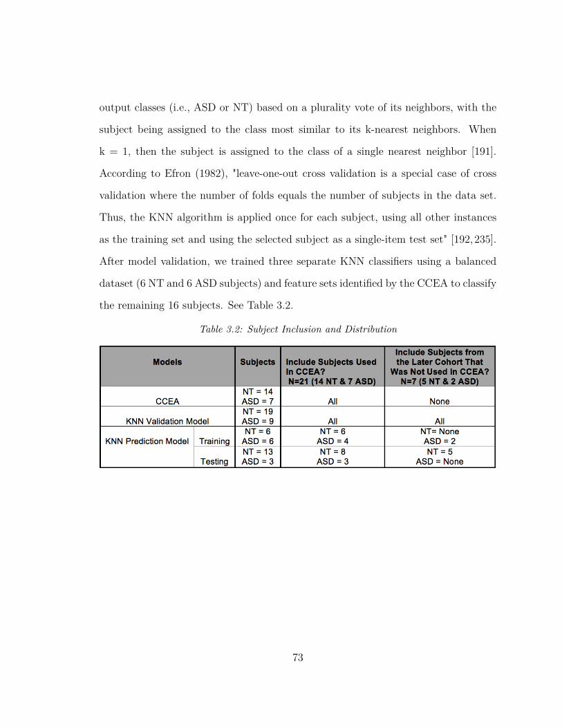
output classes (i.e., ASD or NT) based on a plurality vote of its neighbors, with the
subject being assigned to the class most similar to its k-nearest neighbors. When
k = 1, then the subject is assigned to the class of a single nearest neighbor [191].
According to Efron (1982), "leave-one-out cross validation is a special case of cross
validation where the number of folds equals the number of subjects in the data set.
Thus, the KNN algorithm is applied once for each subject, using all other instances
as the training set and using the selected subject as a single-item test set" [192,235].
After model validation, we trained three separate KNN classifiers using a balanced
dataset (6 NT and 6 ASD subjects) and feature sets identified by the CCEA to classify
the remaining 16 subjects. See Table 3.2.
Table 3.2: Subject Inclusion and Distribution
73
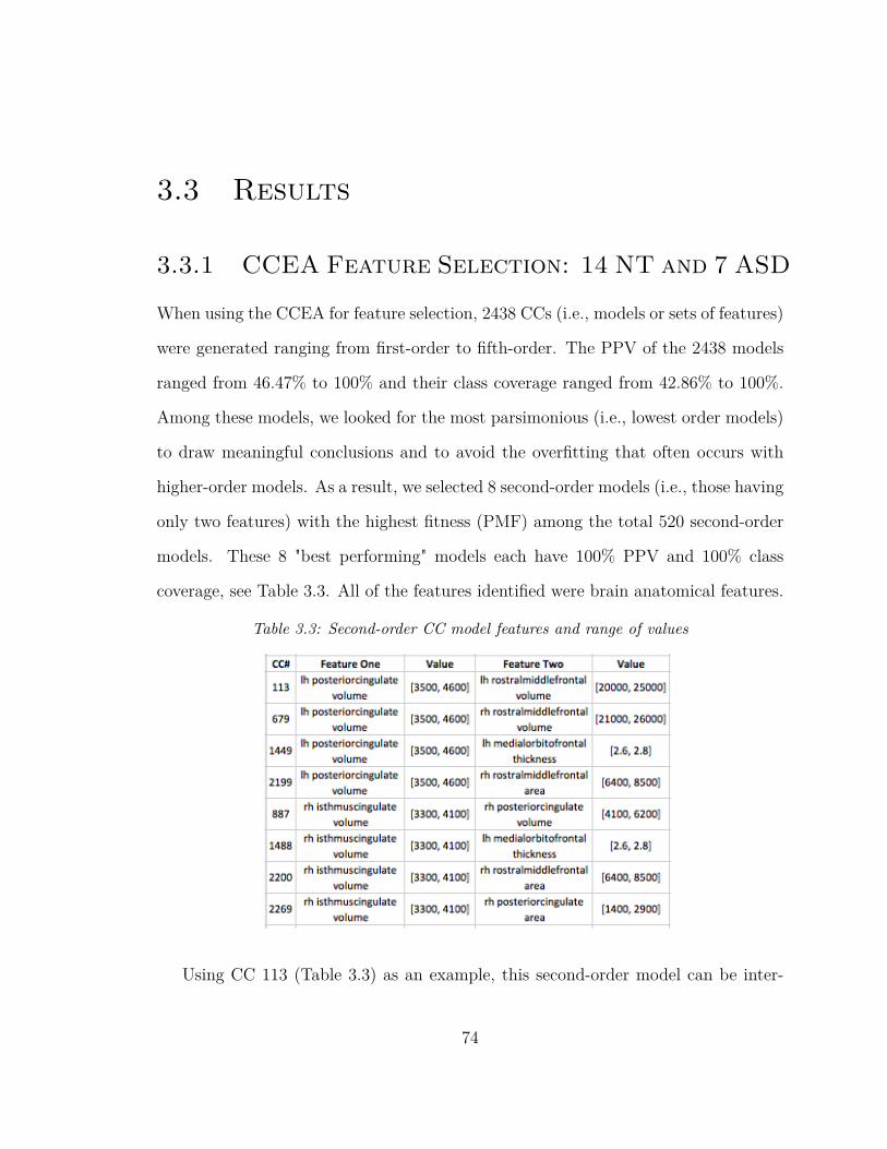
3.3 Results
3.3.1 CCEA Feature Selection: 14 NT and 7 ASD
When using the CCEA for feature selection, 2438 CCs (i.e., models or sets of features)
were generated ranging from first-order to fifth-order. The PPV of the 2438 models
ranged from 46.47% to 100% and their class coverage ranged from 42.86% to 100%.
Among these models, we looked for the most parsimonious (i.e., lowest order models)
to draw meaningful conclusions and to avoid the overfitting that often occurs with
higher-order models. As a result, we selected 8 second-order models (i.e., those having
only two features) with the highest fitness (PMF) among the total 520 second-order
models. These 8 "best performing" models each have 100% PPV and 100% class
coverage, see Table 3.3. All of the features identified were brain anatomical features.
Table 3.3: Second-order CC model features and range of values
Using CC 113 (Table 3.3) as an example, this second-order model can be inter-
74

preted as: any subject whose posterior cingulate gyrus volume was within the range
of 3500 to 4600 mm3 AND left rostral middle frontal gyrus volume was within the
range of 20,000 to 25,000 mm3 would be classified as having ASD. The volume of
the left hemisphere posterior cingulate gyrus and the volume of the right hemisphere
isthmus of the cingulate gyrus were the two features to appear most frequently (i.e.,
four times) across all second-order models, suggesting that the volume of cingulate
gyrus is a potentially important biomarker for ASD. Figure 3.1 provides a 2D visual-
ization for the range of feature values (numerical boundaries) associated with these
models and the placement of each subject within this range.
75

(a) CC113 (b) CC679
(c) CC1449 (d) CC2199
(e) CC887 (f) CC1488
(g) CC2200 (h) CC2269
Figure 3.1: 2D visualization of second-order CC models.
Green dots represent ASD subjects and group together within the rectangle defining
the range of values in Table 3.3
76
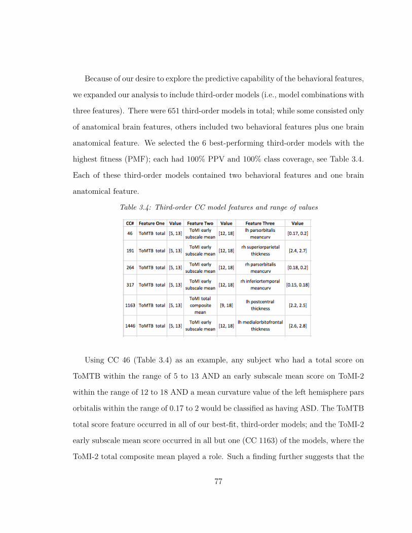
Because of our desire to explore the predictive capability of the behavioral features,
we expanded our analysis to include third-order models (i.e., model combinations with
three features). There were 651 third-order models in total; while some consisted only
of anatomical brain features, others included two behavioral features plus one brain
anatomical feature. We selected the 6 best-performing third-order models with the
highest fitness (PMF); each had 100% PPV and 100% class coverage, see Table 3.4.
Each of these third-order models contained two behavioral features and one brain
anatomical feature.
Table 3.4: Third-order CC model features and range of values
Using CC 46 (Table 3.4) as an example, any subject who had a total score on
ToMTB within the range of 5 to 13 AND an early subscale mean score on ToMI-2
within the range of 12 to 18 AND a mean curvature value of the left hemisphere pars
orbitalis within the range of 0.17 to 2 would be classified as having ASD. The ToMTB
total score feature occurred in all of our best-fit, third-order models; and the ToMI-2
early subscale mean score occurred in all but one (CC 1163) of the models, where the
ToMI-2 total composite mean played a role. Such a finding further suggests that the
77

ToMTB and ToMI-2 might be effective for ASD testing and diagnosis. See Figure
3.2.
78
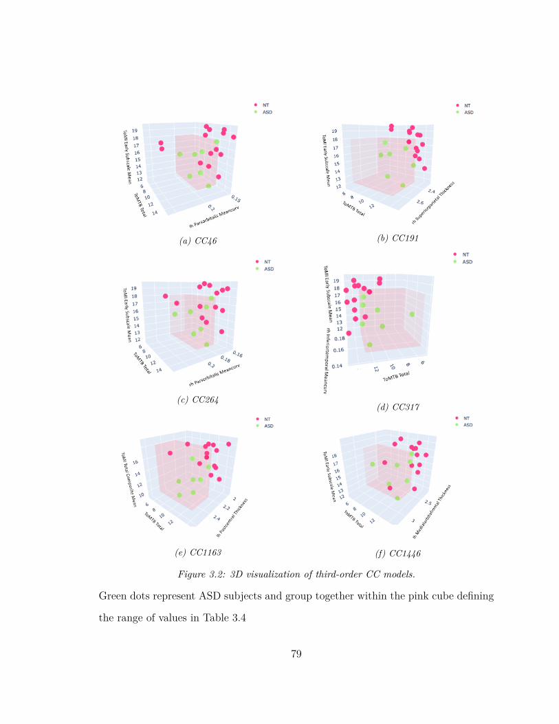
(a) CC46 (b) CC191
(c) CC264(d) CC317
(e) CC1163 (f) CC1446
Figure 3.2: 3D visualization of third-order CC models.
Green dots represent ASD subjects and group together within the pink cube defining
the range of values in Table 3.4
79
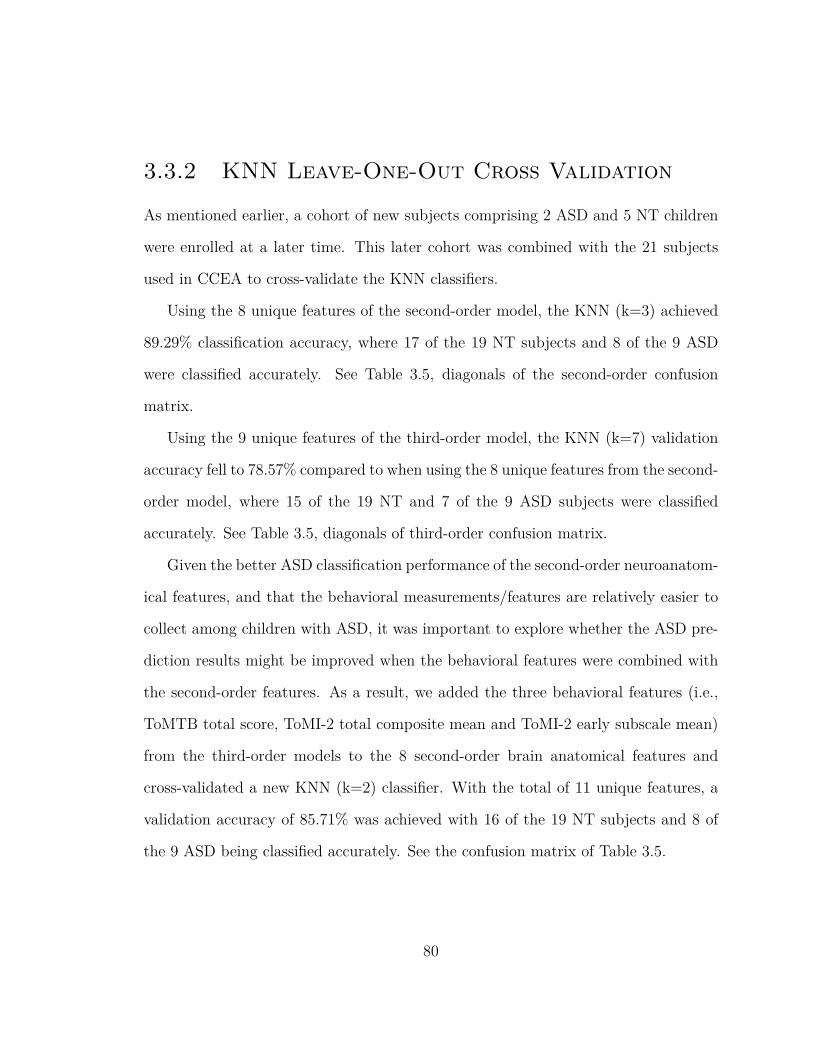
3.3.2 KNN Leave-One-Out Cross Validation
As mentioned earlier, a cohort of new subjects comprising 2 ASD and 5 NT children
were enrolled at a later time. This later cohort was combined with the 21 subjects
used in CCEA to cross-validate the KNN classifiers.
Using the 8 unique features of the second-order model, the KNN (k=3) achieved
89.29% classification accuracy, where 17 of the 19 NT subjects and 8 of the 9 ASD
were classified accurately. See Table 3.5, diagonals of the second-order confusion
matrix.
Using the 9 unique features of the third-order model, the KNN (k=7) validation
accuracy fell to 78.57% compared to when using the 8 unique features from the second-
order model, where 15 of the 19 NT and 7 of the 9 ASD subjects were classified
accurately. See Table 3.5, diagonals of third-order confusion matrix.
Given the better ASD classification performance of the second-order neuroanatom-
ical features, and that the behavioral measurements/features are relatively easier to
collect among children with ASD, it was important to explore whether the ASD pre-
diction results might be improved when the behavioral features were combined with
the second-order features. As a result, we added the three behavioral features (i.e.,
ToMTB total score, ToMI-2 total composite mean and ToMI-2 early subscale mean)
from the third-order models to the 8 second-order brain anatomical features and
cross-validated a new KNN (k=2) classifier. With the total of 11 unique features, a
validation accuracy of 85.71% was achieved with 16 of the 19 NT subjects and 8 of
the 9 ASD being classified accurately. See the confusion matrix of Table 3.5.
80
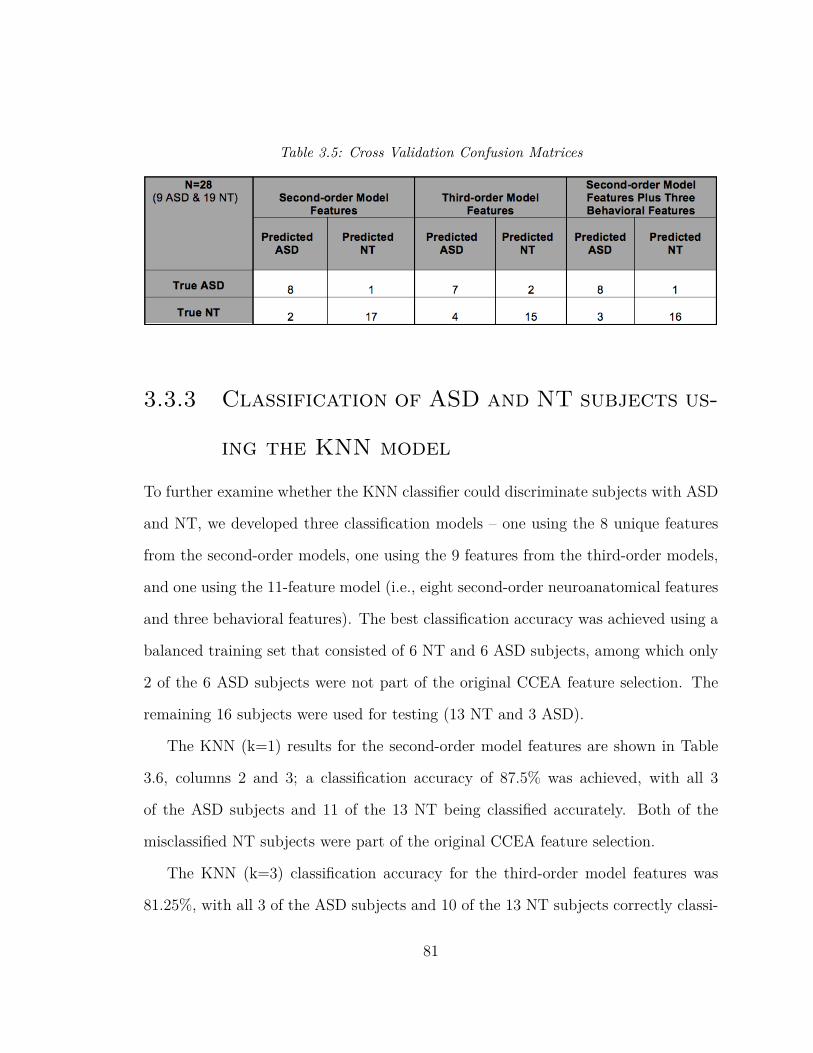
Table 3.5: Cross Validation Confusion Matrices
3.3.3 Classification of ASD and NT subjects us-
ing the KNN model
To further examine whether the KNN classifier could discriminate subjects with ASD
and NT, we developed three classification models – one using the 8 unique features
from the second-order models, one using the 9 features from the third-order models,
and one using the 11-feature model (i.e., eight second-order neuroanatomical features
and three behavioral features). The best classification accuracy was achieved using a
balanced training set that consisted of 6 NT and 6 ASD subjects, among which only
2 of the 6 ASD subjects were not part of the original CCEA feature selection. The
remaining 16 subjects were used for testing (13 NT and 3 ASD).
The KNN (k=1) results for the second-order model features are shown in Table
3.6, columns 2 and 3; a classification accuracy of 87.5% was achieved, with all 3
of the ASD subjects and 11 of the 13 NT being classified accurately. Both of the
misclassified NT subjects were part of the original CCEA feature selection.
The KNN (k=3) classification accuracy for the third-order model features was
81.25%, with all 3 of the ASD subjects and 10 of the 13 NT subjects correctly classi-
81
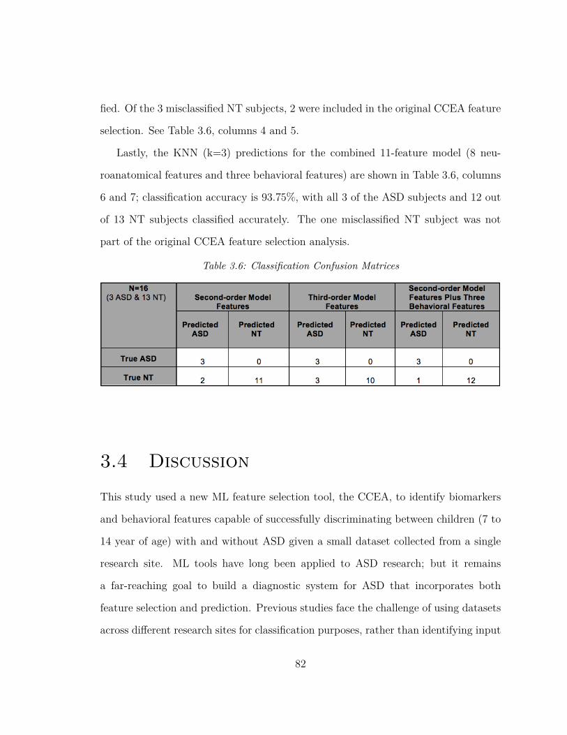
fied. Of the 3 misclassified NT subjects, 2 were included in the original CCEA feature
selection. See Table 3.6, columns 4 and 5.
Lastly, the KNN (k=3) predictions for the combined 11-feature model (8 neu-
roanatomical features and three behavioral features) are shown in Table 3.6, columns
6 and 7; classification accuracy is 93.75%, with all 3 of the ASD subjects and 12 out
of 13 NT subjects classified accurately. The one misclassified NT subject was not
part of the original CCEA feature selection analysis.
Table 3.6: Classification Confusion Matrices
3.4 Discussion
This study used a new ML feature selection tool, the CCEA, to identify biomarkers
and behavioral features capable of successfully discriminating between children (7 to
14 year of age) with and without ASD given a small dataset collected from a single
research site. ML tools have long been applied to ASD research; but it remains
a far-reaching goal to build a diagnostic system for ASD that incorporates both
feature selection and prediction. Previous studies face the challenge of using datasets
across different research sites for classification purposes, rather than identifying input
82
variables most closely associated with ASD (i.e., feature selection) [39, 59, 61, 62,
68, 81, 107, 140, 170, 204, 288]. Additionally, traditional ML algorithms do not work
well with ASD datasets given the large amount of variance and the heterogeneous
nature of the disorder [127, 148]. Meanwhile, it requires a tremendous amount of
effort to include ASD individuals in a research study given the social, cognitive and
language challenges of such a population. Thus, nearly all ASD datasets have a large
number of features with relatively small sample sizes, which despite being unsuitable
for many ML algorithms, often leads to overfitting and poor classification accuracy.
However, the CCEA in this work is able to address such issues by efficiently exploring
large search spaces for feature interactions associated with some nominal outcome
(e.g., ASD or NT). It also adopts built-in tools to prevent overfitting to produce
parsimonious models.
The present study demonstrated exceptionally good performance (i.e., 100% ASD
PPV and 100% class coverage) of the features identified by the CCEA. The selected
CCEA features from the parsimonious second and third order models included vol-
ume, area, cortical thickness, and mean curvature in specific regions around the
cingulate cortex, frontal cortex, and temporal-parietal junction areas as biomark-
ers for ASD (e.g., the pericalcarine cortex, posterior cingulate cortex, isthmus of
the cingulate gyrus, pars orbitalis, etc.). Such findings are consistent with previous
literature suggesting that individuals with ASD have abnormalities in these brain
regions [7,45,78,81,123,163,197,201,219,246,266,270,290]. Additionally, third-order
models from the training set include measurements from the ToMI-2 and the ToMTB
as important features [133,166], which further validates the use of these tools in ASD
assessments.
83
It is impressive that the KNN classifiers are able to achieve such high classification
accuracy given our sample size, and validation of the discriminant features selected
by the CCEA models. In particular, the KNN classifiers perform better using the
second-order neuroanatomical features than the third-order feature models, which
emphasizes the importance of focusing on parsimonious models selected by the CCEA.
In addition, the KNN achieved the highest classification accuracy when adding the
behavioral features from the third-order models to the neuroanatomical features from
the second-order models. In most cases, neuroimaging measurements are conducted
along with behavioral assessments together. As the third-order models are included
to explore the potential role that behavioral features might play along the side of
neuroanatomical features, it is convincing to see the highest classification accuracy are
achieved when combining the behavioral features and the neuroanatomical features.
These findings highlight the heterogeneous and multi-facet characteristics of ASD
itself. Although it is more difficult to implement MRI among children with ASD, such
findings support the idea that neuroanatomical measurements increase confidence
in diagnosis. It also suggests that a good ASD prediction model should consider
including both behavioral and neuroanatomical features.
This study further demonstrates the robustness of the CCEA as a feature selec-
tion methodology. The accuracy of these features when used as input variables in
the KNN classifier suggest their potential to help clinicians and researchers target
specific domains in ToM in treating the social challenges most often seen in children
with ASD. More importantly, CCEA is able to capture accurate characteristics of het-
erogeneous datasets exceptionally well. With the rapid development of "easy-to-use"
neuroimaging techniques, CCEA has great potential to assist clinicians in identifying
84

those individuals who may not be at risk for ASD, hence, shortening the wait-list
for diagnosing those with higher risk in need of immediate intervention. The impli-
cations of our findings for clinical researchers reinforce earlier findings regarding the
brain-behavior connections for children with and without ASD related to ToM un-
derstanding [21, 131,143,166,205,256]. Knowing these connections may guide future
researchers in the assessment of change following intervention at both a behavioral
and neurobiological level. This may also lead to knowledge about which interventions
may be most effective for children with specific neurobiological markers.
The present study has established important biomarker candidates of ASD. These
biomarker candidates support previous research adopting traditional neuroimaging
measurements identifying similar brain regions to explain the abnormalities in ASD
[7,45,78,81,123,163,201,246,266,270]. Importantly, ML methodologies can perform
as well as the traditional approaches in the field of neuroscience and specifically
in our assessment of ASD in selecting neuroanatomical biomarkers. Although ML
techniques have been adopted to help with diagnosis and treatment development in
medicine [3,74,89,243], the heterogeneity in ASD creates challenges. Typically, large,
diverse, and comprehensive datasets are required to extract solid biomarkers, which
can be time-consuming and may be less accurate with traditional approaches. Under
such circumstances, ML techniques as described in this study can help advance the
development of an automatic diagnostic and predictive system for ASD. The present
study provides a new direction for adopting ML techniques in ASD research and other
areas of medicine with similar heterogeneity in disease condition.
85

Chapter 4
Conclusion and Future Direction
Currently available behavioral tests of ToM typical examine one or just a few aspects
of ToM; however, the two behavioral tests used in the current study, ToMI-2 [131]
and ToMTB [134] are multi-faceted tools that cover several aspects of ToM (e.g.,
emotion recognition, false belief, perspective taking etc.), including information from
both parent and the child.
Two novel ToM tasks were developed for this study that can be used as brain
measures of ToM in children. The fToM task was designed to closely resemble an item
on the ToMTB, which directly assesses a child’s understanding of a series of scenarios
tapping ToM. It allowed a direct examination of the correlation between behavioral
performance and brain activation patterns associated with ToM (i.e., desire-based
emotion). This was the first time a behavioral ToM task was recreated in an fMRI
task for comparison of specific emotions in children with ASD. The fER task was
designed to assess surprise and embarrassment that are two later developing and more
complex emotions requiring ToM. Although there is a plethora of research examining
emotion recognition in children with ASD, much of the previous literature examined
86

recognition of basic emotions such as happiness, sadness, anger, and fear; few studies
have examined surprise and embarrassment, and none have investigated the brain
areas underlying recognition of these emotions among children with ASD.
All three emotions targeted in the current study are critical aspects of ToM.
Surprise and embarrassment are particularly difficult for children with ASD to rec-
ognize [121, 132, 265]. While desire-based emotions are easier for children with ASD
to recognize, their understanding of these emotions is delicate and often requires ex-
plicit descriptions [14, 132, 198, 214, 216, 278, 291]. Further, embarrassment has been
studied at a neural level only among adults with ASD [124, 125, 136]; surprise and
desire-based emotions have only been examined at a behavioral level in ASD sub-
jects and little is known about their neural correlates. Thus, the studies presented
here filled a notable gap in the literature by examining these three emotions from
a neuroscience perspective among children with ASD. The current studies were the
first to examine the neural correlates of these emotions requiring ToM and establish
the connections between behavior and brain activities in children with ASD (i.e., 7
to 14 years old). Specifically, the first manuscript, A Pilot Study Using Two Novel
fMRI Tasks: Understanding Theory of Mind and Emotion Recognition Among Chil-
dren With ASD, investigated the neural mechanisms involved in two specific aspects
of ToM: the recognition of basic and "complex" emotions (i.e., surprise, embarrass-
ment), and the understanding of desire-based emotions. Building upon findings from
previous studies, the study provided a deeper and more systematic understanding
of the brain-behavior connections associated with ToM leading to an increased un-
derstanding of the brain regions involved in ToM. With this knowledge, intervention
research can be developed that supports brain-behavior connections leading to nor-
87
malized social performance. Such brain-behavior research might also help predict
those brain-behavior profiles of children that will most likely to benefit from specific
ToM or social cognitive based interventions. This is important as causal modeling in
ASD suggests that interventions need to be delivered at the cognitive level to bridge
behavior with brain function [128]. Further, brain and behavioral characteristics can
be combined with new machine learning (ML) methods to build prediction models
to help detect ASD, and select important biomarkers of ASD population to achieve
personalized interventions.
Due to the heterogeneous nature and early onset of ASD, detection and diagnosis
of ASD often require significant time and costs. Early diagnosis of ASD is crucial
for early intervention so that it can emphasize strategies to address the core social
impairment in autism. A time efficient, low cost, and accurate system to facilitate
the diagnosis of ASD will help determine whether a child needs further assessment,
which will save both human and financial resources, as well as speed up the diag-
nostic process. As an effort to build automatic diagnostic tools for ASD, science has
progressed significantly to classify ASD individuals from NT individuals using ML
approaches. However, challenges in classification performance remain due to data
variance introduced via scanner hardware, imaging protocols, operator characteris-
tics, regional demographics, and other site-specific acquisition factors. In addition,
most studies in the field focus on classification and prediction rather than discriminant
feature selections in ASD. In the second manuscript, Identifying Neuroanatomical and
Behavioral Features for Autism Spectrum Disorder Diagnosis in Children using Ma-
chine Learning, a conjunctive clause evolutionary algorithm (CCEA) was adopted
to identify biomarkers and behavioral features capable of successfully discriminating
88
between children (i.e., 7 to 14 year of age) with and without ASD, given a small
dataset collected from a single research site. This was the first study to successfully
introduce a novel evolutionary algorithm (i.e., CCEA), which identified both behav-
ioral and neuroanatomical biomarkers associated with ASD. More importantly, the
algorithm prevented overfitting and demonstrated robust classification performance
with a small sample size. It provided evidence that ML techniques can be applied
effectively in ASD research and other areas of medicine with similar heterogeneity in
disease condition.
This dissertation work is part of a larger study examining the behavioral and neu-
ral changes surrounding ToM and emotion recognition following a nine-week social
cognitive intervention (i.e., Social Stories) designed to improve social functioning in
children with ASD. Social Stories™(SSs) facilitate the ToM of children with ASD by
reading short stories individually developed based on a child’s successful and/or un-
successful social experiences [154,159,196]. A unique aspect of this intervention is the
ability to emphasize specific instances of emotion recognition and understanding as
well as other aspects of ToM that may be a specific target for a child with ASD. In the
ongoing larger study, SSs are designed following parental discussion of actual social
situations in which a child had difficulty or was successful in understanding surprise,
embarrassment, and desire-based emotion. SSs have been shown to enhance chil-
dren’s social understandings and address challenging behaviors [154, 159, 196]. They
have also been shown to improve ToM, in part due to their focus on emotion recogni-
tion, making them especially promising for children with ASD in whom ToM deficits
contribute to impairments in perceiving and interpreting other’s emotions [236].
Research on interventions to improve social cognition and overall life skills is
89
consistently endorsed as an aspirational research priority in surveys of the ASD com-
munity. The larger study in which this novel dissertation work was developed is an
effort to optimize a treatment program that improves social functioning to ultimately
reduce the economic and social burden of care for individuals with ASD in the fu-
ture. Knowing children with ASD can engage in fMRI tasks as well as behavioral
tasks so that brain-behavior connections can be made is an important contribution
of this dissertation work. The development of two fMRI measures that assess ToM
and are aligned with behavioral measure of ToM is another major contribution to
researchers working to better understand the brain areas involved in ToM and ulti-
mately ASD. These contributions should help facilitate the development of individ-
ualized treatment recommendations and refine how interventions are implemented.
This dissertation work also supports additional efforts to determine the differential
effects of other interventions (e.g., Comic Strip Conversations [129, 130]), including
the great potential to apply ML techniques to explore and define ASD subtypes that
may benefit from specific interventions.
In summary, this dissertation work will contribute to our understanding of the
neural mechanisms underlying ToM related emotion recognition and important neu-
ral anatomical and behavioral biomarkers in ASD. By building such brain-behavior
connections, it will promote the development of more accurate and efficient assess-
ment tools and treatment methods. In addition, it will advance the application of
ML in ASD and neuroscience research to achieve automated predictive and diagnostic
models.
90
Bibliography
[1] Machine learning tutorial-all the essential concepts in single tutorial.https://data-flair.training/blogs/machine-learning-tutorial/.
[2] F. Abell, F. Happe, and U. Frith. Do triangles play tricks? attribution of mentalstates to animated shapes in normal and abnormal development. CognitiveDevelopment, 2000.
[3] Z. Ahmed, K. Mohamed, S. Zeeshan, and X. Dong. Artificial intelligence withmulti-functional machine learning platform development for better healthcareand precision medicine. The journal of biological databases and curation, 2020.
[4] I. Ajzen. The theory of planned behavior. Organ. Behav. Hum, 1991.
[5] M. Aljunied and N. Frederickson. Cognitive indicators of different levels ofspecial educational support needs in autism. Research in Autism SpectrumDisorders, 2011.
[6] D.G. Amaral. Examining the causes of autism. Cerebrum, 2017.
[7] D.G. Amaral, C.M. Schumann, and C.W. Nordahl. Trends neurosci. Neu-roanatomy of autism, 2008.
[8] American Psychiatric Association. Diagnostic and statistical manual of mentaldisorders, 3rd ed. Arlington, VA: American Psychiatric Association, 1980.
[9] American Psychiatric Association. Diagnostic and Statistical Manual of MentalDisorders (DSM-5). 2020.
[10] F. Apicella, F. Sicca, R.R. Federico, G. Campatelli, and F. Muratori. Fusiformgyrus responses to neutral and emotional faces in children with autism spectrumdisorders: A high density erp study. Behavioural Brain Research, 2013.
91

[11] C. Ashwin, E. Chapman, E. Colle, and S. Baron-Cohen. Impaired recognitionof negative basic emotions in autism: A test of the amygdala theory. SocialNeuroscience, 2006.
[12] C. Ashwin et al. Differential activation of the amygdala and the ’social brain’during fearful face-processing in asperger syndrome. Neuropsychologia, 2007.
[13] K.L. Ashwood et al. Predicting the diagnosis of autism in adults using theautism-spectrum quotient (aq) questionnaire. Psychological Medicine, 2016.
[14] J. Astington. The Child’s Discovery of the Mind. Harvard University Press,Cambridge, MA, 1993.
[15] J. Astington and A. Gopnik. Developing understanding of desire and intention.In A. Whiten, editor, Natural theories of mind: Evolution, development, andsimulation of everyday mindreading, pages 39–50. Blackwell, Oxford, 1991.
[16] National Autism Center at May Institute. Autism intervention. 2015.
[17] Autism Society. Asperger’s syndrome. 2021.
[18] J. Avis and P. Harris. Belief-desire reasoning among baka children: Evidencefor a universal conception of mind. Child Dev, 1991.
[19] R. Baillargeon, R.M. Scott, and Z. He. False-belief understanding in infants.Trends in Cognitive Sciences, 2010.
[20] J. Baio. Prevalence of autism spectrum disorders: autism and developmentaldisabilities monitoring network, 14 sites, united states. MMWR Surveill Summ,2012.
[21] S. Baron-Cohen. An essay on autism and theory of mind. MIT Press, 1995.
[22] S. Baron-Cohen. Learning, development, and conceptual change. mindblind-ness: An essay on autism and theory of mind. The MIT Press., 1995.
[23] S. Baron-Cohen, A.M. Leslie, and U. Frith. Does the autistic-child have a theoryof mind. Cognition, 1985.
[24] S. Baron-Cohen, A. Spitz, and P. Cross. Do children with autism recognizesurprise? Cognition and Emotion, 1993.
[25] S. Baron-Cohen and J. Swettenham. Theory of mind in autism: its relationshipto executive function and central coherence. Wiley, 1997.
92
[26] S. Baron-Cohen, S. Wheelwright, J. Hill, Y. Raste, and I. Plumb. The readingthe mind in the eyes’s test revised version: A study with normal adults, andadults with asperger syndrome or high-functioning autism. Journal of ChildPsychology and Psychiatry, 2001.
[27] C. Bastin et al. Feelings of shame, embarrassment and guilt and their neuralcorrelates: A systematic review. Neurosci Biobehav Rev, 2016.
[28] C. Baurain and N.N. Grosbois. Theory of mind, socio emotional problem solv-ing, socio-emotional regulation in children with intellectual disability and intypically developing children. J Autism Dev Disord, 2013.
[29] J.B. Bavelas, L. Coates, and T. Johnson. Listener responses as a collaborativeprocess: the role of gaze. J. Commun, 2002.
[30] A.P. Bayliss, A. Frischen, M.J. Fenske, and S.P. Tipper. Affective evaluationsof objects are influenced by observed gaze direction and emotional expression.Cognition, 2007.
[31] S. Begeer, M. De Rosnay, P. Lunenburg, H. Stegge, and M. Terwogt. Under-standing of emotions based on counterfactual reasoning in children with autismspectrum disorders. Autism, 2014.
[32] M. Bennett. Childrens self-attribution of embarrassment. British Journal ofDevelopmental Psychology, 1989.
[33] M. Bennett and L. Matthews. The role of second-order belief-understandingand social context in childrens self-attribution of social emotions. Social Devel-opment, 2000.
[34] M. Bennett, N. Yuill, R. Banerjee, and S. Thomson. Childrens understandingof extended identity. Developmental Psychology, 1998.
[35] R. Bernier, A. Mao, and J. Yene. Psychopathology, families and culture:Autism. Child Adolesc Psychiatr Clin, 2010.
[36] C. Betancur. Etiological heterogeneity in autism spectrum disorders: More than100 genetic and genomic disorders and still counting. Brain Research, 2011.
[37] X.A. Bi, Y. Wang, Q. Shu, Q. Sun, and Q. Xu. Classification of autism spectrumdisorder using random support vector machine cluster. Front Genet, 2018.
[38] P.F. Bolton, J. Golding, A. Emond, and C.D. Steer. Autism spectrum disor-der and autistic traits in the avon longitudinal study of parents and children:precursors and early signs. J Am Acad Child Adolesc Psychiatry, 2012.
93
[39] D. Bone, M.S. Goodwin, M.P. Black, C.C. Lee, K. Audhkhasi, andS. Narayanan. Applying machine learning to facilitate autism diagnostics: pit-falls and promises. J Autism Dev, 2015.
[40] M. Bornstein and M. Arterberry. Recognition, discrimination, and categoriza-tion of smiling by 5-month-old infants. Developmental Science, 2003.
[41] S. Bosacki and J.W. Astington. Theory of mind in preadolescence: relationsbetween social understanding and social competence. Social Development, 2001.
[42] T. Bourgeron. The genetics and neurobiology of essence: The third birgit olssonlecture. Nord J Psychiatry, 2016.
[43] D.M. Bowler. Theory of mind in asperger syndrome. Journal of Child Psychol-ogy and Psychiatry, 1992.
[44] B. Bracken and R. McCallum. Universal nonverbal intelligence test 2nd edition.Pro-ed, 2016.
[45] P. Brambilla et al. Brain anatomy and development in autism: Review ofstructural mri studies. Brain Res, 2003.
[46] D. Braunschweig. Autism: maternally derived antibodies specific for fetal brainproteins. Neurotoxicology, 2008.
[47] F. Breuer et al. Controlled aliasing in parallel imaging results in higher accel-eration (caipirinha) for multi-slice imaging. Magnetic Resonance in Medicine,2005.
[48] N. Brewer, R.L. Young, and E. Barnett. Erratum to: Measuring theory of mindin adults with autism spectrum disorder. Journal of Autism and DevelopmentalDisorders, 2017.
[49] R.L. Bromley. The prevalence of neurodevelopmental disorders in children pre-natally exposed to antiepileptic drugs. J Neurol Neurosurg Psychiatry, 2013.
[50] S. Burnettand et al. Development during adolescence of the neural processingof social emotion. Journal of Cognitive Neuroscience, 2009.
[51] R.M. Busch et al. Neurobehavioral phenotype of autism spectrum disorder asso-ciated with germline heterozygous mutations in pten. Translational Psychiatry,2019.
94

[52] J.D. Buxbaum et al. Mutation screening of thepten gene in patients with autismspectrum disorders and macrocephaly. American Journal of Medical GeneticsPart B: Neuropsychiatric Genetics, 2007.
[53] L.J. Byom and B. Mutlu. Theory of mind: mechanisms, methods, and newdirections. Frontiers in Human Neuroscience, 2013.
[54] E. Carrow-Woolfolk. Comprehensive assessment of spoken language 2nd edition.Pearson, 2017.
[55] K.W. Cassidy. Three- and four-year-old children’s ability to use desire- andbelief-based reasoning. Cognition, 1998.
[56] F. Castelli. Understanding emotions from standardized facial expressions inautism and normal development. Autism, 2005.
[57] Centers for Disease Control and Prevention. Prevalence of autism spectrumdisorders–autism and developmental disabilities monitoring network, 14 sites,united states, 2008. MMWR Surveill Summ, 2012.
[58] Centers for Disease Control and Prevention. Community report on autism.2020.
[59] G. Chanel et al. Classification of autistic individuals and controls using cross-task characterization of fmri activity. NeuroImage Clin, 2016.
[60] T. Charman, S. Baron-Cohen, J. Swettenham, et al. Testing joint attention,imitation and play as infancy precursors to language and theory of mind. Cog-nitive Development, 2000.
[61] C.P. Chen et al. Diagnostic classification of intrinsic functional connectivityhighlights somatosensory, default mode, and visual regions in autism. Neu-roImage Clin, 2015.
[62] H. Chen et al. Multivariate classification of autism spectrum disorder usingfrequency-specific resting-state functional connectivity. Prog. Neuropsychophar-macol. Biol. Psychiatry, 2016.
[63] R. Chen, Y. Jiao, and E.H. Herskovits. Structural mri in autism spectrumdisorder. Pediatr Res, 2011.
[64] S.A. Collier. Prevalence of self-reported infection during pregnancy among con-trol mothers in the national birth defects prevention study. Birth Defects ResA Clin Mol Teratol, 2009.
95
[65] G.S. Cooper, M.L. Bynum, and E.C. Somers. Recent insights in the epidemiol-ogy of autoimmune diseases: improved prevalence estimates and understandingof clustering of diseases. J Autoimmun, 2009.
[66] B.A. Corbett, V. Carmean, S. Ravizza, C. Wendelken, M.L. Henry, C. Carter,and S.M. Rivera. A functional and structural study of emotion and face pro-cessing in children with autism. Psychiatry Research: Neuroimaging, 2009.
[67] B. Corden, R. Chilvers, and D. Skuse. Avoidance of emotionally arousing stimulipredicts social-perceptual impairment in asperger’s syndrome. Neuropsycholo-gia, 2008.
[68] C. Craddock et al. The neuro bureau preprocessing initiative: open sharing ofpreprocessed neuroimaging data and derivatives. Front. Neuroinform, 2013.
[69] H. Critchley et al. The functional neuroanatomy of social behaviour: changes incerebral blood flow when people with autistic disorder process facial expressions.Brain, 2000.
[70] R. Custrini and R. Feldman. Children’s social competence and nonverbal en-coding and decoding of emotions. Journal of Clinical Child Psychology, 1989.
[71] W. Czyz. Genetic, environmental and stochastic factors in monozygotic twindiscordance with a focus on epigenetic differences. BMC Med, 2012.
[72] E.M. Daly et al. Serotonin and the neural processing of facial emotions in adultswith autism. Arch Gen Psychiatry, 2012.
[73] M. Dapretto et al. Understanding emotions in others: mirror neuron dysfunc-tion in children with autism spectrum disorders. Nature Neuroscience, 2005.
[74] T. Davenport and R. Kalakota. The potential for artificial intelligence in health-care. Future healthcare journal, 2019.
[75] M. DeFilippis and K.D. Wagner. Treatment of autism spectrum disorder inchildren and adolescents. Evidence-Based Medicine, 2016.
[76] V. Demicheli. Vaccines for measles, mumps and rubella in children. 2012.
[77] E. Demurie, M. De Corel, and H. Roeyers. Empathic accuracy in adolescentswith autism spectrum disorders and adolescents with adhd. Research in AutismSpectrum Disorders, 2011.
[78] D.L. Dierker et al. Analysis of cortical shape in children with simplex autism.Cereb Cortex, 2015.
96
[79] C. Dissanayake and K. Macintosh. Mind reading and social functioning inchildren with autistic disorder and asperger’s disorder. 2003.
[80] A. Downs and T. Smith. Emotional understanding, cooperation, and socialbehavior in high-functioning children with autism. Journal of Autism and De-velopmental Disorders, 2004.
[81] M. Duda, R. Ma, N. Haber, and D.P. Wall. Use of machine learning for behav-ioral distinction of autism and adhd. Transl Psychiatry, 2016.
[82] R.S. Duszak. Congenital rubella syndrome–major review. Optometry, 2009.
[83] M. Dyck and E. Denver. Can the emotion recognition ability of deaf childrenbe enhanced? a pilot study. Journal of Deaf Studies and Deaf Education, 2003.
[84] M. Dyck, C. Farrugia, I. Shochet, and M. Holmes-Brown. Emotion recogni-tion/understanding ability in hearing or vision-impaired children: Do sounds,sights, or words make the difference? Journal of Child Psychology Psychiatry,2004.
[85] C. Ecker et al. The effect of age, diagnosis, and their interaction on vertex-basedmeasures of cortical thickness and surface area in autism spectrum disorder. JNeural Transm (Vienna), 2014.
[86] R. Edelmann. Embarrassment: The state of the research. Current PsychologicalReviews, 1981.
[87] R. Edelmann. Social embarrassment: An analysis of the process. Journal ofSocial and Personal Relationships, 1985.
[88] R. Edelmann and S. Hampson. The recognition of embarrassment. Personalityand Social Psychology Bulletin, 1981.
[89] Editorial. Ascent of machine learning in medicine. Nat. Mater., 2019.
[90] H. Eisenbarth and G.W. Alpers. Happy mouth and sad eyes: Scanning emo-tional facial expressions. Emotion, 2011.
[91] P. Ekman. Emotions revealed. Weidenfeld & Nicolson, London, 2003.
[92] M. Elsabbagh et al. Global prevalence of autism and other pervasive develop-mental disorders. 2012.
97
[93] E. Feczko, N. Balba, O. Miranda-Dominguez, M. Cordova, S. Karalunas, L. Ir-win, and D. Fair. Subtyping cognitive profiles in autism spectrum disorder usinga functional random forest algorithm. NeuroImage, 2018.
[94] T.M. Field, R. Woodson, R. Greenberg, and S. Baron-Cohen. Discriminationand imitation of facial expressions by neonates. Science, 1982.
[95] E. Fink, M. de Rosnay, C.C. Peterson, and V. Slaughter. Validation of thepeer social maturity scale for assessing children’s social skills. Infant and ChildDevelopment, 2013.
[96] B. Fischl et al. Whole brain segmentation: automated labeling of neuroanatom-ical structures in the human brain. Neuron, 2002.
[97] M. Fitzgerald. The clinical gestalts of autism: Over 40 years of clinical experi-ence with autism. Open access peer-reviewed chapter, 2017.
[98] J. Flavell, E. Flavell, F. Green, and L. Moses. Young children’s understandingof fact beliefs versus value beliefs. Child Development, 1990.
[99] P.C. Fletcher et al. Other minds in the brain: a functional imaging study oftheory of mind in story comprehension. Cognition, 1995.
[100] E. Fombonne, F. Siddons, S. Achard, et al. Adaptive behaviour and theory ofmind in autism. European Child & Adolescent Psychiatry, 1994.
[101] E. Fox-Edmiston and J. Van de Water. Maternal anti-fetal brain igg autoanti-bodies and autism spectrum disorder: Current knowledge and its implicationsfor potential therapeutics. CNS Drugs, 2015.
[102] FreeSurfer. optseq2. http://surfer.nmr.mgh.harvard.edu/optseq, 2012.
[103] A. Freire, M. Eskritt, and K. Lee. Are eyes windows to a deceiver’s soul:children’s use of another’s eye gaze cues in a deceptive situation. Dev. Psychol,2004.
[104] P. Fusar-Poli et al. Functional atlas of emotional faces processing: a voxel-based meta-analysis of 105 functional magnetic resonance imaging studies. JPsychiatry Neurosci, 2009.
[105] Y.Y. Geng et al. Oxytocin facilitates empathic- and self-embarrassment ratingsby attenuating amygdala and anterior insula responses. Front. Endocrinol, 2018.
[106] M.F. Glasser et al. The minimal preprocessing pipelines for the human connec-tome project. Neuroimage, 2013.
98

[107] I. Gori et al. Gray matter alterations in young children with autism spectrumdisorders: comparing morphometry at the voxel and regional level. J. Neu-roimag, 2015.
[108] C. Gray et al. Attribution of emotions to story characters by severely andprofoundly deaf children. Journal of Developmental and Physical Disabilities,2007.
[109] D. Grelotti et al. fmri activation of the fusiform gyrus and amygdala to cartooncharacters but not to faces in a boy with autism. Neuropsychologia, 2005.
[110] S. Ha et al. Characteristics of brains in autism spectrum disorder: Structure,function and connectivity across the lifespan. Experimental Neurobiology, 2015.
[111] N. Hadjikhani, R. Joseph, J. Snyder, and H. Tager-Flusberg. Abnormal acti-vation of the social brain during face perception in autism. Hum Brain Ma,2007.
[112] J. Hadwin and J. Perner. Pleased and surprised: Childrens cognitive theory ofemotion. British Journal of Developmental Psychology, 1991.
[113] J. Hallmayer. Genetic heritability and shared environmental factors among twinpairs with autism. Arch Gen Psychiatry, 2011.
[114] S. Hamlyn-Wright, R. Draghi-Lorenz, and J. Ellis. Locus of control fails tomediate between stress and anxiety and depression in parents of children witha developmental disorder. Autism, 2007.
[115] J.P. Hanley, D.M. Rizzo, J.S. Buzas, and M.J. Eppstein. A tandem evolutionaryalgorithm for identifying causal rules from complex data. Evol Comput, 2020.
[116] F. Happe. Communicative competence and theory of mind in autism: a test ofrelevance theory. Cognition, 1993.
[117] F. Happe. An advanced test of theory of mind: Understanding of story charac-ters’ thoughts and feelings by able autistic, mentally handicapped, and normalchildren and adults. Journal of Autism and Developmental Disorders, 1994.
[118] F. Happe. Instructions for theory of mind story task. Unpublished documentprovided by F. Happe, 1999.
[119] F. Happe and U. Frith. The neuropsychology of autism. Brain, 1996.
[120] P. Harris, C. Johnson, D. Hutton, G. Andrews, and T. Cooke. Young children’stheory of mind and emotions. Cognition and Emotion, 1989.
99

[121] E. Heerey, D. Keltner, and L. Capps. Making sense of self-conscious emotion:Linking theory of mind and emotion in children with autism. Emotion, 2003.
[122] J. Henry et al. Theory of mind following traumatic brain injury: The role ofemotion recognition and executive dysfunction. Neuropsychologia, 2006.
[123] R. Kucharsky Hiess et al. Corpus callosum area and brain volume in autismspectrum disorder: Quantitative analysis of structural mri from the abidedatabase. J. Autism Dev, 2015.
[124] A. Hillier and L. Allinson. Beyond expectations: Autism, understanding em-barrassment, and the relationship with theory of mind. Autism, 2002.
[125] A. Hillier and L. Allinson. Understanding embarrassment among those withautism: Breaking down the complex emotion of embarrassment among thosewith autism. Journal of Autism and Developmental Disorders, 2002.
[126] M. Hoogenhout and S. Malcolm-Smith. Theory of mind predicts severity levelin autism. Autism, 2016.
[127] W. Huf et al. On the generalizability of resting-state fmri machine learningclassifiers. Frontiers in Human Neuroscience, 2014.
[128] T. Hutchins and P. Prelock. Beyond the theory of mind hypothesis: Usinga causal model to understand the nature and treatment of multiple deficits inautism spectrum disorder. Routledge Handbook of Communication Sciencesand Disorders, 2015.
[129] T.L. Hutchins and P.A. Prelock. Using social stories and comic strip conversa-tions to promote socially valid outcomes for children with autism. Semin SpeechLang, 2006.
[130] T.L. Hutchins and P.A. Prelock. Parents’ perceptions of their children’s socialbehaviors: The social validity of social stories and comic strip conversations. JPositive Behav Interven, 2013.
[131] T.L. Hutchins and P.A. Prelock. Technical manual for the theory of mindinventory-2 and theory of mind task battery. Theoryofmindinventory.com, 2016.
[132] T.L. Hutchins and P.A. Prelock. Theory of mind atlas. theoryofmindinven-tory.com, 2017.
[133] T.L. Hutchins, P.A. Prelock, and L. Bonazinga. Psychometric evaluation of thetheory of mind inventory (tomi): a study of typically developing children andchildren with autism spectrum disorder. J Autism Dev Disord, 2012.
100

[134] T.L. Hutchins, P.A. Prelock, and W. Chase. Test-retest reliability of a theory ofmind task battery for children with autism spectrum disorders. Focus AutismOther Dev Disabil, 2008.
[135] D. Ilzarbe et al. Neural correlates of theory of mind in autism spectrum disorder,attention-deficit/hyperactivity disorder, and the comorbid condition. Front.Psychiatry, 2020.
[136] K.F. Jankowski and H. Takahashi. Cognitive neuroscience of social emotionsand implications for psychopathology: Examining embarrassment, guilt, envyand schadenfreude. Psychiatry and Clinical Neurosciences, 2014.
[137] J. Jenkins and J. Astington. Theory of mind and social behavior. Merrill-Palmer Quarterly, 2000.
[138] J. Jensen, J. Helpern, A. Ramani, H.Z. Lu, and K. Kaczynski. Diffusionalkurtosis imaging: the quantification of non-gaussian water diffusion by meansof magnetic resonance imaging. Magn Reson Med, 2005.
[139] B. Jeurissen et al. Multi-tissue constrained spherical deconvolution for improvedanalysis of multi-shell diffusion mri data. Neuroimage, 2014.
[140] Y. Jin et al. Identification of infants at high-risk for autism spectrum disorderusing multiparameter multiscale white matter connectivity networks. Hum.Brain Mapp, 2015.
[141] A.P. Jones and N. Frederickson. Multi-informant predictors of social inclu-sion for students with autism spectrum disorders attending mainstream school.Journal of Autism and Developmental Disorders, 2010.
[142] Stromland K. Autism in thalidomide embryopathy: a population study. DevMed Child Neurol, 1994.
[143] R.K. Kana, J.O. Maximo, D.L. Williams, T.A. Keller, S.E. Schipul, and V.L.Cherkassky. Aberrant functioning of the theory-of-mind network in childrenand adolescents with autism. Molecular Autism volume, 2015.
[144] L. Kanner. Autistic disturbances of affective contact. Nervous Child, 1943.
[145] Y.C. Kaplan. Prenatal selective serotonin reuptake inhibitor use and the risk ofautism spectrum disorder in children: A systematic review and meta-analysis.Reprod Toxicol, 2016.
101
[146] G.J. Katuwal, S.A. Baum, N.D. Cahill, and A.M. Michael. Divide and conquer:Sub-groupingof asd improves asd detection based on brain morphometry. PLoSOne, 2016.
[147] Z. Ke et al. Neural responses to rapid facial expressions of fear and surprise.Frontiers in Psychology, 2017.
[148] C. Kelly et al. Characterizing variation in the functional connectome: Promiseand pitfalls. Trends in Cognitive Sciences, 2012.
[149] A. Kendon. Some functions of gaze-direction in social interaction. Acta Psychol,1967.
[150] R. Khanna, S.S. Madhavan, M.J. Smith, J.H. Patrick, C. Tworek, andB. Becker-Cottrill. Assessment of health-related quality of life among primarycaregivers of children with autism spectrum disorders. Journal of Autism andDevelopmental Disorders, 2011.
[151] K. Kinast et al. Genetic and pharmacological manipulations of the serotoner-gic system in early life: neurodevelopmental underpinnings of autism-relatedbehavior. Front Cell Neurosci, 2013.
[152] J.M. Kleinman et al. Diagnostic stability in very young children with autismspectrum disorders. J Am Acad Child Adolesc Psychiatry, 2008.
[153] G. Knoblich, S. Butterfill, and N. Sebanz. Psychological research on joint action:theory and data, in the psychology of learning and motivation, eds b. AcademicPress, 2011.
[154] A. Kokina and L. Kern. Social story interventions for students with autismspectrum disorders. J Autism Dev Disabil, 2010.
[155] K. Konrad, C. Firk, and P.J. Uhlhaas. Brain development during adolescence:neuroscientific insights into this developmental period. Deutsches Arzteblattinternational, 2013.
[156] H. Koshino et al. fmri investigation of working memory for faces in autism:visual coding and underconnectivity with frontal areas. Cereb Cortex, 2008.
[157] K. Kuhlthau, E.A. Kovacs, T. Hall, T. Clemmons, F. Orlich, J. Delahaye, et al.Health-related quality of life for children with asd: Associations with behavioralcharacteristics. Research in Autism Spectrum Disorders, 2013.
102

[158] K. Kuhlthau, F. Orlich, T.A. Hall, D. Sikora, E.A. Kovacs, J. Delahaye, et al.Health-related quality of life in children with autism spectrum disorders: Re-sults from the autism treatment network. Journal of Autism and DevelopmentalDisorders, 2010.
[159] H. Kuoch and P. Mirenda. Social story interventions for young children withautism spectrum disorders. Focus Autism Other Dev Disabil, 2003.
[160] S. Kuusikko et al. Emotion recognition in children and adolescents with autismspectrum disorders. Journal of Autism and Developmental Disorders, 1999.
[161] K. Kwanguk and others. A virtual joy-stick study of emotional responses andsocial motivation in children with autism spectrum disorders. Journal of Autismand Developmental Disorders, 2014.
[162] A. Lacroix, M. Guidetti, B. Roge, and J. Reilly. Facial emotion recognition in4- to 8-year-olds with autism spectrum disorder: A developmental trajectoryapproach. Research in Autism Spectrum Disorders, 2014.
[163] N. Lange et al. Longitudinal volumetric brain changes in autism spectrumdisorder ages 6 to 35 years. Autism Res, 2015.
[164] P. Lanka et al. Supervised machine learning for diagnostic classification fromlarge-scale neuroimaging datasets. Brain Imaging and Behavior, 2019.
[165] J.P. Leigh and J. Du. Brief report: Forecasting the economic burden of autismin 2015 and 2025 in the united states. J Autism Dev Disord, 2015.
[166] M. Lerner, T.L. Hutchins, and P.A. Prelock. A brief report: preliminary evalu-ation of the theory of mind inventory and its relationship to measures of socialskills. J Autism Dev Disord, 2011.
[167] P. Levitt. Serotonin and the autisms: a red flag or a red herring? Arch GenPsychiatry, 2011.
[168] S. Levy, M. Duda, N. Haber, and D.P. Wall. Sparsifying machine learningmodels identify stable subsets of predictive features for behavioral detection ofautism. Molecular Autism, 2017.
[169] M. Lewis, C. Stanger, M. Sullivan, and P. Barone. Changes in embarrassmentas a function of age, sex, and situation. British Journal of DevelopmentalPsychology, 1991.
103

[170] D. Li, W. Yang, and S. Wang. Classification of foreign fibers in cotton lintusing machine vision and multi-class support vector machine. Electron. Agric.Comp, 2010.
[171] G.Y. Li et al. High efficiency classification of children with autism spectrumdisorder. PLOS ONE, 2018.
[172] J.E. Libbey. Autistic disorder and viral infections. J Neurovirol, 2005.
[173] C. Lord, S. Risi, and L. Lambrecht. The autism diagnostic observation schedule-generic: a standard measure of social and communication deficits associatedwith the spectrum of autism. J Autism Dev Disord, 2000.
[174] C. Lord, M. Rutter, and A. Le Couteur. A revised version of a diagnosticinterview for caregivers of individuals with possible pervasive developmentaldisorders. J Autism Dev Disord, 1994.
[175] C. Lord, M. Rutter, P. Dilavore, and S. Risi. Autism diagnostic observationschedule-wps edition. Western Psychological Services, 1999.
[176] M. Losh and L. Capps. Understanding of emotional experience in autism:Insights from the personal accounts of high-functioning children with autism.Developmental Psychology, 2006.
[177] J. Lyons, M.C. Cappadocia, and J.A. Weiss. Brief report: social characteristicsof students with autism spectrum disorders across classroom settings. Journalon Developmental Disabilities, 2011.
[178] R. MacLaren and D Olson. Trick or treat: Children’s understanding of surprise.Cognitive Development, 1993.
[179] G. Mancini et al. Recognition of facial emotional expressions among italianpre-adolescents, and their affective reactions. Front. Psychol, 2018.
[180] D.S. Mandell et al. Race differences in the age at diagnosis among medicaid-eligible children with autism. J Am Acad Child Adolesc Psychiatry, 2002.
[181] D.S. Mandell, M.M. Novak, and C.D. Zubritsky. Factors associated with age ofdiagnosis among children with autism spectrum disorders. Pediatrics, 2005.
[182] W. Mandy and M.C. Lai. Annual research review: The role of the environmentin the developmental psychopathology of autism spectrum condition. J ChildPsychol Psychiatry, 2016.
104

[183] C. McAlpine, N. Singh, K. Kendall, and C. Ellis. Recognition of facial ex-pressions by persons with mental retardation: A matched comparison study.Behavior Modification, 1992.
[184] R. McCallum. Handbook of nonverbal assessment. SpringerLink, 2003.
[185] E.B. McClure. A meta-analytic review of sex differences in facial expressionprocessing and their development in infants, children, and adolescents. Psychol.Bull, 2000.
[186] Z. Michalewicz and M. Schoenauer. Evolutionary algorithms. Encyclopedia ofInformation Systems, 2003.
[187] K.L. Mills et al. Developmental changes in the structure of the social brainin late childhood and adolescence. Social Cognitive and Affective NeuroscienceAdvance Access, 2012.
[188] A. Modigliani and S. Blumenfeld. A developmental study of embarrassmentand self-presentation. British Journal of Developmental Psychology, 1979.
[189] C. Monk. Neural circuitry of emotional face processing in autism spectrumdisorders. Journal of Psychiatry and Neuroscience, 2010.
[190] M.J. Morrier, K.L Hess, and L.J. Heflin. Ethnic disproportionality in studentswith autism spectrum disorders. Multicultural Educ Fall, 2008.
[191] A. Mucherino, P.J. Papajorgji, and P.M. Pardalos. k-nearest neighbor classifica-tion. in: Data mining in agriculture. springer optimization and its applications.Springer, New York, NY, 2009.
[192] A. Mucherino, P.J. Papajorgji, and P.M. Pardalos. Cross-validation. encyclo-pedia of machine learning. Springer, Boston, MA, 2011.
[193] L. Muller-Pinzler et al. Neural pathways of embarrassment and their modulationby social anxiety. Neuroimage, 2015.
[194] C. Murphy et al. Anatomy and aging of the amygdala and hippocampus inautism spectrum disorder: an in vivo magnetic resonance imaging study ofasperger syndrome. Autism Res, 2012.
[195] National Institute of Mental Health. The teen brain: 7 things to know. 2020.
[196] S.L. Nichols et al. Review of social storytm interventions for children diagnosedwith autism spectrum disorders. J Evidence-Based Practices Schools, 2006.
105

[197] K. Nickel et al. Volume loss distinguishes between autism and (comorbid)attention-deficit/hyperactivity disorder freesurfer analysis in children. Frontiersin Psychiatry, 2018.
[198] Y. Nicola. Young childrens coordination of motive and outcome in judgments ofsatisfaction and morality. British Journal of Developmental Psychology, 1984.
[199] J. Nomi and L. Uddin. Face processing in autism spectrum disorders: Frombrain regions to brain networks. Neuropsychologia, 2015.
[200] C.W. Nordahl. Brain enlargement is associated with regression in preschool-ageboys with autism spectrum disorders. Proc Natl Acad Sci USA, 2011.
[201] C.W. Nordahl et al. Cortical folding abnormalities in autism revealed by surface-based morphometry. J Neurosci, 2007.
[202] C.W. Nordahl, R. Scholz, X. Yang, M.H. Buonocore, T. Simon, S. Rogers, andD.G. Amaral. Increased rate of amygdala growth in children aged 2 to 4 yearswith autism spectrum disorders. Archives of General Psychiatry, 2012.
[203] R. Nuzzo. Scientific method: Statistical errors. Nature, 2014.
[204] P. Odriozola et al. Insula response and connectivity during social and non-socialattention in children with autism. Soc. Cogn. Affect, 2015.
[205] E. O’Nions, C.L. Sebastian, E. Mccrory, K. Chantiluke, F. Happe, and E. Vid-ing. Neural bases of theory of mind in children with autism spectrum disordersand children with conduct problems and callous-unemotional traits. Develop-mental Science, 2014.
[206] A. Ornoy, L. Weinstein-Fudim, and Z. Ergaz. Prenatal factors associated withautism spectrum disorder (asd). Reprod Toxicol, 2015.
[207] O. Ousley and T. Cermak. Autism spectrum disorder: Defining dimensions andsubgroups. Current Developmental Disorders Reports, 2014.
[208] M.N. Parikh, H. Li, and L. He. Enhancing diagnosis of autism with optimizedmachine learning models and personal characteristic data. Frontiers in Com-putational Neuroscience, 2019.
[209] P.H. Patterson. Maternal infection and immune involvement in autism. TrendsMol Med, 2011.
[210] K. Pelphrey, J. Morris, G. McCarthy, and K. LaBar. Perception of dynamicchanges in facial affect and identity in autism. Soc Cogn Affect Neurosci, 2007.
106

[211] K. Pelphrey, S. Shultz, C. Hudac, and B. Vander Wyk. Research review: Con-straining heterogeneity: The social brain and its development in autism spec-trum disorder. Child Psychology and Psychiatry, 2011.
[212] J Peskin and V Ardino. Representing the mental world in children’s socialbehavior. Social Development, 2003.
[213] C. Peterson, V. Slaughter, C. Moore, and H.M. Wellman. Peer social skills andtheory of mind in children with autism, deafness, or typical development. DevPsychol, 2016.
[214] C. Peterson and H. Wellman. Steps in theory of mind development for childrenwith deafness or autism. Child Development, 2005.
[215] C. Peterson, H. Wellman, and V. V. Slaughter. The mind behind the message:Advancing theory-of- mind scales for typically developing children, and thosewith deafness, autism or asperger syndrome. Child Development, 2012.
[216] W. Phillips, S. Baron-Cohen, and M. Rutter. To what extent can children withautism understand desire? Development and Psychopathology, 1995.
[217] A. Pinkham et al. Neural bases for impaired social cognition in schizophreniaand autism spectrum disorders. Schizophr Res, 2008.
[218] E. Poljac et al. Reduced accuracy and sensitivity in the perception of emotionalfacial expressions in individuals with high autism spectrum traits. Autism, 2012.
[219] M.C. Postema, D. van Rooij, and E. Anagnostou. Altered structural brainasymmetry in autism spectrum disorder in a study of 54 datasets. Nat Commun,2019.
[220] S. Rademacher and B.J. Eickholt. Pten in autism and neurodevelopmentaldisorders. Cold Spring Harbor Perspectives in Medicine, 2019.
[221] V. Rajmohan and E. Mohandas. Mirror neuron system. Indian J Psychiatry,2007.
[222] T.S. Rao and C. Andrade. The mmr vaccine and autism: Sensation, refutation,retraction, and fraud. Indian journal of psychiatry, 2011.
[223] A. Raznahan et al. Compared to what? early brain overgrowth in autism andthe perils of population norms. Biological Psychiatry, 2013.
107
[224] A. Renouf et al. Interactive links between theory of mind, peer victimization,and reactive and proactive aggression. Journal of Abnormal Child Psychology,2010.
[225] R.A. Rhoades, A. Scarpa, and B. Salley. The importance of physician knowledgeof autism spectrum disorders: results of a parent survey. BMC Pediatr, 2007.
[226] K. Richardson and S. Norgate. The equal environments assumption of classicaltwin studies may not hold. Br J Educ Psychol, 2005.
[227] C. Rieffe et al. Understanding atypical emotions among children with autism.Journal of Autism and Developmental Disorders, 2000.
[228] C. Rieffe et al. Preschooler’s appreciation of uncommon desires and subsequentemotions. British Journal of Developmental Psychology, 2011.
[229] C. Rieffe and M. Meerum Terwogt. Deaf children’s understanding of emotions:Desires take precedence. Journal of Child Psychology & Psychiatry, 2000.
[230] S. De Rubeis and J.D. Buxbaum. Genetics and genomics of autism spectrumdisorder: embracing complexity. Hum Mol Genet, 2015.
[231] T. Ruffman and T. R. Keenan. The belief- based emotion of surprise: The casefor a lag in understanding relative to false belief. Developmental Psychology,1996.
[232] K. Rump et al. The development of emotion recognition in individuals withautism. Child Development, 2009.
[233] M. Rutter. Diagnosis and definitions of childhood autism. Journal of Autism& Childhood Schizophrenia, 1978.
[234] M. Rutter et al. Social communication questionnaire. Western PsychologicalServices, 2003.
[235] C. Sammut and G.I. Webb. Leave-one-out cross-validation. Encyclopedia ofMachine Learning. Springer, 2011.
[236] F.J. Sansosti, K.A. Powell-Smith, and D. Kincaid. A research synthesis of socialstory interventions for children with autism spectrum disorders. Focus AutismOther Dev Disabil, 2004.
[237] K.S. Scherf, B. Luna, N. Minshew, and M. Behrmann. Location, location,location: Alterations in the functional topography of face- but not object- orplace-related cortex in adolescents with autism. Front Hum Neurosci, 2010.
108

[238] N. Sebanz, H. Bekkering, and G. Knoblich. Joint action: bodies and mindsmoving together. Trends Cogn. Sci, 2006.
[239] B. Seider et al. A developmental analysis of elementary school-aged childrensconcepts of pride and embarrassment. Child Dev, 1988.
[240] L. Sepeta et al. Abnormal social reward processing in autism as indexed bypupillary responses to happy faces. Journal of Neurodevelopmental Disorders,2012.
[241] K. Setsompop et al. Blipped-controlled aliasing in parallel imaging for simul-taneous multislice echo planar imaging with reduced g-factor penalty. MagnReson Med, 2012.
[242] K. Setsompop et al. Improving diffusion mri using simultaneous multi-slice echoplanar imaging. Neuroimage, 2012.
[243] P. Shah et al. Artificial intelligence and machine learning in clinical develop-ment: a translational perspective. Digit. Med., 2019.
[244] S. Shamay-Tsoory. Recognition of fortune of others emotions in asperger syn-drome and high functioning autism. Journal of Autism and DevelopmentalDisorders, 2008.
[245] L. Shi. Maternal influenza infection causes marked behavioral and pharmaco-logical changes in the offspring. J Neurosci, 2003.
[246] M. Shokouhi, J.H. Williams, G.D. Waiter, and B. Condon. Changes in the sulcalsize associated with autism spectrum disorder revealed by sulcal morphometry.Autism Res, 2012.
[247] V. Slaughter and D. Perez-Zapata. Cultural variations in the development ofmind reading. Child Development Perspectives, 2014.
[248] S. Smith and T. Nichols. Threshold-free cluster enhancement: addressing prob-lems of smoothing, threshold dependence and localisation in cluster inference.Neuroimage, 2009.
[249] De Sonneville et al. Facial identity and facial emotions: speed, accuracy andprocessing strategies in children and adult. J. Clin. Exp. Neuropsychol, 2002.
[250] M.D. Spencer, R.J. Holt, L.R. Chura, J. Suckling, A.J. Calder, E.T. Bull-more, and S. Baron-Cohen. A novel functional brain imaging endophenotypeof autism: the neural response to facial expression of emotion. TranslationalPsychiatry, 2011.
109

[251] L.A. Sroufe. Emotional development. Cambridge University Press, New York,1995.
[252] V.E. Sturm et al. Role of right pregenual anterior cingulate cortex in self-conscious emotional reactivity. SCAN, 2013.
[253] J. Sutton, P. Smith, and J. Swettenham. Social cognition and bullying. BritishJournal of Developmental Psychology, 1999.
[254] H. Tager-Flusberg. Evaluating the theory-of-mind hypothesis of autism. Cur-rent Directions in Psychological Science, 2007.
[255] H. Takahashi et al. Brain activation associated with evaluative processes ofguilt and embarrassment:an fmri study. NeuroImage, 2004.
[256] H.N. Mayumi Takeuchi and Masafumi Harada. Deficiency of theory of mind inautism estimated by fmri. International Congress Series, 2002.
[257] V. Talwar and K. Lee. Development of lying to conceal transgressions. Inter-national Journal of Behavior Development, 2002.
[258] J. Tanaka and A. Sung. The "eye avoidance" hypothesis of autism face process-ing. J Autism Dev Disord, 2017.
[259] J.P. Tangney, R.S. Miller, L. Flicker, and D.H. Barlow. Are shame, guilt, andembarrassment distinct emotions? Journal of Personality and Social Psychol-ogy, 1996.
[260] F. Thabtah and D. Peebles. A new machine learning model based on inductionof rules for autism detection. Health Informatics Journal, 2019.
[261] The Children’s Hospital of Philadelphia. Getting an mri: A cartoon for kids.Youtube, 2016.
[262] A. Thirion-Marissiaux and N. Nader-Grosbois. Theory of mind beliefs, devel-opmental characteristics and social understanding in children and adolescentswith intellectual disabilities. Research in Developmental Disabilities, 2008.
[263] A.F. Thirion-Marissiaux and N. Nader-Grosbois. Theory of mind "emotion",developmental characteristics and social understanding in children and adoles-cents with intellectual disabilities. Res Dev Disabil, 2007.
[264] B. Tick. Autism spectrum disorders and other mental health problems: Explor-ing etiological overlaps and phenotypic causal associations. J Am Acad ChildAdolesc Psychiatry, 2016.
110

[265] M. Uljarevic and A. Hamilton. Recognition of emotions in autism: A formalmeta-analysis. Journal of Autism and Developmental Disorders, 2013.
[266] S.L. Valk et al. Multicenter mapping of structural network alterations in autism.Hum Brain Mapp, 2015.
[267] F.R. Volkmar et al. Handbook of Autism and Pervasive Developmental Disor-ders, Fourth Edition. 2014.
[268] A. Walker-Andrews. Infants perception of expressive behaviors: Differentiationof multimodal information. Psychological Bulletin, 1997.
[269] G. Wallace et al. Diminished sensitivity to sad facial expressions in high func-tioning autism spectrum disorders is associated with symptomology and adap-tive functioning. Journal of Autism and Developmental Disorders, 2011.
[270] G.L. Wallace et al. Longitudinal cortical development during adolescence andyoung adulthood in autism spectrum disorder: increased cortical thinning butcomparable surface area changes. J Am Acad Child Adolesc Psychiatry, 2015.
[271] S. Wallace, M. Coleman, and A. Bailey. An investigation of basic facial expres-sion recognition in autism spectrum disorders. Cognition & Emotion, 2008.
[272] R.L. Wasserstein, A.L. Schirm, and N.A. Lazar. The asa 92s statement onp-values: context, process, and purpose. The American Statistician, 2016.
[273] R.L. Wasserstein, A.L. Schirm, and N.A. Lazar. Moving to a world beyond p<0.05. The American Statistician, 2019.
[274] A. Weimer, J. Sallquist, and R. Bolnick. Young childrens emotion comprehen-sion and theory of mind understanding. Early Education and Development,2012.
[275] H. Wellman, D. Cross, and J. Watson. Meta-analysis of theory of mind devel-opment: The truth about false belief. Child Development, 2011.
[276] H. Wellman, F. Fuxi, and C. Petersonn. Sequential progressions in a theory ofmind scale: Longitudinal perspectives. Child Development, 2011.
[277] H. Wellman and D. Liu. Scaling of theory of mind tasks. Child Dev, 2004.
[278] H.M. Wellman and J. J. Woolley. From simple desires to ordinary beliefs: Theearly development of everyday psychology. Cognition, 1990.
111
[279] S.J. Weng, M. Carrasco, J.R. Swartz, J.L. Wiggins, N. Kurapati, I. Liberzon,and C. Monk. Neural activation to emotional faces in adolescents with autismspectrum disorders. Journal of Child Psychology and Psychiatry, 2010.
[280] L.D. Wiggins, J. Baio, and C. Rice. Examination of the time between firstevaluation and first autism spectrum diagnosis in a population-based sample.J Dev Behav Pediatr, 2006.
[281] D. Williams and F. Happe. Recognizing social and non-social emotions in selfand others: A study of autism. Autism, 2010.
[282] H. Wimmer and J. Perner. Beliefs about beliefs: representation and constrain-ing function of wrong beliefs in young children’s understanding of deception.Cognition, 1983.
[283] A. Winkler et al. Permutation inference for the general linear model. Neuroim-age, 2014.
[284] World Health Organization (who). The icd-9 classification of mental and be-havioural disorders. Clinical descriptions and diagnostic guidelines, 1975.
[285] World Health Organization (who). The icd-10 classification of mental and be-havioural disorders. clinical descriptions and diagnostic guidelines. 1992.
[286] C. Yianni-Coudurier, C. Darrou, P. Lenoir, et al. What clinical characteristicsof children with autism influence their inclusion in regular classrooms? Journalof Intellectual Disability Research, 2008.
[287] O. Zerbo. Association between influenza infection and vaccination during preg-nancy and risk of autism spectrum disorder. JAMA Pediatr, 2016.
[288] Y. Zhang and L. Wu. Classification of fruits using computer vision and amulticlass support vector machine. Sensors, 2012.
[289] X.H. Zhou et al. Association between maternal antidepressant use during preg-nancy and autism spectrum disorder: an updated meta-analysis. Mol Autism,2018.
[290] B.A. Zielinski et al. Longitudinal changes in cortical thickness in autism andtypical development. Brain, 2014.
[291] M. Ziv and D. Frye. The relation between desire and false belief in children’stheory of mind: no satisfaction? Dev Psychol, 2003.
112
Related Documents