Contents lists available at ScienceDirect Brain and Language journal homepage: www.elsevier.com/locate/b&l Dissociable intrinsic functional networks support noun-object and verb- action processing Huichao Yang a , Qixiang Lin a , Zaizhu Han a , Hongyu Li a , Luping Song b , Lingjuan Chen a , Yong He a , Yanchao Bi a,⁎ a National Key Laboratory of Cognitive Neuroscience and Learning & IDG/McGovern Institute for Brain Research, Beijing Normal University, Beijing 100875, China b Rehabilitation College of Capital Medical University, Department of Neurology, China Rehabilitation Research Center, Beijing 100068, China ARTICLE INFO Keywords: Functional connectivity Module Network Nouns Verbs Objects Actions ABSTRACT The processing mechanism of verbs-actions and nouns-objects is a central topic of language research, with robust evidence for behavioral dissociation. The neural basis for these two major word and/or conceptual classes, however, remains controversial. Two experiments were conducted to study this question from the network perspective. Experiment 1 found that nodes of the same class, obtained through task-evoked brain imaging meta- analyses, were more strongly connected with each other than nodes of different classes during resting-state, forming segregated network modules. Experiment 2 examined the behavioral relevance of these intrinsic net- works using data from 88 brain-damaged patients, finding that across patients the relative strength of functional connectivity of the two networks significantly correlated with the noun-object vs. verb-action relative behavioral performances. In summary, we found that verbs-actions and nouns-objects are supported by separable intrinsic functional networks and that the integrity of such networks accounts for the relative noun-object- and verb- action-selective deficits. 1. Introduction Nouns and verbs, commonly referring to two major types of con- cepts of the human mind – objects (entities) and actions (events), are the core components that support syntax for all known human lan- guages (Robins, 1952). While grammatical classes (nouns and verbs) and conceptual classes (objects and actions) could be dissociated - there are nouns and verbs referring to concepts that are beyond objects and actions (e.g., abstract words) - object and action naming and compre- hension tasks have been the common proxy for studying noun and verb processing in the literature. Classical neuropsychological studies have long established that brain damage can lead to relatively selective im- pairment to nouns (objects) or verbs (actions), suggesting that they are supported at least partly by segregated brain systems (Breedin, Saffran, & Schwartz, 1998; Caramazza & Hillis, 1991; Damasio & Tranel, 1993; Daniele, Giustolisi, Silveri, Colosimo, & Gainotti, 1994; Goodglass, Klein, Carey, & Jones, 1966; McCarthy & Warrington, 1985; Miceli, Silveri, Villa, & Caramazza, 1984; Zingeser & Berndt, 1988). The brain basis underlying such behavioral dissociation, however, has been elusive. From the lesion study perspective, there are tendencies that more severe verb-action processing deficit is associated with the left frontal damage, while more severe noun-object processing deficit with damage of the left temporal cortex (Aggujaro, Crepaldi, Pistarini, Taricco, & Luzzatti, 2006; Bates, Chen, Tzeng, Li, & Opie, 1991; Cappa et al., 1998; Damasio & Tranel, 1993; Daniele et al., 1994; Druks, 2002; Glosser & Donofrio, 2001; Lubrano, Filleron, Démonet, & Roux, 2014). Yet there are cases that do not follow this pattern (De Renzi & Di Pellegrino, 1995; Luzzatti, Aggujaro, & Crepaldi, 2006) and that pa- tients with verb-action impairment are rarely caused by a cerebral le- sion limited to the frontal or the parietal lobe (Aggujaro et al., 2006). Neuroimaging studies of healthy populations have reported much more distributed regions for the two word/conceptual classes: Preferential activations by verbs-actions were observed in left inferior frontal gyrus, middle and superior temporal gyrus, precentral area, and right cere- bellum, while noun-object preferential activations were found in left fusiform gyrus, inferior parietal lobe, inferior frontal gyrus, and right cerebellum (Crepaldi, Berlingeri, Paulesu, & Luzzatti, 2011; Vigliocco, Vinson, Druks, Barber, & Cappa, 2011). One hypothesis about verb-action and noun-object processing that readily accommodates the neuropsychological and neuroimaging find- ings is that noun-object or verb-action processing is not attributable to specific circumscribed regions, but rather are supported by networks of many different regions, and it is the integrity of the whole functional http://dx.doi.org/10.1016/j.bandl.2017.08.009 Received 9 July 2016; Received in revised form 24 May 2017; Accepted 24 August 2017 ⁎ Corresponding author. E-mail address: [email protected] (Y. Bi). Brain and Language 175 (2017) 29–41 Available online 18 September 2017 0093-934X/ © 2017 Elsevier Inc. All rights reserved. MARK

Welcome message from author
This document is posted to help you gain knowledge. Please leave a comment to let me know what you think about it! Share it to your friends and learn new things together.
Transcript
Contents lists available at ScienceDirect
Brain and Language
journal homepage: www.elsevier.com/locate/b&l
Dissociable intrinsic functional networks support noun-object and verb-action processing
Huichao Yanga, Qixiang Lina, Zaizhu Hana, Hongyu Lia, Luping Songb, Lingjuan Chena, Yong Hea,Yanchao Bia,⁎
a National Key Laboratory of Cognitive Neuroscience and Learning & IDG/McGovern Institute for Brain Research, Beijing Normal University, Beijing 100875, Chinab Rehabilitation College of Capital Medical University, Department of Neurology, China Rehabilitation Research Center, Beijing 100068, China
A R T I C L E I N F O
Keywords:Functional connectivityModuleNetworkNounsVerbsObjectsActions
A B S T R A C T
The processing mechanism of verbs-actions and nouns-objects is a central topic of language research, with robustevidence for behavioral dissociation. The neural basis for these two major word and/or conceptual classes,however, remains controversial. Two experiments were conducted to study this question from the networkperspective. Experiment 1 found that nodes of the same class, obtained through task-evoked brain imaging meta-analyses, were more strongly connected with each other than nodes of different classes during resting-state,forming segregated network modules. Experiment 2 examined the behavioral relevance of these intrinsic net-works using data from 88 brain-damaged patients, finding that across patients the relative strength of functionalconnectivity of the two networks significantly correlated with the noun-object vs. verb-action relative behavioralperformances. In summary, we found that verbs-actions and nouns-objects are supported by separable intrinsicfunctional networks and that the integrity of such networks accounts for the relative noun-object- and verb-action-selective deficits.
1. Introduction
Nouns and verbs, commonly referring to two major types of con-cepts of the human mind – objects (entities) and actions (events), arethe core components that support syntax for all known human lan-guages (Robins, 1952). While grammatical classes (nouns and verbs)and conceptual classes (objects and actions) could be dissociated - thereare nouns and verbs referring to concepts that are beyond objects andactions (e.g., abstract words) - object and action naming and compre-hension tasks have been the common proxy for studying noun and verbprocessing in the literature. Classical neuropsychological studies havelong established that brain damage can lead to relatively selective im-pairment to nouns (objects) or verbs (actions), suggesting that they aresupported at least partly by segregated brain systems (Breedin,Saffran, & Schwartz, 1998; Caramazza &Hillis, 1991; Damasio & Tranel,1993; Daniele, Giustolisi, Silveri, Colosimo, & Gainotti, 1994;Goodglass, Klein, Carey, & Jones, 1966; McCarthy &Warrington, 1985;Miceli, Silveri, Villa, & Caramazza, 1984; Zingeser & Berndt, 1988). Thebrain basis underlying such behavioral dissociation, however, has beenelusive.
From the lesion study perspective, there are tendencies that moresevere verb-action processing deficit is associated with the left frontal
damage, while more severe noun-object processing deficit with damageof the left temporal cortex (Aggujaro, Crepaldi, Pistarini,Taricco, & Luzzatti, 2006; Bates, Chen, Tzeng, Li, & Opie, 1991; Cappaet al., 1998; Damasio & Tranel, 1993; Daniele et al., 1994; Druks, 2002;Glosser & Donofrio, 2001; Lubrano, Filleron, Démonet, & Roux, 2014).Yet there are cases that do not follow this pattern (De Renzi & DiPellegrino, 1995; Luzzatti, Aggujaro, & Crepaldi, 2006) and that pa-tients with verb-action impairment are rarely caused by a cerebral le-sion limited to the frontal or the parietal lobe (Aggujaro et al., 2006).Neuroimaging studies of healthy populations have reported much moredistributed regions for the two word/conceptual classes: Preferentialactivations by verbs-actions were observed in left inferior frontal gyrus,middle and superior temporal gyrus, precentral area, and right cere-bellum, while noun-object preferential activations were found in leftfusiform gyrus, inferior parietal lobe, inferior frontal gyrus, and rightcerebellum (Crepaldi, Berlingeri, Paulesu, & Luzzatti, 2011; Vigliocco,Vinson, Druks, Barber, & Cappa, 2011).
One hypothesis about verb-action and noun-object processing thatreadily accommodates the neuropsychological and neuroimaging find-ings is that noun-object or verb-action processing is not attributable tospecific circumscribed regions, but rather are supported by networks ofmany different regions, and it is the integrity of the whole functional
http://dx.doi.org/10.1016/j.bandl.2017.08.009Received 9 July 2016; Received in revised form 24 May 2017; Accepted 24 August 2017
⁎ Corresponding author.E-mail address: [email protected] (Y. Bi).
Brain and Language 175 (2017) 29–41
Available online 18 September 20170093-934X/ © 2017 Elsevier Inc. All rights reserved.
MARK

systems that are predictive of noun-object or verb-action processingskills. Damage to any component of the network, including the con-nections among the cortical regions, would affect the functional in-tegration of the system and thus compromise the processing of thecorresponding word/conceptual class.
We here test this hypothesis explicitly by asking two questions: (1)Are the regions showing preferential activation to a particular word/conceptual class (nouns-objects or verbs-actions) intrinsically moretightly connected, i.e., forming functional networks? (2) Is the integrityof the functional network, i.e., the strength of the functional con-nectivity, associated with behavioral performances for the corre-sponding class? In Experiment 1 we employed graph-based brain net-work analysis methods (Newman, 2006) to examine the intrinsicorganization of verb/action- and noun/object-preference brain regionsobtained in previous fMRI studies, using resting-state fMRI data in 146healthy individuals. Experiment 2 tested whether the network func-tional connectivity strength (FCS) associates with noun-object- or verb-action- behavioral deficits in 88 brain-damaged patients.
Given that object and action naming and comprehension tasks arecommonly used in the literature, the conceptual and grammatical ori-gins of the word class distinction in these tasks is difficult to be teasedapart (see Vigliocco et al., 2011 for a review). We use nouns-objects andverbs-actions without committing to either dimension, and use noun-and verb-specific networks when referring to relevant brain networksfor the sake of simplicity. Also note that noun-object- and verb-action-dissociations similar to studies with Indo-European language have beendemonstrated in Chinese using both neuropsychological and neuroi-maging approaches (e.g., Bi, Han, Shu, & Caramazza, 2007; Yu, Bi, Han,Zhu, & Law, 2012; Yu, Law, Han, Zhu, & Bi, 2011), we thus consideredprevious results of both studies using English and those using Chinese inExperiment 1 and tested Chinese speaking patients in Experiment 2.
2. Materials and methods
2.1. Experiment 1: Characterizing the intrinsic functional networkorganization of verb- and noun-preferential regions in healthy subjects
In this experiment, we examined whether the brain regions pre-viously shown to be preferentially activated by verbs or nouns are in-trinsically organized into dissociable functional networks by testing theresting-state functional connectivity (RSFC) pattern using resting-statefMRI data of 146 healthy subjects. First, activation likelihood estima-tion (ALE) meta-analyses were applied to define verb nodes and nounnodes based on the task-based fMRI activation results. We then ex-amined: (1) Are the within-class (i.e., among the ALE-defined-verbnodes and among the ALE-defined-noun nodes) FCS greater than thebetween-class (i.e., between ALE-defined-verb nodes and ALE-defined-noun nodes) FCS? (2) Are the nodes, when pulled together, can bepartitioned into distinct modules on the basis of the FCS pattern?
2.1.1. ParticipantsOne hundred and forty-six right-handed healthy young participants
(76 females; 22.7 ± 2.1 years old; range, 19–30 years old) were re-cruited from Beijing Normal University for this experiment. Fifty-sevenmembers of this group took part in another scanning session withidentical scanning parameters about 6 weeks later, the data of whichwere used in our validation analyses as the retest dataset. All subjectswere from the same cohort reported in our earlier study (Xu, Lin, Han,He, & Bi, 2016). They were native Mandarin speakers with no history ofneurological or psychiatric disorders. Each gave written informedconsent and the research was approved by the Institutional ReviewBoard of the National Key Laboratory of Cognitive Neuroscience andLearning, Beijing Normal University.
2.1.2. Image acquisition and preprocessingStructural and functional imaging data were acquired using a Siemens
TrioTim 3-Tesla scanner at the Beijing Normal University Imaging Centerfor Brain Research. During resting-state fMRI scanning, participants wereasked to stay relaxed and to rest with their eyes closed and not fall asleep.T1-weighted three-dimensional magnetization-prepared rapid gradient echo(3D MPRAGE) images were obtained with the following parameters: re-petition time (TR)= 2530 ms, echo time (TE) = 3.39 ms, flip angle = 7°,slice thickness = 1.3 mm, slice gap= 0.65 mm, slice in-place re-solution= 1.3× 1.0 mm2, field of view (FOV)= 256× 256 mm2, slicenumber= 144. Functional images were acquired using an echo planarimaging (EPI) sequence (TR= 2000 ms, TE= 30 ms, flip angle = 90°,slice thickness = 3.5 mm, slice gap= 0.7 mm, slice in-place re-solution= 3.1× 3.1 mm2, FOV= 200× 200 mm2, slice number= 33,volume number= 200).
Functional imaging data preprocessing was performed using DataProcessing Assistant for Resting-State fMRI (DPARSF, available athttp://rfmri.org/DPARSF, Chao-Gan & Yu-Feng, 2010). The first 10volumes of the functional images were discarded before slice timingand head motion correction. In the main dataset, two participants ex-hibited head motion of >2 mm maximum translation or 2° rotation andwere excluded from the analyses, resulting in 144 remaining subjects(75 females; 22.7 ± 2.3 years old; range, 19–30 years old). Next, eachparticipant’s structural images were co-registered to their mean func-tional images and were subsequently segmented. The functional imageswere normalized to the Montreal Neurological Institute (MNI) space(resampling voxel size was 3 × 3 × 3 mm3) using the parameters ob-tained during segmentation. Next, linear trend removal, band-pass fil-tering (0.01–0.1 Hz) and spatial smoothing (6 mm FWHM Gaussiankernel) were applied to the functional images. Finally, some nuisancecovariates were regressed out, including rigid-body 6 head motionparameters, white matter signal, and cerebrospinal fluid signal. Theresidual time series were used in the subsequent network analysis.
2.1.3. Node definitionALE meta-analyses were used to identify regions showing consistent
preferential activation to nouns or verbs across studies as the followingprocedures.
Literature selection. An influential and comprehensive review writtenby Vigliocco et al. (2011) summarized the verb- and noun-activationpreference results from 26 imaging studies. We selected 20 studies fromthis summary, with six studies excluded for the following reasons: onereported only the ROI analysis results (Palti, Ben-Shachar,Hendler, & Hadar, 2007); one did not report the number of subjects(Martin, Haxby, Lalonde, Wiggs, & Ungerleider, 1995); two did not findany positive results contrasting verbs and nouns (Fujimaki et al., 1999;Vigliocco et al., 2006); and two did not report the coordinates of theactivation differences (Kable, Lease-Spellmeyer, & Chatterjee, 2002; Li,Jin, & Tan, 2004). Two studies that compared noun and verb processingusing Chinese language (Yu et al., 2011, 2012) that were publishedafter the review were additionally included for completeness and forhaving the same language speakers with our current study. As a result,22 articles (containing 16 fMRI and 6 PET studies) were designatedsuitable for the meta-analyses. Twenty of them were used for verb-preference ALE meta-analysis, while 11 were involved in a noun-pre-ference ALE meta-analysis (see Table 1). It is important to note thatdifferent sets of studies are used in verb > noun and noun > verb ac-tivation meta-analysis because many studies do not find both types ofactivations with many cases finding verb > noun activity with nonoun > verb activity. The inconsistency between studies may be re-lated to the complex nature of the noun/verb dissociations (see Section4).
Types of contrast. We focused on the verbs versus nouns directcomparison in the meta-analyses. Simple effects of verbs or nounsversus baseline were not considered for the following two reasons: (1)The baselines used in each study were quite different, ranging from“resting” to “face picture identification”; (2) The activations observedfor the simple effect might be dominated by the cognitive components
H. Yang et al. Brain and Language 175 (2017) 29–41
30
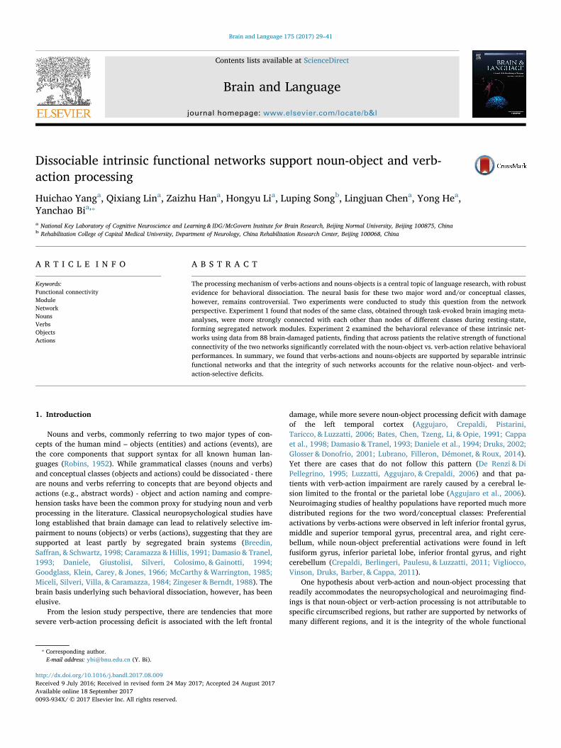
that are shared by nouns and verbs such as visual perception in the caseof a visual task, as proposed by Crepaldi et al. (2011). For the verbs-nouns direct contrast, 20 studies reporting 132 verb-activation foci in287 subjects and 11 studies reporting 59 noun-activation foci in 149subjects were included (Fig. 1A). A large proportion of these activationswere found in the semantic and naming tasks.
Activation likelihood estimation (ALE). All coordinates were trans-formed onto MNI space (Evans et al., 1993) using the Convert Focimenu in the GingerALE software package (available at http://brainmap.org). These foci were organized into a .txt file according todifferent subject groups for verbs and nouns, respectively. The ALEanalyses were implemented in the GingerALE software package withthe settings of the conservative (smaller) mask without excluding focioutside the mask. We chose an ALE algorithm that implements smallcorrections to minimize within-experiment effects, as described byTurkeltaub et al. (2012). The threshold was set at P < 0.05 and wascorrected for multiple comparisons using the false discovery rate (FDR)method along with a minimum cluster size of 50 mm3.
Node extraction. ALE probability maps for verbs and for nouns wereconstructed from the ALE meta-analyses. All of the peaks extracted fromverb- and noun-ALE probability maps were used to form spheres of4 mm radius within a grey matter mask (N voxels = 67541) that wasgenerated by thresholding (cut off = 0.2) the grey matter probabilitymap in SPM. These spheres were labeled as ALE-defined-verb and nounnodes, respectively.
2.1.4. Edge definitionEdges are represented by the strength of RSFC, measured by the
time series correlation coefficients, between the nodes. Specifically, theresidual time-series of all voxels within each node were averaged, andthen in each subject the Fisher-z transformed average residual time-
series correlations across nodes were calculated. The analysis wasperformed with the Resting State fMRI Data Analysis Toolkit (REST,available at http://www.restfmri.net, Song et al., 2011).
2.1.5. Average FCS comparison on the ALE-defined networksWe first examined whether the nodes showing stronger activation to
a particular word class, as obtained from the ALE analyses above, aremore strongly intrinsically connected to each other. We compared themean FCS during the resting-state for within- and between-class nodesacross subjects (see similar methods in He et al., 2007; Van Dijk et al.,2010). For each subject, the FCS for each pair of nodes was computed,and then three values were calculated: the average of the FCS withinthe ALE-defined-noun nodes, that of the FCS within the ALE-defined-verb nodes, and that of the FCS for all the between-class nodes. Thepaired t-test was conducted to test whether the average FCS for eachwithin-class network was significantly different from that of the be-tween networks.
2.1.6. Network modularity analysisAnother way to elucidate whether the ALE-defined-noun and verb
nodes are intrinsically organized as different networks is to explore themodularity structure of these nodes in a data-driven manner. We con-structed the network matrix over all the nodes without a prior role as-signment to perform a modularity analysis (Newman’s spectral opti-mization, Newman, 2006). This approach identifies modules accordingto the functional connectivity structure of the nodes. We then inspectedwhether the intrinsic modular structure roughly corresponded to theALE-defined-verb/noun networks.
Connectivity matrix generation. Averaging strength across subjects foreach edge, the group weighted graph matrix was obtained. Given thatthe negative correlations between two nodes may not be meaningful,
Table 1Overview of the 22 studies included in the ALE meta-analyses.
Paper Subject number Mode Task
Studies used in the verb-preference ALE meta-analysisWarburton et al. (1996) 9 PET Word generationTyler, Russell, Fadili, and Moss (2001) 8 PET Semantic categorizationTyler et al. (2001) 9 PET Lexical decisionTyler et al. (2003) 12 fMRI Semantic categorizationTyler, Bright, Fletcher, and Stamatakis (2004) 12 fMRI Semantic categorizationBedny and Thompson-Schill (2006) 13 fMRI Semantic similarity judgmentDavis, Meunier, and Marslen-Wilson (2004) 10 fMRI One-back synonym-monitoringBedny, Caramazza, Grossman, Pascual-Leone, and Saxe (2008) 12 fMRI Semantic relatedness judgmentDamasio et al. (2001) 20 PET Picture namingTranel, Martin, Damasio, Grabowski, and Hichwa (2005) 10 PET Picture namingSaccuman et al. (2006) 13 fMRI Picture namingLiljeström et al. (2008) 15 fMRI Picture namingBerlingeri et al. (2008) 12 fMRI Picture naming; Grammatical-class switchingLonge, Randall, Stamatakis, and Tyler (2007) 12 fMRI Pleasant judgmentTyler, Randall, and Stamatakis (2008) 15 fMRI Pleasant judgmentPerani et al. (1999) 14 PET Lexical decisionYokoyama et al. (2006) 28 fMRI Lexical decisionShapiro et al. (2005) 12 PET Word inflectionShapiro, Moo, and Caramazza (2006) 10 fMRI Word inflectionYu et al. (2011) 21 fMRI Semantic relatedness judgmentYu et al. (2012) 20 fMRI Semantic associate generation
Studies used in the noun-preference ALE meta-analysisWarburton et al. (1996) 9 PET Word generationBedny and Thompson-Schill (2006) 13 fMRI Semantic similarity judgmentSaccuman et al. (2006) 13 fMRI Picture namingLiljeström et al. (2008) 15 fMRI Picture namingSiri et al. (2008) 12 fMRI Picture namingBerlingeri et al. (2008) 12 fMRI Grammatical-class switchingShapiro et al. (2005) 12 PET Word inflectionShapiro et al. (2006) 10 fMRI Word inflectionBurton, Krebs-Noble, Gullapalli, and Berndt (2009) 12 fMRI Grammaticality judgmentYu et al. (2011) 21 fMRI Semantic relatedness judgmentYu et al. (2012) 20 fMRI Semantic associate generation
H. Yang et al. Brain and Language 175 (2017) 29–41
31
they were converted to zero and the positive correlations were re-mained and analyzed in this study. Thus, an n × n RSFC matrix, inwhich n is the sum number of ALE-defined-verb and noun nodes, wasconstructed.
Modularity analysis. The n × n weighted graph matrix was input to aGraph-theoretical Network Analysis Toolkit in MATLAB (GRETNA,available at https://github.com/sandywang/GRETNA, Wang et al.,2015) for modularity analysis. The modularity Q(p) for a given partitionp of the RSFC network (Newman & Girvan, 2004) is defined as:
∑= ⎡⎣⎢
−⎛⎝
⎞⎠
⎤⎦⎥=
Q p lL
dL
( )2s
Ns s
1
2m
where Nm is the number of modules, L is the total number of networkconnections, ls is the number of connections between nodes in module s,and ds is the sum of the degrees of the nodes in the modules. Themodularity index quantifies the difference between the real number ofintra-module connections in the actual network and that of a network in
which nodes are connected at random. This module detection processallows us to identify the preference partition p that maximizes Q(p).There are several available optimization algorithms; we partitioned theRSFC network using Newman’s spectral optimization algorithm(Newman, 2006). According to Clauset, Newman, and Moore (2004), aQ value above 0.3 is good enough to indicate the significant modules ina network. The difference between Q of the real network and that ofrandom networks was used to generate a Z score, which indicates thesignificance level. The modularity analyses were conducted for sparsitythresholds ranging from 0.22 to 0.50 in increments of 0.01. The lowestthreshold was determined to ensure that the resulting graph was fullyconnected; the highest threshold was set to remove weak correlations sothat only the correlations significantly above zero were included. Notethat because almost all of the connections in the matrix were sig-nificantly above zero, 80% remained when the significance reached theBonferroni-corrected level of 0.001. This connection density is deemedtoo high and we arbitrarily set the highest threshold to 0.50.
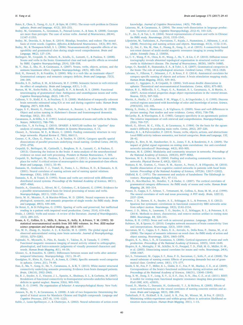
Fig. 1. Node definition. (A) Verb- and noun-activation foci included in themeta-analyses. (B) ALE probability maps of the meta-analyses (threshold:FDR corrected P < 0.05, volume > 50 mm3). (C) Distribution map of the19 ALE-defined-verb nodes and the 15 ALE-defined-noun nodes. Arabicnumbers for the brain regions correspond to those in Table 2. This and thebrain figures below are all visualized with the BrainNet Viewer (Xia,Wang, & He, 2013, http://www.nitrc.org/projects/bnv/).
H. Yang et al. Brain and Language 175 (2017) 29–41
32
2.1.7. Validation analysesWe performed validation analyses using a new test-retest dataset,
preprocessing with global signal removal, and additional head motiontreatment procedures.
A different (retest) dataset. Fifty-seven (27 females; 23.1 ± 2.3 yearsold; range, 19–30 years old) of the 146 participants in Experiment 1took part in another scanning session about 6 weeks (40.94 ± 4.51days) later (i.e., the second scan session in Lin et al., 2015). We usedthis later scan to validate our results. This sub-group retest dataset wasincluded in the “Connectivity-based Brian Imaging Research Database(C-BIRD) at BNU” (http://fcon_1000.projects.nitrc.org/indi/CoRR/html/bnu_1.html, BNU 1). Two subjects were excluded due to ex-cessive head motion and 55 participants (26 females; 23.1 ± 2.3 yearsold; range, 19–30 years old) remained for identical analysis proceduresas the main dataset in Experiment 1.
Global signal removal. It remains controversial whether global signalregression should be performed during resting-state fMRI preprocessing(Fox, Zhang, Snyder, & Raichle, 2009; Murphy, Birn, Handwerker,Jones, & Bandettini, 2009). To test the extent to which that our resultswere reliable across different preprocessing protocols, we performedthe same analyses using data with global signal regression for the maindataset.
Head motion. Head motion has been shown to have a confoundingeffect on RSFC (Power, Barnes, Snyder, Schlaggar, & Petersen, 2012;Power et al., 2014; Van Dijk, Sabuncu, & Buckner, 2012). In order tofurther exclude the possible effects of head motion, we performed a‘scrubbing’ procedure (Power et al., 2012; Yan et al., 2013) duringpreprocessing of the main dataset. In this scrubbing analysis, functionalvolumes were deleted based on a framewise displacement >0.5 mmcompared with the 1 back and 2 forward neighbors (Power et al.,2012). Different time points remained after scrubbing across subjects.To have a sufficient number of time points for meaningful analyses, twosubjects that had too few remaining time points (<150 volumes out ofthe 190 total time points; i.e., <5 min) were excluded, resulting in 142healthy subjects for this analysis.
2.2. Experiment 2: Testing the relationship between functional networkintegrity and behavioral deficits in patients
This experiment further examines the cognitive relevance of theintrinsic noun- and verb-functional networks by testing whether thebreakdowns of these functional networks associate with noun or verbdeficits. We collected resting-state fMRI data and behavioral data from88 patients with brain damage, and correlated the relative FCS for thetwo networks (subtractions of noun network FCS from verb networkFCS) with the relative performances on noun and verb processing tasks(subtracting the noun behavioral score from the verb behavioral score)after controlling for potential confounding variables (following similarprocedures in Nomura et al., 2010). We also examined whether thepotential effect of the FCS is account for by the lesions to the con-stituent nodes. This correlation approach captures the general re-lationship between two continuous variables and is possible because ofthe proper sample size in this Experiment.
2.2.1. ParticipantsEighty-eight patients with brain damage (17 females; 45.1 ± 13.4
years old; range, 19–74 years old; 12.9 ± 3.2 years of education; range,4–19 years of education), all from the China Rehabilitation ResearchCenter, participated in this study voluntarily. Sixty-eight of the patientswere from the cohort reported in our earlier study (Han et al., 2013).All were right-handed (Edinburgh Handedness Inventory; Oldfield,1971). The inclusion criteria were as follows: presenting with braininjury for the first time; at least 1 month elapsed since the onset; noother neurological or psychiatric disease such as schizophrenia or al-cohol abuse; and capable of following task instructions. The majoritysuffered from stroke (n = 71), with remaining suffering from trauma,
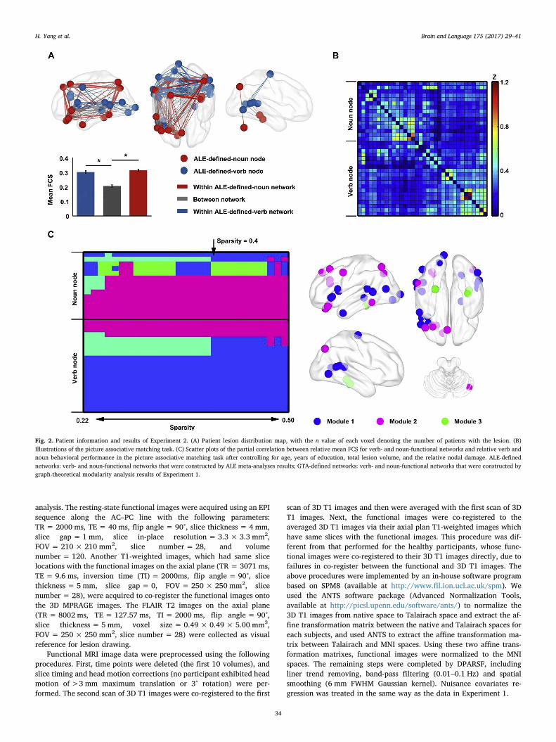
atrophy, gas poisoning, and electric shock. Thirty-five patients had bi-lateral lesions, 35 had left hemisphere lesions, 17 had right hemispherelesions and 1 had brain stem lesion. Detailed information for each pa-tient is shown in the Supplementary Table S1. A lesion distribution mapof the 88 patients is presented in Fig. 2A. All subjects were providedwritten informed consent. This research was approved by the Institu-tional Review Board of the National Key Laboratory of CognitiveNeuroscience and Learning, Beijing Normal University.
2.2.2. Behavioral tests design and data preprocessingTo assess the cognitive processing ability of verbs and nouns, four
tasks were administered, including an oral picture naming task, a pic-ture associative matching task, a word associative matching task and aword-picture verification task. The four tasks covered pictorial andverbal inputs, oral and nonverbal outputs, reflecting primarily abilitiesin semantic and oral production, nonverbal comprehension, verbalcomprehension, and verbal-nonverbal matching, respectively. All sti-muli used in the tasks were words and pictures corresponding to actions(e.g., diving, hugging) and objects (tools, animals, fruits and vegetables,artifacts, and large nonmanipulable objects). Illustrations of pictureassociative matching are shown in Fig. 2B. The complete stimulus list ofall tasks is presented in the Supplementary Table S2. There were nosignificant differences in term of word frequency across various wordcategories in any task (see the Supplementary Table S3). For oral pic-ture naming task, there are no significant differences in phonologicallength across categories (number of syllables, F = 1.272, P = 0.281).For word-picture verification and word associative matching task, therewere no significant differences in word visual complexity across cate-gories (number of strokes, word-picture verification: F= 0.756,P = 0.586; word associative matching: F= 1.153, P = 0.344).
In oral picture naming, participants were instructed to name eachobject or action picture. The word and picture associative matchingtasks had an identical structure: in each trial contained three items andthe participants need to choose which of the bottom two were se-mantically closer to the top item, except that three words were pre-sented in the word version and three pictures were presented in thepicture version. In the word-picture verification task, a word was pre-sented above a picture on the touch screen in each trial. Participantswere instructed to judge whether the picture corresponded with theword by pressing “yes” or “no” on the screen. This task included 2 setsof 60 trials each. The word stimuli were identical across the two sets,but each set of the words was paired separately with a congruent pic-ture or an incongruent picture. Only when both trials of the two setswere correct would the word stimuli be scored as accurate. The re-sponse deadline was one minute for each trial of these tasks.
As the patients were not asked to respond as quickly as possible toavoid pressure, only response accuracies were analyzed. Z-transformedaccuracies for each task were used as the corresponding behavioralscores. We considered two types of contrasts for noun/verb behavioralmeasures: actions versus tool items; actions versus all object items. Thisspecific contrast of actions versus tools was motivated by a line of hy-pothesis that tools are semantically closer than other types of objects toactions in that a core dimension of conceptual representation of toolsare its manipulation properties (Bird, Howard, & Franklin, 2000; Mahonet al., 2007).
2.2.3. Image acquisition and preprocessingStructural and functional imaging data were collected using a 1.5 T
GE SIGNA EXCITE scanner at the China Rehabilitation Research Center.During resting-state functional images scanning, participants were in-structed to keep still with their eyes closed. The 3D MPRAGE T1-weighted images were obtained in the sagittal plane with the para-meters: TR = 1226 ms, TE = 4.2 ms, flip angle = 15°, voxel si-ze = 0.49 × 0.49 × 0.70 mm3, FOV = 250 × 250 mm2, and slicenumber = 248. Two identical sequences of 3D T1 images were col-lected and averaged to improve the signal-to-noise ratio during
H. Yang et al. Brain and Language 175 (2017) 29–41
33
analysis. The resting-state functional images were acquired using an EPIsequence along the AC–PC line with the following parameters:TR = 2000 ms, TE = 40 ms, flip angle = 90°, slice thickness = 4 mm,slice gap = 1 mm, slice in-place resolution = 3.3 × 3.3 mm2,FOV = 210 × 210 mm2, slice number = 28, and volumenumber = 120. Another T1-weighted images, which had same slicelocations with the functional images on the axial plane (TR = 3071 ms,TE = 9.6 ms, inversion time (TI) = 2000ms, flip angle = 90°, slicethickness = 5 mm, slice gap = 0, FOV = 250 × 250 mm2, slicenumber = 28), were acquired to co-register the functional images ontothe 3D MPRAGE images. The FLAIR T2 images on the axial plane(TR = 8002 ms, TE = 127.57 ms, TI = 2000 ms, flip angle = 90°,slice thickness = 5 mm, voxel size = 0.49 × 0.49 × 5.00 mm3,FOV = 250 × 250 mm2, slice number = 28) were collected as visualreference for lesion drawing.
Functional MRI image data were preprocessed using the followingprocedures. First, time points were deleted (the first 10 volumes), andslice timing and head motion corrections (no participant exhibited headmotion of >3 mm maximum translation or 3° rotation) were per-formed. The second scan of 3D T1 images were co-registered to the first
scan of 3D T1 images and then were averaged with the first scan of 3DT1 images. Next, the functional images were co-registered to theaveraged 3D T1 images via their axial plan T1-weighted images whichhave same slices with the functional images. This procedure was dif-ferent from that performed for the healthy participants, whose func-tional images were co-registered to their 3D T1 images directly, due tofailures in co-register between the functional and 3D T1 images. Theabove procedures were implemented by an in-house software programbased on SPM8 (available at http://www.fil.ion.ucl.ac.uk/spm). Weused the ANTS software package (Advanced Normalization Tools,available at http://picsl.upenn.edu/software/ants/) to normalize the3D T1 images from native space to Talairach space and extract the af-fine transformation matrix between the native and Talairach spaces foreach subjects, and used ANTS to extract the affine transformation ma-trix between Talairach and MNI spaces. Using these two affine trans-formation matrixes, functional images were normalized to the MNIspaces. The remaining steps were completed by DPARSF, includingliner trend removing, band-pass filtering (0.01–0.1 Hz) and spatialsmoothing (6 mm FWHM Gaussian kernel). Nuisance covariates re-gression was treated in the same way as the data in Experiment 1.
Fig. 2. Patient information and results of Experiment 2. (A) Patient lesion distribution map, with the n value of each voxel denoting the number of patients with the lesion. (B)Illustrations of the picture associative matching task. (C) Scatter plots of the partial correlation between relative mean FCS for verb- and noun-functional networks and relative verb andnoun behavioral performance in the picture associative matching task after controlling for age, years of education, total lesion volume, and the relative nodal damage. ALE-definednetworks: verb- and noun-functional networks that were constructed by ALE meta-analyses results; GTA-defined networks: verb- and noun-functional networks that were constructed bygraph-theoretical modularity analysis results of Experiment 1.
H. Yang et al. Brain and Language 175 (2017) 29–41
34
For the 3D T1 images, we first co-registered each of the two se-quences on the same native space and then averaged them. The FLAIRT2 images were co-registered and resliced to the native space of theaveraged 3D images. Lesions were manually drawn by two trainedpersonnel on the averaged 3D T1 image slice by slice with visual re-ference to the FLAIR T2 images for each patient. The lesion descriptionwas finally transformed into the MNI space. More details can be ob-tained from our previously research (Han et al., 2013).
2.2.4. Partial correlations between relative FCS and relative behavioralscore
To test the extent to which the difference in terms of the integrity ofthe noun- and verb-functional networks could account for the potentialnoun/verb behavioral dissociations, we carried out a partial correlationanalysis between the relative FCS of the two networks and the relativeof the behavior scores on each noun and verb task, with the total lesionvolume (i.e., total number of damaged voxels across the whole brain),age, and years of education as covariates. The relative noun/verb FCSfor each patient was computed by subtracting the average FCS withinthe noun-functional network from the average FCS within the verb-functional network. The relative behavioral score for each patient oneach task was calculated by subtracting the noun behavior score (z-transformed accuracy) from the verb behavior score.
2.2.5. Controlling for the effects of nodal lesionsTo assess whether the effects of the functional connectivity within
each network on behavior are fully attributable to the extent of lesion tothe relevant nodes, we performed further correlation analyses betweenthe relative noun/verb FCS and the relative noun/verb behavioralscores, including the relative extent of the anatomical damage for thenodes in the two networks as an additional covariate. For each patient,the extent of nodal lesions for each network was calculated by aver-aging the percentage of voxels with lesion each node within the net-work. The relative nodal lesion for the two networks was calculated bysubtracting the extent of nodal lesions of the noun-functional networkfrom the nodal lesions of the verb-functional network.
2.2.6. Validation analysesSimilar to Experiment 1, we carried out validation analyses with
global signal removal and additional head motion data treatment.Furthermore, because the physiological basis of fMRI signal for voxelswith lesion is unclear, we conducted analysis excluding each patient’sdamaged voxels from the RSFC analyses.
Global signal removal. To test whether our results were reliableacross different preprocessing procedures, we performed the sameanalyses using data with global signal regression, using preprocessingprocedures same to those in Experiment 1.
Head motion. Similar to Experiment 1, we performed an additional‘scrubbing’ procedure during preprocessing for the patients group tofurther deal with the potential head motion confound, using proceduresidentical to those of Experiment 1. Seven patients were excluded for nothaving enough remaining time points (<90 volumes out of 110 totaltime points; i.e., <3 min) and the data of 81 patients entered analysesafter scrubbing.
Exclude damaged voxels. We performed a further validation analysesto exclude the damaged voxels within ALE-defined-verb and nounnodes for each patient. There were 41 patients had no lesion in allnodes. We first used this sub-group to validate our main results ofExperiment 2. For the other patients, we excluded their damaged voxelswithin each node one by one. If all of voxels within a node were da-maged, we would exclude this node or set the FCS from this node aszero. Results of both analyses were presented.
3. Results
3.1. Experiment 1: Characterizing the intrinsic functional networkorganization of verb- and noun-preferential regions in healthy subjects
3.1.1. Node definition resultsWe carried out ALE meta-analyses based on 22 imaging studies
where verb processing and noun processing were contrasted acrosstasks, including 20 studies where stronger verb activations were ob-served and 11 where stronger noun activations were observed(Table 1). As presented in Fig. 1B (thresholded at whole brain FDRcorrected P < 0.05, volume > 50 mm3), brain regions that are con-sistently activated more strongly for verbs, i.e., ALE-defined-verbnodes, across the 20 studies include the bilateral superior temporalgyrus (STG), left middle temporal gyrus (MTG), left inferior frontalgyrus (IFG), left insular, bilateral precentral gyrus, left middle occipitalgyrus (MOG), left middle frontal gyrus (MFG), left postcentral gyrus,left lingual gyrus, right inferior temporal gyrus (ITG) and right posteriorcingulate. Brain regions showing consistent noun-preference activationacross the 11 studies, i.e., ALE-defined-noun nodes, were the bilateralparahippocampal gyrus (PHG), bilateral precuneus, left superior par-ietal lobule (SPL), left IFG, left MTG, left medial frontal gyrus, left ITG,left MFG, left SFG, and right cerebellar tonsil. The peaks were extractedfrom the ALE probability maps to form spheres of radius 4 mm. One ofthe verb-preference peaks (coordinates: −10, −42, 80) was excludedbecause it was located outside the pre-defined gray matter mask. In thismanner, we identified 19 ALE-defined-verb nodes and 15 ALE-defined-noun nodes (see details in Table 2 and Fig. 1C).
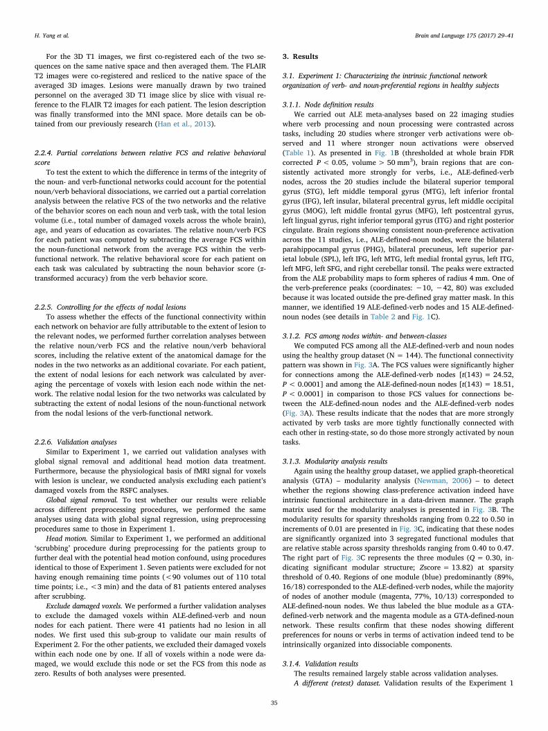
3.1.2. FCS among nodes within- and between-classesWe computed FCS among all the ALE-defined-verb and noun nodes
using the healthy group dataset (N = 144). The functional connectivitypattern was shown in Fig. 3A. The FCS values were significantly higherfor connections among the ALE-defined-verb nodes [t(143) = 24.52,P < 0.0001] and among the ALE-defined-noun nodes [t(143) = 18.51,P < 0.0001] in comparison to those FCS values for connections be-tween the ALE-defined-noun nodes and the ALE-defined-verb nodes(Fig. 3A). These results indicate that the nodes that are more stronglyactivated by verb tasks are more tightly functionally connected witheach other in resting-state, so do those more strongly activated by nountasks.
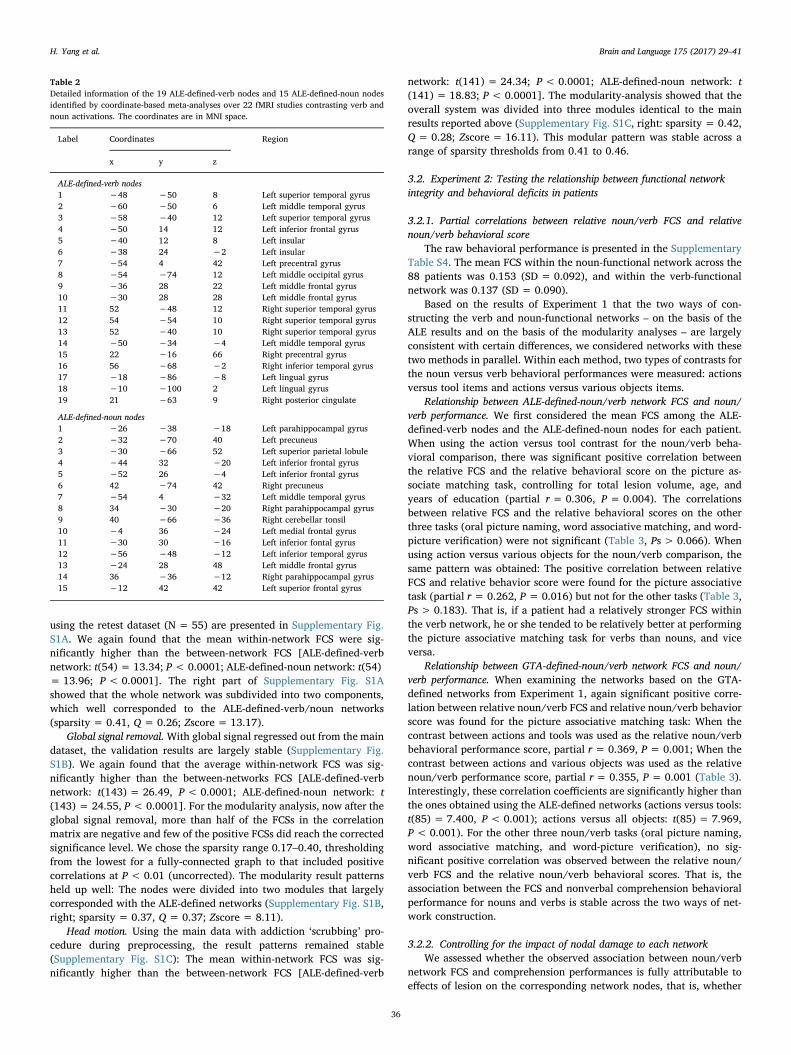
3.1.3. Modularity analysis resultsAgain using the healthy group dataset, we applied graph-theoretical
analysis (GTA) – modularity analysis (Newman, 2006) – to detectwhether the regions showing class-preference activation indeed haveintrinsic functional architecture in a data-driven manner. The graphmatrix used for the modularity analyses is presented in Fig. 3B. Themodularity results for sparsity thresholds ranging from 0.22 to 0.50 inincrements of 0.01 are presented in Fig. 3C, indicating that these nodesare significantly organized into 3 segregated functional modules thatare relative stable across sparsity thresholds ranging from 0.40 to 0.47.The right part of Fig. 3C represents the three modules (Q = 0.30, in-dicating significant modular structure; Zscore = 13.82) at sparsitythreshold of 0.40. Regions of one module (blue) predominantly (89%,16/18) corresponded to the ALE-defined-verb nodes, while the majorityof nodes of another module (magenta, 77%, 10/13) corresponded toALE-defined-noun nodes. We thus labeled the blue module as a GTA-defined-verb network and the magenta module as a GTA-defined-nounnetwork. These results confirm that these nodes showing differentpreferences for nouns or verbs in terms of activation indeed tend to beintrinsically organized into dissociable components.
3.1.4. Validation resultsThe results remained largely stable across validation analyses.A different (retest) dataset. Validation results of the Experiment 1
H. Yang et al. Brain and Language 175 (2017) 29–41
35
using the retest dataset (N = 55) are presented in Supplementary Fig.S1A. We again found that the mean within-network FCS were sig-nificantly higher than the between-network FCS [ALE-defined-verbnetwork: t(54) = 13.34; P< 0.0001; ALE-defined-noun network: t(54)= 13.96; P< 0.0001]. The right part of Supplementary Fig. S1Ashowed that the whole network was subdivided into two components,which well corresponded to the ALE-defined-verb/noun networks(sparsity = 0.41, Q = 0.26; Zscore = 13.17).
Global signal removal.With global signal regressed out from the maindataset, the validation results are largely stable (Supplementary Fig.S1B). We again found that the average within-network FCS was sig-nificantly higher than the between-networks FCS [ALE-defined-verbnetwork: t(143) = 26.49, P< 0.0001; ALE-defined-noun network: t(143) = 24.55, P < 0.0001]. For the modularity analysis, now after theglobal signal removal, more than half of the FCSs in the correlationmatrix are negative and few of the positive FCSs did reach the correctedsignificance level. We chose the sparsity range 0.17–0.40, thresholdingfrom the lowest for a fully-connected graph to that included positivecorrelations at P < 0.01 (uncorrected). The modularity result patternsheld up well: The nodes were divided into two modules that largelycorresponded with the ALE-defined networks (Supplementary Fig. S1B,right; sparsity = 0.37, Q = 0.37; Zscore = 8.11).
Head motion. Using the main data with addiction ‘scrubbing’ pro-cedure during preprocessing, the result patterns remained stable(Supplementary Fig. S1C): The mean within-network FCS was sig-nificantly higher than the between-network FCS [ALE-defined-verb
network: t(141) = 24.34; P < 0.0001; ALE-defined-noun network: t(141) = 18.83; P< 0.0001]. The modularity-analysis showed that theoverall system was divided into three modules identical to the mainresults reported above (Supplementary Fig. S1C, right: sparsity = 0.42,Q = 0.28; Zscore = 16.11). This modular pattern was stable across arange of sparsity thresholds from 0.41 to 0.46.
3.2. Experiment 2: Testing the relationship between functional networkintegrity and behavioral deficits in patients
3.2.1. Partial correlations between relative noun/verb FCS and relativenoun/verb behavioral score
The raw behavioral performance is presented in the SupplementaryTable S4. The mean FCS within the noun-functional network across the88 patients was 0.153 (SD = 0.092), and within the verb-functionalnetwork was 0.137 (SD = 0.090).
Based on the results of Experiment 1 that the two ways of con-structing the verb and noun-functional networks – on the basis of theALE results and on the basis of the modularity analyses – are largelyconsistent with certain differences, we considered networks with thesetwo methods in parallel. Within each method, two types of contrasts forthe noun versus verb behavioral performances were measured: actionsversus tool items and actions versus various objects items.
Relationship between ALE-defined-noun/verb network FCS and noun/verb performance. We first considered the mean FCS among the ALE-defined-verb nodes and the ALE-defined-noun nodes for each patient.When using the action versus tool contrast for the noun/verb beha-vioral comparison, there was significant positive correlation betweenthe relative FCS and the relative behavioral score on the picture as-sociate matching task, controlling for total lesion volume, age, andyears of education (partial r= 0.306, P = 0.004). The correlationsbetween relative FCS and the relative behavioral scores on the otherthree tasks (oral picture naming, word associative matching, and word-picture verification) were not significant (Table 3, Ps > 0.066). Whenusing action versus various objects for the noun/verb comparison, thesame pattern was obtained: The positive correlation between relativeFCS and relative behavior score were found for the picture associativetask (partial r = 0.262, P = 0.016) but not for the other tasks (Table 3,Ps > 0.183). That is, if a patient had a relatively stronger FCS withinthe verb network, he or she tended to be relatively better at performingthe picture associative matching task for verbs than nouns, and viceversa.
Relationship between GTA-defined-noun/verb network FCS and noun/verb performance. When examining the networks based on the GTA-defined networks from Experiment 1, again significant positive corre-lation between relative noun/verb FCS and relative noun/verb behaviorscore was found for the picture associative matching task: When thecontrast between actions and tools was used as the relative noun/verbbehavioral performance score, partial r = 0.369, P = 0.001; When thecontrast between actions and various objects was used as the relativenoun/verb performance score, partial r= 0.355, P= 0.001 (Table 3).Interestingly, these correlation coefficients are significantly higher thanthe ones obtained using the ALE-defined networks (actions versus tools:t(85) = 7.400, P < 0.001); actions versus all objects: t(85) = 7.969,P < 0.001). For the other three noun/verb tasks (oral picture naming,word associative matching, and word-picture verification), no sig-nificant positive correlation was observed between the relative noun/verb FCS and the relative noun/verb behavioral scores. That is, theassociation between the FCS and nonverbal comprehension behavioralperformance for nouns and verbs is stable across the two ways of net-work construction.
3.2.2. Controlling for the impact of nodal damage to each networkWe assessed whether the observed association between noun/verb
network FCS and comprehension performances is fully attributable toeffects of lesion on the corresponding network nodes, that is, whether
Table 2Detailed information of the 19 ALE-defined-verb nodes and 15 ALE-defined-noun nodesidentified by coordinate-based meta-analyses over 22 fMRI studies contrasting verb andnoun activations. The coordinates are in MNI space.
Label Coordinates Region
x y z
ALE-defined-verb nodes1 −48 −50 8 Left superior temporal gyrus2 −60 −50 6 Left middle temporal gyrus3 −58 −40 12 Left superior temporal gyrus4 −50 14 12 Left inferior frontal gyrus5 −40 12 8 Left insular6 −38 24 −2 Left insular7 −54 4 42 Left precentral gyrus8 −54 −74 12 Left middle occipital gyrus9 −36 28 22 Left middle frontal gyrus10 −30 28 28 Left middle frontal gyrus11 52 −48 12 Right superior temporal gyrus12 54 −54 10 Right superior temporal gyrus13 52 −40 10 Right superior temporal gyrus14 −50 −34 −4 Left middle temporal gyrus15 22 −16 66 Right precentral gyrus16 56 −68 −2 Right inferior temporal gyrus17 −18 −86 −8 Left lingual gyrus18 −10 −100 2 Left lingual gyrus19 21 −63 9 Right posterior cingulate
ALE-defined-noun nodes1 −26 −38 −18 Left parahippocampal gyrus2 −32 −70 40 Left precuneus3 −30 −66 52 Left superior parietal lobule4 −44 32 −20 Left inferior frontal gyrus5 −52 26 −4 Left inferior frontal gyrus6 42 −74 42 Right precuneus7 −54 4 −32 Left middle temporal gyrus8 34 −30 −20 Right parahippocampal gyrus9 40 −66 −36 Right cerebellar tonsil10 −4 36 −24 Left medial frontal gyrus11 −30 30 −16 Left inferior fontal gyrus12 −56 −48 −12 Left inferior temporal gyrus13 −24 28 48 Left middle frontal gyrus14 36 −36 −12 Right parahippocampal gyrus15 −12 42 42 Left superior frontal gyrus
H. Yang et al. Brain and Language 175 (2017) 29–41
36

the integrity of the synchronization among the networks has effects forbehavior beyond the damage of the constituent grey matter regions. Wedid this by further computing the partial correlation between relativenoun/verb FCS and relative noun/verb behavioral scores, including therelative extent of the anatomical damage for the nodes in the twonetworks as an additional covariate. Again both the ALE-defined net-works and the GTA-defined networks were considered in parallel in thiscontrol analysis.
Results of the ALE-defined networks. For the ALE-defined networks,after controlling for the extent of lesion to the corresponding nodes ineach network, the positive relationship between the relative noun/verbFCS and the relative noun/verb performances remained significant inthe picture associative matching task (Fig. 2C & Table 3): Using thecontrast of actions versus tools as the relative noun/verb behavioralmeasures: partial r = 0.307, P = 0.004; Using the contrast actions
versus various objects as the relative noun/verb behavioral measure,partial r = 0.265, P = 0.015. That is, the effect of FCS for these twonetworks in predicting the nonverbal comprehension ability is not to befully explained by the lesion of the nodes involved. But rather, the in-tegrity of intrinsic communication capacity within the network hasadditional effects. No relationships were found for other tasks.
Results for GTA-defined networks. The same pattern as above wasobserved when we considered the GTA-defined networks. Significantpositive correlations between relative noun/verb FCS and relativenoun/verb behavior score were found for the picture associativematching task: Using the actions versus tools contrast as the noun/verbrelative performance measures, partial r = 0.370, P = 0.001; Using theactions versus various objects contrast as the noun/verb measures,partial r= 0.358, P = 0.001 (see Fig. 2C & Table 3). Again the partialcorrelation were significantly stronger in the GTA-defined networks
Fig. 3. Results of Experiment 1: The intrinsic network organization of the ALE-defined-noun and verb nodes in the healthy population. (A) Upper panel: The functional connectivitypattern of ALE-defined-verb and noun networks at the sparsity threshold of 0.4. Lower panel: Comparison of the average FCS within and between these networks. Error bars represent theSEM. *: P < 0.0001. (B) Graph matrix used in the modularity analysis. (C) Left: the assignments of areal nodes into subgraphs (colors) across a range of sparsity thresholds from 0.22 to0.50 in 0.01 steps by modularity analyses. Right: subgraph at sparsity threshold of 0.40 is shown for the areal nodes (Q= 0.30; Zscore = 13.82).
Table 3Partial correlation coefficients between the relative mean FCS of verb- and noun-functional networks and the relative verb and noun behavioral performance in each task.
Verb/noun behavioral comparison Network construction Correlation coefficient
Picture associative matching Word associative matching Word-picture verification Oral picture naming
Covariates: age, years of education, and total lesion volumeActions vs. tools ALE-defined networks 0.306 (0.004) −0.201 (0.066) −0.121 (0.271) 0.063 (0.566)
GTA-defined networks 0.369 (0.001) −0.217 (0.046) 0.004 (0.974) −0.037 (0.739)Actions vs. objects ALE-defined networks 0.262 (0.016) −0.146 (0.183) −0.019 (0.860) 0.010 (0.924)
GTA-defined networks 0.355 (0.001) −0.097 (0.376) 0.069 (0.528) −0.031 (0.778)
Covariates: age, years of education, total lesion volume, and nodal damageActions vs. tools ALE-defined networks 0.307 (0.004) −0.202 (0.065) −0.108 (0.327) 0.071 (0.522)
GTA-defined networks 0.370 (0.001) −0.219 (0.045) 0.022 (0.841) −0.029 (0.791)Actions vs. objects ALE-defined networks 0.265 (0.015) −0.143 (0.194) −0.010 (0.931) 0.009 (0.936)
GTA-defined networks 0.358 (0.001) −0.094 (0.394) 0.081 (0.462) −0.033 (0.766)
H. Yang et al. Brain and Language 175 (2017) 29–41
37

than in the ALE-defined networks [actions versus tools contrast t(85)= 7.370, P < 0.001; actions versus objects contrast t(85) = 7.918,P < 0.001].
3.2.3. Validation resultsThe overall results pattern remained stable across validation ap-
proaches. Given that the ALE-defined and GTA-defined noun- and verb-networks converged well in the main results, we here report validationresults for ALE-defined networks.
Global signal removal. The partial correlation between the relativenoun/verb FCS and the relative noun/verb behavioral score in thepicture associate matching task was significant for the actions versusvarious objects contrast (r = 0.221, P= 0.042), and was no longersignificant for the actions versus tools contrast (r = 0.175, P = 0.110).After controlling for the effects of nodal lesions within the networks, thepartial r was 0.177 (P = 0.107) in the actions versus tools contrast andwas 0.231 (P = 0.035) in the actions versus various objects contrast.Details are shown in Supplementary Table S5.
Head motion. The results of the validation of data following the‘scrubbing’ procedure during preprocessing are very similar to our mainresults. Significant positive correlations were found for the picture as-sociative matching task (Supplementary Table S5): The partial corre-lation between relative noun/verb FCS and relative noun/verb beha-vioral score in the actions versus tools contrast was 0.312 (P= 0.005)and in the actions versus various objects contrast was 0.264(P = 0.020). After controlling for the effect of nodal lesions in eachnetwork, the partial r was 0.311 (P = 0.006) in the actions versus toolscontrast and 0.265 (P = 0.020) in the actions versus various objectscontrast.
Excluding damaged voxels. We first performed analyses includingonly the 41 patients who had no lesion in any node. Similar to the mainanalyses results, significant positive correlation between relative noun/verb FCS and relative noun/verb behavioral score was observed for thepicture associative matching task in the actions versus tools contrast(partial r= 0.353, P = 0.030). We then performed analyses includingall patients, while excluding the damaged voxels within the nodes foreach patient, if there were any. If all voxels in a node were damaged, weexcluded this node or set the FCS from this node as zero. When ex-cluding nodes with no voxel left, the correlation between the relativenoun/verb FCS and the relative noun/verb behavioral score in thepicture associate matching task was significant for the actions versustools contrast (partial r = 0.229, P= 0.035); When setting the FCSfrom nodes with no voxel left to zero, the partial r was 0.286(P = 0.008) in the actions versus tools contrast.
4. Discussion
With two experiments we examined the neural underpinning for thedissociation of noun-object and verb-action processing from the net-work perspective. We first established in the healthy population thatthe regions consistently showing preferential activation to the sameclass are intrinsically connected with each other more strongly duringthe resting-state, forming word-class (and/or conceptual-class) specificintrinsic functional networks. Then in patients with brain damage, wefound that the extent to which the two networks’ functional integritydiffered, measured by the mean FCS difference, significantly associatedwith how different the behavioral performances are on the object andaction nonverbal comprehension abilities. That is, if a patient hasweaker within-verb-network FCS relative to within-noun-network FCS,he or she is more likely to suffer from more severe deficit in nonverbalcomprehension of actions compared to objects. These two experiments,together, revealed the functional network basis of noun/verb (object/action) dissociation previously established in the aphasia and neuroi-maging field.
While the regions that are commonly activated by a class of stimuliare often referred to as a network for the class, we here showed that
even at the resting-state without the task engagement, the word-class(and/or conceptual-class) specific regions are tightly connected, pro-viding new aspect of mechanism for the neural integrative basis ofnoun-object or verb-action processing. Such correspondence betweentask-driven activation pattern and the intrinsic functional connectivitypattern during the resting-state extends the previous reports about thesimilar correspondence in auditory, action execution, language andmemory processes (Calhoun, Kiehl, & Pearlson, 2008; Smith et al.,2009) to dissociable structures within a cognitive system, i.e., com-prehension to object and action stimuli. The correlations of sponta-neous BOLD fluctuations across regions are likely to be caused by theirrepeated co-activation during everyday activities due to Hebbian me-chanisms (Hebb, 1949). Note that the correspondence is not perfect. Inthe spontaneous data-driven modularity analyses, three ALE-defined-verb nodes [left precentral gyrus (−54, 4, 42) and left MFG (−36, 28,22; −30, 28, 28)] and two ALE-defined-noun nodes [left IFG (−52, 26,−4) and left anterior middle temporal gyrus (−54, 4, −32)] wereconsistently classified into the other module across different validationresults. Interestingly, these are nodes whose functions tended to becontroversial. In particular, the left inferior frontal gyrus has beensuggested to be modulated by processing demand (Vigliocco et al.,2011) and high-frequency power increases during both verb and noungeneration have been found here using electrocorticography recording(Conner, Chen, Pieters, & Tandon, 2014; Crepaldi et al., 2013). For theanterior middle temporal gyrus node, its lesion is associated withnaming impairments for both nouns and verbs (Glosser & Donofrio,2001). Experiment 2 showed that the FCS for the GTA-defined-networkshad stronger association with behavioral performances than the ALE-defined networks, indicating that categorizing these nodes according tothe modularity analyses may more accurately reflect their functions.
The dissociable intrinsic functional networks have direct cognitiverelevance, as illustrated by the patients’ data in Experiment 2, wherethe disturbance of these networks associated with the correspondingbehavior. Importantly, such effects were not to be fully attributed to thedamage of the constituent brain regions – the strength of the con-nectivity within the system has its unique effects beyond the regionallesion measures. Could the association between the network con-nectivity strength difference and the word class behavioral differencebe explained by the potential confounding psycholinguistic properties?Although stimuli of various categories in the current Experiment 2 didnot differ by word frequency or shallow orthographic/phonologicalvariables (see Section 2), and that the class-specific regions in the targetnetworks were obtained by meta-analysis of a large set of fMRI studieswith varying stimulus properties, there are still dimensions along whichthe two word/conceptual classes may differ (e.g., imageability, con-creteness, familiarity, valence and arousal). The difficulty in matchingthese variables is mainly due to the stimulus limitation of picturenaming and matching tasks. It is unclear what types of psycholinguisticproperty are supported by the network synchronization of these net-works. Worth specific mentioning is a psycholinguistic property specificto the Chinese stimuli used here – Chinese words are rich in com-pounding and we focused on the whole words’ class properties. It hasbeen reported that the compound constituents play an effect in com-pound production in Chinese aphasic patients (Bates et al., 1991), yetlater studies have revealed that the whole word property is more pro-minent in both aphasic and healthy subjects word naming (Bi et al.,2007; Janssen, Bi, & Caramazza, 2008). As stated in the Introduction,given the difficulty in teasing apart these variables, the cognitive ori-gins of noun/verb dissociations have been a long-standing debate, withproposals including that nouns and verbs dissociate on multiple levels(semantic, grammatical, and lexical, Laiacona & Caramazza, 2004), orthat the noun/verb dissociations can all be explained by semanticvariables (Vigliocco et al., 2011). A number of studies have attemptedto dissociate between conceptual and grammatical classes and shownthat there is no dissociation from grammatical class only (Barber,Kousta, Otten, & Vigliocco, 2010; Bedny, Dravida, & Saxe, 2014;
H. Yang et al. Brain and Language 175 (2017) 29–41
38
Moseley & Pulvermüller, 2014). It is beyond the scope of the currentpaper to pinpoint the cognitive origin of the noun-object and verb-ac-tion differences.
The point here is that how the observed dissociable noun/verb (orobject/action) functional network framework reconciles the seemingcontroversies regarding the lesion profiles relating to the noun/verb(object/action) dissociations in the neuropsychological literature.While a general frontal-temporal dichotomy hypothesis was proposed(Damasio & Tranel, 1993), important exceptions have been docu-mented. Aggujaro et al. (2006) reported that lesions covered differentbrain areas in different verb-impaired patients, including the left frontaloperculum, the medio-temporal region and the occipito-temporaljunction; Mätzig, Druks, Masterson, and Vigliocco (2009) reviewed 27lesion studies reporting large differences (30%+) between verb- ornoun-selective deficits in a picture naming task and observed largevariations in lesion profiles. These lesion profiles differences for noun-object selective cases or verb-action selective cases can be readily re-conciled within our network findings. These distributed lesion patternsfor each class actually fell well within the intrinsic verb-/noun- (ac-tion-/object-) functional networks. The whole verb-/action- or noun-/object- functional networks, including not only the nodes, but also theconnections among these nodes, together support the processing of thecorresponding word class. In other words, the noun/verb (object/ac-tion) dissociation behavioral patterns can only be predicted by takinginto consideration the complex network pattern as a whole rather thanany single or combinations of lesion sites in a univariate manner. Dif-ferent lesion sites in two patients may actually affect the network in-tegrity to similar degree; yet two patients with similar lesion site (e.g.temporo-patietal areas) may implicate disruptions of connectionswithin different functional networks to different degrees, leading todifferent behavioral profiles. These findings corroborate the recentfindings of associating network integrity with neurological and psy-chiatric disorders (Lo et al., 2010; Wang et al., 2013), highlighting theneed for the network-based approach for neuropsychological studies.
Our findings, by themselves, do not inform us how exactly the in-formation is represented, processed, and integrated within the noun-/object- and verb-/action- functional networks. Nonetheless, combingthe previous findings about the functional roles of these constituentregions and the patterns that they are connected together, new hy-potheses can be developed. For instance, for the noun/object network,the strongest functional connectivity was found linking the precuneus/SPL with the bilateral PHG, the left ITG, and the IFG/MFG. For theverb/action network, the strongest functional connectivity was foundlinking the MTG/STG with the left IFG/insula and the left MOG. Onepossible scenario is that the regions processing semantic knowledge,centered around the precuneus/SPL for nouns-objects and MTG/STGfor verbs-actions link with sub-regions within IFG regions for syntactic-related processing. Of course, the functions of the specific functionalconnections for these conceptual and word classes are to be explicitlytested.
One intriguing result is that in patients the connectivity strength forthe class-specific networks significantly associated with behavioralperformances only on the nonverbal comprehension task (picture as-sociative matching) and not with performances on verbal comprehen-sion (word associative matching, word-picture verification) or verbalproduction (oral picture naming). While the studies used in our ALEmeta-analyses covered a range of tasks (Table 1), including picturenaming and verbal semantic tasks, it is still possible that the noun/verb(object/action) dissociable functional networks pinpointed here corre-spond to one of these components shared by these tasks, i.e., semanticprocessing. However, this possibility does not readily explain our cur-rent finding of positive effects for only picture associative matching andnot the other tasks, as deficits in semantic processing would also affectthe other tasks (e.g., oral picture naming), which clearly involve thesemantic processes and were the primary tasks used to define the noun/verb regions of interest in the first place. One possibility for the
negative results for the verbal noun/verb tasks is the following. Patientsperformances are determined not only by the semantic processing in-tegrity but also the other cognitive components these tasks entail, suchas lexical access for production or visual word recognition, which eitherdid not distinguish between nouns-objects or verbs-actions or the dis-sociative neural bases are not fully captured by the networks identifiedhere. That is, some of the variance of the noun-/object- or verb-/action-behavioral deficits on these tasks may originate from cognitive pro-cesses that are supported by systems outside of the target networks ofinterest here, and thus overshadowed the noun/verb (object/action)dissociations being predicted from the target network integrity, whichprimarily function for semantic processing. Replication studies thatconsolidate these correlation results between brain network propertyand behavioral pattern are desired.
A few important methodological issues warrant considerations.First, the nodes we used to derive the functional network come fromthose showing differential activation in the two word classes. Thosethat are comparably activated by the two classes were not included.As a consequence, the networks may not constituent the “full” networksfor noun-object or verb-action processing, which presumably includealso elements where the two classes do not dissociate. We in fact alsoconsidered such regions by using the contrast noun versus baseline andverb versus baseline in the ALE analyses, which generated widely dis-tributed in frontal and temporal gyrus for the two classes which werelargely indistinguishable. Modularity analyses on these regions showedthat they tended to form modules that had no correspondence to theclass effects in the task activation analyses. Second, with the develop-ment of human connectomic research, various graph measures havebeen established to quantify the network information processing effi-ciency, including, for instance, global efficiency or clustering coeffi-cient, which measure the information flow and integration of network(Rubinov & Sporns, 2010). Given that the two networks identified herewere relatively small, containing fewer than 20 nodes each, we adopteda simple measure – the mean of FCS. Future studies are desired to testmore specifically the mechanisms of noun-object/verb-action proces-sing relating to these observed networks and to examine different net-work characteristics.
Acknowledgements
We thank all BNU-CN Lab members for data collection and imagingpreprocessing, Ruiwang Huang and Litao Zhu for help with imagingscanning sequence designing, Alfonso Caramazza, Binke Yuan, JunhuaDing, Xi Yu, Nan Lin, and Chenxi He for helpful comments on earlierversions of the manuscript. We also thank Mick Fox for his help in theBrainMap forum.
Funding
This work was supported by the National Basic Research Program ofChina Program (2013CB837300; 2014CB846100), National NaturalScience Foundation of China (Grant No. 31671128), Beijing BrainProjects (Z16110100020000, Z161100000216125).
Appendix A. Supplementary material
Supplementary data associated with this article can be found, in theonline version, at http://dx.doi.org/10.1016/j.bandl.2017.08.009.
References
Aggujaro, S., Crepaldi, D., Pistarini, C., Taricco, M., & Luzzatti, C. (2006). Neuro-anato-mical correlates of impaired retrieval of verbs and nouns: Interaction of grammaticalclass, imageability and actionality. Journal of Neurolinguistics, 19(3), 175–194.
Barber, H. A., Kousta, S. T., Otten, L. J., & Vigliocco, G. (2010). Event-related potentials toevent-related words: Grammatical class and semantic attributes in the representationof knowledge. Brain Research, 1332, 65–74.
H. Yang et al. Brain and Language 175 (2017) 29–41
39
Bates, E., Chen, S., Tzeng, O., Li, P., & Opie, M. (1991). The noun-verb problem in Chineseaphasia. Brain and language, 41(2), 203–233.
Bedny, M., Caramazza, A., Grossman, E., Pascual-Leone, A., & Saxe, R. (2008). Conceptsare more than percepts: The case of action verbs. Journal of Neuroscience, 28(44),11347–11353.
Bedny, M., Dravida, S., & Saxe, R. (2014). Shindigs, brunches, and rodeos: The neuralbasis of event words. Cognitive, Affective, & Behavioral Neuroscience, 14(3), 891–901.
Bedny, M., & Thompson-Schill, S. L. (2006). Neuroanatomically separable effects of im-ageability and grammatical class during single-word comprehension. Brain andLanguage, 98(2), 127–139.
Berlingeri, M., Crepaldi, D., Roberti, R., Scialfa, G., Luzzatti, C., & Paulesu, E. (2008).Nouns and verbs in the brain: Grammatical class and task specific effects as revealedby fMRI. Cognitive Neuropsychology, 25(4), 528–558.
Bi, Y., Han, Z., Shu, H., & Caramazza, A. (2007). Nouns, verbs, objects, actions, and theanimate/inanimate effect. Cognitive Neuropsychology, 24(5), 485–504.
Bird, H., Howard, D., & Franklin, S. (2000). Why is a verb like an inanimate object?Grammatical category and semantic category deficits. Brain and Language, 72(3),246–309.
Breedin, S. D., Saffran, E. M., & Schwartz, M. F. (1998). Semantic factors in verb retrieval:An effect of complexity. Brain and Language, 63(1), 1–31.
Burton, M. W., Krebs-Noble, D., Gullapalli, R. P., & Berndt, R. S. (2009). Functionalneuroimaging of grammatical class: Ambiguous and unambiguous nouns and verbs.Cognitive Neuropsychology, 26(2), 148–171.
Calhoun, V. D., Kiehl, K. A., & Pearlson, G. D. (2008). Modulation of temporally coherentbrain networks estimated using ICA at rest and during cognitive tasks. Human BrainMapping, 29(7), 828–838.
Cappa, S. F., Binetti, G., Pezzini, A., Padovani, A., Rozzini, L., & Trabucchi, M. (1998).Object and action naming in Alzheimer's disease and frontotemporal dementia.Neurology, 50(2), 351–355.
Caramazza, A., & Hillis, A. E. (1991). Lexical organization of nouns and verbs in the brain.Nature, 349(6312), 788–790.
Chao-Gan, Y., & Yu-Feng, Z. (2010). DPARSF: A MATLAB toolbox for “pipeline” dataanalysis of resting-state fMRI. Frontiers in Systems Neuroscience, 4, 13.
Clauset, A., Newman, M. E., & Moore, C. (2004). Finding community structure in verylarge networks. Physical Review E, 70(6), 066111.
Conner, C. R., Chen, G., Pieters, T. A., & Tandon, N. (2014). Category specific spatialdissociations of parallel processes underlying visual naming. Cerebral Cortex, 24(10),2741–2750.
Crepaldi, D., Berlingeri, M., Cattinelli, I., Borghese, N. A., Luzzatti, C., & Paulesu, E.(2013). Clustering the lexicon in the brain: A meta-analysis of the neurofunctionalevidence on noun and verb processing. Frontiers in Human Neuroscience, 7, 303.
Crepaldi, D., Berlingeri, M., Paulesu, E., & Luzzatti, C. (2011). A place for nouns and aplace for verbs? A critical review of neurocognitive data on grammatical-class effects.Brain and Language, 116(1), 33–49.
Damasio, H., Grabowski, T. J., Tranel, D., Ponto, L. L., Hichwa, R. D., & Damasio, A. R.(2001). Neural correlates of naming actions and of naming spatial relations.Neuroimage, 13(6), 1053–1064.
Damasio, A. R., & Tranel, D. (1993). Nouns and verbs are retrieved with differentlydistributed neural systems. Proceedings of the National Academy of Sciences, 90(11),4957–4960.
Daniele, A., Giustolisi, L., Silveri, M. C., Colosimo, C., & Gainotti, G. (1994). Evidence fora possible neuroanatomical basis for lexical processing of nouns and verbs.Neuropsychologia, 32(11), 1325–1341.
Davis, M. H., Meunier, F., & Marslen-Wilson, W. D. (2004). Neural responses to mor-phological, syntactic, and semantic properties of single words: An fMRI study. Brainand Language, 89(3), 439–449.
De Renzi, E., & Di Pellegrino, G. (1995). Sparing of verbs and preserved, but ineffectualreading in a patient with impaired word production. Cortex, 31(4), 619–636.
Druks, J. (2002). Verbs and nouns—A review of the literature. Journal of Neurolinguistics,15(3), 289–315.
Evans, A. C., Collins, D. L., Mills, S., Brown, E., Kelly, R., & Peters, T. M. (1993). 3Dstatistical neuroanatomical models from 305 MRI volumes. In: Proc. IEEE-nuclearscience symposium and medical imaging, pp. 1813–1817.
Fox, M. D., Zhang, D., Snyder, A. Z., & Raichle, M. E. (2009). The global signal andobserved anticorrelated resting state brain networks. Journal of Neurophysiology,101(6), 3270–3283.
Fujimaki, N., Miyauchi, S., Pütz, B., Sasaki, Y., Takino, R., & Tamada, T. (1999).Functional magnetic resonance imaging of neural activity related to orthographic,phonological, and lexico-semantic judgments of visually presented characters andwords. Human Brain Mapping, 8(1), 44–59.
Glosser, G., & Donofrio, N. (2001). Differences between nouns and verbs after anteriortemporal lobectomy. Neuropsychology, 15(1), 39–47.
Goodglass, H., Klein, B., Carey, P., & Jones, K. (1966). Specific semantic word categoriesin aphasia. Cortex, 2(1), 74–89.
Han, Z., Ma, Y., Gong, G., He, Y., Caramazza, A., & Bi, Y. (2013). White matter structuralconnectivity underlying semantic processing: Evidence from brain damaged patients.Brain, 136(10), 2952–2965.
He, B. J., Snyder, A. Z., Vincent, J. L., Epstein, A., Shulman, G. L., & Corbetta, M. (2007).Breakdown of functional connectivity in frontoparietal networks underlies behavioraldeficits in spatial neglect. Neuron, 53(6), 905–918.
Hebb, D. O. (1949). The organization of behavior: A neuropsychological theory. New York:Wiley.
Janssen, N., Bi, Y., & Caramazza, A. (2008). A tale of two frequencies: Determining thespeed of lexical access for Mandarin Chinese and English compounds. Language andCognitive Processes, 23(7–8), 1191–1223.
Kable, J., Lease-Spellmeyer, J., & Chatterjee, A. (2002). Neural substrates of action event
knowledge. Journal of Cognitive Neuroscience, 14(5), 795–805.Laiacona, M., & Caramazza, A. (2004). The noun/verb dissociation in language produc-
tion: Varieties of causes. Cognitive Neuropsychology, 21(2–4), 103–123.Li, P., Jin, Z., & Tan, L. H. (2004). Neural representations of nouns and verbs in Chinese:
An fMRI study. Neuroimage, 21(4), 1533–1541.Liljeström, M., Tarkiainen, A., Parviainen, T., Kujala, J., Numminen, J., Hiltunen, J., et al.
(2008). Perceiving and naming actions and objects. Neuroimage, 41(3), 1132–1141.Lin, Q., Dai, Z., Xia, M., Han, Z., Huang, R., Gong, G., et al. (2015). A connectivity-based
test-retest dataset of multi-modal magnetic resonance imaging in young healthyadults. Scientific Data, 2, 150056.
Lo, C.-Y., Wang, P.-N., Chou, K.-H., Wang, J., He, Y., & Lin, C.-P. (2010). Diffusion tensortractography reveals abnormal topological organization in structural cortical net-works in Alzheimer's disease. The Journal of Neuroscience, 30(50), 16876–16885.
Longe, O., Randall, B., Stamatakis, E. A., & Tyler, L. K. (2007). Grammatical categories inthe brain: The role of morphological structure. Cerebral Cortex, 17(8), 1812–1820.
Lubrano, V., Filleron, T., Démonet, J. F., & Roux, F. E. (2014). Anatomical correlates forcategory-specific naming of objects and actions: A brain stimulation mapping study.Human Brain Mapping, 35(2), 429–443.
Luzzatti, C., Aggujaro, S., & Crepaldi, D. (2006). Verb-noun double dissociation inaphasia: Theoretical and neuroanatomical foundations. Cortex, 42(6), 875–883.
Mahon, B. Z., Milleville, S. C., Negri, G. A., Rumiati, R. I., Caramazza, A., & Martin, A.(2007). Action-related properties shape object representations in the ventral stream.Neuron, 55(3), 507–520.
Martin, A., Haxby, J. V., Lalonde, F. M., Wiggs, C. L., & Ungerleider, L. G. (1995). Discretecortical regions associated with knowledge of color and knowledge of action. Science,270(5233), 102–105.
Mätzig, S., Druks, J., Masterson, J., & Vigliocco, G. (2009). Noun and verb differences inpicture naming: Past studies and new evidence. Cortex, 45(6), 738–758.
McCarthy, R., & Warrington, E. K. (1985). Category specificity in an agrammatic patient:The relative impairment of verb retrieval and comprehension. Neuropsychologia,23(6), 709–727.
Miceli, G., Silveri, M. C., Villa, G., & Caramazza, A. (1984). On the basis for the agram-matic's difficulty in producing main verbs. Cortex, 20(2), 207–220.
Moseley, R. L., & Pulvermüller, F. (2014). Nouns, verbs, objects, actions, and abstractions:Local fMRI activity indexes semantics, not lexical categories. Brain and Language, 132,28–42.
Murphy, K., Birn, R. M., Handwerker, D. A., Jones, T. B., & Bandettini, P. A. (2009). Theimpact of global signal regression on resting state correlations: Are anti-correlatednetworks introduced? Neuroimage, 44(3), 893–905.
Newman, M. E. (2006). Modularity and community structure in networks. Proceedings ofthe National Academy of Sciences, 103(23), 8577–8582.
Newman, M. E., & Girvan, M. (2004). Finding and evaluating community structure innetworks. Physical Review E, 69(2), 026113.
Nomura, E. M., Gratton, C., Visser, R. M., Kayser, A., Perez, F., & D'Esposito, M. (2010).Double dissociation of two cognitive control networks in patients with focal brainlesions. Proceedings of the National Academy of Sciences, 107(26), 12017–12022.
Oldfield, R. C. (1971). The assessment and analysis of handedness: The Edinburgh in-ventory. Neuropsychologia, 9(1), 97–113.
Palti, D., Ben-Shachar, M., Hendler, T., & Hadar, U. (2007). The cortical correlates ofgrammatical category differences: An fMRI study of nouns and verbs. Human BrainMapping, 28, 303–314.
Perani, D., Cappa, S. F., Schnur, T., Tettamanti, M., Collina, S., Rosa, M. M., et al. (1999).The neural correlates of verb and noun processing: A PET study. Brain, 122(12),2337–2344.
Power, J. D., Barnes, K. A., Snyder, A. Z., Schlaggar, B. L., & Petersen, S. E. (2012).Spurious but systematic correlations in functional connectivity MRI networks arisefrom subject motion. Neuroimage, 59(3), 2142–2154.
Power, J. D., Mitra, A., Laumann, T. O., Snyder, A. Z., Schlaggar, B. L., & Petersen, S. E.(2014). Methods to detect, characterize, and remove motion artifact in resting statefMRI. Neuroimage, 84, 320–341.
Robins, R. H. (1952). Noun and verb in universal grammar. Language, 289–298.Rubinov, M., & Sporns, O. (2010). Complex network measures of brain connectivity: Uses
and interpretations. Neuroimage, 52(3), 1059–1069.Saccuman, M. C., Cappa, S. F., Bates, E. A., Arevalo, A., Della Rosa, P., Danna, M., et al.
(2006). The impact of semantic reference on word class: An fMRI study of action andobject naming. Neuroimage, 32(4), 1865–1878.
Shapiro, K. A., Moo, L. R., & Caramazza, A. (2006). Cortical signatures of noun and verbproduction. Proceedings of the National Academy of Sciences, 103(5), 1644–1649.
Shapiro, K. A., Mottaghy, F. M., Schiller, N. O., Poeppel, T. D., Flüß, M. O., Müller, H.-W.,et al. (2005). Dissociating neural correlates for nouns and verbs. Neuroimage, 24(4),1058–1067.
Siri, S., Tettamanti, M., Cappa, S. F., Rosa, P. D., Saccuman, C., Scifo, P., et al. (2008). Theneural substrate of naming events: Effects of processing demands but not of gram-matical class. Cerebral cortex, 18(1), 171–177.
Smith, S. M., Fox, P. T., Miller, K. L., Glahn, D. C., Fox, P. M., Mackay, C. E., et al. (2009).Correspondence of the brain's functional architecture during activation and rest.Proceedings of the National Academy of Sciences, 106(31), 13040–13045.
Song, X.-W., Dong, Z.-Y., Long, X.-Y., Li, S.-F., Zuo, X.-N., Zhu, C.-Z., et al. (2011). REST:A toolkit for resting-state functional magnetic resonance imaging data processing.PLoS ONE, 6(9), e25031.
Tranel, D., Martin, C., Damasio, H., Grabowski, T. J., & Hichwa, R. (2005). Effects ofnoun–verb homonymy on the neural correlates of naming concrete entities and ac-tions. Brain and Language, 92(3), 288–299.
Turkeltaub, P. E., Eickhoff, S. B., Laird, A. R., Fox, M., Wiener, M., & Fox, P. (2012).Minimizing within-experiment and within-group effects in activation likelihood es-timation meta-analyses. Human Brain Mapping, 33(1), 1–13.
H. Yang et al. Brain and Language 175 (2017) 29–41
40
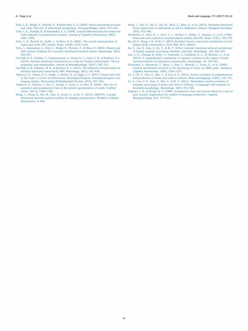
Tyler, L. K., Bright, P., Fletcher, P., & Stamatakis, E. A. (2004). Neural processing of nounsand verbs: The role of inflectional morphology. Neuropsychologia, 42(4), 512–523.
Tyler, L. K., Randall, B., & Stamatakis, E. A. (2008). Cortical differentiation for nouns andverbs depends on grammatical markers. Journal of Cognitive Neuroscience, 20(8),1381–1389.
Tyler, L. K., Russell, R., Fadili, J., & Moss, H. E. (2001). The neural representation ofnouns and verbs: PET studies. Brain, 124(8), 1619–1634.
Tyler, L., Stamatakis, E., Dick, E., Bright, P., Fletcher, P., & Moss, H. (2003). Objects andtheir actions: Evidence for a neurally distributed semantic system. Neuroimage, 18(2),542–557.
Van Dijk, K. R., Hedden, T., Venkataraman, A., Evans, K. C., Lazar, S. W., & Buckner, R. L.(2010). Intrinsic functional connectivity as a tool for human connectomics: Theory,properties, and optimization. Journal of Neurophysiology, 103(1), 297–321.
Van Dijk, K. R., Sabuncu, M. R., & Buckner, R. L. (2012). The influence of head motion onintrinsic functional connectivity MRI. Neuroimage, 59(1), 431–438.
Vigliocco, G., Vinson, D. P., Druks, J., Barber, H., & Cappa, S. F. (2011). Nouns and verbsin the brain: A review of behavioural, electrophysiological, neuropsychological andimaging studies. Neuroscience & Biobehavioral Reviews, 35(3), 407–426.
Vigliocco, G., Warren, J., Siri, S., Arciuli, J., Scott, S., & Wise, R. (2006). The role ofsemantics and grammatical class in the neural representation of words. CerebralCortex, 16(12), 1790–1796.
Wang, J., Wang, X., Xia, M., Liao, X., Evans, A., & He, Y. (2015). GRETNA: A graphtheoretical network analysis toolbox for imaging connectomics. Frontiers in HumanNeuroscience, 9, 386.
Wang, J., Zuo, X., Dai, Z., Xia, M., Zhao, Z., Zhao, X., et al. (2013). Disrupted functionalbrain connectome in individuals at risk for Alzheimer's disease. Biological Psychiatry,73(5), 472–481.
Warburton, E., Wise, R. J., Price, C. J., Weiller, C., Hadar, U., Ramsay, S., et al. (1996).Noun and verb retrieval by normal subjects studies with PET. Brain, 119(1), 159–179.
Xia, M. R., Wang, J. H., & He, Y. (2013). BrainNet Viewer: A network visualization tool forhuman brain connectomics. PLoS ONE, 8(7), e68910.
Xu, Y., Lin, Q., Han, Z., He, Y., & Bi, Y. (2016). Intrinsic functional network architectureof human semantic processing: Modules and hubs. Neuroimage, 132, 542–555.
Yan, C.-G., Cheung, B., Kelly, C., Colcombe, S., Craddock, R. C., Di Martino, A., et al.(2013). A comprehensive assessment of regional variation in the impact of headmicromovements on functional connectomics. Neuroimage, 76, 183–201.
Yokoyama, S., Miyamoto, T., Riera, J., Kim, J., Akitsuki, Y., Iwata, K., et al. (2006).Cortical mechanisms involved in the processing of verbs: An fMRI study. Journal ofCognitive Neuroscience, 18(8), 1304–1313.
Yu, X., Bi, Y., Han, Z., Zhu, C., & Law, S.-P. (2012). Neural correlates of comprehensionand production of nouns and verbs in Chinese. Brain and Language, 122(2), 126–131.
Yu, X., Law, S. P., Han, Z., Zhu, C., & Bi, Y. (2011). Dissociative neural correlates ofsemantic processing of nouns and verbs in Chinese—A language with minimal in-flectional morphology. Neuroimage, 58(3), 912–922.
Zingeser, L. B., & Berndt, R. S. (1988). Grammatical class and context effects in a case ofpure anomia: Implications for models of language production. CognitiveNeuropsychology, 5(4), 473–516.
H. Yang et al. Brain and Language 175 (2017) 29–41
41
Related Documents