Brain activity varies with modulation of dynamic pitch variance in sentence melody Martin Meyer, a,b, * Karsten Steinhauer, c,d Kai Alter, a Angela D. Friederici, a and D. Yves von Cramon a a Max-Planck-Institute of Cognitive Neuroscience, Leipzig, Germany b Department of Neuropsychology, University of Z€ urich, Treichlerstrasse 10, CH-8032 Z€ urich, Switzerland c Brain and Language Lab, Georgetown University, Washington DC, USA d School of Communication Sciences and Disorders, McGill University, Montreal, Canada Accepted 20 August 2003 Abstract Fourteen native speakers of German heard normal sentences, sentences which were either lacking dynamic pitch variation (flattened speech), or comprised of intonation contour exclusively (degraded speech). Participants were to listen carefully to the sentences and to perform a rehearsal task. Passive listening to flattened speech compared to normal speech produced strong brain responses in right cortical areas, particularly in the posterior superior temporal gyrus (pSTG). Passive listening to degraded speech compared to either normal or flattened speech particularly involved fronto-opercular and subcortical (Putamen, Caudate Nucleus) regions bilaterally. Additionally the Rolandic operculum (premotor cortex) in the right hemisphere subserved processing of neat sentence intonation. As a function of explicit rehearsing sentence intonation we found several activation foci in the left inferior frontal gyrus (BrocaÕs area), the left inferior precentral sulcus, and the left Rolandic fissure. The data allow several suggestions: First, both flattened and degraded speech evoked differential brain responses in the pSTG, particularly in the planum temporale (PT) bilaterally indicating that this region mediates integration of slowly and rapidly changing acoustic cues during comprehension of spoken language. Second, the bilateral circuit active whilst participants receive degraded speech reflects general effort allocation. Third, the differential finding for passive perception and explicit rehearsal of intonation contour suggests a right fronto-lateral network for processing and a left fronto-lateral network for producing prosodic information. Finally, it appears that brain areas which subserve speech (frontal operculum) and premotor functions (Rolandic operculum) coincidently support the processing of intonation contour in spoken sentence comprehension. Ó 2003 Elsevier Inc. All rights reserved. Keywords: Functional MRI; Dynamic pitch variation; Sentence prosody; Peri-sylvian cortex; Planum temporale; Frontal operculum; Rolandic operculum; Basal ganglia; Language and motor integration; Auditory rehearsal 1. Introduction Comprehending spoken language includes the de- coding of information from differing linguistic domains, e.g., semantics of words, thematic and structural rela- tions, as well as from nonlinguistic and linguistic acoustical cues, commonly referred to as Õprosody.Õ Prosody describes abstract phonological phenomena such as word stress, sentence accent, and phrasing and refers also to the phonetic attributes used to encode these abstract structures, i.e., intonation, amplitude, duration, etc. Listeners can elicit information from in- tonation, duration, and amplitude to help decode the syntactic and focus structure of the sentences they at- tend to (Steinhauer, 2003; Steinhauer, Alter, & Fried- erici, 1999). Thus, prosody has a linguistic function at many different levels. During speech comprehension it contributes to the interpretation of the linguistic signal. Modulation of prosodic parameters, i.e., of pitch accent, guides syntactic parsing even though pitch accent per se is not a syntactic property. Slow pitch movements which * Corresponding author. Fax: +41-1-634-4342. E-mail address: [email protected] (M. Meyer). 0093-934X/$ - see front matter Ó 2003 Elsevier Inc. All rights reserved. doi:10.1016/S0093-934X(03)00350-X Brain and Language 89 (2004) 277–289 www.elsevier.com/locate/b&l

Welcome message from author
This document is posted to help you gain knowledge. Please leave a comment to let me know what you think about it! Share it to your friends and learn new things together.
Transcript

Brain and Language 89 (2004) 277–289
www.elsevier.com/locate/b&l
Brain activity varies with modulation of dynamic pitchvariance in sentence melody
Martin Meyer,a,b,* Karsten Steinhauer,c,d Kai Alter,a Angela D. Friederici,a
and D. Yves von Cramona
a Max-Planck-Institute of Cognitive Neuroscience, Leipzig, Germanyb Department of Neuropsychology, University of Z€urich, Treichlerstrasse 10, CH-8032 Z€urich, Switzerland
c Brain and Language Lab, Georgetown University, Washington DC, USAd School of Communication Sciences and Disorders, McGill University, Montreal, Canada
Accepted 20 August 2003
Abstract
Fourteen native speakers of German heard normal sentences, sentences which were either lacking dynamic pitch variation
(flattened speech), or comprised of intonation contour exclusively (degraded speech). Participants were to listen carefully to the
sentences and to perform a rehearsal task. Passive listening to flattened speech compared to normal speech produced strong brain
responses in right cortical areas, particularly in the posterior superior temporal gyrus (pSTG). Passive listening to degraded speech
compared to either normal or flattened speech particularly involved fronto-opercular and subcortical (Putamen, Caudate Nucleus)
regions bilaterally. Additionally the Rolandic operculum (premotor cortex) in the right hemisphere subserved processing of neat
sentence intonation. As a function of explicit rehearsing sentence intonation we found several activation foci in the left inferior
frontal gyrus (Broca�s area), the left inferior precentral sulcus, and the left Rolandic fissure. The data allow several suggestions: First,
both flattened and degraded speech evoked differential brain responses in the pSTG, particularly in the planum temporale (PT)
bilaterally indicating that this region mediates integration of slowly and rapidly changing acoustic cues during comprehension of
spoken language. Second, the bilateral circuit active whilst participants receive degraded speech reflects general effort allocation.
Third, the differential finding for passive perception and explicit rehearsal of intonation contour suggests a right fronto-lateral
network for processing and a left fronto-lateral network for producing prosodic information. Finally, it appears that brain areas
which subserve speech (frontal operculum) and premotor functions (Rolandic operculum) coincidently support the processing of
intonation contour in spoken sentence comprehension.
� 2003 Elsevier Inc. All rights reserved.
Keywords: Functional MRI; Dynamic pitch variation; Sentence prosody; Peri-sylvian cortex; Planum temporale; Frontal operculum; Rolandic
operculum; Basal ganglia; Language and motor integration; Auditory rehearsal
1. Introduction
Comprehending spoken language includes the de-
coding of information from differing linguistic domains,
e.g., semantics of words, thematic and structural rela-
tions, as well as from nonlinguistic and linguistic
acoustical cues, commonly referred to as �prosody.�Prosody describes abstract phonological phenomena
such as word stress, sentence accent, and phrasing and
* Corresponding author. Fax: +41-1-634-4342.
E-mail address: [email protected] (M. Meyer).
0093-934X/$ - see front matter � 2003 Elsevier Inc. All rights reserved.
doi:10.1016/S0093-934X(03)00350-X
refers also to the phonetic attributes used to encodethese abstract structures, i.e., intonation, amplitude,
duration, etc. Listeners can elicit information from in-
tonation, duration, and amplitude to help decode the
syntactic and focus structure of the sentences they at-
tend to (Steinhauer, 2003; Steinhauer, Alter, & Fried-
erici, 1999). Thus, prosody has a linguistic function at
many different levels. During speech comprehension it
contributes to the interpretation of the linguistic signal.Modulation of prosodic parameters, i.e., of pitch accent,
guides syntactic parsing even though pitch accent per se
is not a syntactic property. Slow pitch movements which
278 M. Meyer et al. / Brain and Language 89 (2004) 277–289
extend over chunks of utterances longer than just onesegment constitute the intonation contour, i.e., speech
melody. In intonational languages such as German,
French, and English, speakers use different pitch
movements to signal questions vs. statements, where
questions are usually produced with a pitch rise at the
end of the utterance.
Recent brain imaging studies identified distinct brain
regions, especially in left peri-sylvian cortex subservingparticular aspects of sentence-level speech comprehen-
sion (Bookheimer, 2002; Friederici, 2002; Friederici &
Alter, this volume; Friederici, R€uschemeyer, Hahne, &
Fiebach, 2003b; Kaan & Swaab, 2002). Neither the
spectral and temporal features that carry the relevant
attributes of prosody, nor the cerebral substrates of
prosodic parameters (e.g., speech melody) available in
spoken language, however, have been exactly identifiedso far (Lakshminarayanan et al., 2003). Patients suffer-
ing from either left or right hemispheric lesions showed
comprehension deficits for linguistic intonation giving
credence to the view that prosodic processing is medi-
ated by both hemispheres (Pell & Baum, 1997). This
view lends support to a recent model which proposed
that prosodic functions are not localized in either the
right or left hemisphere (Dogil et al., 2002). Accordingto this model the prosodic frame length rather than
prosody per se dictates the lateralization. Prosodic fea-
tures which require a short address frame, (e.g., focused
syllable) are lateralized differently as compared to pro-
sodic elements comprising a long address frame, (e.g.,
intonational phrases). In complement to this, the
�asymmetric sampling in time hypothesis (AST)� arguesin favour of a functional hemispheric difference whichderives from the manner in which auditory signals are
processed at an early stage (P€oppel, 2003). This hy-
pothesis holds that speech processing even at an early
stage generally occurs symmetrically in both the left and
the right hemisphere. However, the signal is elaborated
differentially in the time domain. Left non-primary au-
ditory areas extract information from short (20–50ms),
and right hemisphere homologues pick up informationfrom long temporal integration windows (150–250ms).
Linguistically, the �AST� hypothesis suggests that pro-
sodic processing at the level of lexical stress is lateralized
to the left hemisphere. Additionally, right temporal re-
gions are more proficient at processing prosody at the
level of intonation contour. Empirical evidence sup-
porting this view comes from a recent lesion study on
patients who underwent a resection of right superiortemporal areas and unveiled an impairment at using
pitch contour information (Johnsrude, Penhune, & Za-
torre, 2000). Additionally, an fMRI-study from our lab
demonstrated a stronger right hemisphere involvement,
particularly in the right planum temporale (PT) and the
right Rolandic operculum (ROP) in processing slow
prosodic modulations, (e.g., pure sentence intonation)
(Meyer, Alter, Friederici, Lohmann, & von Cramon,2002). In this study, volunteers heard either normal
sentences or pure sentence intonation degraded speech.
The latter condition was derived from normal sentences
which underwent a special filtering procedure (PURR-
Filter (cf. Section 2) which removes segmental, but
preserves prosodic parameters, (i.e., intonation, dura-
tion, and amplitude envelope) which represent speech
melody. However, in this study we contrasted normalspeech to degraded speech both comprising intonation
contour. In order to elucidate the cerebral regions sub-
serving prosodic variation in spoken language we de-
signed a new fMRI-study.
1.1. The present study
Here, participants were presented with three condi-tions, normal sentences, degraded speech, and flattened
speech. The latter was derived from normal sentences by
selectively removing variations in the intonation contour
(fundamental frequency, or F0), but preserving other
prosodic parameters, (segmental/phonemic information,
amplitude envelope, and duration). This design allowed
us to contrast speech signals comprising intonation
(normal speech, degraded speech) to speech signalswithout any sentence intonation (flattened speech).
Based on the results of our previous study we might
expect larger activation for normal sentences and flat-
tened speech in the peri-sylvian cortex, namely the left
inferior frontal gyrus (IFG) and the superior temporal
gyrus (STG) bilaterally when compared to degraded
speech, since the latter lacks any lexical and syntactic
information. We predicted stronger brain responses todegraded speech in the fronto-opercular areas including
the basal ganglia bilaterally reflecting an interaction
between speech processing and related effort (Meyer
et al., 2002). Additionally we expected activation in the
right inferior premotor cortex for degraded, but not for
normal speech pointing to a role this particular area
plays in implicit sub-articulation whilst participants
heard speech melody. As phonological working memoryhas been found to involve frontal structures particularly
of the left hemisphere (Paulesu, Frith, & Frackowiak,
1993), the right hemisphere activation may shift to left
homologues when volunteers perform an explicit re-
hearsal task. We therefore asked the subjects to explic-
itly hold in memory intonation contour. Separate
analyses of the time windows for passive listening and
for active rehearsal of the sentence contour should en-able to distinguish between the networks for perceiving
and reproducing sentential melodies. Finally, as far as
the two models mentioned above are concerned we in-
tended to test whether the planum temporale (PT) can
be identified as the brain region that mediates the inte-
gration of slowly as well as rapidly changing acoustic
cues during comprehension of spoken language. Even

M. Meyer et al. / Brain and Language 89 (2004) 277–289 279
though the functional role the PT plays has not yet beenspecified there is some evidence that it mediates pro-
cesses of phonetic integration. Generally, the PT is as-
sumed to constitute any complex auditory processing,
including speech, at an early stage including speech
(Binder et al., 2000; Hickok & P€oppel, 2000). In a recent
review paper it has been suggested that the planum
temporale acts as a computational hub for the integration
of spectrotemporal patterns (Griffiths & Warren, 2002).J€ancke, W€ustenberg, Scheich, & Heinze (2002) recently
provided compelling evidence for the view that the PT is
not particularly involved in phonetic analysis per se, but
is rather specialized for the analysis of rapidly changing
cues. Here, we would like to augment this view by elu-
cidating the role of the PT regarding slowly as opposed
to rapidly changing acoustic cues in spoken language. If
the PT is crucial for integration of slow changes (i.e., F0variations), we may expect larger PT activity for pro-
sodically adverse stimuli degraded speech, flattened
speech when compared to normal sentences, reflecting
the violation of prosodic integrity.
2. Materials and methods
2.1. Subjects
Fourteen native German volunteers (6 male, age
range 18–27, mean 22.7) participated in the study after
giving written informed consent in accordance with the
guidelines approved by the Ethics Committee of the
Leipzig University Medical Faculty. Volunteers were
assessed as right-handed according to the Edinburgh
Handedness Inventory (Oldfield, 1971). Participants had
no hearing or neurological disorders and normal struc-
tural MRI scans. They had no prior experience with the
task and were not familiar with the stimulus material.
2.2. Stimuli
The German sentence material consisted of 144stimuli (72 natural sentences and 72 artificially manip-
ulated sentences) varying pitch parameters. All speech
signals were controlled for their duration and loudness.
1. Normal speech. This condition includes three sub-
types of sentences which differ slightly in their intona-
tional contour.1 All sentences were infinitival sentences
containing a control verb such as �promises� and an in-
finitive (see example below) varying the position ofsentence accents which either appeared on the first noun
phrase, on the second noun phrase or on the first verb.
Since the distinct sub-conditions are not assumed to
yield substantial hemodynamic differences, all sub-con-
1 Example sound files are available at ‘‘http://www.psychologie.
unizh.ch/neuropsy/home_mmeyer/YBLRN2956’’.
ditions were included for reasons of a highly desirablecontrolled variability of natural speech input.
PETER verspricht ANNA zu ARBEITEN und das B€uro zu
putzen.
PETER promises ANNA to WORK and to clean the office.
2. Flattened pitch. The flattened pitch condition wasderived by using a special speech re-synthesis procedure2
in order to generate a violation of the sentence prosody.
All normal sentences were manipulated by re-synthesis.
The manipulation is based on an algorithm (WinPitch,
cit.) allowing the re-adjustment of the F0-contour. Thespeech re-synthesis was carried out at the mean F0-valueof the speaker�s voice, i.e., at 200Hz by applying a
simple linear function between onset and offset of eachsentence. This procedure removes the original geomet-
rical characteristics such as linguistically triggered pitch
accents and the declination line. In addition, global slow
modulations had been removed yielding a monotonous
sounding sentence. These modulations concerned the
pitch contour varying over domains which size is larger
than one syllable. Apart from the pitch contour (F0) there-synthesis procedure preserves both syllabic and rap-idly changing sub-syllabic varying information (e.g.,
amplitude envelope, duration) in the speech signal.
Fig. 1 illustrates that the resulting signal does not con-
tain any dynamic pitch variations, i.e., no peaks and
valleys, and is thus flattened globally. On the other hand
the manipulated speech signal contains all information
necessary to perform phoneme detection, lexical access,
syntactic and semantic processing.3. Degraded speech. To achieve a speech signal which
lacks lexical and syntactic information normal sentences
were subjected to PURR-filtering procedure (Sonntag &
Portele, 1998). PURR-manipulated speech stimuli con-
tain only prosodic parameters such as intonation, du-
ration, amplitude envelope, and the second and third
harmonics. From a linguistic point of view, these pho-
netic attributes represent speech melody, the distribu-tion and type of pitch accents and boundary markers of
prosodic domains. In more perceptual terms degraded
speech could be described to sound like speech melody
listened to from behind a door. Fig. 1 demonstrates that
acoustic information exceeding the 3rd harmonic had
been removed from the speech signal.
For technical reasons it was not possible to add a
fourth condition [-prosody] and [-segmental] which issupposed to complete the design as the PURR filter was
disrupting the re-synthesized speech files. All normal
sentences were recorded with a trained female speaker
in a sound proof room (IAC) at a 16 bit/41.1 kHz sam-
pling rate and then digitised. Both flattened and de-
graded speech were derived from normal sentences.
Since the PURR-procedure allows for only a 16 bit/
2 WinPitch 1.89, Pitch Instruments, Toronto, Ont., Canada.
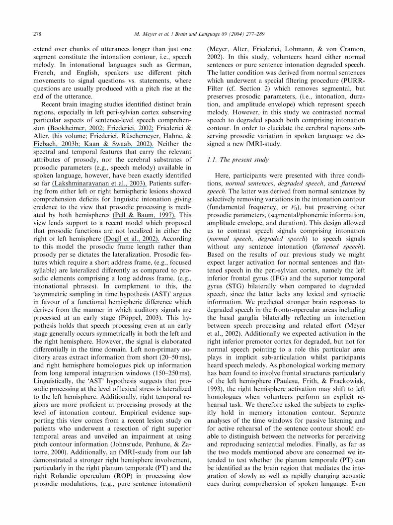
Fig. 1. Acoustic analyses of flattened and degraded speech. Speech signals of sentence derived from resynthesis revealed flattened pitch contour (A),
but unaltered wide band spectrum of frequencies (0–10kHz) (B). The artificial re-synthesis eliminates prosodic cues in an intonational language such
as German, i.e., the typical rising and falling F0 pattern over the whole sentence. The right side of the figure shows normal sentence intonation for
degraded speech (A) whilst the wide band spectrogram illustrates reduced frequency information of a degraded sentence (B).
280 M. Meyer et al. / Brain and Language 89 (2004) 277–289
16 kHz sampling rate, all signals to be presented in the
experiment were downsampled to avoid quality differ-ences across conditions. All stimuli except degraded
signals were normalized in amplitude (70%). Since the
latter were limited in bandwidth compared to the other
three conditions a stronger normalization (85%) was
necessary to guarantee equal loudness. The mean length
of the sentences in the �natural speech� condition was
�SD, 3.61� 0.23 s, in the �flattened pitch� condition
�SD, 3.85� 0.28 s, and in the delexicalized �degradedspeech� condition �SD, 3.81� 0.28 s.
2.3. Procedure
Participants heard the stimuli occurring in pseudo-
random order.3 Sentences were not repeated during the
experiment. The sounds were presented binaurally via
specially constructed headphones. The study employed asingle-trial design to enable an event-related analysis
(D�Esposito, Zarahn, & Aguirre, 1999). To allow the
hemodynamic response to return to baseline level ade-
quately each sentence was followed by an Inter-Trial-
Interval lasting twelve seconds until the onset of the
following trial. The entire experimental session consisted
of two blocks (runs), each comprising 72 trials.
2.4. Task
Participants were asked to perform in a prosody
comparison task. First, they had to closely listen to the
3 A comprehensive report on methods and procedure is avail-
able at ‘‘http://www.psychologie.unizh.ch/neuropsy/home_mmeyer/
YBLRN2956’’.
sentence intonation and to rehearse this percept during
the Inter-Stimulus Interval. Whenever a trial was un-predictably initially marked as a �Compare� trial, subjectshad to judge whether the current and the preceding
sentence shared the same prosodic pattern (yes/no
judgement). The number of �Compare� trials was reducedto 20% (i.e., 7 trials in each condition) in order to avoid
a general influence of this matching task on sentence
perception and not to overtax working memory. �Com-
pare� trials were included in the design only to guaranteethat language stimuli were attended to appropriately.
However, �Compare� trials were excluded from further
analysis in order to avoid confounding between re-
hearsal and template matching processes. At the begin-
ning of the remaining 80% of trials (28 trials in each
condition), the presentation of a sine wave tone
(1000Hz, 100ms) indicates that no explicit comparison
was required.
2.5. MRI data acquisition
MRI data were collected at 3T using a Bruker 30/100
Medspec system (Bruker Medizintechnik GMBMBH, Ett-
lingen, Germany). For each subject, structural and
functional (echo-planar) images were obtained from
eight axial slices parallel to the plane intersecting theanterior and posterior commissures (AC–PC plane). The
most inferior slice was positioned below the AC–PC
plane and the remaining seven slices extended dorsally.
The whole range of slices comprised an anatomical
volume of 46mm and covered all parts of the peri-syl-
vian cortex and extended dorsally to the intraparietal
sulcus. After defining the slices� position a set of two-
dimensional T1 weighted anatomical images (IR-RARE
M. Meyer et al. / Brain and Language 89 (2004) 277–289 281
sequence: TE¼ 20ms, TR¼ 3750ms, in-plane resolu-tion 0.325mm2) were collected in plane with the echo-
planar images, to align the functional images to the
3D-images. A gradient-echo EPI sequence was used with
a TE 30ms, flip angle 90�, TR 2 s, acquisition bandwidth
100 kHz. The matrix acquired was 64� 64 with a FOV
of 19.2 cm, resulting in an in-plane resolution of
3mm� 3mm. The slice thickness was 4mm with an
interslice gap of 2mm. In a separate session high reso-lution whole-head 3D MDEFT brain scans (128 sagittal
slices, 1.5mm thickness, FOV 25.0� 25.0� 19.2 cm,
data matrix of 256� 256 voxels) were acquired addi-
tionally for reasons of improved localization (Lee et al.,
1995; Ugurbil et al., 1993).
2.6. Data analysis
The data processing was performed using the soft-
ware package LIPSIA (Lohmann et al., 2001). Func-
tional data were corrected for slice-time acquisition
differences using sinc-interpolation. In addition, the data
were corrected for motion artefacts. Signal changes and
baseline-drifts were removed by applying a temporal
high-pass filter to remove frequencies lower than 1/48Hz
(2.5 times the length of one complete oscillation). Theanatomical slices were co-registered with the full-brain
scan that resided in the stereotactic coordinate system
and then transformed by linear scaling to a standard
size. The transformation parameters obtained from this
step were subsequently applied to the functional slices so
that the functional slices were also registered into the
stereotactic space. The statistical analysis was based on
a least squares estimation using the general linear model(GLM) for serially autocorrelated observations (Agu-
irre, Zarahn, & D�Esposito, 1997; Bosch, 2000; Friston,1994; Zarahn, Aguirre, & D�Esposito, 1997). The model
equation including the observation data, the design
matrix, and the error term, was linearly smoothed by
convolving it with a Gaussian kernel of dispersion of 4 s
FWHM. The contrast between the different conditions
was calculated using the t statistics. Subsequently, t
values were converted to Z scores. As the individual
functional datasets were all aligned to the same stereo-
tactic reference space a group analysis of fMRI-data was
performed by averaging individual Z maps. The average
SPM was multiplied by a SPM correction factor of
square root of the current number of subjects (N ¼ 14)
(Bosch, 2000). For the purpose of illustration, averaged
data were superimposed onto one normalized 3DMDEFT standard volume (see Fig. 2).
To identify differential brain systems subserving
passive perception and active rehearsal of intonation
contour we modelled the hemodynamic response for the
time window during stimulus presentation (�passiveperception�) and mental task performance (active re-
production) separately.
3. Results
In this section we report brain responses collected
whilst volunteers were listening to speech stimuli (A, B,
and C), or rehearsed sentence intonation (D). Fig. 2 and
Tables 1–4 display the main results of the contrasts
between conditions.
3.1. Normal speech vs. degraded speech
This contrast yielded a pattern of functional brain
activation which is almost identical to the results de-
scribed by Meyer et al. (2002). Listening to normal
sentences involves the left IFG (pars triangularis), large
parts of the left superior temporal gyrus (STG) and
superior temporal sulcus (STS), the left temporo-occip-
ital-parietal (TOP) junction area, and yields only smallright hemisphere responses in the primary auditory ar-
eas (cf. Fig. 2A and Table 2). When compared to normal
sentences degraded speech produced stronger functional
activity in the left and right posterior superior temporal
region, particularly in the planum temporale. Further
areas that show stronger responses to degraded speech
included the left middle frontal gyrus (MFG), the infe-
rior parietal lobe (IPL), the right Rolandic operculum(ventral premotor cortex), and the deep frontal oper-
culum bilaterally. Additionally the basal ganglia (Puta-
men, Caudate Nucleus) turned out to be more strongly
involved whilst participants heard pure intonation
contour.
3.2. Normal speech vs. flattened speech
Several areas showed stronger activation for flattened
speech relative to normal speech including large por-
tions of the right posterior STG and the planum tem-
porale bilaterally (cf. Fig. 2B and Table 2). Further
activation was found in the left MFG and in the left
IPL.
3.3. Flattened speech vs. degraded speech
Like normal speech hearing flattened speech relative
to degraded speech revealed stronger activity in the left
IFG (pars triangularis) and along the entire STG/STS
bilaterally, and in the left temporo–occipital–parietal
(TOP) junction area, (cf. Fig. 2C, Table 3). Analogous
to contrast Fig. 2A the left and right frontal operculum,
the right Rolandic operculum, the right planum parie-tale, and the basal ganglia responded more strongly to
degraded relative to flattened speech.
3.4. Rehearsal: Intonation vs. flattened speech
Fig. 2D and Table 4 shows that active rehearsal of
sentence intonation involved areas only in the left
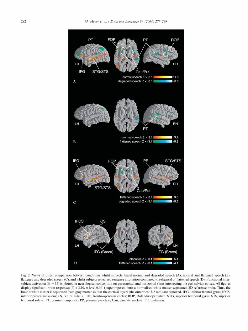
Fig. 2. Views of direct comparison between conditions whilst subjects heard normal and degraded speech (A), normal and flattened speech (B),
flattened and degraded speech (C), and whilst subjects rehearsed sentence intonation compared to rehearsal of flattened speech (D). Functional inter-
subject activation (N ¼ 14) is plotted in neurological convention on parasagittal and horizontal slices intersecting the peri-sylvian cortex. All figures
display significant brain responses (ZP 3:10, a-level 0.001) superimposed onto a normalised white-matter segmented 3D reference brain. Thus, the
brain�s white matter is separated from gray matter so that the cortical layers (the outermost 3–5mm) are removed. IFG, inferior frontal gyrus; IPCS,
inferior precentral sulcus; CS, central sulcus; FOP, fronto-opercular cortex; ROP, Rolandic operculum; STG, superior temporal gyrus; STS, superior
temporal sulcus; PT, planum temporale; PP, planum parietale; Cau, caudate nucleus; Put, putamen.
282 M. Meyer et al. / Brain and Language 89 (2004) 277–289

Table 1
Normal speech vs. degraded speech
Location BA Left hemisphere Right hemisphere
Z score x y z Z score x y z
Normal speech> degraded speech
IFG 45 7.1 )47 25 5 — — — —
IFG 47 — — — — 4.5 40 19 )10mid./post. STG/STS 22/21 10.3 )53 )11 )6 9.3 51 )8 0
Tha — 4.0 )11 )32 7 — — — —
post. GC 31 4.75 )4 )51 30 — — — —
Degraded speech> normal speech
MFG 46 )5.8 )45 43 10 — — — —
FOP — )5.7 )39 13 9 )5.9 26 16 7
ROP 44/6 — — — — )5.4 44 6 10
PrCG 6 )4.6 )53 2 15 — — — —
mid. CG — — — — — )4.06 0 )24 29
post. STG/PT 42/22 )4.4 )51 )33 18 )7.5 56 )34 21
IPL 40/19 )4.4 )29 )48 40 )3.7 37 )40 41
In this table and in Tables 2–4 results of direct comparison of different conditions are listed. Z scores indicate the magnitude of statistical
significance. Localization is based on stereotactic coordinates (Talairach & Tournoux, 1988). These coordinates refer to the location of maximal
activation indicated by the Z score in a particular anatomical structure. Distances are relative, to the intercommissural (AC—PC) line in the
horizontal (x), anterior–posterior (y), and vertical (z) directions. Functional activation was thresholded at jZjP 3:1. The table only lists activation
clusters exceeding a minimal size of 100 voxels. IFG, inferior frontal gyrus; MFG, mid frontal gyrus; aINS, anterior insula; FOP, frontal operculum;
ROP, Rolandic operculum; PrCG, precentral gyrus; IPCS, inferior precentral sulcus; CS, central sulcus (Rolandic fissure); STG, superior temporal
gyrus; STS, temporal sulcus; MTG, middle temporal gyrus; PT, planum temporale; PP, planum parietal; CG, cingulate gyrus; IPL, inferior parietal
lobe; SPL, superior parietal lobe; Tha, Thalamus; Cau, caudate head; Put, putamen.
Table 2
Flattened pitch vs. normal speech
Location BA Left hemisphere Right hemisphere
Z score x y z Z score x y z
Flattened pitch> normal speech
MFG 46 )3.9 )45 43 11 — — — —
IFG 45 — — — — )4.4 39 23 8
ROP 44/6 — — — — )3.7 41 )1 6
mid STG 42 — — — — )4.8 50 )28 16
post. STG 42/22 )4.5 )58 )33 15 )4.1 38 )39 15
PP 40 — — — — )4.7 56 )42 23
IPL 40/19 )4.0 )30 )48 41 )3.9 34 )58 43
SPL 40/7 )3.8 )51 )54 48 — — — —
Functional activation indicated separately for contrasts between conditions. For explanations see Table 1.
M. Meyer et al. / Brain and Language 89 (2004) 277–289 283
hemisphere, namely in Broca�s area, in the ventral pre-
motor and presumably motor cortex. Plausibly, re-
hearsing flattened speech did not produce any significant
activity.
4. Discussion
The study examined whether we can identify brain
regions particularly mediating the processing of into-
national information. Therefore we delivered to partic-
ipants sentences that either carried segmental cues but
lacked dynamic pitch variations (flattened speech), solely
suprasegmental cues (degraded speech), or both seg-
mental and suprasegmental information (normal
speech).
Generally, we found auditory speech stimuli to acti-
vate brain areas including the bilateral peri-sylvian
cortex. However, the different conditions produced dif-
ferential activation patterns pointing to distinct brain
regions which may subserve differential aspects of
speech processing. Processing both normal speech andflattened speech which comprise proper lexical and
syntactic information corresponded to brain activation
in the left IFG, the bilateral STG/STS, and the left TOP
junction area (2A and 2C). The left IFG and the STG
have been comprehensively described as mediating
normal sentence comprehension in the auditory domain

Table 3
Flattened pitch vs. degraded speech
Location BA Left hemisphere Right hemisphere
Z score x y z Z score x y z
Flattened pitch> degraded speech
IFG 47 — — — — 4.0 35 30 )11IFG 45 5.4 )51 24 5 — — — —
ant. STG/MTG 22/21 8.5 )53 )11 )6 9.0 51 )7 )1CS 4/3 — — — — 3.9 56 )8 23
post. MTG 21 — — — — 4.8 55 )49 9
post. STS 39 4.5 )42 )67 24 — — — —
post. CG 31 — — — — 3.8 3 )52 24
Tha — 4.1 )15 )30 3 — — — —
Degraded speech> flattened pitch
aINS/FOP — )3.9 )43 13 8 )5.4 26 16 7
ROP 44/6 — — — — )3.6 43 5 10
Cau — )4.9 )17 14 9 )4.5 14 13 5
Put — — — — )5.2 18 5 8
post. STG/PP 42/22 — — — — )4.2 54 )35 20
Functional activation indicated separately for contrasts between conditions. For explanations see Table 2.
Table 4
Pitch variation speech vs. flattened speech
Location BA Left hemisphere Right hemisphere
Z score x y z Z score x y z
Pitch variation> flattened speech
IFG 44 4.7 )46 9 11 — — — —
IPCS 6 4.69 )52 0 20 — — — —
CS 4 4.0 )46 )14 32 — — — —
MFG 9 4.53 )37 33 26 4.45 28 30 29
Functional activation during rehearsal interval indicated separately for contrasts between sentence stimuli with and without dynamic pitch
variation. For explanations see Table 2.
284 M. Meyer et al. / Brain and Language 89 (2004) 277–289
(Friederici, 2002; Friederici & Alter, this volume; Kaan
& Swaab, 2002). We also found stronger responses to
normal and flattened speech in left and right STG/STS
(2A and C). Whilst the left STG/STS has been associ-
ated with a �pathway for speech comprehension� (Binderet al., 2000; Scott, Blank, Rosen, & Wise, 2000) the
stronger right hemisphere activity for normal and flat-
tened speech demands an explanation. Degraded speechcomprises attenuated spectral complexity. Therefore we
argue that the right STG/STS activity accounts for
processing spectral information. Additionally both
normal and flattened speech activated the left TOP
junction area. This finding accords with a recent model
by Hickok & P€oppel (2000) who proposed that the TOP
junction area constitutes an auditory–conceptual inter-
face. In contrast to degraded speech, processing normaland flattened speech requires access to the mental lexi-
con. In terms of the present study, activity in the TOP
junction area reflects access to meaning-based repre-
sentations during sentence comprehension. Additionally
to normal sentence comprehension which more strongly
recruited left brain regions the study revealed more sa-
lient brain responses in right peri-sylvian areas whilst the
volunteers heard phonetically incongruent sentences ei-
ther lacking dynamic pitch contour (flattened speech) or
comprising melodic intonation (degraded speech). In a
cross-linguistic study which examined the temporal in-
tegration of speech prosody in native speakers of Chi-
nese and English Gandour and colleagues demonstrated
that local pitch contours associated with tones are pro-
cessed in the left hemisphere whereas global pitch con-tours representing intonation are processed in the right
hemisphere (Gandour et al., 2003). The functional
rightward asymmetry was even more prominent when
native speakers of English heard pure intonation
contours (hummed versions of Chinese sentences).
Furthermore the authors propose that native speakers
of intonational languages (like English) are more able to
discern pitch contours which span over longer temporaldomains whereas native speakers of tonal languages
(like Chinese and Thai) are more primed to picking up
pitch contours which span over shorter temporal do-
mains. These observations in compliance with our
present data partially agree with several recent neuro-
cognitive models that sketched a bilateral cerebral im-
plementation of speech perception (Friederici, 2002;
M. Meyer et al. / Brain and Language 89 (2004) 277–289 285
Hickok & P€oppel, 2000) with the right hemisphereplaying a prominent role in processing intonation (Dogil
et al., 2002; Friederici & Alter, this volume; P€oppel,2003). Building on the results of the present study we
can argue against recent views proposing a universal
right hemisphere superiority for propositional prosody
per se (Weintraub & Mesulam, 1981), but we can sup-
port the view that bilaterally distributed mechanisms
may constitute the ability to decode the melodic line ofspeech (Pell & Baum, 1997). However, it appears that
hemispheric specialization is dependent on the �prosodicneeds� of a particular language (Gandour et al., 2002,
2003) and to what extent spectral and temporal pa-
rameters shape the linguistic and emotional interpreta-
tion of the received speech (Lakshminarayanan et al.,
2003).4
Nonetheless, the present finding needs a comprehen-sive discussion: Normal speech signals, (i.e., a sentence)
comprise rapidly changing (formant transitions, high
frequencies) and slowly changing (syllable-seized
changing intonation, fundamental frequency) acoustic
features. In flattened speech we primarily removed slow
modulations of the pitch contour resulting in a monot-
onous sounding utterance. Degraded speech primarily
lacks rapidly changing cues. Brain responses to flattenedand degraded speech were not identical but overlapped
in the posterior STG bilaterally, particularly in the
planum temporale (PT) (2A and B). Functionally, the
PT corresponds to auditory association cortices that are
important in higher order processing of auditory lan-
guage input (Foundas, Leonard, Gilmore, Fennel, &
Heilman, 1994). Morphologically, right-handers dem-
onstrate a significant volumetric PT leftward asymmetry(Foundas, Leonard, & Hanna-Pladdy, 2002; Steinmetz,
Volkmann, J€ancke, & Freund, 1991). However, it ap-
pears that this observed leftward asymmetry reflects a
difference in morphology rather than size of the left and
right PT (Westbury, Zatorre, & Evans, 1999). There has
been an initial tendency to consider the differential
morphologic characteristics of the left PT as the likely
neuroanatomical substrate of language. Meanwhile re-cent findings give argument in favour of the left PT
mediating the analysis of rapidly changing cues relative
to language or even phonetic analysis per se (Celsis
et al., 1999; J€ancke et al., 2001, 2002). Complementary,
a functional rightward asymmetry to extended relative
to rapid formant transitions has been proposed (Belin
et al., 1998). Corroborating evidence stems from several
4 Several recent brain imaging studies tried to identify the brain
regions which are associated with processing emotionally toned speech.
Whilst some studies report stronger right hemisphere responses to
emotional tone available in spoken utterances (Buchanan et al., 2000;
George et al., 1996; Stiller et al., 1997; Wildgruber, Pihan, Erb,
Ackermann, & Grodd, 2002), there are other studies which argue
against a functional rightward asymmetry for emotional prosody
(J€ancke, Buchanan, Lutz, & Shah, 2001; Kotz et al., 2003).
studies that implicate a functional designation of com-puting harmonic information to the right hemisphere
and temporal cues to the left hemisphere (Johnsrude,
Zatorre, Milner, & Evans, 1997; Zatorre & Belin, 2001;
Zatorre, Belin, & Penhune, 2002). It appears that this
neurofunctional assignment holds more for more in-
ferolaterally situated parts of the STG/STS rather than
the PT. Since both flattened and degraded speech acti-
vated both the left and right PT our data do not fullysupport the notion of the left PT processing only rapidly
changing cues and the right PT solely processing slowly
changing cues. The present data rather support a view
which suggests the left and right PT as governing the
integration of rapidly and slowly changing acoustic cues
during speech comprehension. Thus, the starker re-
sponses to flattened or degraded speech are caused by
the phonetically degraded inflowing segmental and su-prasegmental units and reflect detection and compen-
sation of incongruent acoustic information. Our view of
the PT constituting an integrating mechanism is con-
sistent with a recent review article describing the PT as a
�computational hub� which matches spectrotemporal
acoustic patterns (Griffiths & Warren, 2002). The right
planum parietale (PP) also brought on an increase in
activity for impoverished speech, i.e., when degradedand flattened speech is compared to normal speech.
However, based on the current data it cannot be con-
cluded as to whether attentional demands account for
increase in activity observed in the PP (J€ancke et al.,
2001, 1994) or as to whether the right posterior Sylvian
Fissure is generally more concerned with slow prosodic
modulations as available in degraded speech.
In addition to activity in the PT and PP, we found anensemble of bilateral fronto-opercular areas including
basal ganglia and the right ROP more strongly re-
sponding to degraded speech when compared to normal
and flattened speech (2A and 2C) which is a replication
of the results described by Meyer et al. (2002). Briefly
summarized, we suggest that activity in the deep fronto-
opercular cortex reflects an interaction between pro-
cessing unintelligible speech and related effort. We willbe discussing the functions of the different brain struc-
tures in turn, starting with the basal ganglia. Further-
more, we will be discussing the role the intrasylvian
cortex may play during the perception of degraded
speech and its implication for speech motor control.
The data exposed an involvement of basal ganglia
(Putamen, Caudate Nucleus) in processing degraded
relative to normal and flattened speech (see Figs. 2A andC). In combination with the ventral striatum the Puta-
men and the Nucleus Caudatus form the neostriatum
(NS) (Murdoch, 2001). The NS is considered to be a
�multi laned pathway� which integrates diverse input
from the entire cerebral cortex and projects back to
cortical areas via thalamic regions (�basal ganglia-tha-lamicocortical circuits�). Traditionally, there is an

286 M. Meyer et al. / Brain and Language 89 (2004) 277–289
understanding of the basal ganglia playing a role in se-quencing and generating motor functions, particularly
in articulation since the Caudate Nucleus projects to the
inferior ventral premotor cortex which governs supr-
alaryngeal functions of tongue, lips, and jaws (Pickett,
Kuniholm, Protopapas, Friedman, & Lieberman, 1998).
Wise, Murray, & Gerfen (1996) outlined a mechanism
for the basal ganglia�s proposed role in rule potention.
According to this mechanism the basal ganglia activa-tion increase when to alter routine behaviour is needed.
However, the basal ganglia have also been associated
more specifically with functions related to generating,
encoding, modifying, and transferring linguistic infor-
mation (Crosson, 1992). Both views are principally
consistent with the present data. With respect to the
first, the presentation of pure sentence melody might
mean an alteration of normal processing routines whichis reflected by increased basal ganglia activity. The sec-
ond view receives some support from clinical studies
which examined sentence comprehension in patients
suffering from brain lesions and from Parkinson�s dis-
ease, respectively (Friederici, Kotz, Werheid, Hein, &
von Cramon, 2003a; Friederici, von Cramon, & Kotz,
1999). This data suggests that deficient function of the
basal ganglia affects integrational processes which occurat a late stage during sentence comprehension. Here, we
observed that the basal ganglia brought on stronger
activity for pure prosodic contour relative to normal
and flattened speech comprising syntactic information
which might reflect difficulties in integrating segmental
and suprasegmental units. Recent lesion studies also
speak to the issue of basal ganglia involvement in
prosody. These studies provide consistent evidence thatdamage to the basal ganglia causes prosodic compre-
hension deficits (Kempler & Van Lancker, 2002; Lloyd,
1999), particularly when patients were presented with
nonsense sentences (Pell, 1996) or degraded speech
(Speedie, Brake, Folstein, Bowers, & Heilman, 1990).
Even though these observations are not completely in
harmony they might be reconciled by a more general
view which proposes that the basal ganglia performtemporal binding necessary for any kind of integrational
sequencing, that is timing and ordinality in higher cog-
nition including language functions (Pickett et al., 1998).
The authors purport that sequencing can be considered
as an underlying device essential for syntax, prosody,
and cognition. To support this view the authors report a
case study of a patient with damage to Putamen and
Caudate Nucleus who demonstrates deficient syntacticprocessing and disturbed prosodic production. An al-
ternative explanation for the stronger basal ganglia ac-
tivation in degraded speech also considers the stronger
activity in the right ROP whilst participants passively
perceived intonation contour (2A). Meyer et al. (2002)
recently reported selective responses to intonation con-
tour in this particular area combined with stronger basal
ganglia activity. Several recent neuroimaging studiesassociate the ROP with covert speech production and
both covert and overt singing (Perry et al., 1999; Riec-
ker, Ackermann, Wildgruber, Dogil, & Grodd, 2000;
Wildgruber, Ackermann, Klose, Kardatzki, & Grodd,
1996). Here, subjects were asked to carefully listen to
sentence melodies and to silently rehearse these melodies
for several seconds. Plausibly, this representation in-
duced sub-articulatory processes which might accountfor the right premotor and basal ganglia activation.
Corroborating view comes from a recent brain imaging
study which also reported premotor activation whilst
participants processed degraded speech which involved
covert speech processing and consequently strategies
inducing sub-articulation (Kiehl, Laurens, & Liddle,
2002).
From the point of an integration of language andmotor networks it is notable that a network of areas
which have been identified to process auditory input,
has also been described to support producing auditory
output (Ackermann & Riecker, this volume; Dogil
et al., 2002; Indefrey et al., 2001; Indefrey, Hellwig,
Herzog, Seitz, & Hagoort, this volume). Clinical and
neuroimaging studies suggest that a similar network
subserves the production and perception of prosody(Dogil et al., 2002; Pell & Baum, 1997). However, there
seems no single area which can be considered as an
accomodation of the prosody generator. It rather seems
that differential brain areas contribute to the produc-
tion of prosodically modulated speech dependent on
the prosodic frame length (Behrens, 1989; Dogil et al.,
2002) with the left hemisphere subserving prosodic
processing on the syllable level and the right hemi-sphere supporting processing on the level of intona-
tional phrases.
In a recent fMRI study Ackermann and Riecker
demonstrated that (overt) speech tasks activated the left
anterior insula whilst (overt) reproduction of nonlyrical
tune evoked stronger responses in the right anterior in-
sular cortex (Ackermann & Riecker, this volume) which
led the authors to suggest a distributional operationalmode whereby the production of segmental information
recruits the left anterior insula and the production of
intonation contours engages the contralateral homo-
logue. Even though Ackermann and Riecker conclude
that activation of the intrasylvian cortex is tied to overt
task performance, probably reflecting the coordination
of the muscles engaged in articulation and phonation,
there is also evidence in favour of the (right) anteriorinsula as mediating processing rather than production of
auditory and speech signals (Ackermann et al., 2001;
Benson et al., 2001; Meyer et al., 2002; Plante, Creusere,
& Sabin, 2002; Wong et al., 2002). Interestingly, these
studies report exceptionally strong responses when
subjects heard nonspeech sounds which had been de-
rived from speech signals and preserved phonetic and

M. Meyer et al. / Brain and Language 89 (2004) 277–289 287
prosodic information also available in normal speechsounds. To reconcile these observations with the view of
the anterior insula as mediator of pure articulatory
functions as put forward by Dronkers (1996) we favour
the suggestion to consider the anterior insula as the lo-
cation of common pre-articulatory/articulatory neurons
subserving both speech perception and production
(Benson et al., 2001), and possibly supporting functions
of language and motor integration.Furthermore, we observed a shift from right to left
frontal areas for the post stimulus presentation interval
(see Fig. 2D). Due to the instructions participants were
to actively rehearse the sentence intonation which ap-
pears to correspond to a selective activity in Broca�s areaand in the caudally adjacent (pre-)motor cortex (ROP
and central sulcus). A similar shift of functional later-
alization was recently observed by Plante et al. (2002)who discovered larger intensity for rehearsal of lingual
melody in right premotor areas whereas the contralat-
eral cortex was more strongly activated in rehearsing
normal sentences. Supporting evidence springs from
observations which tied the left inferior frontal cortex,
particularly the lateral convexity of Broca�s area, to
verbal rehearsal (Paulesu et al., 1993). Thus, it seems
plausible to assume that verbal information available innormal speech automatically activated the left IFG in
the active rehearsal condition since this region is con-
sidered to mediate sensory-motor and audio-motor in-
tegration systems which are crucial in rehearsing both
segmental and suprasegmental cues (Dronkers, 1996;
Hickok & P€oppel, 2000). Neuroanatomical evidence
also lends credence to this view as the bilateral inferior
ventral premotor cortex represents supralaryngal artic-ulators (lips, tongue) which indisputably play an essen-
tial role in (sub-)articulation (Kolb & Whishaw, 1995).
Based on these converging observations we finally
propose a neural network consisting of brain areas
which are known to reside language (fronto-opercular
region), premotor functions (ROP), and sequencing
functions (basal ganglia and inferior ventral premotor
cortex) to coincidently support the processing of into-nation contour in spoken sentences. However, further
neuroimaging research in compliance with computa-
tional modeling is required to reveal the neural under-
pinnings of language and motor integration in more
detail.
5. Concluding remarks
In this paper we have demonstrated that [1] areas in
the right rather than left peri-sylvian region are more
sensitive to phonetically incongruent speech signals, that
[2] the pSTG bilaterally, particularly the PT constitutes
the integration of slowly and rapidly changing acoustic
cues during comprehension of spoken language, that [3]
processing speech melody per se recruits a bilateral cir-cuit involving the bilateral fronto-opercular cortex, ba-
sal ganglia, and the right premotor cortex. Finally, this
fMRI study showed [4] a right fronto-lateral network
for processing and a left fronto-lateral network for
producing prosodic information. In sum, the data
clearly adds weight to recent neurocognitive models
which propose a differential contribution of right and
left brain areas to prosodic processing rather than a soleright hemispheric responsibility.
Acknowledgments
The authors wish to thank Adam McNamara and
two anonymous reviewers for helpful comments on the
manuscript. The work was supported by the Leibniz
Science Prize awarded to Angela Friederici, a grant from
the German Research Foundation (FR 519/17-3)awarded to Angela Friederici and Kai Alter, and a grant
from the Human Frontier Science Program (HFSP RGP
5300/2002-C102) awarded to Kai Alter.
References
Ackermann, H. & Riecker, A. (this volume). The contribution of the
insula to motor aspects of speech production: A review and a
hypothesis. Brain and Language.
Ackermann, H., Riecker, A., Mathiak, K., Erb, M., Grodd, W., &
Wildgruber, D. (2001). Rate dependent activation of a prefrontal-
insular-cerebellar network during passive listening to trains of click
stimuli: An fMRI study. NeuroReport, 12, 4087–4092.
Aguirre, G. K., Zarahn, E., & D�Esposito, M. (1997). Empirical
analysis of BOLD-fMRI statistics. II. Spatially smoothed data
collected under null-hypothesis and experimental conditions. Neu-
roImage, 5, 199–212.
Behrens, S. (1989). Characterizing sentence intonation in a right-
hemisphere damaged population. Brain and Language, 37, 181–200.
Belin, P., Zilbovicius, M., Crozier, S., Thivard, L., Fontaine, A.,
Masure, M., & Samson, Y. (1998). Lateralization of speech and
auditory temporal processing. Journal of Cognitive Neuroscience,
10, 536–540.
Benson, R. R., Whalen, D. H., Richardson, M., Swainson, B., Clark,
V. P., Lai, S., & Liberman, A. M. (2001). Parametrically dissoci-
ating speech and nonspeech perception in the brain using fMRI.
Brain and Language, 78, 364–396.
Binder, J. R., Frost, J. A., Hammeke, T. A., Bellgowan, P. S. F.,
Springer, J. A., Kaufman, J. N., & Possing, E. T. (2000). Human
temporal lobe activation by speech and nonspeech sounds. Cere-
bral Cortex, 10, 512–528.
Bookheimer, S. (2002). Functional MRI of language: New approaches
to understanding the cortical organization of semantic processing.
Annual Reviews of Neuroscience, 25, 151–188.
Bosch, V. (2000). Statistical analysis of multi-subject fMRI data: The
assessment of focal activations. Journal of Magnetic Resonance
Imaging, 11, 61–64.
Buchanan, T., Lutz, K., Mirzazade, S., Specht, K., Shah, N. J., Zilles,
K., & J€ancke, L. (2000). Recognition of emotional prosody and
verbal components of spoken language: An fMRI study. Cognitive
Brain Research, 9, 227–238.

288 M. Meyer et al. / Brain and Language 89 (2004) 277–289
Celsis, P., Boulanouar, K., Doyon, B., Ranjeva, J. P., Berry, I.,
Nespoulous, J. L., & Chollet, F. (1999). Differential fMRI
responses in the left posterior superior temporal gyrus and left
supramarginal gyrus to habituation and change detection in
syllables and tones. NeuroImage, 9, 135–144.
Crosson, B. (1992). Subcortical functions in language and memory. New
York: Guilford Press.
D�Esposito, M., Zarahn, E., & Aguirre, G. K. (1999). Event-related
functional MRI: Implications for cognitive psychology. Psycho-
logical Bulletin, 125, 155–164.
Dogil,G.,Ackermann,H.,Grodd,W.,Haider,H.,Kamp,H.,Mayer, J.,
Riecker, A., &Wildgruber,D. (2002). The speaking brain: A tutorial
introduction to fMRI experiments in the production of speech,
prosody, and syntax. The Journal of Neurolinguistics, 15, 59–90.
Dronkers, N. (1996). A new brain region for coordinating speech
articulation. Nature, 384, 159–161.
Foundas, A. L., Leonard, C. M., Gilmore, R., Fennel, E., & Heilman,
K. M. (1994). Planum temporale asymmetry and language dom-
inance. Neuropsychologia, 32, 1225–1231.
Foundas, A. L., Leonard, C. M., & Hanna-Pladdy, B. (2002).
Variability in the anatomy of the planum temporale and posterior
ascending ramus: Do right- and left handers differ? Brain and
Language, 83, 403–424.
Friederici, A. D. (2002). Towards a neural basis of auditory sentence
processing. Trends in Cognitive Science, 6, 78–84.
Friederici, A. D. & Alter, K. (this volume). Lateralization of auditory
language functions: A dynamic dual pathway model. Brain and
Language.
Friederici, A. D., Kotz, S. A., Werheid, K., Hein, G., & von Cramon,
D. Y. (2003a). Syntactic comprehension in Parkinson�s disease:
Investigating early automatic and late integrational processes using
ERPs. Neuropsychology, 17, 133–142.
Friederici, A. D., R€uschemeyer, S., Hahne, A., & Fiebach, C. (2003b).
The role of left inferior frontal and superior temporal cortex in
sentence comprehension: Localizing syntactic and semantic pro-
cesses. Cerebral Cortex, 13, 170–177.
Friederici, A. D., von Cramon, D. Y., & Kotz, S. A. (1999). Language
related brain potentials in patients with cortical and subcortical left
hemisphere lesions. Brain, 122, 1033–1047.
Friston, K. J. (1994). Statistical parametric maps in functional
imaging: A general linear approach. Human Brain Mapping, 2,
189–210.
Gandour, J., Dzemidzic, M., Wong, D., Lowe, M., Tong, Y., Hsieh,
L., Satthamnuwong, N., & Lurito, J. (2003). Temporal integration
of speech prosody is shaped by language experience: An fMRI
study. Brain and Language, 84, 318–336.
Gandour, J., Wong, D., Lowe, M., Dzemidzic, M., Satthamnuwong,
N., Tong, Y., & Li, X. (2002). A cross-linguistic fMRI study of
spectral and temporal cues underlying phonological processing.
Journal of Cognitive Neuroscience, 14, 1076–1087.
George, M. S., Priti, P. I., Rosinky, N., Ketter, T. A., Kimbrell, T. A.,
Heilman, K. M., Hersecovitch, P., & Post, R. (1996). Understand-
ing emotional prosody activates right hemisphere regions. Archives
of Neurology, 53, 665–670.
Griffiths, T. D., & Warren, J. D. (2002). The planum temporale as a
computational hub. Trends in Neuroscience, 25, 348–353.
Hickok, G., & P€oppel, D. (2000). Towards a functional neuroanat-
omy of speech perception. Trends in Cognitive Science, 4, 131–
138.
Indefrey, P., Brown, C. M., Hellwig, F., Amunts, K., Herzog, H.,
Seitz, R. J., & Hagoort, P. (2001). A neural correlate of syntactic
encoding during speech production. Proceedings of the National
Academy of Sciences of the United States of America, 98, 5933–
5936.
Indefrey, P., Hellwig, F., Herzog, H., Seitz, R. J. & Hagoort, P. (this
volume). Neural responses to the production and comprehension of
syntax in identical utterances. Brain and Language.
J€ancke, L., Buchanan, T., Lutz, K., & Shah, N. J. S. (2001). Focused
and non-focused attention in verbal and emotional dichotic
listening: An fMRI study. Brain and Language, 78, 349–363.
J€ancke, L., Schlaug, G., Huang, X., & Steinmetz, H. (1994).
Asymmetry of the planum parietale. NeuroReport, 5, 1161–
1163.
J€ancke, L., W€ustenberg, T., Scheich, H., & Heinze, H. (2002). Phonetic
perception and the temporal cortex. NeuroImage, 15, 733–746.
Johnsrude, I. S., Penhune, B., & Zatorre, R. J. (2000). Functional
specificity in the right human auditory cortex for perceiving pitch
direction. Brain, 123, 155–163.
Johnsrude, I. S., Zatorre, R. J., Milner, B. A., & Evans, A. C. (1997).
Left-hemisphere specialization for the processing of acoustic
transients. NeuroReport, 8, 1761–1765.
Kaan, E., & Swaab, T. Y. (2002). The brain circuitry of syntactic
comprehension. Trends in Cognitive Science, 6, 350–356.
Kempler, D., & Van Lancker, D. (2002). Effect of speech task on
intelligibility in Dysarthria: A case study of Parkinson�s disease.
Brain and Language, 80, 449–464.
Kiehl, K. A., Laurens, K. R., & Liddle, P. F. (2002). Reading
anomalous sentences: An event-related fMRI-study of semantic
processing. NeuroImage, 17, 842–850.
Kolb, B., & Whishaw, I. Q. (1995). Fundamentals of human neuropsy-
chology. New York: Freeman.
Kotz, S. A., Meyer, M., Alter, K., Besson, M., von Cramon, D. Y., &
Friederici, A. D. (2003). On the lateralization of emotional
prosody: An event-related functional MR investigation. Brain
and Language, 86, 366–376.
Lakshminarayanan, K., Shalom, D., van Wassenhove, V., Orbelo, D.,
Houde, J., & P€oppel, D. (2003). The effect of spectral manipulation
on the identification of affective and linguistic prosody. Brain and
Language, 84, 250–263.
Lee, J.-H., Garwood, M., Menon, R., Adriany, G., Andersen, P.,
Truwit, C. L., & Ugurbil, K. (1995). High contrast and fast three-
dimensional magnetic resonance imaging at high fields. Magnetic
Resonance in Medicine, 34, 308.
Lloyd, A. J. (1999). Comprehension of prosody in Parkinson�s disease.Cortex, 35, 389–402.
Lohmann, G., M€uller, K., Bosch, V., Mentzel, H., Hessler, S., Chen,
L., Zysset, S., & von Cramon, D. Y. (2001). Lipsia—a new software
system for the evaluation of functional magnetic resonance images
of the human brain. Computerized Medical Imaging and Graphics,
25, 449–457.
Meyer, M., Alter, K., Friederici, A. D., Lohmann, G., & von Cramon,
D. Y. (2002). Functional MRI reveals brain regions mediating slow
prosodic manipulations of spoken sentences. Human Brain Map-
ping, 17, 73–88.
Murdoch, B. E. (2001). Subcortical brain mechanism in speech and
language. Folia Phoniatrica et Logopaedica, 53, 233–251.
Oldfield, R. C. (1971). The assessment and analysis of handedness.
Neuropsychologia, 9, 97–113.
Paulesu, E., Frith, C. D., & Frackowiak, R. S. J. (1993). The neural
correlates of the verbal components of working memory. Nature,
362, 342–345.
Pell, M. D. (1996). On the receptive prosodic loss in Parkinson�sdisease. Cortex, 32, 693–704.
Pell, M. D., & Baum, S. R. (1997). The ability to perceive and
comprehend intonation in linguistic and affective contexts by brain-
damaged adults. Brain and Language, 57, 80–99.
Perry, D. W., Zatorre, R. J., Petrides, M., Alivisatos, B., Meyer, E., &
Evans, A. C. (1999). Localization of cerebral activity during simple
singing. NeuroReport, 11, 3979–3984.
Pickett, E. R., Kuniholm, E., Protopapas, A., Friedman, J., &
Lieberman, P. (1998). Selective speech motor, syntax and
cognitive deficits with bilateral damage to the putamen and the
head of caudate nucleus: A case study. Neuropsychologia, 36,
173–188.

M. Meyer et al. / Brain and Language 89 (2004) 277–289 289
Plante, E., Creusere, M., & Sabin, C. (2002). Dissociating sentential
prosody from sentence processing: Activation interacts with task
demands. NeuroImage, 17, 401–410.
P€oppel, D. (2003). The analysis of speech in different temporal
integration windows: Cerebral lateralization as �asymmetric sam-
pling in time�. Speech and Communication, 41, 245–255.
Riecker, A., Ackermann, H., Wildgruber, D., Dogil, G., & Grodd, W.
(2000). Opposite hemispheric lateralization effects during speaking
and singing at motor cortex, insula and cerebellum. NeuroReport,
11, 1997–2000.
Scott, S. K., Blank, C. C., Rosen, S., & Wise, R. J. S. (2000).
Identification of a pathway for intelligible speech in the left
temproal lobe. Brain, 123, 2400–2406.
Sonntag, G. P., & Portele, T. (1998). PURR—a method for prosody
evaluation and investigation. Journal of Computer Speech and
Language, 12, 437–451.
Speedie, L. J., Brake, N., Folstein, S. E., Bowers, D., & Heilman, K.
M. (1990). Comprehension of prosody in Huntington�s disease. TheJournal of Neurology Neurosurgery and Psychiatry, 53, 607–610.
Steinhauer, K. (2003). Electrophysiological correlates of prosody and
punctuation. Brain and Language, 86, 142–164.
Steinhauer, K., Alter, K., & Friederici, A. D. (1999). Brain potentials
indicate immediate use of prosodic cues in natural speech process-
ing. Nature Neuroscience, 2, 191–196.
Steinmetz, H., Volkmann, J., J€ancke, L., & Freund, H. (1991).
Anatomical left-right aymmetry of language-related temporal
cortex is different in left and right handers. Annals of Neurology,
29, 315–319.
Stiller, D., Gaschler-Markefski, B., Baumgart, F., Schindler, F.,
Tempelmann, C., Heinze, H.-J., & Scheich, H. (1997). Lateralized
processing of speech prosodies in the temporal cortex: A 3-T
functional magnetic resonance imaging study. Magma, 5, 275–284.
Talairach, J., & Tournoux, P. (1988). Co-planar stereotaxic atlas of the
human brain. New York: Thieme.
Ugurbil, K., Garwood, M., Ellermann, J., Hendrich, K., Hinke, R.,
Hu, X., Kim, S.-G., Menon, R., Merkle, H., Ogawa, S., & Salmi,
R. (1993). Magnetic fields: Initial experiences at 4T. Magnetic
Resonance Quarterly, 9, 259.
Weintraub, S., & Mesulam, M.-M. (1981). Disturbances of prosody. A
right-hemisphere contribution to language. Archives of Neurology,
38, 742–744.
Westbury, C. F., Zatorre, R. J., & Evans, A. C. (1999). Quantifying
variability in the planum temporale: A probability map. Cerebral
Cortex, 9, 392–405.
Wildgruber, D., Ackermann, H., Klose, U., Kardatzki, B., &
Grodd, W. (1996). Functional lateralization of speech production
at primary motor cortex: A fMRI study. NeuroReport, 7, 2791–
2795.
Wildgruber, D., Pihan, D., Erb, M., Ackermann, H., & Grodd, W.
(2002). Dynamic brain activation during processing of emotional
intonation: Influence of acoustic parameters, emotional valence,
and sex. NeuroImage, 15, 856–869.
Wise, S. P., Murray, E. A., & Gerfen, C. R. (1996). The frontal cortex -
basal ganglia system in primates. Critical Reviews in Neurobiology,
10, 317–356.
Wong, D., Pisoni, D. B., Learn, J., Gandour, J. T., Miyamoto, R. T.,
& Hutchins, G. D. (2002). PET imaging of differential cortical
activation by monoaural speech and nonspeech stimuli. Hearing
Research, 166, 9–23.
Zarahn, E., Aguirre, G., & D�Esposito, M. (1997). Empirical analysis
of BOLD-fMRI statistics. I. Spatially smoothed data collected
under null-hypothesis and experimental conditions. NeuroImage, 5,
179–197.
Zatorre, R. J., & Belin, P. (2001). Spectral and temporal processing in
human auditory cortex. Cerebral Cortex, 11, 946–953.
Zatorre, R. J., Belin, P., & Penhune, V. B. (2002). Structure and
function of auditory cortex: Music and speech. Trends in Cognitive
Science, 6, 37–46.
Related Documents