Brain abscess is an uncommon, serious infection of the brain parenchyma, and this requires prompt admin- istration of high dose antibiotics and surgical drainage. Brain abscesses frequently arise secondary to hematoge- nous dissemination, by direct inoculation (trauma or surgery), by contiguous dissemination from an extracra- nial site or as a complication of meningitis. However, the development of a brain abscess at the site of a prior intracerebral hemorrhage is extremely rare, and only several sporadic cases have been reported in the med- ical literature (1, 2). Differentiation between a resolving intracerebral hematoma and a brain abscess is mandato- ry for administering the appropriate treatment. We pre- sent here a case with a brain abscess at the site of a prior intracerebral hemorrhage and we provide the MR find- ings that can help differentiate between a brain abscess and a resolving intracerebral hematoma. Case Report A 68-year-old man with a medical history of an old in- farction at the left basal ganglia developed right hemi- plegia and motor aphasia after a fall. There is no exter- nal evidence of head trauma, but CT and MRI revealed multifocal acute intracerebral hemorrhages with fluid- blood levels in the left frontal lobe (Fig. 1, 2A, 2B). He was treated conservatively and rehabilitation was then started. However, on the 14th hospital day, the patient developed a high fever with aggravation of his right hemiplegia and aphasia. The patient’s chest x-rays were normal. The laboratory findings showed a peripheral blood WBC count of 46700/mm 3 , an ESR of 72 mm/hr and a C-reactive protein level of 24.2 mg/dL. A urinary tract infection was present. Three urine cultures yielded J Korean Radiol Soc 2008;58:555-559 ─ 555 ─ Brain Abscess Following Intracerebral Hemorrhage: A Case Report 1 Jin Kyung Oh, M.D., Young Joo Kim, M.D., Eun Deok Chang, M.D. 2 1 Departments of Radiology and 2 Pathology, The Catholic University of Korea Received February 18, 2008; Accepted April 21, 2008 Address reprint requests to : Young Joo Kim, M.D., Department of Radiology, Uijongbu St. Mary’s Hospital, The Catholic University of Korea, 65-1 Kumoh-dong, Uijongbu 480-130, Korea. Tel. 82-31-820-3599 Fax. 82-31-846-3080 E-mail: [email protected] A brain abscess developing at the site of a preceding intracerebral hemorrhage is a rare finding. We report here on a rare case of a brain abscess that developed at the site of an intracerebral hemorrhage after a systemic infection. Index words : Brain abscess Cerebral hemorrhage Fig. 1. The initial non-enhanced CT at the time of injury shows multifocal hemorrhages in the left frontal lobe.

Welcome message from author
This document is posted to help you gain knowledge. Please leave a comment to let me know what you think about it! Share it to your friends and learn new things together.
Transcript
Brain abscess is an uncommon, serious infection ofthe brain parenchyma, and this requires prompt admin-istration of high dose antibiotics and surgical drainage.Brain abscesses frequently arise secondary to hematoge-nous dissemination, by direct inoculation (trauma orsurgery), by contiguous dissemination from an extracra-nial site or as a complication of meningitis. However,the development of a brain abscess at the site of a priorintracerebral hemorrhage is extremely rare, and onlyseveral sporadic cases have been reported in the med-ical literature (1, 2). Differentiation between a resolvingintracerebral hematoma and a brain abscess is mandato-ry for administering the appropriate treatment. We pre-sent here a case with a brain abscess at the site of a priorintracerebral hemorrhage and we provide the MR find-ings that can help differentiate between a brain abscessand a resolving intracerebral hematoma.
Case Report
A 68-year-old man with a medical history of an old in-farction at the left basal ganglia developed right hemi-
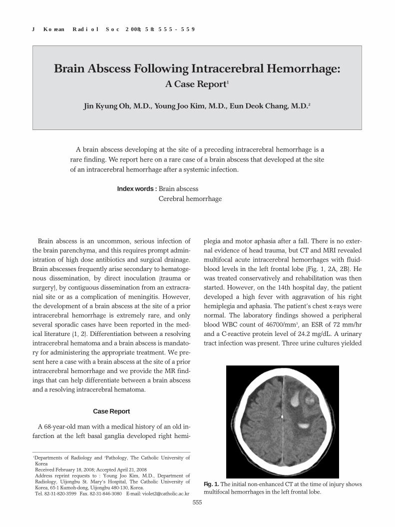
plegia and motor aphasia after a fall. There is no exter-nal evidence of head trauma, but CT and MRI revealedmultifocal acute intracerebral hemorrhages with fluid-blood levels in the left frontal lobe (Fig. 1, 2A, 2B). Hewas treated conservatively and rehabilitation was thenstarted. However, on the 14th hospital day, the patientdeveloped a high fever with aggravation of his righthemiplegia and aphasia. The patient’s chest x-rays werenormal. The laboratory findings showed a peripheralblood WBC count of 46700/mm3, an ESR of 72 mm/hrand a C-reactive protein level of 24.2 mg/dL. A urinarytract infection was present. Three urine cultures yielded
J Korean Radiol Soc 2008;58:555-559
─ 555 ─
Brain Abscess Following Intracerebral Hemorrhage: A Case Report1
Jin Kyung Oh, M.D., Young Joo Kim, M.D., Eun Deok Chang, M.D.2
1Departments of Radiology and 2Pathology, The Catholic University ofKoreaReceived February 18, 2008; Accepted April 21, 2008Address reprint requests to : Young Joo Kim, M.D., Department ofRadiology, Uijongbu St. Mary’s Hospital, The Catholic University ofKorea, 65-1 Kumoh-dong, Uijongbu 480-130, Korea.Tel. 82-31-820-3599 Fax. 82-31-846-3080 E-mail: [email protected]
A brain abscess developing at the site of a preceding intracerebral hemorrhage is arare finding. We report here on a rare case of a brain abscess that developed at the siteof an intracerebral hemorrhage after a systemic infection.
Index words : Brain abscess Cerebral hemorrhage
Fig. 1. The initial non-enhanced CT at the time of injury showsmultifocal hemorrhages in the left frontal lobe.

Pseudomonas aeruginosa. Following administration ofantibiotics for two weeks, the fever subsided and theWBC count returned to normal. However, the patient’sneurologic symptoms did not improve. The follow upMR scan performed on the 27th hospital day showedcystic masses with marked ring enhancement at thesites of the preceding intracerebral hemorrhages, in ad-
dition to the extensive perilesional edema. The center ofthe lesion had high signal intensity on the T2-weightedimages, with a complete dark signal rim, and mixedhigh signal intensity on the T1-weighted images.Diffusion-weighted imaging (DWI) revealed concentricbands of heterogeneous signal intensity with an inverse-ly heterogeneous afferent diffusion coefficient (ADC) at
Jin Kyung Oh, et al : Brain Abscess Following Intracerebral Hemorrhage
─ 556 ─
A B C
D E F
Fig. 2. A, B. The initial MR images at the time of injury. The axial T2-weighted (A) andT1-weighted images (B) show the layered, acute stage hematomas with minimal per-ilesional edema. The lesions are not enhanced (not shown).C-G. The MR images taken 3 weeks after the hemorrhage. The axial T2-weighted im-age (C) shows multiple well-defined hyperintensity lesions with hypointense walls,and these lesions appear hyperintense on the axial T1-weighted image (D) with isoin-tense walls. Note the prominent perilesional edema. The postcontrast T1-weightedimage (E) demonstrates uniform peripheral wall enhancements, which accurately cor-responded to the low signal rim on the T2-weighted image. The diffusion-weightedimage (F) reveals layered heterogeneous signal intensities with inversely heteroge-neous afferent diffusion coefficient values (G) at the center of the lesions and amarked hypointense rim at the periphery. Ring enhancement with a complete hy-pointense rim on both the T2-weighted images and the DWI, and the extensive edemaindicate that brain abscess developed at the site of the preceding hemorrhage.
G

the center of the lesions and a marked hypointense rimat the periphery of the lesions. The low signal rim at theperiphery on the DWI and T2-weighted images accu-rately corresponded to the enhanced rim on the contrastenhanced images (Fig. 2C-G). Given the possibility ofbrain abscesses, the patient underwent surgical drainagevia open craniotomy. A yellowish, cheese like purulentmaterial was aspirated and three well capsulated ab-scesses were removed. The histology revealedmacrophages, a mononuclear infiltrate, revasculariza-tion and gliosis in the wall, and all this was suggestive ofa brain abscess (Fig. 3). The cultures of the surgical spec-imen and the necrotic fluid were negative. The patientwas treated with broad-spectrum antimicrobial cover-age for 6 weeks. Follow up CT was performed 2 weeksafter completion of the antibiotic course and it revealednear complete resolution of the ring enhancing lesionsand the brain edema. The patient has fared well and hehas been followed for 12 months.
Discussion
An intact blood brain barrier in the normal brain pro-vides resistance to infection. Disruption of the bloodbrain barrier by hemorrhage may make the affectedbrain tissue susceptible to infection by blood-borne bac-teria with subsequent abscess formation (1). In the pre-viously reported cases, the first episodes of high fever,which indicated systemic infection and bacteremia, oc-curred 0-90 days after the onset of the intracereberal he-
morrhages. Our patient had an episode of high fever 2weeks after the hemorrhage. A urinary tract infectionwas considered to be the source of hematogenous seed-ing of the infection that spread to the brain. Althoughuniform ring enhancement is an important radiologicfinding for the diagnosis of a brain abscess, it is not aspecific finding for a brain abscess and it must be distin-guished from a necrotic neoplasm and other cystic le-sions. Intracerebral hematomas usually resolve sponta-neously or they form a cavity over several months. Asthe hemorrhage evolves, different characteristic appear-ances can be identified on CT & MRI, depending on theage of the bleed. From 1-6 weeks, peripheral enhance-ment can be seen because there is a breakdown of theblood-brain barrier in the vascularized capsule that sur-rounds the hematoma (3) and this mimics the appear-ance of an abscess. Because a brain abscess is an emer-gency condition that requires prompt administration ofhigh dose antibiotics and surgical drainage, it is manda-tory to differentiate a brain abscess from a resolvinghematoma. However, this differentiation can be diffi-cult due to the overlapping radiological features.
More recently, DWI has demonstrated significant po-tential to further delineate and diagnose ring-enhancingmass lesions (4, 5). Many studies have confirmed thepresence of restricted diffusion in those abscesses withhigh signal intensity in the central cavity and a corre-spondingly low ADC value. The probable factors for therestricted diffusion in brain abscesses are the microscop-ic organization of the tissues, the high viscosity of thepus that’s caused by a high protein level and the differ-ent types of viable or dead cells along with the necrotictissue, bacteria and exuded plasma. Additionally, watermolecules are bound to amino acid groups on the sur-face of macromolecules, which further restrict theirtranslational motion (5). However, these findings are notconfined to an abscess and they might present in variousother brain diseases like hemorrhagic primary or sec-ondary tumors and resolving hematomas (4). It isknown that one DWI finding of hyperacute and latesubacute hematomas is hyperintensity on DWI with alow ADC value (6). The precise biophysical explanationfor the decreased ADC in hyperacute and late subacutehematomas is uncertain. We found layered low andhigh signal intensities on the DWI with an inversely het-erogeneous ADC at the center of the lesions in our case.Correlation of our surgical and pathologic specimensdemonstrated that the central heterogeneous signal in-tensities on the DWI reflected the proteinaceous and
J Korean Radiol Soc 2008;58:555-559
─ 557 ─
Fig. 3. Photomicrograph of the histologic specimen reveals fi-broblasts, mononuclear infiltrates and macrophages associatedwith hemosiderin pigments in the abscess capsule, and amor-phous proteinaceous materials with degenerating erythrocytes(upper left) within the abscess cavity (Hematoxylin-eosin stain,×200).

necrotic debris with a bloody background in the abscesstissues.
The mature abscess often has a rim that gives a signalthat’s similar to or slightly higher than the white matteron the T1-weighted images and a lower signal for therim is seen on the T2-weighted images. These signalproperties have been ascribed to collagen and the para-magnetic free radicals that are released from the phago-cytosing macrophages (7). A low signal rim on T2-weighted images may also be seen in other lesions suchas subacute and chronic hematomas, metastases, granu-lomatous lesions and, on rare occasions, gliomas (8). Inour case, a complete low signal rim was seen on the T2-weighted image and the DWI. Although paramagnetichemosiderin-laden macrophages begin to take up resi-dence at the periphery of the hemorrhage at the latesubacute stage of an intracerebral hematoma, the pres-ence of scant amounts of hemosiderin is unlikely tohave been the primary cause of a complete T2 hy-pointense rim on the subacute hematoma in our case.According to Kang et al, a hypointense rim on both theDWI and the T2-weighted images showed up only at thechronic stage of an intracerebral hematoma (6).Furthermore, a hypointense rim at the periphery on theDWI and T2-weighted images accurately correspondedto the enhanced rim observed on the contrast enhancedimages. In a study on 221 patients reported by Schwartzet al, an abscess was the most common pathology thatmanifested as a ring-enhancing lesion with complete hy-pointense rims on the T2-weighted images (8). Schwartzet al also reported that an intracerebral hemorrhage wasa rare condition with ring enhancement and a T2 hy-pointense border (8). The histology for our case con-firmed the presence of macrophages, a mononuclear in-filtrate, revascularization and gliosis, and hemosiderinin the wall, which all contributed to the complete lowsignal rim seen on T2WI.
In our case of brain abscess, the patient’s brain edemawas aggravated on the follow up imaging study. Afterintracereberal hemorrhage, penetration of the serumprotein from the clot into the surrounding white matter,followed by breakdown of the blood-brain barrier dueto inflammation, have been proposed as mechanismsleading to edema formation in the extracellular compo-nent (9). This edema usually subsides and the mass ef-fect gradually diminishes in cases with a resolving hem-orrhage. It is known that pronounced or persistent ede-ma is one of the signs of hemorrhagic intracranial neo-plasm rather than a benign hemorrhage. In the case of a
brain abscess, exuberant neovascularization around themargin of the necrotic brain is responsible for themarked vasogenic edema. The brain edema may begreater in volume than the abscess itself, and this causesmuch of the associated mass effect. In spite of encapsu-lation, a circumscribed disturbance of the blood-brainbarrier persisted, and this was responsible for the belat-ed resolution of the patient’s edema and a slow decreaseof the intracranial pressure (10).
This case illustrates that a cerebral hematoma can betransformed into an abscess when systemic infectioncomplicates a hematoma. Abscess formation in ahematoma cavity should be considered in the differen-tial diagnosis of patients who deteriorate after a febrileepisode and who also have a history of an intracerebralhemorrhage.
In this case with a brain abscess, the presence of ringenhancement with a complete hypointense rim both onthe T2-weighted images and DWI and the unusual ex-tensive edema along with the clinical findings made usdiagnose a brain abscess rather than a resolving suba-cute hematoma.
References
1. Chen ST, Tang LM, Ro LS. Brain abscess as a complication ofstroke. Stroke 1995;26:696-698
2. Siatouni A, Mpouras T, Boviatsis EJ, Gatzonis S, Stefanatou M,Sakas D. Brain abscess following intracerebral haemorrhage. J ClinNeurosci 2007;14:986-989
3. Zimmerman RD, Leeds NE, Naidich TP. Ring blush associatedwith intracerebral hematoma. Radiology 1977;122:707-711
4. Hartmann M, Jansen O, Heiland S, Sommer C, Munkel K, Sartor K.Restricted diffusion within ring enhancement is not pathognomon-ic for brain abscess. AJNR Am J Neuroradiol 2001; 22:1738-1742
5. Mishra AM, Gupta RK, Jaggi RS, Reddy JS, Jha DK, Husain N, etal. Role of diffusion-weighted imaging and in vivo proton magneticresonance spectroscopy in the differential diagnosis of ring-en-hancing intracranial cystic mass lesions. J Comput Assist Tomogr2004;28:540-547
6. Kang BK, Na DG, Ryoo JW, Byun HS, Roh HG, Pyeun YS.Diffusion-weighted MR imaging of intracerebral hemorrhage.Korean J Radiol 2001;2:183-191
7. Haimes AB, Zimmerman RD, Morgello S, Weingarten K, BeckerRD, Jennis R, et al. MR imaging of brain abscesses. AJR Am JRoentgenol 1989;152:1073-1085
8. Schwartz KM, Erickson BJ, Lucchinetti C. Pattern of T2 hy-pointensity associated with ring-enhancing brain lesions can helpto differentiate pathology. Neuroradiology 2006;48:143-149
9. Wagner KR, Xi G, Hua Y, Kleinholz M, de Courten-Myers GM,Myers RE, et al. Lobar intracerebral hemorrhage model in pigs:rapid edema development in perihematomal white matter. Stroke1996; 27:490-497
10. Wallenfang T, Bohl J, Kretzschmar K. Evolution of brain abscess incats formation of capsule and resolution of brain edema. NeurosurgRev 1980; 3:101-111
Jin Kyung Oh, et al : Brain Abscess Following Intracerebral Hemorrhage
─ 558 ─

J Korean Radiol Soc 2008;58:555-559
─ 559 ─
대한영상의학회지 2008;58:555-559
뇌 실질 내 혈종과 동반된 뇌 농양: 증례 보고1
1가톨릭대학교 영상의학과, 2병리과
오진경·김영주·장은덕2
뇌 실질 내 혈종이 있었던 위치에 뇌 농양이 병발되는 경우는 현재까지 13예가 보고된 매우 드문 증례이다. 저
자들은 뇌 실질 내 혈종이 있었던 위치에 전신 패혈증으로 인한 뇌 농양이 병발되었던 환자 1예를 경험하였기에
영상소견과 함께 보고하는 바이다.
Related Documents


