RESEARCH Open Access Borna disease virus (BDV) infection in psychiatric patients and healthy controls in Iran Elham Mazaheri-Tehrani 1,2* , Nader Maghsoudi 1 , Jamal Shams 3 , Hamid Soori 4 , Hasti Atashi 1 , Fereshteh Motamedi 1 , Liv Bode 5 and Hanns Ludwig 5 Abstract Background: Borna disease virus (BDV) is an evolutionary old RNA virus, which infects brain and blood cells of humans, their primate ancestors, and other mammals. Human infection has been correlated to mood disorders and schizophrenia, but the impact of BDV on mental-health still remains controversial due to poor methodological and cross-national comparability. Method: This first report from the Middle East aimed to determine BDV infection prevalence in Iranian acute psychiatric disorder patients and healthy controls through circulating immune complexes (CIC), antibodies (Ab) and antigen (pAg) in blood plasma using a standardized triple enzyme immune assay (EIA). Samples of 314 subjects (114 psychiatric cases, 69 blood donors, and 131 healthy controls) were assayed and data analyzed quantitatively and qualitatively. Results: CICs revealed a BDV prevalence of one third (29.5%) in healthy Iranian controls (27.5% controls; 33.3% blood donors). In psychiatric patients CIC prevalence was higher than in controls (40.4%) and significantly correlating with bipolar patients exhibiting overt clinical symptoms (p = 0.005, OR = 1.65). CIC values were significantly elevated in bipolar (p = 0.001) and major depressive disorder (p = 0.029) patients as compared to controls, and in females compared to males (p = 0.031). Conclusion: This study supports a similarly high prevalence of subclinical human BDV infections in Iran as reported for central Europe, and provides again an indication for the correlation of BDV infection and mood disorders. Further studies should address the morbidity risk for healthy carriers and those with elevated CIC levels, along with gender disparities. Keyword: Borna disease virus, Circulating immune complexes, Psychiatric disorders, Iranian patients/controls Background Borna disease virus (BDV) holds unique features in terms of its cell biology, molecular properties, preference to old brain areas, broad host spectrum [1], and unusual bio- logical age, dating back to more than 40 million years [2,3]. The outstanding molecular biology of the virus, and its single stranded RNA genome leading to the classification [4] of an own family, Bornaviridae (order Mononegavirales), has been comprehensively reviewed [5]. BDV had first been recognized as an often deadly pathogen of horses and sheep [1,6] with a wide spectrum in other domestic and farm animals. However, BDV’ s non-cytolytic proper- ties, low replication while over-expressing two major proteins, and evidence of modulating neurotransmitter networks [7], pointed to a long-term adaption toward moderate pathogenicity and persistency [1]. Human infection and its putative link to mental disor- ders, first suggested after detection of antibodies [8], be- came a key issue inspiring research groups around the globe. After nucleic acid and antigen could be demon- strated in white blood cells of psychiatrically diseased pa- tients [9], such a link was further strengthened by the finding of specific RNA sequences in post mortem brains * Correspondence: [email protected] 1 Neuroscience Research Center, Shahid Beheshti University of Medical Sciences, P.O. Box 19615–1178, Tehran, Iran 2 Department of Diagnostic, Clinic and Public Health Medicine, University of Modena and Reggio Emilia, Modena, Italy Full list of author information is available at the end of the article © 2014 Mazaheri-Tehrani et al.; licensee BioMed Central Ltd. This is an Open Access article distributed under the terms of the Creative Commons Attribution License (http://creativecommons.org/licenses/by/4.0), which permits unrestricted use, distribution, and reproduction in any medium, provided the original work is properly credited. The Creative Commons Public Domain Dedication waiver (http://creativecommons.org/publicdomain/zero/1.0/) applies to the data made available in this article, unless otherwise stated. Mazaheri-Tehrani et al. Virology Journal 2014, 11:161 http://www.virologyj.com/content/11/1/161

Welcome message from author
This document is posted to help you gain knowledge. Please leave a comment to let me know what you think about it! Share it to your friends and learn new things together.
Transcript

Mazaheri-Tehrani et al. Virology Journal 2014, 11:161http://www.virologyj.com/content/11/1/161
RESEARCH Open Access
Borna disease virus (BDV) infection in psychiatricpatients and healthy controls in IranElham Mazaheri-Tehrani1,2*, Nader Maghsoudi1, Jamal Shams3, Hamid Soori4, Hasti Atashi1, Fereshteh Motamedi1,Liv Bode5 and Hanns Ludwig5
Abstract
Background: Borna disease virus (BDV) is an evolutionary old RNA virus, which infects brain and blood cells ofhumans, their primate ancestors, and other mammals. Human infection has been correlated to mood disorders andschizophrenia, but the impact of BDV on mental-health still remains controversial due to poor methodological andcross-national comparability.
Method: This first report from the Middle East aimed to determine BDV infection prevalence in Iranian acutepsychiatric disorder patients and healthy controls through circulating immune complexes (CIC), antibodies (Ab) andantigen (pAg) in blood plasma using a standardized triple enzyme immune assay (EIA). Samples of 314 subjects(114 psychiatric cases, 69 blood donors, and 131 healthy controls) were assayed and data analyzed quantitativelyand qualitatively.
Results: CICs revealed a BDV prevalence of one third (29.5%) in healthy Iranian controls (27.5% controls; 33.3%blood donors). In psychiatric patients CIC prevalence was higher than in controls (40.4%) and significantlycorrelating with bipolar patients exhibiting overt clinical symptoms (p = 0.005, OR = 1.65). CIC values weresignificantly elevated in bipolar (p = 0.001) and major depressive disorder (p = 0.029) patients as compared tocontrols, and in females compared to males (p = 0.031).
Conclusion: This study supports a similarly high prevalence of subclinical human BDV infections in Iran as reportedfor central Europe, and provides again an indication for the correlation of BDV infection and mood disorders.Further studies should address the morbidity risk for healthy carriers and those with elevated CIC levels, along withgender disparities.
Keyword: Borna disease virus, Circulating immune complexes, Psychiatric disorders, Iranian patients/controls
BackgroundBorna disease virus (BDV) holds unique features in termsof its cell biology, molecular properties, preference to oldbrain areas, broad host spectrum [1], and unusual bio-logical age, dating back to more than 40 million years[2,3]. The outstanding molecular biology of the virus, andits singlestranded RNA genome leading to the classification [4]
of an own family, Bornaviridae (order Mononegavirales),has been comprehensively reviewed [5]. BDV had first
* Correspondence: [email protected] Research Center, Shahid Beheshti University of MedicalSciences, P.O. Box 19615–1178, Tehran, Iran2Department of Diagnostic, Clinic and Public Health Medicine, University ofModena and Reggio Emilia, Modena, ItalyFull list of author information is available at the end of the article
© 2014 Mazaheri-Tehrani et al.; licensee BioMeCreative Commons Attribution License (http:/distribution, and reproduction in any mediumDomain Dedication waiver (http://creativecomarticle, unless otherwise stated.
been recognized as an often deadly pathogen of horsesand sheep [1,6] with a wide spectrum in other domesticand farm animals. However, BDV’s non-cytolytic proper-ties, low replication while over-expressing two majorproteins, and evidence of modulating neurotransmitternetworks [7], pointed to a long-term adaption towardmoderate pathogenicity and persistency [1].Human infection and its putative link to mental disor-
ders, first suggested after detection of antibodies [8], be-came a key issue inspiring research groups around theglobe. After nucleic acid and antigen could be demon-strated in white blood cells of psychiatrically diseased pa-tients [9], such a link was further strengthened by thefinding of specific RNA sequences in post mortem brains
d Central Ltd. This is an Open Access article distributed under the terms of the/creativecommons.org/licenses/by/4.0), which permits unrestricted use,, provided the original work is properly credited. The Creative Commons Publicmons.org/publicdomain/zero/1.0/) applies to the data made available in this

Table 1 Basic data on the population
Groups N Female/Male Mean age + SE Min-Max
Controls 131 83/48 41.08 + 1.009 18-69
Blood donors 69 6/63 29.93 + 1.296 19-58
Mental patients 114 52/62 37.42 + 1.103 17-62
BD* 64 32/32 36.20 + 1.477 17-62
MDD** 12 7/5 43.42 + 3.450 21-57
Schizophrenia 18 3/15 34.56 + 2.689 20-53
Schizoaffective 15 5/10 38.33 + 2.863 22-57
OCD*** 5 5/0 46.20 + 3.967 37-56
Summary 314 141/173 37.30 + 0.688 17-69
*Bipolar disorder.**Major depressive disorder.***Obsessive compulsive disorder.
Mazaheri-Tehrani et al. Virology Journal 2014, 11:161 Page 2 of 9http://www.virologyj.com/content/11/1/161
of psychiatric patients [10] and limbic structures from oldpeople [11].The impact of human infection was significantly sup-
ported by the isolation and sequence characterization ofhuman viruses from psychiatric patients’ blood cells andbrain [12-14], and the recent correlation of neurologicalsymptoms in humans with BDV infection [15]. The latestdiscovery of functional endogenous virus gene pieces inte-grated in the human and primate ancestor germ linesstrongly argued in favor of a long-term co-evolution ofvirus and hosts [2,3,16]. However, a role of BDV, whatso-ever, in human mental-health remained controversial, des-pite of predominantly supportive reports [17-24]. This ismainly due to a great variation in prevalence results largelycaused by methodological disparities, due to different anti-body and/or RNA techniques, affecting as well cross-national comparability. In contrast, BDV-specific circulatingimmune complexes, the most prevalent infection markers[25], have shown to be superior to antibody- or RNA- de-tection. Pilot prevalence studies could demonstrate that theBDV-CIC enzyme immune assay (EIA) is an easy to per-form and robust test format, suitable to conducting com-parable surveys in the general population of differentcountries, as well as longitudinal follow-up studies of pa-tients in clinical cohorts [26-31]. Circulating immune com-plexes are the result of periods of antigenemia over-expressing N- and P-proteins, and antibody induction inthe host, reflecting recent and current virus activity. Evi-dence for a contribution of BDV infection to disease symp-toms has recently been reviewed [32].This is the first report from the Middle East, addressing
the prevalence of BDV in the human population in Iran.The virus in horses has previously been reported by anti-body studies [33]. Here we explore the prevalence of BDVmarkers among Iranian mentally diseased patients, healthycontrols, and blood donors.
MethodIndividual subjectsThree hundred and fourteen Iranian subjects, including114 psychiatric patients, 131 sex and age matched healthycontrols, and 69 blood donors were included in this study.The association between BDV infection markers in bloodplasma and five DSM IV- categorized psychiatric diseases,as well as gender and age of the individuals were analyzed.Basic data are given in Table 1. One hundred and four-
teen acute psychiatric patients, who had been admitted tolocal departments of psychiatry in Tehran, were included.All patients met the Diagnostic and Statistical Manual ofMental Disorders IV (DSM-IV) -criteria on the basis ofinterviews and medical records. They could be dividedinto five main groups and different DSM IV codes, includ-ing 64 bipolar disorder (BD)-, 12 major depressive dis-order (MDD)-, 18 schizophrenia-, 15 schizoaffective- and
5 obsessive compulsive disorder (OCD) patients (Table 2).Additionally, 69 blood donors and 131 sex- and agematched, mentally healthy subjects (based on the supervi-sion of the psychiatrists) were included and regarded ascontrols. All individuals were negative for Hepatitis B-and C-viruses, as well as HIV. The study was approved bythe Ethic Committee of the Neuroscience Research Centerat Shahid Beheshti University of Medical Sciences, and allpatients -or an authorized representative- gave their writ-ten informed consent for participation. Blood samples ofall individuals were collected prior to any medical treat-ment and plasma or sera were kept at −20°C.
Enzyme immune assays (EIAs)The BDV infection markers, circulating immune com-plexes (CICs), virus antigens (N- and P- protein, N/P-complexes; abbreviated Ag), and antibodies (Ab), wereassayed using the triple enzyme immune assay (EIA) sys-tem, as described [25]. According to the double- sandwichformat, two BDV-specific monoclonal antibodies (mAbs),anti-N mAb (W1) and anti-P mAb (Kfu2), were used tobind any BDV-N- and P-protein or N/P -heterodimers inplasma, either circulating antigen bound to virus- specifichost antibodies (CIC- EIA) or free antigen (pAg-EIA).CICs were visualized through alkaline phosphatase (AP)-coupled anti-human IgG and substrate, whereas the Ag-EIA needs a BDV-specific detecting antibody (rabbithyper-immune serum) followed by AP phosphatasecoupled anti-rabbit IgG and substrate. The specificity andsensitivity of the BDV mAbs have been further character-ized [34]. In particular, epitope mapping has revealed thatboth these mAbs are binding to powerful conformationalepitopes on either protein, which are formed through 5binding sites in case of the anti-N mAb (W1) and 3 bind-ing sites in case of the anti-P mAb (Kfu2). None of the W1binding sites are overlapping with P-protein binding do-mains on the N-protein, confirming that commonly oc-curring N/P heterodimers are recognized by W1, as well.
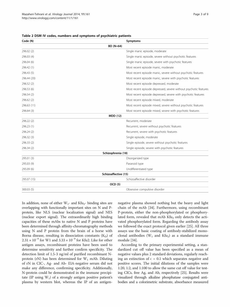
Table 2 DSM IV codes, numbers and symptoms of psychiatric patients
Code (N) Symptoms
BD (N=64)
296.02 (2) Single manic episode, moderate
296.03 (4) Single manic episode, severe without psychotic features
296.04 (6) Single manic episode, severe with psychotic features
296.42 (1) Most recent episode manic, moderate
296.43 (5) Most recent episode manic, severe without psychotic features
296.44 (20) Most recent episode manic, severe with psychotic features
296.52 (2) Most recent episode depressed, moderate
296.53 (6) Most recent episode depressed, severe without psychotic features
296.54 (2) Most recent episode depressed, severe with psychotic features
296.62 (2) Most recent episode mixed, moderate
296.63 (11) Most recent episode mixed, severe without psychotic features
296.64 (3) Most recent episode mixed, severe with psychotic features
MDD (12)
296.22 (2) Recurrent, moderate
296.23 (1) Recurrent, severe without psychotic features
296.24 (2) Recurrent, severe with psychotic features
296.32 (3) Single episode, moderate
296.33 (2) Single episode, severe without psychotic features
296.34 (2) Single episode, severe with psychotic features
Schizophrenia (18)
295.01 (3) Disorganized type
295.03 (9) Paranoid type
295.09 (6) Undifferentiated type
Schizoaffective (15)
295.07 (15) Schizoaffective disorder
OCD (5)
300.03 (5) Obsessive compulsive disorder
Mazaheri-Tehrani et al. Virology Journal 2014, 11:161 Page 3 of 9http://www.virologyj.com/content/11/1/161
In addition, none of either W1- and Kfu2- binding sites areoverlapping with functionally important sites on N and P-protein, like NLS (nuclear localization signal) and NES(nuclear export signal). The extraordinarily high bindingcapacities of these mAbs to native N and P proteins havebeen determined through affinity-chromatography methodsusing N and P protein from the brain of a horse withBorna disease, resulting in dissociation constants (KD) of2.31 × 10−9 for W1 and 3.33 × 10⁻⁹ for Kfu2. Like for otherantigen assays, recombinant proteins have been used todetermine sensitivity and further confirm specificity. Thedetection limit of 1.5-3 ng/ml of purified recombinant N-protein (rN) has been determined for W1 mAb. Dilutingof rN in CIC-, Ag- and Ab- EIA-negative serum did notmake any difference, confirming specificity. Additionally,N-protein could be demonstrated in the immune precipi-tate (IP using W1) of a strongly antigen positive patient’splasma by western blot, whereas the IP of an antigen-
negative plasma showed nothing but the heavy and lightchain of the mAb [34]. Furthermore, using recombinantP-protein, either the non-phosphorylated or phosphory-lated form, revealed that mAb Kfu2 only detects the acti-vated phosphorylated form. Regarding the antibody assaywe followed the exact protocol given earlier [25]. All threeassays use the basic coating of antibody-stabilized mono-clonal antibodies (W1 and Kfu2) as a standard immunemodule [34].According to the primary experimental setting, a stan-
dardized cut off value has been specified as a mean ofnegative values plus 2 standard deviations, regularly reach-ing an extinction of < = 0.1 which separates negative andpositive scores. The initial dilutions of the samples were1:20, 1:2, and 1:100 to allow the same cut off value for test-ing CICs, free Ag, and Ab, respectively [25]. Results werevisualized through alkaline phosphatase conjugated anti-bodies and a colorimetric substrate, absorbance measured
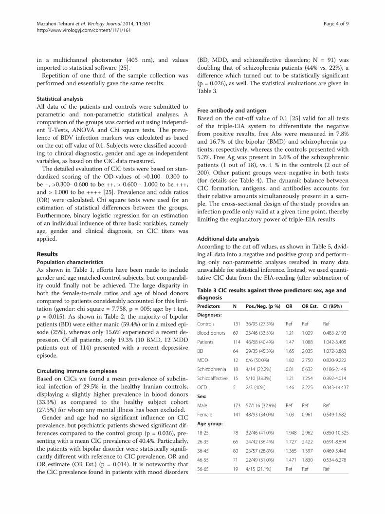
Table 3 CIC results against three predictors: sex, age anddiagnosis
Predictors N Pos./Neg. (p %) OR OR Est. CI (95%)
Diagnoses:
Controls 131 36/95 (27.5%) Ref Ref Ref
Blood donors 69 23/46 (33.3%) 1.21 1.029 0.483-2.193
Patients 114 46/68 (40.4%) 1.47 1.088 1.042-3.405
BD 64 29/35 (45.3%) 1.65 2.035 1.072-3.863
MDD 12 6/6 (50.0%) 1.82 2.750 0.820-9.222
Schizophrenia 18 4/14 (22.2%) 0.81 0.632 0.186-2.149
Schizoaffective 15 5/10 (33.3%) 1.21 1.254 0.392-4.014
OCD 5 2/3 (40%) 1.46 2.225 0.343-14.437
Sex:
Male 173 57/116 (32.9%) Ref Ref Ref
Female 141 48/93 (34.0%) 1.03 0.961 0.549-1.682
Age group:
18-25 78 32/46 (41.0%) 1.948 2.962 0.850-10.325
26-35 66 24/42 (36.4%) 1.727 2.422 0.691-8.894
36-45 80 23/57 (28.8%) 1.365 1.597 0.469-5.440
46-55 71 22/49 (31.0%) 1.471 1.830 0.534-6.278
56-65 19 4/15 (21.1%) Ref Ref Ref
Mazaheri-Tehrani et al. Virology Journal 2014, 11:161 Page 4 of 9http://www.virologyj.com/content/11/1/161
in a multichannel photometer (405 nm), and valuesimported to statistical software [25].Repetition of one third of the sample collection was
performed and essentially gave the same results.
Statistical analysisAll data of the patients and controls were submitted toparametric and non-parametric statistical analyses. Acomparison of the groups was carried out using independ-ent T-Tests, ANOVA and Chi square tests. The preva-lence of BDV infection markers was calculated as basedon the cut off value of 0.1. Subjects were classified accord-ing to clinical diagnostic, gender and age as independentvariables, as based on the CIC data measured.The detailed evaluation of CIC tests were based on stan-
dardized scoring of the OD-values of >0.100- 0.300 tobe +, >0.300- 0.600 to be ++, > 0.600 - 1.000 to be +++,and > 1.000 to be ++++ [25]. Prevalence and odds ratios(OR) were calculated. Chi square tests were used for anestimation of statistical differences between the groups.Furthermore, binary logistic regression for an estimationof an individual influence of three basic variables, namelyage, gender and clinical diagnosis, on CIC titers wasapplied.
ResultsPopulation characteristicsAs shown in Table 1, efforts have been made to includegender and age matched control subjects, but comparabil-ity could finally not be achieved. The large disparity inboth the female-to-male ratios and age of blood donorscompared to patients considerably accounted for this limi-tation (gender: chi square = 7.758, p = 005; age: by t test,p = 0.015). As shown in Table 2, the majority of bipolarpatients (BD) were either manic (59.4%) or in a mixed epi-sode (25%), whereas only 15.6% experienced a recent de-pression. Of all patients, only 19.3% (10 BMD, 12 MDDpatients out of 114) presented with a recent depressiveepisode.
Circulating immune complexesBased on CICs we found a mean prevalence of subclin-ical infection of 29.5% in the healthy Iranian controls,displaying a slightly higher prevalence in blood donors(33.3%) as compared to the healthy subject cohort(27.5%) for whom any mental illness has been excluded.Gender and age had no significant influence on CIC
prevalence, but psychiatric patients showed significant dif-ferences compared to the control group (p = 0.036), pre-senting with a mean CIC prevalence of 40.4%. Particularly,the patients with bipolar disorder were statistically signifi-cantly different with reference to CIC prevalence, OR andOR estimate (OR Est.) (p = 0.014). It is noteworthy thatthe CIC prevalence found in patients with mood disorders
(BD, MDD, and schizoaffective disorders; N = 91) wasdoubling that of schizophrenia patients (44% vs. 22%), adifference which turned out to be statistically significant(p = 0.026), as well. The statistical evaluations are given inTable 3.
Free antibody and antigenBased on the cut-off value of 0.1 [25] valid for all testsof the triple-EIA system to differentiate the negativefrom positive results, free Abs were measured in 7.8%and 16.7% of the bipolar (BMD) and schizophrenia pa-tients, respectively, whereas the controls presented with5.3%. Free Ag was present in 5.6% of the schizophrenicpatients (1 out of 18), vs. 1 % in the controls (2 out of200). Other patient groups were negative in both tests(for details see Table 4). The dynamic balance betweenCIC formation, antigens, and antibodies accounts fortheir relative amounts simultaneously present in a sam-ple. The cross-sectional design of the study provides aninfection profile only valid at a given time point, therebylimiting the explanatory power of triple-EIA results.
Additional data analysisAccording to the cut off values, as shown in Table 5, divid-ing all data into a negative and positive group and perform-ing only non-parametric analyses resulted in many dataunavailable for statistical inference. Instead, we used quanti-tative CIC data from the EIA-reading (after subtraction of

Table 4 Prevalence of free antibodies and antigen
Free antibody Free antigen (N-& P-protein)
Groups Pos./Neg. Prevalence % CI Pos./Neg. Prevalence % CI
Controls 7/124 (5.3%) 1.5-9% 1/130 (0.7%) 0.7-2.3%
Patients 8/106 (7.0%) 2.3-11.7% 1/113 (0.9%) 0-2.6%
BD 5/59 7.8% 1.2-14.4% 0/64 0.0% -
MDD 0/12 0.0% - 0/12 0.0% -
Schizophrenia 3/15 16.7% 0-33.9% 1/17 5.6% 0-16%
Schizoaffective 0/15 0.0% - 0/15 0.0% -
OCD 0/5 0.0% - 0/5 0.0% -
Blood donors 1/68 (1.4%) 0-4.2% 1/68 (1.4%) 0-4.2%
Total 16/298 (5.1%) 2.7-7.5% 3/311 (1%) 0-2%
Mazaheri-Tehrani et al. Virology Journal 2014, 11:161 Page 5 of 9http://www.virologyj.com/content/11/1/161
the OD values for blanks) for parametric statisticalanalyses.A noticeable increase in CIC levels of both, the bipolar
disorder (0.147) and the major depressive disorder (0.163)groups became obvious, being statistically significantwhen compared to control subjects. The values for 95%CI of CICs are illustrated in Figure 1. The CIC levelswithin the total population tend to be elevated amongfemales when compared to males (p = 0.089). Therefore,the influence of sex on CIC extinction values was alsoanalyzed in these patient groups (Figure 2). A significantincrease in CIC levels in female patients was recognizedwhen compared to males (p = 0.031).
Table 5 Distribution of categorized CIC results(neg., +, ++, +++) in subgroups
Subgroups Neg N (%) + ++ +++ Total
Controls 95 (72.5%) 31 (23.7%) 5 (3.8%) 0 131
Blood donors 46 (66.7%) 21 (30.4%) 2 (2.9%) 0 69
Case 68 (59.6%) 36 (31.5%) 8 (7.0%) 2 (1.7%) 114
BD 35 (54.7%) 22 (34.4%) 6 (9.4%) 1 (1.60%) 64
MDD 6 (50.0%) 5 (41.7%) 0 1 (8.30%) 12
Schizophrenia 14 (77.8%) 2 (11.1%) 2 (11.1%) 0 18
Schizoaffective 10 (66.7%) 5 (33.3%) 0 0 15
OCD 3 (60.0%) 2 (40.0%) 0 0 5
Sex
Male 116 (67.1%) 50 (28.9%) 7 (4.0%) 0 173
Female 93 (66.0%) 38 (27.0%) 8 (5.7%) 2 (1.4%) 141
Age groups
18-25 ys 46 (59.0%) 26 (33.3%) 6 (7.7%) 0 78
26-35 ys 42 (63.6%) 20 (30.3%) 4 (6.1%) 0 66
36-45 ys 57 (71.3%) 20 (25.0%) 2 (2.5%) 1 (1.3%) 80
46-55 ys 49 (69.0%) 20 (28.2%) 2 (2.8%) 0 71
56-65 ys 15 (78.9%) 2 (10.5%) 1 (5.3%) 1 (5.3%) 78
OD absorbance is valued as (+): OD absorbance > 0.100 - 0.300, (++): ODabsorbance > 0.300 - 0.600 and (+++): OD absorbance > 0.600.
DiscussionThis is the first study in Iranian people showing a fairlyhigh prevalence of Bornavirus infection in healthy individ-uals including blood donors. The results meet reporteddata from Central Europe of about 30% based on the sameinfection marker (CIC). The study also supports previousfindings that this neurotropic virus infection is moreprevalent in psychiatric patients than in healthy donors.According to trends our results are supporting infectionpatterns in other countries, like Europe, the Americasand Asia which are based on specific antibody- and nu-cleic acid detection [9,10,15,21,35-42], despite of largelydiffering prevalence data. Based on measuring BDV re-leased antigens or antigen-antibody complexes, likeCICs [25,27,29,30], our data showed a much betteragreement.Studies questioning and reporting the absence of BDV
in both normal and psychiatrically diseased people remaininconclusive as long as no other cohorts have been inves-tigated and no other methods have been applied. Amongthose are studies of Na et al. [42] and Hornig et al. [43].The latter group even neglected an own earlier positivestudy with contradictory results from the same country[10]. On the other hand, the existence of a human BDVstrain has recently been independently proven by anin vitro study in brain cells. Only the human virus wasable to reduce proliferation and enhance apoptosis butnot the animal-derived laboratory strain of BDV [44].Our study used an established triple EIA which had
been successfully applied to monitor point- and longitu-dinal prevalence of BDV infection markers in patients[25,26]. In our hands, these EIAs were found to be easy tohandle and to provide robust and reproducible measure-ments. It is unfortunate that general acceptance is stillpending. In this study, consecutive sampling of admittedpatients was not possible. Although the data only refer tocross-sectional sample analysis, BDV markers were signifi-cantly more prevalent in Iranian patients with mentaldiseases than in control subjects. These findings were

Figure 1 Mean and 95% confidence intervals for CIC extinction in the investigated groups. Lower and upper limits of 95% CI in groupsincluding Control: 0.080-0.095, Schizophrenia: 0.068-0.121, Schizoaffective: 0.056-0.080, Bipolar: 0.125-0.167, MDD: 0.096-0.224, OCD: 0.047-0.134 andDonor: 0.083-0.105 based on Table 3. *Significant when compared to controls (p = 0.001, ANOVA). **Significant when compared to controls(p = 0.029, ANOVA). Extinction values refer to 1:20 dilution of plasma in the CIC-ELISA.
Mazaheri-Tehrani et al. Virology Journal 2014, 11:161 Page 6 of 9http://www.virologyj.com/content/11/1/161
similar to data reported from Germany [25,26], Italy [27]Australia [29], the CSSR [30], China (Xia Liu, Peng Xie, pers.communication), and Lithuania (Violeta Mockeliūnienė,Robertas Bunevicius, pers. communication) where the sametest system had been applied.The presence of CIC with or without antibodies indi-
cates a chronic infection; the presence of Ag, with or with-out CICs at the same time, a currently active infection.The finding of free anti-BDV antibody alone (no antigen,no CICs) is thought to indicate previous exposure to theagent, but not a current active infection [34]. As shown inearlier reports CICs represent the major viral markerexplaining the transient disappearance of antibodies andantigens in blood plasma between activated and dormant
Figure 2 Statistical differences between female and malesamples in control and patient groups based on 95% CICabsorbance (p = 0.031). 131 control samples (83 female and 48male) and 114 patient samples (52 female and 62 male) werecalculated by a parametric t-student test.
phases of virus infection, and by this providing also a cluefor the true number of silently infected carriers in ahealthy cohort or population [25,26,34].Iranian psychiatric patients show a clearly elevated
CIC sero-prevalence (40.4%) compared to healthy con-trols (27.5%). It is of special interest that 33.3% of sam-ples from blood donations were silent virus carriers, afinding confirming Australian [29] and German pilot re-ports [17,26], thus being quite in contrast to an earlierreport [45]. Transfusion issues relating to BDV infectionare still awaiting further clarification [46].BD, MDD and OCD patients presented with infection
rates of 45.3%, 50.0% and 40.0%, respectively. However,significance levels were only reached in BD patients.This might be due to the small sample size, but in para-metric data analysis, comparing OD values of absorb-ance (extinction), high levels of CICs in sera from BDand MDD patients were also significant.In contrast to other reports [47,48], we found a rela-
tively high sero-prevalence of free Ab and Ag in schizo-phrenic patients (16.7% and 5.6%, respectively) which isconsistent with a relatively low CIC sero-prevalenceamong those individuals (22.2%, see Table 2). In additionto schizophrenic patients, only BD patients showed freeantibodies in their sera (7.8%). This implies that BDVantibodies are usually bound in immune complexes andare therefore becoming transiently absent in the bloodstream.It is of considerable interest that the CIC sero-
prevalence adversely correlated with the correspondingage groups (linear regression done using age as continu-ous data, R = −0.116, p = 0.042), which means that the
Mazaheri-Tehrani et al. Virology Journal 2014, 11:161 Page 7 of 9http://www.virologyj.com/content/11/1/161
young patients had highest CIC values, although the agelimit includes only adults 18 years and older. This leavesthe question whether younger people are either moreprone to BDV infection or their immune response is moreprominent. It supports a recent finding that young chil-dren (from 4–6 months to 3 years of age) had even muchhigher infection rates, although this pilot study warrantsfurther investigations [28,34], In addition, it has to be fur-ther examined whether and to which extent vertical trans-mission of BDV in the pregnant horse [49], mouse [50]and human [28] contributes to higher infection rates atyoung age. In this regard, high prevalence of BDV in thenormal population, lifelong persistence of the virus in in-fected subjects (patients or healthy people), and the sofar undisclosed function of endogenized BDV genomestretches [2,3,16], might reflect further risk factors war-ranting urgent future investigations.Interestingly, significant differences between female and
male patients could be measured for the first time(Figure 2. middle, p = 0.031), showing a prevalence ofCICs in 42.3% of females, and 38.7% in males. In favor ofthese findings, two female patients, belonging to the BDand MDD groups, had high CIC titers with levels above0.6 (+++) (Table 5). The sero-prevalence among healthycontrols, however, reached only 25.3% in females and31.3% in males.Such sex-related specific differences according to, titers
and prevalence of an antibody response to foreign anti-gens, infectious agents, or even auto antigens are knownfrom the literature [51-55]. Females usually exhibit astronger humoral immune response, as especially knownafter vaccination and infection with microbial agents. Infact, estrogens exert stimulatory effects on B cell prolifera-tion and serum IgG levels, whereas testosterone may sup-press B cell function [56,57].In conclusion, Iranian people seem to fit into the pattern
of BDV infections, so far reported worldwide [5]. More-over, the study benefits from using prevalent infectionmarkers and a highly specific and effective test system[26,34]. The study confirms evidence for a high infectionprevalence, similar to Central Europe, in one third ofhealthy Iranian subjects, contrasting elevated levels in pa-tients with mood disorders. In view of millions of peopleworldwide suffering from depression and the huge relatedhealth care costs [58], this study points again to integrat-ing BDV infection surveillance in psychiatric research [26]rather than to continue in underplaying its impact.
Competing interestsThe authors declared that they have no competing interests.
Authors’ contributionsTehran contributors: The project’s idea was initially conceived by EMT; shealso prepared and pursued the proposal until its final approval, collectedblood samples, separated sera, kept and transported them, conducted all themethods in Berlin and Tehran, and wrote a great deal of the manuscript.
NM, as head of molecular biology lab, checked and approved proposal anddata to receive grant; apart from being involved in its different stages, heorganized and supported the whole project. JS, as a psychiatrist, diagnosedmental diseases, classified symptoms and made folders for furtherepidemiological studies. HS conducted most of the epidemiological testsand analyses. HA tested patients for HIV, HBV and HCV negative. FM, aspresident of Neuroscience Research Center of Shahid Beheshti University ofMedical Sciences, facilitated the reception of grant, the safe-keeping andtransportation of sera as well as access to donor sera, not to mention herinvolvement in the stages of project. Berlin scientists, HL and LB, were majorcontributors in that they, among other things, supplied the essentialmonoclonal antibodies, positive and negative control sera, as well asbackground information on human Borna disease virus infections; they alsoshared their studies on other international groups, provided constant,meaningful advice on manuscript conceptualization and all through itsrevisions and editing. All authors read and approved the final manuscript.
AcknowledgmentThis work was supported by the Iranian Neuroscience Research Network,Ministry of Health [grant number A 162]. We are grateful to Biodaroo Co. forcollecting the plasma donors for this study. Long term grants of the DFG,the EU, and the FU Berlin to HL from the early1990s to 2005 contributed tothe basic Bornavirus research.
Author details1Neuroscience Research Center, Shahid Beheshti University of MedicalSciences, P.O. Box 19615–1178, Tehran, Iran. 2Department of Diagnostic,Clinic and Public Health Medicine, University of Modena and Reggio Emilia,Modena, Italy. 3Behavioral Research Center, Shahid Beheshti University ofMedical Sciences, Tehran, Iran. 4Safety Promotion and Injury PreventionResearch Center, Shahid Beheshti University of Medical Sciences, Tehran, Iran.5Bornavirus Working Group affiliated to the Free University of Berlin, Berlin,Germany.
Received: 24 April 2014 Accepted: 20 August 2014Published: 3 September 2014
References1. Ludwig H, Bode L, Gosztonyi G: Borna Disease: a persistent virus infection
of the central nervous system. Prog Med Virol 1988, 35:107–151.2. Horie M, Honda T, Suzuki Y, Kobayashi Y, Daito T, Oshida T, Ikuta K, Jern P,
Gojobori T, Coffin JM, Tomonaga K: Endogenous non-retroviral RNA viruselements in mammalian genomes. Nature 2010, 463:84–87.
3. Belyi VA, Levine AJ, Skalka AM: Unexpected Inheritance: Multipleintegrations of ancient bornavirus and ebolavirus/marburgvirussequences in vertebrate genomes. PLoS Pathog 2010, 6:e1001030.
4. de la Torre JC, Bode L, Carbone KM, Dietzschold B, Ikuta K, Lipkin WI,Ludwig H, Richt JA, Staeheli P, Stitz L: Family Bornaviridae. In Virustaxonomy 283, classification and nomenclature of viruses. 284 Seventh reportof the International Committee on Taxonomy of Viruses, Volume 10. [Online]285 San Diego: Academic Press, Calif; 2000:531–538.
5. Lipkin WI, Briese T: Bornaviridae. In Fields Virology, Volume II. 5th edition.Edited by Knipe D, Howley P, Griffin D, Lamb R, Martin M, Roizman B, StrausS. Philadelphia: Lippincott Williams and Wilkins; 2007:1829–1851.
6. Vahlenkamp TW, Konrath A, Weber M, Mueller H: Persistence of Bornadisease virus in naturally infected sheep. J Virol 2002, 76:9735–9797.
7. Gosztonyi G, Ludwig H: Neurotransmitter receptors and viralneurotropism. Neuropsychiat Clin 1984, 3:107–114.
8. Rott R, Herzog S, Fleischer B, Winokur A, Amsterdam J, Dyson W, KoprowskiH: Detection of serum antibodies to Borna disease virus in patients withpsychiatric disorders. Science 1985, 228:755–756.
9. Bode L, Zimmermann W, Ferszt R, Steinbach F, Ludwig H: Borna diseasevirus genome transcribed and expressed in psychiatric patients. Nat Med1995, 1:232–236.
10. Salvatore M, Morzunov S, Schwemmle M, Lipkin WI: Borna disease virus inbrains of North American and European people with schizophrenia andbipolar disorder. Lancet 1997, 349:1813–1814.
11. de la Torre JC, Gonzalez-Dunia D, Cubitt B, Mallory M, Mueller-Lantzsch N,Grässer FA: Detection of Borna disease virus antigen and RNA in humanautopsy brain samples from neuropsychiatric patients. Virology 1996,223:272–282.

Mazaheri-Tehrani et al. Virology Journal 2014, 11:161 Page 8 of 9http://www.virologyj.com/content/11/1/161
12. Bode L, Duerrwald R, Rantam FA, Ferszt R, Ludwig H: First isolates ofinfectious human Borna disease virus from patients with mooddisorders. Mol Psychiatry 1996, 1:200–212.
13. de la Torre JC, Bode L, Duerrwald R, Cubitt B, Ludwig H: Sequencecharacterization of human Borna disease virus. Virus Res 1996, 44:33–34.
14. Nakamura Y, Takahashi H, Shoya Y, Nakaya T, Watanabe M, Tomonaga K,Ameno K, Momiyama N, Taniyama H, Sata T, Kurata T, de la Torre JC, Ikuta K,Iwahashi K: Isolation of Borna disease virus from human brain tissue.J Virol 2000, 74:4601–4611.
15. Li Q, Wang Z, Zhu D, Xu M, Chen X, Peng D, Iwata Y, Xie P: Detection andanalysis of Borna disease virus in Chinese patients with neurologicaldisorders. Eur J Neurol 2009, 16:399–403.
16. Feschotte C: Bornavirus enters the genome. Nature News Views 2010,463:39–40.
17. Bode: Human infections with Borna disease virus and potentialpathogenic implications. Curr Top Microbiol Immunol 1995, 190:103–130.
18. Sierra-Honigmann AM, Carbone KM, Yolken RH: Polymerase chain reaction(PCR) search for viral nucleic acid sequences in schizophrenia. Br JPsychiatry 1995, 166:55–60.
19. Taller AM, Asher DM, Pomeroy KL, Eldadah BA, Godec MS, Falkai PG, BogertB, Kleinman JE, Stevens JR, Torrey EF: Search for viral nucleic acidsequences in brain tissues of patients with schizophrenia using nestedpolymerase chain reaction. Arch Gen Psychiatry 1996, 53:32–40.
20. Chen CH, Chiu YL, Shaw CK, Tsa MT, Hwang AL, Hsiao KJ: Detection of Bornadisease virus RNA from pheripheral blood mononuclear cells in schizophreniapatients and mental health workers. Mol Psychiatry 1999, 4:566–571.
21. Rybakowski F, Yamaguchi K, Krzyminski S, Zmyslony F, Biernat J,Kocialkowski M, Tandeck A, Trafarska B, Zalejski M, Sawada T, Naraki T,Czerski P, Rajewski A, Rybakowski JK: Detection of anti-Borna disease virusantibodies in patients hospitalized in psychiatric hospitals located in themid-Western region of Poland. Psychiatr Pol 2001, 35:819–829.
22. Ikuta K, Ibrahim MS, Kobayashi T, Tomonaga K: Borna disease virus andinfection in humans. Front Biosci 2002, 7d:470–495.
23. Miranda HC, Nunes SO, Calvo ES, Suzart S, Itano EN, Watanabe MA:Detection of Borna disease virus p24 RNA in peripheral blood cells fromBrazilian mood and psychotic disorder patients. J Affect Disord 2006,90:43–47.
24. Nunes SO, Itano EN, Amarante MK, Reiche EM, Miranda HC, de Oliveira CE,Matsuo T, Vargas HO, Vatanabe MO: RNA from Borna disease virus inpatients with schizophrenia, schizoaffective patients, and in theirbiological relatives. J Clin Lab Anal 2008, 22:314–320.
25. Bode L, Reckwald P, Severus WE, Stoyloff R, Ferszt R, Dietrich DE, Ludwig H:Borna disease virus-specific circulating immune complexes, antigenemia,and free antibodies–the key marker triplet determining infection andprevailing in severe mood disorders. Mol Psychiatry 2001, 6:481–491.
26. Bode L, Ludwig H: Borna disease virus infection, a human mental-healthrisk. Clin Microbiol Rev 2003, 16:534–545.
27. Patti AM, Vulcano A, Candelori E, Ludwig H, Bode L: Borna disease virusinfection in the population of Latium (Italy). APMIS Suppl 2008, 124:74–76.
28. Scholbach T, Bode L: Borna disease virus infection in young children.APMIS Suppl 2008, 124:83–88.
29. Flower RL, Kamhieh S, McLean L, Bode L, Ludwig H, Ward CM: HumanBorna disease virus infection in Australia: serological markers of infectionin multi-transfused patients. APMIS Suppl 2008, 124:89–93.
30. Rackova S, Janu L, Kabickova H: Borna disease virus circulatingimmunocomplex positivity and psychopathology in psychiatric patientsin the Czech Republic. Neuro Endocrinol Lett 2009, 30:414–420.
31. Thakur R, Sarma S, Sharma B: Role of Borna disease virus inneuropsychiatric illnesses: are we inching closer? Indian J Med Microbiol2009, 27:191–201.
32. Ludwig H, Bode L: From latent herpes viruses to persistent Bornavirus. InFrom the hallowed halls of herpesvirology. Edited by Blaho JA, Baines JD, Atribute to Bernard Roizman. Singapore: World Scientific Publ; 2012:169–186.
33. Bahmani MK, Nowrouzian I, Nakaya T, Nakamura Y, Hagiwara K, Takahashi H,Rad MA, Ikuta K: Varied prevalence of Borna disease virus infection inArabic, thoroughbred and their cross-bred horses in Iran. Virus Res 1996,45:1–13.
34. Bode L: Human Bornavirus infection- towards a valid diagnostic system.APMIS Suppl 2008, 124:21–39.
35. Bode L, Ferszt R, Czech G: Borna disease virus infection and affectivedisorders in man. Arch Virol Suppl 1993, 7:159–167.
36. Iwahashi K, Watanabe M, Nakamura K, Suwaki H, Nakaya T, Nakamura Y,Takahashi H, Ikuta K: Clinical investigation of the relationship betweenBorna disease virus (BDV) infection and schizophrenia in 67 patients inJapan. Acta Psychiatr Scand 1997, 96:412–415.
37. Takahashi H, Nakaya T, Nakamura Y, Asahi S, Onishi Y, Ikebuchi K, TakahashiTA, Katoh T, Sekiquchi S, Takazawa M, Tanaka H, Ikuta K: Higher prevalenceof Borna disease virus infection in blood donors living nearthoroughbred horse farms. J Med Virol Jul 1997, 52:330–335.
38. Iwata Y, Takahashi K, Peng X, Fukuda K, Ohno K, Ogawa T, Gonda K, Mori N,Niwa S, Shigeta S: Detection and sequence analysis of Borna disease virusp24 RNA from peripheral blood mononuclear cells of patients withmood disorders or schizophrenia and of blood donors. J Virol 1998,72:10044–10049.
39. Yamaguchi K, Sawada T, Naraki T, Iqata-Yi R, Shiraki H, Horii Y, Ishii T, Ikeda K, AsouN, Okabe H, Mochizuki M, Takahashi K, Yamada S, Kubo K, Yashiki S, Waltrip RW2nd, Carbone KM: Detection of borna disease virus-reactive antibodies frompatients with psychiatric disorders and from horses by electrochemilumines-cence immunoassay. Clin Diagn Lab Immunol 1999, 6:696–700.
40. Chalmers RM, Thomas DR, Salmon RL: Borna disease virus and theevidence for human pathogenicity: a systematic review. QJM 2005,98:255–274.
41. Kinnunen PM, Billich C, Ek-Kommonen C, Henttonen H, Kallio RK, NiemimaaJ, Palva A, Staeheli P, Vaheri A, Vapalahti O: Serological evidence for Bornadisease virus infection in humans, wild rodents and other vertebrates inFinland. J Clin Virol 2007, 38:64–69.
42. Na KS, Tae SH, Song JW, Kim YK: Failure to detect Borna disease virusantibody and 334 RNA from peripheral blood mononuclear cells ofpsychiatric patients. Psychiatry Investig 2009, 6:306–312.
43. Hornig M, Briese T, Licinio J, Khabbaz RF, Altshuler LL, Potkin SG,Schwemmle M, Siemetzki U, Mintz J, Honkavuori K, Kraemer HC, Egan MF,Whybrow PC, Bunney WE, Lipkin WI: Absence of evidence for bornavirusinfection in schizophrenia, bipolar disorder and major depressivedisorder. Mol Psychiatry 2012, 17:486–493.
44. Li D, Lei Y, Deng J, Zhou C, Zhang Y, Li W, Huang H, Cheng S, Zhang H,Zhang L, Huang R, Liu X, Ma L, Wang X, Li J, Xie P: Human but notlaboratory Borna disease virus inhibits proliferation and inducesapoptosis in human oligodendrocytes in vitro. PLoS ONE 2013, 8:e66623.doi:10.1371/journal.pone.0066623.
45. Davidson F, Lycett C, Petrik J, Fazakerley JK: Investigation of frequency ofactive Borna disease virus infection in Scottish blood donors. Vox Sang2004, 86:148–150.
46. Anand AAP: Is blood transfusion safe? BDV and neuropsychiatric illness.Acta Neuropsychiatrica 2010, 22:208.
47. Horimoto T, Takahashi H, Sakaquchi M, Horikoshi K, Iritani S, Kazamatsuri H,Ikeda K, Tashiro M: A reverse-type sandwich enzyme-linked immunosorbentassay for detecting antibodies to Borna disease virus. J Clin Microbiol 1997,35:1661–1666.
48. Fukuda K, Takahashi K, Iwata Y, Mori N, Gonda K, Ogawa T, Osonoe K, SatoM, Ogata S, Horimoto T, Sawada T, Tashiro M, Yamaguchi K, Niwa S, ShigetaS: Immunological and PCR analyses for Borna disease virus in psychiatricpatients and blood donors in Japan. J Clin Microbiol 2001, 39:419–429.
49. Hagiwara K, Kamitani W, Takamura S, Taniyama H, Nakaya T, Tanaka H,Kirisawa R, Iwai H, Ikuta K: Detection of Borna Disease Virus in a mare andher fetus. Vet Microbiol 2000, 72:207–216.
50. Okamoto M, Hagiwara K, Kamitani W, Sako T, Hirayama K, Kirisawa R, TsujiM, Ishihara C, Iwai H, Kobayashi T, Tomonaga K, Ikuta K, Taniyama H:Experimental vertical transmission of Borna disease virus in the mouse.Arch Virol 2003, 148:1557–1568.
51. Mitchell LA, Zhang T, Tingle AJ: Differential antibody responses to rubellavirus infection in males and females. J Infect Dis 1992, 166:1258–1265.
52. Mitchell LA, Tingle AJ, Decarie D, Laieunesse C: Serologic responses tomeasles, mumps, and rubella (MMR) vaccine in healthy infants: failure torespond to measles and mumps components may influence decisions ontiming of the second dose of MMR. Can J Public Health 1998, 89:325–328.
53. Lahita RG: The role of sex hormones in systemic lupus erythematosus.Curr Opin Rheumatol 1999, 11:352–356.
54. Sieck GC: Genome and hormones: an integrated approach to genderdifferences in physiology. J Appl Physiol 2001, 91:1485–1486.
55. Grimaldi CM: Sex and systemic lupus erythematosus: the role of the sexhormones estrogen and prolactin on the regulation of autoreactive Bcells. Curr Opin Rheumatol 2006, 18:456–461.

Mazaheri-Tehrani et al. Virology Journal 2014, 11:161 Page 9 of 9http://www.virologyj.com/content/11/1/161
56. Rifkind D, Frey JA: Sex difference in antibody response of CFW mice toCandida albicans. Infect Immun 1972, 5:695–698.
57. Jacobson JD, Ansari MA: Immunomodulatory actions of gonadal steroidsmay be mediated by gonadotropin-releasing hormone.Endocrinology 2004, 145:330–336.
58. Sobocki P, Jönsson B, Angst J, Rehnberg C: Cost of depression in Europe.J Ment Health Policy Econ 2006, 9:87–98.
doi:10.1186/1743-422X-11-161Cite this article as: Mazaheri-Tehrani et al.: Borna disease virus (BDV)infection in psychiatric patients and healthy controls in Iran. VirologyJournal 2014 11:161.
Submit your next manuscript to BioMed Centraland take full advantage of:
• Convenient online submission
• Thorough peer review
• No space constraints or color figure charges
• Immediate publication on acceptance
• Inclusion in PubMed, CAS, Scopus and Google Scholar
• Research which is freely available for redistribution
Submit your manuscript at www.biomedcentral.com/submit
Related Documents