October 2008, Vol. 15, No. 4 Cancer Control 295 Introduction The 5-year survival rate for patients with histologically confirmed metastatic ductal adenocarcinoma of the pancreas approaches 0%, with a median overall survival of 5 to 6 months. 1-3 Long-term survival after a diagno- sis of pancreatic cancer is attained only in patients who are diagnosed prior to the development of distant metastasis and are able to undergo complete surgical resection with negative margins. This subset consti- tutes about 15% to 20% of all newly diagnosed pancre- atic cancer patients. 4 Nevertheless, only about 20% of Borderline Resectable Pancreatic Cancer: On the Edge of Survival Gregory M. Springett, MD, PhD, and Sarah E. Hoffe, MD Background: Patients with borderline resectable pancreatic cancer are at high risk of having positive surgical margins due to involvement of the tumor with adjacent vasculature. This article reviews the management of this subset of pancreatic cancer patients. Methods: The authors review the current definitions of borderline resectable pancreatic cancer and how it is diagnosed and staged. The history, current approaches, and future directions in neoadjuvant therapy for borderline resectable pancreatic cancer are also reviewed with emphasis on various chemotherapy regimens that have been used. The application of intensity-modulated radiation therapy and image-guided radiation therapy that accounts for respiratory motion to targeting the gross tumor volume in the pancreas are discussed, and the promise of integrating targeted therapies in neoadjuvant treatment programs is highlighted. Results: The use of neoadjuvant treatment programs that employ gemcitabine-based chemotherapy regimens followed by chemoradiation increases the likelihood of subsequent margin-negative resection in borderline resectable pancreatic cancer. Conclusions: There has been progress in the imaging, staging, surgical technique, and the use of chemotherapy and chemoradiotherapy in the management of borderline resectable pancreatic cancer. Patients can benefit from multidisciplinary management at high-volume pancreatic cancer treatment centers. Patients can benefit from the advances made in the assessment and treatment of borderline resectable pancreatic cancer. Sofía Cáceres. Para Echar Raíces (Growing Roots). Acrylic on canvas, 30′′ × 42′′. From the Gastrointestinal Tumor Program (GMS), Experimental Therapeutics Program (GMS), Drug Discovery Program (GMS), and Radiation Oncology Program (SEH) at the H. Lee Moffitt Cancer Center & Research Institute, Tampa, Florida. Submitted December 3, 2007; accepted June 5, 2008. Address correspondence to Gregory M. Springett, MD, PhD, Gastro- intestinal Tumor Program, Moffitt Cancer Center, 12902 Magnolia Drive,WCB-GI PROG, Tampa, FL 33612. E-mail: gregory.springett@ moffitt.org Ab br e viations used in this paper : PV = portal vein, SMV = superior mesenteric vein, 5-FU = 5-fluorouracil, ECOG = Eastern Cooperative Oncology Group, MTD = maximally tolerated dose, EGFR = epider- mal growth factor receptor, PET = positron-emission tomography.

Welcome message from author
This document is posted to help you gain knowledge. Please leave a comment to let me know what you think about it! Share it to your friends and learn new things together.
Transcript

October 2008, Vol. 15, No. 4 Cancer Control 295
IntroductionThe 5-year survival rate for patients with histologicallyconfirmed metastatic ductal adenocarcinoma of thepancreas approaches 0%,with a median overall survivalof 5 to 6 months.1-3 Long-term survival after a diagno-sis of pancreatic cancer is attained only in patients whoare diagnosed prior to the development of distantmetastasis and are able to undergo complete surgicalresection with negative margins. This subset consti-tutes about 15% to 20% of all newly diagnosed pancre-atic cancer patients.4 Nevertheless, only about 20% of
Borderline Resectable Pancreatic Cancer:On the Edge of Survival
Gregory M. Springett, MD, PhD, and Sarah E. Hoffe, MD
Background: Patients with borderline resectable pancreatic cancer are at high risk of having positive surgical
margins due to involvement of the tumor with adjacent vasculature. This article reviews the management of
this subset of pancreatic cancer patients.
Methods: The authors review the current definitions of borderline resectable pancreatic cancer and how it is
diagnosed and staged. The history, current approaches, and future directions in neoadjuvant therapy for
borderline resectable pancreatic cancer are also reviewed with emphasis on various chemotherapy regimens
that have been used. The application of intensity-modulated radiation therapy and image-guided radiation
therapy that accounts for respiratory motion to targeting the gross tumor volume in the pancreas are discussed,
and the promise of integrating targeted therapies in neoadjuvant treatment programs is highlighted.
Results: The use of neoadjuvant treatment programs that employ gemcitabine-based chemotherapy regimens
followed by chemoradiation increases the likelihood of subsequent margin-negative resection in borderline
resectable pancreatic cancer.
Conclusions: There has been progress in the imaging, staging, surgical technique, and the use of chemotherapy
and chemoradiotherapy in the management of borderline resectable pancreatic cancer. Patients can benefit from
multidisciplinary management at high-volume pancreatic cancer treatment centers.
Patients can benefit from the
advances made in the assessment
and treatment of borderline
resectable pancreatic cancer.
Sofía Cáceres. Para Echar Raíces (Growing Roots). Acrylic on canvas, 30′′ × 42′′.
From the Gastrointestinal Tumor Program (GMS), ExperimentalTherapeutics Program (GMS), Drug Discovery Program (GMS), andRadiation Oncology Program (SEH) at the H. Lee Moffitt Cancer Center & Research Institute, Tampa, Florida.
Submitted December 3, 2007; accepted June 5, 2008.
Address correspondence to Gregory M. Springett, MD, PhD, Gastro-intestinal Tumor Program, Moffitt Cancer Center, 12902 MagnoliaDrive, WCB-GI PROG, Tampa, FL 33612. E-mail: [email protected]
Abbreviations used in this paper: PV = portal vein, SMV = superiormesenteric vein, 5-FU = 5-fluorouracil, ECOG = Eastern CooperativeOncology Group, MTD = maximally tolerated dose, EGFR = epider-mal growth factor receptor, PET = positron-emission tomography.
The Evolving Assessment and Definition ofBorderline Resectable Pancreatic CancerIn the past, the determination of whether a pancreaticcancer was resectable, unresectable, or borderlineresectable was made at surgical exploration. The devel-opment of modern imaging techniques with improvedresolution has allowed for the preoperative staging ofpatients. Institutions vary in the use of these tech-niques and the criteria that are used to stratify patients.There is no definite national consensus on whichapproach is best. The common theme is to use somecombination of complementary imaging modalities todefine the size,geometry,and extent of vascular involve-ment of disease. The Moffitt Cancer Center Multidisci-plinary Pancreatic Cancer Clinic employs a uniform pre-operative assessment algorithm that consists of a multi-detector, thin-section pancreatic protocol computed
tomography (CT) scan, an endoscopicultrasound, a positron-emission tomog-raphy (PET) scan, and a determinationof serum CA19-9. Multidetector CTscan is approximately 80% accurate inpredicting resectability and almost 90%accurate in assessing vascular invasion(Fig 1).6 Endoscopic ultrasound servesa complementary role. It is 75% to 95%accurate in assessing T stage and 74% to 87% accurate in assessing N stage.7-9
It is particularly useful in identifyingsmaller pancreatic tumors that are < 2cm and can confirm the presence ofvascular invasion or venous thrombosis.At our institution we obtain a PET scanto identify potential sites of distantmetastasis that may not be detected bya conventional CT scan of the thorax,abdomen, and pelvis. While this is notyet standard, we have noted instanceswhere patients were spared a pancreati-coduodenectomy after “occult” metas-tases were unmasked on PET scan (Fig2A). PET scan as a modality for the diag-nosis, staging, and monitoring of treat-ment of pancreatic and other cancers iscurrently being assessed nationally in aMedicare outcomes study. Our center isa participant in the National OncologicPET Registry, which has given our insti-tute an opportunity to evaluate thisimaging modality in pancreatic cancer.In a series of 82 patients with poten-tially resectable pancreatic cancer whowere staged with PET/CT and CT scan,the sensitivity and specificity of PETscan were 89% and 88%, respectively.The sensitivity for detecting metastasis
October 2008, Vol. 15, No. 4296 Cancer Control
Fig 1. — Multidetector high-resolution CT scans in staging pancreatic cancer. Coronal (A) and sagit-tal (B) reconstructed images of a patient with an unresectable pancreatic tumor that encases andconstricts the PV (arrows). (C) Teardrop deformation of the PV/SMV that abuts a pancreatictumor for 180°. Varying degrees of SMV abutment (D-F) with tumor (T). (D) No SMV abutment.(E) < 180° SMV abutment. (F) 180° SMV abutment.
A B
C D
E F
this subset (3% to 4% of all patients) will survive beyond5 years. The median overall survival of patients whoundergo complete resection with negative marginsranges between 12 and 26 months.5 Therefore, untilmore effective systemic therapy is discovered,completesurgical resection is the superior treatment modality forpatients with resectable disease. Approximately one-third of patients presenting with locally advanced pan-creatic cancer will be marginally or “borderline”resectable. The application of existing multimodalitytherapies, despite their limited efficacy, can have thegreatest impact on the borderline pancreatic cancerpatient by downstaging them to resectability. In princi-ple, a modest tumor response to neoadjuvant therapymay translate into a potential doubling of survival andpreservation of the opportunity for cure by facilitatingsuccessful resection with negative margins.
October 2008, Vol. 15, No. 4 Cancer Control 297
was 61% for PET/CT scan,59% for CT scan,and 87% forthe combination of the two. Clinical management wasaltered by the PET findings in 11% of cases.10,11 Forpatients who are judged to be borderline and recom-mended to undergo neoadjuvant therapy, having abaseline PET scan and CA19-9 allows for assessment oftumor metabolic response during therapy (Figs 2B-C).
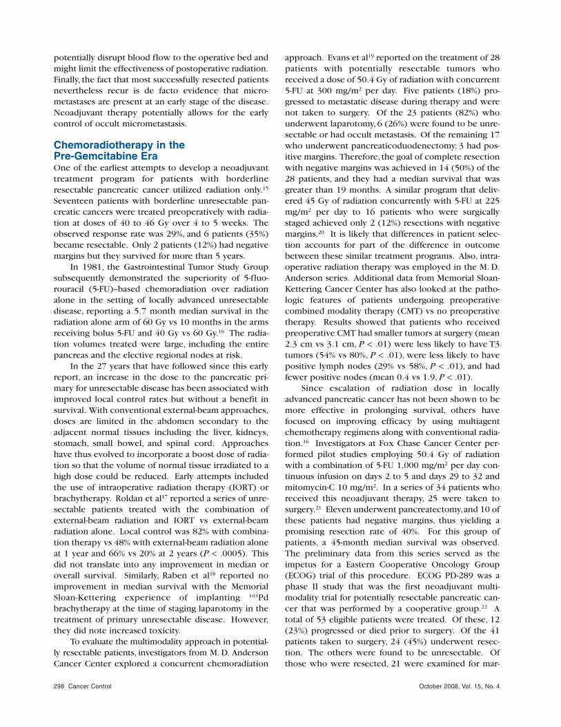
There is general agreement that patients with apatent portal vein (PV), a superior mesenteric vein(SMV),and a fat plane between the tumor and the super-ior mesenteric and celiac arteries without distant metas-tasis are potentially resectable.3 At the Moffitt Multidis-ciplinary Pancreatic Cancer Clinic, these patients arescheduled for immediate surgical resection. Patientswho have encasement of the superior mesenteric artery(SMA), celiac artery, aorta, or inferior vena cava (IVC)are classified as unresectable. PV or SMV encasementby greater than 180° over an extended segment is alsoclassified as unresectable. This is based on the obser-vation that the extent of vessel circumference that is incontact with the tumor correlates with the probabilityof vessel invasion (Table 1).12,13 With ≤ 180° abutment,the likelihood of vessel invasion is overall about 40%.With > 180° abutment, the likelihood of invasion is 80%.When encasement by > 270° is present, vessel invasionis present in almost 100% of cases. Borderline patientsare those who have circumferential tumor abutmentwith the SMV, PV, or SMA over ≤ 180°. Short segment(approximately 1.5 cm) encasement of the PV or SMVthat is amenable to partial vein resection and recon-struction is classified as borderline. Involvement ofboth the PV/SMV and SMA that would require resection
and reconstruction of both arterial and venous systemsis classified as unresectable. Finally, encasement of thegastroduodenal artery up to the origin of the hepaticartery is considered borderline. In these instances, thelikelihood of a resection with positive margins (R1 orR2) is increased. Neoadjuvant therapy is intended tooptimize the likelihood of obtaining an R0 resection. Anadditional criterion is the presence of a teardrop-shapeddeformity of the SMV, which is normally symmetricallyround or oval on CT scan.14 This triangular-shaped pro-trusion from the vessel is a strong indication of vesselinvasion. Its presence, along with other CT scan signs,has a sensitivity of 91%,a specificity of 98% and an accu-racy of 95% in predicting unresectability of pancreaticcancer. Finally, the presence of dense periarterial softtissue portends a low likelihood for resectability,where-as fine periarterial stranding does not.6
Neoadjuvant Therapy for BorderlineResectable Pancreatic Cancer:Past, Present, and FutureThere are a number of theoretical and practical benefitsto neoadjuvant therapy for borderline patients. First,the chief objective is to induce a partial tumor responseand therefore increase the likelihood of completeresection with negative margins. Second, exposure ofthe tumor to chemotherapeutic agents prior to resec-tion allows for the sensitivity of the tumor to thoseagents to be assessed. If the tumor responds, then thesame therapy can be employed if the patient has arecurrence. Third, intensive multiagent multimodalitytreatment programs may be better tolerated by patientsprior to surgery compared to following pancreatico-duodenectomy. Patients who experience major com-plications after surgery may require prolonged recov-ery times during which chemotherapy and radiationmight not be given. Fourth, adequate perfusion shouldallow for the delivery of radiosensitizing oxygen andchemotherapy within the tumor tissue. This shouldincrease the effectiveness of radiation. Surgery may
A B
C
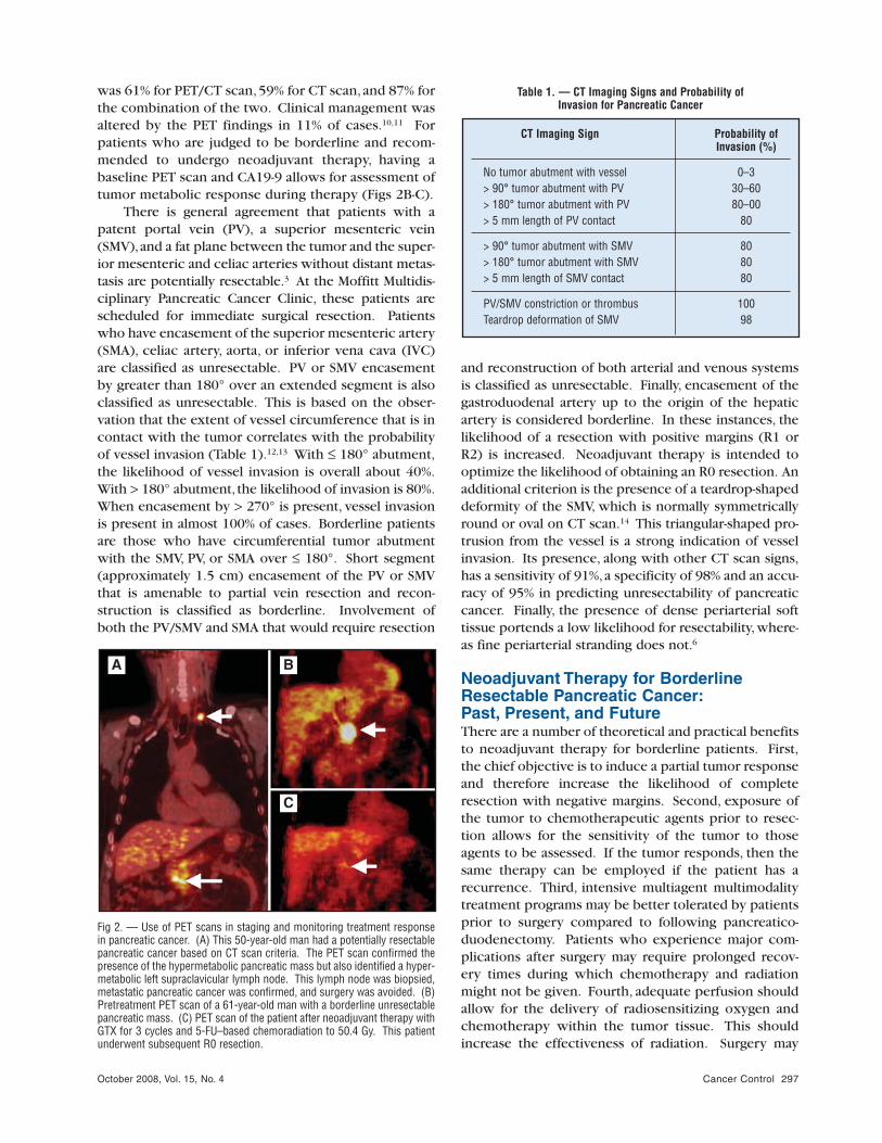
Fig 2. — Use of PET scans in staging and monitoring treatment responsein pancreatic cancer. (A) This 50-year-old man had a potentially resectablepancreatic cancer based on CT scan criteria. The PET scan confirmed thepresence of the hypermetabolic pancreatic mass but also identified a hyper-metabolic left supraclavicular lymph node. This lymph node was biopsied,metastatic pancreatic cancer was confirmed, and surgery was avoided. (B)Pretreatment PET scan of a 61-year-old man with a borderline unresectablepancreatic mass. (C) PET scan of the patient after neoadjuvant therapy withGTX for 3 cycles and 5-FU–based chemoradiation to 50.4 Gy. This patientunderwent subsequent R0 resection.
CT Imaging Sign Probability of Invasion (%)
No tumor abutment with vessel 0–3> 90° tumor abutment with PV 30–60> 180° tumor abutment with PV 80–00> 5 mm length of PV contact 80
> 90° tumor abutment with SMV 80> 180° tumor abutment with SMV 80> 5 mm length of SMV contact 80
PV/SMV constriction or thrombus 100Teardrop deformation of SMV 98
Table 1. — CT Imaging Signs and Probability of Invasion for Pancreatic Cancer
October 2008, Vol. 15, No. 4298 Cancer Control
potentially disrupt blood flow to the operative bed andmight limit the effectiveness of postoperative radiation.Finally, the fact that most successfully resected patientsnevertheless recur is de facto evidence that micro-metastases are present at an early stage of the disease.Neoadjuvant therapy potentially allows for the earlycontrol of occult micrometastasis.
Chemoradiotherapy in the Pre-Gemcitabine EraOne of the earliest attempts to develop a neoadjuvanttreatment program for patients with borderlineresectable pancreatic cancer utilized radiation only.15
Seventeen patients with borderline unresectable pan-creatic cancers were treated preoperatively with radia-tion at doses of 40 to 46 Gy over 4 to 5 weeks. Theobserved response rate was 29%, and 6 patients (35%)became resectable. Only 2 patients (12%) had negativemargins but they survived for more than 5 years.
In 1981, the Gastrointestinal Tumor Study Groupsubsequently demonstrated the superiority of 5-fluo-rouracil (5-FU)–based chemoradiation over radiationalone in the setting of locally advanced unresectabledisease, reporting a 5.7 month median survival in theradiation alone arm of 60 Gy vs 10 months in the armsreceiving bolus 5-FU and 40 Gy vs 60 Gy.16 The radia-tion volumes treated were large, including the entirepancreas and the elective regional nodes at risk.
In the 27 years that have followed since this earlyreport, an increase in the dose to the pancreatic pri-mary for unresectable disease has been associated withimproved local control rates but without a benefit insurvival. With conventional external-beam approaches,doses are limited in the abdomen secondary to theadjacent normal tissues including the liver, kidneys,stomach, small bowel, and spinal cord. Approacheshave thus evolved to incorporate a boost dose of radia-tion so that the volume of normal tissue irradiated to ahigh dose could be reduced. Early attempts includedthe use of intraoperative radiation therapy (IORT) orbrachytherapy. Roldan et al17 reported a series of unre-sectable patients treated with the combination ofexternal-beam radiation and IORT vs external-beamradiation alone. Local control was 82% with combina-tion therapy vs 48% with external-beam radiation aloneat 1 year and 66% vs 20% at 2 years (P < .0005). Thisdid not translate into any improvement in median oroverall survival. Similarly, Raben et al18 reported noimprovement in median survival with the MemorialSloan-Kettering experience of implanting 103Pdbrachytherapy at the time of staging laparotomy in thetreatment of primary unresectable disease. However,they did note increased toxicity.
To evaluate the multimodality approach in potential-ly resectable patients, investigators from M. D. AndersonCancer Center explored a concurrent chemoradiation
approach. Evans et al19 reported on the treatment of 28patients with potentially resectable tumors whoreceived a dose of 50.4 Gy of radiation with concurrent5-FU at 300 mg/m2 per day. Five patients (18%) pro-gressed to metastatic disease during therapy and werenot taken to surgery. Of the 23 patients (82%) whounderwent laparotomy, 6 (26%) were found to be unre-sectable or had occult metastasis. Of the remaining 17who underwent pancreaticoduodenectomy, 3 had pos-itive margins. Therefore, the goal of complete resectionwith negative margins was achieved in 14 (50%) of the28 patients, and they had a median survival that wasgreater than 19 months. A similar program that deliv-ered 45 Gy of radiation concurrently with 5-FU at 225mg/m2 per day to 16 patients who were surgicallystaged achieved only 2 (12%) resections with negativemargins.20 It is likely that differences in patient selec-tion accounts for part of the difference in outcomebetween these similar treatment programs. Also, intra-operative radiation therapy was employed in the M. D.Anderson series. Additional data from Memorial Sloan-Kettering Cancer Center has also looked at the patho-logic features of patients undergoing preoperativecombined modality therapy (CMT) vs no preoperativetherapy. Results showed that patients who receivedpreoperative CMT had smaller tumors at surgery (mean2.3 cm vs 3.1 cm, P < .01) were less likely to have T3tumors (54% vs 80%, P < .01), were less likely to havepositive lymph nodes (29% vs 58%, P < .01), and hadfewer positive nodes (mean 0.4 vs 1.9, P < .01).
Since escalation of radiation dose in locallyadvanced pancreatic cancer has not been shown to bemore effective in prolonging survival, others havefocused on improving efficacy by using multiagentchemotherapy regimens along with conventional radia-tion.16 Investigators at Fox Chase Cancer Center per-formed pilot studies employing 50.4 Gy of radiationwith a combination of 5-FU 1,000 mg/m2 per day con-tinuous infusion on days 2 to 5 and days 29 to 32 andmitomycin-C 10 mg/m2. In a series of 34 patients whoreceived this neoadjuvant therapy, 25 were taken tosurgery.21 Eleven underwent pancreatectomy,and 10 ofthese patients had negative margins, thus yielding apromising resection rate of 40%. For this group ofpatients, a 45-month median survival was observed.The preliminary data from this series served as theimpetus for a Eastern Cooperative Oncology Group(ECOG) trial of this procedure. ECOG PD-289 was aphase II study that was the first neoadjuvant multi-modality trial for potentially resectable pancreatic can-cer that was performed by a cooperative group.22 Atotal of 53 eligible patients were treated. Of these, 12(23%) progressed or died prior to surgery. Of the 41patients taken to surgery, 24 (45%) underwent resec-tion. The others were found to be unresectable. Ofthose who were resected, 21 were examined for mar-
October 2008, Vol. 15, No. 4 Cancer Control 299
gin status, and 7 had positive margins. Therefore, thegoal of resection with negative margins was achievedin a maximum of 17 of the original 53 patients (32%).The overall resection rate (45%) was comparable to theFox Chase experience, but the median survival for theresected patients was lower (16 months vs 20 months).
The Pancreaticobiliary Treatment Group of NewYork employed a three-drug regimen of 5-FU continu-ous infusion 1,000 mg/m2 per day over 108 hours, withcisplatin 100 mg/m2 every 28 days and streptozotocin300 mg/m2 on days 1 through 3 every 28 days.23 Radio-therapy was given to a dose of 54 Gy. The neoadjuvantprogram was given to 68 patients who were unre-sectable based on CT imaging, endoscopic ultrasound,laparoscopy, or laparotomy. It was associated with a27% incidence of grade 3 and 4 toxicity. Of the 68patients, 22 (32%) either progressed during neoadju-vant therapy or did not have an objective radiologicresponse. Of the remaining 46 patients, 22 weredeemed eligible for resection. Six patients (9%) out of 68 had a pathologic complete response. Apparently,1 of 20 resected patients had a positive margin. There-fore, resection with negative margins was achieved for19 of the original 68 patients (28%). The median sur-vival of this group was approximately 32 months.
The available data emphasize the importance ofoptimal patient selection for trimodality therapy inorder to enhance the potential conversion to an R0resection. Results have consistently shown that conver-sion of an unresectable cancer to a resectable one is lesslikely than conversion of a borderline lesion. Massuccoet al24 reported on 28 patients who were treated with aneoadjuvant chemoradiation approach with the resultthat only 1 unresectable tumor was converted success-fully vs 7 out of 18 that were borderline resectable.They further reported that only R0 resections in bothgroups gave the chance of disease-free survival longerthan 24 months. In a series from Duke University Med-ical Center incorporating a median dose of 45 Gy alongwith 5-FU–based chemotherapy, White et al25 reportedthat 28 patients (53%) who were potentially resectableand 11 patients (19%) who were deemed locallyadvanced were able to be converted after chemoradia-tion. They achieved negative surgical margins in 72%and found negative lymph nodes in 70% of resectedpatients. Additional data revealed no increase in mor-bidity or mortality following pancreaticoduodenectomywith neoadjuvant combined-modality therapy.26 In fact,investigators found a marked reduction in the incidenceof pancreatic leak.
Neoadjuvant Chemoradiotherapy in theGemcitabine EraGemcitabine was approved by the US Food and DrugAdministration (FDA) in 1998 and became the standardof care for patients with advanced pancreatic cancer.
This was based on the results of seminal studies per-formed by Burris et al27 that demonstrated improve-ments in median survival (5.6 vs 4.4 months) and clini-cal benefit response (24% vs 5%) when gemcitabine wascompared to bolus 5-FU.27 Response rates to single-agent gemcitabine are typically between 5% and 15% inpatients with locally advanced unresectable andmetastatic pancreatic cancer. Given its superiority inmetastatic pancreatic cancer, it is not surprising that theefficacy of gemcitabine would then be explored in theneoadjuvant setting. The rationale for this strategy wasstrengthened when preclinical studies using humanpancreatic and colon cancer cell lines demonstratedthat gemcitabine was a potent radiosensitizer.28,29
In the late 1990s, efforts to integrate gemcitabineinto chemoradiotherapy programs for locally advancedpancreatic cancer commenced with phase I dose esca-lation trials that were aimed at identifying the maxi-mally tolerated dose (MTD) of gemcitabine when givenwith 50.4 Gy of radiation delivered in 1.8 Gy frac-tions.30 Weekly doses of gemcitabine were escalatedfrom 200 mg/m2 to 700 mg/m2. Dose-limiting gastro-intestinal and hematologic toxicities occurred at 700mg/m2. Responses were seen at doses greater than 500mg/m2. However, doses greater than 400 mg/m2 werealso associated with late duodenal strictures.
A retrospective series of 53 patients with unre-sectable pancreatic cancer treated with gemcitabine250 to 300 mg/m2 per week for 7 weeks with concur-rent hypofractionated radiation (30 to 33 Gy in 10 fractions) was compared to 61 patients treated withconcurrent continuous infusion 5-FU and radiation.31
The radiotherapy volume was large and included notonly the primary tumor but also the regional lymphat-ics in the porta hepatis, celiac axis, and superiormesenteric vessels. Here again, the group receivinggemcitabine as a radiosensitizer experienced greatertoxicities (23% vs 2%) such as mucosal ulceration,hem-orrhage, and hospital admission for supportive care for5 or more days. Two of these patients developed lateduodenal complications, which were ulceration andbowel obstruction. Downstaging to resectability wasaccomplished for 5 (9%) of 53 patients who receivedgemcitabine and 1 (2%) of 61 patients who received 5-FU. There was no significant difference in median sur-vival (11 vs 9 months). Since it appears to be neces-sary to de-escalate the dose of gemcitabine to below400 mg/m2 to reduce toxicity, this approach adminis-ters what is likely a suboptimal dose of gemcitabine forsystemic control of disease in order to maintain con-ventional radiation doses.
Investigators at the University of Michigan Com-prehensive Cancer Center have pursued an alternateapproach to developing a gemcitabine-based chemora-diotherapy.32 The standard dose of gemcitabine usedfor systemic therapy (1,000 mg/m2) was maintained,
October 2008, Vol. 15, No. 4300 Cancer Control
and the dose of conformal radiation was escalated from24 Gy to 42 Gy in 15 fractions of 1.6 to 2.8 Gy. The radi-ation was directed to the primary tumor volume butwithout elective prophylactic nodal coverage. Thirty-four patients with unresectable pancreatic cancer weretreated. At the 42 Gy dose, 2 of 6 patients experienceddose-limiting toxicity. One patient had grade 4 vomit-ing and the other had duodenal ulceration. There were2 complete responses and 8 partial responses. Therewas no evidence of increased nodal failure in thesepatients who were treated without prophylactic elec-tive nodal coverage as only 3 of 67 experienced nodalprogression. In order to reduce the toxicity of the reg-imen, the recommended phase II dose of radiationgoing forward is 36 Gy. Phase II studies evaluating theefficacy of gemcitabine-based chemoradiation areunderway. Most of these are being performed in local-ly advanced unresectable patients rather than the selectsubset of potentially resectable borderline patients.
Another strategy to maintain the systemic efficacyof gemcitabine-based chemoradiotherapy programs isto use combination chemotherapy regimens withdrugs that have synergy with gemcitabine. In the treat-ment of patients with stage IV pancreatic cancer, plat-inum compounds have displayed in vitro, in vivo, andclinical synergy when combined with gemcitabine.Based on this observation, investigators at the Universi-ty Hospital Grosshadern in Munich evaluated the com-bination of gemcitabine and cisplatin in 47 patientswith locally advanced unresectable pancreatic cancer.33
The patients were treated with gemcitabine 300 mg/m2
and cisplatin 30 mg/m2 on days 1, 8, 22, and 29. Thiswas given concurrently with 45 to 50 Gy in 1.8 to 2.0Gy fractions, with the radiation volume consisting ofthe tumor and regional nodes. Restaging with CT scansfound that 27 (60%) of 45 patients responded suffi-ciently to meet criteria for potential resectability. Ofthe 25 patients taken to surgery, 13 had R0 resections,7 had R1 resections, and surgery was aborted at explor-ation in 5 patients due to unresectable disease or metas-tasis. Therefore, the goal of downstaging to resectabilitywith negative margins was accomplished in 13 (29%)of 45 patients. The median survival of the 13 patientswho had R0 resections was 24.2 months comparedwith 8.5 months for those who had R1 resections.Patients who were not downstaged had a median sur-vival of 7.4 months. The main toxicities observed wereneutropenia (68%) and thrombocytopenia (61%).Eleven patients developed cholangitis.
Gemcitabine has also been evaluated in combina-tion with paclitaxel. The Brown University OncologyGroup performed a phase I trial to identify the MTD ofgemcitabine when given with paclitaxel 40 mg/m2
weekly for 6 weeks.34 The dose of gemcitabine wasescalated from 75 mg/m2 to 150 mg/m2. Radiation wasgiven concurrently to a large field encompassing the
primary tumor and the regional peripancreatic, celiac,and porta hepatis nodes to a dose of 45 Gy in 25 frac-tions followed by a 3-fraction boost to the primary plusa 1- to 1.5-cm margin, so that the total dose receivedwas 50.4 Gy. The MTD of gemcitabine was 75 mg/m2.Toxicities included grade 3 and 4 diarrhea, anorexia,dehydration,and pneumonitis. Of 10 patients treated atthe MTD, 4 had radiographic responses and 5 had sta-ble disease. Two of the responders and 1 of those withstable disease were able to undergo resection with neg-ative margins. Therefore, downstaging to resectabilitywith negative margins was accomplished in 3 (30%) of10 patients, and median survival was 11.5 months. Thisprogram is being evaluated in a Radiation TherapyOncology Group phase II trial, RTOG-PA-0200, that hasenrolled 174 patients.
The incorporation of gemcitabine in neoadjuvantconcurrent chemoradiotherapy regimens is feasible.However, either the gemcitabine dose or the radiationdose/volume must be decreased to reduce the inci-dence of severe acute and late gastrointestinal toxicity.There appears to be a trend toward higher responserates with gemcitabine-based chemoradiation that mustbe confirmed in multicenter trials. The optimal doseand schedule of gemcitabine alone or combined withadditional chemotherapy agents and concurrent radia-tion remain to be established.
The optimal radiation field volume, dose, and tech-nique must also be determined. Conventional fieldshave been large, including not only the grossly enlargedprimary tumor and nodes but also the elective nodalregions. The University of Michigan investigators32 havereported no significant risk of nodal failure by targeting
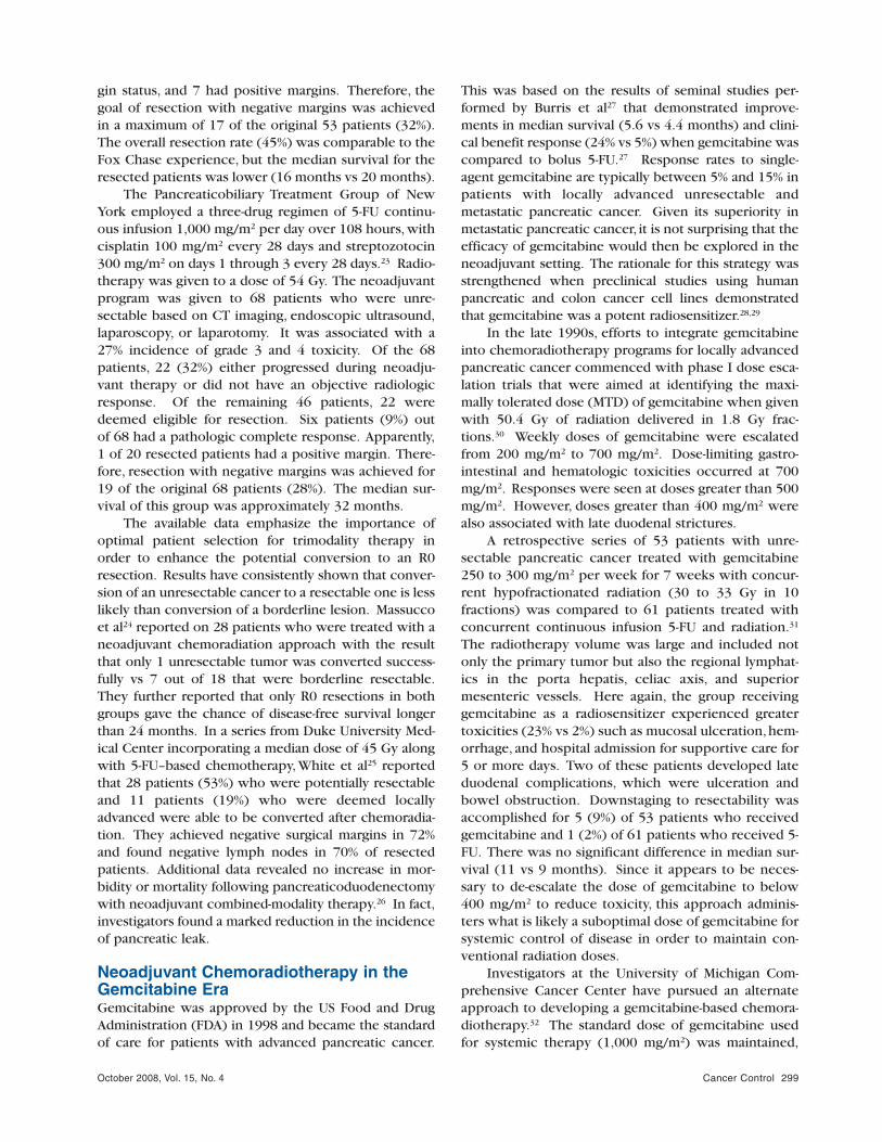
Fig 3. — Representation of the external beams of radiation used to irradiatea patient with disease involving the pancreatic head. Note that the patientis immobilized in the supine position with her arms over her head. Thisallows multiple beam angles to conform the high-dose volume to thepatient’s disease.
October 2008, Vol. 15, No. 4 Cancer Control 301
known disease only. With the advent of PET scanningfor pancreatic cancer patients, radiation oncologistswill be able to incorporate biologic data into delin-eation of the target volume as well. Surgical data havereinforced the concept for omitting elective nodal irra-diation since a series from Johns Hopkins has shownthat an extended retroperitoneal lymphadenectomyresulted in increased complications but no increasedbenefit.35,36 By designing smaller radiation volumeswith less mucosa in the path of the beams,more aggres-sive combined regimens may be feasible. The RadiationTherapy Oncology Group has begun excluding unin-volved lymphatics from the treatment volumes on itsrecent protocols, such as RTOG-0411.
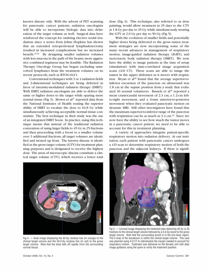
Conventional techniques with 3 or 4 radiation fieldsand 3-dimensional techniques are being deferred infavor of intensity-modulated radiation therapy (IMRT).With IMRT, radiation oncologists are able to deliver thesame or higher doses to the target while sparing morenormal tissue (Fig 3). Brown et al37 reported data fromthe National Institutes of Health touting the superiorability of IMRT to escalate the dose to 64.8 Gy whilesimultaneously achieving acceptable normal tissue con-straints. The best technique in their study was the useof an integrated IMRT boost. In practice,using this tech-nique means that instead of the traditional radiationconvention of using larger fields to 45 Gy in 25 fractionsand then proceeding with a boost to a smaller volumeover 3 additional fractions, all target volumes are identi-fied and treated up front. The known disease is identi-fied as the gross target volume (GTV) for treatment plan-ning purposes and is designated to receive the highestdose. The areas of microscopic disease constitute a clin-ical target volume (CTV), which receives a lower total
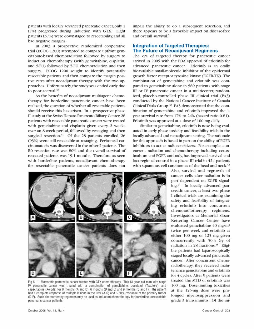
dose (Fig 4). This technique, also referred to as dosepainting, would allow treatment in 25 days to the CTVat 1.8 Gy per day to 45 Gy while simultaneously treatingthe GTV at 2.0 Gy per day to 50 Gy (Fig 5).
With the evolution of smaller fields and potentiallyhigher doses being delivered to the gross tumor, treat-ment strategies are now incorporating some of themany recent advances in management of respiratorymotion, image-guided radiation therapy (IGRT), andstereotactic body radiation therapy (SBRT). We nowhave the ability to image patients at the time of setup(simulation) with time-correlated image acquisitionscans (4-D CT). These scans are able to image thetumor in the upper abdomen as it moves with respira-tion. Bryan et al38 found that the average superior-to-inferior excursion of the pancreas on ultrasound was1.8 cm in the supine position from a study that evalu-ated 36 normal volunteers. Bussels et al39 reported amean craniocaudal movement of 2.3 cm, a 1.2-cm left-to-right movement, and a 6-mm anterior-to-posteriormovement when they evaluated pancreatic motion ondynamic MRI. Still other investigators have found thatthe maximum superior-to-inferior range of the pancreaswith respiration can be as much as 3.4 cm.40 Since wenow have the ability to see how much the tumor movesin a pancreatic cancer patient, we need to be able toaccount for this in treatment planning.
A variety of approaches integrate patient-specificrespiratory motion into radiation delivery. At our insti-tution, each patient with pancreatic cancer undergoesa 4-D scan to determine respiratory motion of both thepancreas and the adjacent kidneys. If there is signifi-
Fig 5. — Coronal image displaying the treatment plan delivering 45 Gy in 25fractions to the clinical target volume followed by a 5.4-Gy boost to the grosstarget volume. Note that the surrounding bowel is in the low-dose region.The C-loop of the duodenum is within the clinical target volume. This casewas planned using 4-D CT to individualize the margin needed to account forrespiratory motion. Treatment was delivered on the Novalis unit with dailyimage guidance using the spine to verify the treatment position.
Fig 4. — Axial image displaying the 45-Gy isodose line (in orange) to theclinical target volume and the 50.4-Gy isodose line (in red) to the gross target volume. Note that the dose falls off rapidly from the surroundingnormal tissue.

October 2008, Vol. 15, No. 4302 Cancer Control
cant motion, the target volumes from the maximuminspiration and expiration scans are delineated andfused together to create what is called an internal tar-get volume. An additional planning volume margin isthen created around the internal target volume. Thisway, the pancreatic target, no matter where it is at anygiven time, will receive the prescribed dose. Otherinstitutions are developing gated technology wherebythe radiation delivery machine turns “on” only whenthe tumor is in a certain phase of the breathingcycle.41,42 There are also a variety of patient breath holdtechniques, including those that train patients to holdtheir breath by themselves or with the aid of an activebreathing control device that occludes airflow duringexhalation while the beam is on.43,44
Other techniques to account for motion incorpo-rate the implantation of fiducial markers directly intothe pancreas (either by CT or at surgical exploration)and the ability to track the markers during treatment.This approach takes advantage of newer IGRT capa-bilities so that the markers can be imaged on a dailybasis to ensure that the intended target receives theintended dose of radiation. It is in this setting thatSBRT or extracranial stereotactic radiosurgery (SRS) isbeing explored. This concept originated with intracra-nial SRS where a single fraction was delivered withhigh precision to a fixed target. With the advent ofextracranial delivery systems, sites can now be treatedin the lung, liver, and pancreas. SRS refers to single-fraction treatment and SBRT refers to delivery in mul-tiple fractions.
Koong et al45 have shown that 100% local control ispossible with a single fraction of extracranial radio-surgery. In their phase I trial, they evaluated locallyadvanced tumors less than 7.5 cm in greatest dimen-sion and did not include elective sites. Gold fiducialmarkers were implanted directly into the tumor by CT,laparoscopy,or open laparotomy. The stereotactic treat-ment was then delivered with the CyberKnife (AccurayInc, Sunnyvale, California), with escalating doses froman initial 15 Gy, then 20 Gy,and finally 25 Gy. With com-plete local control at the 25-Gy dose, the trial wasstopped. The rate of distant progression was high, andthus the median overall survival was only 11 months.
Koong et al46 have sought to improve these resultsby adding a course of conventionally fractionatedtreatment to the SRS boost. In this phase II trial, theydesigned an IMRT field targeting the tumor andregional nodes (peripancreatic, celiac, superiormesenteric, porta hepatic, and retroperitoneal) to 45Gy in 25 fractions. Patients received current 5-FU–based chemotherapy during irradiation. One monthafter the combined modality therapy, a 25-Gy SRSboost was delivered with the CyberKnife. A 94% localcontrol rate was reported, with distant progression asthe first site in all cases. Median overall survival was
33 weeks. The trial reported more gastrointestinal tox-icity with RTOG grade 3 events acutely and with thedevelopment of symptomatic duodenal ulcers 4 to 6months following treatment.
Therefore, to design an optimal chemoradiationprogram, the goal would be to define the gross diseaseas precisely as possible, determine its motion with res-piration, limit the volume of adjacent normal tissue toallow more potent radiosensitizing agents to be givenconcurrently, and validate on a daily basis that the vol-ume intended to be irradiated is actually receiving thedose. In the next few years, additional data should beavailable to guide further selection and integration ofthese treatment techniques.
Induction Chemotherapy Followed byChemoradiationAs noted in many of the studies discussed previously,between 10% and 30% of patients will experience dis-ease progression during chemoradiation. The presenceof metastatic sites of disease frequently becomes mani-fest in locations outside of the radiation field. Again,this is a reminder that micrometastatic disease is usual-ly already present at initial diagnosis despite the local-ized appearance on imaging studies. Understandingthis reality, investigators at the M. D. Anderson CancerCenter hypothesized that a period of inductionchemotherapy might allow for the selection of thoselocally advanced patients who might truly benefit fromsubsequent chemoradiation. Patients who developmetastasis during induction chemotherapy can go onto other chemotherapy or clinical trials. Nonprogress-ing patients could be treated for 2 to 3 months andthen given chemoradiation. Krishnan et al47 conducteda retrospective analysis of 323 patients with locallyadvanced pancreatic cancer who received eitherupfront chemoradiation (n = 247) or induction chemo-therapy with a gemcitabine-based regimen (mostlygemcitabine and cisplatin, n = 76) for a median of 2.5months. Median overall survival was superior in theinduction chemotherapy group compared to theupfront chemoradiation group (11.0 vs 8.5 months, P <.001). There was no difference between the twogroups in the proportion of patients who progressedlocally vs at distant sites. This suggests that the briefcourse of gemcitabine and cisplatin was not effective inclearing micrometastatic sites.
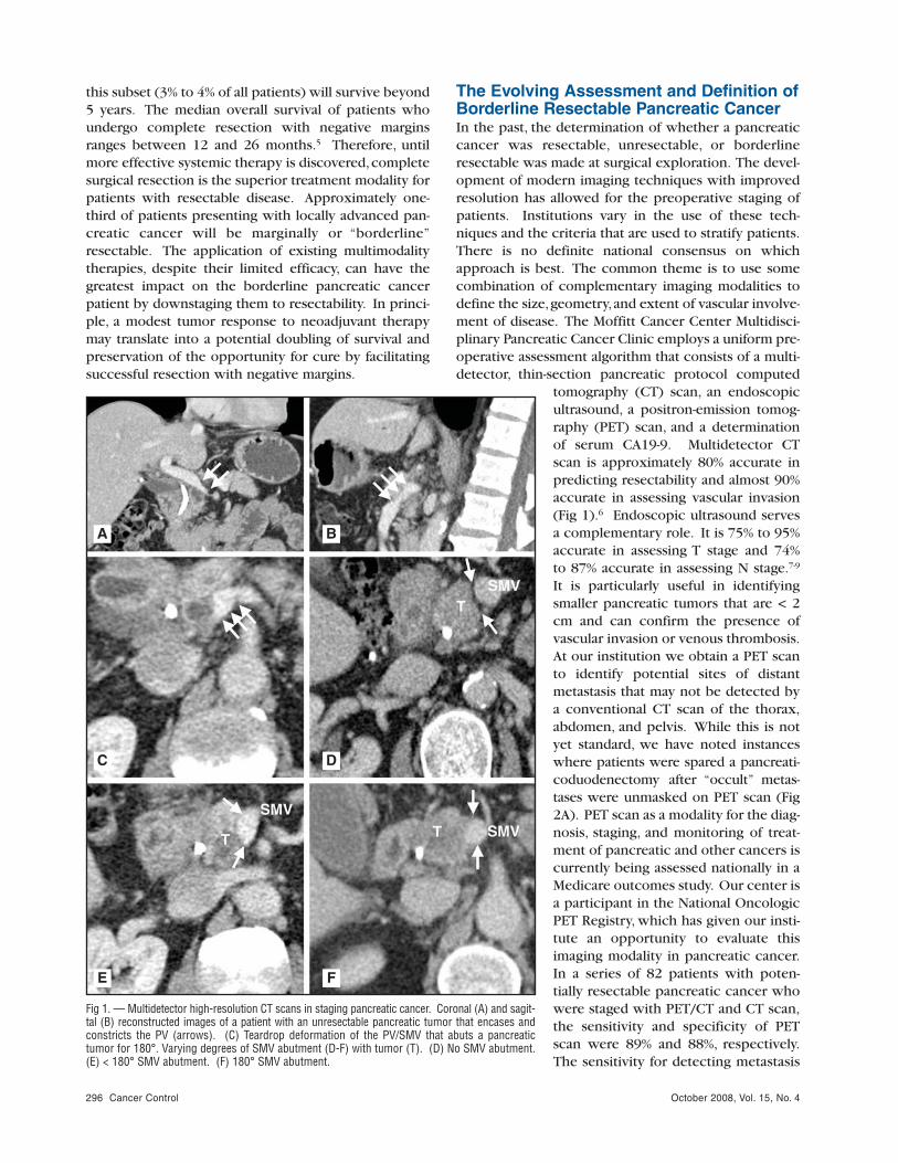
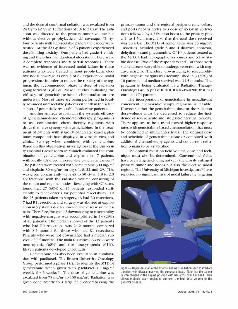
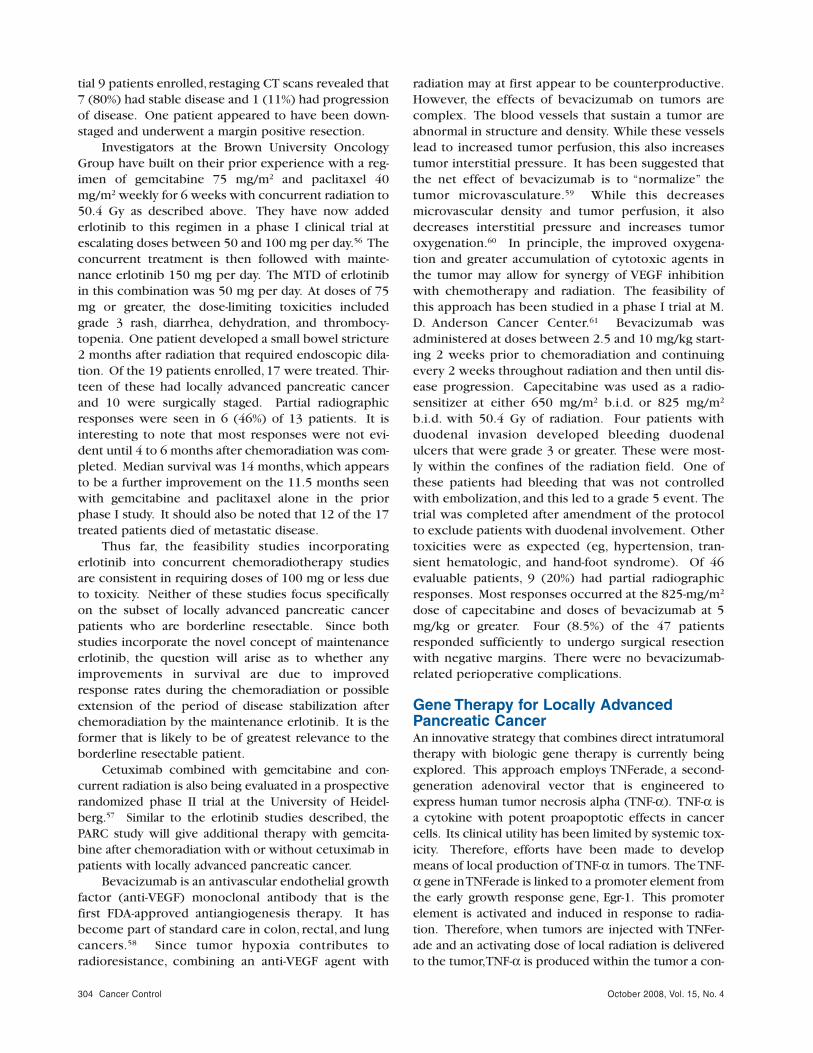
A similar strategy was employed by Columbia Uni-versity using the GTX regimen consisting of gemcitabine,docetaxel (Taxotere), and capecitabine (Xeloda) as in-duction chemotherapy for 3 cycles over 9 weeks fol-lowed by gemcitabine-based chemoradiation.48 GTXwas developed based on the demonstration of preclini-cal in vitro synergy of the combination. In the metastat-ic setting, the regimen reportedly has a response ratethat approaches 30% to 40% (Fig 6).49 In a series of 14
October 2008, Vol. 15, No. 4 Cancer Control 303
patients with locally advanced pancreatic cancer, only 1(7%) progressed during induction with GTX. Eightpatients (57%) were downstaged to resectability, and allhad negative margins.
In 2003, a prospective, randomized cooperativetrial (ECOG 1200) attempted to compare upfront gem-citabine-based chemoradiation followed by surgery toinduction chemotherapy (with gemcitabine, cisplatin,and 5-FU) followed by 5-FU chemoradiation and thensurgery. ECOG 1200 sought to identify potentiallyresectable patients and then compare the margin posi-tive rates after neoadjuvant therapy with the two ap-proaches. Unfortunately, the study was ended early dueto poor accrual.50
As the benefits of neoadjuvant multiagent chemo-therapy for borderline pancreatic cancer have beenrealized, the question of whether all resectable patientsshould receive this has arisen. In a prospective phaseII study at the Swiss Hepato-Pancreato-Biliary Center,28patients with resectable pancreatic cancer were treatedwith gemcitabine and cisplatin given every 2 weeksover an 8-week period, followed by restaging and thensurgical resection.51 Of the 28 patients enrolled, 26(93%) were still resectable at restaging. Peritoneal car-cinomatosis was discovered in the other 2 patients. TheR0 resection rate was 80% and the overall survival ofresected patients was 19.1 months. Therefore, as seenwith borderline patients, neoadjuvant chemotherapyfor resectable pancreatic cancer patients does not
impair the ability to do a subsequent resection, andthere appears to be a favorable impact on disease-freeand overall survival.51
Integration of Targeted Therapies:The Future of Neoadjuvant RegimensThe era of targeted therapy for pancreatic cancerarrived in 2005 with the FDA approval of erlotinib foradvanced pancreatic cancer. Erlotinib is an orallybioavailable small-molecule inhibitor of the epidermalgrowth factor receptor tyrosine kinase (EGFR-TK). Thecombination of gemcitabine and erlotinib was com-pared to gemcitabine alone in 569 patients with stageIII or IV pancreatic cancer in a multicenter, random-ized, placebo-controlled phase III clinical trial (PA3)conducted by the National Cancer Institute of CanadaClinical Trials Group.52 PA3 demonstrated that the com-bination of gemcitabine and erlotinib improved the 1-year survival rate from 17% to 24% (hazard ratio 0.81).Erlotinib was approved at a dose of 100 mg daily.
Similar to gemcitabine, erlotinib is now being eval-uated in early-phase toxicity and feasibility trials in thelocally advanced and neoadjuvant setting. The rationalefor this approach is based in part on the ability of EGFRinhibitors to act as radiosensitizers. For example, con-current radiation and chemotherapy including cetux-imab, an anti-EGFR antibody, has improved survival andlocoregional control in a phase III trial in 424 patientswith squamous cell carcinomas of the head and neck.53
Also, survival and regrowth ofcancer cells after radiation is inpart dependent on EGFR signal-ing.54 In locally advanced pan-creatic cancer, at least two phaseI clinical trials are examining thesafety and feasibility of integrat-ing erlotinib into concurrentchemoradiotherapy regimens.Investigators at Memorial Sloan-Kettering Cancer Center haveevaluated gemcitabine 40 mg/m2
twice per week and erlotinib ateither 100 mg or 125 mg givenconcurrently with 50.4 Gy ofradiation in 28 fractions.55 Eligi-ble patients had laparoscopicallystaged locally advanced pancreaticcancer. After concurrent chemo-radiotherapy, they received main-tenance gemcitabine and erlotinibfor 4 cycles. After 9 patients weretreated, the MTD of erlotinib was100 mg. Dose-limiting toxicitiesat the 125-mg dose were pro-longed myelosuppression andgrade 3 transaminitis. Of the ini-
A
D
B
E
C
F
Fig 6. — Metastatic pancreatic cancer treated with GTX chemotherapy. This 64-year-old man with stageIV pancreatic cancer was treated with a combination of gemcitabine, docetaxel (Taxotere), andcapecitabine (Xeloda) for 0 months (A and D), 6 months (B and E) and 9 months (C and F). The patienthad a complete response of multiple lesions in the liver (A-C) and > 50% response of the primary tumor(D-F). Such chemotherapy regimens may be used as induction chemotherapy for borderline unresectablepancreatic cancer patients.
October 2008, Vol. 15, No. 4304 Cancer Control
tial 9 patients enrolled, restaging CT scans revealed that7 (80%) had stable disease and 1 (11%) had progressionof disease. One patient appeared to have been down-staged and underwent a margin positive resection.
Investigators at the Brown University OncologyGroup have built on their prior experience with a reg-imen of gemcitabine 75 mg/m2 and paclitaxel 40mg/m2 weekly for 6 weeks with concurrent radiation to50.4 Gy as described above. They have now addederlotinib to this regimen in a phase I clinical trial atescalating doses between 50 and 100 mg per day.56 Theconcurrent treatment is then followed with mainte-nance erlotinib 150 mg per day. The MTD of erlotinibin this combination was 50 mg per day. At doses of 75mg or greater, the dose-limiting toxicities includedgrade 3 rash, diarrhea, dehydration, and thrombocy-topenia. One patient developed a small bowel stricture2 months after radiation that required endoscopic dila-tion. Of the 19 patients enrolled, 17 were treated. Thir-teen of these had locally advanced pancreatic cancerand 10 were surgically staged. Partial radiographicresponses were seen in 6 (46%) of 13 patients. It isinteresting to note that most responses were not evi-dent until 4 to 6 months after chemoradiation was com-pleted. Median survival was 14 months, which appearsto be a further improvement on the 11.5 months seenwith gemcitabine and paclitaxel alone in the priorphase I study. It should also be noted that 12 of the 17treated patients died of metastatic disease.
Thus far, the feasibility studies incorporatingerlotinib into concurrent chemoradiotherapy studiesare consistent in requiring doses of 100 mg or less dueto toxicity. Neither of these studies focus specificallyon the subset of locally advanced pancreatic cancerpatients who are borderline resectable. Since bothstudies incorporate the novel concept of maintenanceerlotinib, the question will arise as to whether anyimprovements in survival are due to improvedresponse rates during the chemoradiation or possibleextension of the period of disease stabilization afterchemoradiation by the maintenance erlotinib. It is theformer that is likely to be of greatest relevance to theborderline resectable patient.
Cetuximab combined with gemcitabine and con-current radiation is also being evaluated in a prospectiverandomized phase II trial at the University of Heidel-berg.57 Similar to the erlotinib studies described, thePARC study will give additional therapy with gemcita-bine after chemoradiation with or without cetuximab inpatients with locally advanced pancreatic cancer.
Bevacizumab is an antivascular endothelial growthfactor (anti-VEGF) monoclonal antibody that is thefirst FDA-approved antiangiogenesis therapy. It hasbecome part of standard care in colon, rectal, and lungcancers.58 Since tumor hypoxia contributes toradioresistance, combining an anti-VEGF agent with
radiation may at first appear to be counterproductive.However, the effects of bevacizumab on tumors arecomplex. The blood vessels that sustain a tumor areabnormal in structure and density. While these vesselslead to increased tumor perfusion, this also increasestumor interstitial pressure. It has been suggested thatthe net effect of bevacizumab is to “normalize” thetumor microvasculature.59 While this decreasesmicrovascular density and tumor perfusion, it alsodecreases interstitial pressure and increases tumoroxygenation.60 In principle, the improved oxygena-tion and greater accumulation of cytotoxic agents inthe tumor may allow for synergy of VEGF inhibitionwith chemotherapy and radiation. The feasibility ofthis approach has been studied in a phase I trial at M.D. Anderson Cancer Center.61 Bevacizumab wasadministered at doses between 2.5 and 10 mg/kg start-ing 2 weeks prior to chemoradiation and continuingevery 2 weeks throughout radiation and then until dis-ease progression. Capecitabine was used as a radio-sensitizer at either 650 mg/m2 b.i.d. or 825 mg/m2
b.i.d. with 50.4 Gy of radiation. Four patients withduodenal invasion developed bleeding duodenalulcers that were grade 3 or greater. These were most-ly within the confines of the radiation field. One ofthese patients had bleeding that was not controlledwith embolization, and this led to a grade 5 event. Thetrial was completed after amendment of the protocolto exclude patients with duodenal involvement. Othertoxicities were as expected (eg, hypertension, tran-sient hematologic, and hand-foot syndrome). Of 46evaluable patients, 9 (20%) had partial radiographicresponses. Most responses occurred at the 825-mg/m2
dose of capecitabine and doses of bevacizumab at 5mg/kg or greater. Four (8.5%) of the 47 patientsresponded sufficiently to undergo surgical resectionwith negative margins. There were no bevacizumab-related perioperative complications.
Gene Therapy for Locally Advanced Pancreatic CancerAn innovative strategy that combines direct intratumoraltherapy with biologic gene therapy is currently beingexplored. This approach employs TNFerade, a second-generation adenoviral vector that is engineered toexpress human tumor necrosis alpha (TNF-α). TNF-α isa cytokine with potent proapoptotic effects in cancercells. Its clinical utility has been limited by systemic tox-icity. Therefore, efforts have been made to developmeans of local production of TNF-α in tumors. The TNF-α gene in TNFerade is linked to a promoter element fromthe early growth response gene, Egr-1. This promoterelement is activated and induced in response to radia-tion. Therefore, when tumors are injected with TNFer-ade and an activating dose of local radiation is deliveredto the tumor,TNF-α is produced within the tumor a con-

October 2008, Vol. 15, No. 4 Cancer Control 305
centrations that can induce apoptosis and cellular necro-sis. The safety, toxicity,and preliminary efficacy was eval-uated in a phase I study in 36 patients with varioustumor types.62 The most frequent toxicities were fever,injection site pain, and chills. No dose-limiting toxicitiesoccurred. Seventy percent of the treated patients had anobjective radiologic response of the treated lesion,including 5 patients who had complete responses. Fivepatients with pancreatic cancer were treated and 3 hadresponses. Based on these findings, a large randomized,controlled multicenter phase II/III trial of TNFerade, des-ignated PACT, was initiated in patients with locallyadvanced pancreatic cancer. This trial will enroll 330patients. The results of an interim analysis of the first 51patients has been reported.63 Currently, the 1-year sur-vival rate is 70.5% for the TNFerade arm compared with28% for the control arm. If the efficacy of this strategy isconfirmed, it may also be integrated into the manage-ment of borderline pancreatic cancer patients.
ConclusionsThere has been improvement in the noninvasive assess-ment and neoadjuvant treatment of borderlineresectable pancreatic cancer. The complementary useof high-resolution, multidetector pancreatic protocolCT scans and endoscopic ultrasound has allowed forimproved selection of patients who are likely to benefit
from neoadjuvant therapy. PET scan is currently beingevaluated as a means to identify metastasis and assessmetabolic response to therapy. It also has the potentialof confirming smaller volumes for radiation therapy tar-geting. Over the past 30 years, neoadjuvant therapy hasevolved from conventional radiation therapy alone tocombined modality therapy with multiagent chemo-therapy regimens employing potent radiosensitizersalong with optimized radiotherapy (Table 2). Novelradiation imaging techniques and planning capabilitiesare providing the framework for limiting normal tissuetoxicities without compromising dose. Improvementsin local control have the potential to be maximized andintegrated with the best sensitizing agents to achieve R0 resection in this borderline patient population. Theweight of available evidence suggests that the rate ofsuccessful resections and survival can be improved. Thenotion that locally advanced pancreatic cancer cannotbe downstaged is false. The optimal regimen for neoad-juvant therapy remains to be defined. Prospective con-trolled trials are needed in patients who meet carefullydefined modern imaging criteria to compare the variousapproaches that are under development. Pancreaticcancer should be a high-priority disease for testing themany targeted agents now being developed. As newmolecular targets are discovered, validated, and movedto clinical translation, the opportunities for more effec-
Trial Radiation Treatment Chemotherapy Treatment % Resectability With Negative Margins
Pilepich 198015 40–46 Gy None 12 (2/17)
Evans 199219 50.4 Gy Concurrent 5-FU 300 mg/m2/day 50 (14/28)
Jessup 199320 45 Gy Concurrent 5-FU 225 mg/m2/day 12 (2/16)
Hoffman 199521 50.4 Gy Concurrent 5-FU 1000 mg/m2/day 29 (10/34)Mitomycin-C 10 mg/m2/day
Hoffman 199822 50.4 Gy Concurrent 5-FU 1000 mg/m2/day 32 (17/53)Mitomycin-C 10 mg/m2/day
Snady 200023 54 Gy Concurrent 5-FU 1000 mg/m2/day 28 (19/68)Cisplatin 100 mg/m2/day Streptozotocin 300 mg/m2
Crane 200231 30–33 Gy Concurrent gemcitabine 9 (5/53)250-300 mg/m2/wk for 7 wks
Wilkowski 200433 45–50 Gy Concurrent gemcitabine 300 mg/m2 29 (13/45)Cisplatin 30 mg/m2
Safran 200234 50.4 Gy Concurrent gemcitabine 75 mg/m2 30 (3/10)Paclitaxel 40 mg/m2
Fogelman 200748 45 Gy Induction chemotherapy: 57 (8/14)Gemcitabine 750 mg/m2
Docetaxel 30 mg/m2
Capecitabine 750 mg/m2 then:
Concurrent Gemcitabine 250 mg/m2
Crane 200661 50.4 Gy Concurrent capecitabine 825 mg/m2 8 (4/47)Bevacizumab 2.5–10 mg/kg
Table 2. — Selected Clinical Trials Relevant to Borderline Unresectable Pancreatic Cancer
October 2008, Vol. 15, No. 4306 Cancer Control
tive therapy of borderline patients can only increase.After all, these patients are already on the edge ofachieving longer survival.
DisclosuresDr Springett receives grant support from the Robert Wood JohnsonFoundation, Amos Faculty Development Program, NIH grant2U19CA067771-11, and Genentech Inc. Dr Hoffe receives grantsupport and honoraria from GE Healthcare.
References1. Varadhachary GR, Tamm EP, Abbruzzese JL, et al. Borderline
resectable pancreatic cancer: definitions, management, and role of preop-erative therapy. Ann Surg Oncol. 2006;13(8):1035-1046. Epub 2006 Jul 24.
2. Quiros RM, Brown KM, Hoffman JP, et al. Neoadjuvant therapy inpancreatic cancer. Cancer Invest. 2007;25(4):267-273.
3. NCCN practice guidelines for pancreatic cancer. Oncology (WillistonPark). 1997;11(11A):41-55.
4. Yendluri V, Centeno B, Springett GM, et al. Pancreatic cancer pre-senting as a Sister Mary Joseph’s nodule: case report and update of the lit-erature. Pancreas. 2007;34(1):161-164.
5. Mehta VK, Fisher G, Ford JA, et al. Preoperative chemoradiation formarginally resectable adenocarcinoma of the pancreas. J GastrointestSurg. 2001;5(1):27-35.
6. Valls C, Andía E, Sanchez A, et al. Dual-phase helical CT of pan-creatic adenocarcinoma: assessment of resectability before surgery. AJRAm J Roentgenol. 2002;178(4):821-826.
7. Legmann P, Vignaux O, Dousset B, et al. Pancreatic tumors: com-parison of dual-phase helical CT and endoscopic sonography. AJR Am JRoentgenol. 1998;170(5):1315-1322.
8. DeWitt J, Devereaux B, Chriswell M, et al. Comparison of endo-scopic ultrasonography and multidetector computed tomography for detect-ing and staging pancreatic cancer. Ann Intern Med. 2004;141(10):753-763.
9. Michl P, Pauls S, Gress TM. Evidence-based diagnosis and staging ofpancreatic cancer. Best Pract Res Clin Gastroenterol. 2006;20(2):227-251.
10. Farma JM, Melis M, Santillan A, et al. Positron emission tomogra-phy/computed tomography fusion scans enhances preoperative treatmentplanning in patients with pancreatic cancer. In: Proceedings from the 61stAnnual Society of Surgical Oncology Meeting; March 13-16, 2008; Chica-go, IL. Abstract 10.
11. Farma JM, Santillan AA, Melis M, et al. PET/CT fusion scan enhancesCT staging in patients with pancreatic neoplasms. Ann Surg Oncol. 2008 Jun13. Epub ahead of print.
12. Lu DS, Reber HA, Krasny RM, et al. Local staging of pancreatic can-cer: criteria for unresectability of major vessels as revealed by pancreatic-phase, thin-section helical CT. AJR Am J Roentgenol. 1997;168(6):1439-1443.
13. Schima W, Ba-Ssalamah A, Kölblinger C, et al. Pancreatic adeno-carcinoma. Eur Radiol. 2007;17(3):638-649. Epub 2006 Oct 5.
14. Hough TJ, Raptopoulos V, Siewert B, et al. Teardrop superior mesen-teric vein: CT sign for unresectable carcinoma of the pancreas. AJR Am JRoentgenol. 1999;173(6):1509-1512.
15. Pilepich MV, Miller HH. Preoperative irradiation in carcinoma of thepancreas. Cancer. 1980;46(9):1945-1949.
16. Moertel CG, Frytak S, Hahn RG, et al. Therapy of locally unre-sectable pancreatic carcinoma: a randomized comparison of high dose(6000 rads) radiation alone, moderate dose radiation (4000 rads + 5-fluo-rouracil), and high dose radiation + 5-fluorouracil. The GastrointestinalTumor Study Group. Cancer. 1981;48(8):1705-1710.
17. Roldan GE, Gunderson LL, Nagorney DM, et al. External beam ver-sus intraoperative and external beam irradiation for locally advanced pan-creatic cancer. Cancer. 1988;61(6):1110-1116.
18. Raben A, Mychalczak B, Brennan MF, et al. Feasibility study of thetreatment of primary unresectable carcinoma of the pancreas with 103Pdbrachytherapy. Int J Radiat Oncol Biol Phys. 1996;35(2):351-356.
19. Evans DB, Rich TA, Byrd DR, et al. Preoperative chemoradiation andpancreaticoduodenectomy for adenocarcinoma of the pancreas. Arch Surg.1992;127(11):1335-1339.
20. Jessup JM, Steele G Jr, Mayer RJ, et al. Neoadjuvant therapy forunresectable pancreatic adenocarcinoma. Arch Surg. 1993;128(5):559-564.
21. Hoffman JP, Weese JL, Solin LJ, et al. A pilot study of preoperativechemoradiation for patients with localized adenocarcinoma of the pancreas.Am J Surg. 1995;169(1):71-77; discussion 77-78.
22. Hoffman JP, Lipsitz S, Pisansky T, et al. Phase II trial of preoperativeradiation therapy and chemotherapy for patients with localized, resectableadenocarcinoma of the pancreas: an Eastern Cooperative Oncology GroupStudy. J Clin Oncol. 1998;16(1):317-323.
23. Snady H, Bruckner H, Cooperman A, et al. Survival advantage ofcombined chemoradiotherapy compared with resection as the initial treat-ment of patients with regional pancreatic carcinoma: an outcomes trial.
Cancer. 2000;89(2):314-327.24. Massucco P, Capussotti L, Magnino A, et al. Pancreatic resections
after chemoradiotherapy for locally advanced ductal adenocarcinoma: analy-sis of perioperative outcome and survival. Ann Surg Oncol. 2006;13(9):1201-1208. Epub 2006 Sep 6.
25. White RR, Hurwitz HI, Morse MA, et al. Neoadjuvant chemoradiationfor localized adenocarcinoma of the pancreas. Ann Surg Oncol. 2001;8(10):758-765.
26. Cheng TY, Sheth K, White RR, et al. Effect of neoadjuvant chemora-diation on operative mortality and morbidity for pancreaticoduodenectomy.Ann Surg Oncol. 2006;13(1):66-74. Epub 2006 Jan 1.
27. Burris HA 3rd, Moore MJ, Andersen J, et al. Improvements in sur-vival and clinical benefit with gemcitabine as first-line therapy for patientswith advanced pancreas cancer: a randomized trial. J Clin Oncol. 1997;15(6):2403-2413.
28. Lawrence TS, Chang EY, Hahn TM, et al. Radiosensitization of pan-creatic cancer cells by 2’,2’-difluoro-2’-deoxycytidine. Int J Radiat Oncol BiolPhys. 1996;34(4):867-872.
29. Shewach DS, Lawrence TS. Gemcitabine and radiosensitization inhuman tumor cells. Invest New Drugs. 1996;14(3):257-263.
30. McGinn CJ, Zalupski MM. Radiation therapy with once-weekly gem-citabine in pancreatic cancer: current status of clinical trials. Int J RadiatOncol Biol Phys. 2003;56(4 suppl):10-15.
31. Crane CH, Abbruzzese JL, Evans DB, et al. Is the therapeutic indexbetter with gemcitabine-based chemoradiation than with 5-fluorouracil-based chemoradiation in locally advanced pancreatic cancer? Int J RadiatOncol Biol Phys. 2002;52(5):1293-1302.
32. McGinn CJ, Zalupski MM, Shureiqi I, et al. Phase I trial of radiationdose escalation with concurrent weekly full-dose gemcitabine in patientswith advanced pancreatic cancer. J Clin Oncol. 2001;19(22):4202-4208.
33. Wilkowski R, Thoma M, Schauer R, et al. Effect of chemoradiother-apy with gemcitabine and cisplatin on locoregional control in patients withprimary inoperable pancreatic cancer. World J Surg. 2004;28(10):1011-1018. Epub 2004 Sep 29.
34. Safran H, Dipetrillo T, Iannitti D, et al. Gemcitabine, paclitaxel, andradiation for locally advanced pancreatic cancer: a phase I trial. Int J Radi-at Oncol Biol Phys. 2002;54(1):137-141.
35. Yeo CJ, Abrams RA, Grochow LB, et al. Pancreaticoduodenectomyfor pancreatic adenocarcinoma: postoperative adjuvant chemoradiationimproves survival. A prospective, single-institution experience. Ann Surg.1997;225(5):621-633; discussion 633-636.
36. Riall TS, Cameron JL, Lillemoe KD, et al. Pancreaticoduodenectomywith or without distal gastrectomy and extended retroperitoneal lym-phadenectomy for periampullary adenocarcinoma. Part 3: update on 5-yearsurvival. J Gastrointest Surg. 2005;9(9):1191-1204; discussion 1204-1206.
37. Brown MW, Ning H, Arora B, et al. A dosimetric analysis of doseescalation using two intensity-modulated radiation therapy techniques inlocally advanced pancreatic carcinoma. Int J Radiat Oncol Biol Phys. 2006;65(1):274-283.
38. Bryan PJ, Custar S, Haaga JR, et al. Respiratory movement of thepancreas: an ultrasonic study. J Ultrasound Med. 1984;3(7):317-320.
39. Bussels B, Goethals L, Feron M, et al. Respiration-induced move-ment of the upper abdominal organs: a pitfall for the three-dimensional con-formal radiation treatment of pancreatic cancer. Radiother Oncol. 2003;68(1):69-74.
40. Bhasin DK, Rana SS, Jahagirdar S, et al. Does the pancreas movewith respiration? J Gastroenterol Hepatol. 2006;21(9):1424-1427.
41. Gierga DP, Brewer J, Sharp GC, et al. The correlation between inter-nal and external markers for abdominal tumors: implications for respiratorygating. Int J Radiat Oncol Biol Phys. 2005;61(5):1551-1558. Erratum in: IntJ Radiat Oncol Biol Phys. 2005;62(4):1257.
42. Shirato H, Shimizu S, Kunieda T, et al. Physical aspects of a real-time tumor-tracking system for gated radiotherapy. Int J Radiat Oncol BiolPhys. 2000;48(4):1187-1195.
43. Eccles C, Brock KK, Bissonnette JP, et al. Reproducibility of liverposition using active breathing coordinator for liver cancer radiotherapy. IntJ Radiat Oncol Biol Phys. 2006;64(3):751-759.
44. Kini VR, Vedam SS, Keall PJ, et al. Patient training in respiratory-gated radiotherapy. Med Dosim. 2003;28(1):7-11.
45. Koong AC, Le QT, Ho A, et al. Phase I study of stereotactic radio-surgery in patients with locally advanced pancreatic cancer. Int J RadiatOncol Biol Phys. 2004;58(4):1017-1021.
46. Koong AC, Christofferson E, Le QT, et al. Phase II study to assessthe efficacy of conventionally fractionated radiotherapy followed by a stereo-tactic radiosurgery boost in patients with locally advanced pancreatic can-cer. Int J Radiat Oncol Biol Phys. 2005;63(2):320-323.
47. Krishnan S, Rana V, Janjan NA, et al. Induction chemotherapy selectspatients with locally advanced, unresectable pancreatic cancer for optimal ben-efit from consolidative chemoradiation therapy. Cancer. 2007;110(1):47-55.
48. Fogelman DR, Schreibman S, Sherman W, et al. Neoadjuvant GTXand radiation for unresectable pancreatic cancer: a prospective phase IItrial. In: Proceedings from the 4th Annual Gastrointestinal Cancers Sym-posium; January 19-21, 2007; Orlando, FL. Abstract 143.

October 2008, Vol. 15, No. 4 Cancer Control 307
49. Fine RL, Fogelman DR, Schreibman SM, et al. The gemcitabine,docetaxel, and capecitabine (GTX) regimen for metastatic pancreatic can-cer: a retrospective analysis. Cancer Chemother Pharmacol. 2008;61(1):167-175. Epub 2007 Apr 18.
50. Landry J, Catalano P, Hoffman J, et al. ECOG 1200: a randomizedphase II trial of gemcitabine plus radiotherapy vs gemcitabine, 5-fluorouraciland cisplatin followed by radiotherapy and 5-fluorouracil in patients withlocally advanced, potentially resectable pancreatic adenocarcinoma. Int JRadiat Oncol Biol Phys. 2006;66(3):S272. Abstract 2111.
51. Heinrich S, Pestalozzi BC, Schäfer M, et al. Prospective phase II trial ofneoadjuvant chemotherapy with gemcitabine and cisplatin for resectable ade-nocarcinoma of the pancreatic head. J Clin Oncol. 2008;26(15):2526-2531.
52. Moore MJ, Goldstein D, Hamm J, et al. Erlotinib plus gemcitabinecompared with gemcitabine alone in patients with advanced pancreatic can-cer: a phase III trial of the National Cancer Institute of Canada Clinical Tri-als Group. J Clin Oncol. 2007;25(15):1960-1966. Epub 2007 Apr 23.
53. Bonner JA, Harari PM, Giralt J, et al. Radiotherapy plus cetuximabfor squamous-cell carcinoma of the head and neck. N Engl J Med. 2006;354(6):567-578.
54. Sartor CI. Mechanisms of disease: radiosensitization by epidermalgrowth factor receptor inhibitors. Nat Clin Pract Oncol. 2004;1(2):80-87.
55. Kortmansky JS, O’Reilly EM, Minsky BD, et al. A phase I trial oferlotinib, gemcitabine and radiation for patients with locally-advanced, unre-sectable pancreatic cancer. J Clin Oncol. 2005 ASCO Annual Meeting Pro-ceedings. 2005;23(16S, Part I of II June 1 Suppl):4107. Abstract.
56. Iannitti D, Dipetrillo T, Akerman P, et al. Erlotinib and chemoradiationfollowed by maintenance erlotinib for locally advanced pancreatic cancer: aphase I study. Am J Clin Oncol. 2005;28(6):570-575.
57. Krempien R, Muenter MW, Huber PE, et al. Randomized phase II:study evaluating EGFR targeting therapy with cetuximab in combination withradiotherapy and chemotherapy for patients with locally advanced pancreaticcancer. PARC: study protocol [ISRCTN56652283]. BMC Cancer. 2005;5:131.
58. de Gramont A, Van Cutsem E. Investigating the potential of beva-cizumab in other indications: metastatic renal cell, non-small cell lung, pan-creatic and breast cancer. Oncology. 2005;69(suppl 3):46-56. Epub 2005Nov 21.
59. Dings RP, Loren M, Heun H, et al. Scheduling of radiation with angio-genesis inhibitors anginex and Avastin improves therapeutic outcome viavessel normalization. Clin Cancer Res. 2007;13(11):3395-3402.
60. Willett CG, Boucher Y, di Tomaso E, et al. Direct evidence that theVEGF-specific antibody bevacizumab has antivascular effects in human rec-tal cancer. Nat Med. 2004;10(2):145-147. Epub 2004 Jan 25.
61. Crane CH, Ellis LM, Abbruzzese JL, et al. Phase I trial evaluating thesafety of bevacizumab with concurrent radiotherapy and capecitabine inlocally advanced pancreatic cancer. J Clin Oncol. 2006;24(7):1145-1151.
62. Senzer N, Mani S, Rosemurgy A, et al. TNFerade biologic, an ade-novector with a radiation-inducible promoter, carrying the human tumornecrosis factor alpha gene: a phase I study in patients with solid tumors. JClin Oncol. 2004;22(4):592-601. Epub 2004 Jan 15.
63. Posner M, Chang KJ, Rosemurgy A, et al. Multi-center phase II/IIIrandomized controlled clinical trial using TNFerade combined with chemora-diation in patients with locally advanced pancreatic cancer (LAPC). J ClinOncol. 2007 ASCO Annual Meeting Proceedings Part I. 2007;25(18S June20 suppl):4518. Abstract.
Related Documents