Behavioral/Cognitive Boosting Learning Efficacy with Noninvasive Brain Stimulation in Intact and Brain-Damaged Humans X Florian Herpich, 1,2,3 Michael D. Melnick, 4 X Sara Agosta, 1 Krystel R. Huxlin, 4 * X Duje Tadin, 4 * and X Lorella Battelli, 1,5,6 * 1 Center for Neuroscience and Cognitive Systems@UniTn, Istituto Italiano di Tecnologia, 38068 Rovereto (TN), Italy, 2 Center for Mind/Brain Sciences, University of Trento, 38068 Rovereto, Italy, 3 kbo-Klinikum-Inn-Salzach, 83512 Wasserburg am Inn, Germany, 4 Department of Brain and Cognitive Sciences, Flaum Eye Institute and Center for Visual Science, University of Rochester, Rochester, New York 14642, 5 Berenson-Allen Center for Noninvasive Brain Stimulation and Department of Neurology, Beth Israel Deaconess Medical Center, Harvard Medical School, Boston, Massachusetts 02215, and 6 Cognitive Neuropsychology Laboratory, Harvard University, Cambridge, Massachusetts 02138 Numerous behavioral studies have shown that visual function can improve with training, although perceptual refinements generally require weeks to months of training to attain. This, along with questions about long-term retention of learning, limits practical and clinical applications of many such paradigms. Here, we show for the first time in female and male human participants that just 10 d of visual training coupled with transcranial random noise stimulation (tRNS) over visual areas causes dramatic improvements in visual motion perception. Relative to control conditions and anodal stimulation, tRNS-enhanced learning was at least twice as fast, and, crucially, it persisted for 6 months after the end of training and stimulation. Notably, tRNS also boosted learning in patients with chronic cortical blindness, leading to recovery of motion processing in the blind field after just 10 d of training, a period too short to elicit enhancements with training alone. In sum, our results reveal a remarkable enhancement of the capacity for long-lasting plastic and restorative changes when a neuromodulatory intervention is coupled with visual training. Key words: cortical plasticity; noninvasive brain stimulation; perceptual learning; stroke recovery; tRNS; visual areas Introduction The human brain changes throughout life (Li et al., 2004; Gilbert and Li, 2012). Visual training is a well known tool for inducing such changes, improving sensory performance in healthy adults (Sagi, 2011; Li, 2016; Wang et al., 2016; Dosher and Lu, 2017) and in various clinical populations (Deveau et al., 2013; Melnick et al., 2016; Nyquist et al., 2016), a phenomenon referred to as visual perceptual learning (VPL). The specific role of different cortical visual areas during VPL is still openly debated, with several mech- anisms likely contributing to learning. For instance, neurophys- iological studies have shown that perceptual learning selectively modifies the signal strength of neurons responding to relevant stimulus features, while concurrently suppressing the activity of task-irrelevant information (Yan et al., 2014). Other studies sug- gest that learning stems from better readout mechanisms in higher-level visual areas (Law and Gold, 2009). Psychophysical studies have suggested that boosting subthreshold, stimulus- Received Dec. 28, 2018; revised April 10, 2019; accepted May 8, 2019. Author contributions: F.H., M.D.M., K.R.H., D.T., and L.B. designed research; F.H., M.D.M., and S.A. performed research; F.H., M.D.M., and D.T. analyzed data; K.R.H., D.T., and L.B. wrote the paper. The present study was funded by the Autonomous Province of Trento, called the “Grandi Progetti 2012,” project “Characterizing and improving brain mechanisms of attention–ATTEND (F.H., S.A., L.B.); “Fondazione Caritro– Bando Ricerca e Sviluppo Economico” (F.H.) and Caritro Bando 2016 “per progetti di ricerca scientifica svolti da giovani ricercatori post-doc” (S.A.); National Institutes of Health (Grants R01-EY-027314 and R01-EY-021209; and CVS training Grant T32-EY-007125), and by an unrestricted grant from the Research to Prevent Blindness Founda- tion to the Flaum Eye Institute. We thank Valeria Piombino for data collection with neurological patients. *K.R.H., D.T., and L.B. are coequal senior authors. K.R.H. is coinventor on U.S. Patent No. 7,549,743 and has founder’s equity in Envision Solutions LLC, which licensed this patent from the University of Rochester. The University of Rochester also possesses equity in Envision Solutions LLC. The remaining authors declare no competing financial interests. Correspondence should be addressed to Lorella Battelli at [email protected]. https://doi.org/10.1523/JNEUROSCI.3248-18.2019 Copyright © 2019 the authors Significance Statement Our work demonstrates that visual training coupled with brain stimulation can dramatically reduce the training period from months to weeks, and lead to fast improvement in neurotypical subjects and chronic cortically blind patients, indicating the potential of our procedure to help restore damaged visual abilities for currently untreatable visual dysfunctions. Together, these results indicate the critical role of early visual areas in perceptual learning and reveal its capacity for long-lasting plastic changes promoted by neuromodulatory intervention. The Journal of Neuroscience, July 10, 2019 • 39(28):5551–5561 • 5551

Welcome message from author
This document is posted to help you gain knowledge. Please leave a comment to let me know what you think about it! Share it to your friends and learn new things together.
Transcript
Behavioral/Cognitive
Boosting Learning Efficacy with Noninvasive BrainStimulation in Intact and Brain-Damaged Humans
X Florian Herpich,1,2,3 Michael D. Melnick,4 X Sara Agosta,1 Krystel R. Huxlin,4* X Duje Tadin,4*and X Lorella Battelli,1,5,6*1Center for Neuroscience and Cognitive Systems@UniTn, Istituto Italiano di Tecnologia, 38068 Rovereto (TN), Italy, 2Center for Mind/Brain Sciences,University of Trento, 38068 Rovereto, Italy, 3kbo-Klinikum-Inn-Salzach, 83512 Wasserburg am Inn, Germany, 4Department of Brain and CognitiveSciences, Flaum Eye Institute and Center for Visual Science, University of Rochester, Rochester, New York 14642, 5Berenson-Allen Center for NoninvasiveBrain Stimulation and Department of Neurology, Beth Israel Deaconess Medical Center, Harvard Medical School, Boston, Massachusetts 02215, and6Cognitive Neuropsychology Laboratory, Harvard University, Cambridge, Massachusetts 02138
Numerous behavioral studies have shown that visual function can improve with training, although perceptual refinements generallyrequire weeks to months of training to attain. This, along with questions about long-term retention of learning, limits practical andclinical applications of many such paradigms. Here, we show for the first time in female and male human participants that just 10 d ofvisual training coupled with transcranial random noise stimulation (tRNS) over visual areas causes dramatic improvements in visualmotion perception. Relative to control conditions and anodal stimulation, tRNS-enhanced learning was at least twice as fast, and,crucially, it persisted for 6 months after the end of training and stimulation. Notably, tRNS also boosted learning in patients with chroniccortical blindness, leading to recovery of motion processing in the blind field after just 10 d of training, a period too short to elicitenhancements with training alone. In sum, our results reveal a remarkable enhancement of the capacity for long-lasting plastic andrestorative changes when a neuromodulatory intervention is coupled with visual training.
Key words: cortical plasticity; noninvasive brain stimulation; perceptual learning; stroke recovery; tRNS; visual areas
IntroductionThe human brain changes throughout life (Li et al., 2004; Gilbertand Li, 2012). Visual training is a well known tool for inducing
such changes, improving sensory performance in healthy adults(Sagi, 2011; Li, 2016; Wang et al., 2016; Dosher and Lu, 2017) andin various clinical populations (Deveau et al., 2013; Melnick et al.,2016; Nyquist et al., 2016), a phenomenon referred to as visualperceptual learning (VPL). The specific role of different corticalvisual areas during VPL is still openly debated, with several mech-anisms likely contributing to learning. For instance, neurophys-iological studies have shown that perceptual learning selectivelymodifies the signal strength of neurons responding to relevantstimulus features, while concurrently suppressing the activity oftask-irrelevant information (Yan et al., 2014). Other studies sug-gest that learning stems from better readout mechanisms inhigher-level visual areas (Law and Gold, 2009). Psychophysicalstudies have suggested that boosting subthreshold, stimulus-
Received Dec. 28, 2018; revised April 10, 2019; accepted May 8, 2019.Author contributions: F.H., M.D.M., K.R.H., D.T., and L.B. designed research; F.H., M.D.M., and S.A. performed
research; F.H., M.D.M., and D.T. analyzed data; K.R.H., D.T., and L.B. wrote the paper.The present study was funded by the Autonomous Province of Trento, called the “Grandi Progetti 2012,” project
“Characterizing and improving brain mechanisms of attention–ATTEND (F.H., S.A., L.B.); “Fondazione Caritro–Bando Ricerca e Sviluppo Economico” (F.H.) and Caritro Bando 2016 “per progetti di ricerca scientifica svolti dagiovani ricercatori post-doc” (S.A.); National Institutes of Health (Grants R01-EY-027314 and R01-EY-021209; andCVS training Grant T32-EY-007125), and by an unrestricted grant from the Research to Prevent Blindness Founda-tion to the Flaum Eye Institute. We thank Valeria Piombino for data collection with neurological patients.
*K.R.H., D.T., and L.B. are coequal senior authors.K.R.H. is coinventor on U.S. Patent No. 7,549,743 and has founder’s equity in Envision Solutions LLC, which
licensed this patent from the University of Rochester. The University of Rochester also possesses equity in EnvisionSolutions LLC. The remaining authors declare no competing financial interests.
Correspondence should be addressed to Lorella Battelli at [email protected]://doi.org/10.1523/JNEUROSCI.3248-18.2019
Copyright © 2019 the authors
Significance Statement
Our work demonstrates that visual training coupled with brain stimulation can dramatically reduce the training period frommonths to weeks, and lead to fast improvement in neurotypical subjects and chronic cortically blind patients, indicating thepotential of our procedure to help restore damaged visual abilities for currently untreatable visual dysfunctions. Together, theseresults indicate the critical role of early visual areas in perceptual learning and reveal its capacity for long-lasting plastic changespromoted by neuromodulatory intervention.
The Journal of Neuroscience, July 10, 2019 • 39(28):5551–5561 • 5551
related cortical activity can promote perceptual learning (Seitzand Dinse, 2007), with attention and reinforcement (provided byreward) increasing stimulus-related neuronal activity and facili-tating learning (Ahissar, 2001; Seitz and Watanabe, 2005; Pas-cucci et al., 2015).
In parallel, increasing effort is being directed at applying visualperceptual training approaches to rehabilitate patients with var-ious types of vision loss, including cortical blindness (CB), am-blyopia (Polat et al., 2004; Huang et al., 2008; Levi and Li, 2009; Liet al., 2011, 2013), macular degeneration (Liu et al., 2007; Baker etal., 2008; Kwon et al., 2012), myopia (Tan and Fong, 2008; Ca-milleri et al., 2014a,b), and even keratoconus (Sabesan et al.,2017). Two critical factors that limit practical applications of VPLare as follows: (1) the long duration of training usually requiredfor adequate performance enhancement (e.g., in patients withchronic CB; Huxlin et al., 2009); and (2) persistence of visuallearning and/or recovered abilities after training ends. Noninva-sive brain stimulation coupled with perceptual training hasemerged as a potentially promising solution for both of theselimitations in healthy adults (Fertonani et al., 2011; Falcone et al.,2012; Cappelletti et al., 2013; Sehm et al., 2013; Snowball et al.,2013; Chesters et al., 2017; Zoefel and Davis, 2017).
In CB, a form of vision loss caused by primary visual cortex(V1) damage, one approach shown to recover vision involvestraining on motion integration tasks in the blind field (Huxlin etal., 2009; Das et al., 2014; Vaina et al., 2014; Cavanaugh andHuxlin, 2017; Fig. 1). However, the training required to restorenormal performance on this task in the blind field of patients with
CB typically involves months of daily practice, and is thus diffi-cult to attain and sustain. As such, this represents an ideal taskwith which to ask whether noninvasive brain stimulation of earlyvisual cortex during training can enhance and speed up the resul-tant perceptual learning.
We used the following two forms of direct current stimulationto modulate cortical functioning and boost performance duringlearning: transcranial random noise (tRNS) and anodal transcra-nial direct current stimulation (a-tDCS).
tRNS was first shown to enhance cortical excitability in themotor cortex (Terney et al., 2008), and subsequent studies re-ported that it can improve perceptual functions when deliveredover the visual cortex (Pirulli et al., 2013; Campana et al., 2014;Tyler et al., 2018; van der Groen et al., 2018), while the effect ofa-tDCS is less clear (Miniussi and Ruzzoli, 2013; Ding et al.,2016).
The present experiments asked whether brain stimulationcould improve visual learning when administered during train-ing in visually intact humans and whether these improvementspersist. We then examined the translational potential of this ap-proach to promote visual recovery in patients with chronic CB.Early visual areas of the brain were targeted for stimulation be-cause of their apparent role in mediating training-induced visualplasticity in physiological, imaging, and brain stimulation studies(Schwartz et al., 2002; Yang and Maunsell, 2004; Rokem andSilver, 2010; Kang et al., 2014; Camilleri et al., 2016; Gratton et al.,2017; Barbot et al., 2018).
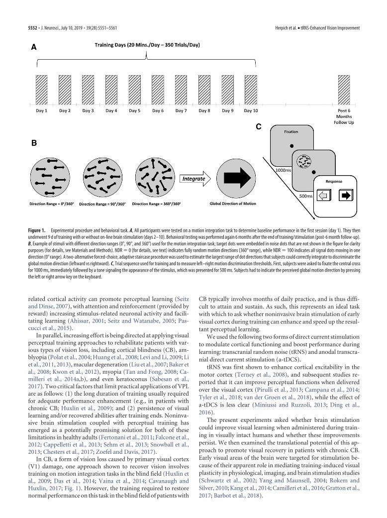
Figure 1. Experimental procedure and behavioral task. A, All participants were tested on a motion integration task to determine baseline performance in the first session (day 1). They thenunderwent 9 d of training with or without on-line brain stimulation (days 2–10). Behavioral testing was performed again 6 months after the end of training/stimulation (post-6 month follow-up).B, Example of stimuli with different direction ranges (0°, 90°, and 360°) used for the motion integration task; target dots were embedded in noise dots that are not shown in the figure for claritypurposes (for details, see Materials and Methods). NDR � 0 (for details, see text) indicates fully random motion directions (360° range), while NDR � 100 indicates all signal dots moving in onedirection (0° range). A two-alternative forced-choice, adaptive staircase procedure was used to estimate the largest range of dot directions that subjects could correctly integrate to discriminate theglobal motion direction (leftward vs rightward). C, Trial sequence used for training and to measure left–right motion discrimination thresholds. First, subjects were asked to fixate the central crossfor 1000 ms, immediately followed by a tone signaling the appearance of the stimulus, which was presented for 500 ms. Subjects had to indicate the perceived global motion direction by pressingthe left or right arrow key on the keyboard.
5552 • J. Neurosci., July 10, 2019 • 39(28):5551–5561 Herpich et al. • tRNS-Enhanced Vision Improvement

Materials and MethodsExperiment 1: tRNS-mediated learning in healthy participantsRegulatory approval. The study was approved by the ethical committee ofthe University of Trento.
Subjects. A total of 45 subjects participated in the experiment (meanage, 19.9 years; age range, 19 –36 years; 32 females and 13 males). Allsubjects were right handed, neurologically normal, with normal orcorrected-to-normal vision and gave written, informed consent beforethe beginning of the study, according to the ethical standards of theDeclaration of Helsinki.
Study design. Subjects were randomly assigned to one of five groups.This included two experimental groups. In the first group, tRNS wasdelivered over early visual areas (electrodes positioned bilaterally, cen-tered over O1 and O2 of the EEG system coordinates, for the left and righthemisphere respectively). In the second group, a-tDCS was deliveredover the occipital cortex (the anode and the cathode were positioned overOz and Cz, respectively). While we used bilateral occipital montage forthe tRNS condition to match the positioning of other successful studiesthat found improved performance with tRNS and likely increased excit-ability in the visual cortex (Romanska et al., 2015; Herpich et al., 2018),particularly with motion discrimination tasks (van der Groen et al.,2018), we chose unilateral montage for the a-tDCS condition, the opti-mal montage to increase cortical excitability with anodal stimulation ofthe visual cortex (Antal et al., 2004). Stimulation was concurrent with thetraining task. There were also three control groups: a sham control; ano-stimulation control; and an active control, where bilateral tRNS wasapplied over parietal cortex (over P3 and P4, regions likely involved inbut not critical for global motion discrimination (Greenlee and Smith,1997; Battelli et al., 2001). Over 10 d, all subjects were trained to discrim-inate the left or right global direction of random dot motion stimuli (350trials/session/d). Day 1 was considered the pretraining session, while day10 was used as the post-training session (Fig. 1). Finally, a long-termfollow-up was performed 6 months after the post-training session. Dur-ing this follow-up, participants repeated the behavioral baseline tests.Critically, no stimulation was delivered at this time.
Apparatus and procedures. For subjects who underwent brain stimula-tion, all experiments took place in the same room, under the same lightand noise conditions, and with the same apparatus. During each session,participants were positioned on a chinrest/forehead bar combination tostabilize their heads, and to place their eyes 57 cm from the stimulus-presenting computer monitor. Visual stimuli were generated on a Mac-Book Pro computer running software based on the PsychophysicsToolbox (Brainard, 1997; Pelli, 1997) in Matlab (MathWorks). Stimuliwere presented on a linearized SensEye 3 LED 24 inch monitor (BenQ)with a refresh rate of 120 Hz, and it was luminance calibrated withgamma � 1 using a professional monitor calibrator (Spyder 5, Data-color). Eye fixation for all subjects was controlled in real time using anEyeLink 1000 Plus Eye Tracking System (SR Research) whose infraredcamera monitored the pupil center and corneal reflection of the left eye.Limits were set so that if the participant’s eye moved �1.5° in any direc-tion away from the fixation spot during stimulus presentation, loudtones sounded, and the currently displayed trial was aborted and ex-cluded from the final analysis.
Global direction discrimination testing and training. We first measureddirection range (DR) thresholds for left–right motion discrimination ofcircular stimuli that contained a limited percentage of signal dots (New-some and Pare, 1988; Huxlin and Pasternak, 2004; Levi et al., 2015) andwere centered at [�5°, 5°] in the visual periphery. To match initial taskdifficulty across observers, motion coherence (Newsome and Pare, 1988)was calibrated for each subject individually, as previously reported (Leviet al., 2015). The motion coherence of the stimulus was chosen based onpreliminary testing aimed to identify a motion signal level that allowedparticipants to perform the discrimination task just above chance (50%correct). For all but three subjects, random dot stimuli contained 40%motion signal. Three subjects were trained with a stimulus containing30% coherent motion. Adding coherence noise to our stimuli ensuredthat all subjects started at about the same difficulty level and, more im-
portantly, allowed plenty of room for improvement for our healthy par-ticipants. Once a motion signal level was selected for each participant, thetask used a QUEST adaptive staircase (Watson and Pelli, 1983) to esti-mate the broadest distribution of dot directions that subjects could cor-rectly integrate to discriminate the global direction of motion as leftwardor rightward. During training, task difficulty was adaptively modulatedby adjusting DR of signal dots (Huxlin and Pasternak, 2004) using 12randomly interleaved 25-trial Quest staircases in each daily session.These adaptive Quest staircases were used to ensure that the stimulipresented to participants are around their threshold performance. Be-cause we collected 300 trials per session, we were able to fit full psycho-metric functions to the data. Here, we fit single-trial data with theWeibull function (where percentage correct � 1 � (1 � chance) �
exp(�(k � x/threshold) slope), and k � (� log((1 � 0.82)/(1 �chance))) (1/slope)). As customary, we used the 82% threshold criterion asthat is close to the steepest point of the Weibull function. To illustrate thequality of data fits, Figure 2 shows data for an example subject. Thresh-olds reported in the study, corresponding to 82% correct, were takenfrom these estimated Weibull functions and are reported as normalizedDR (NDR) thresholds, such that a NDR of 0% equals fully randommotion (360° range of dot directions) and a NDR of 100% indicates allsignal dots moving in one direction (0° range). The random dot stimuliwere presented within a circular aperture 5° in diameter at a density of 2.6dots/° 2. Each dot had a diameter of 0.06° and moved at a speed of 10°/swith a lifetime of 250 ms. The stimulus duration was 500 ms. Each par-ticipant started training with DR in the random dot stimulus set to 0°.
The trial sequence was as follows: participants were asked to fixate ona central cross for 1000 ms, immediately followed by a tone signaling theappearance of the stimulus, which was presented for 500 ms. Once thestimulus disappeared, participants had to indicate the perceived globaldirection of motion by pressing the left or right arrow keys on the key-board. The two motion directions (leftward and rightward) were ran-domized across trials. Auditory feedback was provided indicating thecorrectness of the response on each trial.
During training, stimuli were presented monocularly to the left eye for10 d (one session/d from Monday to Friday, for 2 consecutive weeks; Fig.1), while subjects received active, sham, or no stimulation. We chosemonocular presentation to closely match the procedure used by Huxlinet al. (2009) on patients and by Levi et al. (2015) on healthy participantsusing the same visual stimuli. Subjects performed 350 trials/d for a totalof 3500 trials by the end of the 2 weeks of training. The total duration ofthe daily training session for each group was set to last �20 min.
Stimulation protocols. tDCS and tRNS were delivered using a battery-driven stimulator (DC-Stimulator-Plus, NeuroConn) through a pair ofsaline-soaked conductive rubber electrodes (35 cm 2). Each subject wasrandomly assigned to one of the five stimulation groups, as describedearlier (see Study design). The electrodes were bilaterally placed over thetarget areas identified following the 10 –20 EEG reference system. Sub-jects wore a Lycra swimmers cap to keep the electrode in place, and weensured that the skin and hair between the electrodes were completelydry, otherwise preventing the current from reaching the brain. The in-tensity of stimulation was set to 1.0 mA, and was delivered for 20 minwith a fade in/out period of 20 s. For the a-tDCS group, the polarity of theactive electrode was anodal. For the tRNS condition, the random noisestimulation was applied with a 0 mA offset at frequencies of alternatingcurrent ranging from 101 to 640 Hz (high-frequency tRNS). For thesham stimulation group, the stimulation (using the same electrode mon-tage as in the tRNS condition) was shut down after 20 s. At the end of eachsession, we asked all subjects to fill out a questionnaire about potentialdiscomfort or any unusual sensation they experienced during the stim-ulation. Only minor side effects were reported by the tDCS group (twosubjects reported slight itching under the electrode, one subject reporteda slight subjective temperature increase under the electrode), whereasnone of the tRNS group participants reported any sensation of beingstimulated.
Data analysis. The Shapiro–Wilk test was used to control for the nor-mality of data distribution. Data sphericity was addressed using Mauch-ly’s test, and a Greenhouse–Geisser correction was used in case of thenonsphericity of the data. Levene’s test was used to address the assump-
Herpich et al. • tRNS-Enhanced Vision Improvement J. Neurosci., July 10, 2019 • 39(28):5551–5561 • 5553

tion of equality of variances. The p values were considered significant at�0.05. To correct for multiple comparisons in post hoc testing, we usedTukey’s HSD correction. The effect sizes are reported as the partial �p 2
values.
Experiment 2: tRNS-mediated visual recovery in patients withcortical blindnessRegulatory approval. The study on patients was approved by the ethicalcommittee for clinical experimentation of the “Azienda Provinciale per iServizi Sanitari” (APSS) and by the Institutional Review Board of theUniversity of Rochester Medical Center. The work was conducted afterobtaining written, informed consent from each patient.
Cortically blind patients. Eleven patients participated in the study: fiverecruited at the Center for Neurocognitive Rehabilitation (CeRiN) affil-iated to the University of Trento and the Rehabilitation Hospital “VillaRosa” in Pergine, Italy, and six recruited at the Flaum Eye Institute of theUniversity of Rochester Medical Center. Patients were recruited 2.5–108months after damage to their early visual areas; as such, all but onepatient (U6) were in their chronic, poststroke phase (Table 1; medianpoststroke time, 15 months). Both the location of damage and the hom-onymous visual defects were confirmed by neurological reports, neuro-radiological examinations, and automated visual perimetry (Optopol
PTS 1000 Visual Field, Canon; or Humphrey Field Analyzer HFA II 750,Carl Zeiss Meditec). The patients gave written, informed consent at theirrespective study site before participating.
Ten of the patients (RNS1–2, Sham1–2, U1– 6) suffered from strokeinvolving the territory of the posterior cerebral artery, as confirmed byradiological examinations and reports (Table 1, Fig. 3). One patient(RNS3) suffered from traumatic brain injury. Although from the neuro-radiological report V1 was not directly affected by trauma, there wereindications of visual fields defects, and his visual perimetry showed aclear, homonymous, bilateral upper quadrantanopia; hence, we decidedto enroll him in the training procedure (note that data for each patientwere computed and shown individually). None of the patients had his-tory or evidence of degenerative or psychiatric disorders. All participantswere right handed, with normal or corrected-to-normal visual acuity,and none exhibited visual or other forms of neglect, as determined byneurological examination.
Study design. All cortically blind patients underwent 10 d of training,following the same day 1 to day 10 procedures as neurotypical subjects(Fig. 1). Patients in Italy were randomly assigned to one of the followingtwo experimental groups: three patients received tRNS over early visualareas during training, whereas two patients received sham stimulationduring training, with electrodes placed in the same locations as for tRNS.
Figure 2. Example psychometric data fits. Here, we selected the tRNS subject whose data were closest to the average of all nine tRNS subjects (going from 94% NDR to 30% NDR over 10 sessions).For each session (1–10), blue symbols show all individual trial data (correct trials are at the top and incorrect trials are the bottom of each panel). Psychometric function fits are shown by the red lines.For illustration purposes, individual trials are binned into 10 30-trial bins (red circles).
Table 1. Demographic data for patients with CB
Patient SexAge at testing(years) Visual defect Lesion
Time sincelesion (months)
RNS1 F 59 Left lateral homonymous hemianopia Stroke affecting right fronto-parieto-occipital lobe 36RNS2 M 66 Left lateral homonymous quadrantanopia Stroke affecting right posterior capsule-thalamic and occipital lobe 15RNS3 M 53 Bilateral upper homonymous quadrantanopia Traumatic brain injury affecting Left fronto-temporo-parietal region 108Sham1 M 69 Right lateral homonymous hemianopia Stroke affecting left occipital lobe and left posterior capsule-thalamic area 24Sham2 M 72 Right lateral homonymous quadrantanopia Stroke affecting left occipital lobe and internal capsule 9U1 F 63 Right homonymous hemianopia Stroke affecting left occipital lobe 10U2 F 67 Left lower homonymous quadrantanopia Stroke (hemorrhagic) affecting right occipital lobe 26U3 F 54 Left homonymous hemianopia Stroke affecting right occipital cortex 7U4 M 47 Bilateral hemianopia Bilateral stroke damage affecting occipital lobes 16U5 M 53 Left homonymous hemianopia Stroke affecting right occipital lobe 12U6 F 44 Left homonymous hemianopia Stroke affecting right occipital lobe 2.5
Visual fields defects were assessed with automated perimetry. The last column indicates the time between stroke and in-laboratory testing. Patient RNS3 had a traumatic brain injury, while all others experienced strokes. F, Female; M, male.
5554 • J. Neurosci., July 10, 2019 • 39(28):5551–5561 Herpich et al. • tRNS-Enhanced Vision Improvement

Patients in the United States underwent global direction discriminationtraining without brain stimulation.
Apparatus and procedures. We used the same apparatus as in experi-ment 1, except that stimuli were presented on a CRT monitor (48.5 �31.5 cm, 1024 � 640 pixel resolution, 120 Hz frame rate; model 7217 A,HP) calibrated with a ColorCal II automatic calibration system (Cam-bridge Research Systems) for the patients in the United States. Eye fixa-tion for all patients was controlled in real time using an EyeLink 1000Plus Eye Tracking System (SR Research).
Global direction discrimination testing and training in patients. First, wespatially mapped motion discrimination performance in each patient toidentify a blind field location where training should be performed. Weused the same task described in experiment 1 for neurotypical subjects,with the following two adjustments intended to make the task easier forthe patients: coherence and NDR were set to 100% (the easiest possiblesettings), and the number of trials per training day was lowered to 250.Fixation was enforced, as in visually intact subjects, and each trial wasinitiated by fixation of a small circle in the center of the screen. Duringmapping, stimuli were first presented in the intact field, at locations closeto the border with the blind visual field, and patients performed 100 trialsof the global direction discrimination task per location. This allowed usto ensure that each patient understood task demands and to assess nor-mal baseline performance on an individual basis. Stimulus location wasthen moved progressively into the blind field, with 100 trials of the globaldirection discrimination task performed at each location, until global
motion direction discrimination dropped below chance (Huxlin et al.,2009); this was selected as the training location, and care was taken toensure that it was situated fully inside the perimetrically defined blindfield (Fig. 3, blue circles).
For comparison purposes, we also included data from six patientstrained in the Huxlin laboratory at the Flaum Eye Institute of the Uni-versity of Rochester with the same behavioral protocol, but without anybrain stimulation. Five of six unstimulated patients, trained with 300trials/training day, while one trained with 225 trials/d. Thus, on average,unstimulated patients completed 15% more trials than tRNS/sham-stimulated patients, which made them a conservative comparison group.
On Day 1, all patients performed considerably worse on global motionintegration in their blind field compared with neurotypical subjects, evenat the easiest stimulus level (100% coherence; NDR � 100%). On day 10,no patient performed better than 85% correct at a NDR of 100% (i.e.,with all dots going in the same direction). Specifically, at 100% coher-ence, patients’ global motion direction range (i.e., NDR) threshold aver-aged 232 � 24.3° in their intact hemifield and 9.25 � 15.91° in their blindfield (paired Student’s t test, p � 8.07 � 10 5), indicating that when theglobal motion was �9° around the right or leftward vector none of thesubjects were able to discriminate the global direction of motion ofthe dot stimuli. Thus, given the range of performance by patients, wechose to report the percentage correct at 100% NDR as the measure ofperformance. This allowed us to avoid issues with noisy thresholdestimates for subthreshold performance, while still retaining a suffi-
Figure 3. Neuroradiological images and visual perimetries of CB patients. All patients sustained damage of early visual areas or the optic radiations, resulting in homonymous visual field defectsas shown by the visual field perimetries, next to each brain image. Within the perimetry images (patients in top two rows: Sham1, Sham2, RNS1, RNS2, and RNS3): red marks and shading areasindicate the patients’ blind field. Bottom two rows: Humphrey visual field maps for each of the unstimulated patient (U1– 6), with superimposed shading indicating the blind field and numbersindicating the luminance detection sensitivity in the given position expressed in decibels. For all patients, the blue circles indicate the training location and size (for details, see Global directiondiscrimination testing and training in patients, in Materials and Methods). Radiological images were not available for patients RNS3 and U6.
Herpich et al. • tRNS-Enhanced Vision Improvement J. Neurosci., July 10, 2019 • 39(28):5551–5561 • 5555

cient dynamic range to capture training-induced improvements inperformance.
Stimulation protocols. We performed the exact same stimulation pro-tocols (sham and tRNS) as used with the visually intact participants inexperiment 1.
Data analysis. To analyze data from individual patients, we performedthe following bootstrap analysis. First, for each subject and each trainingday, we generated 10,000 bootstrap samples by selecting, with replace-ment, from the set of available individual trials. Then we fit Weibullfunctions to all 10,000 samples, and, from the resultant fits, we computedthe percentage correct performance at 100% NDR (the easiest difficultylevel). Because most of the individual trials for patients were collectednear 100% NDR, these percentage correct estimates were more robustthan threshold estimates, which in many cases were estimated to be�100% NDR. Thus, for each training day, we had 10,000 estimates foreach patient’s percentage correct performance, allowing us to estimate95% confidence intervals (see Fig. 5). From this set of estimates, wecreated 100,000 full datasets for each patient (random sampling withreplacement). This allowed us to estimate p values for learning slope andthe amount of learning analyses reported in the Results. For the slopeanalysis, we simply computed the proportion of datasets that had nega-tive learning slopes, multiplying results by 2 to get two-tailed p values.For the amount of learning analysis, we computed the proportion ofdatasets where day 1–2 performance was better than day 9 –10 perfor-mance, multiplying results by 2 to get two-tailed p values.
ResultsImpact of pairing brain stimulation with training in visuallyintact subjectsLearning of motion integration in control groupsSubjects recruited for the present experiments were divided intofive training groups. Two experimental groups (bilateral tRNSand anodal stimulation) received stimulation over early visualareas. Their results were compared with the following three con-trol groups: bilateral tRNS over parietal cortex, a sham control,and a no-stimulation control. Before the onset of training, therewere no significant differences in NDR thresholds, a measure ofdirection integration performance, among the five traininggroups (F(4,40) � 1.15, p � 0.35). This confirmed that all fivegroups started with relatively similar levels of performance. Asexpected, all five groups benefited from perceptual training—foreach group, performance at day 10 was better than at day 1 (all t(8)
� 2.7, all p � 0.027). This result is consistent with well establishedeffects of training on visual perception (Watanabe and Sasaki,2015; Levi et al., 2015). However, no significant differences inlearning were observed among three of the groups—no stimula-tion (training only), sham stimulation training, and tRNS overparietal cortex training. The lack of difference was observedregardless of whether learning was expressed as a raw change inthresholds (NDRDay1 � NDRDay10; F(2,24) � 1.58, p � 0.23), a per-centage change in thresholds [(NDRDay1 � NDRDay10)/NDRDay1;F(2,24) � 2.93, p � 0.072] or learning speed (linear regressionslope, F(2,24) � 2.36, p � 0.12). We further performed an addi-tional control analysis on the amount of learning between thefirst and the last session (day 1 to day 10). We ran a repeated-measures t test, which showed that the tRNS group (NDR, 56.7 �16.2, mean � SD) differed significantly from each of the follow-ing control conditions: sham (34.9 � 19.6; t(8) � 2.57, p � 0.01),behavioral (25.8 � 23.6; t(8) � �2,19, p � 0.02), and parietaltRNS (21.3 � 15.8; t(8) � 4.68, p � 0.001). To minimize thenumber of multiple comparisons between experimental and con-trol groups, data from these three control groups were thus com-bined into a single control dataset for all subsequent analyses.
tRNS, but not a-tDCS, enhances learningComparison of the control dataset with tRNS training anda-tDCS training revealed large differences in learning (Fig.4A,B). In addition to the expected main effect of training day(F(3.4,144.1) � 34.7, p � 10�18), we found a main effect of group(F(2,42) � 3.35, p � 0.045) and, notably, a significant group by dayinteraction (F(6.9,144.1) � 4.01, p � 0.01). As suggested by thissignificant interaction, the amount of learning differed amongthe three groups (Fig. 4A; F(2,42) � 9.39, p � 0.0004). Specifically,tRNS training induced stronger learning than both the com-bined control group (p � 0.002) and a-tDCS training (p �0.001; all post hoc tests are Tukey’s HSD), whereas a-tDCS out-comes did not differ significantly from those attained by the com-bined control group (p � 0.53). The same pattern of results wasobserved when we considered group differences in terms of thepercentage of improvement from pretest to post-test (Fig. 4B;F(2,42) � 10.8, p � 0.00016). Again, tRNS training resulted inlarger percentage improvement than that attained by controlsubjects (p � 0.001) and the a-tDCS training group (p �0.0002). Once again, performance following a-tDCS trainingdid not differ significantly from that in the combined controlgroup (p � 0.28). In all groups, learning was well described by alinear trend (Fig. 4A). Slopes, however, differed among groups(Fig. 4C; F(2,42) � 7.8, p � 0.001), with tRNS training generat-ing faster learning than in the combined control group (p �0.008) or a-tDCS training group (p � 0.001). In contrast,a-tDCS did not induce significantly faster learning than that at-tained by the combined control group (p � 0.30).
As tRNS administered during training appeared to causefaster learning, we analyzed at what time point the tRNS groupbegan to diverge from the other two groups. This occurred on day6 (F(2,42) � 5.03, p � 0.01), at which point the tRNS groupshowed stronger learning than both the combined control group(p � 0.03; Tukey’s HSD) and the a-tDCS training group (p �0.01; Tukey’s HSD).
In sum, we found strong evidence for enhanced learning in thetRNS training group, with faster learning than both the com-bined control group and a-tDCS training group. As detailedabove, this finding was supported regardless of how learning wasdefined. While it may seem that a-tDCS, as administered in ourstudy, might actually hinder learning (Fig. 4A,B), this effect wasnot statistically significant.
Persistence of stimulation-enhanced perceptual learningNext, we asked whether the observed enhancement of perceptuallearning by tRNS remained stable over an extended period oftime. To address this question, we retested participants 6 monthsafter completing the 10 d of training with and without the differ-ent forms of stimulation. The post-test, however, was performedwithout brain stimulation. The subjects retested at 6 months in-cluded 37 of 45 original participants (n � 8 for tRNS traininggroup; n � 22 for the combined control group; and n � 7 fora-tDCS training group). Figure 4D contrasts the amount oflearning at day 10 (NDRDay 1 � NDRDay 10) with that exhibited 6months after the end of training (NDRDay 1 � NDR6-months).There was a small, nonsignificant loss in performance for thethree groups (no main effect of testing day; F(1,34) � 3.32, p � 0.8)and no interaction (F(2,34) � 0.88, p � 0.43). We only found amain effect of group, confirming that the group differences at theend of 10 d of training remained unaltered 6 months after train-ing (F(2,34) � 4.68, p � 0.02). Thus, it appears that tRNS en-hanced perceptual learning over the long term—at least 6 monthsafter the end of both training and brain stimulation. Moreover,
5556 • J. Neurosci., July 10, 2019 • 39(28):5551–5561 Herpich et al. • tRNS-Enhanced Vision Improvement

this persistent enhancement was observed without brain stimu-lation at the 6 month follow-up. This suggested that the enhance-ment of perceptual learning by tRNS was not due to on-line orshort-term optimization of visual processing, but instead, re-sulted in consolidated sensory learning.
tRNS boosts training-induced visual recovery in corticallyblind patientsGiven our finding that tRNS, but not a-tDCS, over occipital cor-tex considerably enhances perceptual learning in neurotypicalsubjects, we next asked whether tRNS is also able to enhancetraining-induced visual recovery in chronic, V1-damaged pa-tients with CB. To the best of our knowledge, tRNS has not beenattempted in brain-damaged patients. Moreover, whether tRNSover early visual areas could enhance learning in CB patients is anopen question, as learning in this patient population can exhibitproperties not found in neurotypical subjects (Das et al., 2014;Vaina et al., 2014; Cavanaugh and Huxlin, 2017), and since, bydefinition, the part of early visual cortex that would normally bestimulated is damaged. Hence, we sought to perform a prelimi-nary, proof-of-concept study in five patients with occipital dam-age resulting in homonymous visual field defects measured withvisual perimetry (Table 1, Fig. 3).
Visual perimetry was used to identify the blind field bordersand select training locations in the blind field (Fig. 3). We ran-
domly assigned five patients from our Italian study site to eithertRNS training (n � 3, RNS1–3) or sham stimulation train-ing (n � 2, Sham1–2). Data from an additional six patients withCB who trained identically, but without brain stimulation (un-stimulated, U1– 6), at our U.S. study site, were also analyzed forcomparison. All patients, at both study sites, trained for 10 dusing random dot stimuli, as in neurotypical subjects (Fig. 1).Because patients have difficulty seeing motion, their stimuli, un-like those for neurotypical subjects, did not include noise dots.
As expected (Huxlin et al., 2009; Das et al., 2014; Cavanaugh etal., 2016; Cavanaugh and Huxlin, 2017), all patients performedconsiderably worse on global motion integration in their blindfield compared with neurotypical subjects. This was the case evenat the easiest stimulus level (NDR � 100, with all dots moving inthe same direction), where none of the patients exhibited ceilinglevel performance in their blind field. As such, we used the per-centage correct at 100 NDR as the measure of performance (fordetails, see Materials and Methods). Sham-stimulated patientsexhibited no significant change in performance across the 10 d oftraining (Fig. 5A), as evidenced by learning slopes that were notsignificantly different from 0 (all p � 0.63; for bootstrap proce-dure used to analyze data from individual patients, see Materialsand Methods). This was comparable to the lack of learning ob-served in the six unstimulated patients, who also did not exhibit asignificant learning slope over their first 10 d of training (Fig. 5C;
Figure 4. Effects of brain stimulation on perceptual learning in visually intact subjects. A, NDR thresholds for the control groups, tRNS group, and a-tDCS group. Dashed lines are linear fits,indicating the learning slope. B, Same data as in A, but expressed as the percentage improvement relative to day 1 thresholds. C, Learning index computed in three different ways. The tRNS groupexhibited a significantly stronger amount of learning (day 1 � day 10; F(2,42) � 9.39, p � 0.0004; all Tukey’s HSD, p � 0.002), percentage improvement (100 * (day 1 � day 10)/day 1; F(2,42) �10.8, p � 0.00016; all Tukey’s HSD, p � 0.001), and learning slope (F(2,42) � 7.8, p � 0.001; all Tukey’s HSD, p � 0.008) than both the control and a-tDCS groups. D, Amount of learning, definedas the difference from day 1 thresholds, at the end of the training (left) and 6 months after (right). Error bars are �1 SEM.
Herpich et al. • tRNS-Enhanced Vision Improvement J. Neurosci., July 10, 2019 • 39(28):5551–5561 • 5557

all p � 0.13). In contrast, tRNS coupled with training enhancedthe rate of global motion discrimination learning in CB patients(Fig. 5B), who exhibited significantly positive learning slopes (allp � 0.0048). We also examined the change in performance fromdays 1–2 to days 9 –10, averaging results over 2 consecutive daysto minimize the effects of day-by-day fluctuations. The analysisshowed significant change only for patients trained with tRNS(Fig. 5D; tRNS, all p � 0.0002; sham, all p � 0.24; unstimulated,all p � 0.080).
DiscussionIn the present study, we asked whether brain stimulation overearly visual cortex could boost and speed up visual perceptuallearning in healthy humans and in those affected by corticallyinduced blindness. In healthy humans, we also asked whetherenhancements attained from training coupled with stimulationpersisted over long periods. Results from experiment 1 show thattRNS applied bilaterally over healthy, early visual cortex speedsup and boosts performance during visual perceptual learning.Over 10 d, neurotypical subjects in the tRNS training groupexhibited an �60% improvement in motion integration thresh-
olds (Fig. 4B), which was two and three times as strong as learningattained by the control and a-tDCS groups, respectively. Thisfinding was supported regardless of whether learning was definedas a raw change in NDR threshold, a percentage improvement, orthe slope of a linear fit to the data. The observed benefit of tRNSover training alone or sham stimulation training, or tDCS overparietal cortex is consistent with evidence that tRNS is especiallyeffective at promoting plasticity when coupled with a relevantstimulus, and when it is applied over relevant (in our case, occip-ital) brain areas (Cappelletti et al., 2013). In contrast, we found nosuch benefit of a-tDCS over occipital cortex. Finally, we providefor the first time preliminary evidence that tRNS enhances visionrecovery in patients with CB with V1 damage. Moreover, wedemonstrate seemingly safe usage of this technique in a class ofstroke patients, with no side effects reported. Notably, with re-spect to training-induced recovery, tRNS enabled improvementsin visual task performance of patients with chronic CB in theirblind field, over a tiny fraction of the training days typically re-quired to induce such improvements in the absence of brainstimulation (Huxlin et al., 2009; Das and Huxlin, 2010; Das et al.,
Figure 5. The effects of brain stimulation on perceptual learning in patients with CB. A–C, Task performance over 10 training days for patients who underwent sham stimulation (A), those whoreceived tRNS (B), and six unstimulated patients (C). The raw percentage of correct performance was normalized by subtracting the average percentage correct for the first 2 training days. D,Comparison of the raw percentage correct averaged over the first 2 d against the raw percentage correct for the final 2 training days. Significant learning was observed only for patients who trainedwith tRNS. All error bars indicate 95% confidence intervals. For A–C, all lines are linear fits, indicating the learning slope.
5558 • J. Neurosci., July 10, 2019 • 39(28):5551–5561 Herpich et al. • tRNS-Enhanced Vision Improvement

2014; Melnick et al., 2016; on average, 72– 80 training days arerequired to recover global direction discrimination performanceat a given blind field location (Cavanaugh and Huxlin, 2017).Indeed, we observed a steady and significant increase in perfor-mance for three patients trained with tRNS over 10 d, with nosuch effects in patients who trained with sham stimulation. Pa-tients with chronic CB are a population that would especiallybenefit from enhanced perceptual learning because vision recov-ery using conventional training methods usually takes manymonths of daily training (Huxlin et al., 2009; Das and Huxlin,2010; Das et al., 2014; Melnick et al., 2016; Cavanaugh and Hux-lin, 2017).
Interestingly, while the position of the stimulating electrodescorresponded to early visual areas that included V1, improve-ment occurred despite V1 being damaged in the patients with CB.This might suggest that the neuromodulatory benefit of tRNSlikely impacts any residual V1, but also adjacent visual areas suchas V2 and V3, which might have also supported visual learning, asindicated in imaging studies in CB patients (Henriksson et al.,2007; Raemaekers et al., 2011; Martin et al., 2012; Ajina et al.,2015; Barbot et al., 2018). V2 and V3 are usually spared in CBpatients, and they might have played a pivotal role in supportingrecovery during training. We cannot address whether some formof cortical reorganization took place in 2 weeks of training in ourpatients, as we did not collect any imaging data. However, wecould speculate that since we delivered bilateral stimulation, thiseither boosted cortical functioning within the affected hemi-sphere, promoting activity in the damaged early visual cortex inresponse to concurrent visual/brain stimulation or, alternatively,visual areas in the healthy hemisphere, homologs to the damagedareas, might have promoted recovery (Henriksson et al., 2007).These remain interesting hypotheses to be further tested in futurestudies. Interestingly, physiological studies have shown thatV2/V3 inactivation may degrade the ability of cortical motion-sensitive areas to work efficiently (Ponce et al., 2011), while theymight support learning when they function normally (Law andGold, 2008). In summary, while plasticity of spared visual circuitsis generally believed to underlie visual recovery, the specific neu-ral mechanisms involved remain unclear.
An additional finding in the present study was the observationthat the enhancement of perceptual learning induced by tRNScan persist over an extended period of time (6 months in thepresent study) beyond the end of stimulation and training. This isimportant since stimulation-enhanced perceptual learningwould have limited practical use if its beneficial effects dimin-ished over time. Additionally, because all subjects performed thebehavioral task with no stimulation at the follow-up, 6 monthtime point, we infer that the benefits of tRNS go beyond an on-line enhancement of visual processing and likely involve plasticchanges that persist within the visual system, allowing it to moreeffectively process global motion stimuli. As such, we concludethat consolidation of learning occurred in our subjects. The per-sistence effects observed here are particularly notable, as manyVPL studies failed to see long-lasting effects and/or the transfer oflearning to other tasks (Dosher and Lu, 2017).
Some important questions arising from the present results areas follows: (1) how does brain stimulation enhance the effects ofperceptual learning, and (2) why do some forms of stimulationprove effective and others not? a-tDCS did not exert a beneficialeffect in our study—a surprising result given the results fromprevious studies stimulating the early visual cortex (Antal et al.,2004). One possible explanation is that the strongest effects oftDCS were reported off-line, where a-tDCS was delivered prior to
the measured behavior (Pirulli et al., 2013). Another possibility isthat a-tDCS is not the ideal neuromodulatory technique for re-peated sessions. Although it alters membrane potentials andhence exerts increased excitability, it may also engage inhibitoryhomeostatic mechanisms during repeated sessions (Fertonani etal., 2011; Peters et al., 2013).
Our observation that visual enhancements persist long afterboth training and tRNS ended does constrain the possible mech-anistic explanations. Multiple types of transcranial electricalstimulation have been shown to alter excitability in cortex, andthe longer time course of direct current stimulation effects hasbeen suggested to relate to homeostatic changes in membranepotential (Liebetanz et al., 2002; Nitsche et al., 2003; Ardolino etal., 2005; Terney et al., 2008) or gate threshold (Bikson et al.,2013). However, direct evidence that transcranial stimulation al-ters the dynamics of networks known to be related to perceptuallearning, such as dopaminergic reward networks (Seitz and Wa-tanabe, 2005; Pascucci et al., 2015), has not yet been provided. Allwe can state here is that tRNS does not appear to globally affectreward networks, as there was no boost in visual performance orlearning seen from stimulation over parietal cortex.
An interesting hypothesis is that tRNS-related visual perfor-mance improvements might derive from the state of the neuronsat the time of stimulation (Silvanto et al., 2008), and that addingnoise to the cortex might enhance sensory detection, in particularwhen stimuli are presented at threshold and embedded in noise(Abrahamyan et al., 2015). Several short-term mechanisms havealso been proposed to explain the effects of tRNS, and a favoredhypothesis involves stochastic resonance, whereby random fre-quency stimulation in tRNS appears to boost the responses ofneural populations to weak inputs; thus, the stochastic effect isexpected to be highest for stimuli presented below or just abovethreshold, as in our task (Moss et al., 2004; Schwarzkopf et al.,2011; Miniussi and Ruzzoli, 2013; Pirulli et al., 2013; van derGroen and Wenderoth, 2016; Herpich et al., 2018; van der Groenet al., 2018). An alternative hypothesis proposes temporal sum-mation of excitatory signals between visual stimulation and elec-trical stimulation (Terney et al., 2008; Fertonani et al., 2011;Pirulli et al., 2013), and selective enhancement of active neuralnetworks (Bikson et al., 2013; Miniussi et al., 2013; Luft, 2014;Fertonani and Miniussi, 2017). It is important to note that one ormore of these short-term mechanisms may be the first step in alonger-term cascade that results in a persistence of learning. Forinstance, stronger activation of task-relevant neurons due to tem-poral summation or stochastic resonance may encourage a shifttoward greater plasticity in sensory processing and/or readout.However, the time course of effects observed in the present study,and especially their persistence, suggests that on-line phenomena(i.e., during stimulation or shortly thereafter) are not the onlyones at play with respect to learning enhancements induced bytRNS. Interestingly, studies on perceptual learning in animalmodels have shown that learning might boost the modulation inneuronal tuning to stimulus components relevant to the task (Liuand Pack, 2017). If learning was associated with changes in thetuning characteristics of neurons (for review, see Gilbert and Li,2012), we could speculate that tRNS coupled with behavioraltraining might facilitate and consolidate this plastic change,which could then persist across months (Snowball et al., 2013).
Regardless of its precise mechanism of action, here we provideempirical evidence for the potential usefulness of tRNS coupledwith visual training on a patient population that requires percep-tual learning to attain visual recovery. V1-damaged patients withchronic CB are able to recover some visual abilities within their
Herpich et al. • tRNS-Enhanced Vision Improvement J. Neurosci., July 10, 2019 • 39(28):5551–5561 • 5559

scotoma, but only after intensive and repetitive training overmany months of daily practice (Huxlin et al., 2009; Das et al.,2014). The application of safe, painless neurostimulation in situ-ations like this, where perceptual learning is directly proportionalto the quantity of vision recovered (Cavanaugh and Huxlin,2017), has the potential to dramatically improve quality of lifeand treatment outcomes (Cavanaugh et al., 2016). Therefore,results from our experiment with CB patients suggest that tRNSmight be a viable adjunct procedure to speed up the recoveryprocess. Remarkably, even though the physiological effects oftRNS upon the damaged early visual cortex are currently un-known, our data show that tRNS can help overcome reducedand/or partially absent functionality and boost learning in theblind field.
ReferencesAbrahamyan A, Clifford CW, Arabzadeh E, Harris JA (2015) Low intensity
TMS enhances perception of visual stimuli. Brain Stimul 8:1175–1182.Ahissar M (2001) Perceptual training: a tool for both modifying the brain
and exploring it. Proc Natl Acad Sci U S A 98:11842–11843.Ajina S, Pestilli F, Rokem A, Kennard C, Bridge H (2015) Human blindsight
is mediated by an intact geniculo-extrastriate pathway. Elife 4:e08935.Antal A, Kincses TZ, Nitsche MA, Bartfai O, Paulus W (2004) Excitability
changes induced in the human primary visual cortex by transcranial di-rect current stimulation: direct electrophysiological evidence. InvestOphthalmol Vis Sci 45:702–707.
Ardolino G, Bossi B, Barbieri S, Priori A (2005) Non-synaptic mechanismsunderlie the after-effects of cathodal transcutaneous direct current stim-ulation of the human brain. J Physiol 568:653– 663.
Baker CI, Dilks DD, Peli E, Kanwisher N (2008) Reorganization of visualprocessing in macular degeneration: replication and clues about the roleof foveal loss. Vision Res 48:1910 –1919.
Barbot A, Melnick M, Cavanaugh M, Das A, Merriam E, Heeger D, Huxlin KR(2018) Visual recovery in chronic cortically-blind patients relies onspared cortical activity and increased V1 coverage of the blind field (ab-stract). J Vis 18:1074.
Battelli L, Cavanagh P, Intriligator J, Tramo MJ, Henaff MA, Michel F, BartonJJ (2001) Unilateral right parietal damage leads to bilateral deficit forhigh-level motion. Neuron 32:985–995.
Bikson M, Name A, Rahman A (2013) Origins of specificity during tDCS:anatomical, activity-selective, and input-bias mechanisms. Front HumNeurosci 7:688.
Brainard DH (1997) The psychophysics toolbox. Spat Vis 10:433– 436.Camilleri R, Pavan A, Ghin F, Battaglini L, Campana G (2014a) Improve-
ment of uncorrected visual acuity and contrast sensitivity with perceptuallearning and transcranial random noise stimulation in individuals withmild myopia. Front Psychol 5:1234.
Camilleri R, Pavan A, Ghin F, Campana G (2014b) Improving myopia viaperceptual learning: is training with lateral masking the only (or the most)efficacious technique? Atten Percept Psychophys 76:2485–2494.
Camilleri R, Pavan A, Campana G (2016) The application of on-line trans-cranial random noise stimulation and perceptual learning in the improve-ment of visual functions in mild myopia. Neuropsychologia 89:225–231.
Campana G, Camilleri R, Pavan A, Veronese A, Lo Giudice G, Lo (2014)Improving visual functions in adult amblyopia with combined perceptualtraining and transcranial random noise stimulation (tRNS): a pilot study.Front Psychol 5:1402.
Cappelletti M, Gessaroli E, Hithersay R, Mitolo M, Didino D, Kanai R, CohenKadosh R, Walsh V (2013) Transfer of cognitive training across magni-tude dimensions achieved with concurrent brain stimulation of the pari-etal lobe. J Neurosci 33:14899 –14907.
Cavanaugh MR, Huxlin KR (2017) Visual discrimination training improveshumphrey perimetry in chronic cortically induced blindness. Neurology88:1856 –1864.
Cavanaugh MR, Lilley S, Melnick MD, Reisner A, Huxlin KR (2016) Visualdiscrimination training shrinks cortically blind fields and improves qual-ity of life in chronic stroke patients (abstract). J Vis 16:31.
Chesters J, Watkins KE, Mottonen R (2017) Investigating the feasibility ofusing transcranial direct current stimulation to enhance fluency in peoplewho stutter. Brain Lang 164:68 –76.
Das A, Huxlin KR (2010) New approaches to visual rehabilitation for corti-cal blindness: outcomes and putative mechanisms. Neuroscientist16:374 –387.
Das A, Tadin D, Huxlin KR (2014) Beyond blindsight: properties of visualrelearning in cortically blind fields. J Neurosci 34:11652–11664.
Deveau J, Lovcik G, Seitz AR (2013) The therapeutic benefits of perceptuallearning. Curr Trends Neurol 7:39 – 49.
Ding Z, Li J, Spiegel DP, Chen Z, Chan L, Luo G, Yuan J, Deng D, Yu M,Thompson B (2016) The effect of transcranial direct current stimula-tion on contrast sensitivity and visual evoked potential amplitude inadults with amblyopia. Sci Rep 6:19280.
Dosher B, Lu ZL (2017) Visual perceptual learning and models. Annu RevVis Sci 3:343–363.
Falcone B, Coffman BA, Clark VP, Parasuraman R (2012) Transcranial di-rect current stimulation augments perceptual sensitivity and 24-hour re-tention in a complex threat detection task. PLoS One 7:e34993.
Fertonani A, Miniussi C (2017) Transcranial electrical stimulation: what weknow and do not know about mechanisms. Neuroscientist 23:109 –123.
Fertonani A, Pirulli C, Miniussi C (2011) Random noise stimulation im-proves neuroplasticity in perceptual learning. J Neurosci 31:15416 –15423.
Gilbert CD, Li W (2012) Adult visual cortical plasticity. Neuron 75:250 –264.
Gratton C, Yousef S, Aarts E, Wallace DL, D’Esposito M, Silver MA (2017)Cholinergic, but not dopaminergic or noradrenergic, enhancementsharpens visual spatial perception in humans. J Neurosci 37:4405– 4415.
Greenlee MW, Smith AT (1997) Detection and discrimination of first- andsecond-order motion in patients with unilateral brain damage. J Neurosci17:804 – 818.
Henriksson L, Raninen A, Nasanen R, Hyvarinen L, Vanni S (2007)Training-induced cortical representation of a hemianopic hemifield.J Neurol Neurosurg Psychiatry 78:74 – 81.
Herpich F, Conto F, van Koningsbruggen M, Battelli L (2018) Modulatingthe excitability of the visual cortex using a stimulation priming paradigm.Neuropsychologia 119:165–171.
Huang CB, Zhou Y, Lu ZL (2008) Broad bandwidth of perceptual learningin the visual system of adults with anisometropic amblyopia. Proc NatlAcad Sci U S A 105:4068 – 4073.
Huxlin KR, Pasternak T (2004) Training-induced recovery of visual motionperception after extrastriate cortical damage in the adult cat. Cereb Cortex14:81–90.
Huxlin KR, Martin T, Kelly K, Riley M, Friedman DI, Burgin WS, Hayhoe M(2009) Perceptual relearning of complex visual motion after V1 damagein humans. J Neurosci 29:3981–3991.
Kang JI, Huppe-Gourgues F, Vaucher E (2014) Boosting visual cortex func-tion and plasticity with acetylcholine to enhance visual perception. FrontSyst Neurosci 8:172.
Kwon M, Ramachandra C, Satgunam P, Mel BW, Peli E, Tjan BS (2012)Contour enhancement benefits older adults with simulated central fieldloss. Optom Vis Sci 89:1374 –1384.
Law CT, Gold JI (2008) Neural correlates of perceptual learning in asensory-motor, but not a sensory, cortical area. Nat Neurosci 11:505–513.
Law CT, Gold JI (2009) Reinforcement learning can account for associativeand perceptual learning on a visual-decision task. Nat Neurosci 12:655– 663.
Levi A, Shaked D, Tadin D, Huxlin KR (2015) Is improved contrast sensi-tivity a natural consequence of visual training? J Vis 15:4.
Levi DM, Li RW (2009) Perceptual learning as a potential treatment foramblyopia: a mini-review. Vision Res 49:2535–2549.
Li J, Thompson B, Deng D, Chan LY L, Yu M, Hess RF (2013) Dichoptictraining enables the adult amblyopic brain to learn. Curr Biol 23:R308 –R309.
Li RW, Ngo C, Nguyen J, LeviDM (2011) Video-game play induces plasticityin the visual system of adults with amblyopia. PLoS Biol 9:e1001135.
Li W (2016) Perceptual learning: use-dependent cortical plasticity. AnnuRev Vis Sci 2:109 –130.
Li W, Piech V, Gilbert CD (2004) Perceptual learning and top-down influ-ences in primary visual cortex. Nat Neurosci 7:651– 657.
Liebetanz D, Nitsche MA, Tergau F, Paulus W (2002) Pharmacological ap-proach to the mechanisms of transcranial DC-stimulation-induced after-effects of human motor cortex excitability. Brain 125:2238 –2247.
5560 • J. Neurosci., July 10, 2019 • 39(28):5551–5561 Herpich et al. • tRNS-Enhanced Vision Improvement

Liu L, Kuyk T, Fuhr P (2007) Visual search training in subjects with severe toprofound low vision. Vision Res 47:2627–2636.
Liu LD, Pack CC (2017) The contribution of area MT to visual motionperception depends on training. Neuron 95:436 – 446.e3.
Luft CD (2014) Learning from feedback: the neural mechanisms of feed-back processing facilitating better performance. Behav Brain Res261:356 –368.
Martin T, Das A, Huxlin KR (2012) Visual cortical activity reflects fasteraccumulation of information from cortically blind fields. Brain 135:3440 –3452.
Melnick MD, Tadin D, Huxlin KR (2016) Relearning to see in corticalblindness. Neuroscientist 22:199 –212.
Miniussi C, Ruzzoli M (2013) Transcranial stimulation and cognition. In:Handbook of clinical neurology, Vol 116, Ed 1, pp 739 –750. Amsterdam:Elsevier.
Miniussi C, Harris JA, Ruzzoli M (2013) Modelling non-invasive brainstimulation in cognitive neuroscience. Neurosci Biobehav Rev 37:1702–1712.
Moss F, Ward LM, Sannita WG (2004) Stochastic resonance and sensoryinformation processing: a tutorial and review of application. Clin Neuro-physiol 115:267–281.
Newsome WT, Pare EB (1988) A selective impairment of motion percep-tion following lesions of the middle temporal visual area (MT). J Neurosci8:2201–2211.
Nitsche MA, Liebetanz D, Antal A, Lang N, Tergau F, Paulus W (2003)Modulation of cortical excitability by weak direct current stimulation—technical, safety and functional aspects. Suppl Clin Neurophysiol.56:255–276.
Nyquist JB, Lappin JS, Zhang R, Tadin D (2016) Perceptual training yieldsrapid improvements in visually impaired youth. Sci Rep 6:37431.
Pascucci D, Mastropasqua T, Turatto M (2015) Monetary reward modu-lates task-irrelevant perceptual learning for invisible stimuli. PLoS One10:e0124009.
Pelli DG (1997) The VideoToolbox software for visual psychophysics:transforming numbers into movies. Spat Vis 10:437– 442.
Peters MA, Thompson B, Merabet LB, Wu AD, Shams L (2013) AnodaltDCS to V1 blocks visual perceptual learning consolidation. Neuropsy-chologia 51:1234 –1239.
Pirulli C, Fertonani A, Miniussi C (2013) The role of timing in the inductionof neuromodulation in perceptual learning by transcranial electric stim-ulation. Brain Stimul 6:683– 689.
Polat U, Ma-Naim T, Belkin M, Sagi D (2004) Improving vision in adultamblyopia by perceptual learning. Proc Natl Acad Sci U S A 101:6692–6697.
Ponce CR, Hunter JN, Pack CC, Lomber SG, Born RT (2011) Contributionsof indirect pathways to visual response properties in macaque middletemporal area MT. J Neurosci 31:3894 –3903.
Raemaekers M, Bergsma DP, van Wezel RJ, van der Wildt GJ, van den BergAV (2011) Effects of vision restoration training on early visual cortex inpatients with cerebral blindness investigated with functional magneticresonance imaging. J Neurophysiol 105:872– 882.
Rokem A, Silver MA (2010) Cholinergic enhancement augments magni-tude and specificity of visual perceptual learning in healthy humans. CurrBiol 20:1723–1728.
Romanska A, Rezlescu C, Susilo T, Duchaine B, Banissy MJ (2015) High-frequency transcranial random noise stimulation enhances perception offacial identity. Cereb Cortex 25:4334 – 4340.
Sabesan R, Barbot A, Yoon G (2017) Enhanced neural function in highly
aberrated eyes following perceptual learning with adaptive optics. VisionRes 132:78 – 84.
Sagi D (2011) Perceptual learning in vision research. Vision Res 51:1552–1566.
Schwartz S, Maquet P, Frith C (2002) Neural correlates of perceptual learn-ing: a functional MRI study of visual texture discrimination. Proc NatlAcad Sci U S A 99:17137–17142.
Schwarzkopf DS, Silvanto J, Rees G (2011) Stochastic resonance effects re-veal the neural mechanisms of transcranial magnetic stimulation. J Neu-rosci 31:3143–3147.
Sehm B, Schnitzler T, Obleser J, Groba A, Ragert P, Villringer A, Obrig H(2013) Facilitation of inferior frontal cortex by transcranial direct cur-rent stimulation induces perceptual learning of severely degraded speech.J Neurosci 33:15868 –15878.
Seitz A, Watanabe T (2005) A unified model for perceptual learning. TrendsCogn Sci 9:329 –334.
Seitz AR, Dinse HR (2007) A common framework for perceptual learning.Curr Opin Neurobiol 17:148 –153.
Silvanto J, Muggleton N, Walsh V (2008) State-dependency in brain stimu-lation studies of perception and cognition. Trends Cogn Sci 12:447– 454.
Snowball A, Tachtsidis I, Popescu T, Thompson J, Delazer M, Zamarian L,Zhu T, Cohen Kadosh R (2013) Long-term enhancement of brain func-tion and cognition using cognitive training and brain stimulation. CurrBiol 23:987–992.
Tan DT, Fong A (2008) Efficacy of neural vision therapy to enhance contrastsensitivity function and visual acuity in low myopia. J Cataract RefractSurg 34:570 –577.
Terney D, Chaieb L, Moliadze V, Antal A, Paulus W (2008) Increasing hu-man brain excitability by transcranial high-frequency random noise stim-ulation. J Neurosci 28:14147–14155.
Tyler SC, Contò F, Battelli L (2018) Rapid Improvement on a TemporalAttention Task within a Single Session of High-frequency TranscranialRandom Noise Stimulation. J Cogn Neurosci 30:656 – 666.
Vaina LM, Soloviev S, Calabro FJ, Buonanno F, Passingham R, Cowey A(2014) Reorganization of retinotopic maps after occipital lobe infarc-tion. J Cogn Neurosci 26:1266 –1282.
van der Groen O, Wenderoth N (2016) Transcranial random noise stimu-lation of visual cortex: stochastic resonance enhances central mechanismsof perception. J Neurosci 36:5289 –5298.
van der Groen O, Tang MF, Wenderoth N, Mattingley JB (2018) Stochasticresonance enhances the rate of evidence accumulation during combinedbrain stimulation and perceptual decision-making. PLoS Comput Biol14:e1006301.
Wang R, Wang J, Zhang JY, Xie XY, Yang YX, Luo SH, Yu C, Li W (2016)Perceptual learning at a conceptual level. J Neurosci 36:2238 –2246.
Watanabe T, Sasaki Y (2015) Perceptual learning: toward a comprehensivetheory. Annu Rev Psychol 66:197–221.
Watson AB, Pelli DG (1983) QUEST: a Bayesian adaptive psychometricmethod. Percept Psychophys 33:113–120.
Yang T, Maunsell JH (2004) The effect of perceptual learning on neuronalresponses in monkey visual area V4. J Neurosci 24:1617–1626.
Yan Y, Rasch MJ, Chen M, Xiang X, Huang M, Wu S and Li W (2014)Perceptual training continuously refines neuronal population codes inprimary visual cortex. Nat Neurosci 17:1380 –1387.
Zoefel B, Davis MH (2017) Transcranial electric stimulation for the inves-tigation of speech perception and comprehension. Lang Cogn Neurosci32:910 –923.
Herpich et al. • tRNS-Enhanced Vision Improvement J. Neurosci., July 10, 2019 • 39(28):5551–5561 • 5561
Related Documents