BONES AND BONE TISSUES CHAPTER 6 9/16/07

BONES AND BONE TISSUES CHAPTER 6 9/16/07. Introduction One of the most remarkable tissues of the human body Far from inert and lifeless, bones are living,
Dec 17, 2015
Welcome message from author
This document is posted to help you gain knowledge. Please leave a comment to let me know what you think about it! Share it to your friends and learn new things together.
Transcript
BONES AND BONE TISSUES
CHAPTER 69/16/07
Introduction One of the most remarkable tissues of the human body
Far from inert and lifeless, bones are living, dynamic structures
Bones serve a wide variety of very diverse functions within us
Noted for their strength and resiliency during life, bones will remain long after we are gone

Chapter Outline
Skeletal cartilages Bones Disorders of bones The skeleton throughout life
Location and Basic Structure
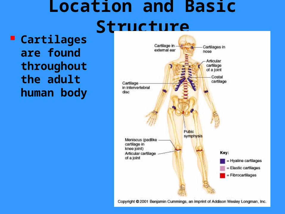
Cartilages are found throughout the adult human body
Location and Basic Structure
Initially our skeleton is made up of fast growing cartilages and fibrous membranes
Gradually our skeletal cartilages are replaced by bone
Upon reaching adulthood the skeleton becomes almost fully ossified
Only a few cartilages remain in the adult skeleton
Location and Basic Structure
A typical cartilage is composed of connective tissue cartilage
It contains no nerves or blood vessels
It is surrounded by a layer of dense irregular connective tissue called the perichondrium which resists outward expansion of the tissue when subjected to pressure
Location and Basic Structure
Each type of cartilage contains a high proportion of water which makes them resilient after compression
Cartilage is 60-80% water The water allows nutrients to diffuse rapidly through a loose matrix

Basic structure, type & location
There are three types of cartilage tissue: hyaline, elastic, and fibrocartilage
Each type consists of chondrocytes living in an extracellular matrix
Each contains a matrix of jellylike ground substance of complex sugar molecules that attract and hold water that is laced with connective tissue fibers
Hyaline cartilages
The most prevalent type of cartilage
Its high proportion of collagen fibers give it flexibility and resilience while providing support
Upon examination the tissue appears white, frosted, and smooth
Hyaline cartilages
The chondrocytes appear spherical
Each chondrocyte occupies a cavity in the matrix called a lacuna
The only type of fiber in the matrix is a collagen unit fibril
Hyaline cartilage locations
Articular - covers the end of bones
Costal - connect ribs to breastbone
Laryngeal - skeleton of larynx Tracheal & bronchial - reinforce the respiratory passages
Fetal - forms the embryonic skeleton

Elastic cartilage Elastic cartilage is similar to hyaline cartilage but its matrix contains many more elastic fibers in addition to collagen fibers
Its elastic fibers enable it to withstand repeated bending
Found only in the external ear and the epiglottis

Fibrocartilage The tissue consists of parallel rows of thick collagen fibers alternating with rows of chondrocytes
Tissue is highly compressible and has great tensile strength
Found in thick pad-like structures like the menisci of the knee or the discs of the vertebral column
Growth of Cartilage A cartilage grows in two ways Appositional growth occurs when cells in the surrounding perichondrium secrete new matrix next to existing cartilage tissue (growth from the outside)
Interstitial growth occurs when the chondrocytes within the cartilage divide and secrete new matrix, expanding the cartilage (growth from within)

Growth of Cartilage Cartilage stops growing in the late teens when the skeleton itself stops growing
Chondrocytes stop dividing and growth stops
Cartilage regenerates poorly in adults with most of the “healing” reflecting the ability of the remaining chondrocytes to secrete additional extracellular matrix
BONES
SECTION II
Bones Bones of the skeleton are organs that contain several different tissues
Bones are dominated by bone tissue but also contain – Nervous tissue and nerves– Blood tissue and vessels– Cartilage in articular cartilages– Epithelial tissue lining the blood vessels

Function of Bones:
Bones perform several important functions:– Support– Movement – Protection– Mineral storage – Blood cell formation and energy storage
Function of Bones
Support Bones provide a hard framework that supports the body
Bones provide support for internal organs

Function of Bone
Movement Skeletal muscle attached to bones use the bones as levers to move the body
Arrangement of bones and joints determine the movements possible

Function of Bone
Protection Fused bones provide a brain case that protects this vital tissue
Spinal cord is surrounded by vertebrae
Rib cage protects vital organs

Function of Bones
Mineral Storage Bone serves as a mineral reservoir
Phosphate and calcium ions can be released into the blood steam for distribution
Deposition and removal are ongoing

Function of Bones
Blood cell formation
Hematopoiesis occurs within the red marrow cavities of the long bones
The yellow marrow cavities are involved in fat storage
CLASSIFICATION OF BONE
SECTION III

Classification of Bone:
Bones vary in shape and size The unique shape of each bone fulfills a particular need
Bones are classified by their shape as long, short, flat, or irregular bone
Bones differ in the distribution of compact and spongy osseous tissues

Classification of Bones
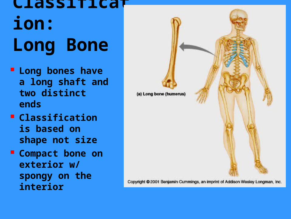
Classification:Long Bone
Long bones have a long shaft and two distinct ends
Classification is based on shape not size
Compact bone on exterior w/ spongy on the interior
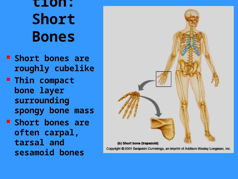
Classification:Short Bones
Short bones are roughly cubelike
Thin compact bone layer surrounding spongy bone mass
Short bones are often carpal, tarsal and sesamoid bones

Classification:
Flat Bones Flat bones are thin, flattened and usually curved
Parallel layer of compact bone with spongy bone layer between
Skull, sternum and ribs are examples

Classification:
Irregular Bone
Irregular bones don’t fit into the previous categories
Complicated shapes
Consist of spongy bone with a thin layer of compact
Examples are hip bones & vertebrae

Gross Anatomy of Bones
SECTION IV

Gross Anatomy Landmarks
– Diaphysis – Proximal epiphysis
– Distal epiphysis
Membranes– Periosteum
– Endosteum
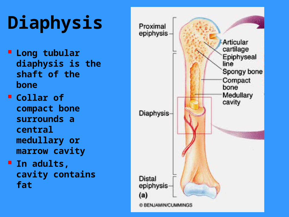
Diaphysis Long tubular diaphysis is the shaft of the bone
Collar of compact bone surrounds a central medullary or marrow cavity
In adults, cavity contains fat

Epiphysis The epiphyses are the ends of the bone
The joint surface of the epiphysis is covered with articular cartilage
Epiphyseal line separate diaphysis and epiphysis
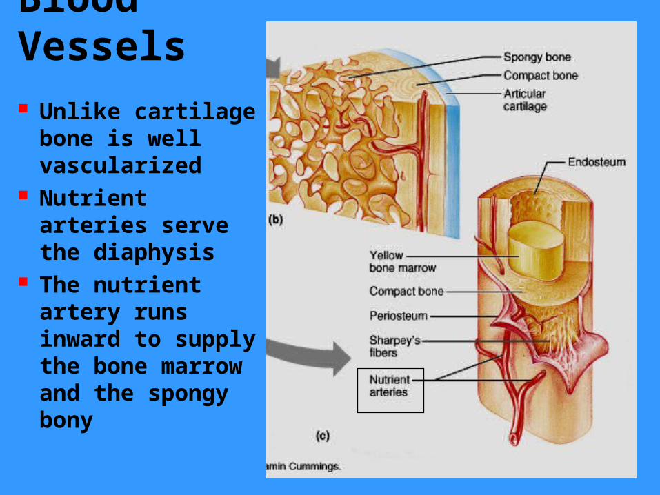
Blood Vessels Unlike cartilage bone is well vascularized
Nutrient arteries serve the diaphysis
The nutrient artery runs inward to supply the bone marrow and the spongy bony

Medullary cavity The interior of all bones consists largely of spongy bone
The very center of the bone is an open or marrow cavity
The cavity is filled with yellow bone marrow

Membranes Periosteum covers outer bone surfaces except the ends of the epiphysis
The membrane has two sublayers– Superficial layer
– Osteogenic layer
Membranes The superficial layer consists of dense irregular connective tissue which resists tension placed on a bone during bending
The osteogenic layer abuts the compact bone and contains bone-depositing cells called osteoblasts and osteoclasts that are responsible for bone remodeling
Membranes During periods of bone growth or deposition the osteogenic cells differentiate into osteoblasts
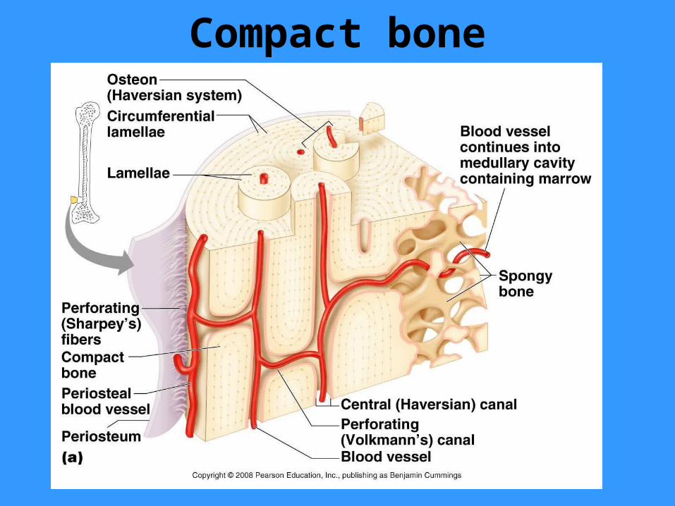
Osteoblasts produce the bone tissue that forms the circumferential lamellae that encircle the perimeter of the bone

Membranes Periosteum is richly supplied with nerves and blood vessels
The periosteum is supplied by branches of the nutrient artery and epiphyseal vessels
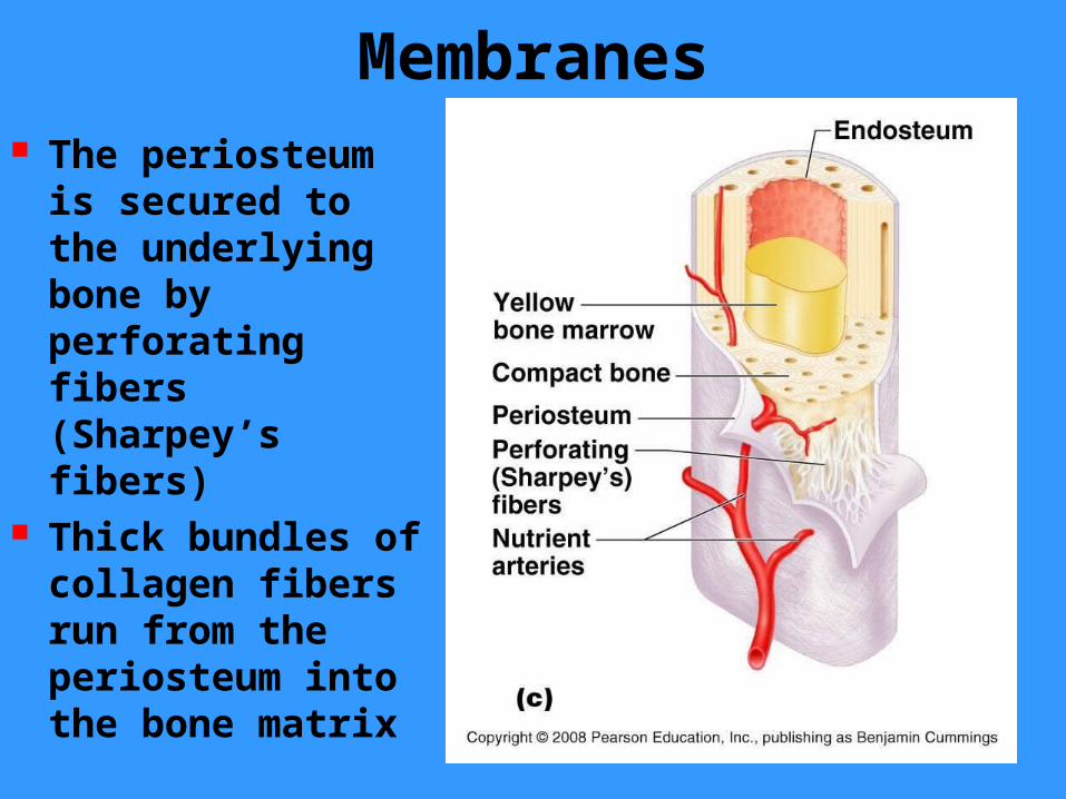
Membranes The periosteum is secured to the underlying bone by perforating fibers (Sharpey’s fibers)
Thick bundles of collagen fibers run from the periosteum into the bone matrix

Membranes Internal bone structures are covered by a thinner connective tissue membrane the endosteum
It also contains the osteoclasts and osteoblasts necessary for bone remodeling

Membranes The endosteum covers the trabeculae of spongy bone and lines the central canals of osteons
Short, Irregular and Flat Bones
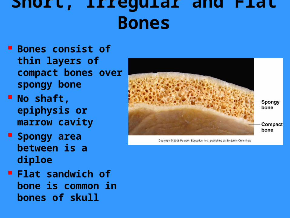
Bones consist of thin layers of compact bones over spongy bone
No shaft, epiphysis or marrow cavity
Spongy area between is a diploe
Flat sandwich of bone is common in bones of skull
Bone Design and Stress The internal anatomy of each bone reflects the stresses most commonly placed upon it
Bones are subjected to compressive forces in weight bearing and tension forces when muscle pulls upon them
Often weight bearing loads are applied off center which threatened to bend the bone

Bone Design and Stress Bending compresses the bone on one side and compresses it on the other
Compression and tension are greatest at the external surfaces of the bone

Bone Design and Stress Compact bone occurs at the external surfaces to resist these tension and compression forces
Internal bone structures are not subjected to these forces and thus spongy bone is sufficient
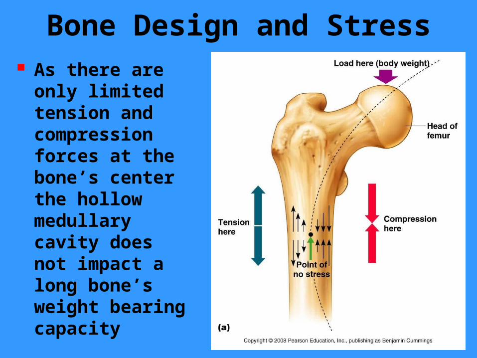
Bone Design and Stress As there are only limited tension and compression forces at the bone’s center the hollow medullary cavity does not impact a long bone’s weight bearing capacity

Bone Design and Stress Spongy bone is not a random network of bone fragments
The trabeculae align along stress lines in an organized patterns of tiny struts that provide internal support for the bone

Bone Markings Bones are shaped by the tissues that act upon and around them
Bones display bulges, depressions and holes which serve as sites of muscle, ligament and tendon attachment, points of articulation, or as conduits for blood vessels and nerves
Projections from the bone surface include heads, trochanters, spines, and others
Depressions include fossae, sinuses, foramina, and grooves


Bone Markings Tuberosity - a large rounded projection which may be roughened– tibial tuberosity
Bone Markings
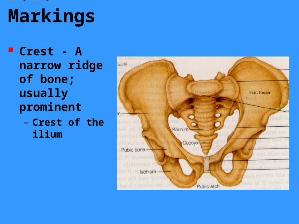
Crest - A narrow ridge of bone; usually prominent– Crest of the ilium

Bone Markings Trochanter - A very large, blunt, irregularly shaped process– Greater trochanter of femur
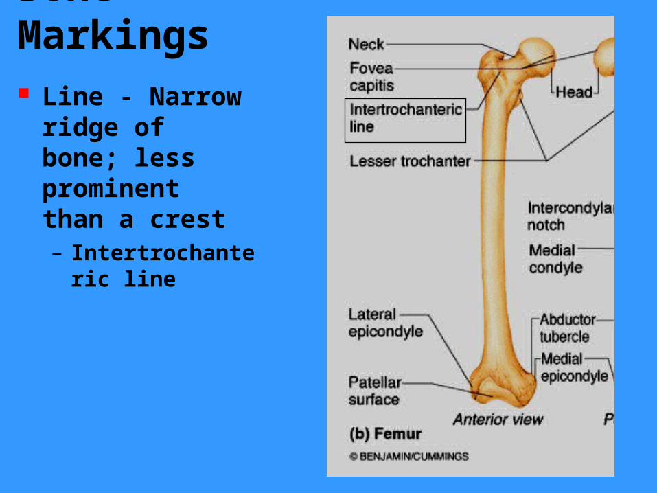
Bone Markings Line - Narrow ridge of bone; less prominent than a crest– Intertrochanteric line

Bone Markings Tubercle - Small rounded projection or process– adductor tubercle
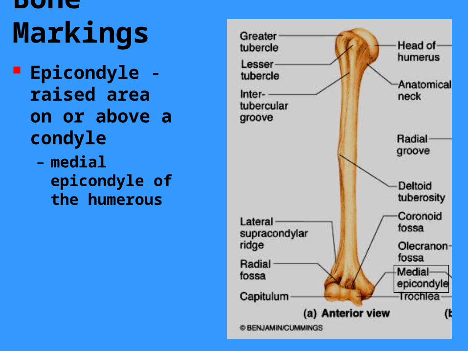
Bone Markings Epicondyle - raised area on or above a condyle– medial epicondyle of the humerous
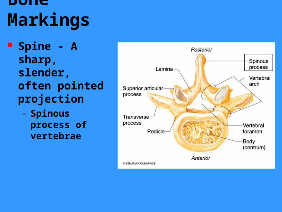
Bone Markings Spine - A sharp, slender, often pointed projection– Spinous process of vertebrae

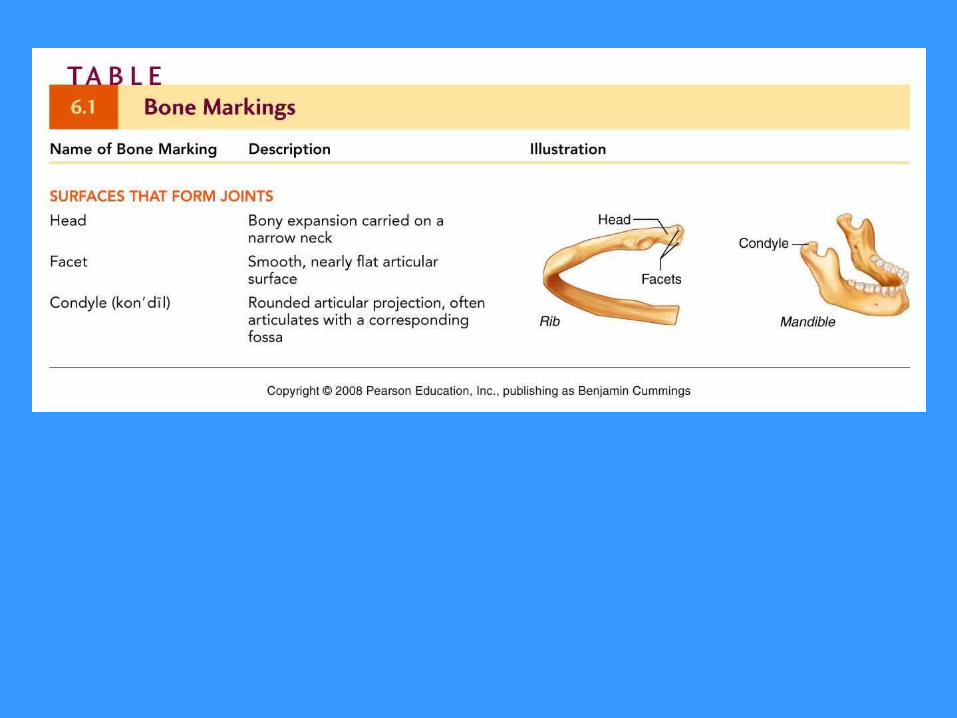
Bone Markings Head - Bony expansion carried on a narrow neck– head of the humerus

Bone Markings Facet - Smooth, nearly flat articular surface
– facet on transverse process of thoracic vertebrae
Facet

Bone Markings Condyle - Rounded articular projection– lateral condyle of femur
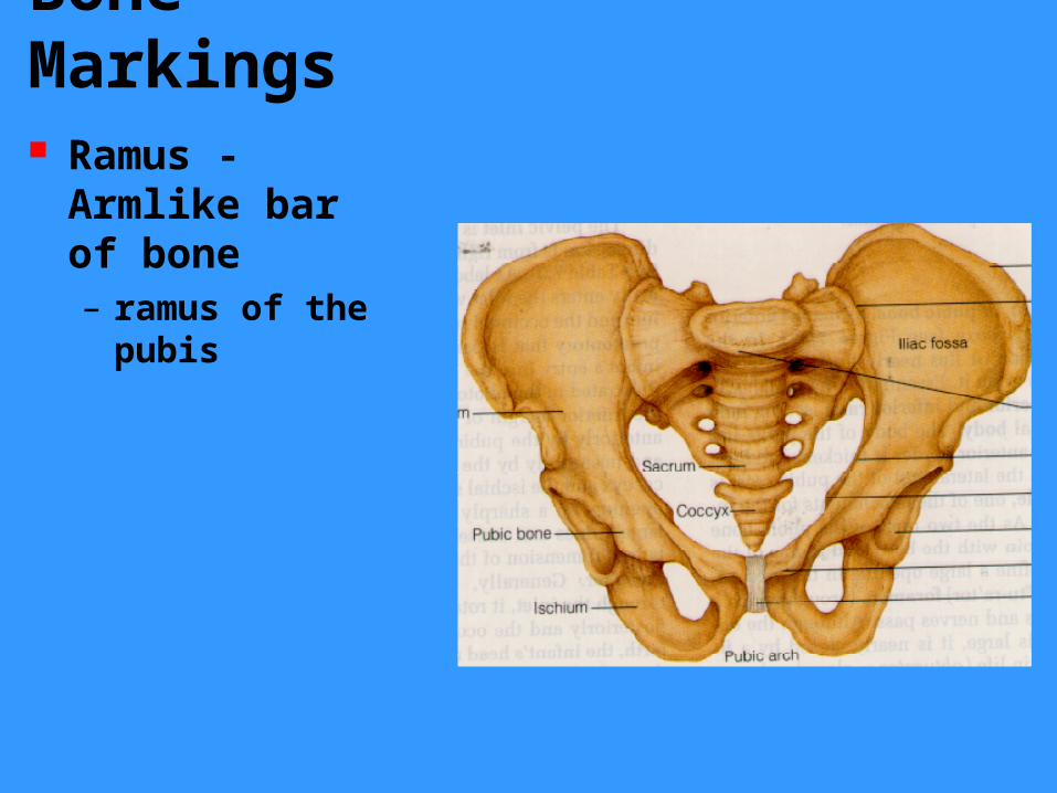
Bone Markings Ramus - Armlike bar of bone– ramus of the pubis

Bone Markings
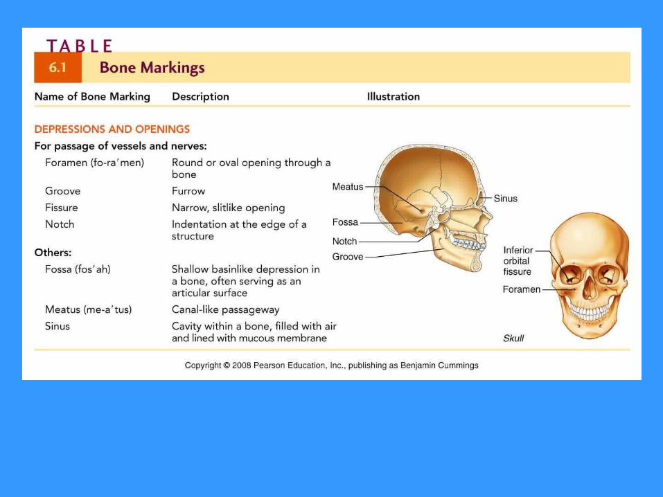
Meatus - canal-like passageway– External auditory meatus

Bone Markings Sinus - Cavity within a bone, filled with air and lined with mucous membrane– nasal sinus
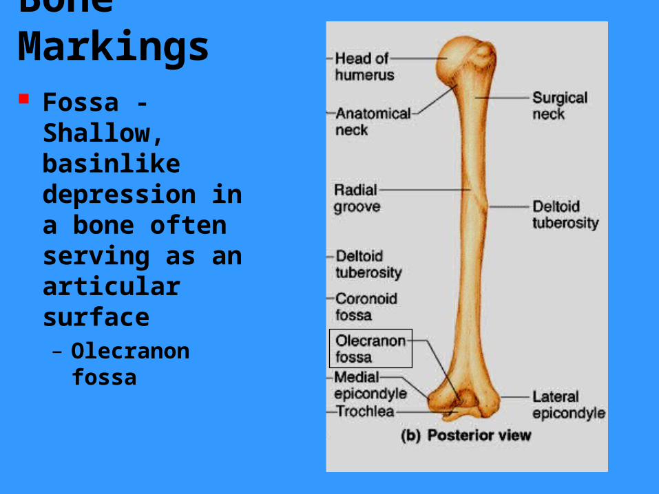
Bone Markings Fossa - Shallow, basinlike depression in a bone often serving as an articular surface– Olecranon fossa

Bone Markings Groove - a narrow furrow in the surface of the bone– radial groove

Bone Markings Fissure - Narrow, slitlike opening
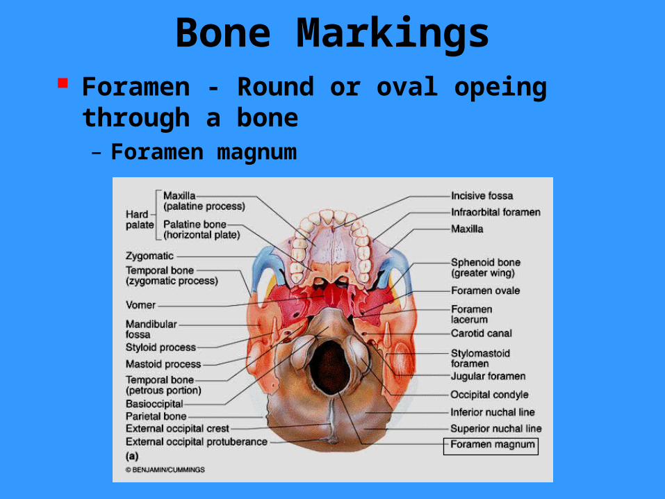
Bone Markings Foramen - Round or oval opeing through a bone– Foramen magnum
Compact Bone Compact bone appears very dense It actually contains canals and passageways that provide access for nerves, blood vessels, and lymphatic ducts
The structural unit of compact bone is the osteon or Haversian system
Each osteon is an elongated cylinder running parallel to the long axis of the bone
Functinally each osteon represents a weight bearing pillar
Compact bone
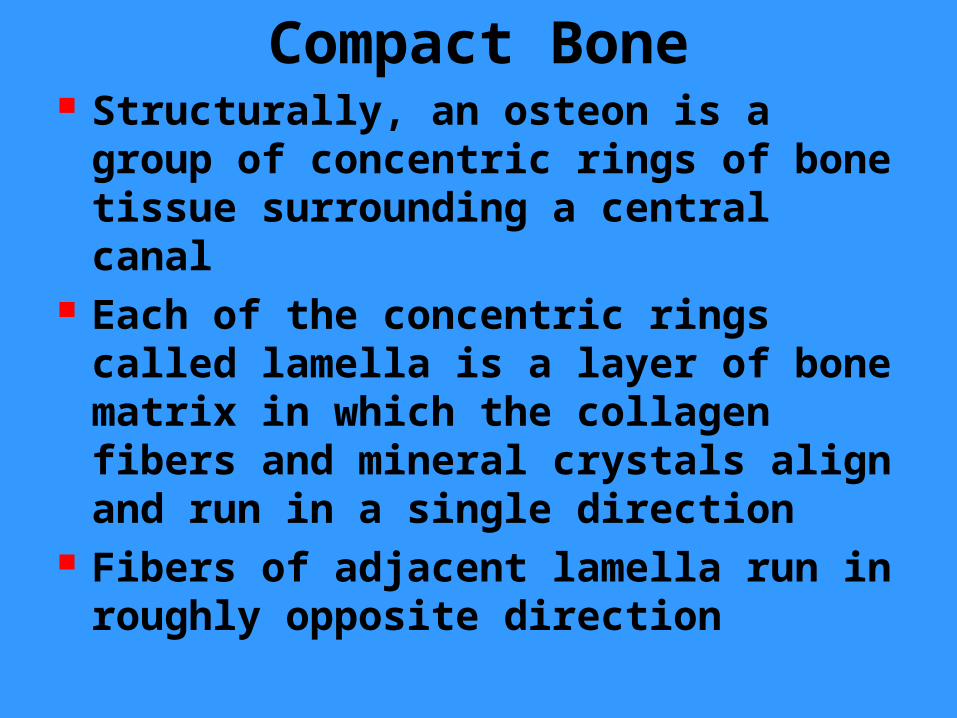
Compact Bone Structurally, an osteon is a group of concentric rings of bone tissue surrounding a central canal
Each of the concentric rings called lamella is a layer of bone matrix in which the collagen fibers and mineral crystals align and run in a single direction
Fibers of adjacent lamella run in roughly opposite direction
Compact bone

Compact bone
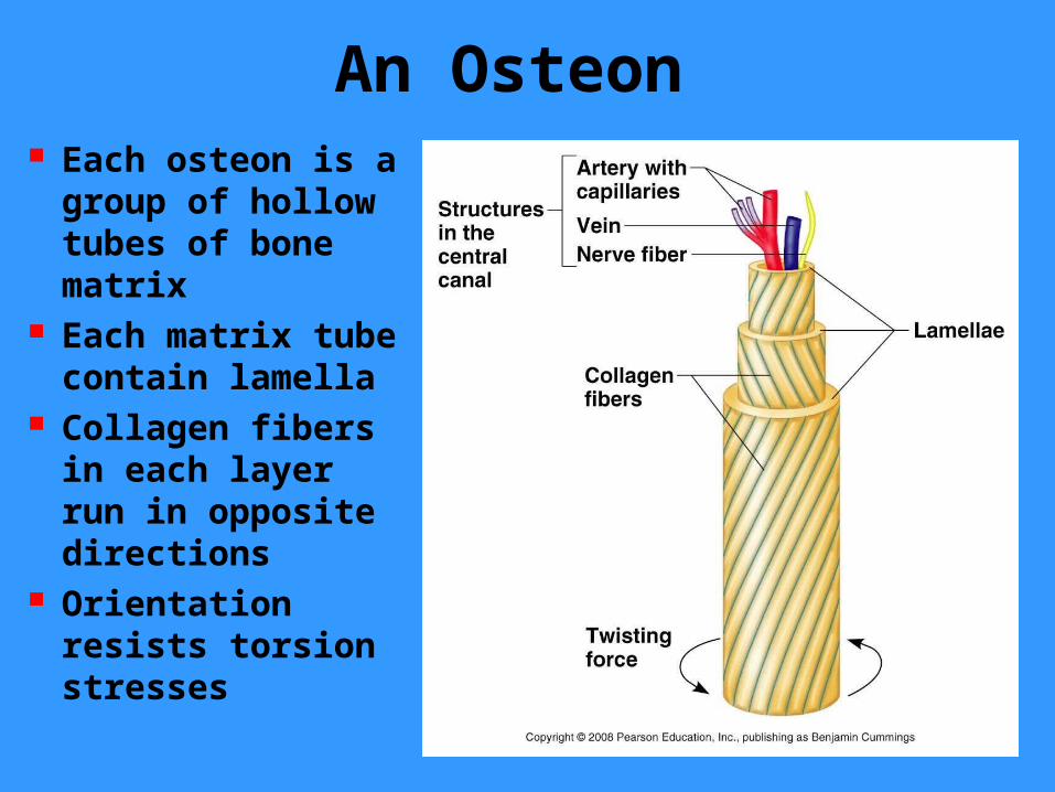
An Osteon Each osteon is a group of hollow tubes of bone matrix
Each matrix tube contain lamella
Collagen fibers in each layer run in opposite directions
Orientation resists torsion stresses
Compact Bone The alternating pattern of lamella orientation is optimal for withstanding torsion, stresses
The lamella of bone also inhibit crack propagation
When a crack reaches the edge of a lamella, the forces causing the crack are dispersed around lamellar boundaries, thus preventing the crack from progressing into deeper parts of the bone and causing fracture
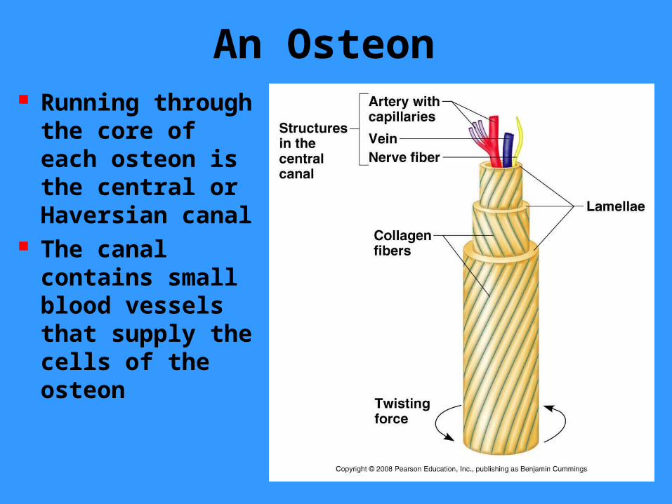
An Osteon Running through the core of each osteon is the central or Haversian canal
The canal contains small blood vessels that supply the cells of the osteon
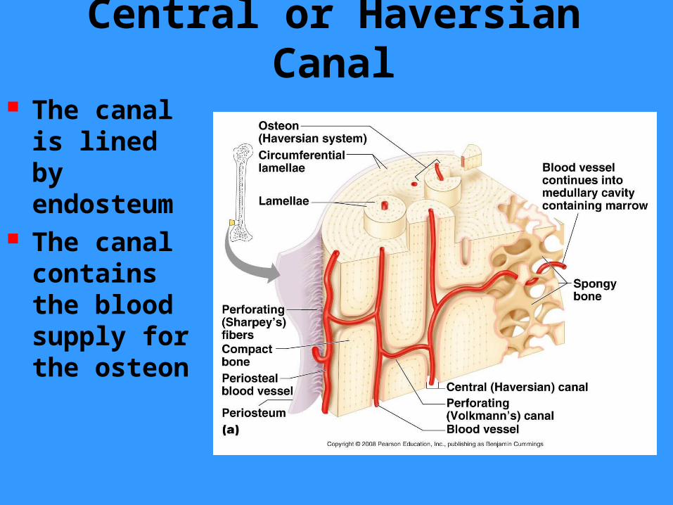
Central or Haversian Canal
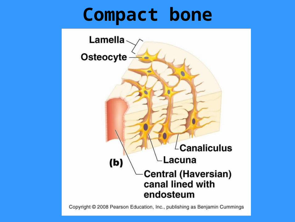
The canal is lined by endosteum
The canal contains the blood supply for the osteon

Perforating (Volkmann’s) Canal
Canals lie at right angles to long axis of bone
Connect the vascular supply of the periosteum to those of the central canal and medullary cavity

Compact Bone Osteocytes are the mature bone cells occupying the small spaces in the solid matrix called lacuna
Thin tubes called canaliculi run through the matrix connecting
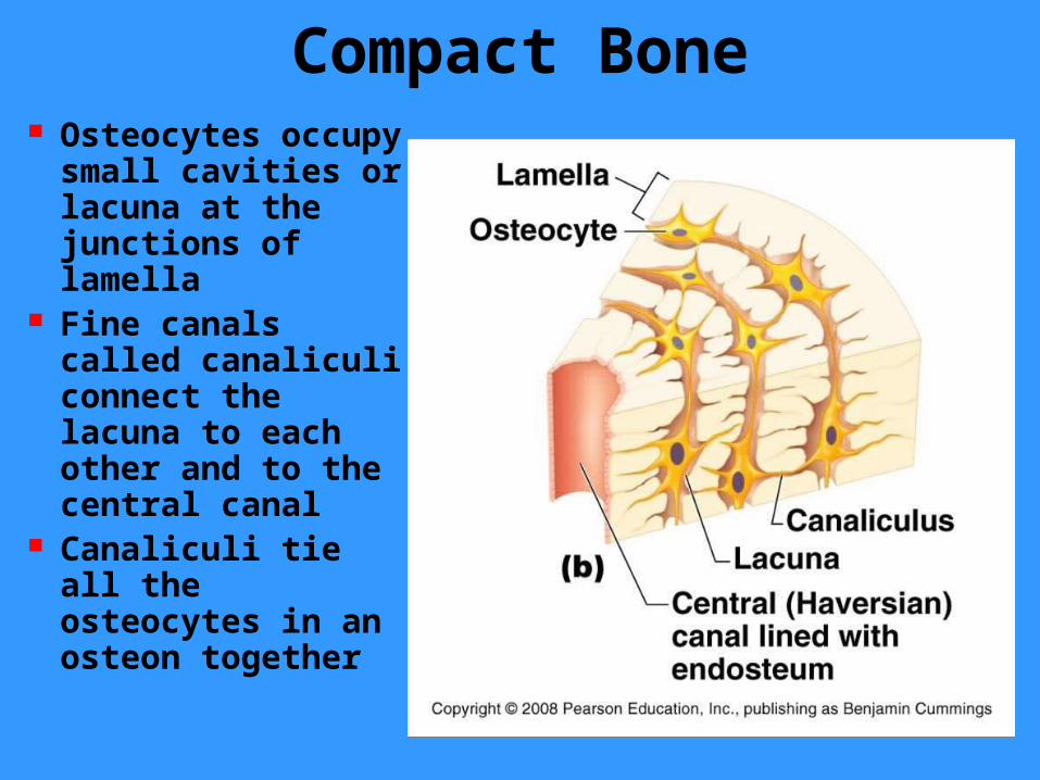
Compact Bone Osteocytes occupy small cavities or lacuna at the junctions of lamella
Fine canals called canaliculi connect the lacuna to each other and to the central canal
Canaliculi tie all the osteocytes in an osteon together

Compact Bone Canaliculi run through the matrix connecting neighboring lacunae to one another and to the nearest capillaries such as those in the central canal
Within the canaliculi, the extensions of neighboring osteocytes touch each other and form gap junctions

Compact Bone Gap junctions allow nutrients diffusing from the capillaries to cross these junctions
Nutrients are then passed from one osteocyte to the next
Compact Bone The passage of nutrients through gap junctions occurs throughout an entire osteon
This direct transfer from cell to cell is the only way to supply osteocytes with nutrients as the intervening bone matrix is too solid and impermeable to act as a diffusion medium
Compact Bone Osteocytes remain in the matrix they have secreted
Live cells appear to be needed to maintain the matrix
Loss of osteocytes from the matrix results resorbtion of the matrix

Compact Bone Not all lamellae in compact bone occur in osteons
Interstitial lamellae are incomplete lamellae lying between the cylindrical osteons
These represent remnants old osteons cut by bone remodeling
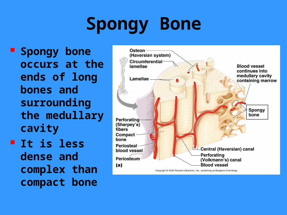
Spongy Bone Spongy bone occurs at the ends of long bones and surrounding the medullary cavity
It is less dense and complex than compact bone
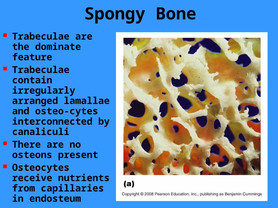
Spongy Bone Trabeculae are the dominate feature
Trabeculae contain irregularly arranged lamallae and osteo-cytes interconnected by canaliculi
There are no osteons present
Osteocytes receive nutrients from capillaries in endosteum

Spongy Bone Trabeculae align along lines of stress
Function as struts of bone

Chemical Composition of Bone
The organic components of bone are:– Osteoblasts (bud cells) – Osteocytes (mature cells) – Osteoclasts (large cells which resorb matrix)
– Osteoid (organic part of the matrix)•Osteoid makes up 1/3 of the matrix•Includes proteogylcans, glycoproteins, & collagen
•These components, particularly collagen contribute to the flexibility and tensile strength of bone to resist stretching and twisting
Chemical Composition of Bone The inorganic components of bone
(65% by mass) consist of hydroxyapatites or mineral salts, largely calcium phosphate
Tiny crystals of calcium salts are deposited in and around the collagen fibers of the extracellular matrix
The crystals are exceptionally hard and resist compression
Organic and inorganic components of matrix allows a bone to be strong but not brittle

Bone Development Osteogenesis and ossification refer to the process of bone formation
Osteogeneis begins in the embryo and continues until adulthood
Remodeling is bone resorption and deposition in response to stress and repair of bone

Bone Development Before week 8 the skeleton of the human embryo is made entirely from hyaline cartilage and mesenchyme membranes
At 8 weeks bone begins to appear and eventually replaces most cartilage and mesenchymal membranes

Bone Development Bones that develop from mesenchymal membranes are called membrane bones
Membrane bones develop from a fibrous membrane in a process called intra-membranous ossification
Other bones develop as hyaline cartilage initially, which is replaced through a process called endochondrial ossification
These are referred to as endochondrial bones or cartilage replacement bones

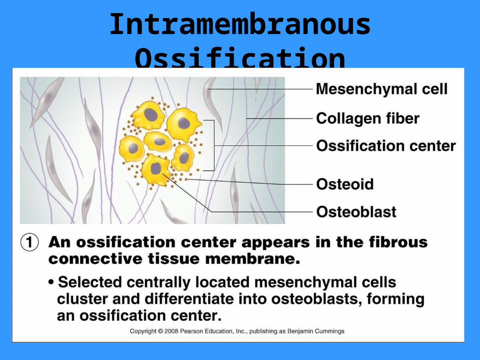
Intramembranous Ossification
Membrane bones form directly from mesenchyme without being modeled in cartilage
All bones of the skull (except a few at the base of skull) are membrane bones
The clavicles are also membrane bones
Note that most of these bones are flat bones
Intramembranous Ossification

Intramembranous Ossification

Intramembranous Ossification

Intramembranous Ossification
Endochondral Ossification
Most bones (except clavicles and most skull bones) form by the process of endochondral ossification
The bones are first modeled in hyaline cartilage, which is then gradually replaced by bone tissue
This process uses hyaline cartilage “bones” as the pattern for bone construction
Endochondral Ossification
Endochondral ossification begins late in the second month of development and continues into early adulthood when the skeleton is fully ossified
In endochondral ossification the cartilage model of the bone is replaced by bone
Growing endochondral bones increase in length and in width

Endochondral Ossification
Cartilage bones are surrounded by a perichondrium
At the 8th week of development, the perichondrium (fibrous connective tissue layer) becomes infiltrated by blood vessels converting it to a vascularized, bone forming periosteum
The increase in nutrition enables the mesenchyme cells to differentiate into osteoblasts that form a collar of bone

Endochondrial Ossification

Endochondral Ossification
Formation of a bone collar around diaphysis of cartilage model
Osteoblasts of the new periosteum secrete osteoid against the hyaline cartilage along the length of the diaphysis
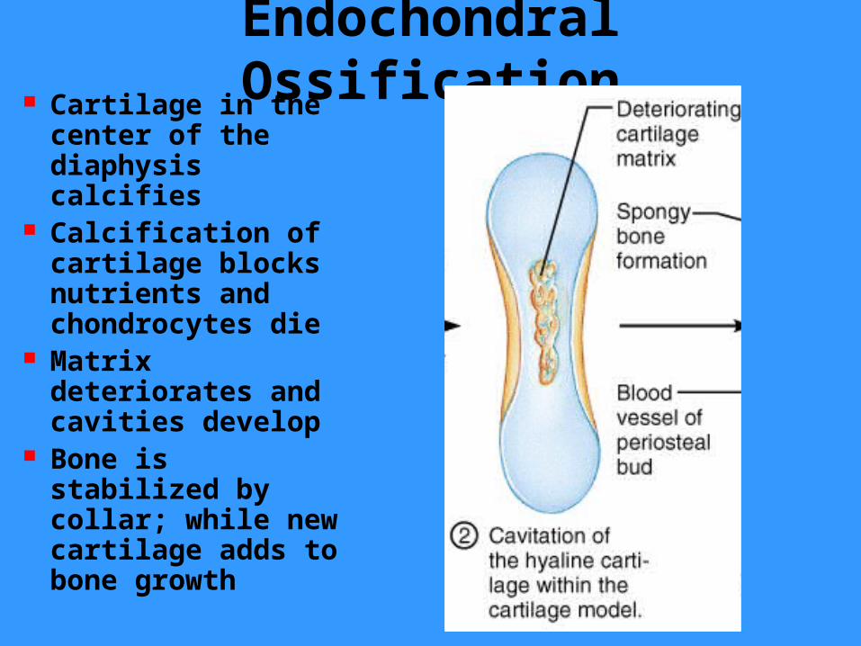
Endochondral Ossification Cartilage in the
center of the diaphysis calcifies
Calcification of cartilage blocks nutrients and chondrocytes die
Matrix deteriorates and cavities develop
Bone is stabilized by collar; while new cartilage adds to bone growth
Endochondral Ossification
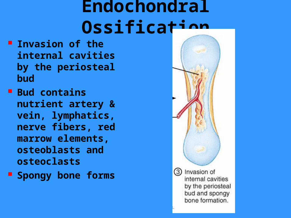
Invasion of the internal cavities by the periosteal bud
Bud contains nutrient artery & vein, lymphatics, nerve fibers, red marrow elements, osteoblasts and osteoclasts
Spongy bone forms

Endochondral Ossification
Formation of the medullary cavity as ossification continues
Secondary ossification centers form in epiphyses
Cartilage in epiphyses calcifies and deteriorates opening cavities for entry of periosteal bud
Endochondral Ossification
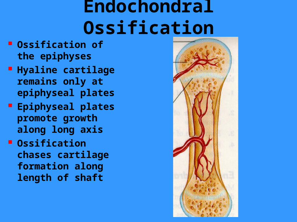
Ossification of the epiphyses
Hyaline cartilage remains only at epiphyseal plates
Epiphyseal plates promote growth along long axis
Ossification chases cartilage formation along length of shaft
Endochondral Ossification
After the secondary ossification sites have appeared and epiphyses have largely ossified, hyaline cartilage remains on– Epiphyseal surfaces where it forms articular cartilages
– Between the diaphysis and the epiphysis where it forms the epiphyseal plates
– The epiphyseal plates or growth plates are responsible for lengthening of bones during the two decades following birth
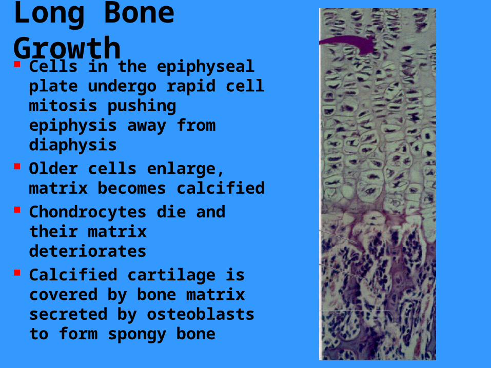
Long Bone Growth Cells in the epiphyseal plate undergo rapid cell mitosis pushing epiphysis away from diaphysis
Older cells enlarge, matrix becomes calcified
Chondrocytes die and their matrix deteriorates
Calcified cartilage is covered by bone matrix secreted by osteoblasts to form spongy bone

Epiphyseal Growth Areas
In the epiphysis of the fetus and the epiphyseal plates are organized to allow bones to grow quickly & efficiently
The cartilage cells nearest the epiphysis (quiescent zone) are relatively inactive

Epiphyseal Growth Areas
The cartilage cells form tall columns in the proliferation zone
The rapid division of chondroblasts push the epiphysis away from the diaphysis
The growth here lengthens the entire long bong
Epiphyseal Growth Areas
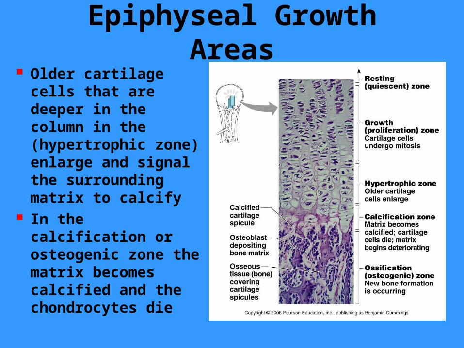
Older cartilage cells that are deeper in the column in the (hypertrophic zone) enlarge and signal the surrounding matrix to calcify
In the calcification or osteogenic zone the matrix becomes calcified and the chondrocytes die

Epiphyseal Growth Areas
The process of ossification leaves long spicules (trabeculae) of calcified cartilage on the diaphysis side
The spicules are then covered with bone tissue by osteoblasts
Osteoclasts complete the remodeling of the bone

Postnatal Bone Growth During childhood and adolescence bone growth occurs entirely by growth at the epiphyseal plates
In growing bones cartilage is replaced with bone tissue on the diaphysis side about as quickly as it grows
The epiphyseal plate remains a constant thickness while the overall length of the bone increases
Postnatal Bone Growth As the end adolescence approaches, the chondroblasts in the epiphyseal plate divide less often
The epiphyseal plates become thinner, eventually exhausting their supply of mitotically active catilage cells
The cartilage stops growing and is replaced by bone tissue
The epiphyseal plate fuses and growth is done (18 female, 21 male)
Postnatal Bone Growth Bones grow in width by appositional growth
Osteoblasts in the periosteum add bone tissue to the external surface of the diaphysis
Osteoclasts in the endosteum remove bone from the internal surface of the diaphysis wall
These two processes occur at roughly the same rate

Postnatal Bone Growth Other types of endochondial bones grow in slightly different patterns
Bone growth is regulated by several hormones– Pituitary simulates growth at plates
– Thyroid regulates growth to ensure that the skeleton retains proper proportions
– Sex hormones influence growth at adolescent growth spurts

Growth and Remodeling
Bone Remodeling Bone is dynamic and active tissue Long bone growth is accompanied by almost continuous remodeling in order to maintain proper proportions
Large amounts of bone matrix and thousands of osteocytes are being continually removed and replaced
The small scale architecture of bones changes constantly
Bone Remodeling The spongy bone of the skeleton is replaced every 3 years
The compact bone is replaced every 10 years
The remodeling process is not uniform as some parts experiencing more stress are replaced at a faster rate (every 5-6 months) while other areas change more slowly
Bone Remodeling Bone remodeling involves both bone formation and resorption
Remodeling occurs at the periosteal and endosteal sufaces
Bone formation is done by osteoblasts and bone resorption is done by osteoclasts

Bone Remodeling
Bone remodeling is coordinated by cohorts of adjacent osteoclasts called remodeling units
Osteoclasts crawl along the bone surfaces digging pits as they break down bone surfaces
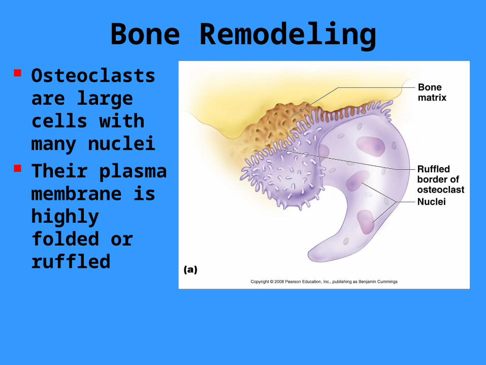
Bone Remodeling Osteoclasts are large cells with many nuclei
Their plasma membrane is highly folded or ruffled
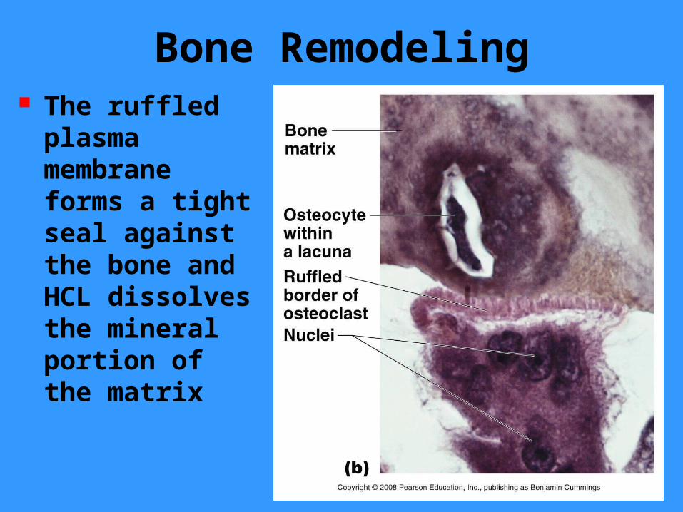
Bone Remodeling The ruffled plasma membrane forms a tight seal against the bone and HCL dissolves the mineral portion of the matrix

Bone Remodeling Osteoclasts release calcium ions (Ca2+) and phosphate ions (PO4
3-) that enters the tissue fluid and the bloodstream
Lysosomal enzymes are also released by the osteoclasts and digest the organic part of the bone matrix
Finally, osteoclasts take up collagen and dead osteocytes by phagocytosis
Bone Remodeling
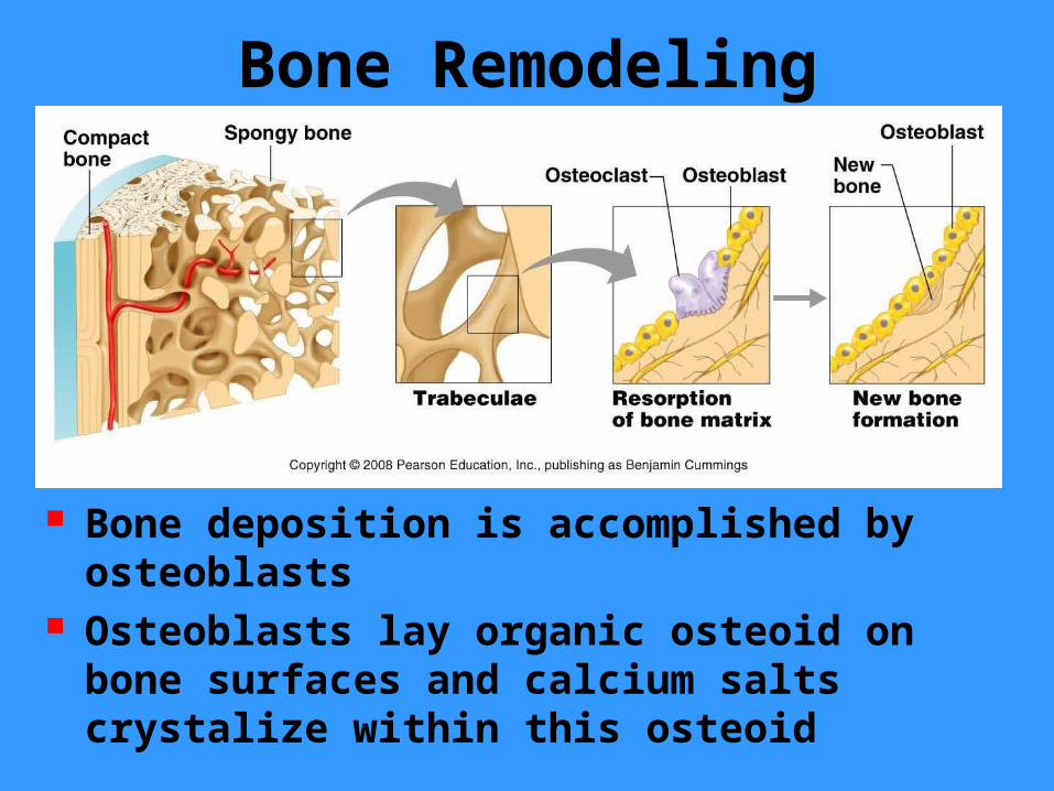
Bone deposition is accomplished by osteoblasts
Osteoblasts lay organic osteoid on bone surfaces and calcium salts crystalize within this osteoid

Bone Remodeling Bone forming osteoblasts form from mesenchyme-like stem cells located in the periosteum, endosteum, and the connective tissue of nearby bone marrow
Osteoclasts form in bone marrow from immature blood cells called hematopoietic stem cells
Many of these stem cells fuse together to form each osteoclast, thus their multinucleate structure
Bone Remodeling Bone of the skeleton are continually remodeled for 2 reasons– Bone remodeling helps maintain constant concentrations of Ca2+ and PO4
3- in bodily fluids– Bones are remodeled in response to the mechanical stress it experiences•Osteons of compact bone and the trabeculae of spongy bone are constantly replaced by new osteons and trabeculae that are more precisely aligned with newly experienced compressive and tensile forces
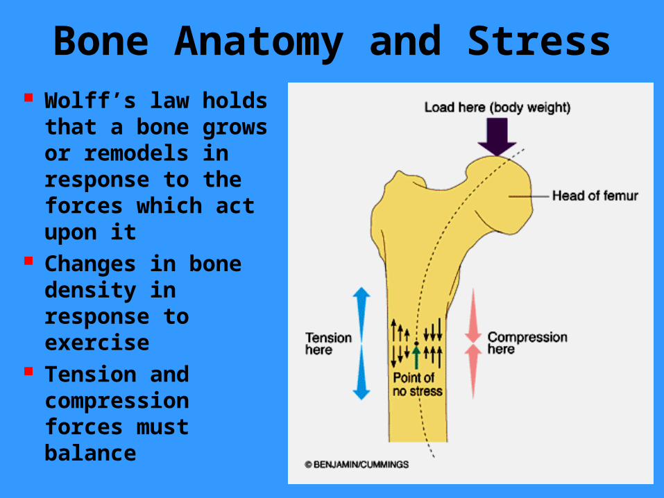
Bone Anatomy and Stress Wolff’s law holds that a bone grows or remodels in response to the forces which act upon it
Changes in bone density in response to exercise
Tension and compression forces must balance

Healing of a Bone Fracture
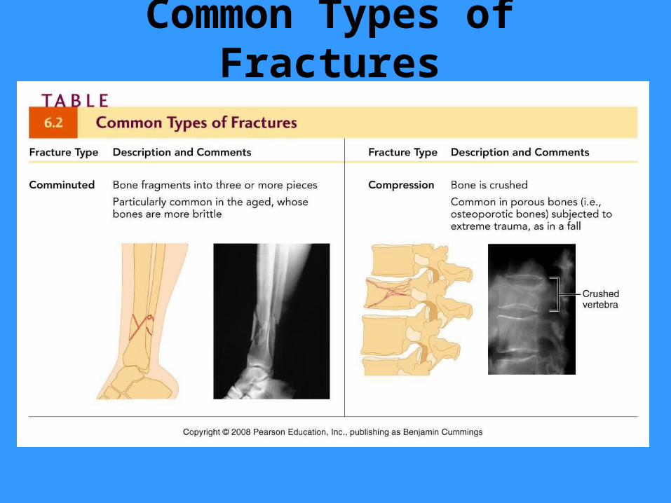
Common Types of Fractures

Common Types of Fractures
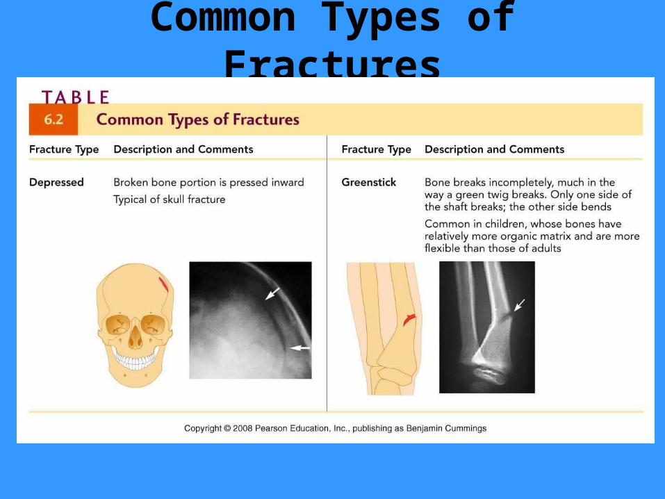
Common Types of Fractures
Related Documents