Bone marrow-derived recipient cells in murine transplanted hearts: potential roles and the effect of immunosuppression Nana Rezai 1 , Stephane Y Corbel 2 , Darya Dabiri 1 , Alexandra Kerjner 1 , Fabio MV Rossi 2 , Bruce M McManus 1 and Thomas J Podor 1 1 Department of Pathology and Laboratory Medicine, The James Hogg iCAPTURE Centre for Cardiovascular and Pulmonary Research, St Paul’s Hospital-University of British Columbia, Vancouver, BC, Canada and 2 Biomedical Research Centre, University of British Columbia, Vancouver, BC, Canada Currently, there is intense debate regarding the origin of reparative cells in injured hearts and vasculature. To determine the contribution of recipient bone marrow (BM)-derived cells to the regeneration of cells in the vasculature of transplanted hearts and to examine the effect of immunosuppression on this phenomenon, we evaluated the fate of green fluorescent protein (GFP)-positive recipient BM cells in non-GFP-expressing cardiac allografts. C57BL/6 BM-GFP chimeric recipients underwent cardiac transplantation. Allografts were immuno- suppressed with tacrolimus for 14 or 30 days post-transplantation or were saline treated. Hearts were excised and stained with markers for endothelial cells (EC) or smooth muscle cells (SMC). Colocalization with BM- derived recipient cells was evaluated using confocal microscopy with three-dimensional image analysis. Immunosuppression with tacrolimus did not affect the frequency of recipient BM-derived cell chimerism as EC or SMC phenotypes. A higher frequency of EC chimerism was found at 14 days as compared to 30 days post- transplantation in allograft hearts. BM-derived recipient cells are recruited to areas of donor vascular injury with intercalation of recipient EC and SMC in the setting of ongoing alloimmune recognition of the allograft. Our findings confirm that immunosuppression with tacrolimus does not affect the frequency of recipient BM- derived cell repopulation at an early time point 14 days post-transplantation. EC repopulation by BM-derived recipient cells was found to be an early event in transplanted allograft hearts, which decreased in frequency over time. Laboratory Investigation (2005) 85, 982–991. doi:10.1038/labinvest.3700302; published online 20 June 2005 Keywords: bone marrow; transplant heart; immunosuppression; endothelial cell; smooth muscle cell Heart transplantation is a life-prolonging procedure that has become an accepted surgical treatment for patients with end-stage congestive heart failure. Despite improvements in outcomes over the last two decades, both acute and chronic rejection continue to limit survival and quality of life in heart transplant recipients. 1 Transplant vascular disease (TVD), as an expression of chronic rejection, is a rapidly progressive form of atherosclerosis that occurs in the vessels of all solid organ transplants, including the heart. Cardiac TVD is estimated to affect more than 40% of recipients who survive beyond four years after transplantation. 1,2 Recent studies have suggested that undifferen- tiated progenitor cells may emigrate from the recipient to the graft, contributing to vascular remodeling and possibly contributing to the athero- sclerotic process of TVD. 3–5 However, the degree of recipient cell repopulation of cells of the vessel wall is currently a matter of intense debate 6 since there are discrepancies in reported rates of chimerism in damaged vessels and hearts. 3–5,7–10 These discrepan- cies may be attributed to confounding factors, including different assays and techniques used to detect cell differentiation or colocalization events, as well as variations in models used to examine chimerism rates. To address the fate of endogenous recipient bone marrow (BM)-derived progenitor cells in the vascu- lature of transplanted (Tx) hearts, we performed heterotopic heart transplantation using chimeric Received 4 March 2005; revised and accepted 5 May 2005; published online 20 June 2005 Correspondence: Dr TJ Podor, PhD, Department of Pathology and Laboratory Medicine, The James Hogg iCAPTURE Center for Cardiovascular and Pulmonary Research, St Paul’s Hospital- University of British Columbia, Rm 161, 1081 Burrard St., Vancouver, BC, Canada V6Z 1Z3. E-mail: [email protected] Laboratory Investigation (2005) 85, 982–991 & 2005 USCAP, Inc All rights reserved 0023-6837/05 $30.00 www.laboratoryinvestigation.org

Welcome message from author
This document is posted to help you gain knowledge. Please leave a comment to let me know what you think about it! Share it to your friends and learn new things together.
Transcript

Bone marrow-derived recipient cells in murinetransplanted hearts: potential roles and theeffect of immunosuppression
Nana Rezai1, Stephane Y Corbel2, Darya Dabiri1, Alexandra Kerjner1, Fabio MV Rossi2,Bruce M McManus1 and Thomas J Podor1
1Department of Pathology and Laboratory Medicine, The James Hogg iCAPTURE Centre for Cardiovascularand Pulmonary Research, St Paul’s Hospital-University of British Columbia, Vancouver, BC, Canada and2Biomedical Research Centre, University of British Columbia, Vancouver, BC, Canada
Currently, there is intense debate regarding the origin of reparative cells in injured hearts and vasculature. Todetermine the contribution of recipient bone marrow (BM)-derived cells to the regeneration of cells in thevasculature of transplanted hearts and to examine the effect of immunosuppression on this phenomenon, weevaluated the fate of green fluorescent protein (GFP)-positive recipient BM cells in non-GFP-expressing cardiacallografts. C57BL/6 BM-GFP chimeric recipients underwent cardiac transplantation. Allografts were immuno-suppressed with tacrolimus for 14 or 30 days post-transplantation or were saline treated. Hearts were excisedand stained with markers for endothelial cells (EC) or smooth muscle cells (SMC). Colocalization with BM-derived recipient cells was evaluated using confocal microscopy with three-dimensional image analysis.Immunosuppression with tacrolimus did not affect the frequency of recipient BM-derived cell chimerism as ECor SMC phenotypes. A higher frequency of EC chimerism was found at 14 days as compared to 30 days post-transplantation in allograft hearts. BM-derived recipient cells are recruited to areas of donor vascular injury withintercalation of recipient EC and SMC in the setting of ongoing alloimmune recognition of the allograft. Ourfindings confirm that immunosuppression with tacrolimus does not affect the frequency of recipient BM-derived cell repopulation at an early time point 14 days post-transplantation. EC repopulation by BM-derivedrecipient cells was found to be an early event in transplanted allograft hearts, which decreased in frequencyover time.Laboratory Investigation (2005) 85, 982–991. doi:10.1038/labinvest.3700302; published online 20 June 2005
Keywords: bone marrow; transplant heart; immunosuppression; endothelial cell; smooth muscle cell
Heart transplantation is a life-prolonging procedurethat has become an accepted surgical treatment forpatients with end-stage congestive heart failure.Despite improvements in outcomes over the lasttwo decades, both acute and chronic rejectioncontinue to limit survival and quality of life inheart transplant recipients.1 Transplant vasculardisease (TVD), as an expression of chronic rejection,is a rapidly progressive form of atherosclerosis thatoccurs in the vessels of all solid organ transplants,including the heart. Cardiac TVD is estimated to
affect more than 40% of recipients who survivebeyond four years after transplantation.1,2
Recent studies have suggested that undifferen-tiated progenitor cells may emigrate from therecipient to the graft, contributing to vascularremodeling and possibly contributing to the athero-sclerotic process of TVD.3–5 However, the degree ofrecipient cell repopulation of cells of the vessel wallis currently a matter of intense debate6 since thereare discrepancies in reported rates of chimerism indamaged vessels and hearts.3–5,7–10 These discrepan-cies may be attributed to confounding factors,including different assays and techniques used todetect cell differentiation or colocalization events,as well as variations in models used to examinechimerism rates.
To address the fate of endogenous recipient bonemarrow (BM)-derived progenitor cells in the vascu-lature of transplanted (Tx) hearts, we performedheterotopic heart transplantation using chimeric
Received 4 March 2005; revised and accepted 5 May 2005;published online 20 June 2005
Correspondence: Dr TJ Podor, PhD, Department of Pathology andLaboratory Medicine, The James Hogg iCAPTURE Center forCardiovascular and Pulmonary Research, St Paul’s Hospital-University of British Columbia, Rm 161, 1081 Burrard St.,Vancouver, BC, Canada V6Z 1Z3.E-mail: [email protected]
Laboratory Investigation (2005) 85, 982–991& 2005 USCAP, Inc All rights reserved 0023-6837/05 $30.00
www.laboratoryinvestigation.org

recipient mice expressing green fluorescent protein(GFP)-positive BM in order to track the migration ofthe BM-derived recipient cells. Moreover, we mea-sured the effect of immunosuppression vs none onthe frequency of recipient GFP-labeled BM-derivedendothelial cells (EC) and smooth muscle cells(SMC) in the coronary arteries of the Tx heart.
Materials and methods
Generation of BM-Chimeric Mice
The study protocol was approved by the Universityof British Columbia Office of Research ServicesCommittee on Animal Care. C57BL/6 (CD45.2) andC57BL/6 (GFP/CD45.1) mice were bred in-house andmaintained in a pathogen-free environment. TheC57BL/6 (GFP/CD45.1) mice express GFP driven bythe CMV enhancer-chicken beta-actin hybrid pro-moter. They were a kind gift of Dr I Weissman.11
C57BL/6 (GFP/CD45.1) mice (8 weeks old) wereused as donors and 8- to 10-week-old C57BL/6(CD45.2) mice as recipients.
Cells
BM was flushed out of the femurs and the tibias ofC57BL/6 (GFP/CD45.1) mice and single-cell suspen-sions were made. Red cells were lysed, cells wereenumerated and 5� 106 cells were injected into thetail vein of lethally (950 rad) irradiated recipientC57BL/6 mice.
Peripheral Blood Analysis of BM-Chimeric Mice
At various time points after transplantation, 150 mlof peripheral blood were collected from the tails ofrecipient BM-chimeric mice. Reconstitution of thehematopoietic system was analyzed by flow cyto-metry on a FACScan (Becton Dickinson) using aprotocol as described previously.7 Mice with morethan 80% of GFP-positive peripheral blood leuko-cytes were used as recipient mice for the heterotopicheart transplant experiments 2 months after BMtransplantation.
Heterotopic Cardiac Transplantation
Heterotopic cardiac transplantation was performedas described.12 Hearts of 10-week-old 129SV/j micewere transplanted into the reconstituted GFP-BM-chimeric C57BL/6 mice described above. Controlsyngrafts consisted of 10-week-old C57BL/6 donorhearts transplanted into C57BL/6 GFP-BM-chimericmice. Allograft mice were treated with tacrolimus(Fujisawa, Osaka, Japan) administered intraperito-neally (i.p.) (6 mg/kg) daily for a period of 14 (n¼ 5)and 30 (n¼ 5) days post-transplantation. A subset ofallograft mice did not receive any immunosuppres-sive therapy, and were treated with daily i.p.injections of saline (n¼ 5) for a period of 14 days
post-transplantation. Mice were euthanized at 14and 30 days post-transplantation.
Immunofluorescence and Histochemical Staining
Native (Nat) and Tx hearts were excised from theallograft and control syngraft groups and routinelyprocessed for cryosections. Immunofluorescencestaining was performed using a modified protocolas described previously13 and other sections werestained with hematoxylin and eosin (H&E). Briefly,hearts in all treatment groups were perfusion fixedwith 2% paraformaldehyde and OCT embedded.Frozen midventricular sections were incubated withprimary antibodies, anti-smooth muscle (SM) a-actin(DAKO, Carpinteria, CA, USA), von Willebrandfactor (vWf) (DAKO) and anti-CD45 (BD Pharmingen,Mississauga, ON, Canada). Alexa 594-conjugatedsecondary antibody was used and nuclei werecounterstained with Hoechst 33342 (MolecularProbes, Eugene, OR, USA).
Confocal Microscopic Analysis and Quantitation ofthe Recipient BM-Derived Cells
All images were obtained using a Leica AOBSt SP2confocal microscope. In all, 10mm sections of the Txand Nat hearts were stained for vWf, SM a-actin andCD45, and analyzed. The number of GFP-positiveBM-derived recipient cells in the hearts was quanti-tated at 14 and 30 days post-transplantation in allallograft and syngraft groups. Briefly, 10 representa-tive microscopic fields at 1.4� 10�3 mm3 from thesame anatomical location in each myocardial sectionwere taken and Image-Pros Plus software (Media-Cyberneticss, San Diego, CA, USA) was used tocount the number of nuclei and nucleated GFP-positive recipient cells in the confocal opticalsections. For vWf and SM a-actin quantitation,analysis for colocalization with BM-derived recipientGFP-positive cells was performed on volumes of5.3� 10�4 mm3 optical stacks of vessels, 60–200mmwide in diameter, found in representative midven-tricular sections from Tx and Nat hearts. Stacks werereconstructed using Volocityt (Improvisions, Boston,MA, USA) and Metamorphs (Universal ImagingCorporationt, Downingtown, PA, USA) image soft-wares. Two independent, blinded observers reviewedeach reconstructed vessel. Nucleated recipient BM-derived GFP-positive cells seeded within the vesselwalls were quantitated and the number of morpho-logically distinguishable cells that stained dualpositive for GFP and either vWf or SM a-actin inthe vessel wall was scored. Cells were consideredendothelial in origin if they were positioned on theluminal side of the vessel, thin and elongate in shape,and demonstrated intracellular granular vWf staining.Cells were considered vascular SMC if they were inthe subendothelium or media, were spindle shaped,blunt ended and exhibited intracellular SM a-actin
Bone marrow-derived recipient cells in cardiac allograftsN Rezai et al
983
Laboratory Investigation (2005) 85, 982–991
staining. Each chimerism count was expressed as apercentage of total EC or SMC, respectively. Inaddition, to confirm the presence of GFP withinindividual cells separated from the autofluorescentbackground, spectrophotometric wavelength lambdascanning was performed on tissue sections. Algo-rithms were then applied to further separate thespectral fingerprint for GFP (510 nm emission maxi-mum) from the autofluorescence fingerprint (535–545 nm emission maximum) in order to ensureaccurate recipient BM-derived GFP cell detection.
Statistical Analyses
Data for the quantitation of recipient cell seeding aregiven as mean7s.e.m. Comparison between groupswas made using ANOVA and a post hoc Tukey’stest was used to determine statistical differencesbetween groups. The frequency of vWf or SM a-actinand GFP double-positive cells in the vessel wall areshown as mean7s.e.m. of the total recipient EC orSMC. A P-value of less than 0.05 was consideredsignificant.
Results
Recipient Cell Seeding to the Tx Heart and the Effectof Immunosuppression
We utilized chimeric GFP-BM recipient mice in aheterotopic heart transplant model to identify the
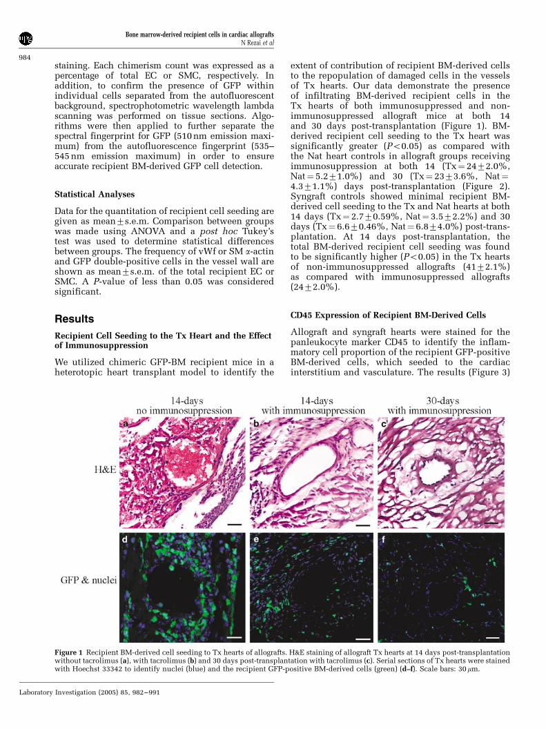
extent of contribution of recipient BM-derived cellsto the repopulation of damaged cells in the vesselsof Tx hearts. Our data demonstrate the presenceof infiltrating BM-derived recipient cells in theTx hearts of both immunosuppressed and non-immunosuppressed allograft mice at both 14and 30 days post-transplantation (Figure 1). BM-derived recipient cell seeding to the Tx heart wassignificantly greater (Po0.05) as compared withthe Nat heart controls in allograft groups receivingimmunosuppression at both 14 (Tx¼ 2472.0%,Nat¼ 5.271.0%) and 30 (Tx¼ 2373.6%, Nat¼4.371.1%) days post-transplantation (Figure 2).Syngraft controls showed minimal recipient BM-derived cell seeding to the Tx and Nat hearts at both14 days (Tx¼ 2.770.59%, Nat¼ 3.572.2%) and 30days (Tx¼ 6.670.46%, Nat¼ 6.874.0%) post-trans-plantation. At 14 days post-transplantation, thetotal BM-derived recipient cell seeding was foundto be significantly higher (Po0.05) in the Tx heartsof non-immunosuppressed allografts (4172.1%)as compared with immunosuppressed allografts(2472.0%).
CD45 Expression of Recipient BM-Derived Cells
Allograft and syngraft hearts were stained for thepanleukocyte marker CD45 to identify the inflam-matory cell proportion of the recipient GFP-positiveBM-derived cells, which seeded to the cardiacinterstitium and vasculature. The results (Figure 3)
Figure 1 Recipient BM-derived cell seeding to Tx hearts of allografts. H&E staining of allograft Tx hearts at 14 days post-transplantationwithout tacrolimus (a), with tacrolimus (b) and 30 days post-transplantation with tacrolimus (c). Serial sections of Tx hearts were stainedwith Hoechst 33342 to identify nuclei (blue) and the recipient GFP-positive BM-derived cells (green) (d–f). Scale bars: 30mm.
Bone marrow-derived recipient cells in cardiac allograftsN Rezai et al
984
Laboratory Investigation (2005) 85, 982–991

indicate that in the Tx hearts, CD45 was coexpressedby a large proportion of the GFP-positive BM-derived recipient cells found in the perivascular
space and cardiac interstitium in allograft hearts at14 and 30 days post-transplantation, both with andwithout immunosuppression.
0
0.05
0.1
0.15
0.2
0.25
0.3
0.35
0.4
0.45
0.5
Allograft 14-day NoImmunosuppression
Allograft 14-day WithImmunosuppression
Syngraft 14-day Allograft 30-day WithImmunosuppression
Syngraft 30-day
Mouse Transplant Group
Rat
io o
f R
ecip
ien
t B
M C
ells
/To
tal C
ard
iac
Nu
clei
Transplant heart
Native heart* p<0.05
*
**
**
**
Figure 2 Quantification of recipient BM-derived GFP-positive cell seeding. GFP-positive recipient BM-derived cells seeded within Txand Nat hearts of all allograft and syngraft groups were quantified and expressed as a fraction of total cardiac nuclei and graphicallyrepresented as the mean7s.e.m. from animals per group. *Significant difference, Po0.05.
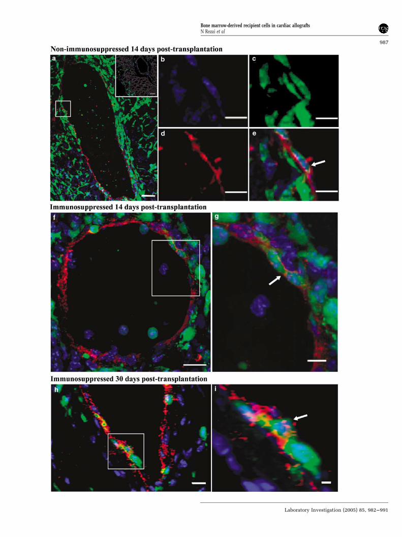
Figure 3 vWf staining of EC in Tx hearts. Confocal micrographs of (a, f and h) allograft vessels. (b) Hoechst 33342 nuclear staining (blue),(c) GFP-positive recipient BM-derived cells (green), (d) vWf-positive EC (red) and (e, g and i) merged images at higher magnification showdual-positive granular staining for vWf within the GFP cytoplasm of some cells (arrows). Inset (a) shows control immunostain withspecific antibody isotype control. Scale bars¼ (a) 40mm, (b–e and g) 10mm and (f) 20mm.
Bone marrow-derived recipient cells in cardiac allograftsN Rezai et al
985
Laboratory Investigation (2005) 85, 982–991
EC Differentiation in Tx Hearts
In all Tx hearts of allograft groups, we found GFP-vWf dual-positive cells in the endothelial lining ofvessels (Figure 4). The frequency of GFP-vWf dual-positive cells out of the total EC within the vesselwall was 11.872.5 and 4.071.2% for 14 and 30days post-transplantation, respectively (Table 1).The results indicate that the frequency of differ-entiation events for recipient BM-derived cells intoan EC phenotype within the vessel wall of Tx heartsappears to be higher at 14 days post-transplantationthan at 30 days (P¼ 0.04). The frequency of GFP-vWf dual-positive cells was similar in the vessels ofTx hearts in both the non-immunosuppressed(9.472.1%) and the immunosuppressed (11.872.5%) allograft groups at 14 days post-transplant-ation. These findings suggest that 14 days ofimmunosuppressive therapy does not significantlyaffect the frequency of recipient BM-derived cellchimerism in vessels of Tx hearts. Nat heartsdemonstrated a low level of GFP-vWf dual-positivecells only in the vessels of non-immunosuppressedallograft hearts, and no dual-positive cells werefound in any other allograft or syngraft recipient Nathearts.
SMC Differentiation in Tx Hearts
SM a-actin was used as a phenotypic marker forvascular SMC in Tx and Nat hearts. The results(Table 1) indicate that at 14 days post-transplant-ation, the frequency of SM a-actin-GFP dual-positivecells out of the total number of SMC within thevessel wall is not significantly different in theTx hearts of the allograft groups (nonimmuno-suppressed¼ 0.970.89% and immunosuppressed¼ 2.072.0%). This suggests that the frequency ofdifferentiation events into an SMC phenotype is notaffected by immunosuppressive treatment withtacrolimus (Figure 5). Further, there appears to beno significant difference in the number of SMa-actin-GFP dual-positive cells in Tx hearts ofimmunosuppressed allografts at 14 and 30 dayspost-transplantation (2.072.0 and 0.470.4%,respectively). No vessels in the Nat heart controlscontained SM a-actin-GFP dual-positive cells.
Discussion
TVD is one of the leading causes of graft failure anddeath after the first perioperative year and is nowthe most important problem in clinical organtransplantation. The original concept of TVD and
vascular remodeling assumed that vascular EC andSMC in TVD originate from graft tissue and aretherefore donor derived.1,14 In recent years, severalgroups have reported data both supporting9,15,16
and discounting17,18 repopulation of graft vesselsby recipient-derived cells after solid organ trans-plantation. However, several questions remain un-answered, including: (1) whether there is significantrepopulation of cells of the vessel wall withrecipient-derived cells, and if so, what the originof these cells is, (2) if the repopulation of vascularcells is either an early or late event, or a continuousprocess throughout TVD progression and (3) whatthe effect of immunosuppression is on recipient cellrepopulation of cells of the vessel wall of Tx hearts.The possibilities for the origin of recipient cellsrecruited to damaged vessels include circulatingvessel wall-derived adult EC, tissue resident pro-genitor cells or recipient BM-derived cells. In thisstudy, we investigate these questions by elucidatingthe contribution of recipient BM-derived cells in therepopulation of cells of the vessel wall at both anearly (14 days) and late (30 days) time-point post-transplantation as well as examining the effect ofimmunosuppressive therapy with tacrolimus on thefrequency of recipient BM-derived cell differentia-tion into vascular cell phenotypes.
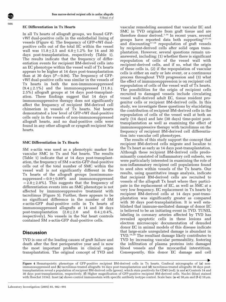
The results of this study support the concept thatrecipient BM-derived cells migrate and localize tothe Tx heart as early as 14 days post-transplantation.Although these recipient BM-derived cells predo-minantly consisted of inflammatory cell subsets, wewere particularly interested in examining the role ofnon-inflammatory recipient cell populations foundto seed sites within vessels of the Tx hearts. Ourresults, using quantitative image analysis, indicatethat recipient BM-derived cells are recruited tovessels of the allograft Tx heart where they partici-pate in the replacement of EC, as well as SMC at avery low frequency. EC replacement in Tx hearts byrecipient BM-derived cells at 14 days post-trans-plantation was significantly greater as comparedwith 30 days post-transplantation. It is well esta-blished that immune-mediated damage of donor ECis believed to be an initiating event in TVD. TUNELlabeling in coronary arteries affected by TVD hasrevealed apoptotic cells in these lesions andelectron microscopic documentation of denudeddonor EC in animal models of this disease indicatethat large-scale unregulated damage is abundant inTVD.19,20 The resultant damage likely contributes toTVD by increasing vascular permeability, fosteringthe infiltration of plasma proteins into damagedblood vessels and the myocardial interstitium.Consequently, this donor EC damage and cell
Figure 4 Hematopoietic phenotype of GFP-positive recipient BM-derived cells in Tx hearts. Confocal micrographs of (a) non-immunosuppressed and (b) tacrolimus-treated allografts 14 days post-transplantation and (d) tacrolimus-treated allografts 30 days post-transplantation reveal a population of recipient BM-derived cells (green), which stain positively for CD45 (red). (c and e) Controls 14 and30 days post-transplantation, respectively. (f) Higher magnification of GFP-positive recipient BM-derived cells. Nuclei (blue) stainedwith Hoechst 33342. Inset (a) shows control immunostain with specific antibody isotype control. Scale bars: (a–e) 50mm and (f–i) 10mm.
Bone marrow-derived recipient cells in cardiac allograftsN Rezai et al
986
Laboratory Investigation (2005) 85, 982–991
Bone marrow-derived recipient cells in cardiac allograftsN Rezai et al
987
Laboratory Investigation (2005) 85, 982–991

death early post-transplantation may lead to therelease of a variety of cytokines and other signals inthe vessel wall, which in turn lead to the homing ofrecipient BM-derived cells. The precise signalsthat attract these recipient BM-derived cells to thesites of vascular damage in TVD remain unclear.This early EC apoptosis in TVD may account forwhy there is a decrease in EC replacement in thevessels of Tx hearts from 14 to 30 days post-transplantation.
SMC chimerism was consistently at a low levelover time (14 and 30 days post-transplantation). Aswell, at the 14-day time point, the results suggestthat immunosuppression with tacrolimus does notsignificantly affect the EC and SMC replacement byrecipient BM-derived cells in the Tx heart ofallografts. Moreover, the higher levels of BM-derivedrecipient cell chimerism found in Tx as compared to
Nat control hearts supports the concept of prefer-ential seeding of these cells to sites of vasculardamage.
Uncovering the true extent of EC and SMCreplacement by recipient cells in TVD is animportant biological objective that has yet to beaddressed adequately. In order to determine theprecise contribution of recipient cells to EC andSMC replacement in TVD, it is vital to utilize andcompare findings between the same models ofvascular disease, that is, cardiac allograft vasculo-pathy, as other models in which there is endothelialinjury may lead to variable findings due to differ-ences in pathogenetic mechanisms. Our studydepends on a well-established heterotopic cardiacallograft model to study the role of recipient BM-derived cells in EC and SMC replacement. However,even in similar animal models of TVD, there
Figure 5 SM a-actin staining in vessels of allografts. Confocal micrographs of (a) Tx heart 14 days post-transplantation. (b) Hoechst 33342nuclear staining (blue), (c) GFP-positive recipient BM-derived cells (green), (d) SM a-actin-stained SMC (red) and (e) merged at highermagnification reveal green/red dual-positive staining of rare recipient cells. Inset (a) shows control immunostain with specific antibodyisotype control. Scale bars¼ (a–e) 10mm.
Table 1 Chimerism frequencies of recipient cells in vessel walls of Tx and Nat hearts
Transplant group EC SMC
Tx Nat Tx Nat
Allograft 14 days with tacrolimus 11.872.5%* 0 2.072.0% 0Allograft 14 days no tacrolimus 9.472.1% 2.072.0% 0.9070.89% 0Syngraft 14 days 0 0 0 0Allograft 30 days with tacrolimus 4.071.2%* 0 0.470.4% 0Syngraft 30 days 0 0 0 0
Mean7s.e.m.; Tx¼ transplanted heart; Nat¼native heart.*P¼0.04.
Bone marrow-derived recipient cells in cardiac allograftsN Rezai et al
988
Laboratory Investigation (2005) 85, 982–991

remains large discrepancies and debate regardingthe frequency of recipient cell replacement of cellsof the vessel wall and particularly the contributionof BM-derived cells. Some studies report that almostall cells in the vessel wall are recipient derived inmodels of aortic transplantation,3,16,21,22 vein graft-ing23 and cardiac allografts,4,22 while other studieshave reported contrary results suggesting thatrecipient cells only contribute minimally, if at all,to vascular cell replacement in similar models ofallograft vasculopathy. Attempts at explaining thesediscrepancies have suggested that immunosuppres-sants may potentially affect the pathogenesis ofTVD, while others suggest that certain models mayunderestimate the potential contribution of reci-pient cells in vascular remodeling. Furthermore, thecontribution may vary in different tissues as sug-gested by one study showing that recipient-derivedEC replaced donor endothelium in aortic but notcardiac allografts.22
Recent studies propose that imaging artifactsrelated to the lack of stringent methodologicalapproaches may explain the major discrepancies inthe reported contribution of BM-derived recipientcells as a major source of SMC in graft vasculopathy(5%21–82.5%4).24 We have utilized more rigorousimaging techniques and analytical strategies toconfirm the specificity of the GFP spectral trackingand three-dimensional imaging than those em-ployed before when investigators have reported bothhigh4 and low21 rates of EC and SMC replacement byBM-derived recipient cells. Several studies haveutilized both conventional light microscopy3,10,25
and confocal microscopy4,9 to examine the fate ofrecipient cells within Tx hearts. With respect toconfocal microscopy, although this technique doesallow the power to analyze thicker tissue sections ascompared to conventional light microscopy, there isalso a risk in relying solely on the color overlap oftwo fluorophores in a given thickness of tissue todetermine the phenotype of cells of interest. Con-focal microscopy increases the capability of distin-guishing adjacent structures in thicker sections oftissue if it is utilized to its full potential. Byobtaining optical sections of a given tissue, confocalmicroscopy enables users to produce three-dimen-sional reconstructions and volume renderings inorder to examine target cells in that given volume oftissue in a three-dimensional, more accurate view.In this study, we have used 10 mm sections andobtained 0.4–0.5mm optical sections of tissuestained for EC and SMC markers. These opticalsections were then reconstructed to produce avolume rendering of each vessel and were examinedby segmentation analysis of the three color channelsand by rotating the volume through 1801 at incre-ments of 11. In this manner, we anticipate accuratedetermination of colocalization events betweencellular markers in the exact three-dimensionalplane (x, y and z planes) of the tissue. In addition,a wavelength (lambda) scan of the GFP-expressing
recipient BM-derived cells was performed using aconfocal laser scanning microscope to confirm thepresence or absence of GFP within individual cells.We believe the present observations bring us closerto the true frequency of recipient BM-derived cellchimerism in TVD.
Thus, although our results for the frequency ofdifferentiation events into both an EC and SMCphenotype in murine heart allografts is consistentwith some recent findings,25,26 we report lower ratesof recipient cell replacement of EC and SMC incardiac allografts as compared to other studies,which utilized confocal microscopic analysis.3,4,9
As mentioned above, these lower rates may beattributed to our utilization of a rigorous method ofimage analysis for all tissue sections, therebyenhancing our identification of true EC and SMCof recipient BM origin, and diminishing the chanceof both falsely identifying inflammatory cells as ECor SMC in the vessel wall and falsely identifyingsubadjacent cells as colocalization events. Anotherfactor that may contribute to our lower rates ofrecipient cell chimerism is our selective examina-tion of only BM-derived recipient cell contributionsto chimerism in the vessels of Tx hearts. Indeed, theBM is likely not the only source of cells thatcontribute to this phenomenon and numerousreports have in fact shown that vascular cells areheterogeneous and that both EC and SMC invascular lesions are composed of cells of diverseorigins.27,28 In addition, there may be a population ofradiation-resistant BM progenitor cells that alsocontribute to vascular remodeling, which wouldnot be labeled with the GFP marker following BMreconstitution, and thus, would not be detected byour stringent image analysis technique.
To address the question of whether immunosup-pression may affect the recipient cell repopulationof EC and SMC in TVD, we compared the effect ofimmunosuppressive treatment with tacrolimus vsno treatment on the rates of recipient BM-derivedcell chimerism in the vessels of our heterotopicheart transplant model. Our findings confirm thateven as early as 14 days post-transplantation,immunosuppression in a cardiac allograft modeldoes not significantly affect the frequency ofrecipient BM-derived cell repopulation of EC andSMC. Hence, our data suggest that discrepancies inthe reported rates of recipient cell chimerism atearly time points in vessels of Tx hearts to date arenot due to an effect of immunosuppressive treat-ment on progenitor cell populations in the BM.
To our knowledge, this study is among the first toexamine the effect of immunosuppressive therapyon BM-derived recipient cell chimerism in the Txheart. Interestingly, we found that although immuno-suppression with tacrolimus led to a significantdecrease in the total number of BM-derived recipi-ent cells seeding to sites within the Tx heart ofallografts at 14 days post-transplantation, it didnot appear to affect the frequency of recipient
Bone marrow-derived recipient cells in cardiac allograftsN Rezai et al
989
Laboratory Investigation (2005) 85, 982–991
BM-derived cell repopulation of EC and SMC. Onepossible reason for this is that the time point used inthis study was not long enough to see differences inchimerism rates between the immunosuppressed vsthe non-immunosuppressed allograft hearts. Themurine heterotopic heart transplant model utilizedin this study does not allow survival of the Tx heartpast 14 days without immunosuppressive therapy,as the non-immunosuppressed Tx heart ceases tobeat past this time period, and it is possible that wewould begin to see differences in rates of chimerismdue to immunosuppression at the 30 day time point.However, there is a possibility that a progenitorcell population within the BM is not affected byimmunosuppressive therapy with tacrolimus, andthat this treatment only influences the inflammatorycell response to the heart of allografts.
The findings reported here are novel in the field ofcardiac TVD and the study of vascular repopulationevents. This is the first report which demonstratesthat immunosuppression with tacrolimus does notaffect the frequency of repopulation of damagedvessels in the donor heart by recipient BM-derivedcells in a murine model of TVD. We demonstratethat EC replacement is an early event, found to occurat 14 days post-transplantation, but at a rate thatdiminishes with time up to 30 days post-transplant-ation. We found that the frequency of recipient BM-derived cell chimerism events at an early time pointof 14 days post-transplantation, into both EC andSMC phenotypes, remains constant irrespectiveof immunosuppressive treatment with tacrolimus.These data suggest that precursor cells are recruitedearly following transplantation to areas of donorvascular dysfunction in a process of attemptedrepair in the context of ongoing donor–recipientalloimmune interactions. Since cardiac TVD isprimarily a vascular disorder affecting both intra-myocardial and epicardial coronary arteries andveins14 and is characterized predominantly by ECdamage and intimal proliferation, the results of thismanuscript focus specifically on the role andcontribution of recipient BM-derived cells in therepopulation of EC and SMC in the vasculature ofcardiac TVD. However, there is also considerableinterest and continued controversy surrounding theissue of whether BM-derived cells can directly orindirectly facilitate functional cardiac cell regenera-tion. Although the murine heterotopic hearttransplant model is not the optimal model ofcardiomyocyte injury to evaluate the potential ratesof recipient BM-derived cells differentiation intocardiomyocytes, we have conducted preliminarystudies and our results using desmin immunolabel-ing indicate that this phenomenon occurs at a verylow frequency in this model. Additional studies willalso be required to determine the degree to whichother, non-BM-derived sources of recipient cellpopulations contribute to this replacement processand to identify the factors that modulate thisprocess.
Acknowledgements
This work was funded by grants from the CanadianInstitutes of Health Research (BMM, TJP) and theHeart and Stroke Foundation of British Columbiaand Yukon (BMM). NR is the recipient of a Heartand Stroke Foundation of Canada Doctoral ResearchAward, the University of British Columbia Univer-sity Graduate Fellowship and the Harry and Flor-ence Dennison Fellowship in Medical Research.
References
1 Aranda Jr JM, Hill J. Cardiac transplant vasculopathy.Chest 2000;118:1792–1800.
2 Gao SZ, Schroeder JS, Alderman EL, et al. Clinical andlaboratory correlates of accelerated coronary arterydisease in the cardiac transplant patient. Circulation1987;76:V56–V61.
3 Shimizu K, Sugiyama S, Aikawa M, et al. Hostbone-marrow cells are a source of donor intimalsmooth-muscle-like cells in murine aortic transplantarteriopathy. Nat Med 2001;7:738–741.
4 Sata M, Saiura A, Kunisato A, et al. Hematopoieticstem cells differentiate into vascular cells that partici-pate in the pathogenesis of atherosclerosis. Nat Med2002;8:403–409.
5 Saiura A, Sata M, Hirata Y, et al. Circulating smoothmuscle progenitor cells contribute to atherosclerosis.Nat Med 2001;7:382–383.
6 Goodell MA, Brose K, Paradis G, et al. Isolation andfunctional properties of murine hematopoietic stemcells that are replicating in vivo. J Exp Med 1996;183:1797–1806.
7 Corbel SY, Lee A, Yi L, et al. Contribution ofhematopoietic stem cells to skeletal muscle. Nat Med2003;9:1528–1532.
8 Jackson KA, Majka SM, Wang H, et al. Regeneration ofischemic cardiac muscle and vascular endothelium byadult stem cells. J Clin Invest 2001;107:1395–1402.
9 Quaini F, Urbanek K, Beltrami AP, et al. Chimerism ofthe transplanted heart. N Engl J Med 2002;346:5–15.
10 Laflamme MA, Myerson D, Saffitz JE, et al. Evidencefor cardiomyocyte repopulation by extracardiac pro-genitors in transplanted human hearts. Circ Res 2002;90:634–640.
11 Wright DE, Wagers AJ, Gulati AP, et al. Physiologicalmigration of hematopoietic stem and progenitor cells.Science 2001;294:1933–1936.
12 Corry RJ, Winn HJ, Russell PS. Heart transplantationin congenic strains of mice. Transplant Proc 1973;5:733–735.
13 Wellington CL, Walker EK, Suarez A, et al. ABCA1mRNA and protein distribution patterns predict multi-ple different roles and levels of regulation. Lab Invest2002;82:273–283.
14 Billingham ME. Pathology and etiology of chronicrejection of the heart. Clin Transplant 1994;8:289–292.
15 Hillebrands J, van den Hurk BM, Klatter FA, et al.Recipient origin of neointimal vascular smooth musclecells in cardiac allografts with transplant arterio-sclerosis. J Heart Lung Transplant 2000;19:1183–1192.
16 Hu Y, Davison F, Zhang Z, et al. Endothelial replace-ment and angiogenesis in arteriosclerotic lesions of
Bone marrow-derived recipient cells in cardiac allograftsN Rezai et al
990
Laboratory Investigation (2005) 85, 982–991
allografts are contributed by circulating progenitorcells. Circulation 2003;108:3122–3127.
17 Hruban RH, Long PP, Perlman EJ, et al. Fluorescencein situ hybridization for the Y-chromosome can beused to detect cells of recipient origin in allograftedhearts following cardiac transplantation. Am J Pathol1993;142:975–980.
18 Atkinson C, Horsley J, Rhind-Tutt S, et al. Neointimalsmooth muscle cells in human cardiac allograftcoronary artery vasculopathy are of donor origin.J Heart Lung Transplant 2004;23:427–435.
19 Dong C, Wilson JE, Winters GL, et al. Human trans-plant coronary artery disease: pathological evidencefor Fas-mediated apoptotic cytotoxicity in allograftarteriopathy. Lab Invest 1996;74:921–931.
20 Lai JC, Tranfield EM, Walker DC, et al. Ultrastructuralevidence of early endothelial damage in coronaryarteries of rat cardiac allografts. J Heart Lung Trans-plant 2003;22:993–1004.
21 Hu Y, Davison F, Ludewig B, et al. Smooth muscle cellsin transplant atherosclerotic lesions are originatedfrom recipients, but not bone marrow progenitor cells.Circulation 2002;106:1834–1839.
22 Hillebrands JL, Klatter FA, van den Hurk BM, et al.Origin of neointimal endothelium and alpha-actin-
positive smooth muscle cells in transplant arterio-sclerosis. J Clin Invest 2001;107:1411–1422.
23 Hu Y, Mayr M, Metzler B, et al. Both donor andrecipient origins of smooth muscle cells in vein graftatherosclerotic lesions. Circ Res 2002;91:e13–20.
24 Jackson KA, Snyder DS, Goodell MA. Skeletal musclefiber-specific green autofluorescence: potential forstem cell engraftment artifacts. Stem Cells 2004;22:180–187.
25 Glaser R, Lu MM, Narula N, et al. Smooth muscle cells,but not myocytes, of host origin in transplanted humanhearts. Circulation 2002;106:17–19.
26 Simper D, Wang S, Deb A, et al. Endothelial progenitorcells are decreased in blood of cardiac allograftpatients with vasculopathy and endothelial cells ofnoncardiac origin are enriched in transplant athero-sclerosis. Circulation 2003;108:143–149.
27 Li J, Han X, Jiang J, et al. Vascular smooth muscle cellsof recipient origin mediate intimal expansion afteraortic allotransplantation in mice. Am J Pathol 2001;158:1943–1947.
28 Tanaka K, Sata M, Hirata Y, et al. Diverse contributionof bone marrow cells to neointimal hyperplasiaafter mechanical vascular injuries. Circ Res 2003;93:783–790.
Supplementary Information accompanies the paper on Laboratory Investigation website (http://www.nature.com/labinvest)
Bone marrow-derived recipient cells in cardiac allograftsN Rezai et al
991
Laboratory Investigation (2005) 85, 982–991
Related Documents