Bone Marrow–Derived Mesenchymal Stem Cells in Repair of the Injured Lung Mauricio Rojas, Jianguo Xu, Charles R. Woods, Ana L. Mora, Willy Spears, Jesse Roman, and Kenneth L. Brigham Division of Pulmonary, Allergy and Critical Care Medicine; Center for Translational Research of the Lung, McKelvey Center for Lung Transplantation, Department of Medicine, Emory University School of Medicine; and Atlanta Veterans Affairs Medical Center, Atlanta, Georgia We sought to determine whether an intact bone marrow is essential to lung repair following bleomycin-induced lung injury in mice, and the mechanisms of any protective effects conferred by bone marrow–derived mesenchymal stem cell (BMDMSC) transfer. We found that myelosupression increased susceptibility to bleomycin injury and that BMDMSC transfer was protective. Protection was associated with the differentiation of engrafted BMDMSC into spe- cific and distinct lung cell phenotypes, with an increase in circulating levels of G-CSF and GM-CSF (known for their ability to promote the mobilization of endogenous stem cells) and with a decrease in inflammatory cytokines. In vitro, cells from injured, but not from normal, mouse lung produced soluble factors that caused BMDMSC to proliferate and migrate toward the injured lung. We conclude that bone marrow stem cells are important in the repair of bleomycin- injured lung and that transfer of mesenchymal stem cells protects against the injury. BMDMSC localize to the injured lung and assume lung cell phenotypes, but protection from injury and fibrosis also involves suppression of inflammation and triggering production of reparative growth factors. Keywords: stem cells; lung injury; bleomycin An emerging concept of tissue repair holds that target organ injury is “sensed” by bone marrow stem cells that migrate to the site of damage and undergo differentiation promoting struc- tural and functional repair (1). Pluripotent adult stem cells, nor- mally resident in the bone marrow, are divided into at least two distinct populations: hematopoietic stem cells (HSC) and bone marrow–derived mesenchymal stem cells (BMDMSC). HSC are nonadherent cells negative for lineage-specific markers (2). BMDMSC are a group of plastic adherent cells, negative for CD45 and CD11b, that have been shown capable of differentiat- ing into a variety of cell types, including endothelial, epithelial, and neuronal cells as well as adipocytes, depending on the culture conditions (3). When BMDMSC are infused into mice they can be found in the liver, muscle, heart, intestine, and lung, with phenotypic characteristics of cells in the organ where they reside (4). In the lung, BMDMSC have been detected as type I and type II alveolar epithelial cells, endothelial cells, fibroblasts, and bronchial epithelial cells (5). It seems certain that BMDMSC have the capacity to localize to injured lung and differentiate into specific cell types (6). How- (Received in original form October 25, 2004 and in final form April 7, 2005) This research was supported by grant number 5 P01 HL0669496-02 from the National Heart Lung and Blood Institute and the McKelvey Center for Lung Trans- plantation at Emory University. Correspondence and requests for reprints should be addressed to Mauricio Rojas, Division of Pulmonary, Allergy and Critical Care Medicine, Center for Translational Research of the Lung, Emory University School of Medicine, Atlanta, GA 30322. E-mail: [email protected] Am J Respir Cell Mol Biol Vol 33. pp 145–152, 2005 Originally Published in Press as DOI: 10.1165/rcmb.2004-0330OC on May 12, 2005 Internet address: www.atsjournals.org ever, the origin and nature of the factors that determine localization and differentiation are yet to be determined, and the quantitative importance of this process in repairing injured lung is not yet clear. In animal experiments in which lung repair appears to be augmented by stem cell transplantation, the magnitude of the effect on repair appears out of proportion to the numbers of donor-derived parenchymal cells engrafting in the lungs (7). In mice either myelosuppressed with a single intraperitoneal dose of busulfan or not, we administered bleomycin intratra- cheally in a dose that is reported to be non-lethal (8). In some animals from both groups, we administered BMDMSC from a green fluorescent protein (GFP)- donor 6 h after bleomycin. All of the normal but only one-third of the myelosuppressed animals survived to Day 14 after bleomycin; administration of BMDMSC after bleomycin resulted in 100% survival. Transplanted stem cells localized to the injured lung and assumed parenchymal cell phenotypes. Stem cell transplant prevented increased lung expression of immune-related cytokines and caused circulating levels of growth factors that can mobilize endogenous stem cells from bone marrow to increase. Cells from injured lungs produced humoral factors that both stimulated proliferation of mesenchy- mal stem cells and caused such cells to migrate toward the lung. We conclude that marrow-derived stem cells are important to recovery of the lungs from injury and that administration of BMDMSC can hasten lung repair. The ability of stem cell transplantation to decrease persistent bleomycin-induced lung injury and fibrosis is a result not only of supplying stem cells to the lung, but includes attenuation of lung inflammation and increased production of growth factors that may mobilize endoge- nous stem cells. Lung repair includes specific signals from injured lung that cause mesenchymal cells to proliferate and migrate to sites of injury. MATERIALS AND METHODS Animal Maintenance C57BL/6 mice (6–8 wk old; Jackson Laboratories, Bar Harbor, ME) were used in all experiments. They were randomized into various groups, weighed, and blood samples were collected. Animals were main- tained in the animal care facility at Emory University. Approval of the experimental protocol by the IACUC was obtained before conducting the experiments. Bleomycin Administration Mice were anesthetized by isofluorane inhalation, the trachea exposed using sterile technique, and 4 U/kg bleomycin (Sigma, St. Louis, MO) in 100 l of phosphate-buffered saline (PBS) were injected into the tracheal lumen. Results from our group and other groups have demon- strated that this dose causes fibrosis with a low mortality (9). After inoculation, the incision was closed and the animals were allowed to recover. Bone Marrow Suppression by Busulfan Administration Mice were anesthetized by isofluorane inhalation, and 20 mg/kg of busulfan was administrated intraperitoneally (Orphan Medical,

Welcome message from author
This document is posted to help you gain knowledge. Please leave a comment to let me know what you think about it! Share it to your friends and learn new things together.
Transcript
Bone Marrow–Derived Mesenchymal Stem Cellsin Repair of the Injured LungMauricio Rojas, Jianguo Xu, Charles R. Woods, Ana L. Mora, Willy Spears, Jesse Roman,and Kenneth L. Brigham
Division of Pulmonary, Allergy and Critical Care Medicine; Center for Translational Research of the Lung, McKelvey Centerfor Lung Transplantation, Department of Medicine, Emory University School of Medicine; and Atlanta Veterans AffairsMedical Center, Atlanta, Georgia
We sought to determine whether an intact bone marrow is essentialto lung repair following bleomycin-induced lung injury in mice,and the mechanisms of any protective effects conferred by bonemarrow–derived mesenchymal stem cell (BMDMSC) transfer. Wefound that myelosupression increased susceptibility to bleomycininjury and that BMDMSC transfer was protective. Protection wasassociated with the differentiation of engrafted BMDMSC into spe-cific and distinct lung cell phenotypes, with an increase in circulatinglevels of G-CSF and GM-CSF (known for their ability to promotethe mobilization of endogenous stem cells) and with a decrease ininflammatory cytokines. In vitro, cells from injured, but not fromnormal, mouse lung produced soluble factors that caused BMDMSCto proliferate and migrate toward the injured lung. We conclude thatbone marrow stem cells are important in the repair of bleomycin-injured lung and that transfer of mesenchymal stem cells protectsagainst the injury. BMDMSC localize to the injured lung and assumelung cell phenotypes, but protection from injury and fibrosis alsoinvolves suppression of inflammation and triggering production ofreparative growth factors.
Keywords: stem cells; lung injury; bleomycin
An emerging concept of tissue repair holds that target organinjury is “sensed” by bone marrow stem cells that migrate tothe site of damage and undergo differentiation promoting struc-tural and functional repair (1). Pluripotent adult stem cells, nor-mally resident in the bone marrow, are divided into at least twodistinct populations: hematopoietic stem cells (HSC) and bonemarrow–derived mesenchymal stem cells (BMDMSC). HSC arenonadherent cells negative for lineage-specific markers (2).BMDMSC are a group of plastic adherent cells, negative forCD45 and CD11b, that have been shown capable of differentiat-ing into a variety of cell types, including endothelial, epithelial,and neuronal cells as well as adipocytes, depending on the cultureconditions (3). When BMDMSC are infused into mice they canbe found in the liver, muscle, heart, intestine, and lung, withphenotypic characteristics of cells in the organ where they reside(4). In the lung, BMDMSC have been detected as type I andtype II alveolar epithelial cells, endothelial cells, fibroblasts, andbronchial epithelial cells (5).
It seems certain that BMDMSC have the capacity to localizeto injured lung and differentiate into specific cell types (6). How-
(Received in original form October 25, 2004 and in final form April 7, 2005)
This research was supported by grant number 5 P01 HL0669496-02 from theNational Heart Lung and Blood Institute and the McKelvey Center for Lung Trans-plantation at Emory University.
Correspondence and requests for reprints should be addressed to Mauricio Rojas,Division of Pulmonary, Allergy and Critical Care Medicine, Center for TranslationalResearch of the Lung, Emory University School of Medicine, Atlanta, GA 30322.E-mail: [email protected]
Am J Respir Cell Mol Biol Vol 33. pp 145–152, 2005Originally Published in Press as DOI: 10.1165/rcmb.2004-0330OC on May 12, 2005Internet address: www.atsjournals.org
ever, the origin and nature of the factors that determine localizationand differentiation are yet to be determined, and the quantitativeimportance of this process in repairing injured lung is not yetclear. In animal experiments in which lung repair appears to beaugmented by stem cell transplantation, the magnitude of theeffect on repair appears out of proportion to the numbers ofdonor-derived parenchymal cells engrafting in the lungs (7).
In mice either myelosuppressed with a single intraperitonealdose of busulfan or not, we administered bleomycin intratra-cheally in a dose that is reported to be non-lethal (8). In someanimals from both groups, we administered BMDMSC from agreen fluorescent protein (GFP)- donor 6 h after bleomycin. Allof the normal but only one-third of the myelosuppressed animalssurvived to Day 14 after bleomycin; administration of BMDMSCafter bleomycin resulted in 100% survival. Transplanted stemcells localized to the injured lung and assumed parenchymalcell phenotypes. Stem cell transplant prevented increased lungexpression of immune-related cytokines and caused circulatinglevels of growth factors that can mobilize endogenous stem cellsfrom bone marrow to increase. Cells from injured lungs producedhumoral factors that both stimulated proliferation of mesenchy-mal stem cells and caused such cells to migrate toward the lung.
We conclude that marrow-derived stem cells are importantto recovery of the lungs from injury and that administrationof BMDMSC can hasten lung repair. The ability of stem celltransplantation to decrease persistent bleomycin-induced lunginjury and fibrosis is a result not only of supplying stem cells tothe lung, but includes attenuation of lung inflammation andincreased production of growth factors that may mobilize endoge-nous stem cells. Lung repair includes specific signals from injuredlung that cause mesenchymal cells to proliferate and migrate tosites of injury.
MATERIALS AND METHODS
Animal Maintenance
C57BL/6 mice (6–8 wk old; Jackson Laboratories, Bar Harbor, ME)were used in all experiments. They were randomized into variousgroups, weighed, and blood samples were collected. Animals were main-tained in the animal care facility at Emory University. Approval of theexperimental protocol by the IACUC was obtained before conductingthe experiments.
Bleomycin Administration
Mice were anesthetized by isofluorane inhalation, the trachea exposedusing sterile technique, and 4 U/kg bleomycin (Sigma, St. Louis, MO)in 100 �l of phosphate-buffered saline (PBS) were injected into thetracheal lumen. Results from our group and other groups have demon-strated that this dose causes fibrosis with a low mortality (9). Afterinoculation, the incision was closed and the animals were allowed torecover.
Bone Marrow Suppression by Busulfan Administration
Mice were anesthetized by isofluorane inhalation, and 20 mg/kgof busulfan was administrated intraperitoneally (Orphan Medical,
146 AMERICAN JOURNAL OF RESPIRATORY CELL AND MOLECULAR BIOLOGY VOL 33 2005
Minnetonka, MN) in a single dose of 200 �l. Experiments were begun24 h later.
Generation and Administration of Mesenchymal Stem Cells
BMDMSC were generated from male donor mice expressing a GFPtransgene driven by a �-actin promoter described previously (10). Freshbone marrow cells were isolated by flushing Dulbecco’s modified Eagle’smedium (DMEM; ATCC, Manassas, MD) containing 1% penicillin-streptomycin (ATCC) through the medullary cavity of mouse femurs.The cells were washed once with DMEM (ATCC) and plated at 1 �106 cells per 100-mm cell culture dish (Corning, Corning, NY) in DMEM(ATCC) media containing 10% fetal calf serum (ATCC) supplementedwith HEPES (ATCC), nonessential aminoacids (Cellgro, Herndon,VA), and pyruvate (Invitrogen, Carlsbad, CA), and cultured at 37 �Cin 5% CO2. After 48 h, nonadherent cells were removed, fresh mediawas added, and the culture was maintained for 7 d. Cells were harvestedand using magnetic beads (Miltenyi Biotech, Auburn, CA), macro-phages were depleted with anti-CD11b (BD, Palo Alto, CA) antibody,and hematopoietic cells were removed using an anti-CD45 (BD) anti-body. Before infusion, cells were washed twice with PBS and resus-pended at a concentration of 5 � 106cells/ml. Mice were anesthetizedby inhalation of isofluorane and 0.1 ml of cell suspension was infusedthrough a tail vein puncture. The cells were administered 6 h afterintratracheal administration of bleomycin.
Histopathology
Frozen lung sections were fixed with 4% paraformadehyde for 30 minand treated with 1% bovine serum albumin plus 0.1% triton for another30 min. Sections were then blocked with normal donkey serum (Sigma)for 30 min at room temperature. For double labeling, sections were incu-bated with monoclonal anti-GFP antibody (Molecular Probes, Eugene,OR) and polyclonal goat anti-SPC antibody (Santa Cruz Biotech., SantaCruz, CA), polyclonal rabbit anti-aquaporin 5 antibody (Chemicon,Temecula, CA), polyclonal rabbit anti-� smooth muscle actin antibody(Spring Bioscience, Fremont, CA), or polyclonal goat anti-vimentinantibody (Santa Cruz Biotech). FITC-conjugated donkey anti-mouseand Rhodamine-conjugated donkey anti-rabbit, or goat IgG (JacksonImmunoresearch laboratories, West Grove, PA) were used as secondantibodies. Each one of the experiments included a control for thesecondary antibody to demonstrate the specificity of the reaction; thesecontrols were negative. Endothelial cells were detected using Lectinfrom Ulex europaeus TRITC conjugated (Sigma); sections were coun-terstained with DAPI (Molecular Probes). Photographs were takenin an Olympus EX41 fluorescence microscope (Olympus America,Melville, NY) using �100 and �40 lenses with an Olympus MagnaFirecamera. For quantitation of percentage of cells that were GFP� anddouble positive for GFP/rhodamine, twenty �40 magnification fieldswere randomly selected and the cells counted. Cells that were GFP�,GFP�, and double positive for GFP/rhodamine were enumerated sepa-rately. The percentage of GFP� cells � (number of GFP� cells)/[(number of GFP� cells) � (number of GFP� cells)]. The percentagedouble positive cells for GFP/rhodamine � (number of cells doublepositive for GFP/rhodamine)/(number of GFP� cells). Formalin-fixedlung specimens were used for preparing hematoxylin and eosin–stainedsections and for immunohistochemical detection of GFP.
Morphometric Analysis
To assess lung tissue damage quantitatively, photographs were takenthrough an Olympus EX41 fluorescence microscope (Olympus America,Melville, NY) with an Olympus MagnaFire camera and the tissue density,as an indicator of injury, was determined quantitatively by determiningdensity from ten images at �2 magnification, which includes the totallung section, using Scion Image Software (Frederick, MD). To plot thedata, we express the differences by subtracting from each single valuethe mean from the control group.
Detection of GM-CSF and G-CSF
Concentrations of G-CSF and GM-CSF in blood were determined usinga Luminex system (Luminex, Austin, TX) with an anti-mouse kit ob-tained from Linco (St. Charles, MO). Well filters were prewashed, and1:1 diluted samples were applied to each well. Specific antibody–coatedbeads were added to the wells and incubated for 18 h at 4 �C tempera-
ture. After incubation, the plate was washed twice. Biotinylated anti-bodies against the growth factors were added and the mixture wasincubated for 1 h. Afterwards, the cytokine–antibody complexes weredetected by adding streptavidin coupled to PE. The number of positivecomplexes was determined by reading each sample in a Luminex XYPplatform. Data were analyzed using a MasterPlex 1.2 from Mai-Rabio,and data related to concentration expressed in pg/ml.
Co-Culture Experiments
The entire lung was placed in ice-cold PBS in a 100-mm cell cultureplate and the tissue mechanically macerated to create a suspension. Thecells were pelleted, washed twice with DMEM (ATCC), resuspended ina final volume of 2 ml, and counted. A quantity of 5 � 105 lung cellsfrom mice treated with bleomycin or untreated were placed in a 3-�mpore size membrane (Millipore, Billerica, MA) and inserted into awell of a 6-well plate (Millipore) containing 50% confluent BMDMSCobtained from GFP-expressing mice.
Cytokine Expression in the Lung
Cytokine expression in the lungs obtained 14 d after bleomycin wasdetermined using RT-PCR. Total RNA was extracted using a Qiagen kitaccording to the manufacturer’s recommendations (Qiagen, Valencia,CA).cDNAwasgeneratedfrom5�g of totalRNAusingoligodTprimersand superscript II reverse transcriptase (Invitrogen), and RT-PCR wasperformed using primers specific for the cytokines of interest. Quantifi-cation of the products was measured by the amount cDNA amplifiedand using amplification reactions of a 200-bp fragment from �-actincDNA as control. The amount was normalized using �-actin as a stan-dard. The primers used were: interleukin (IL)-1�, 5-GCAACTGTTCCTGAACTCA and 5-CTCGGAGCCTGTAGTGCAG; interferon(IFN)-, 5-AACGCTACACACTGCATCT and 5-GAGCTCATTGAATGCTTGG; IL-2, 5-AACAGCGCACCCACTTCAA and 5-TTGAGATGATGCTTTGACA; IL-4: 5-TAGTTGTCATCCTGCTCTTand 5-CTACGAGTAATCCATTTGC; �-actin, 5-ATGGATGACGATATCGCT and 5-ATGAGGTAGTCTGTCAGGT.
Statistical Methods
For comparisons between groups, paired or unpaired t test and repeatedmeasures ANOVA (three way ANOVA) tests were used (P values �0.05 were considered significant). We used GraphPad Prism and GraphPadInStat (GraphPad Software, Inc., San Diego, CA) to calculate the statistics.
RESULTS
Animal Survival and Lung Histology
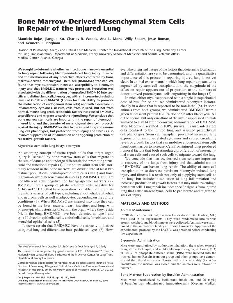
The dose of bleomycin that we used was not lethal in nonmyelo-supressed animals. Although different percentages of mortalityare reported in the literature, in our hand this dose of bleomycinis not lethal in the strain of mice used (9, 11). Figure 1a shows sur-vival at 14 d after bleomycin in each of our experimental groups.All animals receiving bleomycin alone survived, but only onethird of the myelosuppressed animals receiving bleomycin sur-vived to Day 14. Administration of BMDMSC after bleomycinresulted in 100% survival of the busulfan-treated animals as wellas the animals without bone marrow suppression.
Representative histologic sections from lungs of animals fromeach experimental group are shown in Figures 1b–1m. Lungsfrom animals receiving busulfan but no bleomycin appearedhistologically normal. Lungs from animals given bleomycin andno other intervention demonstrated the changes typical of thismodel 14 d after administering the drug (12). There is markedalteration in lung architecture, with increased cellularity andfibrosis. In animals treated with busulfan before administrationof bleomycin, we were able to examine only the lungs of theanimals that survived for 14 d (two thirds of the animals diedbefore that time), so that the lung histology in this group is likelyless severe than in the group as a whole. In these survivinganimals, however, the qualitative effect of bleomycin was similarto that in animals with intact bone marrow, but the severity of
Rojas, Xu, Woods, et al.: Stem Cells in Repair of the Injured Lung 147
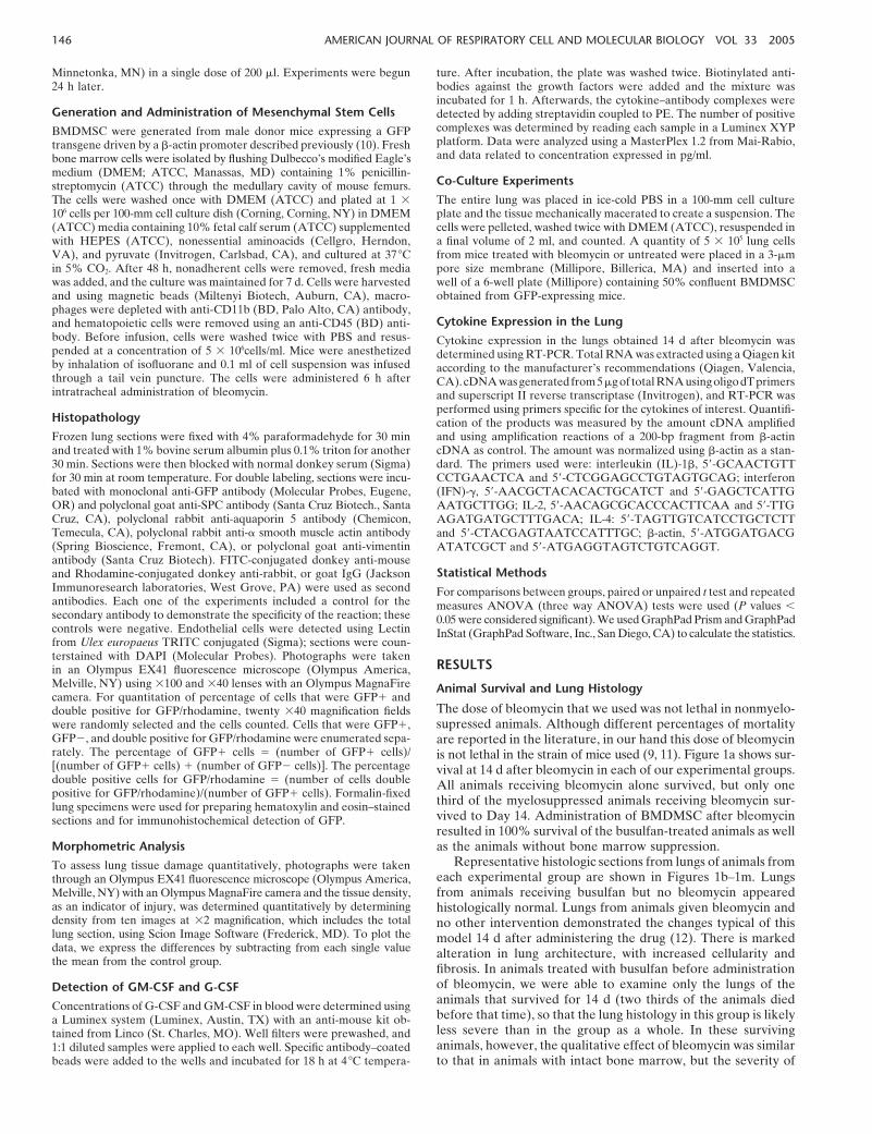
the persistent injury appeared to be greater subjectively. Infusionof BMDMSC after bleomycin administration resulted in lesspersistent injury and fibrosis whether or not the animals weremyelosupressed. However, in bone marrow–suppressed animals,there was still evidence of a substantial bleomycin effect, whereasin the bone marrow–sufficient animals receiving BMDMSCtransplant, there was minimal evidence of lung injury 14 d afterbleomycin. Morphometric analysis of the histologic sections,demonstrated a significant decrease in the bleomycin-inducedlung injury in animals receiving a BMDMSC infusion (Figure 2).
�Figure 1. (a ) Infusion of BMDMSC prevented mortality in mice myelo-suppressed with busulfan before bleomycin. Sixty-six percent of myelo-suppressed mice (n � 6) given bleomycin died without BMDMSC transfer.There was no mortality in any of the other experimental groups (n � 5animals per group). Histologic sections of lungs obtained from: (b, c )normal C57BL/6 mice; (d, e ) 14 d after 4 U/kg busulfan (lungs appearnormal); (f, g ) 14 d after bleomycin (increased cellularity and fibrosistypical of bleomycin injury); (h, i ) 14 d after bleomycin following busul-fan myelosuppression (apparently more extensive increased cellularityand fibrosis compared with f and g ); (j, k ) 14 d after bleomycin followedby BMDMSC transfer (minimal alterations in lung architecture com-pared with f and g ); (l, m ) 14 d after bleomycin followed by BMDMSCtransfer in myelosuppressed mice showing apparent protection (com-pare with h and i ), but more abnormalities than present in the BMDMSCtransplanted mice without myelosuppression (compare with j and k ).
On average, this measure of injury was worse in the myelosup-pressed animals than in nonmyelosuppressed animals givenbleomycin, but, again, these are only the animals that survivedfor 14 d and likely had less injury than the animals that diedearlier. When myelosuppressed animals given bleomycin alsoreceived BMDMSC, they all survived to 14 d and the quantitativemeasure of injury was less on average even than in the animalsthat survived without BMDMSC.
Donor-Derived Cells in the Lungs after BMDMSC Transplant
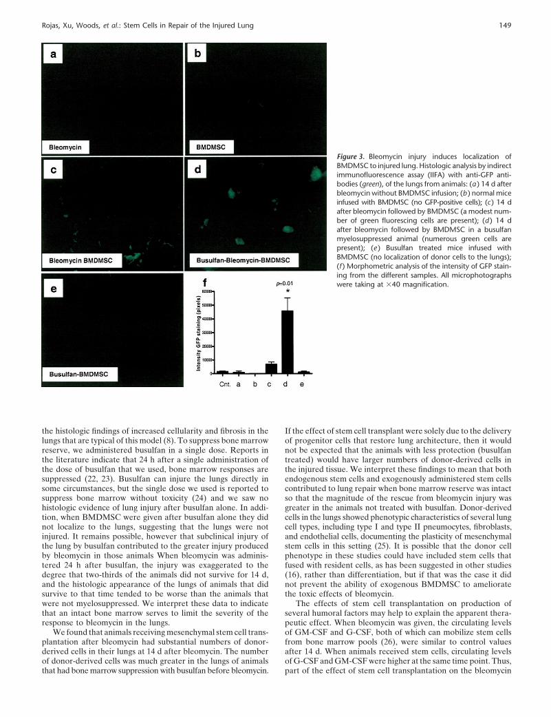
To demonstrate localization of the infused BMDMSC to thelung, we identified donor cells by staining the sections with ananti-GFP–specific antibody (Figure 3). Lungs from control ani-mals that did not receive BMDMSC infusions showed minimalbackground staining. Lungs from animals that received busulfanand BMDMSC transfer without receiving bleomycin also showedminimal deposition of donor cells. Donor-derived cells weredetected in the lungs of animals 14 d after bleomycin, and inanimals that received busulfan, large numbers of donor cellswere present in the lungs at that time. If the therapeutic effectof stem cell transfer were solely due to the delivery of cells thatreconstitute lung architecture, then it would not be expectedthat the animals with less protection (busulfan treated) wouldhave larger numbers of donor-derived cells in the injured tissue.These findings could imply that both endogenous stem cells andexogenously administered stem cells contributed to lung repairwhen bone marrow reserve was intact so that myelosuppressedanimals had more persistent injury and less “therapeutic” effectof BMDMSC transfer.
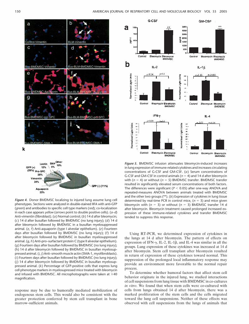
We examined tissue sections by immunofluorescent stainingfor cell type–specific markers so that co-localization of green fluo-rescence (indicating GFP-positive donor cells) with cell-specificmarkers would indicate the phenotype of the donor cells. Figure4 shows a series of fluorescent photomicrographs illustratingfibroblast (vimentin-positive [13], Figures 4a–4d), type I alveolarepithelial (aquaporin [14] antibody–positive, Figures 4e–4f),type II alveolar epithelial (pro-SPC [15] antibody–positive, Figures4g–4h), or myofibroblast (SMA-1 [13]-positive, Figures 4i–4j)phenotypes. These data suggest that BMDMSC can assume phe-notypic characteristics of the major cell types that compose lungparenchyma, including fibroblasts. Before the cells were infused,immunostaining for the cell specific markers was negative (datanot shown). Quantitative estimates of donor cell localization inthe lungs and the phenotypes of these cells are shown in Figure4k for animals receiving bleomycin after myelosuppression. Inthis circumstance, � 29% of the lung cells were donor-derived.Less than 5% of cells were donor-derived in bleomycin-treatedanimals with intact bone marrow. No donor cells were detected
148 AMERICAN JOURNAL OF RESPIRATORY CELL AND MOLECULAR BIOLOGY VOL 33 2005
Figure 2. Infusion of BMDMSC reduces lung damage induce by bleo-mycin. Morphometric analysis of histologic sections of total left lungwas done to determine the percentage of the lung that was affected.Pictures (magnification: �2) were analyzed using AxioVision 4.2 soft-ware (Carl Zeiss, Thornwood, NY). The graphic represents the averagefrom 5–9 histologic lung sections. Bleomycin caused substantial injuryand BMDMSC infusion reduced bleomycin-induced injury (*P � 0.05,bleomycin-BMDMSC versus bleomycin, versus busulfan-bleomycin, andversus busulfan-bleomycin-BMDMSC). In myelosuppressed animals, thismeasure of injury was on average worse than in nonmyelosuppressedanimals and on average less severe when animals received BMDMSC.Since only the lungs of the one-third of the myelosuppressed animalsthat received BMDMSC survived to 14 d were analyzed, the histologicmeasurements likely underestimate the degree of injury in the entiregroup.
in animals that did not receive bleomycin whether or not theywere myelosuppressed. The donor-derived cells before the infu-sion were negative for all of the lung cell markers that we studied.After transplant, cells were fairly evenly distributed among allcell types for which we tested. It is possible that the donor cellphenotype in these studies included stem cells that fused withresident cells, as has been suggested in other studies (16), ratherthan differentiation (17). We cannot rule out that possibility,but if that was the case it did not prevent the ability of exogenousBMDMSC to ameliorate the toxic effects of bleomycin.
Circulating G-CSF and GM-CSF
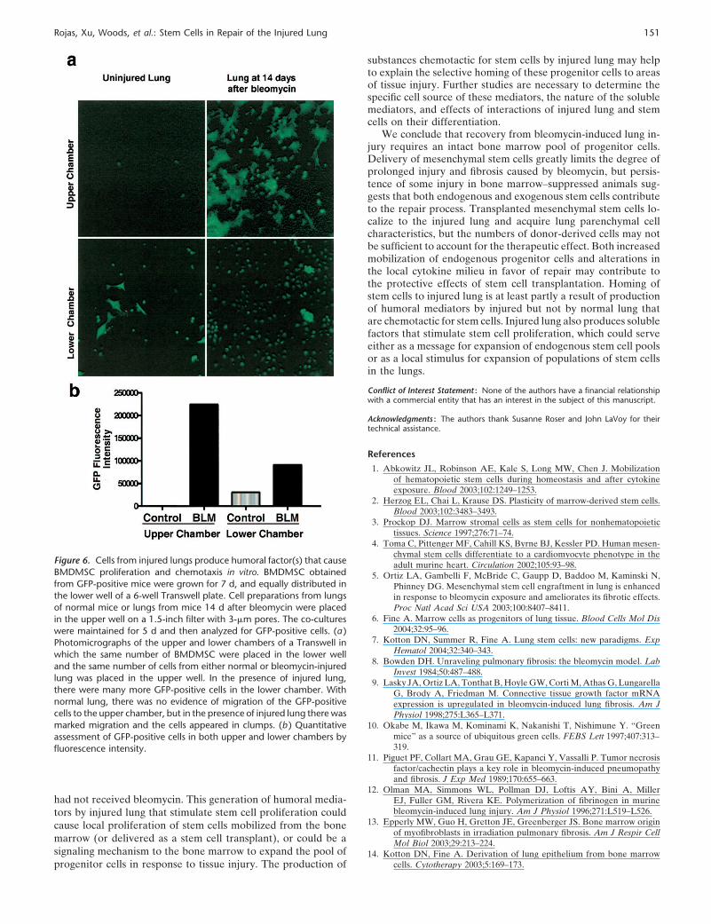
The effects of stem cell transplantation on production of severalhumoral factors may help to explain the apparent therapeuticeffect. We determined the circulating concentrations of G-CSFand GM-CSF at 14 d after bleomycin administration. As shownin Figure 5A, when bleomycin was given, the circulating levelsof GM-CSF and G-CSF, both of which can mobilize stem cellsfrom bone marrow pools (18), were not increased 14 d afterbleomycin. When animals received stem cells, there was amarked and significant increase in circulating levels of G-CSFand GM-CSF at the same time point. Thus, part of the effectof stem cell transplantation on the bleomycin response may bedue to mobilization of endogenous stem cells. This would alsobe consistent with the greater protection conferred by stem celltransplant in the bone marrow sufficient animals.
Expression of Immune Related Cytokines in the Lungs
To determine effects of BMDMSC transfer on the local inflam-matory milieu in the lungs, we determined expression of severalimmune system related cytokines in lung tissue by real-time PCR(Figure 5B). Lung expression of IFN-, IL-2, IL-1�, and IL-4
was increased at 14 d after bleomycin and stem cell transplantresulted in return of expression of these cytokines toward nor-mal. This suppression of the prolonged inflammatory responsein the lungs (19) may provide an environment more favorableto normal repair.
In Vitro Effects of Cells from Injured Lungs onMesenchymal Stem Cell Behavior
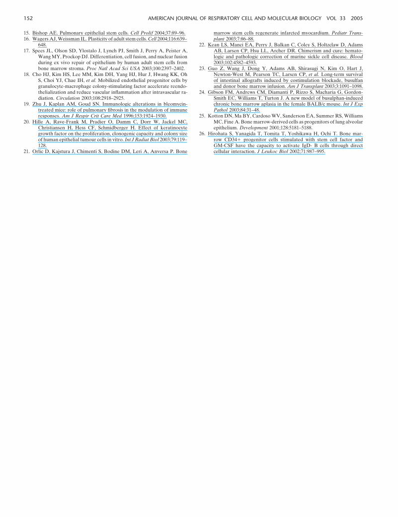
To determine whether humoral factors that affect stem cell be-havior originate in the injured lung, we measured the effectsof cell suspensions prepared from lung harvested 14 d afterbleomycin administration on the proliferation and migration ofGFP-expressing BMDMSC in co-culture experiments. Figure6A shows fluorescent photomicrographs of the upper surface ofthe filter separating the lung and stem cells and of the lowerchamber that contained only GFP-expressing stem cells after5 d in culture. When the upper chamber contained cells frominjured lung, there was a marked increase in the number ofGFP-expressing stem cells in the lower chamber compared withexperiments in which cells from uninjured lung were in theupper chamber. When the upper chamber contained cells fromuninjured lung, there was no evidence of migration of the GFP-positive stem cells from the lower chamber toward the lung cells.However, when cells from bleomycin-injured lungs were in theupper chamber, numerous GFP-positive stem cells migrated tothe upper chamber. The migrating cells also appeared differentmorphologically, with many large cells and cell clusters. Figure6B summarizes quantitative measurements of fluorescence inthe lower chamber and in the filter as an estimate of the locationof the GFP-expressing BMDMSC.
DISCUSSION
One theory of tissue repair holds that organ injury is “sensed”by distant stem cells that migrate to the site of damage anddifferentiate into organ-specific cells, promoting structural andfunctional repair (1, 20). Several recent studies derive from thatgeneral concept. For example, stem cells obtained from bonemarrow are reported to be capable of homing selectively toinfarcted myocardium and differentiating into cardiac muscle,endothelial cells, and vascular smooth muscle cells (21), decreas-ing infarct size and improving cardiac function. Mobilization ofendogenous progenitor cells by administration of G-CSF andGM-CSF in mice is reported to cause tissue regeneration in anarea of experimental myocardial infarct (21) with new myocytesand arterioles and capillaries connected to the circulation of theunaffected ventricle.
Bleomycin is one of the most extensively studied and repro-ducible models for lung fibrosis in mice. When bleomycin isgiven into the airway, it produces lung epithelial injury, followedby an inflammatory response over several days that is followedby lung fibrosis that eventually resolves (8). Ortiz and colleaguesdelivered purified BMDMSC from bleomycin-resistant mice tosusceptible mice after bleomycin administration (5). They foundthat the donor cells homed to the injured lung and adoptedepithelial phenotypes, including that of type II alveolar epithelialcells. Because the numbers of donor-derived cells engrafting thelung did not appear sufficient to account for the therapeuticresponse, they suggest that donor stem cells may have otherlocal effects. The quantitative importance of this process is notknown, and the source of signals that are responsible for mobili-zation and homing of endogenous stem cells remains to be de-fined. Effects of stem cell transplant other than provision ofpluripotent cells to the area of injury that may contribute to theapparent therapeutic effect also remain to be determined.
In our studies, mice given bleomycin survived and showed
Rojas, Xu, Woods, et al.: Stem Cells in Repair of the Injured Lung 149
Figure 3. Bleomycin injury induces localization ofBMDMSC to injured lung. Histologic analysis by indirectimmunofluorescence assay (IIFA) with anti-GFP anti-bodies (green), of the lungs from animals: (a ) 14 d afterbleomycin without BMDMSC infusion; (b ) normal miceinfused with BMDMSC (no GFP-positive cells); (c) 14 dafter bleomycin followed by BMDMSC (a modest num-ber of green fluorescing cells are present); (d ) 14 dafter bleomycin followed by BMDMSC in a busulfanmyelosuppressed animal (numerous green cells arepresent); (e ) Busulfan treated mice infused withBMDMSC (no localization of donor cells to the lungs);(f ) Morphometric analysis of the intensity of GFP stain-ing from the different samples. All microphotographswere taking at �40 magnification.
the histologic findings of increased cellularity and fibrosis in thelungs that are typical of this model (8). To suppress bone marrowreserve, we administered busulfan in a single dose. Reports inthe literature indicate that 24 h after a single administration ofthe dose of busulfan that we used, bone marrow responses aresuppressed (22, 23). Busulfan can injure the lungs directly insome circumstances, but the single dose we used is reported tosuppress bone marrow without toxicity (24) and we saw nohistologic evidence of lung injury after busulfan alone. In addi-tion, when BMDMSC were given after busulfan alone they didnot localize to the lungs, suggesting that the lungs were notinjured. It remains possible, however that subclinical injury ofthe lung by busulfan contributed to the greater injury producedby bleomycin in those animals When bleomycin was adminis-tered 24 h after busulfan, the injury was exaggerated to thedegree that two-thirds of the animals did not survive for 14 d,and the histologic appearance of the lungs of animals that didsurvive to that time tended to be worse than the animals thatwere not myelosuppressed. We interpret these data to indicatethat an intact bone marrow serves to limit the severity of theresponse to bleomycin in the lungs.
We found that animals receiving mesenchymal stem cell trans-plantation after bleomycin had substantial numbers of donor-derived cells in their lungs at 14 d after bleomycin. The numberof donor-derived cells was much greater in the lungs of animalsthat had bone marrow suppression with busulfan before bleomycin.
If the effect of stem cell transplant were solely due to the deliveryof progenitor cells that restore lung architecture, then it wouldnot be expected that the animals with less protection (busulfantreated) would have larger numbers of donor-derived cells inthe injured tissue. We interpret these findings to mean that bothendogenous stem cells and exogenously administered stem cellscontributed to lung repair when bone marrow reserve was intactso that the magnitude of the rescue from bleomycin injury wasgreater in the animals not treated with busulfan. Donor-derivedcells in the lungs showed phenotypic characteristics of several lungcell types, including type I and type II pneumocytes, fibroblasts,and endothelial cells, documenting the plasticity of mesenchymalstem cells in this setting (25). It is possible that the donor cellphenotype in these studies could have included stem cells thatfused with resident cells, as has been suggested in other studies(16), rather than differentiation, but if that was the case it didnot prevent the ability of exogenous BMDMSC to amelioratethe toxic effects of bleomycin.
The effects of stem cell transplantation on production ofseveral humoral factors may help to explain the apparent thera-peutic effect. When bleomycin was given, the circulating levelsof GM-CSF and G-CSF, both of which can mobilize stem cellsfrom bone marrow pools (26), were similar to control valuesafter 14 d. When animals received stem cells, circulating levelsof G-CSF and GM-CSF were higher at the same time point. Thus,part of the effect of stem cell transplantation on the bleomycin
150 AMERICAN JOURNAL OF RESPIRATORY CELL AND MOLECULAR BIOLOGY VOL 33 2005
Figure 4. Donor BMDMSC localizing to injured lung assume lung cellphenotypes. Sections were analyzed in double-stained IIFA with anti-GFP(green) and antibodies to specific cell type markers (red); co-localizationin each case appears yellow (arrows point to double positive cells). (a–d)Anti-vimentin (fibroblast). (a ) Normal control; (b ) 14 d after bleomycin;(c ) 14 d after busulfan followed by BMDMSC (no lung injury); (d ) 14 dafter bleomycin followed by BMDMSC in a busulfan myelosuppressedanimal. (e, f ) Anti-aquaporin (type I alveolar epithelium). (e ) Fourteendays after busulfan followed by BMDMSC (no lung injury); (f ) 14 dafter bleomycin followed by BMDMSC in busulfan myelosuppressedanimal. (g, h ) Anti-pro–surfactant protein C (type II alveolar epithelium).(g ) Fourteen days after busulfan followed by BMDMSC (no lung injury);(h) 14 d after bleomycin followed by BMDMSC in busulfan myelosup-pressed animal. (i, j ) Anti–smooth muscle actin (SMA-1, myofibroblasts).(i ) Fourteen days after busulfan followed by BMDMSC (no lung injury);(j ) 14 d after bleomycin followed by BMDMSC in busulfan myelosup-pressed animal. (k ) Percentage of GFP-positive cells that express lungcell phenotype markers in myelosupressed mice treated with bleomycinand infused with BMDMSC. All microphotographs were taken at �40magnification.
response may be due to humorally mediated mobilization ofendogenous stem cells. This would also be consistent with thegreater protection conferred by stem cell transplant in bonemarrow–sufficient animals.
Figure 5. BMDMSC infusion attenuates bleomycin-induced increasesin lung expression of immune-related cytokines and increases circulatingconcentrations of G-CSF and GM-CSF. (a ) Serum concentrations ofG-CSF and GM-CSF in control animals (n � 4) and 14 d after bleomycinwith (n � 4) or without (n � 5) BMDMSC transfer. BMDMSC transferresulted in significantly elevated serum concentrations of both factors.The differences were significant (P � 0.05) after one-way ANOVA andrepeated-measures ANOVA between animals treated with BMDMSCand the other two groups (**). (b ) Expression of cytokines in lung tissuedetermined by real-time PCR in control mice, (n � 3) and mice givenbleomycin with (n � 3) or without (n � 3) BMDMSC transfer 14 dafter bleomycin. Bleomycin treatment caused prolonged increased ex-pression of these immune-related cytokines and transfer BMDMSCtended to suppress this response.
Using RT-PCR, we determined expression of cytokines inthe lungs at 14 d after bleomycin. The pattern of effects onexpression of IFN-, IL-2, IL-1�, and IL-4 was similar in all thegroups. Lung expression of these cytokines was increased at 14 dafter bleomycin. Stem cell transplant after bleomycin resultedin return of expression of these cytokines toward normal. Thissuppression of the prolonged local inflammatory response mayprovide an environment more favorable to the normal repairprocess.
To determine whether humoral factors that affect stem cellbehavior originate in the injured lung, we studied interactionsof cell suspensions from lung tissue with BMDMSC in co-culturesin vitro. We found that when stem cells were co-cultured withcells from lungs obtained 14 d after bleomycin, there was amarked proliferation of the stem cells and the cells migratedtoward the lung cell suspensions. Neither of these effects wasobserved with cell suspensions from the lungs of animals that
Rojas, Xu, Woods, et al.: Stem Cells in Repair of the Injured Lung 151
Figure 6. Cells from injured lungs produce humoral factor(s) that causeBMDMSC proliferation and chemotaxis in vitro. BMDMSC obtainedfrom GFP-positive mice were grown for 7 d, and equally distributed inthe lower well of a 6-well Transwell plate. Cell preparations from lungsof normal mice or lungs from mice 14 d after bleomycin were placedin the upper well on a 1.5-inch filter with 3-�m pores. The co-cultureswere maintained for 5 d and then analyzed for GFP-positive cells. (a )Photomicrographs of the upper and lower chambers of a Transwell inwhich the same number of BMDMSC were placed in the lower welland the same number of cells from either normal or bleomycin-injuredlung was placed in the upper well. In the presence of injured lung,there were many more GFP-positive cells in the lower chamber. Withnormal lung, there was no evidence of migration of the GFP-positivecells to the upper chamber, but in the presence of injured lung there wasmarked migration and the cells appeared in clumps. (b ) Quantitativeassessment of GFP-positive cells in both upper and lower chambers byfluorescence intensity.
had not received bleomycin. This generation of humoral media-tors by injured lung that stimulate stem cell proliferation couldcause local proliferation of stem cells mobilized from the bonemarrow (or delivered as a stem cell transplant), or could be asignaling mechanism to the bone marrow to expand the pool ofprogenitor cells in response to tissue injury. The production of
substances chemotactic for stem cells by injured lung may helpto explain the selective homing of these progenitor cells to areasof tissue injury. Further studies are necessary to determine thespecific cell source of these mediators, the nature of the solublemediators, and effects of interactions of injured lung and stemcells on their differentiation.
We conclude that recovery from bleomycin-induced lung in-jury requires an intact bone marrow pool of progenitor cells.Delivery of mesenchymal stem cells greatly limits the degree ofprolonged injury and fibrosis caused by bleomycin, but persis-tence of some injury in bone marrow–suppressed animals sug-gests that both endogenous and exogenous stem cells contributeto the repair process. Transplanted mesenchymal stem cells lo-calize to the injured lung and acquire lung parenchymal cellcharacteristics, but the numbers of donor-derived cells may notbe sufficient to account for the therapeutic effect. Both increasedmobilization of endogenous progenitor cells and alterations inthe local cytokine milieu in favor of repair may contribute tothe protective effects of stem cell transplantation. Homing ofstem cells to injured lung is at least partly a result of productionof humoral mediators by injured but not by normal lung thatare chemotactic for stem cells. Injured lung also produces solublefactors that stimulate stem cell proliferation, which could serveeither as a message for expansion of endogenous stem cell poolsor as a local stimulus for expansion of populations of stem cellsin the lungs.
Conflict of Interest Statement : None of the authors have a financial relationshipwith a commercial entity that has an interest in the subject of this manuscript.
Acknowledgments : The authors thank Susanne Roser and John LaVoy for theirtechnical assistance.
References
1. Abkowitz JL, Robinson AE, Kale S, Long MW, Chen J. Mobilizationof hematopoietic stem cells during homeostasis and after cytokineexposure. Blood 2003;102:1249–1253.
2. Herzog EL, Chai L, Krause DS. Plasticity of marrow-derived stem cells.Blood 2003;102:3483–3493.
3. Prockop DJ. Marrow stromal cells as stem cells for nonhematopoietictissues. Science 1997;276:71–74.
4. Toma C, Pittenger MF, Cahill KS, Byrne BJ, Kessler PD. Human mesen-chymal stem cells differentiate to a cardiomyocyte phenotype in theadult murine heart. Circulation 2002;105:93–98.
5. Ortiz LA, Gambelli F, McBride C, Gaupp D, Baddoo M, Kaminski N,Phinney DG. Mesenchymal stem cell engraftment in lung is enhancedin response to bleomycin exposure and ameliorates its fibrotic effects.Proc Natl Acad Sci USA 2003;100:8407–8411.
6. Fine A. Marrow cells as progenitors of lung tissue. Blood Cells Mol Dis2004;32:95–96.
7. Kotton DN, Summer R, Fine A. Lung stem cells: new paradigms. ExpHematol 2004;32:340–343.
8. Bowden DH. Unraveling pulmonary fibrosis: the bleomycin model. LabInvest 1984;50:487–488.
9. Lasky JA, Ortiz LA, Tonthat B, Hoyle GW, Corti M, Athas G, LungarellaG, Brody A, Friedman M. Connective tissue growth factor mRNAexpression is upregulated in bleomycin-induced lung fibrosis. Am JPhysiol 1998;275:L365–L371.
10. Okabe M, Ikawa M, Kominami K, Nakanishi T, Nishimune Y. “Greenmice” as a source of ubiquitous green cells. FEBS Lett 1997;407:313–319.
11. Piguet PF, Collart MA, Grau GE, Kapanci Y, Vassalli P. Tumor necrosisfactor/cachectin plays a key role in bleomycin-induced pneumopathyand fibrosis. J Exp Med 1989;170:655–663.
12. Olman MA, Simmons WL, Pollman DJ, Loftis AY, Bini A, MillerEJ, Fuller GM, Rivera KE. Polymerization of fibrinogen in murinebleomycin-induced lung injury. Am J Physiol 1996;271:L519–L526.
13. Epperly MW, Guo H, Gretton JE, Greenberger JS. Bone marrow originof myofibroblasts in irradiation pulmonary fibrosis. Am J Respir CellMol Biol 2003;29:213–224.
14. Kotton DN, Fine A. Derivation of lung epithelium from bone marrowcells. Cytotherapy 2003;5:169–173.
152 AMERICAN JOURNAL OF RESPIRATORY CELL AND MOLECULAR BIOLOGY VOL 33 2005
15. Bishop AE. Pulmonary epithelial stem cells. Cell Prolif 2004;37:89–96.16. Wagers AJ, Weissman IL. Plasticity of adult stem cells. Cell 2004;116:639–
648.17. Spees JL, Olson SD, Ylostalo J, Lynch PJ, Smith J, Perry A, Peister A,
Wang MY, Prockop DJ. Differentiation, cell fusion, and nuclear fusionduring ex vivo repair of epithelium by human adult stem cells frombone marrow stroma. Proc Natl Acad Sci USA 2003;100:2397–2402.
18. Cho HJ, Kim HS, Lee MM, Kim DH, Yang HJ, Hur J, Hwang KK, OhS, Choi YJ, Chae IH, et al. Mobilized endothelial progenitor cells bygranulocyte-macrophage colony-stimulating factor accelerate reendo-thelialization and reduce vascular inflammation after intravascular ra-diation. Circulation 2003;108:2918–2925.
19. Zhu J, Kaplan AM, Goud SN. Immunologic alterations in bleomycin-treated mice: role of pulmonary fibrosis in the modulation of immuneresponses. Am J Respir Crit Care Med 1996;153:1924–1930.
20. Hille A, Rave-Frank M, Pradier O, Damm C, Dorr W, Jackel MC,Christiansen H, Hess CF, Schmidberger H. Effect of keratinocytegrowth factor on the proliferation, clonogenic capacity and colony sizeof human epithelial tumour cells in vitro. Int J Radiat Biol 2003;79:119–128.
21. Orlic D, Kajstura J, Chimenti S, Bodine DM, Leri A, Anversa P. Bone
marrow stem cells regenerate infarcted myocardium. Pediatr Trans-plant 2003;7:86–88.
22. Kean LS, Manci EA, Perry J, Balkan C, Coley S, Holtzclaw D, AdamsAB, Larsen CP, Hsu LL, Archer DR. Chimerism and cure: hemato-logic and pathologic correction of murine sickle cell disease. Blood2003;102:4582–4593.
23. Guo Z, Wang J, Dong Y, Adams AB, Shirasugi N, Kim O, Hart J,Newton-West M, Pearson TC, Larsen CP, et al. Long-term survivalof intestinal allografts induced by costimulation blockade, busulfanand donor bone marrow infusion. Am J Transplant 2003;3:1091–1098.
24. Gibson FM, Andrews CM, Diamanti P, Rizzo S, Macharia G, Gordon-Smith EC, Williams T, Turton J. A new model of busulphan-inducedchronic bone marrow aplasia in the female BALB/c mouse. Int J ExpPathol 2003;84:31–48.
25. Kotton DN, Ma BY, Cardoso WV, Sanderson EA, Summer RS, WilliamsMC, Fine A. Bone marrow-derived cells as progenitors of lung alveolarepithelium. Development 2001;128:5181–5188.
26. Hirohata S, Yanagida T, Tomita T, Yoshikawa H, Ochi T. Bone mar-row CD34� progenitor cells stimulated with stem cell factor andGM-CSF have the capacity to activate IgD- B cells through directcellular interaction. J Leukoc Biol 2002;71:987–995.
Related Documents


