Appl. Phys. B56, 104-112 (1993) Al::gzlied "~'* physics Physics B ""'"" Chemistw © Springer-Verlag1993 Bone-Ablation Mechanism Using CO2 Lasers of Different Pulse Duration and Wavelength M. Forrer I, M. Frenz 1, V. Romano 1, H.J. Altermatt 2, H.P. Weber 1, A. Silenok 3, M. Istomyn 3, and V.I. Konov 3 1 Institute of Applied Physics, University of Berne, Sidlerstrasse 5, CH-3012 Berne, Switzerland (Fax: +41-31/653765) 2 Institute of Pathology, University of Berne, CH-3012 Berne, Switzerland 3 General Institute of Physics, Russian Academy of Sciences, Moscow, Russia Received 29 September 1992/Accepted 15 October 1992 Abstract. Bone ablation using different pulse parameters and four emission lines of 9.3, 9.6, 10.3, and 10.6 gin of the CO 2 laser exhibits effects which are caused by the thermal properties and the absorption spectrum of bone material. The ablation mechanism was investigated with light- and electron-microscopy at short laser-pulse durations of 0.9 and 1.8 gs and a long pulse of 250 gs. It is shown that different processes are responsible for the ablation mechanism either using the short or the long pulse durations. In the case of short pulse durations it is shown that, although the mineral components are the main absorber for CO 2 radiation, water is the driving force for the ablation process. The destruction of material is based on explosive evaporation of water with an ablation energy of 1.3 kJ/cm 3. Histological examination revealed a minimal zone of 10-15 gm of thermally altered material at the bottom of the laser drilled hole. Within the investigated spectral range we found that the ablation threshold at 9.3 and 9.6 gm is lower than at 10.3 and 10.6 gin. In comparison the ablation with a long pulse duration is determined by two processes. On the one side, the heat lost by heat conduction leads to carbonization of a surface layer, and the absorption of the CO 2 radiation in this carbonized layer is the driving force of the ablation process. On the other side, it is shown that up to 60% of the pulse energy is absorbed in the ablation plume. Therefore, a long pulse duration results in an eight-times higher specific ablation energy of 10 kJ/cm 3. PACS: 61.80.Ba, 79.20.Ds, 87.50.Eg, 87.55.Hb, 87.80.+s Infrared laser radiation has found its way to medical appli- cations such as cutting and removal of tissue material [1-3]. The success of infrared lasers is based on the characteristic absorption spectrum of water which has strong absorption lines at 1.95, 2.94, 6.09 ~tm and a broad absorption around 10 gm [4]. In view of an increasing interest in contact-free ablation methods in the fields of orthopedy, dentistry and otorhinolaryngology, infrared lasers have also been used in ablation of hard tissue materials with a relatively low water content. Ablation of hard tissue materials has already been reported with different infrared wavelengths of solid-state and gas lasers [5-8]. Investigations of the ablation process in the infrared wavelength region have confirmed that explo- sive ablation of the bone material can explain the ablation mechanism [7, 9]. In this process the water in the bone un- dergoes rapid heating and carries away the bone material in an explosive expansion. Comparison of the infrared lasers in terms of efficiency and threshold for bone ablation was carried out with varying pulse durations as well as in the continuous-wave mode. With respect to efficiency and ther- mal damage, the pulsed erbium laser at a wavelength of 2.94 gm showed the best results for bone ablation, whereas the CO 2 laser at 10.6 gm is generally quoted to lead to high thermal strain and charring of the bone material connected with deteriorated healing properties [7, 10]. In this study the potential of the CO 2 laser for ablation with minimal carbonization and very small thermal damage is demonstrated. We have focused our attention on the influ- ence of wavelength and pulse duration on the ablation mech- anism. The ablation has been studied by ablation dosimetry, histology and electron microscopy of the ablation site as well as of the debris material. The effect of wavelength on the ablation mechanism was investigated at different wave- lenghts of a TEA-CO 2 laser with a short pulse duration (~-p0.9 gs). We found that the ablation thresholds change sig- nificantly in the investigated spectral range. The influence of the pulse duration on the ablation mechanism was com- pared at the wavelength of 10.6 gm with a short (~-pl.8 gs, TEA) and a long (~-p250gs, longitudinally pumped) laser pulse. The electron microscopic and histological investiga- tions evidence the thermal influence of the pulse duration. The results are compared in view of the thermodynamical properties of the bone material, which have been determined by differential scanning calorimetry. In the case of the short pulse duration it is shown that the ablation is based mainly on explosive evaporation of water, whereas the ablation with the long pulse duration is strongly influenced by additional energy absorption in the ablation plume.

Welcome message from author
This document is posted to help you gain knowledge. Please leave a comment to let me know what you think about it! Share it to your friends and learn new things together.
Transcript
Appl. Phys. B56, 104-112 (1993) Al::gzlied " ~ ' * physics
Physics B ""'"" Chemistw © Springer-Verlag 1993
Bone-Ablation Mechanism Using CO2 Lasers of Different Pulse Duration and Wavelength M. Forrer I, M. Frenz 1, V. Romano 1, H.J. Altermatt 2, H.P. Weber 1, A. Silenok 3, M. Istomyn 3, and V.I. Konov 3
1 Institute of Applied Physics, University of Berne, Sidlerstrasse 5, CH-3012 Berne, Switzerland (Fax: +41-31/653765) 2 Institute of Pathology, University of Berne, CH-3012 Berne, Switzerland 3 General Institute of Physics, Russian Academy of Sciences, Moscow, Russia
Received 29 September 1992/Accepted 15 October 1992
Abstract. Bone ablation using different pulse parameters and four emission lines of 9.3, 9.6, 10.3, and 10.6 gin of the CO 2 laser exhibits effects which are caused by the thermal properties and the absorption spectrum of bone material. The ablation mechanism was investigated with light- and electron-microscopy at short laser-pulse durations of 0.9 and 1.8 gs and a long pulse of 250 gs. It is shown that different processes are responsible for the ablation mechanism either using the short or the long pulse durations. In the case of short pulse durations it is shown that, although the mineral components are the main absorber for CO 2 radiation, water is the driving force for the ablation process. The destruction of material is based on explosive evaporation of water with an ablation energy of 1.3 kJ/cm 3. Histological examination revealed a minimal zone of 10-15 gm of thermally altered material at the bottom of the laser drilled hole. Within the investigated spectral range we found that the ablation threshold at 9.3 and 9.6 gm is lower than at 10.3 and 10.6 gin. In comparison the ablation with a long pulse duration is determined by two processes. On the one side, the heat lost by heat conduction leads to carbonization of a surface layer, and the absorption of the CO 2 radiation in this carbonized layer is the driving force of the ablation process. On the other side, it is shown that up to 60% of the pulse energy is absorbed in the ablation plume. Therefore, a long pulse duration results in an eight-times higher specific ablation energy of 10 kJ/cm 3.
PACS: 61.80.Ba, 79.20.Ds, 87.50.Eg, 87.55.Hb, 87.80.+s
Infrared laser radiation has found its way to medical appli- cations such as cutting and removal of tissue material [1-3]. The success of infrared lasers is based on the characteristic absorption spectrum of water which has strong absorption lines at 1.95, 2.94, 6.09 ~tm and a broad absorption around 10 gm [4]. In view of an increasing interest in contact-free ablation methods in the fields of orthopedy, dentistry and otorhinolaryngology, infrared lasers have also been used in ablation of hard tissue materials with a relatively low water
content. Ablation of hard tissue materials has already been reported with different infrared wavelengths of solid-state and gas lasers [5-8]. Investigations of the ablation process in the infrared wavelength region have confirmed that explo- sive ablation of the bone material can explain the ablation mechanism [7, 9]. In this process the water in the bone un- dergoes rapid heating and carries away the bone material in an explosive expansion. Comparison of the infrared lasers in terms of efficiency and threshold for bone ablation was carried out with varying pulse durations as well as in the continuous-wave mode. With respect to efficiency and ther- mal damage, the pulsed erbium laser at a wavelength of 2.94 gm showed the best results for bone ablation, whereas the CO 2 laser at 10.6 gm is generally quoted to lead to high thermal strain and charring of the bone material connected with deteriorated healing properties [7, 10].
In this study the potential of the CO 2 laser for ablation with minimal carbonization and very small thermal damage is demonstrated. We have focused our attention on the influ- ence of wavelength and pulse duration on the ablation mech- anism. The ablation has been studied by ablation dosimetry, histology and electron microscopy of the ablation site as well as of the debris material. The effect of wavelength on the ablation mechanism was investigated at different wave- lenghts of a TEA-CO 2 laser with a short pulse duration (~-p0.9 gs). We found that the ablation thresholds change sig- nificantly in the investigated spectral range. The influence of the pulse duration on the ablation mechanism was com- pared at the wavelength of 10.6 gm with a short (~-pl.8 gs, TEA) and a long (~-p250 gs, longitudinally pumped) laser pulse. The electron microscopic and histological investiga- tions evidence the thermal influence of the pulse duration. The results are compared in view of the thermodynamical properties of the bone material, which have been determined by differential scanning calorimetry. In the case of the short pulse duration it is shown that the ablation is based mainly on explosive evaporation of water, whereas the ablation with the long pulse duration is strongly influenced by additional energy absorption in the ablation plume.
Bone-Ablation Mechanism Using CO 2 Lasers
1 Materials and Methods
1.1 Lasers
In this investigation two different lasers were used. A TEA- CO 2 laser was modified for wavelength selectivity with a blazed reflection grating (75 lines/mm) as outcoupling mirror. The first order reflection was the laser channel, the minus first order reflection the outcoupling channel. A CO 2 laser has four laser-amplification bands determined by the rotationally splitted vibronic energy levels of the molecules. We tuned the laser to the four strongest lines at the wavelengths 9.3 btm, 9.6 btm, 10.3 btm, and 10.6 btm and controlled the laser mode with an intracavity diaphragm. The total pulse length Ttot could be adjusted from 3.5 bts to 12.5 gs by appropriate modification of the gas mixture. Pulse energies ranged up to 13mJ. The temporal pulse profile always showed the maximum intensity during the first quarter of the total pulse length and had a weak trailing edge. Aproximation of this profile with a rectangular-shaped profile of maximum intensity of the same total energy resulted in an alternative definition of the pulse duration Up := f I@)dt/Ima x.
The relation between the total pulse length Tto t and the calculated pulse duration rp is as follows: Ttot = 3.5 ~ts corresponds to rp = 0.9gs, and Ttot = 12.5 bts corresponds to rp = 1.8 gs.
The second laser system, a longitudinally excited CO 2 laser, emitting bell-shaped pulses of rp = 250gs duration (Trot = 350bts) with a maximum pulse energy of 130mJ, was always operated at 10.6 gm. It enabled us to compare different ranges of pulse duration. Both lasers worked in their fundamental transverse mode. The laser pulses were focused to the application area with a ZnSe lens. The focus diameter of the intensity-profile for the short pulses was between 150 btm and 260 btm, controlled by an intracavity diaphragm, and 220btm for the long pulse duration. The size was determined by introducing filters of equal absorption (Ca-F 2 plates) into the beam path and measuring the diameter etched on thermographic paper. The method assumes a Gaussian intensity distribution and determines the diameter of the intensity profile from the slope of the data in a logarithmic plot [ 11 ].
1.2 Bone Material
Bone material consists approximately of 13% water, 27% collagen and 60% hydroxyapatite and calcium phosphate. The mineral component of bone material is found in the form
105
of hydroxyapatite crystallites, which is a compound form of calcium phosphate. The crystallites are surrounded by amorphous calcium phosphate and embedded in a collagen matrix. They reach a maximum size of 50nm and are clustered along the collagen fibrils in distances of 6 0 7 0 nm. The clusters size up to a few micron. The melting point of the minerals ia at T m ~- 1500 ° C.
Opposed to the absorption at the wavelength of 2.94 gm, which is mainly due to the water content, the absorption in the wavelength range of the CO 2 laser is strongest for the mineral component. As seen in Table 1, the absorption of water is of the same order as that of collagen. The absorption maximum of the mineral component is found at 9.6btm [12, 13]. Water is the only component of bone which shows an increase in absorption when increasing the wavelength from 9.3 gm to 10.6~tm. This fact is used to discriminate between the influence of the individual components on the ablation mechanism.
For the ablation experiments we used fresh pig rib bone from the slaughterhouse which had been stored no longer than 48 h at liquid nitrogen temperature. Before laser treatment the rib bone was cut into small samples. The soft surface tissue and the periosteum were removed with a razor blade. The samples were than kept in a 0.9% NaC1 physiological saline for no longer than one houre to reach approximately the water content the bone would have in vivo. The cortical part of the bone was exposed to the laser radiation.
1.3 Ablation Dosimetry
The ablation processes have been investigated, qualitatively, in ablation experiments with the sample surface placed at different positions along the caustic of the CO2qaser beam. With the divergence of the beam known it was possible to estimate the fluence and the intensity thresholds for processes such as drying, carbonization and permanent ablation of fresh and dry bone samples.
For quantitative ablation dosimetry the focal point was placed on the surface of the bone samples. The craters were drilled in arrays in order to permit good identification of near-threshold holes. The fluence to the surface was changed with absorber plates introduced in the beam path. The repetition rate was maintained at 0.5 Hz, low enough to obtain thermal relaxation between the pulses.
The depth of the craters was determined with a high- power optical microscope by measuring the sample stage translation between focusing on the surface and the bottom of the crater. This method reaches an accuracy of -4- 5 btm.
Table 1. Comparison of the absorbance properties of the bone components at the different wavelengths of the CO 2 and the erbium laser a Values recalculated from transmission data
Absorbance in the individual pure bone component [cm- l ]
Relative Wavelength [btm] abundance
2.94 9.3 9.6 10.3 10.6 Ref.
Collagen 27% 1330 502 556 212 222 [14] Water 13% 11850 554 577 709 817 [15] Minerals 60% 648 5 2 0 0 5 4 9 4 4572 3475 [12, 13 a]
106
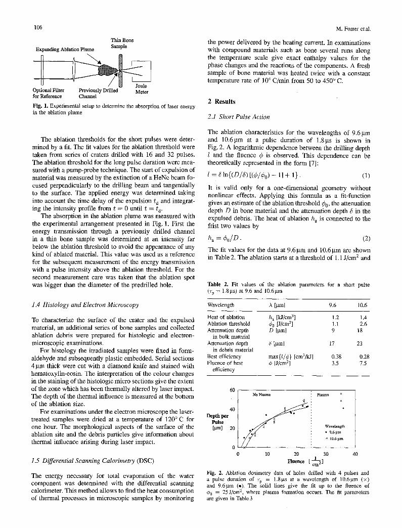
Thin Bone Expanding Ablation Plume Sample
Optional Filter Previously Drilled Meter for Reference Channel
Fig. 1. Experimental setup to determine the absorption of laser energy in the ablation plume
M. Forrer et al.
the power delivered by the heating current. In examinations with compound materials such as bone several runs along the temperature scale give exact enthalpy values for the phase changes and the reactions of the components. A fresh sample of bone material was heated twice with a constant temperature rate of 10 ° C/min from 50 to 450 ° C.
2 Resu l t s
2.1 Short Pulse Action
The ablation thresholds for the short pulses were deter- mined by a fit. The fit values for the ablation threshold were taken from series of craters drilled with 16 and 32 pulses. The ablation threshold for the long pulse duration were mea- sured with a pump-probe technique. The start of expulsion of material was measured by the extinction of a HeNe beam fo- cused perpendicularly to the drilling beam and tangentially to the surface. The applied energy was determined taking into account the time delay of the expulsion t d and integrat- ing the intensity profile from t = 0 until t = Q.
The absorption in the ablation plume was measured with the experimental arrangement presented in Fig. 1. First the energy transmission through a previously drilled channel in a thin bone sample was determined at an intensity far below the ablation threshold to avoid the appearance of any kind of ablated material. This value was used as a reference for the subsequent measurement of the energy transmission with a pulse intensity above the ablation threshold. For the second measurement care was taken that the ablation spot was bigger than the diameter of the predrilled hole.
1.4 Histology and Electron Microscopy
To characterize the surface of the crater and the expulsed material, an additional series of bone samples and collected ablation debris were prepared for histologic and electron- microscopic examinations.
For histology the irradiated samples were fixed in form- aldehyde and subsequently plastic embedded. Serial sections 4 gm thick were cut with a diamond knife and stained with hematoxylin-eosin. The interpretation of the colour changes in the staining of the histologic micro sections give the extent of the zone which has been thermally altered by laser impact. The depth of the thermal influence is measured at the bottom of the ablation size.
For examinations under the electron microscope the laser- treated samples were dried at a temperature of 120°C for one hour. The morphological aspects of the surface of the ablation site and the debris particles give information about thermal influence arising during laser impact.
1.5 Differential Scanning Calorimetry (DSC)
The energy necessary for total evaporation of the water component was determined with the differential scanning calorimeter. This method allows to find the heat consumption of thermal processes in microscopic samples by monitoring
The ablation characteristics for the wavelengths of 9.6 gm and 10.6~tm at a pulse duration of 1.8gs is shown in Fig. 2. A logarithmic dependence between the drilling depth 1 and the fluence q~ is observed. This dependence can be theoretically represented in the form [7]:
l : ~In{(D/6) [(qS/qS0) - 1] + 1}. (1)
It is valid only for a one-dimensional geometry without nonlinear effects. Applying this formula as a fit-function gives an estimate of the ablation threshold ¢o, the attenuation depth D in bone material and the attenuation depth ~ in the expulsed debris. The heat of ablation h a is connected to the frist two values by
h a = ColD. (2)
The fit values for the data at 9.6 gin and 10.6 gm are shown in Table 2. The ablation starts at a threshold of 1.1 J/cm 2 and
Table 2. Fit values of the ablation parameters for a short pulse (~-p = 1.8gs) at 9.6 and 10.6~tm
Wavelength )~ [gm] 9.6 10.6
Heat of ablation Ablation threshold Attenuation depth
in bulk material Attenuation depth
in debris material Best efficiency Fluence of best
efficiency
h a [kJ/cm 3] 1.2 1.4 ¢0 [ J/cm2] 1.1 2.6 D [gm] 9 18
6 [gm] 17 23
max{l/C} [cm3/kj] 0.38 0.28 ¢ [J/cm 2] 3.5 7.5
60 No P lasma P l a sma ×
×
Depth per 40 Pulse [gin] 20 Wavelength
• 9.6 I.tm × 10.6 lain
0 0 10 20 30 40
Fluenee [ c ~ ]
Fig. 2. Ablation dosimetry data of holes drilled with 4 pulses and a pulse duration of ~-p = 1.8gs at a wavelength of 10.6gm (x) and 9.6gm (,). The solid lines give the fit up to the fluence of ¢0 = 25 J/cm 2, where plasma formation occurs. The fit parameters are given in Table 3
Bone-Ablation Mechanism Using CO; Lasers
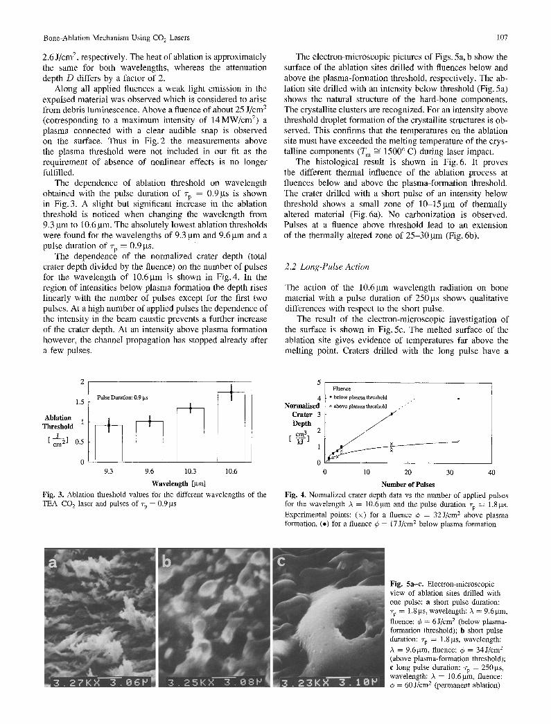
2.6 J/cm 2, respectively. The heat of ablation is approximately the same for both wavelengths, whereas the attenuation depth D differs by a factor of 2.
Along all applied fluences a weak light emission in the expulsed material was observed which is considered to arise from debris luminescence. Above a fluence of about 25 J/cm a (corresponding to a maximum intensity of 14MW/cm 2) a plasma connected with a clear audible snap is observed on the surface• Thus in Fig. 2 t h e measurements above the plasma threshold were not included in our fit as the requirement of absence of nonlinear effects is no longer fulfilled.
The dependence of ablation threshold on wavelength obtained with the pulse duration of rp = 0.9 gs is shown in Fig. 3. A slight but significant increase in the ablation threshold is noticed when changing the wavelength from 9.3 ~tm to 10.6 gm. The absolutely lowest ablation thresholds were found for the wavelengths of 9.3 gm and 9.6 gm and a pulse duration of rp = 0.9 gs.
The dependence of the normalized crater depth (total crater depth divided by the fluence) on the number of pulses for the wavelength of 10.6gm is shown in Fig. 4. In the region of intensities below plasma formation the depth rises linearly with the number of pulses except for the first two pulses. At a high number of applied pulses the dependence of the intensity in the beam caustic prevents a further increase of the crater depth. At an intensity above plasma formation however, the channel propagation has stopped already after a few pulses•
107
The electron-microscopic pictures of Figs. 5a, b show the surface of the ablation sites drilled with fluences below and above the plasma-formation threshold, respectively. The ab- lation site drilled with an intensity below threshold (Fig. 5a) shows the natural structure of the hard-bone components. The crystallite clusters are recognized. For an intensity above threshold droplet formation of the crystallite structures is ob- served. This confirms that the temperatures on the ablation site must have exceeded the melting temperature of the crys- talline components (T m ~ 1500 ° C) during laser impact•
The histological result is shown in Fig. 6. It proves the different thermal influence of the ablation process at fluences below and above the plasma-formation threshold. The crater drilled with a short pulse of an intensity below threshold shows a small zone of 10-15 gm of thermally altered material (Fig. 6a). No carbonization is observed. Pulses at a fluence above threshold lead to an extension of the thermally altered zone of 25-30 gm (Fig. 6b).
2.2 Long-Pulse Action
The action of the 10.6gm wavelength radiation on bone material with a pulse duration of 250 gs shows qualitative differences with respect to the short pulse.
The result of the electron-microscopic investigation of the surface is shown in Fig. 5c: The melted surface of the ablation site gives evidence of temperatures far above the melting point. Craters drilled with the long pulse have a
1 . 5 " : "
Ablation 1 Threshold
[ c ~ ] 0.5
0 9.3 9.6 10.3 10.6
Wavelength [/am] Fig. 3. Ablation threshold values for the different wavelengths of the TEACO 2 laser and pulses of rp = 0.9 gs
4 Normalised
Crater 3 Depth cm . -ffj
F l u e n c e
• be low p l a s m a th re sho ld
x above p l a s m a th resho ld . . "
1
0 0 10 20 30 40
Number of Pulses Fig. 4. Normalized crater depth data vs the number of applied pulses for the wavelength A = 10.6 gm and the pulse duration rp = 1.8 gs. Experimental points: (×) for a fluence ¢ = 32J/cm 2 above plasma formation, (e) for a fluence 4) = 17 J/cm 2 below plasma formation
Fig. 5a-c. Electron-microscopic view of ablation sites drilled with one pulse: a short pulse duration: rp = 1.8 gs, wavelength: )~ = 9.6 gin, fluence: q5 = 6 J/cm 2 (below plasma- formation threshold); b short pulse duration: 7-p = 1.8 gs, wavelength: )~ = 9.6~tm, fluence: ~b = 34J/cm 2 (above plasma-formation threshold); e long pulse duration: rp = 250gs, wavelength: ~ = 10.6 ktm, fluence: q~ = 60 J/cm 2 (permanent ablation)
108 M. Forrer et al.
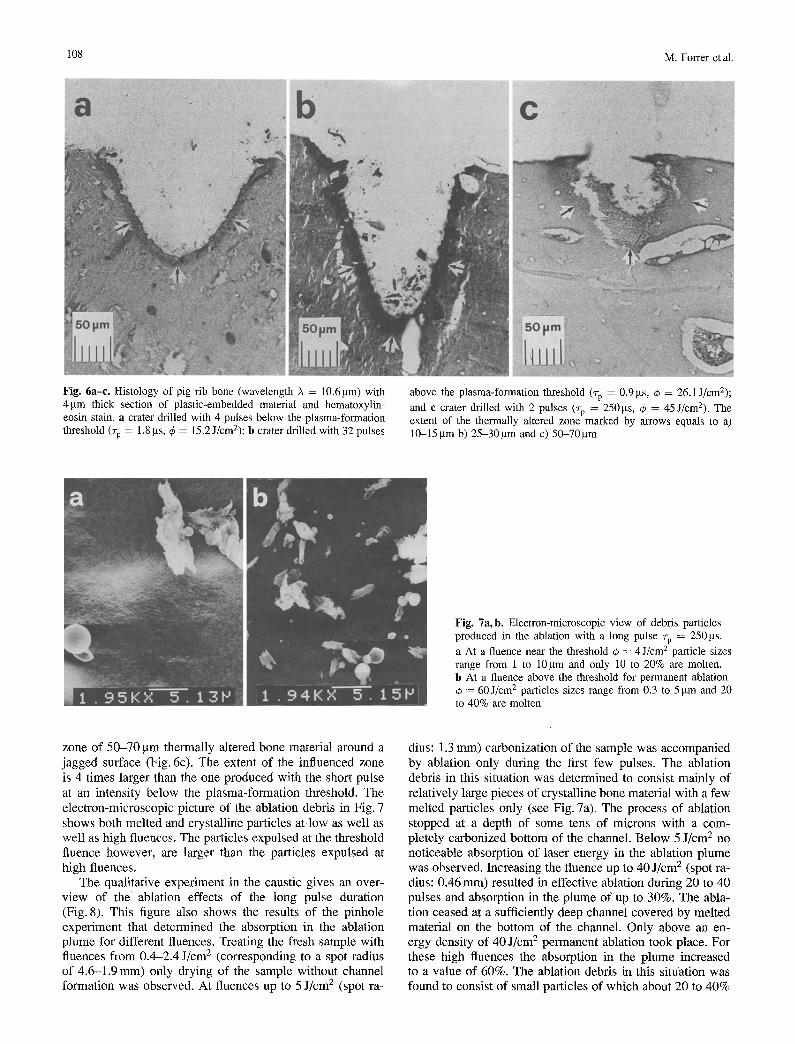
Fig. 6a-c. Histology of pig rib bone (wavelength A = 10.6gm) with 4gin thick section of plastic-embedded material and hematoxylin- eosin stain, a crater drilled with 4 pulses below the plasma-formation threshold (to = 1.8 p.s, q5 = 15.2 J/cm2); b crater drilled with 32 pulses
above the plasma-formation threshold (rp = 0.9bts, ~b = 26.1 J/cm2); and e crater drilled with 2 pulses (rp = 250 gs, ~b = 45 J/cm2). The extent of the thermally altered zone marked by arrows equals to a) 10-15 btm b) 25-30gm and c) 50-70gin
Fig. 7a, b. Electron-microscopic view of debris particles produced in the ablation with a long pulse rp = 250 gs. a At a fluence near the threshold 4~ = 4 J/cm 2 particle sizes range from 1 to 10gm and only 10 to 20% are molten. b At a fluence above the threshold for permanent ablation
= 60J/cm 2 particles sizes range from 0.3 to 5 gm and 20 to 40% are molten
zone of 50-70 btm thermally altered bone material around a jagged surface (Fig. 6c). The extent of the influenced zone is 4 times larger than the one produced with the short pulse at an intensity below the plasma-formation threshold. The electron-microscopic picture of the ablation debris in Fig. 7 shows both melted and crystalline particles at low as well as well as high fluences. The particles expulsed at the threshold fluence however, are larger than the particles expulsed at high fluences.
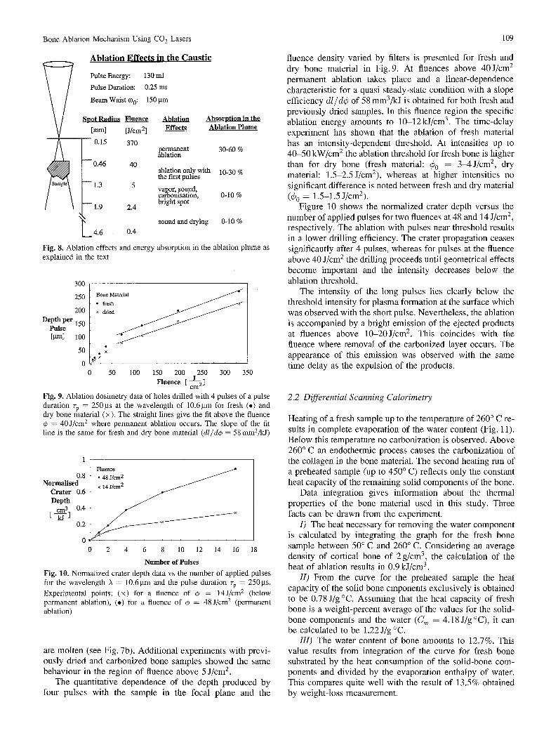
The qualitative experiment in the caustic gives an over- view of the ablation effects of the long pulse duration (Fig. 8). This figure also shows the results of the pinhole experiment that determined the absorption in the ablation plume for different fluences. Treating the fresh sample with ltuences from 0.4-2.4 J/cm 2 (corresponding to a spot radius of 4.6-1.9 mm) only drying of the sample without channel formation was observed. At fluences up to 5 J/cm 2 (spot ra-
dius: 1.3 mm) carbonization of the sample was accompanied by ablation only during the first few pulses. The ablation debris in this situation was determined to consist mainly of relatively large pieces of crystalline bone material with a few melted particles only (see Fig. 7a). The process of ablation stopped at a depth of some tens of microns with a com- pletely carbonized bottom of the channel. Below 5 J/cm 2 no noticeable absorption of laser energy in the ablation plume was observed. Increasing the fluence up to 40 J/cm 2 (spot ra- dius: 0.46 mm) resulted in effective ablation during 20 to 40 pulses and absorption in the plume of up to 30%. The abla- tion ceased at a sufficiently deep channel covered by melted material on the bottom of the channel. Only above an en- ergy density of 40 J/cm 2 permanent ablation took place. For these high fluences the absorption in the plume increased to a value of 60%. The ablation debris in this situation was found to consist of small particles of which about 20 to 40%
Bone-Ablation Mechanism Using CO 2 Lasers 109
A b l a t i o n E f f e c t s in the C a u s t i c
Pulse Energy: 130 mJ
Pulse Duration: 0.25 ms
Beam Waist ¢Oo: 150 gm
Spot Radius Fluence Ablation [mini [J/cm 2] Effects
0.15 370
- - 0.46 40
- -1 .3 5
1.9 2.4
4.6 0.4
Absorption in the Ablation Plume
permanent 30-60 % ablation
ablation only with 10-30 % the first pulses
vapor, sound, carbonisation, 0-10 % bright spot
sound and drying 0-10 %
Fig. 8. Ablation effects and energy absorption in the ablation plume as explained in the text
300
250
200 Depth per 150
Pulse [gm] 100
50
0
Bone Material
• fresh ~ /
x
50 100 150 200 250 300 350 Fluence [ c ~ ]
Fig. 9. Ablation dosimetry data of holes drilled with 4 pulses of a pulse duration ~-p = 250gs at the wavelength of 10.6gm for fresh (.) and dry bone material (x). The straight lines give the fit above the ftuence
= 40 J/cm 2 where permanent ablation occurs. The slope of the fit line is the same for fresh and dry bone material (dl/d~ = 58 mm3/kJ)
Flnence 0.8 • 48 J/cm 2
Normalised × 14 J / e m 2 ~ Crater 0.6 Depth
cm 3 0.4
E ~-1 02 ~ ~ X ~ ~
0 0 2 4 6 8 10 12 14 16 18
Number of Pulses Fig. 10. Normalized crater depth data vs the number of applied pulses for the wavelength A = 10.6gm and the pulse duration ~-p = 250gs. Experimental points: (x) for a fluence of ~b = 14J/cm 2 (below permanent ablation), (e) for a fluence of ~b = 48 J/cm 2 (permanent ablation)
are molten (see Fig. 7b). Addit ional experiments with previ- ously dried and carbonized bone samples showed the same behaviour in the region of fluence above 5 J/cm 2.
The quantitative dependence of the depth produced by four pulses with the sample in the focal plane and the
fluence density varied by filters is presented for fresh and dry bone material in Fig. 9. At fluences above 40J/cm 2 permanent ablation takes place and a l inear-dependence characteristic for a quasi steady-state condition with a slope efficiency dl/dO of 58 mm3/kJ is obtained for both fresh and previously dried samples. In this fluence region the specific ablation energy amounts to 10-12kJ/cm 3. The t ime-delay experiment has shown that the ablation of fresh material has an intensity-dependent threshold. At intensities up to 40-50 kW/cm 2 the ablation threshold for fresh bone is higher than for dry bone (fresh material: 40 = 3-4J/cm2, dry material: 1.5-2.5 J/cm2), whereas at higher intensities no significant difference is noted between fresh and dry material (60 = 1.5-1.5 J/cm2).
Figure 10 shows the normalized crater depth versus the number of applied pulses for two fluences at 48 and 14 J/cm 2, respectively. The ablation with pulses near threshold results in a lower drilling efficiency. The crater propagation ceases significantly after 4 pulses, whereas for pulses at the fluence above 40 J/cm 2 the drilling proceeds until geometrical effects become important and the intensity decreases below the ablation threshold.
The intensity of the long pulses lies clearly below the threshold intensity for plasma formation at the surface which was observed with the short pulse. Nevertheless, the ablation is accompanied by a bright emission of the ejected products at fluences above 10-20J/cm 2. This coincides with the fluence where removal of the carbonized layer occurs. The appearance of this emission was observed with the same time delay as the expulsion of the products.
2.2 Differential Scanning Calorimetry
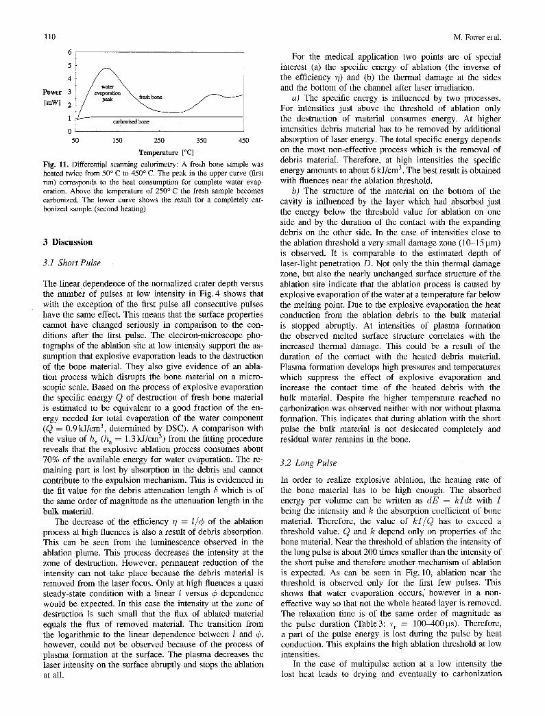
Heating of a fresh sample up to the temperature of 260 ° C re- sults in complete evaporation of the water content (Fig. 11). Below this temperature no carbonization is observed. Above 260 ° C an endothermic process causes the carbonization of the collagen in the bone material. The second heating run of a preheated sample (up to 450 ° C) reflects only the constant heat capacity of the remaining solid components of the bone.
Data integration gives information about the thermal properties of the bone material used in this study. Three facts can be drawn from the experiment.
I) The heat necessary for removing the water component is calculated by integrating the graph for the fresh bone sample between 50 ° C and 260 ° C. Considering an average density of cortical bone of 2 g/cm ~, the calculation of the heat of ablation results in 0.9 kJ/cm 3.
II) From the curve for the preheated sample the heat capacity of the solid bone cmponents exclusively is obtained to be 0.78 J/g °C. Assuming that the heat capacity of fresh bone is a weight-percent average of the values for the solid- bone components and the water (C w = 4.18 J/g °C), it can be calculated to be 1.22J/g °C.
Ill) The water content of bone amounts to 12.7%. This value results from integration of the curve for fresh bone substrated by the heat consumption of the solid-bone com- ponents and divided by the evaporation enthalpy of water. This compares quite well with the result of 13.5% obtained by weight-loss measurement.
l l0
6
5
4
Power 3
[mW] 2
1 carbonise.d bone
0
50 150 250 350 450
Temperature [°C]
Fig. 11. Differential scanning calorimetry: A fresh bone sample was heated twice from 50 ° C to 450 ° C. The peak in the upper curve (first run) corresponds to the heat consumption for complete water evap- oration. Above the temperature of 250 ° C the fresh sample becomes carbonized. The lower curve shows the result for a completely car- bonized sample (second heating)
3 Discussion
3.1 Short Pulse
The linear dependence of the normalized crater depth versus the number of pulses at low intensity in Fig. 4 shows that with the exception of the first pulse all consecutive pulses have the same effect. This means that the surface properties cannot have changed seriously in comparison to the con- ditions after the first pulse. The electron-microscope pho- tographs of the ablation site at low intensity support the as- sumption that explosive evaporation leads to the destruction of the bone material. They also give evidence of an abla- tion process which disrupts the bone material on a micro- scopic scale. Based on the process of explosive evaporation the specific energy Q of destruction of fresh bone material is estimated to be equivalent to a good fraction of the en- ergy needed for total evaporation of the water component (Q = 0.9 kJ/cm 3, determined by DSC). A comparison with the value of h a (h a = 1.3 kJ/cm 3) from the fitting procedure reveals that the explosive ablation process consumes about 70% of the available energy for water evaporation. The re- maining part is lost by absorption in the debris and cannot contribute to the expulsion mechanism. This is evidenced in the fit value for the debris attenuation length 5 which is of the same order of magnitude as the attenuation length in the bulk material.
The decrease of the efficiency ~ = I/¢ of the ablation process at high fluences is also a result of debris absorption. This can be seen from the luminescence observed in the ablation plume. This process decreases the intensity at the zone of destruction. However, permanent reduction of the intensity can not take place because the debris material is removed from the laser focus. Only at high ftuences a quasi steady-state condition with a linear 1 versus ¢ dependence would be expected. In this case the intensity at the zone of destruction is such small that the flux of ablated material equals the flux of removed material. The transition from the logarithmic to the linear dependence between 1 and ¢, however, could not be observed because of the process of plasma formation at the surface. The plasma decreases the laser intensity on the surface abruptly and stops the ablation at all.
M. Forrer et al.
For the medical application two points are of special interest (a) the specific energy of ablation (the inverse of the efficiency r/) and (b) the thermal damage at the sides and the bottom of the channel after laser irradiation.
a) The specific energy is influenced by two processes. For intensities just above the threshold of ablation only the destruction of material consumes energy. At higher intensities debris material has to be removed by additional absorption of laser energy. The total specific energy depends on the most non-effective process which is the removal of debris material. Therefore, at high intensities the specific energy amounts to about 6 kJ/cm 3. The best result is obtained with fluences near the ablation threshold.
b) The structure of the material on the bottom of the cavity is influenced by the layer which had absorbed just the energy below the threshold value for ablation on one side and by the duration of the contact with the expanding debris on the other side. In the case of intensities close to the ablation threshold a very small damage zone (10-15 gm) is observed. It is comparable to the estimated depth of laser-light penetration D. Not only the thin thermal damage zone, but also the nearly unchanged surface structure of the ablation site indicate that the ablation process is caused by explosive evaporation of the water at a temperature far below the melting point. Due to the explosive evaporation the heat conduction from the ablation debris to the bulk material is stopped abruptly. At intensities of plasma formation the observed melted surface structure correlates with the increased thermal damage. This could be a result of the duration of the contact with the heated debris material. Plasma formation develops high pressures and temperatures which suppress the effect of explosive evaporation and increase the contact time of the heated debris with the bulk material. Despite the higher temperature reached no carbonization was observed neither with nor without plasma formation. This indicates that during ablation with the short pulse the bulk material is not desiccated completely and residual water remains in the bone.
3.2 Long Pulse
In order to realize explosive ablation, the heating rate of the bone material has to be high enough. The absorbed energy per volume can be written as dE = kIdt with I being the intensity and k the absorption coefficient of bone material. Therefore, the value of k I / Q has to exceed a threshold value. Q and k depend only on properties of the bone material. Near the threshold of ablation the intensity of the long pulse is about 200 times smaller than the intensity of the short pulse and therefore another mechanism of ablation is expected. As can be seen in Fig. 10, ablation near the threshold is observed only for the first few pulses. This shows that water evaporation occurs,' however in a non- effective way so that not the whole heated layer is removed. The relaxation time is of the same order of magnitude as the pulse duration (Table3: ~-r = 100-400gs). Therefore, a part of the pulse energy is lost during the pulse by heat conduction. This explains the high ablation threshold at low intensities.
In the case of multipulse action at a low intensity the lost heat leads to drying and eventually to carbonization
Bone-Ablation Mechanism Using CO 2 Lasers 111
Table 3. Thermophysical properties of bone relevant for the ablation with CO2-1aser radiation
Origin Value Unit
Attenuation depth d = qSo/h a 10-20 gm Heat conductivity [16] k 6.0 × 10 - 3 W/cm °C Thermal diffusivity ~ = k/~oc 2.5 × 10 - 3 c m 2 / s
Thermal relaxation time (Tr) ~'r = D2/4~ 100400 gs
of the bone material. Subsequent pulses only increase the thickness of the carbonized and desiccated layer and channel propagation is stopped, because the intensity is too low to ablate the carbonized layer. In contrast, the qualitative experiment has shown that with fluences above 40J/cm 2 the surface of the drilled crater shows no carbonized layer after ablation. We assume that for high fluences the heat at the beginning of the pulse leads to carbonization of a surface layer, which, however, is thin enough to be ablated by the peak intensity of the same pulse. The existence of a carbonized layer does not mean absence of explosive evaporation for these high fluences. The heating rate in the carbonized material affects deeper layers of material by heat conduction so fast that explosive evaporation can
take place. Supposing carbonization on the surface and total absorption of the laser energy in this thin layer of carbonized material, the thickness A of the layer heated by thermal conduction can be estimated by A = 4~/v, with ~ = 2.5 × 10 -3 cmZ/s being the thermal diffusivity of bone (Table 3) and v = 40 cm/s the drilling velocity (Fig. 8). Inserting these values results in A = 2.5 gm, much less than the laser-light penetration depth in fresh bone. As a result, the heating rate of the layer is high enough to cause explosive evaporation. This leads to a significantly higher drilling efficiency (cf. Fig. 10) and explains the mixture of melted and crystalline particles observed in the electron-microscopic investigation of the debris (Fig. 7). The carbonized layer therefore is a prerequisite for efficient drilling to occur with the long pulse. The high absorption in the thin carbonized layer also renders possible bone and tissue ablation with lasers of an initially low absorption in fresh material (e.g. argon laser at A = 514.5 nm).
The comparative experiments between fresh and dry bone material show that above the fluence of 40 J/cm 2 the slope efficiency dl/d¢ is the same, although the specific ablation energy ¢/1 is smaller for fresh material. On the one side this shows the positive effect of the water component on the overall drilling efficiency, although the process of destruction is covered by low-effective removal. On the other side ablation is also possible on previously dried material. This phenonemon evidences that the evaporation of the organic bone components can also initiate the process of destruction. The equivalent slope efficiency for fresh and dry bone material shows that the process of acceleration of the debris does not depend on the water content. Therefore, the absorption of pulse energy in non-vaporized debris material can be responsible for the increasing pressure and the removal of the products. The increase in absorption at higher intensities could be connected to the appearance of non-melted products due to explosive evaporation.
Pulse Signal Pulse Signal
Heat without on (AE
Start of the\
Expulsion ~ E x t J ~ ct'c n Signal
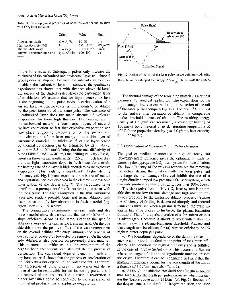
Fig. 12. Action of the tail of the laser pulse on the bulk material. After
the ablation has stopped the energy AE = 7 I(t)dt heats the surface TO
The thermal damage of the remaining material is a critical parameter for medical application. The explanation for the high damage observed can be found in the action of the tail of the laser pulse (compare Fig. 12). The heat A E applied to the surface after cessation of ablation is comparable to the threshold fluence of ablation. The resulting energy density of 1.5 J/cm 2 can reasonably account for heating of 150gin of bone material to its denaturation temperature of 60 ° C (bone properties: density • = 2.0 g/cm 3, heat capacity c = 1.22J/g°C).
3.3 Optimization of Wavelength and Pulse Duration
The goal of medical treatment with high efficiency and low-temperature influence gives the optimization path for choosing the appropriate CO 2 laser system for bone ablation. The low efficiency of the process responsible for removing the debris during the ablation with the long pulse and the large thermal damage observed inhibit the use of a longitudinally pumped low pressure CO 2 laser system, which can only produce a pulse duration longer than 100-150 gs.
The short pulse from a TEA-CO 2 laser system is prefer- able due to the low thermal damage and high ablation effi- ciency produced by the explosive ablation process. Because the efficiency of drilling is decreased abruptly and thermal damage is increased when a plasma is formed, the pulse in- tensity has to be chosen to lie below the plasma-formation threshold. Therefore a pulse duration of a few microseconds is advantageous because it allows to work with higher flu- ences below the plasma-formation threshold. Fhience and wavelength can be chosen for (a) highest efficiency or (b) highest crater depth per pulse.
a) The logarithmic dependence of the depth l versus flu- ence ~b can be used to calculate the point of maximum effi- ciency. The condition for highest efficiency I/6 is fulfilled in the case of (l/C) - (dl/d~) = 0. This occurs at a fluence where the tangential line to the logarithmic function crosses the origin. Therefore it can be recognized in Fig. 2 that the maximum efficiency occurs for the wavelength of 9.6 gm at a fluence of 3.5 J/cm 2 (see also Table 2).
b) Although the ablation threshold for 10.6 gin is higher than for 9.6 gm, the depth per pulse increases when increas- ing the fluence above about 11 J/cm 2 (cf. Fig. 2). Because of the deeper penetration depth of 10.6 gm radiation, the laser
112 M. Forrer et al.
energy is dispersed in a larger volume and therefore a higher intensity is needed to reach the ablation threshold. If the in- tensity, however, is high enough to ablate the whole volume in an explosive way, a faster drilling velocity is obtained. In order to maximise the crater depth per pulse, an intensity just below the plasma-formation threshold at a wavelength of 10.6 btm should be used. The ablation thresholds are the same for 9.3 and 9.6 btm radiation within the experimental error. Nevertheless, the maximum drilling efficiency occurs at the wavelength of 9.6 gm. This is explained by the fact that the absorption maximum of bone material in the in- frared lies close to the wavelength of 9.6 btm (Table 1). The ablation threshold at this wavelength and a pulse duration of 0.9 bts corresponds to the bone ablation threshold found with 300 ns pulses of a HF laser at a wavelength of 2,91 btm [7]. It seems that the higher absorption of water at 2.91 gm (see Table 1) is compensated by additional absorption of the mineral components of bone material at the wavelengths of 9.3 and 9.6 gm. This is also supported by the fact that the zone of thermally altered material around holes drilled Oeith short CO2-1aser pulses (Fig. 6a) is of the same order as damage zones found with the Q-switched erbium laser in bone material (5-10 gm) [10].
This comparison shows that the bone ablation mechanism with a pulse duration of a few microseconds is based on explosive evaporation of water at both the wavelength of 9.6 btm as well as the wavelength of 2.91 gm. Although the total absorption in fresh bone material is higher at 9.6 btm, the ablation is not more efficient than at 2.91 btm. This is caused by the fact that at 9.6 gm the radiation is absorbed mainly in the minerals and the water is heated only indirectly by heat conduction, whereas at 2.91 btm the radiation is absorbed 6. directly in the water. This difference becomes most apparent with the pulse duration longer than the thermal relaxation 7. time of bone material. At 9 .6gm the heat produced in the mineral structure is dissipated fast which reduces the 8. heating rate in the irradiated volume below the threshold 9. for explosive evaporation: in contrast to holes produced with the wavelength of 2.91 gm, the craters drilled with the 10. wavelength of 9.6 btm therefore show a ring of carbonization around the top of the crater.
4 Conclusion
Ablation dosimetry, electron microscopy and histology in the application of CO2-1aser osteotomy have led to a clas- sification of ablation mechanisms being dependent on pulse duration and wavelength. The ablation mechanism is based on the absorption of laser radiation in the minerals. For the short pulse this leads to a fast heating rate and explosive evaporation of the water component. With the pulse dura-
tion longer than 100 gs a part of the absorbed laser energy is lost by heat conduction to the surrounding material, which becomes desiccated and subsequently carbonized. With the long pulse duration therefore permanent ablation can only occur with the intensity high enough to ablate this carbonized layer of material.
It was evidenced that with a correctly chosen set of laser parameters the CO2-1aser can be a useful instrument for laser osteotomy. From a medical point of view the best result is obtained by using a short pulse (1-5 gs) at a wavelength of 9.6 gin and an intensity below the plasma- formation threshold (10-20 MW/cm2).
Acknowledgements. The authors wish to express their gratitude to Th. Schaffner and Chr. Wirz for their valuable discussions. We thank also A. Friedrich, E. Krahenbtihl and S. Binggeli for their technical assistance. This work was supported by the Swiss National Science Foundation.
References
1. M.L. Wolbarsht: IEEE J. QE-20, 1427-1432 (1984) 2. A.D. Zweig, B. Meierhofer, O.M. Mtiller, C. Mischler, V. Romano,
M. Frenz, H.P. Weber: Lasers Surg. Med. 10, 262-274 (1990) 3. M. Frenz, A.D. Zweig, V. Romano, H.P. Weber: In Laser-Tissue
Interaction, ed. by S.L. Jacques (Proc. SPIE 1202, 1990) pp. 22-33 4. V.M. Zolotgarev, B.A. Mikhailov, L.I. Alperovich, S.I. Popov:
Opt. Spectrosc. 26, 430-432 (1969) 5. D.R. Meyer, C. Scholz, A. Bfichle, M. Grothues-Spork, G.J.
Mtiller, W. Siebert, F. Dinkelaker, T. Cierpinski: Lasers Med. Surg. 6, 150-155 (1990) R.C. Nuss, R.L. Fabian, S. Rajabrata, C.A. Puliafito: Lasers Med. Surg. 8, 381-391 (1988) J.A. Izatt, N.D. Sankey, F. Partovi, M. Fitzmaurice, R.P~ Rava, I. Itzkan, M.S. Feld: IEEE J. QE-26, 2261-2270 (1990) T. Schomacker, Y. Domankevitz, T.J. Flone, T.F. Deutsch: Lasers Med. Surg. 11, 141-151 (1991) J.T. Walsh, T.J. Flotte, R.R. Anderson, T.F. Deutsch: Lasers Med. Surg. 8, 108-118 (1988) J.T. Walsh: Pulsed Laser Ablation of Tissue: Analysis of the removal process and tissue healing. Ph.D. Thesis, MIT, Boston (1988)
11. J.E. Decker, W. Xiong, F. Yergeau, S.L. Chin: Appl. Opt. 31, 1912-1913 (1992)
12. R.A. Nyquist, R.O. Kagel: Infrared spectra of inorganic com- pounds (Academic, New York 1971) pp. 162, 163, 492, 493
13. C. Scholz, M. Grothues-Spork, F. Dinkelaker, G. Mtiller, R. Rah- mandzadeh: In Optoelectronics in Medicine, ed. by W. Waidelich (Springer, Berlin, Heidelberg 1990) pp. 67-78
14. I.V. Yannas: J. Macromol. Sci. Rev. Macromol. Chem. 1, 49-104 (1972)
15. C.W. Robertson, D. Williams: J. Opt. Soc. Am. 61, 1316-1320 (1971)
16. H.F. Bowman, E.G. Cravalho, M. Woods: Annu. Rev. Biophys. Bioeng. 4, 43-79 (1975)
Related Documents