RESEARCH Open Access Body electrical loss analysis (BELA) in the assessment of visceral fat: a demonstration Kim H Blomqvist 1* , Jesper Lundbom 2 , Nina Lundbom 2 and Raimo E Sepponen 1 * Correspondence: kim.h. [email protected] 1 Department of Electronics, Aalto University, PO Box 13340, 00076 Aalto, Finland Full list of author information is available at the end of the article Abstract Background: Body electrical loss analysis (BELA) is a new non-invasive way to assess visceral fat depot size through the use of electromagnetism. BELA has worked well in phantom measurements, but the technology is not yet fully validated. Methods: Ten volunteers (5 men and 5 women, age: 22-60 y, BMI: 21-30 kg/m 2 , waist circumference: 73-108 cm) were measured with the BELA instrument and with cross-sectional magnetic resonance imaging (MRI) at the navel level, navel +5 cm and navel -5 cm. The BELA signal was compared with visceral and subcutaneous fat areas calculated from the MR images. Results: The BELA signal did not correlate with subcutaneous fat area at any level, but correlated significantly with visceral fat area at the navel level and navel +5 cm. The correlation was best at level of navel +5 cm (R 2 = 0.74, P < 0.005, SEE = 29.7 cm 2 , LOOCV = 40.1 cm 2 ), where SEE is the standard error of the estimate and LOOCV is the root mean squared error of leave-one-out style cross-validation. The average estimate of repeatability of the BELA signal observed through the study was ±9.6 %. One of the volunteers had an exceptionally large amount of visceral fat, which was underestimated by BELA. Conclusions: The correlation of the BELA signal with the visceral but not with the subcutaneous fat area as measured by MRI is promising. The lack of correlation with the subcutaneous fat suggests that subcutaneous fat has a minor influence to the BELA signal. Further research will show if it is possible to develop a reliable low-cost method for the assessment of visceral fat either using BELA only or combining it, for example, with bioelectrical impedance measurement. The combination of these measurements may help assessing visceral fat in a large scale of body composition. Before large-scale clinical testing and ROC analysis, the initial BELA instrumentation requires improvements. The accuracy of the present equipment is not sufficient for such new technology. Background Accumulation of visceral fat (VF) has been shown to be important in the development of Type 2 diabetes and cardiovascular disease, while subcutaneous fat (SF) carries minor risks [1,2]. Excessive visceral fat may predispose even young children to these adult age diseases [3]. Therefore, VF and SF depots must be differentiated in interventional studies. The size of the visceral adipose depot can be accurately measured by magnetic resonance imaging (MRI) [4] or computed tomography (CT) [5], but the high measurement costs and CT’s radiation dose make these methods unsuitable for large studies or repeated Blomqvist et al. BioMedical Engineering OnLine 2011, 10:98 http://www.biomedical-engineering-online.com/content/10/1/98 © 2011 Blomqvist et al; licensee BioMed Central Ltd. This is an Open Access article distributed under the terms of the Creative Commons Attribution License (http://creativecommons.org/licenses/by/2.0), which permits unrestricted use, distribution, and reproduction in any medium, provided the original work is properly cited.

Welcome message from author
This document is posted to help you gain knowledge. Please leave a comment to let me know what you think about it! Share it to your friends and learn new things together.
Transcript
RESEARCH Open Access
Body electrical loss analysis (BELA) in theassessment of visceral fat: a demonstrationKim H Blomqvist1*, Jesper Lundbom2, Nina Lundbom2 and Raimo E Sepponen1
* Correspondence: [email protected] of Electronics, AaltoUniversity, PO Box 13340, 00076Aalto, FinlandFull list of author information isavailable at the end of the article
Abstract
Background: Body electrical loss analysis (BELA) is a new non-invasive way to assessvisceral fat depot size through the use of electromagnetism. BELA has worked well inphantom measurements, but the technology is not yet fully validated.
Methods: Ten volunteers (5 men and 5 women, age: 22-60 y, BMI: 21-30 kg/m2,waist circumference: 73-108 cm) were measured with the BELA instrument and withcross-sectional magnetic resonance imaging (MRI) at the navel level, navel +5 cmand navel -5 cm. The BELA signal was compared with visceral and subcutaneous fatareas calculated from the MR images.
Results: The BELA signal did not correlate with subcutaneous fat area at any level,but correlated significantly with visceral fat area at the navel level and navel +5 cm.The correlation was best at level of navel +5 cm (R2 = 0.74, P < 0.005, SEE = 29.7cm2, LOOCV = 40.1 cm2), where SEE is the standard error of the estimate and LOOCVis the root mean squared error of leave-one-out style cross-validation. The averageestimate of repeatability of the BELA signal observed through the study was ±9.6 %.One of the volunteers had an exceptionally large amount of visceral fat, which wasunderestimated by BELA.
Conclusions: The correlation of the BELA signal with the visceral but not with thesubcutaneous fat area as measured by MRI is promising. The lack of correlation withthe subcutaneous fat suggests that subcutaneous fat has a minor influence to theBELA signal. Further research will show if it is possible to develop a reliable low-costmethod for the assessment of visceral fat either using BELA only or combining it, forexample, with bioelectrical impedance measurement. The combination of thesemeasurements may help assessing visceral fat in a large scale of body composition.Before large-scale clinical testing and ROC analysis, the initial BELA instrumentationrequires improvements. The accuracy of the present equipment is not sufficient forsuch new technology.
BackgroundAccumulation of visceral fat (VF) has been shown to be important in the development of
Type 2 diabetes and cardiovascular disease, while subcutaneous fat (SF) carries minor
risks [1,2]. Excessive visceral fat may predispose even young children to these adult age
diseases [3]. Therefore, VF and SF depots must be differentiated in interventional studies.
The size of the visceral adipose depot can be accurately measured by magnetic resonance
imaging (MRI) [4] or computed tomography (CT) [5], but the high measurement costs
and CT’s radiation dose make these methods unsuitable for large studies or repeated
Blomqvist et al. BioMedical Engineering OnLine 2011, 10:98http://www.biomedical-engineering-online.com/content/10/1/98
© 2011 Blomqvist et al; licensee BioMed Central Ltd. This is an Open Access article distributed under the terms of the CreativeCommons Attribution License (http://creativecommons.org/licenses/by/2.0), which permits unrestricted use, distribution, andreproduction in any medium, provided the original work is properly cited.
individual use. Anthropometric methods, such as waist circumference (WC) and waist-to-
hip ratio (WHR), are frequently used as alternative indices for predicting VF related risks.
However, these methods are known to be non-specific [6] because they include SF. Con-
ventional bioelectrical impedance analysis (BIA) has also difficulties in measuring VF due
to the influence of SF [7], and therefore various abdominal BIA methods have been intro-
duced to measure VF more accurately [7-12]. Efforts have even been put into the use of
ultrasound in the assessment of VF [13-17].
We are constructing an instrumentation to measure visceral adiposity with a radio-
frequency coil, relying on the measurement of electrical losses in the subject through
the use of electromagnetism. The idea of the measurement, called body electrical loss
analysis (BELA), has been tested with phantom measurements [18]. The experiment
does not expose the patient to ionizing radiation, and the strength of the magnetic
field is below ICNIRP’s (International Commission on Non-Ionizing Radiation Protec-
tion) guidelines [19] for general public exposure. Because of its simplicity, the BELA
measurement could also be used to measure VF in children, which would be an impor-
tant aid in preventive care [20].
The aim of this pilot study was to evaluate, how the BELA signal and the initial
instrumentation perform in determining the size of the VF depot in humans, and to
validate the results using MRI.
Subjects and methodsThe coordinating ethics committee of Helsinki University Central Hospital approved
the study and an informed written consent was taken from the volunteers. All volun-
teers were measured with the BELA instrument and imaged with MRI within one
week. Ten healthy volunteers (5 men, 5 women) covering the range of BMI of 21-30
kg/m2 and the range of waist circumference of 73 to 108 cm were recruited from
laboratory personnel and acquaintances. More descriptive data is shown in figure 1.
Measurement of fat distribution by MRI
Fat distribution was measured on a clinical 1.5 Tesla MR imager (Avanto, Siemens,
Erlangen, Germany) using a T1-weighted gradient echo sequence with selective fat
excitation for optimal fat contrast [4]. A stack of sixteen axial image slices with 1 cm
nominal thickness was centered at the L4/L5 intervertebral disk to determine the fat
distribution at the waist level. The MR images were analyzed using a segmentation
algorithm (SliceOmatic v4.3) to yield the areas of visceral and subcutaneous fat [4].
BELA measurement
During the BELA measurement, the volunteer crouches into the radio frequency coil -
so that the coil surrounds the individual at the abdomen - and stands still hands over
head, with shoes and belt removed. The apparatus is shown in figure 2. A time-varying
magnetic field produced by the coil will induce so called eddy currents into the patient.
These induced currents produce electrical losses in the patient, which can be observed
as changes in the electrical resonance quality factor of the coil. Because the electrical
conductivity of lean tissue is over ten times higher than that of fat tissue across the
frequency range from 1 kHz to 1 MHz [21], lean tissue (e.g. muscle) will produce a
larger change in the coil’s quality factor.
Blomqvist et al. BioMedical Engineering OnLine 2011, 10:98http://www.biomedical-engineering-online.com/content/10/1/98
Page 2 of 9

By inducing variations in the excitation frequency, the major signal contribution can
be altered. At higher frequencies, the signal tends to concentrate on the outer shell
volume of the subject, allowing the assessment of fat distribution. For example, in an
individual who has a small amount of VF, the loss changing rate as a function of fre-
quency will also be small. This conclusion is based on the rough body models [18],
where the trunk consists of three separate areas: interior (fatty or non-fatty), muscles
and SF. From an electrical point of view, only the interior and the conductive volume
20 25 30 35 40 45 50 55 600
1
2
3
4
Age (y)
Num
ber
of s
ubje
cts
21 22 23 24 25 26 27 28 29 300
1
2
3
4
BMI (kg/m2)
70 75 80 85 90 95 100 105 1100
1
2
3
4
WC (cm)
Num
ber
of s
ubje
cts
60 65 70 75 80 85 90 95 1001050
1
2
3
4
Weight (kg)
Figure 1 Description of subjects. The age, BMI, WC and weight histograms of the subjects.
Figure 2 BELA feasibility model. A coil large enough for a human body to be surrounded at theabdomen. (1) An electrostatic shield, (2) measurement electronics, (3) multistrand litz wire [28].
Blomqvist et al. BioMedical Engineering OnLine 2011, 10:98http://www.biomedical-engineering-online.com/content/10/1/98
Page 3 of 9

(of muscles) surrounding the interior interact in the BELA measurement. The influ-
ence of SF is considered to be small because of its low conductivity.
The loss changing rate is calculated with the following equation
mloss =�Vi,H − �Vi,L
fH − fL(1)
where ΔVi,H and ΔVi,L are the measured voltage changes across the coil between
empty and loaded coil at high and low (fH and fL) frequencies, respectively. The sub-
script i indicates the in-phase signals [18].
In this study the BELA measurements were taken at 103 kHz and 185 kHz frequen-
cies at navel level, navel +5 cm and navel -5 cm. All measurements were repeated five
times at every level. In addition, this session was repeated for one subject on three
consecutive days.
The effect of the positioning was studied by taking measurements when standing
three centimetres forward, rightward and leftward from the marked standing position
(the middle of the coil). This test was repeated ten times for every positioning.
Statistical analysis
Data were analyzed using the Pearson product-moment correlation coefficient and
leave-one-out style cross-validation. The repeatability of the BELA signal was estimated
as
�mloss =mloss,max − mloss,min
2=
σH + σL
fH − fL(2)
where sH and sL are the standard deviations of the measured voltage changes at fHand fL, respectively. The effect of the frequency measurement was insignificant (< 5 ·
10-3) and is thus ignored in equation 2.
ResultsThe calculated correlation coefficients between BELA and MRI at three different levels
of navel are shown in tables 1 and 2. The measured mean values of the loss changing
rates (mloss) did not correlate with SF in MRI at any level (R2 < 0.14). Significant corre-
lations between mloss and MRI were found for VF (R2 = 0.74, P < 0.005) at the navel
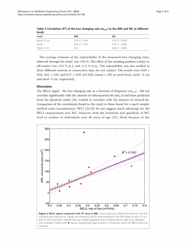
+5 cm level and (R2 = 0.43, P < 0.05) at the navel level. Figure 3 presents the scatter
plot of the best correlation (SEE = 29.7 cm2, LOOCV = 40.1 cm2) between the mloss
and VF area. SEE is the standard error of the estimate and LOOCV is the root mean
squared error of leave-one-out style cross-validation. One measurement point, marked
with a square, was exceptional and thus ignored in the calculations of correlation coef-
ficients, see below figure 4A.
Table 1 Correlation (R2) of the loss changing rate (mloss) to the VF, SF, VF/SF and SF/VFat different levels
Level VF SF VF/SF SF/VF
Navel +5 cm 0.74, P < 0.005 0.005 0.38, P < 0.05 0.35, P < 0.05
Navel 0.43, P < 0.05 0.14 0.06 0.03
Navel -5 cm 0.19 0.002 0.08 0.19
Blomqvist et al. BioMedical Engineering OnLine 2011, 10:98http://www.biomedical-engineering-online.com/content/10/1/98
Page 4 of 9
The average estimate of the repeatability of the measured loss changing rates,
observed through the study, was ±9.6 %. The effect of the standing position (center or
off-center) was ±2.5 % at fL and ±1.5 % at fH. The repeatability was also studied in
three different sessions in consecutive days for one subject. The results were 0.60 ±
0.03, 0.61 ± 0.01 and 0.57 ± 0.01 mV/kHz (mean ± SD) at navel level, navel +5 cm
and navel -5 cm, respectively.
DiscussionThe BELA signal - the loss changing rate as a function of frequency (mloss) - did not
correlate significantly with the amount of subcutaneous fat and, as had been predicted
from the phantom study [18], tended to correlate with the amount of visceral fat.
Comparison of the correlations found in this study to those found for a much simpler
method waist circumference (WC) [22,23] do not suggest much advantage for the
BELA measurement over WC. However, both the sensitivity and specificity of WC
tend to weaken in individuals over 40 years of age [23], likely because of the
Table 2 Correlation (R2) of the loss changing rate (mloss) to the BMI and WC at differentlevels
Level BMI WC
Navel +5 cm 0.33, P < 0.05 0.75, P < 0.005
Navel 0.35, P < 0.05 0.74, P < 0.005
Navel -5 cm 0.32 0.68, P < 0.005
0.3 0.35 0.4 0.45 0.5 0.55 0.6 0.65 0.7 0.75 0.8−50
0
50
100
150
200
250
300
BELA, rate of loss (mV/kHz)
MR
I, vi
scer
al fa
t are
a (c
m2 )
R2 = 0.7357
Figure 3 BELA signal compared with VF area in MRI. Linear regression (bold line) between the losschanging rate measured as voltage per frequency and VF area measured in the MR image at navel +5 cmlevel in nine volunteers. Dashed lines are multiple regression lines of leave-one-out style cross validation.One volunteer, marked with ■, had an exceptionally large amount of visceral fat, which the BELA could notrecognize.
Blomqvist et al. BioMedical Engineering OnLine 2011, 10:98http://www.biomedical-engineering-online.com/content/10/1/98
Page 5 of 9

accumulation of SF. The low sensitivity of BELA for SF could make it more specific in
this age group.
The comparison of the BELA correlations to those found for abdominal BIA and
ultrasonography (US) do not show much advantage either. However, large scale studies
in abdominal BIA [9,10,12] and US [13] do not report receiver operating characteristics
(ROC) graphs, which we think are important when the performance of the method is
estimated. Furthermore, one may question the performance of the bioelectrical impe-
dance measurement in the abdominal BIA [24], because the final analysis commonly
includes several additional parameters like sex, age [11] and body shape [8,10,11], or
WC [12]. The nature of the eddy currents may justify including WC in the BELA ana-
lysis, but there is no need to use the other parameters often combined with BIA. Thus,
in some groups of individuals, BELA will potentially perform better than BIA. BELA
would also be more convenient in medical check-ups and repeated individual use than
US, because BELA measurements do not need medical personnel to perform the mea-
surement or interpret the results.
The loss changing rate mloss is a relative quantity. It does not include information on
the bulk losses caused by the subject but rather the rate of loss as a function of fre-
quency. Based on this, we assumed that even if larger subjects introduce more losses
than smaller ones, mloss would not be sensitive to the body size. However, a strong
correlation to WC at all levels suggests that mloss is sensitive to body geometrics,
which has to be targeted in further studies to make sure that the sensitivity to changes
in fat content is sufficiently larger than the sensitivity to changes in WC.
The BELA setup used in this study was similar to that used in [18], except the
doubled field strength (1V across the coil) and the electrostatic shield made to cover
the coil’s inner surface. By doubling the field strength, the signal from human subjects
corresponded to what was observed with phantoms in [18]. This change was necessary,
because at low frequency, the losses would have been too small. The electrostatic
shield was included because, in human subjects but not in phantoms, losses were
observed to be somewhat higher without shielding. The shield’s purpose is to reduce
capacitive coupling with the subject inside the coil. Therefore, only the inner surface of
the coil was shielded. Shielding the outer surface would only reduce the coil’s quality
factor unnecessarily.
Figure 4 Axial MR images from two volunteers. One volunteer had an exceptionally large amount of VF(A) but smaller amount of SF than a volunteer with a reasonable amount of VF (B). The relative amount ofconductive tissue (muscle) compared with poorly conductive fatty tissue is so small in (A) that the BELAcould not differentiate VF from VF+SF.
Blomqvist et al. BioMedical Engineering OnLine 2011, 10:98http://www.biomedical-engineering-online.com/content/10/1/98
Page 6 of 9

The losses were measured at 103 kHz and 185 kHz frequencies. The frequency selec-
tion is not critical and therefore these frequencies were not tuned precisely to some
more intuitive frequencies like 100 kHz and 200 kHz by using trimmer capacitors but
discrete components. It is important to have two separate frequencies low enough so
that the wave length is clearly above the subject’s diameter. Moreover, frequencies
from 100 kHz to 200 kHz are low enough to give signal also from the interior of the
subject. This was tested with air core and filled phantoms in [18] - a similar test than
in [25].
One of the volunteers had an exceptionally large amount of VF, which the BELA
measurement could not differentiate. The axial MR image, shown in figure 4A, indi-
cated that in this volunteer, the VF and SF pools came into close contact with each
other due to very thin or near non-existent abdominal muscles, thus significantly redu-
cing the conductive volume (of muscle) between SF and VF pools. Since the BELA
measurement needs an electrically conductive volume between the SF and VF pools,
the measurement could not differentiate between the two fat pools, leading to underes-
timation of the VF and delaying possible treatment.
Individuals who do not exercise and maintain normal BMI through diet only may
have a considerable proportion of VF. This “slim but fat inside” or thin-on-the-outside
fat-on-the-inside (TOFI) body constitution carries a metabolic risk [26] and needs to
be recognized. For BELA to perform well in a large range of body compositions, addi-
tional parameters are needed to give more information on the total conductivity of the
measured area. Combining bioelectrical impedance measured at the waist level with
BELA may allow a more reliable assessment of VF across a large range of body compo-
sitions. This combination of measurements should give enough information on sub-
ject’s fat distribution so that TOFIs and cases similar to the one shown in figure 4A
can be correctly classified. Practically, in these subjects, bioelectrical impedance should
show exceptionally large impedance while the rate of loss remains small.
Both the accuracy and resolution of the BELA instrumentation need to be improved
before performing evaluations in larger and more heterogeneous test groups. The mea-
sured voltage changes across the coil map to changes in the coil resistance. This
change of resistance is very small, below mΩ at low frequency [18], which makes the
measurement challenging. To improve the accuracy, we have developed a new pre-
amplifier especially with the BELA measurement in mind [27]. The usage of this
amplifier allows reducing the signal processing and provides higher gains in an analog
front-end design. In this study, the accuracy and repeatability was limited by 10-bit
analog-to-digital converter, which had an absolute accuracy of ±2 LSB. With 1.1V vol-
tage reference, this is ±2.15 mV which corresponds the average standard deviations of
the measured voltage changes through the study, 1.71 mV and 1.89 mV for low and
high frequencies, respectively. Ten times better accuracy should be achievable which
would suffice for clinical measurements. In addition, the average relative difference
between the mloss at different levels of navel was only ~ 3.3 %, and the effective slice
thickness of the initial version of the BELA setup was ~ 5 cm. To achieve a thinner
slice, and thus better spatial resolution, the form of the magnetic field has to be chan-
ged. Work is now under progress towards a new instrumentation that will allow more
extensive clinical testing.
Blomqvist et al. BioMedical Engineering OnLine 2011, 10:98http://www.biomedical-engineering-online.com/content/10/1/98
Page 7 of 9
ConclusionsThis paper demonstrates the potential of the body electrical loss analysis (BELA) in the
assessment of visceral fat (VF) accumulation. The novel quantity mloss measured by
BELA correlated significantly with the VF but not with the subcutaneous fat (SF) area
as measured by MRI at the navel level and navel +5 cm. The lack of correlation with
SF suggests that SF has a minor influence to the BELA signal allowing more VF speci-
fic measurement. Further research will show if it is possible to develop a reliable low-
cost method for the assessment of VF either using BELA only or combining it, for
example, with bioelectrical impedance measurement.
AcknowledgementsThe work was funded by a special government subsidy for health sciences research for Helsinki University Hospital(TLD8100073) and supported in part by the Graduate School of Electrical and Communications Engineering and theFinnish Society of Electronics Engineers.
Author details1Department of Electronics, Aalto University, PO Box 13340, 00076 Aalto, Finland. 2Department of Radiology, Universityof Helsinki and HUS Radiology (Medical Imaging Center), PO Box 340, 00027 HUS, Finland.
Authors’ contributionsKB constructed the BELA measurement setup, made the measurements, performed the statistical analysis and draftedthe manuscript. JL carried out the MRI measurements, performed the segmentation and helped in the statisticalanalysis. NL designed and coordinated the MRI measurements, and helped to draft the manuscript. The original ideaof the BELA measurement comes from RS who participated in the design of the study and revised the manuscriptcritically. All authors read and approved the final manuscript.
Competing interestsThe authors declare that they have no competing interests.
Received: 17 June 2011 Accepted: 10 November 2011 Published: 10 November 2011
References1. Wajchenberg BL: Subcutaneous and Visceral Adipose Tissue: Their Relation to the Metabolic Syndrome. Radiology
2000, 21:697-738.2. Kopelman PG: Obesity as a medical problem. Nature 2000, 404:635-43.3. Speiser PW, Rudolf MC, Anhalt H, Camacho-Hubner C, Chiarelli F, Eliakim A: Childhood obesity. J Clin Endochrinol
Metab 2005, 90:1871-87.4. Lundbom J, Hakkarainen A, Söderlund S, Westerbacka J, Lundbom N, Taskinen M: Long-TE 1H MRS suggests that liver
fat is more saturated than subcutaneous and visceral fat. NMR Biomed 2010, 24:238-45.5. Yoshizumi T, Nakamura T, Yamane M, Islam AH, Menju M, Yamasaki K, Arai T, Kotani K, Funahashi T, Yamashita S,
Matsuzawa Y: Abdominal fat: standardized technique for measurement at CT. Radiology 1999, 211:283-6.6. Berker D, Koparal S, Isik S, Pasaoglu L, Aydin Y, Erol K, Delibasi T, Güler S: Compatibility of different methods for the
measurement of visceral fat in different body mass index strata. Diagn Interv Radiol 2010, 16:99-105.7. Shiga T, Oshima Y, Kanai T, Hirata M, Hosoda K, Nakao K: A simple measurement method of visceral fat accumulation
by bioelectrical impedance analysis. In IFMBE Proc vol 17, ICEBI: 29 August - 2 September 2007; Graz, Austria. Edited by:Scharfetter H, Merwa R. Springer; 2007:687-90.
8. Nakajima H, Tasaki H, Tsuchiya N, Hamaguchi T, Shiga T: Visceral fat estimation method by bioelectrical impedanceanalysis and causal analysis. In Proc of SPIE vol 8058, Independent Component Analyses, Wavelets, Neural Networks,Biosystems, and Nanoengineering IX: 27 April 2011; Orlando, Florida, USA. Edited by: Szu H, Dai L. Springer; 2011:80580Z.
9. Nagai M, Komiya H, Mori Y, Ohta T, Kasahara Y, Ikeda Y: Estimating visceral fat area by multifrequency bioelectricalimpedance. Diabetes care 2010, 33(5):1077-9.
10. Shiga T, Hamaguchi T, Oshima Y, Kanai H, Hirata M, Hosoda K, Nakao K: A new simple measurement system ofvisceral fat accumulation by bioelectrical impedance analysis. In IFMBE Proc vol. 25/7, World Congress on MedicalPhysics and Biomedical Engineering: 7-12 September 2009; Munich, Germany. Edited by: Döossel O, Schlegel WC. Springer;2009:338-41.
11. Nagai M, Komiya H, Mori Y, Ohta T, Kasahara Y, Ikeda Y: Development of a New Method for Estimating Visceral FatArea with Multi-Frequency Bioelectrical Impedance. Tohoku J Exp Med 2008, 214:105-12.
12. Ryo M, Maeda K, Onda T, Katashima M, Okumiya A, Nishida M: A new simple method for the measurement ofvisceral fat accumulation by bioelectrical impedance. Diabetes Care 2005, 28:451-53.
13. Bazzocchi A, Filonzi G, Ponti F, Sassi C, Salizzoni E, Battista G, Canini R: Accuracy, reproducibility and repeatability ofultrasonography in the assessment of abdominal adiposity. Acad Radiol 2011, 18:1133-43.
14. Vlachos IS, Hatziioannou A, Perelas A, Perrea DN: Sonographic assessment of regional adiposity. American RoentgenRay Society 2007, 189:1545-53.
15. Koda M, Senda M, Kamba M, Kimura K, Murawaki Y: Sonographic subcutaneous and visceral fat indices represent thedistribution of body fat volume. Abdom Imaging 2007, 32:387-92.
Blomqvist et al. BioMedical Engineering OnLine 2011, 10:98http://www.biomedical-engineering-online.com/content/10/1/98
Page 8 of 9
16. Hirooka M, Kumagi T, Kurose K, Nakanishi S, Michitaka K, Matsuura B, Horikee N, Onji M: A Technique for theMeasurement of Visceral Fat by Ultrasonography: Comparison of Measurements by Ultrasonography andComputed Tomography. Internal Medicine 2005, 44:794-9.
17. Ribeiro-Filho FF, Faria AN, Azjen S, Zanella MT, Ferreira SR: Methods of estimation of visceral fat: advantages ofultrasonography. Obes Res 2003, 11:1488-94.
18. Blomqvist KH, Sepponen RE: A feasibility study of altered spatial distribution of losses induced by eddy currents inbody composition analysis. Biomed Eng Online 2010, 9:69.
19. International Commission on Non-Ionizing Radiation Protection: Guidelines for Limiting Exposure to Time-VaryingElectric, Magnetic, and Electromagnetic Fields (up to 300 GHz). Health Physics 1998, 74:495-522.
20. Taylor G: Science to practice: Good fat, bad fat-Does location matter? Radiology 2007, 242:645-6.21. Furse C, Christensen DA, Durney CH: Appendix A: Electrical Properties of the Human Body. Basic introduction to
bioelectromagnetics. 2 edition. Broken Sound Parkway, NW: CRC Press; 2009, 254.22. Chan DC, Watts GF, Barret PHR, Burke V: Waist circumference, waist-to-hip ratio and body mass index as predictors
of adipose tissue compartments in men. Q J Med 2003, 96:441-7.23. Rankinen T, Kim SY, Pérusse L, Després JP, Bouchard C: The prediction of abdominal visceral fat level from body
composition and anthropometry: ROC analysis. International Journal of Obesity 1999, 23:801-9.24. Watson S, Blundell HL, Evans WD, Griffiths H, Newcombe RG, Rees DA: Can abdominal bioelectrical impedance refine
the determination of visceral fat from waist circumference? Physiol Meas 2009, 30:N53-8.25. Sutcliffe JF, Smye SW, Smith MA: An investigation of an electromagnetic method for the measurement of body
composition. Phys Med Biol 1994, 39:1501-7.26. Thomas EL, Parkinson JR, Frost GS, Goldstone AP, Doré CJ, McCarthy JP, Collins AL, Fitzpatrick JA, Durighel G, Taylor-
Robinson SD, Bell JD: The Missing Risk: MRI and MRS Phenotyping of Abdominal Adiposity and Ectopic Fat. Obesity2011.
27. Blomqvist KH, Eskelinen P, Sepponen RE: Instrumentation amplifier implements second-order active low-pass filterwith high gain factor. Meas Sci Technol 2011, 22:057002.
28. McLyman CWT: Window utilization, magnet wire, and insulation. Transformer and Inductor Design Handbook. 3edition. NY: CRC Press; 2004, 169.
doi:10.1186/1475-925X-10-98Cite this article as: Blomqvist et al.: Body electrical loss analysis (BELA) in the assessment of visceral fat: ademonstration. BioMedical Engineering OnLine 2011 10:98.
Submit your next manuscript to BioMed Centraland take full advantage of:
• Convenient online submission
• Thorough peer review
• No space constraints or color figure charges
• Immediate publication on acceptance
• Inclusion in PubMed, CAS, Scopus and Google Scholar
• Research which is freely available for redistribution
Submit your manuscript at www.biomedcentral.com/submit
Blomqvist et al. BioMedical Engineering OnLine 2011, 10:98http://www.biomedical-engineering-online.com/content/10/1/98
Page 9 of 9
Related Documents