ORIGINAL ARTIC LE Early changes in biochemical markers of bone turnover and their relationship with bone mineral density changes after 24 months of treatment with teriparatide A. Blumsohn & F. Marin & T. Nickelsen & K. Brixen & G. Sigurd sson & J. González de la Vera & S. Boonen & S. Liu-Léage & C. Barker & R. Eastell & EUROFORS Study Group Received: 29 April 2010 /Accepted: 16 August 2010 /Published online: 12 October 2010 # The Author(s ) 2010. This artic le is publi shed with open access at Sprin gerli nk.com Abstract Summary We report the changes in biochemical markers of bone formation during the first 6 months of teriparatide therapy in postmenopausal women with osteoporosis according to previous antiresorptive treatment. Prior therapy does not adverse ly affect the response to ter iparatide tre atment . Simila r bone mar ker s levels are rea ched after 6 months of treatment. Introduction The response of biochemical markers of bone tur nover with ter ipa rati de the rapy in subject s who have previously received osteoporosis drugs is not fully eluci- dated. We examined biochemical markers of bone forma- tion in women with osteoporosis treated with teriparatide and determined: (1) whether the response is associated with prior osteoporosis therapy, (2) which marker shows the best performance for detecting a response to therapy, and (3) the cor rela tions bet ween ear ly cha nges in bon e mark ers and sub sequ ent bon e mineral den sity (BMD) cha nge s aft er 24 months of teriparatide. Methods We conducted a prospective, open-label, 24-month study at 95 centers in 10 countries in 758 postmenopausal women with est abl ish ed ost eopo ros is (n= 181 trea tmen t-na ïve ) who had at least one post-baseline bone marker determination. T eripar atide (20 μ g/ da y) wa s admi ni st er ed for up to 24 mo nth s. We mea sured procol lagen type I N-te rminal propeptide (PINP), bone-specific alkaline phosphatase (b-ALP), and total alkali ne phosphatas e (t-ALP) at baselin e, 1 and 6 months , and change in BMD at the lum ba r spine, tot al hi p andfemor al nec k from baseline to 24 months. Results Signif icant increases in formatio n markers occurred after 1 month of teriparatide regardless of prior osteoporosis the rapy . The absol ute increa se at 1 month was lower in previously treated versus treatment-naïve patients, but after 6 months all groups reached similar levels. PINP showed the bes t signal -to- noi se rat io. Bas elin e PINP cor rela ted pos it iv ely and si gni fi can tl y wi th BMD re spo nse at 24 months. Conclusions This study suggests that the long-term respon- siveness of bone formation markers to teriparatide is not The EUROFORS Principal Investigators are listed in the Appendix. Preliminary results of this study were presented at the 27th Annual Meeting of The American Society for Bone and Mineral Research, September 23rd-27th, 2005, Nashville, Tennessee, USA A. Blums ohn (*) Department of Clinical Biochemistry, Sheffield Teaching Hospitals NHS Foundation Trust, Sheffield, UK e-mail: [email protected] F. Marin : T. Nickelsen : S. Liu-Léage : C. Barker Lilly Research Center, Windlesham, UK K. Brixen Department of Endocrinology, Universitethospital, Odense, Denmark G. Sigurdsson Landspitalinn University Hospital, Reykjavik, Iceland J. González de la Vera Department of Rheumatology, Hospital Universitario Virgen Macarena, Sevilla, Spain S. Boonen Leuven University Division of Geriatric Medicine, Leuven, Belgium R. Eastell University of Sheffield, Sheffield, UK Osteoporos Int (2011) 22:1935 – 1946 DOI 10.1007/s00198-010-1379-y

Welcome message from author
This document is posted to help you gain knowledge. Please leave a comment to let me know what you think about it! Share it to your friends and learn new things together.
Transcript
7/31/2019 Blumsohn Et Al_Teriparatide _ Bone Markers (Eurofors)_OI(June2011)
http://slidepdf.com/reader/full/blumsohn-et-alteriparatide-bone-markers-euroforsoijune2011 1/12
ORIGINAL ARTICLE
Early changes in biochemical markers of bone turnover
and their relationship with bone mineral density changes
after 24 months of treatment with teriparatide
A. Blumsohn & F. Marin & T. Nickelsen & K. Brixen &
G. Sigurdsson & J. González de la Vera & S. Boonen &
S. Liu-Léage & C. Barker & R. Eastell &
EUROFORS Study Group
Received: 29 April 2010 /Accepted: 16 August 2010 /Published online: 12 October 2010# The Author(s) 2010. This article is published with open access at Springerlink.com
AbstractSummary We report the changes in biochemical markers of
bone formation during the first 6 months of teriparatide
therapy in postmenopausal women with osteoporosis
according to previous antiresorptive treatment. Prior therapy
does not adversely affect the response to teriparatidetreatment. Similar bone markers levels are reached after
6 months of treatment.
Introduction The response of biochemical markers of bone
turnover with teriparatide therapy in subjects who have
previously received osteoporosis drugs is not fully eluci-
dated. We examined biochemical markers of bone forma-
tion in women with osteoporosis treated with teriparatide
and determined: (1) whether the response is associated with
prior osteoporosis therapy, (2) which marker shows the best
performance for detecting a response to therapy, and (3) the
correlations between early changes in bone markers and
subsequent bone mineral density (BMD) changes after
24 months of teriparatide.
Methods We conducted a prospective, open-label, 24-month
study at 95 centers in 10 countries in 758 postmenopausal
women with established osteoporosis (n= 181 treatment-naïve)
who had at least one post-baseline bone marker determination.
Teriparatide (20μ g/day) was administered for up to 24 months.
We measured procollagen type I N-terminal propeptide
(PINP), bone-specific alkaline phosphatase (b-ALP), and total
alkaline phosphatase (t-ALP) at baseline, 1 and 6 months, and
change in BMD at the lumbar spine, total hip and femoral neck
from baseline to 24 months.
Results Significant increases in formation markers occurredafter 1 month of teriparatide regardless of prior osteoporosis
therapy. The absolute increase at 1 month was lower in
previously treated versus treatment-naïve patients, but after
6 months all groups reached similar levels. PINP showed
the best signal-to-noise ratio. Baseline PINP correlated
positively and significan tly with BMD respo nse at
24 months.
Conclusions This study suggests that the long-term respon-
siveness of bone formation markers to teriparatide is not
The EUROFORS Principal Investigators are listed in the Appendix.
Preliminary results of this study were presented at the 27th Annual
Meeting of The American Society for Bone and Mineral Research,
September 23rd-27th, 2005, Nashville, Tennessee, USA
A. Blumsohn (*)
Department of Clinical Biochemistry,
Sheffield Teaching Hospitals NHS Foundation Trust,Sheffield, UK
e-mail: [email protected]
F. Marin : T. Nickelsen : S. Liu-Léage : C. Barker
Lilly Research Center,
Windlesham, UK
K. Brixen
Department of Endocrinology, Universitethospital,
Odense, Denmark
G. Sigurdsson
Landspitalinn University Hospital,
Reykjavik, Iceland
J. González de la Vera
Department of Rheumatology,
Hospital Universitario Virgen Macarena,
Sevilla, Spain
S. Boonen
Leuven University Division of Geriatric Medicine,
Leuven, Belgium
R. Eastell
University of Sheffield,
Sheffield, UK
Osteoporos Int (2011) 22:1935 – 1946
DOI 10.1007/s00198-010-1379-y

7/31/2019 Blumsohn Et Al_Teriparatide _ Bone Markers (Eurofors)_OI(June2011)
http://slidepdf.com/reader/full/blumsohn-et-alteriparatide-bone-markers-euroforsoijune2011 2/12
affected in subjects previously treated with antiresorptive
drugs.
Keywords BMD . Bone markers . Bone turnover .
Osteoporosis . Postmenopausal women . Teriparatide
Introduction
Teriparatide (recombinant human parathyroid hormone,
rhPTH [1 – 34]) is a bone anabolic agent for the treatment
of osteoporosis. Teriparatide induces new bone formation
and increases trabecular connectivity as well as cortical
bone thickness [1 – 4]. This results in favorable changes in
bone strength at the spine [5] and cortical bone assessed at
the distal radius [6] and proximal femur, both in primates
and humans [7, 8]. Treatment with teriparatide for 18 months
reduces the risk of vertebral and nonvertebral fractures in
postmenopausal women with osteoporosis as shown in the
Fracture Prevention Trial [9], and shows superior BMD andfracture efficacy results compared with alendronate in
subjects with glucocorticoid-induced osteoporosis [10].
Monitoring of changes in biochemical markers of bone
turnover induced by bone active drugs plays an important
role in characterizing drug effects on the basic multicellular
units, and bone marker changes can be seen earlier than
changes in BMD. Teriparatide treatment in patients with
osteoporosis results in increases in markers of both bone
formation and bone resorption, although the increase in
bone formation markers derived from bone matrix proteins,
such as type I procollagen terminal extensions and
osteocalcin, occurs earlier and is larger than the increase
in bone resorption markers [11, 12]. Data from a subset of
osteoporosis treatment-naïve women in the Fracture Pre-
vention Trial showed that early increases in bone formation
markers had modest correlations with the BMD response to
teriparatide [13] and with improvements in bone structure
[14].
Currently, teriparatide is often used as a second-line
treatment for patients with severe osteoporosis who have
already received other osteoporosis therapies. Therefore,
many patients receiving teriparatide have previously been
treated with antiresorptive agents that may affect the bone
marker response to teriparatide. Several clinical studies
have shown that previous or concurrent treatment with
alendronate reduces the bone marker and BMD response to
teriparatide or full-length PTH(1-84) [15−17]. However,
not all studies in patients previously treated with osteopo-
rosis medications have shown this [18, 19], and direct
comparisons of the bone marker response to teriparatide
therapy in patients with and without prior antiresorptive
therapy have not been performed. Moreover, although there
are numerous biochemical markers of bone formation and
bone resorption, they exhibit significant within-subject and
between-subject variability [20], and it remains unclear
which is the best bone marker for measuring the response to
teriparatide therapy.
The European Study of Forsteo (EUROFORS) was a 2-
year, prospective, randomized trial which enrolled 868
postmenopausal women with established osteoporosis and
was designed to investigate various sequential treatments of teriparatide. During the first year, all patients received
teriparatide treatment, which was continued for 24 months
in a subgroup of 503 patients [21]. Of the remaining
patients who continued in the second year of the study, 100
were randomized to raloxifene treatment and 102 to no
active antiresorptive treatment [22]. The dual-energy x-ray
absorptiometry (DXA) and quantitative computerized to-
mography BMD and safety results of the patients who
received teriparatide for 24 months have been published
previously [21, 23, 24].
The objectives of the present planned analysis of
EUROFORS were: (i) to compare the bone marker response during the first 6 months of teriparatide therapy
in three distinct, predefined subgroups of patients with
respect to prior antiresorptive treatment; (ii) to examine the
responses of three biochemical markers of bone formation
to teriparatide therapy and to determine which marker can
most reliably detect a response to this therapy; and (iii) to
determine whether early changes in bone markers are
predictive of subsequent BMD changes.
Subjects and methods
Study design
EUROFORS was a multinational, multicenter, prospective,
controlled, randomized, open-label, 2 year clinical trial in
postmenopausal women with severe osteoporosis. Its
primary objective was to compare the effects of three
sequential treatments of teriparatide. The study was
conducted at 95 centers in 10 European countries (Austria,
Belgium, Denmark, France, Germany, Greece, Iceland,
Portugal, Spain and United Kingdom), and consisted of
two substudies and two treatment phases, which have been
described in detail elsewhere [21, 22] and are summarized
only briefly below.
Following a 1-month screening period, during which the
patients' eligibility for enrolment was determined, all
participants (n= 868) received once-daily subcutaneous self-
injections of teriparatide (20 μ g/day) together with supple-
ments of calcium (500 mg/day) and vitamin D (400 – 800 IU/
day) throughout the first year of treatment (treatment phase
1). At 12 months post-baseline, patients entered treatment
phase 2 and were either randomized to teriparatide (n= 305),
1936 Osteoporos Int (2011) 22:1935 – 1946
7/31/2019 Blumsohn Et Al_Teriparatide _ Bone Markers (Eurofors)_OI(June2011)
http://slidepdf.com/reader/full/blumsohn-et-alteriparatide-bone-markers-euroforsoijune2011 3/12
raloxifene (n=100) or no active antiresorptive treatment (n=
102) for 12 months (substudy 1), or continued open-label
teriparatide without randomization (n= 199) for 12 months
(substudy 2) [21, 22].
The study was approved by ethical review boards at each
clinical center, and all subjects provided written informed
consent before participating in the study. All study methods
and procedures were conducted in accordance with theethical standards of the Declaration of Helsinki.
Participants
Ambulatory women (aged ≥ 55 years) who were at least
2 years postmenopausal were enrolled if they had a T-score
of −2.5 or less for BMD at the lumbar spine, total hip or
femoral neck, and at least one documented vertebral or
nonvertebral fragility fracture in the past 3 years. Eligible
women also had to have baseline levels of serum
parathyroid hormone, alkaline phosphatase and calcium
within the reference ranges of the local laboratory wherethe sample was measured, and had to be free of severe or
chronically disabling conditions other than osteoporosis. At
least two of the lumbar vertebrae from L2 to L4 had to be
evaluable for BMD.
Women were excluded if they were taking drugs or had
diseases known to cause secondary forms of osteoporosis,
or had contraindications to treatment with teriparatide or
raloxifene, as described previously [21, 22].
Prior use of any antiresorptive (AR) drugs (including
bisphosphonates, raloxifene, estrogens and estrogen/pro-
gestin therapy, calcitonin and vitamin D metabolites) was
allowed without restrictions or washout periods, but these
drugs had to be discontinued at baseline. Details of each
subject ’s medical history and previous medication use were
recorded, including dosages, start and stop dates of
previous antiresorptive agents, dates, scanner types and
results of historic BMD assessments, and a precise fracture
history. Historic BMD results of the total hip obtained on
Hologic, Lunar and Norland scanners were converted to
standardized values, and historic BMD results of the lumbar
spine and femoral neck obtained on Lunar and Norland
scanners were converted to Hologic values using published
and validated formulae [25, 26]. Based on these data,
participating women were divided into three subgroups: (i)
treatment-naïve; (ii) AR pretreated, and (iii) inadequate AR
responders. Treatment-naïve patients had not received any
anti-osteoporosis medications before entering the study.
Women were classified as inadequate AR responders if they
met one of the following criteria: (a) sustained at least one
new vertebral or nonvertebral fragility fracture despite prior
prescription of an antiresorptive therapy for at least
12 months; (b) had a lumbar spine, total hip or femoral
neck BMD T-score −3.0 or less after documented prior
antiresorptive treatment for at least 24 months; and/or (c)
experienced a decrease of ≥3.5% in BMD at any one of the
skeletal sites despite documented prescription of an
antiresorptive agent in the preceding 24 months. All other
women who had previously received antiresorptive treat-
ment and who did not meet any of these criteria were
assigned to the AR pretreated subgroup. For patients who
had previously experienced an inadequate response to prior antiresorptive treatment, it was considered potentially
unethical to randomize them to no active treatment or
raloxifene; thus, these patients were given the option to be
enrolled into substudy 2, where they continued on teripara-
tide (20 μ g/day) for the second year without randomization.
It should be noted that the patients were not randomly
distributed in the three study subgroups, but that they were
assigned to the respective subgroups as observational
cohorts.
Biochemical markers of bone formation
Serum concentrations of three biochemical markers of bone
formation were measured at baseline and after 1 and 6 months
of teriparatide treatment: (1) procollagen type I N-terminal
propeptide (PINP); (2) bone-specific alkaline phosphatase (b-
ALP); and (3) total alkaline phosphatase (t-ALP). Blood
samples (10 ml) were collected at any time between 7 am and
4 pm, then serum samples were prepared and stored at – 20°C
or lower at the study site for up to 4 months before being sent
to a central laboratory (Clinical Sciences Centre, University of
Sheffield) for storage at – 80°C and processing. All samples
from an individual were assayed in a single analytical batch.
Serum PINP was measured by immunoassay on the
Elecsys 2010 automated immunoanalyser (Roche Diagnostics
GmbH, Mannheim, Germany). The interassay (within day)
analytical coefficient of variation (CV) was less than 1.1%
over the reference interval. Serum b-ALP was measured by
immunoassay using the Access Ostase Assay (Beckman
Access, Beckman Coulter Inc., Fullerton, CA, USA). The
interassay (within day) analytical CV was less than 4% over
the reference interval. Cross reactivity of liver alkaline
phosphatase in this assay is estimated to be about 10%. t-
ALP was measured using an enzyme kinetic assay using a
dry-slide technique (Vitros 250, Ortho Clinical Diagnostics,
Rochester, NY, USA). The interassay CV was 4.1%.
To assess the reproducibility of biochemical assessment for
the calculation of the signal-to-noise ratio in patients taking
teriparatide, an additional serum sample was collected within
3 – 14 days after the 6-month visit in 83 women from the UK
participant study sites. The 6-month visit rather than the
baseline visit was chosen to avoid any systematic confounders
due to the multiple therapeutic changes that occurred around
the time of baseline (withdrawal of prior antiresorptive
treatment, initiation of calcium supplementation). These
Osteoporos Int (2011) 22:1935 – 1946 1937
7/31/2019 Blumsohn Et Al_Teriparatide _ Bone Markers (Eurofors)_OI(June2011)
http://slidepdf.com/reader/full/blumsohn-et-alteriparatide-bone-markers-euroforsoijune2011 4/12
additional samples were assayed within the same analytical
batch as other samples from the same participant. The 6-
month visit was selected as the appropriate time point for this
assessment because bone formation markers were expected to
have reached their peak value by this time.
Assessment of BMD
Areal BMD at the lumbar spine (LS; L1 – L4) and hip (total
hip and femoral neck) was assessed by DXA (using
Hologic, Lunar or Norland scanners) at baseline and at 6,
12, 18 and 24 months of teriparatide treatment [for details
see: 21, 27, 28]. Quality assessments and evaluations were
performed by a central reader (Bioimaging Technologies,
Leiden, The Netherlands).
Statistical analysis
The bone marker analysis of this nonrandomized cohort
was based on a full analysis set and included all patientswho took at least one dose of study medication and had at
least one post-baseline bone marker determination (n= 758).
All non-missing data were included and no imputations for
missing data were performed. In addition, a per protocol
analysis was completed, which included 651 subjects who
were >80% compliant with the study medication in the first
6 months (when the bone markers were assessed) and had
all three measurements of the bone markers available for
analysis. For the Spearman correlations with BMD and the
relationship with incident fractures, the analysis included
those patients who received daily teriparatide treatment for
up to 24 months (n=468).
Baseline patient demographic characteristics of the three
defined subgroups (treatment-naïve, AR-pretreated, and inad-
equate AR responders) were compared using ANOVA. The
duration of previous medication was compared between the
AR-pretreated and inadequate AR responder subgroups.
The biochemical bone markers have a log normal
distribution; therefore, the data were transformed before
analysis. Mixed model repeated measure (MMRM) was
used to assess the within-patient change from baseline and
the between-group differences in bone markers. Within-
patient changes at each visit were assumed to be correlated
but no assumptions regarding the stru cture of these
correlations were made. The MMRM assumes data are
missing at random; all non-missing data contribute to the
model. This model assumes that the bone markers of those
patients with missing data would behave in a similar way to
those of patients with non-missing data.
Change in BMD to 24 months was modeled using
ANOVA. The amount of variance in the change in BMD to
24 months was modeled. A stepwise selection method of
PINP concentration at baseline, and change in PINP to
1 month and 6 months was used to find the optimal model,
which was defined by the largest proportion of variance
explained (the highest R 2).
The signal-to-noise ratio (S/N) was determined for each
bone marker using the results of the 83 UK-based patients
with duplicate measurements, where the "signal" was the
absolute change of log-transformed values while on
therapy, and the "noise" was the within-subject biologicalvariability of the measurement (standard deviation of log-
transformed measurements on therapy calculated from the
duplicate differences on the subset). Data were analyzed by
Eli Lilly and Company using SAS software, version 9.0
(SAS Institute, Inc., Cary, North Carolina, USA), and
independently by the first author (AB).
Results
Patient disposition
Of the 868 patients enrolled in the study, two were
excluded from all analyses because they had no post-
baseline data. Of the 866 evaluable patients at baseline, 758
(87.5%) had at least one evaluable post-baseline bone
marker measurement and were included in the analysis:
treatment-naïve (n= 181), AR pretreated (n= 209), and
inadequate AR responders (n=368) (Fig. 1). Of these 758
patients, 468 in the three subgroups together continued with
a second year of teriparatide treatment, and 443 completed
the second year of teriparatide treatment (Fig. 1).
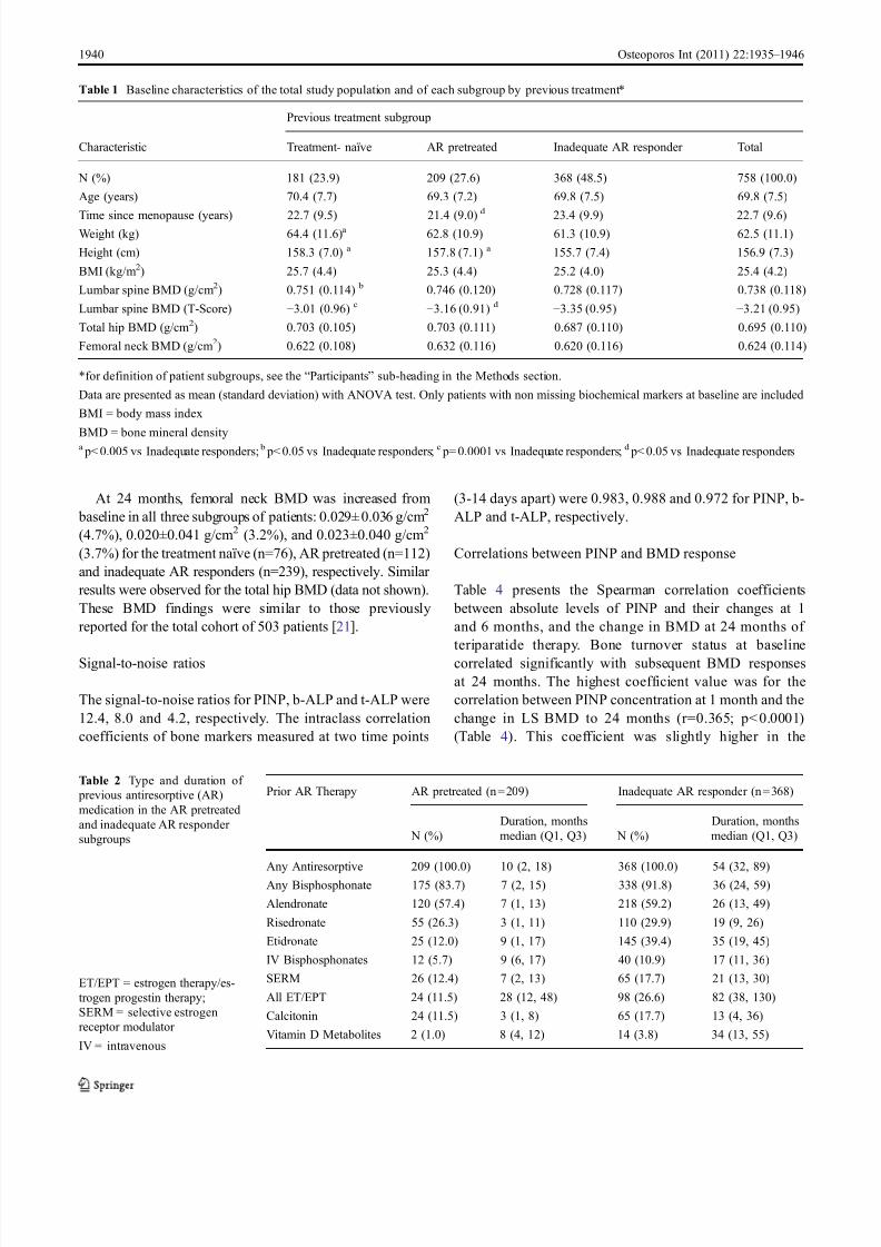
Baseline characteristics
The baseline characteristics of the 758 patients by previous
antiresorptive treatment subgroup are given in Table 1. The
three subgroups did not differ in age, BMI, or BMD at the
hip. Pairwise comparisons showed that LS BMD and height
were significantly lower in the inadequate AR responder
group than in the other two groups (Table 1). We also
observed some variability in weight, height and years since
menopause among the subgroups, but these differences are
probably a consequence of the non-randomized way the
patients were assigned to the subgroups.
Table 2 summarizes the type and duration of previous
antiresorptive medications. Among the AR pretreated
group, 83.7% used a bisphosphonate for a median of
7 months, whereas 91.8% of inadequate AR responders had
used a bisphosphonate for a median of 36 months. The
median lag time between stopping the last antiresorptive
treatment and starting teriparatide was 28 days (interquartile
range: 18−115 days) for the AR pretreated subgroup, and
29 days (interquartile range: 17−56 days) for the inadequate
AR responder subgroup.
1938 Osteoporos Int (2011) 22:1935 – 1946

7/31/2019 Blumsohn Et Al_Teriparatide _ Bone Markers (Eurofors)_OI(June2011)
http://slidepdf.com/reader/full/blumsohn-et-alteriparatide-bone-markers-euroforsoijune2011 5/12
Bone formation markers response to teriparatide
Table 3 shows the bone marker values at baseline, 1 month
and 6 months in the three subgroups. Pairwise comparisons
showed that both the AR pretreated and inadequate AR
responder groups had significantly lower baseline values of
bone markers than the treatment-naïve group. In response to
teriparatide treatment, serum levels of PINP, b-ALP and t-
ALP increased significantly in all subgroups at 1 and
6 months. MMRM analysis showed that the concentrations
of bone markers differed among the subgroups (Table 3).
Thus, at 1 month, there were no significant differences
between AR pretreated and inadequate AR responders for
any of the bone markers, but these two subgroups had PINP
values approximately 30% lower and b-ALP values
approximately 15% lower than the treatment-naïve patients.
However, by 6 months, there were no significant differ-
ences between the treatment-naïve and previously treated
subgroups for any of the bone formation markers (Table 3).
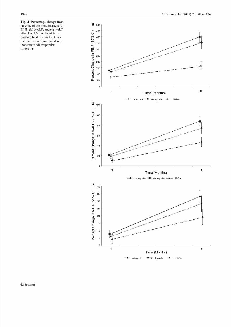
Figure 2 shows the percentage change from baseline for
each of the three bone markers in the three subgroups.
Because of baseline differences in bone marker levels due
to prior AR treatment, calculation as percentage change
provides a different impression of the data. This should be
taken into account when interpreting such data.
The analysis of the bone marker results in the per
protocol population (n =651) yielded similar results to the
full analysis cohort.
BMD response to teriparatide
The mean percent increase in lumbar spine BMD from
baseline to 24 months in the analyzed cohort was, on
average, 10.3% for the total group of teriparatide-treated
patients. The absolute change (mean ± SD) in lumbar spine
BMD from baseline was 0.097±0.052 g/cm2 (13.1%) in
the treatment-naïve subgroup (n=80), 0.077±0.048 g/cm2
(10.7%) in the AR pretreated subjects (n =115), and 0.068
±0.049 g/cm2 (9.4%) in the inadequate AR responder
group (n=245).
Screened
N=1169
Evaluable
N=866
Enrolled
N=868
1 post-baseline bone marker measurement
N=758
Not eligible
N=301
No post-baseline data
N=2
Treatment naive
N=181AR pretreated
N=209
Inadequate ARresponders
N=368
Assigned to 2nd year ofteriparatide
N=125
Completed 2nd year onteriparatide
N=116
Discontinued 1st year
N=29
Patient decision (n=12)
Adverse event (n=7)
Entry criteria not met (n=4)
Protocol violation (n=2)
Death (n=1)
Sponsor decision (n=1)
Physician decision (n=1)
Noncompliance (n=1)
Discontinued 1st year
N=31
Patient decision (n=10)
Entry criteria not met (n=8)
Adverse event (n=7)
Sponsor decision (n=2)
Death (n=1)
Lost to follow-up (n=1)
Moved away (n=1)
Physician decision (n=1)
Discontinued 1st year
N=44
Adverse event (n=21)
Patient decision (n=15)
Entry criteria not met (n=3)
Physician decision (n=2)
Death (n=1)
Sponsor decision (n=1)
Noncompliance (n=1)
Assigned to 2nd year ofteriparatide
N=84
Assigned to 2nd year ofteriparatide
N=259
Completed 2nd year onteriparatide
N=81
Completed 2nd year onteriparatide
N=246
Discontinued 2ndyear
N=3
Death (n=1)
Moved away (n=1)
Noncompliance (n=1)
Discontinued 2ndyear
N=9
Patient decision (n=7)
Adverse event (n=1)
Physician decision (n=1)
Discontinued 2ndyear
N=13
Adverse event (n=7)
Patient decision (n=3)
Death (n=2)
Protocol violation (n =1)
Assigned to othertreatments
N=55
Assigned to othertreatments
N=66
Assigned to other
treatmentsN=65
Fig. 1 Patient disposition
Osteoporos Int (2011) 22:1935 – 1946 1939
7/31/2019 Blumsohn Et Al_Teriparatide _ Bone Markers (Eurofors)_OI(June2011)
http://slidepdf.com/reader/full/blumsohn-et-alteriparatide-bone-markers-euroforsoijune2011 6/12
At 24 months, femoral neck BMD was increased from
baseline in all three subgroups of patients: 0.029± 0.036 g/cm2
(4.7%), 0.020±0.041 g/cm2 (3.2%), and 0.023±0.040 g/cm2
(3.7%) for the treatment naïve (n=76), AR pretreated (n=112)
and inadequate AR responders (n=239), respectively. Similar
results were observed for the total hip BMD (data not shown).
These BMD findings were similar to those previously
reported for the total cohort of 503 patients [21].
Signal-to-noise ratios
The signal-to-noise ratios for PINP, b-ALP and t-ALP were
12.4, 8.0 and 4.2, respectively. The intraclass correlation
coefficients of bone markers measured at two time points
(3-14 days apart) were 0.983, 0.988 and 0.972 for PINP, b-
ALP and t-ALP, respectively.
Correlations between PINP and BMD response
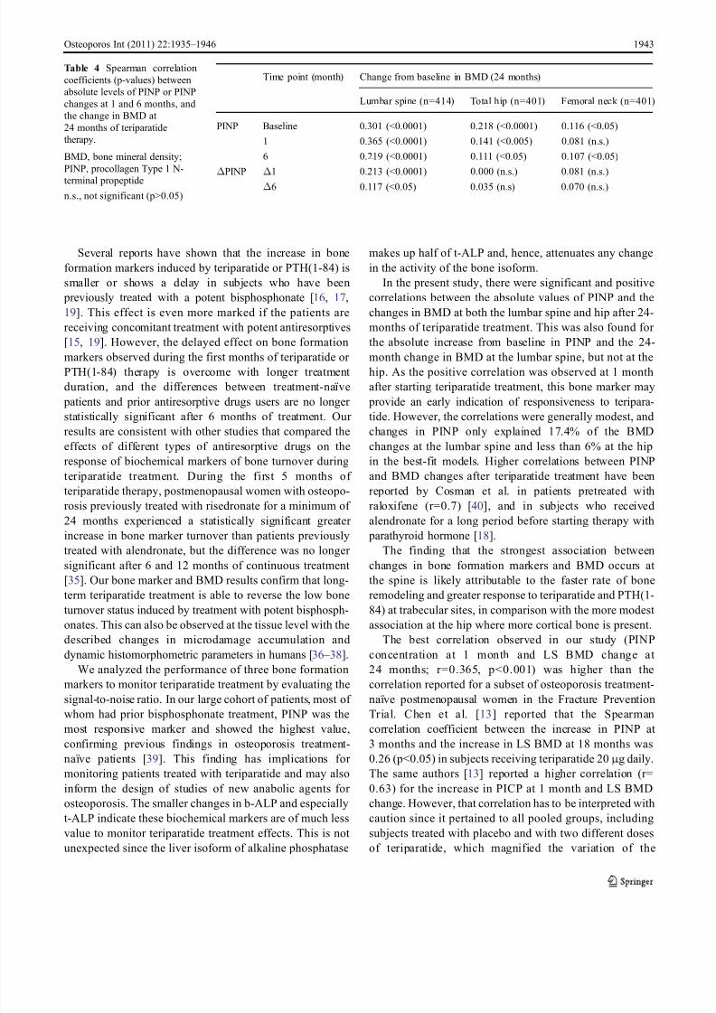
Table 4 presents the Spearman correlation coefficients
between absolute levels of PINP and their changes at 1
and 6 months, and the change in BMD at 24 months of
teriparatide therapy. Bone turnover status at baseline
correlated significantly with subsequent BMD responses
at 24 months. The highest coefficient value was for the
correlation between PINP concentration at 1 month and the
change in LS BMD to 24 months (r=0.365; p< 0.0001)
(Table 4). This coefficient was slightly higher in the
Table 1 Baseline characteristics of the total study population and of each subgroup by previous treatment*
Previous treatment subgroup
Characteristic Treatment- naïve AR pretreated Inadequate AR responder Total
N (%) 181 (23.9) 209 (27.6) 368 (48.5) 758 (100.0)
Age (years) 70.4 (7.7) 69.3 (7.2) 69.8 (7.5) 69.8 (7.5)
Time since menopause (years) 22.7 (9.5) 21.4 (9.0)d
23.4 (9.9) 22.7 (9.6)Weight (kg) 64.4 (11.6)a 62.8 (10.9) 61.3 (10.9) 62.5 (11.1)
Height (cm) 158.3 (7.0) a 157.8 (7.1) a 155.7 (7.4) 156.9 (7.3)
BMI (kg/m2) 25.7 (4.4) 25.3 (4.4) 25.2 (4.0) 25.4 (4.2)
Lumbar spine BMD (g/cm2) 0.751 (0.114) b 0.746 (0.120) 0.728 (0.117) 0.738 (0.118)
Lumbar spine BMD (T-Score) −3.01 (0.96) c−3.16 (0.91) d
−3.35 (0.95) −3.21 (0.95)
Total hip BMD (g/cm2) 0.703 (0.105) 0.703 (0.111) 0.687 (0.110) 0.695 (0.110)
Femoral neck BMD (g/cm2) 0.622 (0.108) 0.632 (0.116) 0.620 (0.116) 0.624 (0.114)
*for definition of patient subgroups, see the “Participants” sub-heading in the Methods section.
Data are presented as mean (standard deviation) with ANOVA test. Only patients with non missing biochemical markers at baseline are included
BMI = body mass index
BMD = bone mineral densitya p< 0.005 vs Inadequate responders; b p< 0.05 vs Inadequate responders; c p= 0.0001 vs Inadequate responders; d p< 0.05 vs Inadequate responders
Prior AR Therapy AR pretreated (n = 209) Inadequate AR responder (n = 368)
Duration, months Duration, months
N (%) median (Q1, Q3) N (%) median (Q1, Q3)
Any Antiresorptive 209 (100.0) 10 (2, 18) 368 (100.0) 54 (32, 89)
Any Bisphosphonate 175 (83.7) 7 (2, 15) 338 (91.8) 36 (24, 59)
Alendronate 120 (57.4) 7 (1, 13) 218 (59.2) 26 (13, 49)
Risedronate 55 (26.3) 3 (1, 11) 110 (29.9) 19 (9, 26)
Etidronate 25 (12.0) 9 (1, 17) 145 (39.4) 35 (19, 45)
IV Bisphosphonates 12 (5.7) 9 (6, 17) 40 (10.9) 17 (11, 36)
SERM 26 (12.4) 7 (2, 13) 65 (17.7) 21 (13, 30)
All ET/EPT 24 (11.5) 28 (12, 48) 98 (26.6) 82 (38, 130)
Calcitonin 24 (11.5) 3 (1, 8) 65 (17.7) 13 (4, 36)
Vitamin D Metabolites 2 (1.0) 8 (4, 12) 14 (3.8) 34 (13, 55)
Table 2 Type and duration of
previous antiresorptive (AR)
medication in the AR pretreated
and inadequate AR responder
subgroups
ET/EPT = estrogen therapy/es-
trogen progestin therapy;
SERM = selective estrogen
receptor modulator
IV = intravenous
1940 Osteoporos Int (2011) 22:1935 – 1946

7/31/2019 Blumsohn Et Al_Teriparatide _ Bone Markers (Eurofors)_OI(June2011)
http://slidepdf.com/reader/full/blumsohn-et-alteriparatide-bone-markers-euroforsoijune2011 7/12
subgroup of osteoporosis treatment-naïve patients (r=
0.405; p<0.0001) (data not shown). The coefficient values
were lower for the changes in total hip and femoral neck
BMD (Table 4).
The best-fit model for predicting change from baseline in
LS BMD for all patients contained prior duration of
antiresorptive treatment, increases in PINP after 1 month,
and PINP concentrations at 1 and 6 months, and accounted
for 17.4% of the total variation in change in LS BMD to
24 months. In this model, prior duration of antiresorptive
treatment was negatively associated with BMD changes at
the LS, as previously described [21]. The different models
explored for predicting change from baseline in total hip or
femoral neck BMD to 24 months accounted for a maximum
of 5.6% of the total variation in the best-fit model which
included duration of prior antiresorptive treatment and
PINP concentration at 1 month.
Forty-nine subjects experienced an incident fracture
during follow-up. No relationship between baseline levels
or changes in PINP concentrations after 1 and 6 months of
treatment with teriparatide and the overall risk of clinical
fractures was found (p>0.05).
Discussion
Our results showed that teriparatide 20 μ g/day was
associated with significant early increases in biochemical
markers of bone formation at 1 month, and that these
changes were increased further after 6 months of
therapy. The increases in bone markers occurred regard-
less of previous antiresorptive therapy, although the
absolute values after 1 month of teriparatide treatment
were lower in subjects who had received previous
antiresorptive therapy than in treatment-naïve subjects.
This delayed increase in bone formation markers in
pretreated patients is likely associated with the bone
turnover inhibition induced by long-term antiresorptive
therapy as shown with the lower values of the bone
markers at the baseline visit compared with treatment-
naïve individuals. However, as early as 6 months,
teriparatide overcomes the inhibition of bone remodel-
ling induced by prior antiresorptive therapy.
Previous studies investigated the changes in various
biochemical markers of bone turnover during treatment
with teriparatide or PTH(1-84) in osteoporosis treatment-
naïve subjects. They reported significant increases in bone
formation markers as early as 1 month after starting
teriparatide or PTH(1-84) therapy in postmenopausal
women with osteoporosis [11, 13, 14, 29 – 31], in patients
with glucocorticoid-induced osteoporosis [10, 32], and in
men with idiopathic and hypogonadal osteoporosis receiv-
ing teriparatide [17, 33, 34]. The changes in PINP, b-ALP
and t-ALP during the first 6 months of teriparatide
treatment in the present study are consistent with those
reported previously in treatment-naïve subjects.
Table 3 Mean bone marker valuesa (95% confidence intervals) at baseline, 1 month and 6 months in the treatment naïve, AR pretreated and
inadequate AR responder subgroups
Treatment naive AR pretreated Inadequate AR responder p-value b
AR pretreated
vs. naive
Inadequate
AR responder
vs. naive
AR pretreated
vs. inadequate
AR responder
PINP (μ g/L)
Baseline 48.2 (43.8 – 53.1) 26.1(23.8 – 28.5) 27.5 (25.7 – 29.4) <0.0001 <0.0001 0.363
1 month 85.5 (78.0 – 93.6) 56.6 (52.0 – 61.6) 62.2 (58.4 – 66.3) <0.0001 <0.0001 0.079
6 months 129.1 (116.1 – 143.5) 118.2 (106.9 – 130.6) 136.6 (126.8 – 147.2) 0.235 0.387 0.022
b-ALP (μ g/L)
Baseline 12.9 (12.1 – 13.7) 10.1 (9.6 – 10.7) 10.2 (9.8 – 10.7) <0.0001 <0.0001 0.775
1 month 14.3 (13.5 – 15.2) 12.0 (11.4 – 12.7) 12.4 (11.9 – 12.9) <0.0001 <0.0001 0.374
6 months 18.9 (17.6 – 20.3) 17.6 (16.5 – 18.8) 19.2 (18.3 – 20.2) 0.152 0.749 0.045
t-ALP (μ g/L)
Baseline 69.6 (66.5 – 72.9) 64.1 (61.4 – 66.9) 63.3 (61.3 – 65.4) 0.010 0.001 0.655
1 month 72.5 (69.4 – 75.7) 67.9 (65.2 – 70.8) 68.0 (65.9 – 70.1) 0.034 0.019 0.976
6 months 82.9 (79.0 – 87.0) 82.1 (78.5 – 85.9) 84.1 (81.3 – 87.0) 0.777 0.630 0.407
a Adjusted by baseline P1NP concentration and BMD values, and duration of prior AR treatment b MMRM of log-transformed data
AR = antiresorptive; PINP = procollagen Type 1 N-terminal propeptide; b-ALP = bone-specific alkaline phosphatase; t-ALP = total alkaline
phosphatase
Osteoporos Int (2011) 22:1935 – 1946 1941
7/31/2019 Blumsohn Et Al_Teriparatide _ Bone Markers (Eurofors)_OI(June2011)
http://slidepdf.com/reader/full/blumsohn-et-alteriparatide-bone-markers-euroforsoijune2011 8/12
Adequate inadequate Naïve
0
50
100
150
200
250
300
350
400
450
500
1 6
P e r c e n t C h a n g e
i n P I N P ( 9 5 % C
I )
Time (Months)
0
20
40
60
80
100
120
P e r c e n t C h a n g e i n b - A L P ( 9 5 % C
I )
1 6
Adequate inadequate Naïve
Time (Months)
0
5
10
15
20
25
30
35
40
P e r c e n t
C h a n g e i n t - A L P ( 9 5 % C
I )
1 6
Adequate inadequate Naïve
Time (Months)
c
b
aFig. 2 Percentage change from
baseline of the bone markers (a)
PINP, (b) b-ALP, and (c) t-ALP
after 1 and 6 months of teri-
paratide treatment in the treat-
ment naïve, AR pretreated and
inadequate AR responder
subgroups
1942 Osteoporos Int (2011) 22:1935 – 1946
7/31/2019 Blumsohn Et Al_Teriparatide _ Bone Markers (Eurofors)_OI(June2011)
http://slidepdf.com/reader/full/blumsohn-et-alteriparatide-bone-markers-euroforsoijune2011 9/12
Several reports have shown that the increase in bone
formation markers induced by teriparatide or PTH(1-84) is
smaller or shows a delay in subjects who have been
previously treated with a potent bisphosphonate [16, 17,
19]. This effect is even more marked if the patients are
receiving concomitant treatment with potent antiresorptives
[15, 19]. However, the delayed effect on bone formation
markers observed during the first months of teriparatide or PTH(1-84) therapy is overcome with longer treatment
duration, and the differences between treatment-naïve
patients and prior antiresorptive drugs users are no longer
statistically significant after 6 months of treatment. Our
results are consistent with other studies that compared the
effects of different types of antiresorptive drugs on the
response of biochemical markers of bone turnover during
teriparatide treatment. During the first 5 months of
teriparatide therapy, postmenopausal women with osteopo-
rosis previously treated with risedronate for a minimum of
24 months experienced a statistically significant greater
increase in bone marker turnover than patients previouslytreated with alendronate, but the difference was no longer
significant after 6 and 12 months of continuous treatment
[35]. Our bone marker and BMD results confirm that long-
term teriparatide treatment is able to reverse the low bone
turnover status induced by treatment with potent bisphosph-
onates. This can also be observed at the tissue level with the
described changes in microdamage accumulation and
dynamic histomorphometric parameters in humans [36 – 38].
We analyzed the performance of three bone formation
markers to monitor teriparatide treatment by evaluating the
signal-to-noise ratio. In our large cohort of patients, most of
whom had prior bisphosphonate treatment, PINP was the
most responsive marker and showed the highest value,
confirming previous findings in osteoporosis treatment-
naïve patients [39]. This finding has implications for
monitoring patients treated with teriparatide and may also
inform the design of studies of new anabolic agents for
osteoporosis. The smaller changes in b-ALP and especially
t-ALP indicate these biochemical markers are of much less
value to monitor teriparatide treatment effects. This is not
unexpected since the liver isoform of alkaline phosphatase
makes up half of t-ALP and, hence, attenuates any change
in the activity of the bone isoform.
In the present study, there were significant and positive
correlations between the absolute values of PINP and the
changes in BMD at both the lumbar spine and hip after 24-
months of teriparatide treatment. This was also found for
the absolute increase from baseline in PINP and the 24-
month change in BMD at the lumbar spine, but not at thehip. As the positive correlation was observed at 1 month
after starting teriparatide treatment, this bone marker may
provide an early indication of responsiveness to teripara-
tide. However, the correlations were generally modest, and
changes in PINP only explained 17.4% of the BMD
changes at the lumbar spine and less than 6% at the hip
in the best-fit models. Higher correlations between PINP
and BMD changes after teriparatide treatment have been
reported by Cosman et al. in patients pretreated with
raloxifene (r=0.7) [40], and in subjects who received
alendronate for a long period before starting therapy with
parathyroid hormone [18].The finding that the strongest association between
changes in bone formation markers and BMD occurs at
the spine is likely attributable to the faster rate of bone
remodeling and greater response to teriparatide and PTH(1-
84) at trabecular sites, in comparison with the more modest
association at the hip where more cortical bone is present.
The best correlation observed in our study (PINP
concentration at 1 month and LS BMD change at
24 months; r=0.365, p<0.001) was higher than the
correlation reported for a subset of osteoporosis treatment-
naïve postmenopausal women in the Fracture Prevention
Trial. Chen et al. [13] reported that the Spearman
correlation coefficient between the increase in PINP at
3 months and the increase in LS BMD at 18 months was
0.26 (p<0.05) in subjects receiving teriparatide 20 μ g daily.
The same authors [13] reported a higher correlation (r=
0.63) for the increase in PICP at 1 month and LS BMD
change. However, that correlation has to be interpreted with
caution since it pertained to all pooled groups, including
subjects treated with placebo and with two different doses
of teriparatide, which magnified the variation of the
Time point (month) Change from baseline in BMD (24 months)
Lumbar spine (n=414) Total hip (n=401) Femoral neck (n=401)
PINP Baseline 0.301 (<0.0001) 0.218 (<0.0001) 0.116 (<0.05)
1 0.365 (<0.0001) 0.141 (<0.005) 0.081 (n.s.)
6 0.219 (<0.0001) 0.111 (<0.05) 0.107 (<0.05)
ΔPINP Δ1 0.213 (<0.0001) 0.000 (n.s.) 0.081 (n.s.)
Δ6 0.117 (<0.05) 0.035 (n.s) 0.070 (n.s.)
Table 4 Spearman correlation
coefficients (p-values) between
absolute levels of PINP or PINP
changes at 1 and 6 months, and
the change in BMD at
24 months of teriparatide
therapy.
BMD, bone mineral density;
PINP, procollagen Type 1 N-
terminal propeptide
n.s., not significant (p>0.05)
Osteoporos Int (2011) 22:1935 – 1946 1943
7/31/2019 Blumsohn Et Al_Teriparatide _ Bone Markers (Eurofors)_OI(June2011)
http://slidepdf.com/reader/full/blumsohn-et-alteriparatide-bone-markers-euroforsoijune2011 10/12
measured change and, hence, increased the correlation
coefficient. In another analysis with the full-length peptide
PTH(1-84) in patients from the PaTH trial, Bauer et al. [29]
showed that each standard deviation (SD) increase in 3-
month change in PINP was positively associated with a
4.0% increase in spine BMD and a 1.3% increase in hip
BMD measured using DXA. These associations were even
more striking when BMD changes were measured byquantitative computerized tomography (QCT): thus, each
SD increase in the 3-month change in PINP was associated
with a 21.2% increase in spine QCT trabecular BMD and a
7.0% increase in hip QCT trabecular BMD.
There are several limitations of this study. First, it was
open-label and did not include a placebo or control group.
However, biochemical markers of bone turnover and BMD
are unlikely to be influenced by a lack of blinding.
Moreover, the central laboratory personnel who performed
the analyses were blind to the patients’ treatment assign-
ments and previous medication history. Second, because
data on prior osteoporosis treatments were obtainedretrospectively at baseline, we do not have accurate details
on adherence and compliance to those treatments. Third,
only bone formation markers and not bone resorption
markers were measured; therefore, we do not get a full
picture of bone turnover. Fourth, the number of fractures
observed in this cohort was small. Thus, the lack of a
significant relationship between changes in biochemical
markers and fracture risk should be interpreted with
caution. Further studies are needed to define the role of
biochemical markers as predictors of fracture risk during
teriparatide therapy. Finally, the subjects of this study were
not randomized to the three analysis subgroups, which
represent observational cohorts.
The strength of this study lies in its external validity. We
included women with severe postmenopausal osteoporosis
regardless of prior antiresorptive treatment and their
response (or lack of response) to it. By keeping the
inclusion and exclusion criteria broad, it was possible to
recruit almost all women for whom teriparatide was
indicated, thereby assembling a study cohort whose
properties are similar to those of patients suitable for
treatment with teriparatide in routine care. Of note, we only
analyzed patients who had stopped their prior antiresorptive
therapy before starting teriparatide; therefore, our results
may differ from those studies where patients continued the
antiresorptive concomitantly with teriparatide [15, 19].
In conclusion, teriparatide treatment is associated with a
significant increase in biochemical markers of bone
formation at 1 and 6 months. The bone formation marker
response in patients does not seem to be adversely
influenced by prior antiresorptive therapy, and can be
detected at 1 month of therapy. After 6 months of treatment,
bone formation markers are at a similar level regardless of
prior osteoporosis treatment. Although indices of bone
formation or change in formation were only modestly
predictive of change in BMD at the spine or total hip at
24 months, and were not correlated with fracture outcomes,
PINP appears to be the most sensitive bone marker to
assess a therapeutic response to teriparatide.
Acknowledgements The authors thank Simon Cleall, MSc, for providing statistical advice; Deirdre Elmhirst, PhD, Elmhirst Medical
Writing Services, for assisting in the formatting and language of the
manuscript, and the following individuals for providing technical
assistance in conducting the study: Petra Ochs (study coordination),
Ruth Alonso, David López, and Laura Briones (data management),
and Alain Frix (study drug coordination). Dr Elmhirst ’s work on the
manuscript was funded by the study sponsor. Steve Boonen is senior
clinical investigator of the Fund for Scientific Research and is holder
of the Leuven University Chair in Metabolic Bone Diseases. The
authors thank the women who participated in this study; the doctors,
study nurses, and support staff at the local sites; and the monitors and
study managers in the participating countries. Funding was provided
by Lilly Research Center, Europe
Conflicts of interest AB received funding from Eli Lilly to performassays of bone turnover for this study. He has no other conflicts of
interest and has received no personal funding from any pharmaceutical
or diagnostic company. KB has served as consultant, received research
grants from and has served on speakers’ bureau for Eli Lilly. SB has
received research funding and consulting fees from Eli Lilly. RE has
previously consulted and received lecture fees from Eli Lilly and
received grant support from 1998 to 2005. FM, TN, CB, SL-L are
employees of Eli Lilly. GS, JG have nothing to declare.
Open Access This article is distributed under the terms of the
Creative Commons Attribution Noncommercial License which per-
mits any noncommercial use, distribution, and reproduction in any
medium, provided the original author(s) and source are credited.
Appendix: EUROFORS principal investigators
Austria: B. Obermayer-Pietsch, Lkh-Universitätsklinikum
Graz; L. Erlacher, Krankenhaus der Elisabethinen, Klagenfurt;
G. Finkenstedt, Landeskrankenhaus-Universitätskliniken,
Innsbruck; Belgium: P. Geusens, Limburgs Universitair
Centrum, Diepenbeek; F. Raeman, Jan Palfijn Ziekenhuis,
Merksem; F. van den Bosch, Elisabethziekenhuis, Damme; Y.
Boutson, Cliniques Universitaires de Mont Godinne, Yvoir;
J.-M. Kaufman, Universitair Ziekenhuis Gent; S. Boonen,
Universitair Ziekenhuis Gasthuisberg Leuven; Denmark: K.
Brixen, University Hospital, Odense; B. Langdahl, Aarhus
Amtssygehus; J.-E. B. Jensen, Hvidovre Hospital; Hvidovre;
France: M. Audran, CHU d’Angers; C. Alexandre, Hôpital
Bellevue, Saint Etienne; C. Roux, Hôpital Cochin, Paris; C.L.
Benhamou, Hôpital Porte Madeleine, Orleans; C. Ribot,
Hôpital Paule de Viguier, Toulouse; C. Cormier, Hôpital
Cochin, Paris; J-L. Kuntz, Hôpital de Hautepierre, Strasbourg;
A. Daragon, CHU de Bois Guillaume, Rouen; B. Cortet,
1944 Osteoporos Int (2011) 22:1935 – 1946
7/31/2019 Blumsohn Et Al_Teriparatide _ Bone Markers (Eurofors)_OI(June2011)
http://slidepdf.com/reader/full/blumsohn-et-alteriparatide-bone-markers-euroforsoijune2011 11/12
Hôpital Roger Salengro, Lille; M. Laroche, Hôpital de
Rangueil, Toulouse; M.C. de Vernejoul, Hôspital Lariboisiere,
Paris; P. Fardellone, Hôpital Sud, Amiens; G. Weryha, Chu de
Nancy Hôpital D’Adultes de Brabois, Vandoeuvre Les
Nancy; Germany: H.W. Minne, Klinik - Der Fürstenhof,
Bad Pyrmont; H-J. Heberling, Robert-Koch-Klinik,
Leipzig; K. Badenhoop, Klinikum der Johann Wolfgang
Goethe-Universität Frankfurt; H.G. Fritz, Berlin; J.Kekow, Krankenhaus Vogelsang, Vogelsang/Gommern;
H . M o en i g , K l i ni k u m d e r C h r is t i an - A l br e c ht s -
Universitäts zu Kiel; T. Brabant, Krankenhaus St. Josef
Stift Bremen; H-P. Kruse, Univeritäts-Krankenhaus
Eppendorf, Hamburg; W. Spieler, Zefor, Zerbst; R.
Möricke, Magdeburg; A. Wagenitz, Berlin; F. Flohr,
Universitätsklinikum Freiburg; J. Semler, Immanuel
Krankenhaus Rheuma Klinik Berlin Wannsee; P. Hadji,
Klinikum der Phillips- Universität, Marburg; P. Kaps,
Braunfels; T. Hennigs, Osteoporose Studiengesellschaft
bR, Frankfurt; R.R. Fritzen, Med.Klinik für Endokrino-
logie des Universitätsklinikums Düsseldorf; J. Feldkamp,Städtische Kliniken, Bielefeld; G. Hein, Klinikum der
Friedrich-Schiller-Universität, Jena; U. Haschke, Osnabrück;
C. Kasperk, Universitätsklinkum Heidelberg; J.D. Ringe,
Klinikum Leverkusen; H. Radspieler, Osteoporose-Diagnostik
und Therapiezentrum München; N. Vollmann, München; E.
Blind, Klinikum der Universität Würzburg; M. Runge, Aerpah-
Klinik Esslingen-Kennenburg; F. Jakob, Orthopädische Klinik
König-Ludwig-Haus, Würzburg; H-G. Dammann, Klinikische
Forschung Hamburg; S. Scharla, Bad Reichenhall; Greece: G.
Lyritis, K.A.T. Hospital Of Athens, Kifissia; A. Avramides,
Ippokratio Hospital, Thessaloniki; Iceland: G. Sigurdsson,
Landspitalinn Haskólasjúkrahús, Reykjavik; B. Gudbjörnsson,
Fjordungssjukrahusid Akureyri; Portugal: M.E. Simões,
Instituto Portugues De Reumatologia, Lisboa; J. Melo-
Gomes, Servimed, Lisboa; J.C. Branco, Hospital Egas Moniz,
Lisboa; A. Malcata, Hospitais da Universidade, Coimbra;
Spain: C. Díaz-Lopez, J. Farrerons, Hospital Santa Creu i
Sant Pau, Barcelona; J. González de la Vera , H.U. Virgen
Macarena, Sevilla; J.A. Román, H.U. Dr. Pesset, Valencia; X.
Sans, Ciutat Sanitaria Vall D’Hebron, Barcelona; A. Laffón
Hospital de la Princesa, Madrid; E. Rejón, H.U. Nuestra
Señora de Valme, Sevilla; J. del Pino, Hospital Clínico,
Salamanca; J. de Toro, Hospital Juan Canalejo, A Coruña; J.
Babio, Hospital de Cabueñes, Gijón; C. González, Hospital
Gregorio Marañón, Madrid; United Kingdom: C. Cooper,
University of Southampton; I. Fogelman, Kings’ College,
London; S. Doherty, D. Purdie, Hull and East Yorkshire
Hospitals NHS Trust; D. Reid, Grampian University Hospitals
NHS Trust; M. Stone, Cardiff and Vale NHS Trust; S. Orme,
P. Belchetz, Leeds Teaching Hospital NHS Trust; R. Eastell,
University of Sheffield; W. Fraser, University of Liverpool; D.
Hosking, Nottingham City Hospital NHS Trust; T. O’ Neill,
Salford Hospital NHS Trust; J. Compston, J. Reeve,
Addenbrookes NHS Trust; K. Adams, Bolton Hospitals
NHS Trust; H. Taggart, Belfast City Hospitals Trust; A.
Bhalla, Royal National Hospital for Rheumatic Diseases NHS
Trust; M. Brown, Nuffield Orthopaedic Centre NHS Trust; T.
Palferman, East Somerset NHS Trust; A. Woolf, Royal
Cornwall Hospitals NHS Trust; T. Wheatley, Brighton and
Sussex University Hospitals NHS Trust; P. Thompson, Poole
Hospital NHS Trust; R. Keen, Royal National OrthopaedicHospital NHS Trust; P. Ryan, The Medway NHS Trust; P.
Selby, Manchester University Hospitals NHS Trust.
References
1. Dempster DW, Cosman F, Kurland ES, Zhou H, Nieves J,
Woelfert I, Shane E, Plavetic K, Muller R, Bilezikian J, Lindsay
R (2001) Effects of daily treatment with parathyroid hormone on
bone microarchitecture and turnover in patients with osteoporosis:
a paired biopsy study. J Bone Miner Res 16:1846 – 1853
2. Jiang Y, Zhao JJ, Mitlak BH, WangO, Genant HK, Eriksen EF (2003)
Recombinant human parathyroid hormone (1 – 34) [Teriparatide]
improves both cortical and cancellous bone structure. J Bone Miner
Res 18:1932 – 1941
3. Ma YL, Zeng Q, Donley DW, Ste-Marie LG, Gallagher JC,
Dalsky GP, Marcus R, Eriksen EF (2006) Teriparatide increases
bone formation in modeling and remodeling osteons and enhances
IGF-II immunoreactivity in postmenopausal women with osteo-
porosis. J Bone Miner Res 21:855 – 864
4. Lindsay R, Zhou H, Cosman F, Nieves J, Dempster DW, Hodsman
AB (2007) Effects of a one-month treatment with PTH(1 – 34) on
bone formation on cancellous, endocortical, and periosteal surfaces
of the human ilium. J Bone Miner Res 22:495 – 502
5. Keaveny TM, Donley D, Hoffmann PF, Mitlak BH, Glass EV, San
Martin JA (2007) Effects of teriparatide and alendronate on
vertebral strength as assessed by finite element modeling of QCT
scans in women with osteoporosis. J Bone Miner Res 21:149 – 157
6. Zanchetta JR,Bogado CE, Ferretti JL,Wang O, Wilson MG, Sato M,
Gaich GA, Dalsky GP, Myers SL (2003) Effects of teriparatide
[recombinant human parathyroid hormone (1 – 34)] on cortical bone
in postmenopausal women with osteoporosis. J Bone Miner Res
18:539 – 543
7. Sato M, Westmore M, Ma YL, Schmidt A, Zeng QQ, Glass EV, Vahle
J, Brommage R, Jerome CP, Turner CH (2004) Teriparatide [PTH(1 –
34)] strengthens the proximal femur of ovariectomized nonhuman
primates despite increasing porosity. J Bone Miner Res 19:623 – 629
8. Uusi-Rasi K, Semanick LM, Zanchetta JR, Bogado CE, Eriksen
EF, Sato M, Beck TJ (2005) Effects of teriparatide [rhPTH(1 – 34)]
treatment on structural geometry of the proximal femur in elderly
osteoporotic women. Bone 36:948 – 958
9. Neer RM, Arnaud CD, Zanchetta JR, Prince R, Gaich GA,Reginster JY, Hodsman AB, Eriksen EF, Ish-Shalom S, Genant
HK, Wang O, Mitlak BH (2001) Effect of parathyroid hormone
(1 – 34) on fractures and bone mineral density in postmenopausal
women with osteoporosis. N Engl J Med 344:1434 – 1441
10. Saag KG, Shane E, Boonen S, Marin F, Donley DW, Taylor KA,
Dalsky GP, Marcus R (2007) Teriparatide compared with
alendronate for the treatment of glucocorticoid-induced osteopo-
rosis. N Engl J Med 357:2028 – 2039
11. McClung MR, San Martin J, Miller PD, Civitelli R, Bandeira F,
Omizo M, Donley DW, Dalsky GP, Eriksen EF (2005) Opposite
bone remodeling effects of teripar atide and alendro nate in
increasing bone mass. Arch Intern Med 165:1762 – 1768
Osteoporos Int (2011) 22:1935 – 1946 1945
7/31/2019 Blumsohn Et Al_Teriparatide _ Bone Markers (Eurofors)_OI(June2011)
http://slidepdf.com/reader/full/blumsohn-et-alteriparatide-bone-markers-euroforsoijune2011 12/12
12. Canalis E, Giustina A, Bilezikian JP (2007) Mechanisms of
anabolic therapies for osteoporosis. N Engl J Med 357:905 – 916
13. Chen P, Satterwhite JH, Licata AA, Lewiecki EM, Sipos AA, Misurski
DM, Wagman RB (2005) Early changes in biochemical markers of
bone formation predict BMD response to teriparatide in postmeno-
pausal women with osteoporosis. J Bone Miner Res 20:962 – 970
14. Dobnig H, Sipos A, Jiang Y, Fahrleitner-Pammer A, Ste-Marie L-G,
Gallagher JC, Pavo I, Wang J, Eriksen EF (2005) Early changes in
biochemical markers of bone formation correlate with improvements
in bone structure during teriparatide therapy. J Clin EndocrinolMetab 90:3970 – 3977
15. Black DM, Greenspan SL, Ensrud KE, Palermo L, McGowan JA,
Lang TF, Garnero P, Bouxsein ML, Bilezkian JP, Rosen CJ, for
the PaTH Study Investigators (2003) The effects of parathyroid
hormone and alendronate alone or in combination in postmeno-
pausal osteoporosis. N Engl J Med 349:1207 – 1215
16. Ettinger B, San Martin J, Crans G, Pavo I (2004) Differential
effects of teriparatide on BMD after treatment with raloxifene or
alendronate. J Bone Miner Res 19:745 – 751
17. Finkelstein JS, Leder BZ, Burnett SA, Wyland JJ, Lee H, de la
Paz V, Gibson K, Neer RM (2006) Effects of teriparatide,
alendronate, or both on bone turnover in osteoporotic men. J Clin
Endocrinol Metab 91:2882 – 2887
18. Cosman F, Nieves J, Zion M, Woelfert L, Luckey M, Lindsay R
(2005) Daily and cyclic parathyroid hormone in women receiving
alendronate. N Engl J Med 353:566 – 575
19. Cosman F, Wermers RA, Recknor C, Mauck KF, Xie L, Glass EV,
Krege JH (2009) Effects of teriparatide in postmenopausal women
with osteoporosis on prior alendronate or raloxifene: differences
between stopping and continuing the antiresorptive agent. J Clin
Endocrinol Metab 94:3772 – 3780
20. Seibel MJ (2005) Biochemical markers of bone turnover. Part 1:
biochemistry and variability. Clin Biochem Rev 26:97 – 122
21. Obermayer-Pietsch BM, Marin F, McCloskey EV, Hadji P, Farrerons
J, Boonen S, Audran M, Barker C, Anastasilakis AD, Fraser WD,
Nickelsen T, EUROFORS Investigators (2008) Effects of two years
of daily teriparatide treatment on bone mineral density in postmen-
opausal women with severe osteoporosis with and without prior
antiresorptive treatment. J Bone Miner Res 23:1591 – 1600
22. Eastell R, Nickelsen T, Marin F, Barker C, Hadji P, Farrerons J,
Audran M, Boonen S, Brixen K, Melo-Gomes J, Obermayer-Pietsch
BM, Avramidis A, Sigurdsson G, Glüer C-C (2009) Sequential
treatment of severe postmenopausal osteoporosis following teripara-
tide: final results of the randomized, controlled European Study of
Forsteo (EUROFORS). J Bone Miner Res 24:726 – 736
23. Graeff C, Chevalier Y, Charlebois M, Varga P, Pahr D, Nickelsen
TN, Morlock MM, Glueer CC, Zysset PK (2009) Improvements
in vertebral body strength under teriparatide treatment assessed in
vivo by finite element analysis: results from the EUROFORS
Study. J Bone Miner Res 24:1672 – 1680
24. Borggrefe J, Graeff C, Nickelsen TN, Marin F, Glüer CC (2010)
Quantitative computed tomography assessment of the effects of
24 months of teriparatide treatment on 3-D femoral neck bone
distribution, geometry and bone strength: results from the EURO-
FORS study. J Bone Miner Res 25:472 – 481. doi:10.1359/
JBMR.090820
25. Genant HK, Grampp S, Glüer CC, Faulkner KG, Jergas M,
Hagiwara S, van Kuijk C (1994) Universal standardisation for
dual x-ray absorptiometry: patient and phantom cross-calibration
results. J Bone Miner Res 9:1503 – 1514
26. Hanson J (1997) Standardization of femur bone mineral density. J
Bone Miner Res 12:1316 – 1317
27. Graeff C, Timm W, Nickelsen TN, Farrerons J, Marin F, Barker C,
Glüer C-C, for the EUROFORS High Resolution Quantitative
Computed Tomography Substudy Group (2007) Monitoring
teriparatide associated changes in vertebral microstructure by
high-resolution computed tomography in vivo: results from the
EUROFORS study. J Bone Miner Res 22:1426 – 1433
28. Boonen S, Marin F, Obermayer-Pietsch B, Simoes ME, Barker C,
Glass EV, Hadji P, Lyritis G, Oertel H, Nickelsen T, McCloskey EV,
EUROFORS Investigators (2008) Effects of previous antiresorptive
therapy on the bone mineral density response to two years of
teriparatide treatment in postmenopausal women with osteoporosis. J
Clin Endocrinol Metab 93:852 – 86029. Bauer DC, Garnero P, Bilezikian JP, Greenspon SL, Ensrud KE,
Rosen CJ, Palermo L, Black DM, for the PTH and Alendronate
(PaTH) Research Group (2006) Short-term changes in bone
turnover markers and bone mineral density response to parathy-
roid hormone in postmenopausal women with osteoporosis. J Clin
Endocrinol Metab 91:1370 – 1375
30. Black DM, Bilezikian JP, Ensrud KE, Greenspan SL, Palermo L,
Hue T, Lang TF, McGowan JA, Rosen CJ, for the PaTH Study
Investigators (2005) One year of alendronate after one year of
parathyroid hormone (1-84) for osteoporosis. N Engl J Med
353:555 – 565
31. Greenspan SL, Bone HG, Ettinger MP, Hanley DA, Lindsay R,
Zanchetta JR, Blosch CM, Mathisen AL, Morris SA, Marriott TB,
for the Treatment of Osteoporosis with Parathyroid Hormone Study
Group (2007) Effect of recombinant human parathyroid hormone (1-
84) on vertebral fracture and bone mineral density in postmenopausal
women with osteoporosis. Ann Intern Med 146:326 – 339
32. Lane NE, Sanchez S, Genant HK, Jenkins DK, Arnaud CD (2000)
Short-term increases in bone turnover markers predict parathyroid
hormone-induce spinal bone mineral density gains in postmeno-
pausal women with glucocorticoid-induced osteoporosis. Osteo-
poros Int 11:434 – 442
33. Orwoll ES, Scheele WH, Paul S, Adami S, Syversen U, Diez-
Perez A, Kaufman JM, Clancy AD, Gaich GA (2003) The effect
of teriparatide [human parathyroid hormone (1 – 34)] therapy on
bone density in men with osteoporosis. J Bone Miner Res 18:9 – 17
34. Finkelstein JS, Hayes A, Hunzelman JL, Wyland JJ, Lee H, Neer
RM (2003) The effects of parathyroid hormone, alendronate, or
both in men with osteoporosis. N Engl J Med 349:1216 – 1226
35. Miller PD, Delmas PD, Lindsay R, Watts NB, Luckey M, Adachi
J, Saag K, Greenspan SL, Seeman E, Boonen S, Meeves S, Lang
TF, Bilezikian JP (2008) Early responsiveness of women with
osteoporosis to teriparatide after therapy with alendronate or
risedronate. J Clin Endocrinol Metab 93:3785 – 3793
36. Dobnig H, Stepan JJ, Burr DB, Li J, Michalska D, Sipos A, Petto
H, Fahrleitner-Pammer A, Pavo I (2009) Teriparatide reduces
bone microdamage accumulation in postmenopausal women
previously treated with alendronate. J Bone Miner Res 24:1998 –
2006
37. Stepan JJ, Burr DB, Li J, Ma YL, Petto H, Sipos A, Dobnig H,
Fahrleitner-Pammer A, Michalska D, Pavo I (2010) Histomorpho-
metric changes by teriparatide in alendronate-pretreated women with
osteoporosis. Osteoporos Int. doi:10.1007/s00198-009-1168-7
38. Lindsay R, Cosman F, Zhou H, Nieves JW, Bostrom M, Barbuto
N, Dempster DW (2007) Prior alendronate treatment does not
inhibit the early stimulation of osteoblast activity in response to
teriparatide. J Bone Miner Res 22(Suppl):S124, Abstract
39. Eastell R, Krege JH, Chen P, Glass EV, Reginster JY (2006)
Development of an algorithm for using PINP to monitor treatment
of patients with teriparatide. Curr Med Res Opin 22:61 – 66
40. Cosman F, Nieves JW, Zion M, Barbuto N, Lindsay R (2008)
Effect of prior and ongoing raloxifene therapy on response to PTH
and maintenance of BMD after PTH therapy. Osteoporos Int
19:529 – 535
1946 Osteoporos Int (2011) 22:1935 – 1946
Related Documents