International Journal of Computer Engineering and Applications, Volume XII, Issue I, Jan. 18, www.ijcea.com ISSN 2321-3469 Ajit Danti , Kumari K 16 BLOOD REGIONS SEGMENTATION FOR AUTOMATIC BLOOD GROUP IDENTIFICATION Ajit Danti 1 , Kumari K 2 Dept of Computer Applications JNN College of Engineering, Shimoga [email protected] 1 ; [email protected] 2 ABSTRACT: Recently, automatic blood group identification is receiving major attention in the field of medical research. There is not enough work carried out for automation of blood group identification to help the doctors in critical situations .The blood group of an human is mainly classified into 8 groups in that 4 of them are negatives and other four are positive these 8 groups are popularly called as ABO groups .These groups are classified on the basis of reaction with the antigens .The antigens are classified into three categories namely anti-A, anti-B, anti-D.In this paper, the blood groups are detected and segmented on the basis of color & geometrical properties of the regions by using digital image processing approaches for the automation of blood group identification. In the proposed methodology the features from blood sample images are extracted like shape, area, and size of an each region in an image. Experimental result shows the efficiency of the proposed approach. Keywords: Image-processing, Feature extraction, Segmentation, Blood group, Pre processing. [1] INTRODUCTION In recent days, automatic blood group identification is getting very good scope in the medical research. Blood group classification refers to the activity which is on the basis of the blood sample reacted with each antigen. In the context of the reaction the blood group is easily

Welcome message from author
This document is posted to help you gain knowledge. Please leave a comment to let me know what you think about it! Share it to your friends and learn new things together.
Transcript
International Journal of Computer Engineering and Applications, Volume XII, Issue I, Jan. 18, www.ijcea.com ISSN 2321-3469
Ajit Danti , Kumari K 16
BLOOD REGIONS SEGMENTATION FOR AUTOMATIC BLOOD
GROUP IDENTIFICATION
Ajit Danti1, Kumari K2
Dept of Computer Applications JNN College of Engineering, Shimoga
[email protected] 1 ; [email protected]
ABSTRACT:
Recently, automatic blood group identification is receiving major attention in the field of medical research. There is not enough work carried out for automation of blood group identification to help the doctors in critical situations .The blood group of an human is mainly classified into 8 groups in that 4 of them are negatives and other four are positive these 8 groups are popularly called as ABO groups .These groups are classified on the basis of reaction with the antigens .The antigens are classified into three categories namely anti-A, anti-B, anti-D.In this paper, the blood groups are detected and segmented on the basis of color & geometrical properties of the regions by using digital image processing approaches for the automation of blood group identification. In the proposed methodology the features from blood sample images are extracted like shape, area, and size of an each region in an image. Experimental result shows the efficiency of the proposed approach.
Keywords: Image-processing, Feature extraction, Segmentation, Blood group, Pre processing.
[1] INTRODUCTION
In recent days, automatic blood group identification is getting very good scope in the
medical research. Blood group classification refers to the activity which is on the basis of the
blood sample reacted with each antigen. In the context of the reaction the blood group is easily
BLOOD REGIONS SEGMENTATION FOR AUTOMATIC BLOOD GROUP IDENTIFICATION
Ajit Danti , Kumari K 17
classified. In which pre-processing steps are done for the classification of blood groups by
segmenting the regions from a given image, morphological operations are used for classifying
the blood regions based on statistical features such as area, shape, size of the segmented regions.
For detecting the blood groups classification it needs certain human efforts and it is time
consuming, the process of classifying the blood group has following steps. Initially they take
the blood sample of a patient, and is mixed with three types of chemicals namely, AntiA, Anti-
B, Anti-D. Based on that blood sample reacted with these they determine the blood groups.
Blood groups are classified into 8 groups they are, A-Positive, B-Positive, O-Positive, AB-
Positive, A-Negative, B-Negative, O-Negative, AB-Negative
For example: The sample which reacts with Anti-A and Anti-D that sample is belongs to
the A-Positive group. Like that each blood group sample has its own reaction with these three
types of chemicals .The different samples gives the different reaction with different chemicals,
and the all the same samples will gives the same result with these chemicals.
The process of manual blood group identification is time consuming in critical situation
like patients at in serious condition, to overcome this problem it needs another system to
automate this manual process. On the basis of these information’s we segment the regions from
each image by performing the pre-processing steps by using some morphological operations.
[2] LITERATURE SURVEY
In medical field lot of research work is done. For example: Lung cancer detection using
digital Image processing On CT scan Images (Anita Chaudhary and SonitSukhraj Singh,2012),
Leukemia Cancer Cell Detection using Image Processing(Ashwini Rejintal ,Aswini.N,2016),
Brain tumor detection using image processing (Amruta pramod hebli and sudha gupta,2017),
Cancer Cells Detection Using Digital Image Processing Methods (Bhagyashri G. Patil and
Sanjeev N. Jain ,2014), Image processing based abnormal blood cells detection (Deepika N.
Patil1 and Uday P. Khot,2015), Edge Preservation with Noise Reduction in Arthritis Image
(Devkant Sen and Neha Tiwari, 2015), A Study of Various Bone Fracture Detection
Techniques (Irfan Khatik, 2017), Segmentation techniques on mammograms to detect breast
abnormality (Kamalakannan.J and Rajasekhara Babu ,2016),
Analysis and Edge Detection of Lung Cancer – Survey (C. Jeya Bharathi and P. Kabilan, 2016),
Compression And Canny Edge Detection Based Methods For Image
Segmentation(S.Muthuselvi and P.Prabhu,2016), Breast Cancer Mass Detection in
Mammograms using K-means and Fuzzy C-means Clustering (Nalini Singh, Ambarish G
Mohapatra and Gurukalyan Kanungo,2011),Image Processing Based Approach for Automated
Detection of Chronic Leukemia (Neha Sharma and Dr Supreet Kaur,2016), Breast Cancer
Detection using Image Processing Techniques (PrannoyGiri and K.Saravanakumar 2017 ),
Brain Tumor Detection Using 3D Visualization (Prachi Dhikale and Madhuri Joshi,2017),
Human Skin Cancer Recognition and Classification by Unified Skin Texture and Color
Features (Shubhangi D C and Nagaraj ,2013), Cancer Cell Detection Using Mathematical
Morphology (Sivappriya T and Muthukumaran K, 2014 ), Image Segmentation and
Identification of Brain Tumor from MRI Image (Sonam S, Gavhande and S.B.Jadhav,2013),
Image Processing for Identifying Brain Tumor using Intelligent System
International Journal of Computer Engineering and Applications, Volume XII, Issue I, Jan. 18, www.ijcea.com ISSN 2321-3469
Ajit Danti , Kumari K 18
(Somashekhar Swamy and P.K.Kulkarni,2015), Visualization Of 3D View Of Detected Brain
Tumor And Calculation Of Its Volume (Sushma Laxman Wakchaure and Anil Khandekar,
2015), Leukemia Detection using Image Processing (V.Venmathi and et.al,2017).
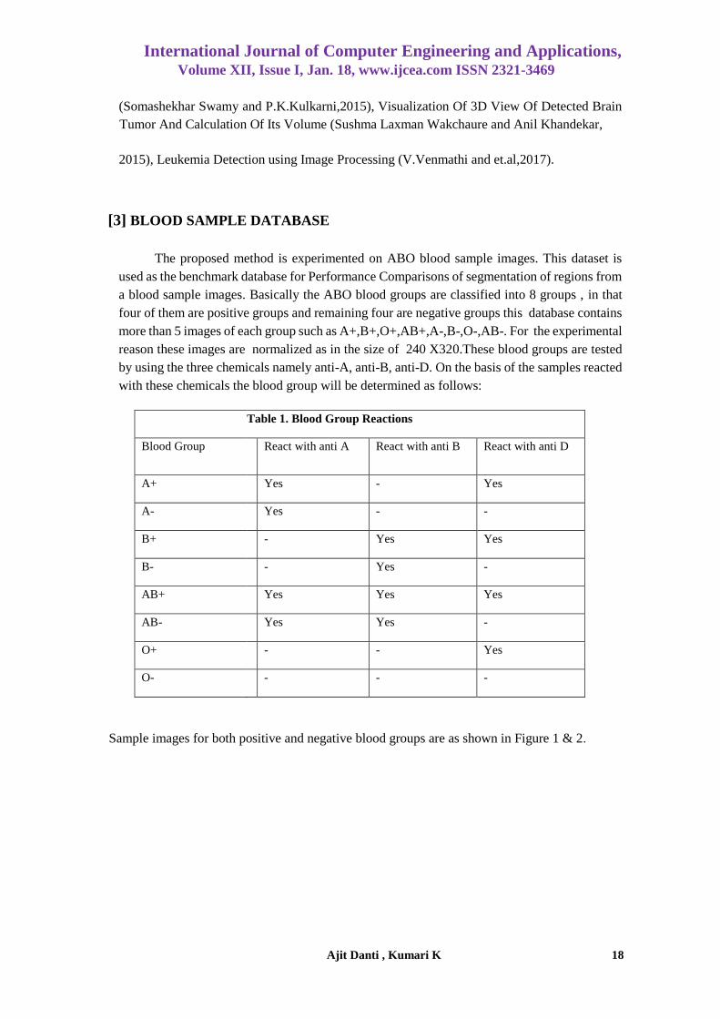
[3] BLOOD SAMPLE DATABASE
The proposed method is experimented on ABO blood sample images. This dataset is
used as the benchmark database for Performance Comparisons of segmentation of regions from
a blood sample images. Basically the ABO blood groups are classified into 8 groups , in that
four of them are positive groups and remaining four are negative groups this database contains
more than 5 images of each group such as A+,B+,O+,AB+,A-,B-,O-,AB-. For the experimental
reason these images are normalized as in the size of 240 X320.These blood groups are tested
by using the three chemicals namely anti-A, anti-B, anti-D. On the basis of the samples reacted
with these chemicals the blood group will be determined as follows:
Table 1. Blood Group Reactions
Blood Group React with anti A React with anti B React with anti D
A+ Yes - Yes
A- Yes - -
B+ - Yes Yes
B- - Yes -
AB+ Yes Yes Yes
AB- Yes Yes -
O+ - - Yes
O- - - -
Sample images for both positive and negative blood groups are as shown in Figure 1 & 2.

BLOOD REGIONS SEGMENTATION FOR AUTOMATIC BLOOD GROUP IDENTIFICATION
Ajit Danti , Kumari K 19
A+ B+ O+ AB+
Fig 1: Sample images of Positive blood groups
A- B- O- AB-
Fig 2: Sample images of Negative blood groups
International Journal of Computer Engineering and Applications, Volume XII, Issue I, Jan. 18, www.ijcea.com ISSN 2321-3469
Ajit Danti , Kumari K 20
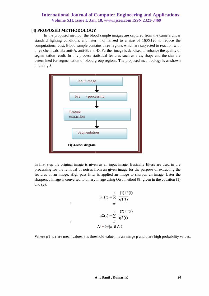
[4] PROPOSED METHODOLOGY
In the proposed method the blood sample images are captured from the camera under
standard lighting conditions and later normalized to a size of 160X120 to reduce the
computational cost. Blood sample contains three regions which are subjected to reaction with
three chemicals like anti-A, anti-B, anti-D. Further image is denoised to enhance the quality of
segmentation result. In this process statistical features such as area, shape and the size are
determined for segmentation of blood group regions. The proposed methodology is as shown
in the fig 3
In first step the original image is given as an input image. Basically filters are used in pre
processing for the removal of noises from an given image for the purpose of extracting the
features of an image. High pass filter is applied an image to sharpen an image. Later the
sharpened image is converted to binary image using Otsu method [8] given in the equation (1)
and (2).
t (1) iP(i)
μ1(t) = ∑
i
t (2) iP(i)
μ2(t) = ∑
i
Ac {w|w ∉ A }
Where μ1 μ2 are mean values, t is threshold value, i is an image p and q are high probability values.
(3)
Fig 3.Block diagram
Input image
Pre - processing
Feature
extraction
Segmentation

BLOOD REGIONS SEGMENTATION FOR AUTOMATIC BLOOD GROUP IDENTIFICATION
Ajit Danti , Kumari K 21
w=pixel value of input image
A= set.
Opening process [8] is performed to remove unwanted regions from an image and also it removes all
connected components that have fewer than P pixels from the binary image using equation (3).
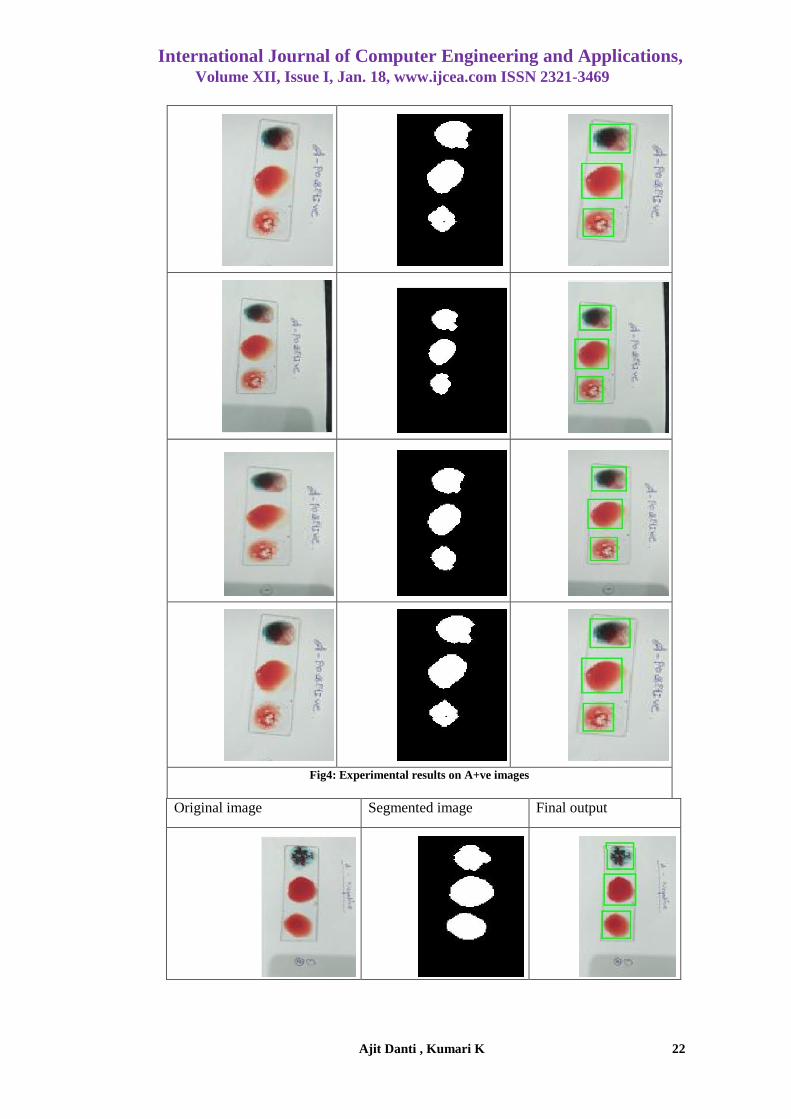
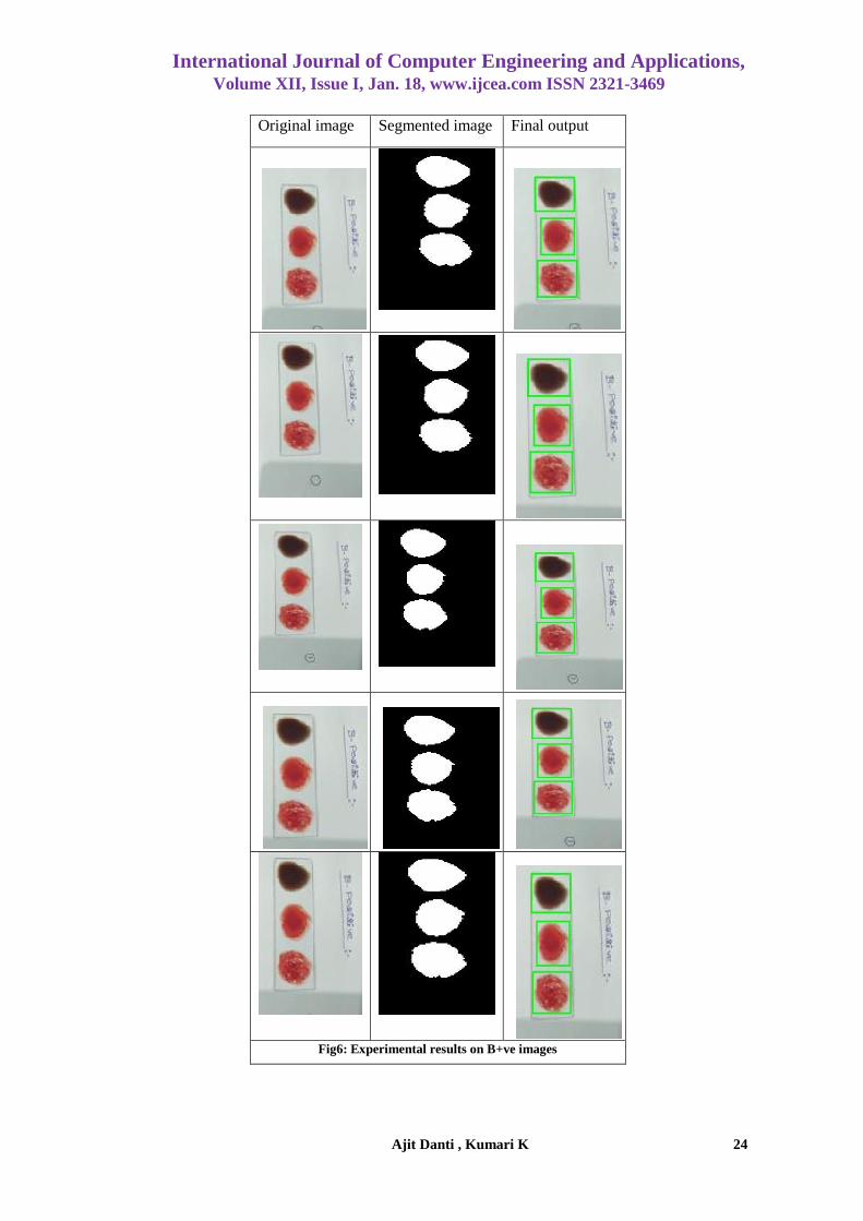
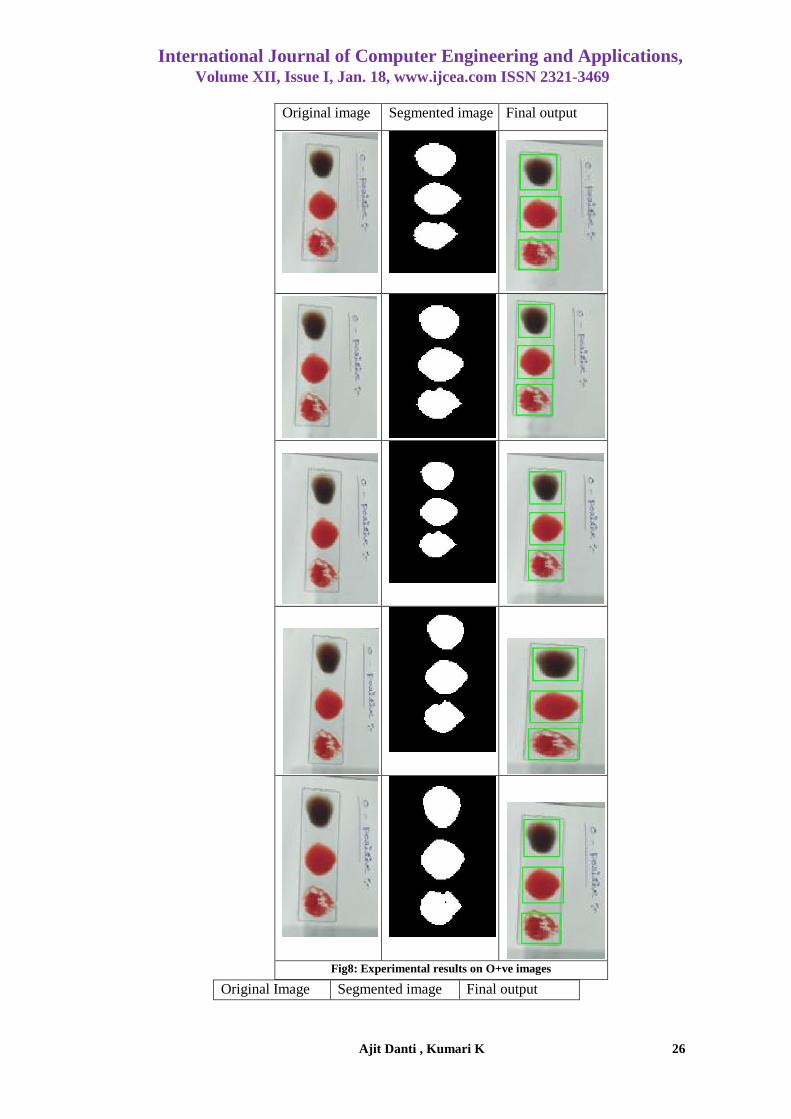
[5] EXPERIMENTAL RESULTS
In this work, experimentation is carried out on blood sample images containing eight blood
groups as given in Table 1. Basically the blood groups are classified into 8 categories such as
A+,B+,O+,AB+,A-,B-,AB-,O-. The data set contains 10 images of each blood group sample. The
segmentation by the proposed method is found to be more efficient in terms of speed and accuracy.
Each of the segmented region is then searched for the features using areas, size and shape to recognize
the blood groups in it.
The proposed approach is implemented using MATLAB software R2016b(9.1.0.441655). on
Core i3 @ 2.40GHz. The test images are expected to contain the different size of the regions which
are on the basis of the blood groups. The different blood group images have been segmented
successfully. In the experimental results, 158 images are properly segmented out of 168 images leading
to success rate is 94.04% (approximately). The average time taken to segment regions is about 0.178
second. The experimental results are shown in the below figures.
Original image Segmented image Final output
International Journal of Computer Engineering and Applications, Volume XII, Issue I, Jan. 18, www.ijcea.com ISSN 2321-3469
Ajit Danti , Kumari K 22
Fig4: Experimental results on A+ve images
Original image Segmented image Final output

BLOOD REGIONS SEGMENTATION FOR AUTOMATIC BLOOD GROUP IDENTIFICATION
Ajit Danti , Kumari K 23
Fig5: Experimental results on A-ve images
International Journal of Computer Engineering and Applications, Volume XII, Issue I, Jan. 18, www.ijcea.com ISSN 2321-3469
Ajit Danti , Kumari K 24
Original image Segmented image Final output
Fig6: Experimental results on B+ve images

BLOOD REGIONS SEGMENTATION FOR AUTOMATIC BLOOD GROUP IDENTIFICATION
Ajit Danti , Kumari K 25
Original Image Segmented image Final output
Fig7: Experimental results on AB+ve images
International Journal of Computer Engineering and Applications, Volume XII, Issue I, Jan. 18, www.ijcea.com ISSN 2321-3469
Ajit Danti , Kumari K 26
Original Image Segmented image Final output
Original image Segmented image Final output
Fig8: Experimental results on O+ve images

BLOOD REGIONS SEGMENTATION FOR AUTOMATIC BLOOD GROUP IDENTIFICATION
Ajit Danti , Kumari K 27
Fig9: Experimental results O-ve images
Original Image Segmented image Final output

International Journal of Computer Engineering and Applications, Volume XII, Issue I, Jan. 18, www.ijcea.com ISSN 2321-3469
Ajit Danti , Kumari K 28
Fig10: Experimental results on AB-ve images

BLOOD REGIONS SEGMENTATION FOR AUTOMATIC BLOOD GROUP IDENTIFICATION
Ajit Danti , Kumari K 29
Original image Segmented image Final output
Fig11: Experimental results on B-ve images

International Journal of Computer Engineering and Applications, Volume XII, Issue I, Jan. 18, www.ijcea.com ISSN 2321-3469
Ajit Danti , Kumari K 30
[6] CONCLUSION
In this paper, an effective segmentation of the blood regions is done using morphological
operations. The proposed approach exhibited 94.04% accuracy for our dataset. In future
by using these segmented images automation of the blood groups classification will be
carried in the future work.
REFERENCES
[1] Ashwini Rejintal and Aswini.N, Leukemia Cancer Cell Detection using Image Processing,
International Journal of Advanced Research in Electrical, Electronics and Instrumentation
Engineering, vol-06, Page No:34-38, Issue 06,ISSN: 2278 – 8875 , July 2016.
[2] Anita Chaudhary and Sonit Sukhraj Singh, Lung cancer detection International Conference14-15,
Page No:142-146, Issue 14 , ISBN: 978-0-7695-4817-3, September 2012.
[3] Amruta pramod hebli and sudha gupta, Brain tumor detection using image processing: A survey,
International Journal of Industrial Electronics and Electrical Engineering,vol-5, Page No:41-44,
Issue 1, ISSN: 2347-6982, Jan 2017.
[4] Bhagyashri G.Patil and Sanjeev N. Jain ,Research on Cancer Cells Detection, International Journal
of Latest Trends in Engineering and Technology (IJLTET), vol-3, Page No:45-49, Issue 04, ISSN:
2278-621X , march 2014.
[5] Deepika N. Patil and Uday P. Khot, Image processing based abnormal blood cells detection,
International Journal of Technical Research and Applications,Page No:37-43, Issue 31, e-ISSN:
2320-8163, September 2015.
[6] Devkant Sen and Neha Tiwari, Edge Preservation with Noise Reduction in Arthritis Image,
International Journal for Scientific Research & Development, vol-2,Page No:371375,Issue 12, ISSN
(online): 2321-0613,2015.
[7] Rafael C. Gonzalez, Richard E.Woods, Digital image processing, 2edition, Pearson,
[8] Irfan Khatik, A Study of Various Bone Fracture Detection Techniques, International Journal Of
Engineering And Computer Science,vol-6, Page No.21418-21423, Issue 05 , ISSN:2319-7242, may
2017.
[9] C. Jeya Bharathi and P. Kabilan, Analysis and Edge Detection of Lung Cancer – Survey,
International Journal on Recent and Innovation Trends in Computing and Communication,vol-
04,Page No:390-392,Issue 05, ISSN: 2321-8169,may 2016.
[10] Kamala kannan.J and Rajasekhara Babu, Segmentation techniques on mammograms to detect breast
abnormality, International Journal of Pharmacy & Technology,vol-8, Page No:16089-16099 , Issue
25, ISSN: 0975-766X , Aug 2016.
[11] S.Muthuselvi and P.Prabhu, Compression And Canny Edge Detection Based Methods For Image
Segmentation, International Journal of Advanced Research Trends in Engineering and Technology,
Page No:63-67, Issue 20, ISSN 2394-3785, April 2016.
[12] Nalini Singh, Ambarish G Mohapatra and Gurukalyan Kanungo, Breast Cancer Mass Detection in
Mammograms using K-means and Fuzzy C-means Clustering, International Journal of Computer
Applications (0975-8887), vol-22–No2 ,Page No:15-21,Issue 15 , May 2011
[13] Neha Sharma and Dr Supreet Kaur , Image Processing Based Approach for Automated Detection
of Chronic Leukemia, International Journal of Innovative Research in Computer and

BLOOD REGIONS SEGMENTATION FOR AUTOMATIC BLOOD GROUP IDENTIFICATION
Ajit Danti , Kumari K 31
Communication Engineering, vol-04, Page No:15038-15045, Issue 08, ISSN(O): 2320-9801, ISSN
(P): 2320-9798, Aug 2016.
[14] Parul Parmar and Vinay Thakur, A Review on Tumor Detection in Medical Images, International
Research Journal of Engineering and Technology (IRJET),vol-04 ,Page No:2714-2717 ,Issue 05, e-
ISSN: 2395 -0056,p-ISSN: 2395-0072, may 2017.
[15] Prachi Dhikale and Madhuri Joshi, Brain Tumor Detection Using 3D Visualization, International
Journal of Computer Science and Network, vol-06,Page No:170-177, Issue 2, ISSN (Online) : 2277-
5420, April 2017.
[16] Sergios Theodoridis Koutroumbas, Pattern Recognition 4th Edition, Page No:1-711.
[17] Shubhangi D C and Nagaraj ,Human Skin Cancer Recognition and Classification by Unified Skin
Texture and Color Features, IOSR Journal of Computer Engineering,vol-12, Page No.42-49, Issue
4,e-ISSN: 2278-0661, p-ISSN: 2278-8727, Jul-Aug 2013.
[18] Sivappriya T and Muthukumaran K, Cancer Cell Detection Using Mathematical Morphology,
International Journal of Innovative Research in Computer and
Communication Engineering, vol-2, Page No:3717-3725,Issue 1, ISSN(O): 2320-9801, March 2014
[19] Sonam S, Gavhande and S.B.Jadhav, Image Segmentation and Identification of Brain Tumor from
MRI Image, International Research Journal of Engineering and Technology,vol-02, Page No:167-
170,Issue 02,e-ISSN: 2395-0056,p-ISSN: 2395-072, May 2013.
[20] Somashekhar Swamy and P.K.Kulkarni, Image Processing for Identifying Brain Tumor using
Intelligent System , International Journal of Innovative Research in Science, Engineering and
Technology, vol-04, Page No:10937-10943, Issue 11, ISSN(O): 23198753, November 2015.
[21] Sushma Laxman Wakchaure and Anil Khandekar, Visualization Of 3D View Of Detected Brain
Tumor And Calculation Of Its Volume, International Journal of Technical Research and
Applications, vol-3, Page No:120-126,Issue 6, November-December, 2015.
[22] V.Venmathi and et.al, Leukemia Detection using Image Processing , International Journal for
Scientific Research & Development, vol-5,Page No:804-808, Issue 01, ISSN (O): 2321-0613, 2017.
[23] https://medlineplus.gov › Medical Encyclopedia.
[24] ]https://homehealth-uk.com › ... › Blood Group Type Test ABO and Rhesus (D)
Related Documents