BLOOD CHAPTER 10

BLOOD CHAPTER 10. FUNCTIONS OF BLOOD BLOOD is in charge of homeostasis in 3 ways 1.BY TRANSPORTATION- -deliver nutrients, oxygen and hormones to cells.
Jan 01, 2016
Welcome message from author
This document is posted to help you gain knowledge. Please leave a comment to let me know what you think about it! Share it to your friends and learn new things together.
Transcript

BLOOD
CHAPTER 10

FUNCTIONS OF BLOOD
BLOOD is in charge of homeostasis in 3 ways
1. BY TRANSPORTATION-
-deliver nutrients, oxygen and hormones to cells
• -carry away waste, carbon dioxide, nitrogen substances (urea and uric acid) and the secretions of the cells

FUNCTIONS OF BLOOD
2. BY PROTECTION- - fights off harmful substances with white
blood cells via phagocytosis and antibodies-protects against fluid loss- (clotting mechanism)
3. BY REGULATION-regulates acid-base balance-regulates body temperature

PROPERTIES OF BLOOD-
COLOR-
HEMOGLOBIN (the protein pigment in red blood cells) causes it to be red- contains the element IRON.
Oxygen binds to the hemoglobin causing a bright crimson-red color.
When blood has low oxygen levels blood appears dark-red with a slightly bluish tinge. -
This is the color of our veins seen through the skin. We do not have BLUE or GREEN blood

PROPERTIES OF BLOOD-
VOLUME- typically 8% of our body weight
Typical male has 5-6 liters
Typical female has 4-5 liters
PROPERTIES OF BLOOD-
VISCOSITY- (resistance to flow) flows 5 times more slowly than water because blood is thicker, denser and more sticky than water
PROPERTIES OF BLOOD-
pH- between 7.35 and 7.45-buffer system in place to keep narrow pH range
Too much acid (6.0) would lead to ACIDOSIS- where the body cells stop functioning
ALKALOSIS- or too little acid also occurs but is rare

PARTS OF BLOOD
PLASMA- (55%) liquid portion of blood-92% water and 8% solutes (which are mainly proteins but can also be nutrients, electrolytes, and hormones)
PLASMA PROTEINS- produced by the liver1. ALBUMIN- 55%- thickens blood-
increases osmotic pressure, Ph buffer2. GLOBULIN- 38% - antibodies in the
immune system3. FIBRINOGEN-7%- precursor to protein
fibrin used for blood clotting
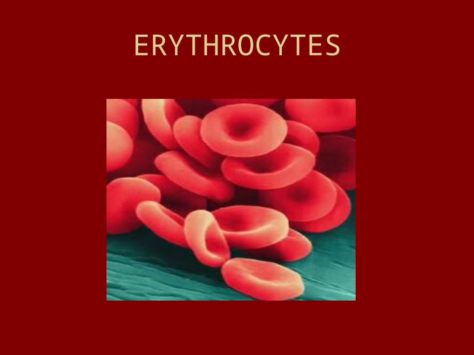
ERYTHROCYTES

PARTS OF BLOODFORMED ELEMENTS• RED BLOOD CELLS- (45%) ERYTHROCYTESCOUNT – called a HEMATOCRIT-4-6 million per cubic
millimeter
FUNCTION- carry oxygen to cells from lungs
STRUCTURE- -biconcave discs-no nucleus (anucleate) and no organelles - 120 DAY lifespan- phagocytized by WBC-HEMOGLOBIN- iron containing protein that transports oxygen (occupies 1/3 volume of the RBC)

ERYTHROCYTES
DISORDERS-
ANEMIA- lower than normal oxygen due to lack of RBC or abnormal hemoglobin
SICKLE CELL ANEMIA-
IRON DEFICIENCY ANEMIA

PARTS OF BLOOD
WHITE BLOOD CELLS- LEUKOCYTES-
COUNT -4,000- 11,000 per cubic millimeter (less than 1%
of total volume)
STRUCTURE -contain a nucleus and can change shape to travel outside the circulatory system
FUNCTION -control diseases

LEUKOCYTES-
3 TYPES OF WBC-
1.GRANULOCYTES- cytoplasm contains granules that stain
2.AGRANULOCYTES- no granules in cytoplasm
3.PLATELETS- cell fragments of various shapes

White Blood Cells

GRANULOCYTES-(312)
1. NEUTROPHILS- (stain pink) phagocytes at infection site- (make up 40-70% of WBC)
2. EOSINOPHILS-(stain red) phagocytes of parasitic worms, might help w/ allergies (make up 1-4% of WBC)
3. BASOPHILS-(stain blue) discharges histamine which allows blood to release from blood vessels-(swelling) (less than 1%)

AGRANULOCYTES
1. LYMPHOCYTES-
B lymphocytes- produce antibodies
T lymphocytes- help recognize previous infections- involved in graft rejection
2. MONOCYTES - phagocytize and clean up tissue
AGRANULOCYTES
PLATELETS- involved in blood clotting

BLOOD TYPING
ANTIGENS - surface proteins on the plasma membrane of red blood cells. These are genetically determined.
ANTIBODIES (AGGLUTININS) - proteins in the plasma that will react with the antigens and cause the blood to clump. These proteins are automatically formed early in life.

ABO SYSTEM-
ANTIGENS ANTIBODIES
A BLOOD A antibody to B
B BLOOD B antibody to A
AB BLOOD A and B NO ANTIBODIES
O BLOOD NO ANTIGENS antibody to A and B
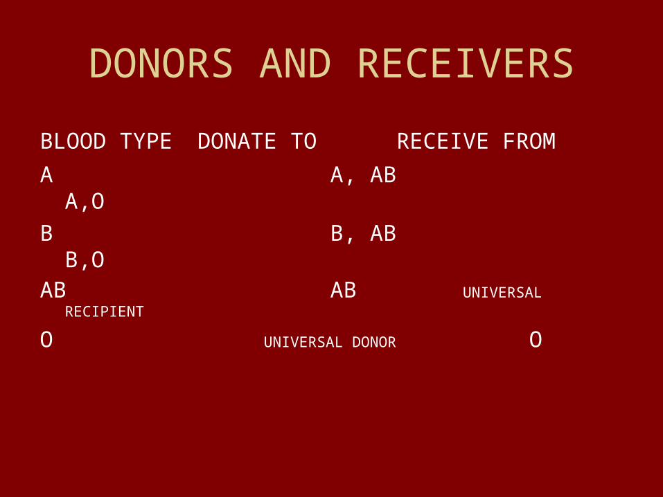
DONORS AND RECEIVERS
BLOOD TYPE DONATE TO RECEIVE FROM
A A, AB A,O
B B, AB B,OAB AB UNIVERSAL
RECIPIENT
O UNIVERSAL DONOR O

Rh BLOOD TYPING SYSTEM
Rh POSITIVE- have Rh antigens (most people born this way)
Rh NEGATIVE no Rh antigens
• Antibodies for Rh antigens are not formed until a person has become sensitized. (example- a blood transfusion of an Rh negative person with Rh positive blood, a Rh negative mother delivering an Rh positive child)
• The is no reaction to the first exposure but from then on after agglutination of the blood will occur.

Blood Clotting
3 stages needed to prevent blood loss
1. Platelet plug stage
2. Vascular spasm
3. Coagulation

Platelet plug stage
Platelets which are repelled by endothelium (inner layer of blood vessel) are attracted to underlying collagen fibers.
Platelets also release chemicals to attract more platelets to injury site.
Eventually enough platelets pile up to form a PLATELET PLUG

VASCULAR SPASMS
Platelet plug releases chemical (serotonin) that causes the blood vessel to go into spasms and narrow.

COAGULATION
Chemicals are released which convert some plasma proteins to THROMBIN with binds with FIBRINOGEN PROTEINS in plasma to create a long hairlike fiber FIBRIN which will form a mesh to catch red blood cells

Clotting problems
HEMOPHILIA- “bleeder’s disease”- can be caused by lack of any the clotting factors
THROMBUS- blood clot in a healthy blood vessel that may travel (called an EMBOLUS) to heart or brain

HEMATOPOIESIS- blood cell formation
Red blood cells (WBC and platelets) are formed in the red bone marrow found in spongy bone.
Red blood cells are formed with nuclei then as they mature (3 to 5 days) the produce huge amounts of hemoglobin which eventually pushes the nucleus and organelles out of the cell.

HEMOGLOBIN
• Each red blood cell contains about 250 Million hemoglobin molecules.
• Each hemoglobin molecule can carry bind to 4 molecules of oxygen.
• EACH RED BLOOD CELL can carry 1 BILLION MOLECULES OF OXYGEN

KIDNEYS responsibility in HEMATOPOIESIS
When blood oxygen levels drop then kidneys produce a hormone called ERYTHROPOIETIN that targets the red bone marrow for production of RBC.

How much blood can we lose?
Clots usually form in 3 to 6 minutes to stop bleeding
30% of blood volume lost will cause severe shock.
Blood banks can store blood for up to 35 days.


Related Documents