J. Biomater. Sci. Polymer Edn, Vol. 17, No. 9, pp. 985– 996 (2006) VSP 2006. Also available online - www.vsppub.com Blood cell and plasma protein repellent properties of Star-PEG-modified surfaces JAN HOFFMANN 1 , JÜRGEN GROLL 2,3 , JEAN HEUTS 3 , HAITAO RONG 2 , DORIS KLEE 3 , GERHARD ZIEMER 1 , MARTIN MOELLER 3 and HANS P. WENDEL 1,∗ 1 Department of Thoracic, Cardiac and Vascular Surgery, University of Tübingen, Calwerstr. 7/1, D-72076 Tübingen, Germany 2 SusTech GmbH & Co. KG, Petersenstrasse 20, D-64287 Darmstadt, Germany 3 Department of Textile and Macromolecular Chemistry, University of Technology Aachen, Aachen, Germany Received 29 August 2005; accepted 30 March 2006 Abstract—The implantation of biomaterials, medical devices or prostheses can instigate a rejection response or initiate an undesirable adsorption of plasma proteins, as well as blood cells on the implant surface, thus triggering diverse defense mechanisms against the supposed pathologic invader. The extent of this inflammatory reaction depends in part on the biocompatibility of the used materials or coatings. Although adsorption and coagulation responses can appear during the total in vivo lifetime of the implant, they are initially and crucially formed within the first 2–4 weeks of implantation. This early phase is of decisive importance for the consecutive in-growth and healing process. The present study was intended to elucidate the effects of blood contact to surfaces modified with reactive six-arm star-shaped poly(ethylene glycol-stat-propylene glycol) pre-polymers (Star PEG). Taken together, for Star-PEG-covered substrates we could demonstrate a profound reduction of various blood–biomaterial interactions compared to non-coated substrates, indicating the promising potential of this material as future coating for biomaterials with blood contact. Key words: Haemocompatibility; polyethylene glycol; platelets; plasma proteins. INTRODUCTION It is of paramount importance that biomaterials used for human application exhibit a maximum of biocompatibility towards the surrounding tissue. Consequently, the development of synthetic devices with little or without any unwanted blood or pro- tein interactions is a major focus in the field of biomaterials research. The evolution ∗ To whom correspondence should be addressed. Tel.: (49-7071) 298-6605. Fax: (49-7071) 295- 369. E-mail: [email protected]

Welcome message from author
This document is posted to help you gain knowledge. Please leave a comment to let me know what you think about it! Share it to your friends and learn new things together.
Transcript
J. Biomater. Sci. Polymer Edn, Vol. 17, No. 9, pp. 985–996 (2006) VSP 2006.Also available online - www.vsppub.com
Blood cell and plasma protein repellent propertiesof Star-PEG-modified surfaces
JAN HOFFMANN 1, JÜRGEN GROLL 2,3, JEAN HEUTS 3, HAITAO RONG 2,DORIS KLEE 3, GERHARD ZIEMER 1, MARTIN MOELLER 3
and HANS P. WENDEL 1,∗1 Department of Thoracic, Cardiac and Vascular Surgery, University of Tübingen, Calwerstr. 7/1,
D-72076 Tübingen, Germany2 SusTech GmbH & Co. KG, Petersenstrasse 20, D-64287 Darmstadt, Germany3 Department of Textile and Macromolecular Chemistry, University of Technology Aachen,
Aachen, Germany
Received 29 August 2005; accepted 30 March 2006
Abstract—The implantation of biomaterials, medical devices or prostheses can instigate a rejectionresponse or initiate an undesirable adsorption of plasma proteins, as well as blood cells on the implantsurface, thus triggering diverse defense mechanisms against the supposed pathologic invader. Theextent of this inflammatory reaction depends in part on the biocompatibility of the used materials orcoatings. Although adsorption and coagulation responses can appear during the total in vivo lifetimeof the implant, they are initially and crucially formed within the first 2–4 weeks of implantation. Thisearly phase is of decisive importance for the consecutive in-growth and healing process. The presentstudy was intended to elucidate the effects of blood contact to surfaces modified with reactive six-armstar-shaped poly(ethylene glycol-stat-propylene glycol) pre-polymers (Star PEG). Taken together, forStar-PEG-covered substrates we could demonstrate a profound reduction of various blood–biomaterialinteractions compared to non-coated substrates, indicating the promising potential of this material asfuture coating for biomaterials with blood contact.
Key words: Haemocompatibility; polyethylene glycol; platelets; plasma proteins.
INTRODUCTION
It is of paramount importance that biomaterials used for human application exhibita maximum of biocompatibility towards the surrounding tissue. Consequently, thedevelopment of synthetic devices with little or without any unwanted blood or pro-tein interactions is a major focus in the field of biomaterials research. The evolution
∗To whom correspondence should be addressed. Tel.: (49-7071) 298-6605. Fax: (49-7071) 295-369. E-mail: [email protected]
986 J. Hoffmann et al.
of surface modifications has already led to an optimized haemocompatibility. How-ever, the so far available biomaterials and biomaterial coatings display differentbiocompatibilities depending on the implant location. Therefore, the aim of thisstudy was to develop a synthetic biomaterial with two vital characteristics: (i) lit-tle or no non-specific adhesion during contact with whole blood and (ii) multipletissue compatibility by uncomplicated functionalization with any desired bioactivemolecule in order to introduce specific biological activities.
Due to their defined structure and their high functionality, star-shaped moleculesoffer interesting possibilities. In combination with this aspect of molecular archi-tecture, poly(ethylene glycol) (PEG) is a material that has widely been used forbiomaterial applications [1] and is particularly suitable for the preparation of bio-compatible coatings. In terms of applicability as coating precursors, the moleculararchitecture of star-shaped molecules implicates a high density of functional groupsat the interface due to surface segregation of the end-groups [2]. This effect hasbeen utilized to prepare surfaces with high ligand density at the surface and therebya high binding capacity (for example, antibody binding to star-shaped PEG mole-cules functionalized with immunoreactive groups) [3, 4]. In addition, star-shapedmolecules allow a high surface coverage of the polymer on the substrate when mole-cules with cross-linkable end-groups are employed [5]. Apart from hydrophilicitythis high surface coverage is the most important parameter when it comes to theprevention of non-specific interactions with biological systems [6–11]. We coulddemonstrate previously that functional coatings prepared from isocyanate termi-nated, six-armed star-shaped poly(ethylene glycol-stat-propylene glycol) molecules(Star PEG) prevent the non-specific adsorption of proteins [12] and cells [13] veryefficiently. In addition, the functionality of these layers can be used to generate spe-cific biological interactions by incorporation of ligands for biological targets intothe coatings [12–14].
Today there are several well-established biomarkers for the analysis of theinteraction between biomaterials and blood: the detection of the platelet surfaceantigen CD41 (GPIIb), which is known to be a common indicator for plateletactivation after contact with artificial materials [15]; the increase in the plasmaconcentration of the coagulation marker TAT as another test system to verify plateletalteration [16]; the measurement of β-thromboglobulin plasma concentration asa representative platelet factor released from activated platelets [17]; and finallythe quantitation of the complement activation marker SC5b-9, giving preciseinformation about the actual immunodefensive situation (increased levels are foundduring infections, sepsis or phagocytosis) [18].
The present study was focused on the haemocompatibility of Star PEG coatingmaterials prepared on glass substrates in different thicknesses. The thickness of thelayers was measured by ellipsometry on corresponding silicon samples and the filmhomogeneity was assessed by optical microscopy and scanning force microscopy(SFM). These films were then checked for their haemocompatibility by bringing
Blood cell and plasma protein repellent properties of Star-PEG-modified surfaces 987
them into contact with whole blood and analysing different parameters of cell orprotein adhesion and coagulation activity.
MATERIALS AND METHODS
Test materials
Small glass discs with a radius of 5 mm were used as substrates. The Star PEGlayers were spin-coated onto such discs with thicknesses of 15 nm (group 1) and30 nm (group 2), whereas uncoated glass substrates served as control samples(group 0).
Six-arm star-shaped NCO terminated pre-polymers with a backbone of statisti-cally co-polymerized 80% ethylene oxide and 20% propylene oxide (MW = 12kg/mol, PDI = 1.15) were obtained from Sustech (Darmstadt, Germany). Siliconwafers (100) were purchased from CrysTec (Berlin, Germany). Glass substrateswere purchased from Vetter (Ammerbuch, Germany). Acetone, isopropanol andethanol (Selectipur, Merck, Darmstadt, Germany) were stored in the clean roomand used as received. THF and toluene were dried over LiAlH4, distilled underargon and transferred into a glove box. 97% N-[3-(trimethoxysilyl) propyl] eth-ylenediamine (Sigma-Aldrich, St. Louis, MO, USA) was stored in the glove boxand filtered before use. Biocytin (Sigma-Aldrich) was stored at −20◦C and usedas received. Syringe filters with pore size 0.02 µm were purchased from Whatman(Brentford, UK).
Substrate preparation
Cutting and cleaning of the substrates was performed in a class 100 clean room.Silicon substrates were cut with a RV-125 diamond cutting device from ATVTechnologie (Vaterstetten, Germany) into pieces of 14 × 14 mm. Glass and siliconsamples were cleaned by sonication in acetone, water and isopropanol for 1 mineach, followed by drying in a stream of nitrogen. Then the substrates were activatedwith UV/ozone using a 40-W UV lamp (main emission 185 nm; UV-TechnikSpeziallampen, Wümbach, Germany) in an oxygen stream of 350 ml/min witha sample distance of 5 mm to the lamp for 12 min. The substrates were thenimmediately used for amino-functionalization.
Aminosilylation of the substrates
After activation, the substrates were transferred into an Unilab glove box (MBraun)and immersed into a solution of 0.3 ml N-[3-(trimethoxysilyl)propyl] ethylenedi-amine in 50 ml dry toluene for 2 h. The samples were then washed several timeswith dry toluene and stored under dry toluene until further use.
988 J. Hoffmann et al.
Spin coating
Star PEG films were generated using a CONVAC ST 146 spin coater (Convac,Wiernsheim, Germany). The desired amount of Star PEG pre-polymer is dissolvedunder an inert gas atmosphere in dry THF. This solution is transferred to the cleanroom and 9-times excess Millipore water is added. The concentration of the polymerin the final solvent mixture was varied from 1 to 10 mg/ml. After 5 min the solutionswere filtered through 0.2-µm syringe filters and used for spin coating. For spincoating, the amino-functionalized substrates were placed on the spin coater, coveredby the corresponding solution and then accelerated within 5 s to the final rotationspeed of 2500 rpm and kept rotating for 40 s. The resulting films were storedovernight in ambient atmosphere to allow for proper cross-linking of the films.
For biotinylated coatings, 100 mg Star PEG pre-polymer was dissolved in dryTHF and mixed with 2 mg biocytin dissolved in 9 ml deionized water prior to spincasting. The protocol is the same for unmodified layers.
Layer thickness determination
Layer thicknesses were examined using a MM-SPEL-VIS spectral ellipsometer(OMT, Ulm, Germany) in the wavelength range from 450 to 900 nm. The azimuthalangle was kept at 15 degrees and the integration time was dependent on the layerthickness and the resulting signal intensity. In order to minimize systematic errorsin the data collection, in a series of experiments always one substrate was justcleaned and activated and one was just cleaned, activated and aminosilylated. Thesetwo substrates were measured as references, and thicknesses of the hydrogel filmswere obtained as relative values to the aminosilylated substrate. Each sample wasmeasured at 5 different places and the presented data are the average values of eachsample.
Homogeneity of the layers
Light microscopy was performed by means of an Axioplan2 Imaging microscopefrom Zeiss (Jena, Germany). Scanning force microscopy (SFM) investigations wereachieved with a nanoscope IIIa (Digital Instruments, Santa Barbara, CA, USA)operating in tapping ModeTM. The oscillation frequency for Tapping Mode™ wasset in the range of 320–360 kHz, depending on the Si cantilever (k approx. 50 N/m;Nanosensors, Neuchatel, Switzerland).
Rocking platform
This device serves to mimic the contact effect of artificial surfaces with blood initi-ating the different cascade reactions of the human haemostatic system (coagulation,inflammation, cell alteration and protein adsorption). Eight discs for each group ofsurface modifications were incubated in 48-well suspension-culture plates (Greiner,Frickenhausen, Germany) with 750 µl fresh human platelet-rich plasma (PRP) per
Blood cell and plasma protein repellent properties of Star-PEG-modified surfaces 989
disc for 1 h at 37◦C on the rocking platform at 25 rpm (Polymax 1040, Heidolph,Schwabach, Germany). PRP without disc contact served as control.
Blood donors
Healthy volunteers between 20 and 30 years with an activated partial thromboplas-tin time (aPTT) in the normal range were written informed and consents were ob-tained from all donors. The blood quality used for these experiments is of decisiveimportance and therefore following exclusion criteria were strictly fulfilled: smok-ing, drug-taking in the last two weeks, especially haemostasis-affecting agents, likeacetylsalicylic acid, oral contraceptives and non-steroidal antiphlogistics.
Blood was immediately drawn carefully by venipuncture in sterile and pre-anticoagulated (3 IU/ml Liquemin; Hoffmann La-Roche, Basel, Switzerland) con-tainers (Sarstedt, Nümbrecht, Germany). PRP was immediately prepared by cen-trifugation at 150 × g for 10 min at room temperature.
Scanning electron microscopy (SEM)
After rinsing the PRP-treated discs in physiological saline, they were fixed in 2%glutaraldehyde (Serva, Heidelberg, Germany) for 1 h followed by dehydration stepswith increasing concentrations of ethanol (30–96%). After storage in absoluteethanol the discs were completely dried by critical point drying and sputtered withgold-palladium. Analyses were performed with the Cambridge Stereoscan 250 Mk2(Cambridge Instruments, Cambridge, UK).
Protein adsorption (ELISA)
The detection of CD41 (antibody: mouse anti human CD41; Serotec, Düsseldorf,Germany) and fibrinogen (antibody: goat anti human fibrinogen; Sigma) wasbased on the sandwich principle. After fixation with 4% para-formaldehyde, theresidual aldehyde groups were inactivated with 0.05 M glycine buffer (Merck).The blocking of non-specific binding was done overnight at 4◦C. Afterwards thesamples were rinsed twice and the primary antibody was added for 1 h at 37◦C.The excess primary antibody was then washed five times. The enzyme-conjugatedsecond antibody (for fibrinogen: donkey anti-sheep IgG-AP, Sigma; for CD41:goat anti mouse IgG-AP, Immunotech, Marseille, France) binds during the nextreaction for 1 h at 37◦C. The unbound enzyme-conjugated antibody was removedby five washing steps. The following enzymatic reaction of the substrate (Sigmafast™ pNPP substrate tablet set, Sigma) at room temperature was terminated after3–10 min by the addition of 3 M NaOH (Merck).
The optical densities of the samples were measured photometrically in a microtiterplate using a MR 7000 microplate reader (Dynatech Deutschland, Denkendorf,Germany) at 405 nm wavelength.
990 J. Hoffmann et al.
Thrombogenicity examinations
Coagulation activation was quantified by an ELISA kit for thrombin-antithrombin-III complexes supplied by Dade Behring (Marburg, Germany). The platelet con-centration was measured with a standard haematology analyzer (Micros 60, ABXDiagnostics, Montpellier, France). The β-thromboglobulin plasma concentrationwas also investigated by ELISA (ELISA kit, Diagnostica Stago, Asnières, France).
RESULTS
Preparation and analysis of the samples
Star PEG films have been prepared by spin casting two different concentrationsof Star PEG solutions, resulting in 15- and 30-nm-thick layers. These layers werechecked for homogeneity by optical microscopy and scanning force microscopy andproved to be homogeneous and smooth (the root mean square roughness (rms) wasless than 0.6 nm for a 1 × 1 µm scan by SFM for both layers). The two differentlayer thicknesses enabled us to check whether the thickness has an effect on theprevention of non-specific interaction with human blood.
Coagulation activation
The contact of blood with artificial surfaces leads to activation and further alterationof platelets. In order to determine the extent of platelet adhesion to Star-PEG-coated substrates compared to non-coated glass after the contact with whole blood,surface adsorbed platelet receptor CD41 was measured by ELISA as described in
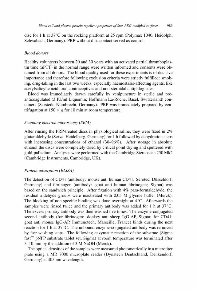
Figure 1. This ELISA shows the amount of adsorbed CD41 protein. It is diagrammed referring everytime to a control disc, which was not in contact with blood or plasma. The protein adsorption on the15 nm Star PEG layer (group 1) is significantly (∗∗P < 0.01) lower compared to the uncoated glasssubstrate (group 0). Also the protein adsorption on the 30 nm Star PEG layer (group 2) is significantly(∗P < 0.05) lower compared to the uncoated glass substrate (group 0). This low value demonstratesthat the Star PEG coating shows no antibody binding, especially in the 15 nm Star PEG layer group.The error bar indicates the mean standard deviation.
Blood cell and plasma protein repellent properties of Star-PEG-modified surfaces 991
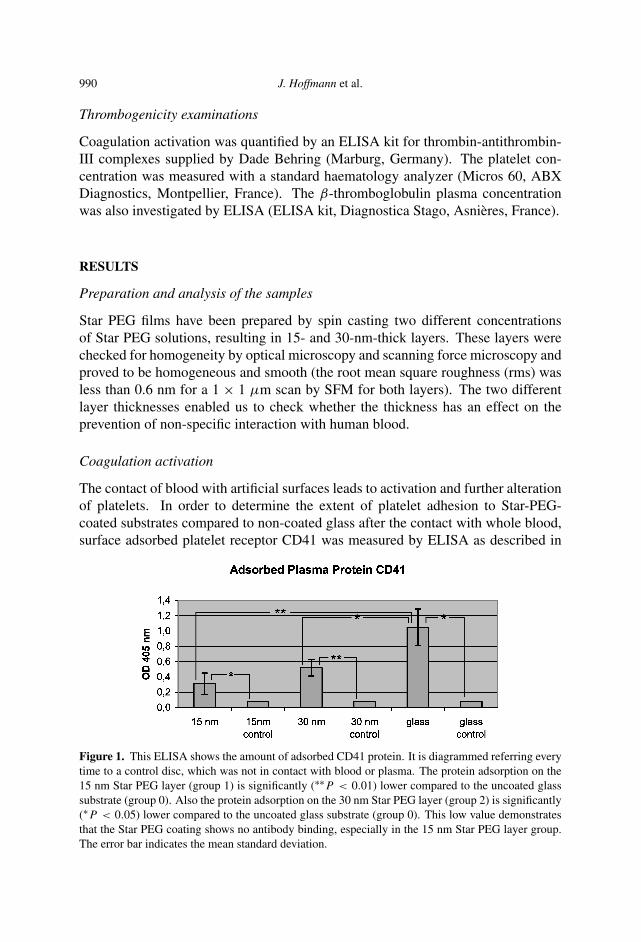
Figure 2. The coagulation marker TAT was measured referring to non-exposed plasma (control).Both the 15 nm Star PEG layer (group 1) and the 30 nm Star PEG layer (group 2) show a highlysignificant difference (∗∗P < 0.01) to the uncoated glass substrate. The error bar indicates the meanstandard deviation.
Figure 3. The β-thromboglobulin concentration in plasma was measured referring to non-exposedplasma (control). We found that the 15 nm Star PEG layer (group 1) shows a highly significant change(∗∗P < 0.01) compared to the uncoated glass substrate (group 0). Also the 30 nm Star PEG layer(group 2) shows even higher significant differences (∗∗∗P < 0.001) compared to the uncoated glasssubstrate (group 0). The error bar indicates the mean standard deviation.
materials and methods. The highest amount of surface adsorbed CD41 (Fig. 1)could be detected in the glass control group 0 (1.05 ± 0.24). In this case, theextent of platelet adsorption was significantly larger than in the 30 nm Star PEGgroup 2 (0.52 ± 0.11) and highly significantly larger than in the 15 nm StarPEG group 1 (0.31 ± 0.14), suggesting that Star PEG coating can prevent theadhesion of platelets to the surface of the glass discs. To further strengthen thisobservation in a second approach we measured the plasma concentration of thecoagulation marker TAT after blood-biomaterial interaction. Figure 2 clearly showsthat the highest plasma TAT concentration again was observed in case of the non-coated glass group 0 (1823.4 ± 593.2 µg/l) and that this was highly significantlyincreased in comparison to both Star PEG coating groups 1 (4.3 ± 1.3 µg/l) and2 (3.8 ± 0.4 µg/l). The Star PEG coating, thus, seems to reduce profoundly
992 J. Hoffmann et al.
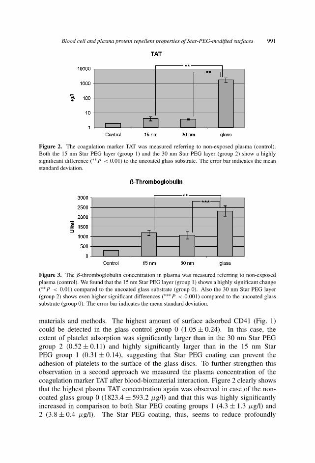
Figure 4. Star-PEG-coated discs were exposed to human plasma for 1 h. Subsequently, the amount ofplatelets was measured referring to non-incubated plasma (control). We set this value as 100%. In thiscase we show that the platelet concentration is reduced in all three cases: in the 15 nm Star PEG layer(group 1), in the 30 nm Star PEG layer (group 2) and especially in the uncoated glass control substrate(group 0). We observed no statistically significant differences between group 1 or 2 and group 0, butthe lowest platelet concentrations appear, as expected, in the uncoated glass control group.
any coagulation on the glass surface. To elucidate this phenomenon in furtherdetail we next examined the concentration of β-thromboglobulin (Fig. 3), a factorbeing released during platelet alteration. Again, the Star PEG groups 1 (1198.7 ±128.5 IU/ml) and 2 (1072.5 ± 184 IU/ml at 30 nm) displayed a highly-significantreduced β-thromboglobulin plasma concentration when compared to the non-coatedglass group 0 (2320.3 ± 264.6 IU/ml). Subsequently, we quantified the number ofplatelets in human plasma before and after the contact with the different substratediscs in order to measure platelet aggregation (Fig. 4). Unfortunately no significantreduction could be notified, but the counted values indicate that more platelets wereadhered to the discs without Star PEG modifications (76.8 ± 4.5%) compared togroup 1 (84.2 ± 4.4%) and group 2 (85.7 ± 6.4%). Finally, the evaluation of thecomplement activation marker SC5b-9 showed a strong increase in all three groupsreferring to the ‘non-contact’-control, but no relevant differences between groups 0(1551.8 ± 650.6 ng/ml), 1 (1654.5 ± 748.2 ng/ml) and 2 (1698.9 ± 593 ng/ml)could be detected.
SEM analyses
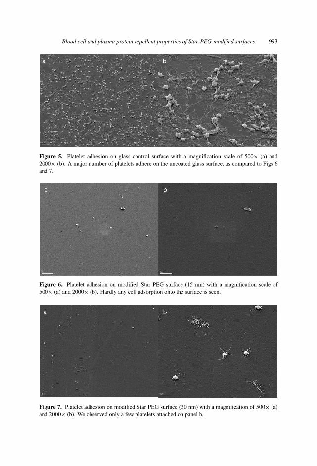
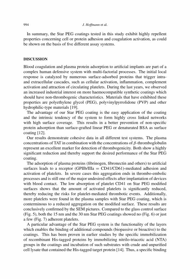
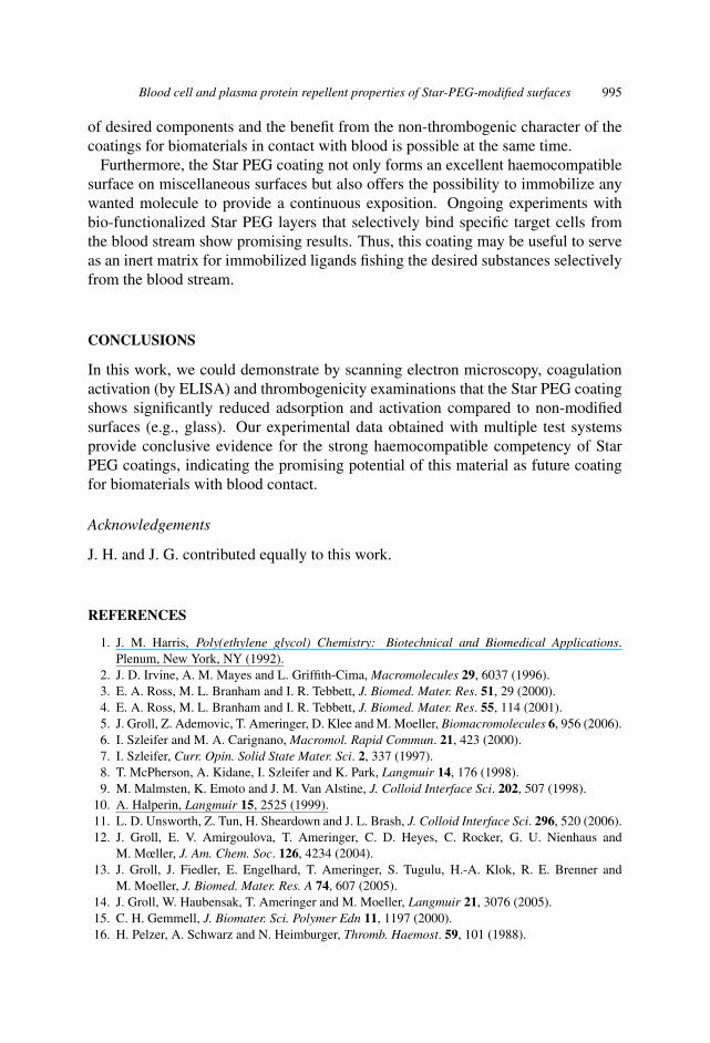
In order to illustrate platelet adhesion to the different substrate discs more plasti-cally, we performed SEM analyses of the discs after the contact with whole blood.Figures 5–7 demonstrate, very convincingly, that the Star-PEG-coated surfaces(Figs 6 and 7) showed hardly any platelet adhesion compared to the non-coatedglass control surface, where a vivid platelet adhesion could be detected (Fig. 5).These data confirm conclusively the results on platelet activation and coagulationdescribed above.
Blood cell and plasma protein repellent properties of Star-PEG-modified surfaces 993
Figure 5. Platelet adhesion on glass control surface with a magnification scale of 500× (a) and2000× (b). A major number of platelets adhere on the uncoated glass surface, as compared to Figs 6and 7.
Figure 6. Platelet adhesion on modified Star PEG surface (15 nm) with a magnification scale of500× (a) and 2000× (b). Hardly any cell adsorption onto the surface is seen.
Figure 7. Platelet adhesion on modified Star PEG surface (30 nm) with a magnification of 500× (a)and 2000× (b). We observed only a few platelets attached on panel b.
994 J. Hoffmann et al.
In summary, the Star PEG coatings tested in this study exhibit highly repellentproperties concerning cell or protein adhesion and coagulation activation, as couldbe shown on the basis of five different assay systems.
DISCUSSION
Blood coagulation and plasma protein adsorption to artificial implants are part of acomplex human defensive system with multi-factorial processes. The initial localresponse is catalyzed by numerous surface-adsorbed proteins that trigger intra-and extracellular cascades, such as cellular activation, inflammation, complementactivation and attraction of circulating platelets. During the last years, we observedan increased industrial interest on more haemocompatible synthetic coatings whichshould have non-thrombogenic characteristics. Materials that have exhibited theseproperties are polyethylene glycol (PEG), polyvinylpyrrolidone (PVP) and otherhydrophilic-type materials [19].
The advantage of our Star PEG coating is the easy application of the coatingand the intrinsic tendency of the system to form highly cross linked networkswith high surface coverage. This results in a better prevention of non-specificprotein adsorption than surface-grafted linear PEG or denaturated BSA as surfacecoating [12].
Our results demonstrate cohesive data in all different test systems. The plasmaconcentrations of TAT in combination with the concentrations of β-thromboglobulinrepresent an excellent marker for detection of thrombogenicity. Both show a highlysignificant reduction and thereby support the desired performance of the Star PEGcoating.
The adsorption of plasma proteins (fibrinogen, fibronectin and others) to artificialsurfaces leads to a receptor (GPIIb/IIIa = CD41/CD61)-mediated adhesion andactivation of platelets. In severe cases this aggregation ends in thrombo-embolicprocesses and is still one of the major undesired effects after implantation of deviceswith blood contact. The low absorption of platelet CD41 on Star PEG modifiedsurfaces shows that the amount of activated platelets is significantly reduced,thereby reducing the risks for platelet-mediated thrombotic events. Additionally,more platelets were found in the plasma samples with Star PEG coating, which isconterminous to a reduced aggregation on the modified surface. These results areconclusively confirmed by the SEM pictures. Compared to the glass control surface(Fig. 5), both the 15 nm and the 30 nm Star PEG coatings showed no (Fig. 6) or justa few (Fig. 7) adherent platelets.
A particular advantage of the Star PEG system is the functionality of the layerswhich enables the binding of additional compounds (biopassive or bioactive) to thecoatings. This has been proven in earlier studies by the specific immobilizationof recombinant His-tagged proteins by immobilizing nitrilo-triacetic acid (NTA)groups in the coatings and incubation of such substrates with crude and unpurifiedcell lysate that contained the His-tagged target protein [14]. Thus, a specific binding
Blood cell and plasma protein repellent properties of Star-PEG-modified surfaces 995
of desired components and the benefit from the non-thrombogenic character of thecoatings for biomaterials in contact with blood is possible at the same time.
Furthermore, the Star PEG coating not only forms an excellent haemocompatiblesurface on miscellaneous surfaces but also offers the possibility to immobilize anywanted molecule to provide a continuous exposition. Ongoing experiments withbio-functionalized Star PEG layers that selectively bind specific target cells fromthe blood stream show promising results. Thus, this coating may be useful to serveas an inert matrix for immobilized ligands fishing the desired substances selectivelyfrom the blood stream.
CONCLUSIONS
In this work, we could demonstrate by scanning electron microscopy, coagulationactivation (by ELISA) and thrombogenicity examinations that the Star PEG coatingshows significantly reduced adsorption and activation compared to non-modifiedsurfaces (e.g., glass). Our experimental data obtained with multiple test systemsprovide conclusive evidence for the strong haemocompatible competency of StarPEG coatings, indicating the promising potential of this material as future coatingfor biomaterials with blood contact.
Acknowledgements
J. H. and J. G. contributed equally to this work.
REFERENCES
1. J. M. Harris, Poly(ethylene glycol) Chemistry: Biotechnical and Biomedical Applications.Plenum, New York, NY (1992).
2. J. D. Irvine, A. M. Mayes and L. Griffith-Cima, Macromolecules 29, 6037 (1996).3. E. A. Ross, M. L. Branham and I. R. Tebbett, J. Biomed. Mater. Res. 51, 29 (2000).4. E. A. Ross, M. L. Branham and I. R. Tebbett, J. Biomed. Mater. Res. 55, 114 (2001).5. J. Groll, Z. Ademovic, T. Ameringer, D. Klee and M. Moeller, Biomacromolecules 6, 956 (2006).6. I. Szleifer and M. A. Carignano, Macromol. Rapid Commun. 21, 423 (2000).7. I. Szleifer, Curr. Opin. Solid State Mater. Sci. 2, 337 (1997).8. T. McPherson, A. Kidane, I. Szleifer and K. Park, Langmuir 14, 176 (1998).9. M. Malmsten, K. Emoto and J. M. Van Alstine, J. Colloid Interface Sci. 202, 507 (1998).
10. A. Halperin, Langmuir 15, 2525 (1999).11. L. D. Unsworth, Z. Tun, H. Sheardown and J. L. Brash, J. Colloid Interface Sci. 296, 520 (2006).12. J. Groll, E. V. Amirgoulova, T. Ameringer, C. D. Heyes, C. Rocker, G. U. Nienhaus and
M. Mœller, J. Am. Chem. Soc. 126, 4234 (2004).13. J. Groll, J. Fiedler, E. Engelhard, T. Ameringer, S. Tugulu, H.-A. Klok, R. E. Brenner and
M. Moeller, J. Biomed. Mater. Res. A 74, 607 (2005).14. J. Groll, W. Haubensak, T. Ameringer and M. Moeller, Langmuir 21, 3076 (2005).15. C. H. Gemmell, J. Biomater. Sci. Polymer Edn 11, 1197 (2000).16. H. Pelzer, A. Schwarz and N. Heimburger, Thromb. Haemost. 59, 101 (1988).

996 J. Hoffmann et al.
17. K. L. Kaplan and J. Owen, Blood 57, 199 (1981).18. T. E. Mollnes, Vox Sang 74 (Suppl. 2), 303 (1998).19. S. Sandhu and A. Luthra, Med. Device Technol. 13, 16 (2002).
Related Documents