Welcome message from author
This document is posted to help you gain knowledge. Please leave a comment to let me know what you think about it! Share it to your friends and learn new things together.
Transcript

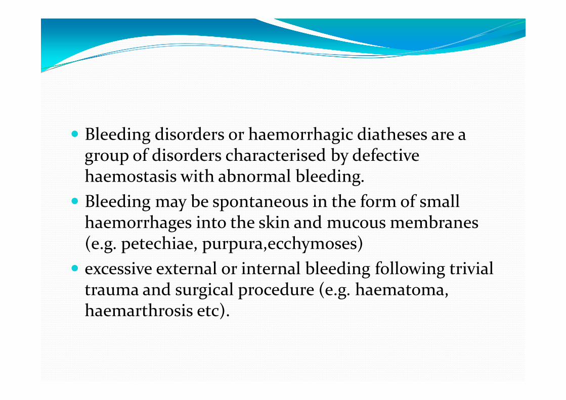
Bleeding disorders or haemorrhagic diatheses are a group of disorders characterised by defective haemostasis with abnormal bleeding.
Bleeding may be spontaneous in the form of small haemorrhages into the skin and mucous membranes (e.g. petechiae, purpura,ecchymoses)
excessive external or internal bleeding following trivial trauma and surgical procedure (e.g. haematoma, haemarthrosis etc).
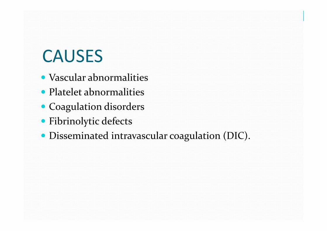
CAUSES Vascular abnormalities Platelet abnormalities Coagulation disorders Fibrinolytic defects Disseminated intravascular coagulation (DIC).
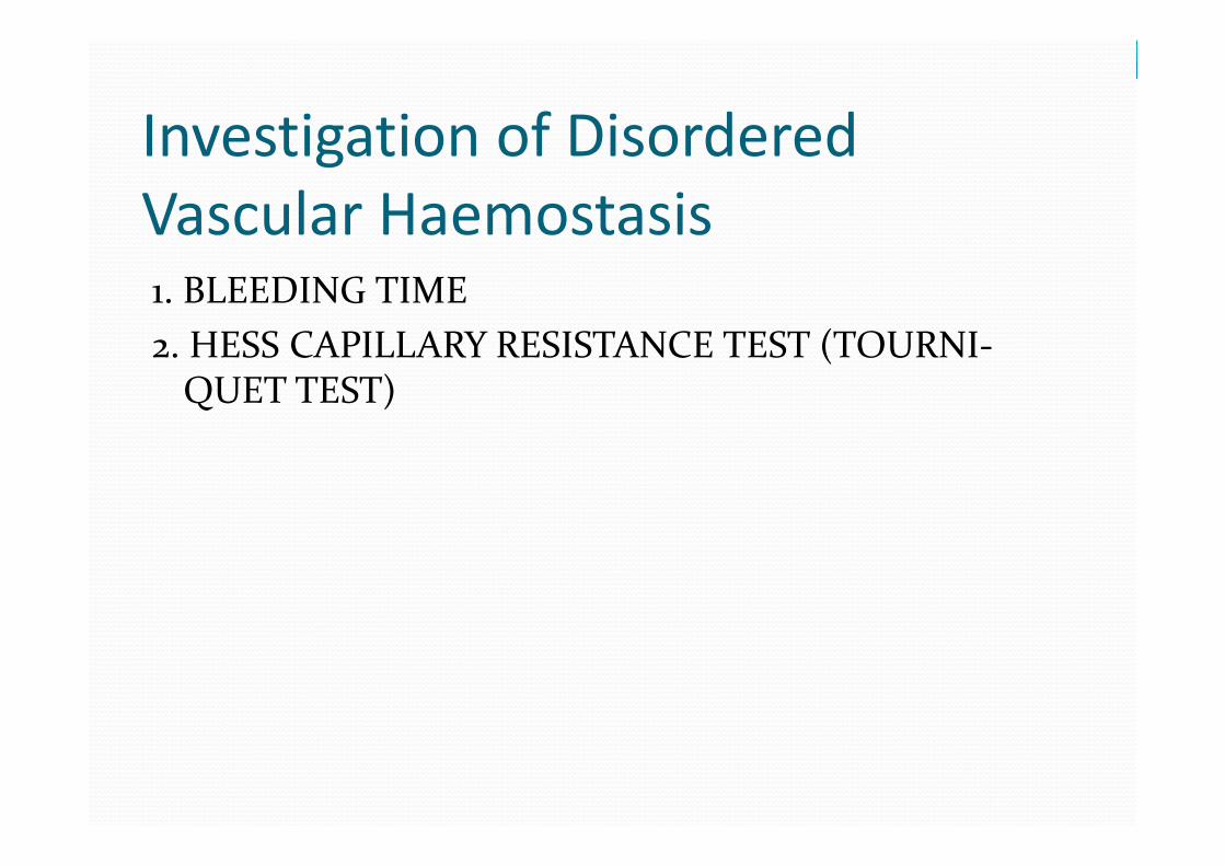
Investigation of Disordered Vascular Haemostasis 1. BLEEDING TIME
2. HESS CAPILLARY RESISTANCE TEST (TOURNI‐QUET TEST)
BLEEDING TIME Based on the principle of formation of haemostatic plug following a standard incision on skin.
Three methods: 1. Duke’s method: ear lobe puncture 2. Ivy’s method: 2‐3 punctures on volar aspect of forearm
with a lancet ( Cutting depth‐ 2‐2.5mm) under standardized venous pressure of 40mm Hg.
3. Template method: Larger cut, 6‐9mm long and 1mm deep.
The time the incision takes to stop bleeding is measured. Normal range is 3‐8 minutes.
A prolonged bleeding time may be due to following causes:
i) Thrombocytopenia. ii) Disorders of platelet function. iii) von Willebrand’s disease. iv) Vascular abnormalities (e.g. in Ehlers‐Danlos syndrome).
v) Severe deficiency of factor V and XI.

HESS CAPILLARY RESISTANCE TEST (TOURNIQUET TEST) This test is done by tying sphygmomanometer cuff to the upper arm and raising the pressure in it between diastolic and systolic for 5 minutes.
After deflation, the number of petechiae appearing in the next 5 minutes in 3 cm2 area over the cubital fossa are counted.
Presence of more than 20 petechiae is considered a positive test.
The test is positive in increased capillary fragility thrombocytopenia.


Investigation of Platelets and Platelet Function 1. SCREENING TESTS Skin bleeding time Peripheral blood platelet count.
Thrombocytopenia (<1.5lakhs/cumm) Hematological malignancies Ingestion of certain drugs DIC ITP Anaemia‐Megaloblastic anaemia, aplastic anaemia.
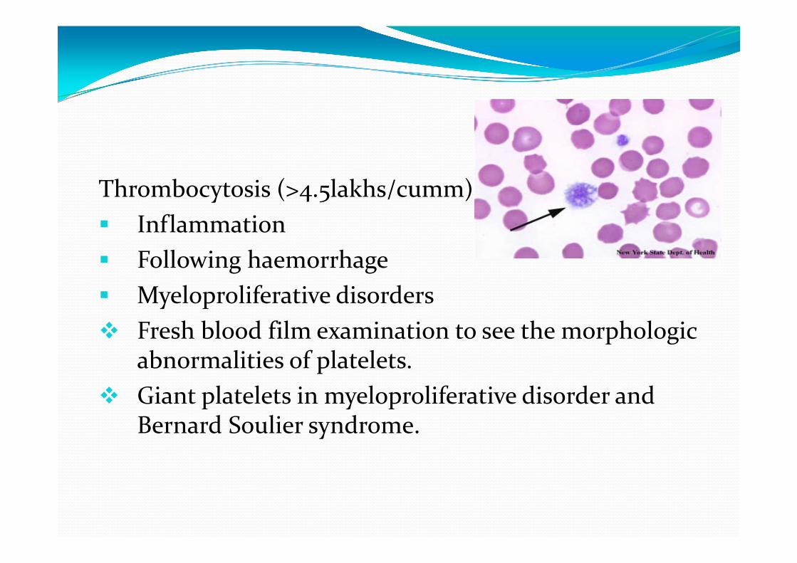
Thrombocytosis (>4.5lakhs/cumm) Inflammation
Following haemorrhage
Myeloproliferative disorders Fresh blood film examination to see the morphologic
abnormalities of platelets. Giant platelets in myeloproliferative disorder and
Bernard Soulier syndrome.
SPECIAL TESTS
Platelet adhesion test: Whole blood is made to pass over a column of non‐
siliconised glass beads. Proportion of platelets retained are assessed from
platelet count done before and after the passage of blood.
Less than 25% retention is usually observed in Von willebrand disease.
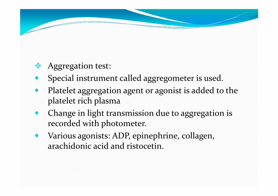
Aggregation test: Special instrument called aggregometer is used. Platelet aggregation agent or agonist is added to the
platelet rich plasma
Change in light transmission due to aggregation is recorded with photometer.
Various agonists: ADP, epinephrine, collagen, arachidonic acid and ristocetin.
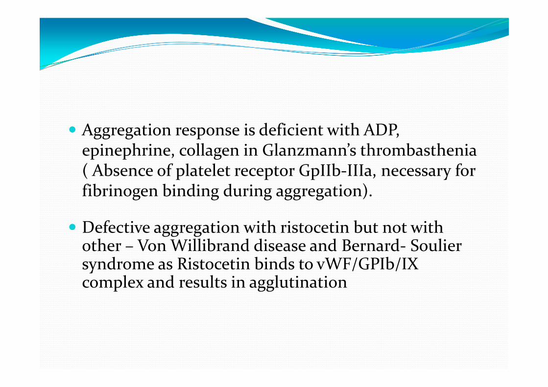
Aggregation response is deficient with ADP, epinephrine, collagen in Glanzmann’s thrombasthenia ( Absence of platelet receptor GpIIb‐IIIa, necessary for fibrinogen binding during aggregation).
Defective aggregation with ristocetin but not with other – Von Willibrand disease and Bernard‐ Soulier syndrome as Ristocetin binds to vWF/GPIb/IX complex and results in agglutination
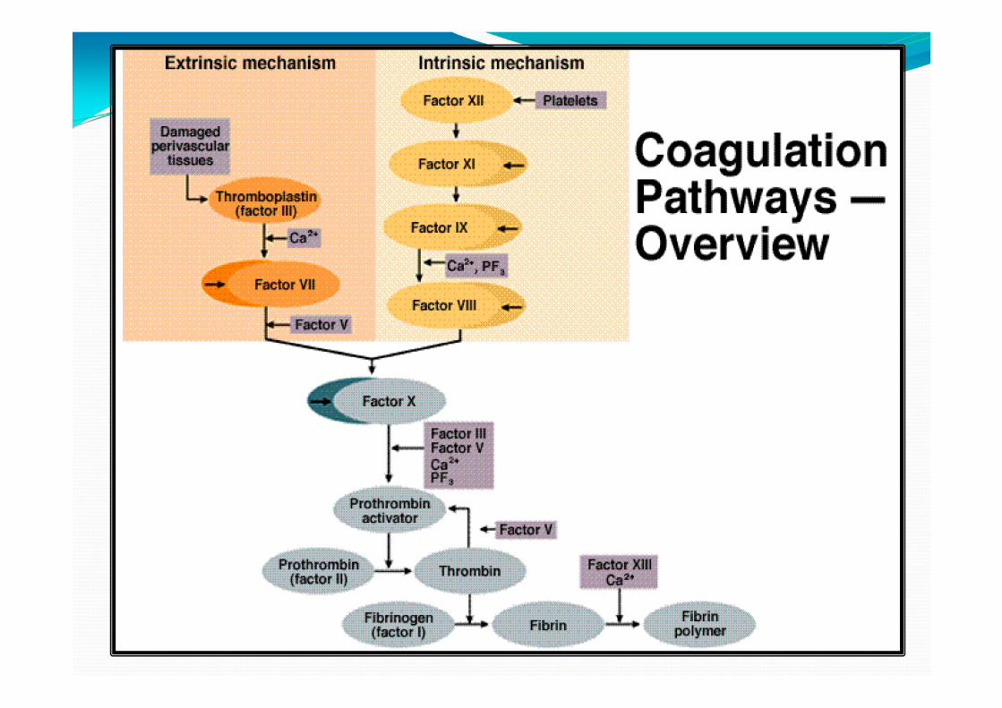

Investigation of Blood Coagulation 1. SCREENING TESTS Whole blood coagulation time done by various capillary and tube methods limited value, since it is an insensitive and
nonspecific test. Normal range is 4‐9 minutes at 37°C. One‐stage prothrombin time (PT) Activated partial thromboplastin time (APTT) or
partial thromboplastin time with kaolin (PTTK) Measurement of fibrinogen

One‐stage prothrombin time (PT) Extrinsic and common pathway
Tissue thromboplastin and calcium are added to platelet poor plasma.
Clotting time of mixture is noted. Commercial tissue thromboplastin is prepared from rabbit brain or lung.
Normal values depend upon the thromboplastin used With most rabbit thromboplastins the range of PT 11‐16 sec
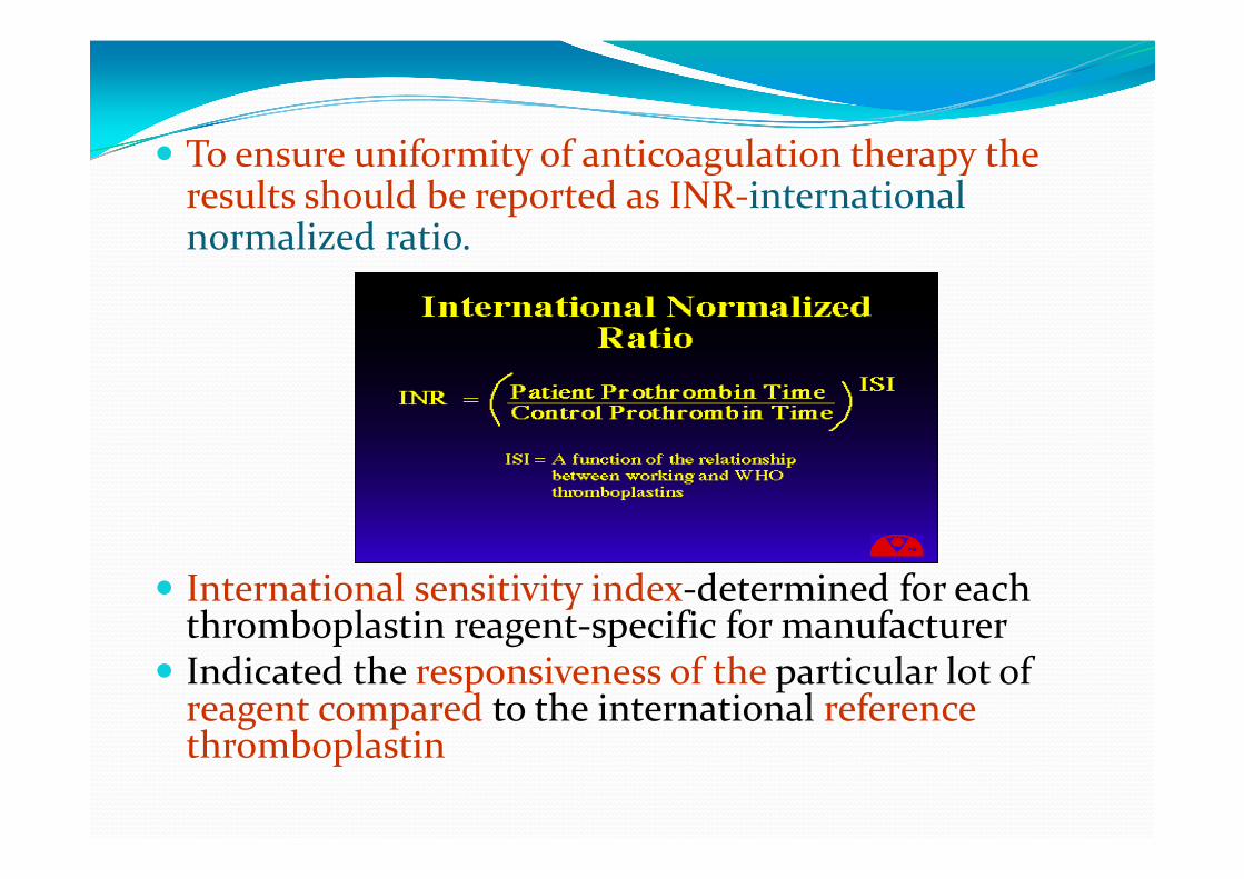
Nor·1nalized Ratio
INR (Patient Prothrombin Time_) ISi Control Prothrombin Time../
ISi = A function of the relationship betv.·een v.·orking and \ 'VHO tluo1nboplastins
To ensure uniformity of anticoagulation therapy the results should be reported as INR‐international normalized ratio.
International sensitivity index‐determined for each thromboplastin reagent‐specific for manufacturer
Indicated the responsiveness of the particular lot of reagent compared to the international reference thromboplastin
The common causes of prolonged one‐stage PT are as under:
i) Administration of oral anticoagulant drugs. ii) Liver disease, especially obstructive liver disease. iii) Vitamin K deficiency. iv) Disseminated intravascular coagulation.
Activated partial thromboplastin time (APTT) Monitor intrinsic and common pathway
Platelet poor plasma is incubated with an activator‐Kaolin, celite and silica.
Phospholipid or partial thromboplastin and calcium are then added.
Normal range is 30‐40 sec.
The common causes of a prolonged PTTK (or APTT) are as follows:
i) Parenteral administration of heparin. ii) Disseminated intravascular coagulation. iii) Liver disease. iv) Inherited def of F VIII or F IX
Thrombin time Thrombin reagent is added to platelet poor plasma. Time required for clot formation is noted. Normal range is 8‐12 sec. Prolongation occurs in
i) Disorder of fibrinogen: afibrinogenaemia, dysfibrinogemaemia.
ii) Chronic liver disease
iii) Increased level of fibrin degradation products.

SPECIAL TESTS 1. Coagulation factor assays: PT or APTT is performed using mixture of patient’s plasma and factor deficient plasma ( contain all coagulation factors, except one to be assayed).
The unknown level of the factor activity is compared with a standard control plasma with a known level of activity.
Results are expressed as percentage of normal activity. Normal level for all coagulation factors is 50‐150%

2. Quantitative assays. The coagulation factors can be quantitatively assayed by immunological and other chemical methods.

Investigation of Fibrinolytic System SCREENING TEST
1. Estimation of fibrinogen. 2. Fibrin degradation products (FDP) in the serum. 3. Ethanol gelation test. 4. Euglobin or whole blood lysis time.

Latex Agglutination test A suspension of latex particles coated with anti fibrinogen antibodies or with anti FDP is mixed with serum on a glass slide.
Agglutination of latex particles‐ positive test.

Specific tests Functional assays Immunological assays by ELISA
Chromogenic assays of plasminogen activators, plasminogen and FDP.
Related Documents


![Marinov - Anemia and haemorrhagic diatheses 2016 [Eng].ppt fileANEMIA Time Anemias due to impaired erythroproduction Impaired synthesis of haemoglobin Fe deficit Altered DNA synthesis](https://static.cupdf.com/doc/110x72/5e1cf6b432081363e478e702/marinov-anemia-and-haemorrhagic-diatheses-2016-engppt-time-anemias-due-to-impaired.jpg)