Bleeding Disorders. MS4.27.10.15 Abdallah Abbadi.MD.FRCP

Welcome message from author
This document is posted to help you gain knowledge. Please leave a comment to let me know what you think about it! Share it to your friends and learn new things together.
Transcript

Bleeding Disorders. MS4.27.10.15
Abdallah Abbadi.MD.FRCP

Hemostasis
BV Injury
PlateletAggregation
PlateletActivation
Blood VesselConstriction
CoagulationCascade
Stable Hemostatic Plug
Fibrin formation
Reduced
Blood flow
Damage/contact.
Primary hemostatic plug
NeuralContact

Laboratory Evaluation of the Coagulation Pathways
Partial thromboplastin time(PTT)
Prothrombin time(PT)
Intrinsic pathway Extrinsic pathway
Common pathwayThrombin time (TT)
Thrombin
Surface activating agent
(Ellagic acid, kaolin)PhospholipidCalcium
ThromboplastinTissue factorPhospholipid
Calcium
Fibrin clot F.XIII A

Bleeding Time(BT)
• 5-10% of patients have a prolonged bleeding time• Most of the prolonged bleeding times are due to aspirin
or drug ingestion• Prolonged bleeding time does not predict excess
surgical blood loss• Not recommended for routine testing in preoperative
patients• Prolonged in vWD• Prolonged in thrombocytopenia• Prolonged in Qualitative platelet disorders

Case 519 yr old male complains of repeated attacks of large joint painful swelling especially in his knees for several years, with limitation of movement of the l knee joint. P/E shown. His maternal uncle has similar condition.PT 14/14 s, PTT 80/31s, with mixing 40/32s. TT 12/12s, Plt 220K, BT 5mnts. F VIII <1%. F IX 100%.No VIII inhibitors. Genetic testing INT 22 INVS.
HA.

Case 5: Management & Follow-up1- Treat acute attack: FVIII* 30u/kg/ IV q 12 hrs x 2 days, then daily until it subsides. + Analgesics.2- Evaluate for ? Synovectomy (chemical or radio-isotope or surgical).Or Joint replacement.3- Consider for long term prophylaxis 20u/kg x 2 per week indefinitely.4- Education/ rehabilitation5- genetic counseling.6- Family screening and registration7- Screen for inhibitors x 2 per yr since therapy is different.
*FVIII: recombinant or ?plasma derived

Hemophilia A and B
Hemophilia A Hemophilia B
Coagulation factor deficiency Factor VIII Factor IX
Inheritance X-linked X-linkedrecessive recessive
Incidence 1/10,000 males 1/50,000 males
Severity Related to factor level<1% - Severe - spontaneous bleeding1-5% - Moderate - bleeding with mild injury5-25% - Mild - bleeding with surgery or trauma
Complications Soft tissue bleeding

Clinical Features of Bleeding Disorders
Platelet Coagulation Factors Disorders
Site of bleeding Skin Deep in soft tissuesMucous membranes (joints, muscles)
(epistaxis, gum,vaginal, GI tract)
Petechiae Yes No
Ecchymoses (“bruises”) Small, superficial Large, deep
Hemarthrosis / muscle bleeding Extremely rare Common
Bleeding after cuts & scratches Yes No
Bleeding after surgery or trauma Immediate, Delayed (1-2 days),usually mild often severe

Coagulation factor disorders
• Inherited bleeding disorders– Hemophilia A and B– vonWillebrands
disease– Other factor
deficiencies
• Acquired bleeding disorders– Liver disease– Vitamin K
deficiency/warfarin overdose
– DIC

The F8 gene
Human F8 gene maps to the most distal band (Xq28) of the long arm of the X chromosome
The gene is 186 Kb in length and comprises 26 exons.An intron 22 inversion is responsible for 45% of severe
hemophilia A and intron 1 inversion is responsible for 3 %of severe hemophilia A.
other reported mutation include deletion, insertion and point mutations causing nonsense, missense or splice site mutation.
F8 Intron 22 InversionThe F8 gene intron 22 inversion mutation arises from
homology recombination between copies of the intron 22 homology region(int22h-1”F8A+F8B”) and repeated telomeric DNA sequences outside the F8 gene ( int22h-2,int22h-3) on the long arm of chr.X
These two copies are located approximately 500Kb distal and telomeric to the F8gene.The int22h-1 and h2-, h-3 regions have 99% homology with one another.

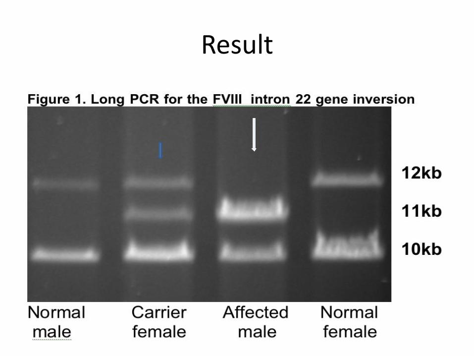
Result

Genetic Screening: Results for Hemophilia A (HA)
Type of Mutation Number of Families
Numberof Patients
% of families
% of patients
Itnron 22 inversion 25 70 53% 38.8Intron 1 inversion 1 1 2.4% 0.5
Missense 16 95 36% 52.7Frameshift
(Insertion/deletion)
4 14 9.5% 7
Total 46 180
F8 gene mutation profile of all Jordanian hemophilia A patients examined.
• HA causative mutations identified in tested patients
*Awidi A et al: Haemophilia. 2010
16(1):136-42

SerialNO. Disorder Total
NO. M F M F MSEVERE
<1MODERATE
1-5MILD>5
1 Haemophilia-A 259 259 0 8 0 2.3 150 35 74
2 Haemophilia-B 66 66 0 7.1 0 4.45 55 5 6
3 VWD 151 62 89 132221.5 17.25 __ ___ ___
4 Glanzmann 112 47 65 12 8.2 10.1 __ ___ ___
5
TOTAL NO. OF COMMON-BLEEDING DISORDER
588 434 145 ___ ___ ___ 205 40 80
Thrombosis&Haemostas lab (THL)Medical school / Jordan University
Common Bleeding Disorders End of July 2015x̅ Age/Yr of diagnosis at THL Severity %

Treatment of Severe/ Moderate hemophilia
1- On demand/hospital based2- On demand/home based
3- Prophylactic/ home/ intermittent X 2 per week4- Treatment of target joint5- Physiotherapy6- Genetic counseling 7- Education

Dosing guidelines for hemophilia A• Mild bleeding
– Target: 30% dosing q8-12h; 1-2 days (15U/kg)– Hemarthrosis, oropharyngeal or dental, epistaxis, hematuria
• Major bleeding– Target: 80-100% q8-12h; 7-14 days (50U/kg)– CNS trauma, hemorrhage, lumbar puncture– Surgery– Retroperitoneal hemorrhage– GI bleeding
• Adjunctive therapy– Tranexemic acid(Cyclokapron) or DDAVP (for mild disease only)

Complications of therapy• Formation of inhibitors (antibodies)
– 10-15% of severe hemophilia A patients– 1-2% of severe hemophilia B patients
• Viral infections-Hepatitis B -Human parvovirus-Hepatitis C -Hepatitis A-HIV -Others (Prion disease or BSE)

Treatment of hemophilia B
• Agent – High purity factor IX– Recombinant human factor IX
• Dose– Initial dose: 100U/kg– Subsequent: 50 U/kg every 24 hours
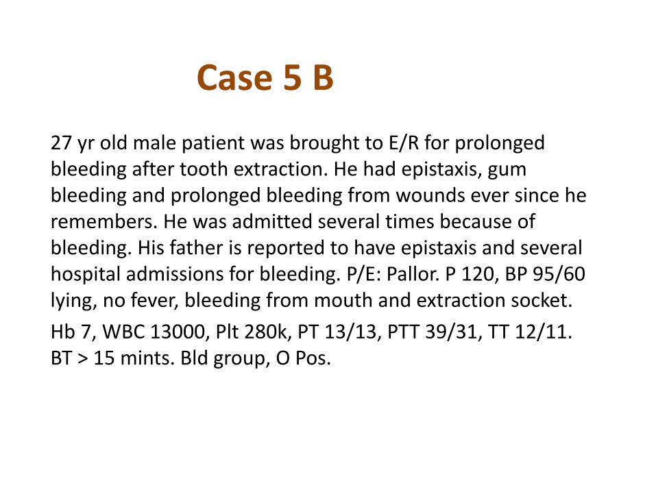
Case 5 B 27 yr old male patient was brought to E/R for prolonged bleeding after tooth extraction. He had epistaxis, gum bleeding and prolonged bleeding from wounds ever since he remembers. He was admitted several times because of bleeding. His father is reported to have epistaxis and several hospital admissions for bleeding. P/E: Pallor. P 120, BP 95/60 lying, no fever, bleeding from mouth and extraction socket.Hb 7, WBC 13000, Plt 280k, PT 13/13, PTT 39/31, TT 12/11. BT > 15 mints. Bld group, O Pos.
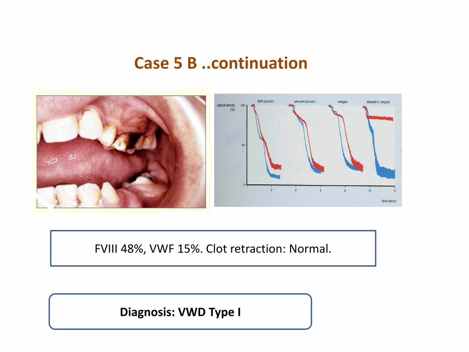
Case 5 B ..continuation
FVIII 48%, VWF 15%. Clot retraction: Normal.
Diagnosis: VWD Type I

Case 5 B VWF Multimers
NP 1 2B 32A 2Aplt

Management of Case 5 B
1- Cryoprecipitate 1 bag/ per 10 kg body weight x 2 day for 3-4 days then daily for 3 more days.2- Dental consultation/ mouth hygiene & care.3-Education and counseling.4- Screening of family. 5- ?? DDAVP for therapy of mild bleeding

von Willebrand Disease: Clinical Features
• von Willebrand factor– Synthesis in endothelium and megakaryocytes– Forms large multimer – Carrier of factor VIII– Anchors platelets to subendothelium– Bridge between platelets
• Inheritance - autosomal dominant• Incidence - 1/10,000• Clinical features - mucocutaneous bleeding

VWF VWF VWF VWF
VIII
t1/2 = 12-20 h
Normal Blood Clotting
Normal level of VIII
t1/2 ≤ 2 h
Bleeding
VIII
≤ 10% of normal VIII
VWF and Factor VIII Survival
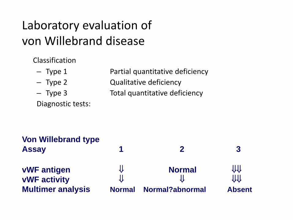
Laboratory evaluation of von Willebrand disease
Classification– Type 1 Partial quantitative deficiency– Type 2 Qualitative deficiency– Type 3 Total quantitative deficiencyDiagnostic tests:
Von Willebrand typeAssay 1 2 3
vWF antigen ⇓ Normal ⇓⇓vWF activity ⇓ ⇓ ⇓⇓Multimer analysis Normal Normal?abnormal Absent

VWF Multimers
NP 1 2B 32A 2Aplt

Treatment of von Willebrand diseaseVaries by Classification
• Cryoprecipitate– Source of fibrinogen, factor VIII and VWF– Only plasma fraction that consistently contains VWF multimers– Correction of bleeding time is variable
• DDAVP (Deamino-8-arginine vasopressin)– Increases plasma VWF levels by stimulating secretion from
endothelium– Duration of response is variable– Used for type 1 disease– Dosage 0.3 µg/kg q 12 hr IV
• Factor VIII concentrate (Humate-P)– Virally inactivated product– Used for type 2 and 3

Immune-mediated:• Lymphoproliferative disease (MM, MGUS)• Autoimmune disease (SLE)• May respond to DDAVP, IVIG, factor concentrate
Proteolysis-mediated:• Thrombocytosis (reactive or myeloproliferative
disorders)• Shear-induced (aortic stenosis)• Correct the underlying disorder
Acquired von Willebrand Syndrome

Case 5 C
37 yr old lady was admitted with high fever, chills, rigors and severe dysuria.P/E shown. Temp 40.5,BP 80/50, P122 regular, low volume. Bleeding from needle puncture sites and bruising. Hb 9g/dl, retcs 6%, bilirubin 5 (d1), WBC 19k, Plt 25k, PT >50s, PTT > 100s, TT >30s, D-Dimer +++, Creatinine 2.3. Bld film shown.Fibrinogen. 30mg/dl.

Pathogenesis of DIC

Common clinical conditions associated withDisseminated Intravascular Coagulation
• Sepsis
• Trauma– Head injury– Fat embolism
• Malignancy
• Obstetrical complications– Amniotic fluid embolism– Abruptio placentae
• Vascular disorders
• Reaction to toxin (e.g. snake venom, drugs)
• Immunologic disorders– Severe allergic reaction– Transplant rejection
Activation of both coagulation and fibrinolysisTriggered by

Disseminated Intravascular Coagulation (DIC)Mechanism
Systemic activationof coagulation
Intravasculardeposition of fibrin
Depletion of plateletsand coagulation factors
BleedingThrombosis of smalland midsize vessels
with organ failure

Pathogenesis of DIC
Coagulation Fibrinolysis
Fibrinogen
FibrinMonomers
FibrinClot
(intravascular)
Fibrin(ogen)Degradation
Products
Plasmin
Thrombin Plasmin
Release of thromboplastic
material intocirculation
Consumption ofcoagulation factors;
presence of FDPs↑ aPTT↑ PT↑ TT
↓ Fibrinogen
Presence of plasmin↑ FDP
Intravascular clot↓ Platelets
Schistocytes

Case 5 C: treatment and follow-up
1- Treat vigorously with IV antibiotics after blood, urine culture and septic work-up2- Hydrate and ensure adequate urine output3- ? ICU care4- Replace missing clotting factors: FFP 10 ml/kg frequency to be determined as needed5-Plt replacement6- Monitor PT, PTT, D-Dimer and fbgn, Plt count7- Investigate cause of uro-sepsis.8- TTP can easily be excluded.

Disseminated Intravascular Coagulation:Treatment approaches
• Treatment of underlying disorder
• Platelet transfusion
• Fresh frozen plasma
• Coagulation inhibitor concentrate (ATIII)• Anticoagulation with heparin

Disseminated Intravascular CoagulationTreatment approaches
• Treatment of underlying disorder
• Platelet transfusion
• Fresh frozen plasma
• ?Coagulation inhibitor concentrate (ATIII)• ??Anticoagulation with heparin
Related Documents