Citation: Awadelkareem, A.M.; Al-Shammari, E.; Elkhalifa, A.O.; Adnan, M.; Siddiqui, A.J.; Patel, M.; Khan, M.I.; Mehmood, K.; Ashfaq, F.; Badraoui, R.; et al. Biosynthesized Silver Nanoparticles from Eruca sativa Miller Leaf Extract Exhibits Antibacterial, Antioxidant, Anti-Quorum-Sensing, Antibiofilm, and Anti-Metastatic Activities. Antibiotics 2022, 11, 853. https:// doi.org/10.3390/antibiotics11070853 Academic Editors: Delia Muntean and Silvana Vulpie Received: 7 June 2022 Accepted: 23 June 2022 Published: 25 June 2022 Publisher’s Note: MDPI stays neutral with regard to jurisdictional claims in published maps and institutional affil- iations. Copyright: © 2022 by the authors. Licensee MDPI, Basel, Switzerland. This article is an open access article distributed under the terms and conditions of the Creative Commons Attribution (CC BY) license (https:// creativecommons.org/licenses/by/ 4.0/). antibiotics Article Biosynthesized Silver Nanoparticles from Eruca sativa Miller Leaf Extract Exhibits Antibacterial, Antioxidant, Anti-Quorum-Sensing, Antibiofilm, and Anti-Metastatic Activities Amir Mahgoub Awadelkareem 1 , Eyad Al-Shammari 1 , AbdElmoneim O. Elkhalifa 1 , Mohd Adnan 2 , Arif Jamal Siddiqui 2 , Mitesh Patel 3 , Mohammad Idreesh Khan 4 , Khalid Mehmood 5 , Fauzia Ashfaq 6 , Riadh Badraoui 2 and Syed Amir Ashraf 1, * 1 Department of Clinical Nutrition, College of Applied Medical Sciences, University of Hail, Hail P.O. Box 2440, Saudi Arabia; [email protected] (A.M.A.); [email protected] (E.A.-S.); [email protected] (A.O.E.) 2 Department of Biology, College of Science, University of Hail, Hail P.O. Box 2440, Saudi Arabia; [email protected] (M.A.); [email protected] (A.J.S.); [email protected] (R.B.) 3 Department of Biotechnology, Parul Institute of Applied Sciences and Centre of Research for Development, Parul University, Vadodara 391760, Gujarat, India; [email protected] 4 Department of Clinical Nutrition, College of Applied Health Sciences in Arras, Qassim University, Buraydah 58883, Saudi Arabia; [email protected] 5 Department of Pharmaceutics, College of Pharmacy, University of Hail, Hail P.O. Box 2440, Saudi Arabia; [email protected] 6 Department of Clinical Nutrition, College of Applied Medical Sciences, Jazan University, Jazan 45142, Saudi Arabia; [email protected] * Correspondence: [email protected] Abstract: Worldwide, the primary problem today is the proliferation of cancer and secondary bacterial infections caused by biofilms, as they are the principal causes of death due to the lack of effective drugs. A great deal of biological activities of silver nanoparticles (AgNPs) have made them a brilliant choice for the development of new drugs in recent years. The present study was conducted to evaluate the anticancer, antibacterial, anti-QS, and antibiofilm effects of AgNPs synthesized from Eruca sativa (E. sativa) leaf extract. The ultraviolet–visible (UV–Vis) spectra showed a peak of surface plasmon resonance at 424 nm λmax, which corresponded to AgNP formation. The Fourier transform infrared spectroscopy (FT-IR) confirmed that biological moieties are involved for the development of AgNPs. Moreover, transmission electron microscopy (TEM) analyses confirmed the spherical shape and uniform size (8.11 to 15 nm) of the AgNPs. In human lung cancer cells (A549), the anticancer potential of AgNPs was examined by the MTT [3-dimethylthiazol-2-yl)-2,5-diphenyltetrazolium bromide] assay, scratch assay, and invasion assay. The results indicated that AgNPs inhibit the migration of A549 cells. The synthesized AgNPs showed MIC values of 12.5 μg/mL against Chromobacterium violaceum (C. violaceum) and 25 μg/mL against Pseudomonas aeruginosa (P. aeruginosa), which demonstrated their antibacterial abilities. Biological compounds that disable the QS system are being investigated as potential strategies for preventing bacterial infections. Thus, we analyzed the potential effectiveness of synthesized AgNPs in inhibiting QS-regulated virulence factors and biofilm formation in both strains of bacteria. In C. violaceum, the synthesized AgNPs significantly inhibited both violacein (85.18% at 1/2 × MIC) and acyl homoserine lactone (78.76% at 1/2 × MIC). QS inhibitory activity was also demonstrated in P. aeruginosa at a sub-MIC concentration (1/2 × MIC) by a reduction in pyocyanin activity (68.83%), total protease (68.50%), LasA activity (63.91%), and LasB activity (56.40%). Additionally, the exopolysaccharide production was significantly reduced in both C. violaceum (65.79% at 1/2 × MIC) and P. aeruginosa (57.65% at 1/2 × MIC). The formation of biofilm was also significantly inhibited at 1/2 × MIC in C. violaceum (76.49%) and in P. aeruginosa (65.31%). Moreover, a GC–MS analysis confirmed the presence of different classes of bioactive phytochemical constituents present in the leaf extract of E. sativa. On the basis of our results, we conclude that biologically synthesized Antibiotics 2022, 11, 853. https://doi.org/10.3390/antibiotics11070853 https://www.mdpi.com/journal/antibiotics

Welcome message from author
This document is posted to help you gain knowledge. Please leave a comment to let me know what you think about it! Share it to your friends and learn new things together.
Transcript
Citation: Awadelkareem, A.M.;
Al-Shammari, E.; Elkhalifa, A.O.;
Adnan, M.; Siddiqui, A.J.; Patel, M.;
Khan, M.I.; Mehmood, K.; Ashfaq, F.;
Badraoui, R.; et al. Biosynthesized
Silver Nanoparticles from Eruca sativa
Miller Leaf Extract Exhibits
Antibacterial, Antioxidant,
Anti-Quorum-Sensing, Antibiofilm,
and Anti-Metastatic Activities.
Antibiotics 2022, 11, 853. https://
doi.org/10.3390/antibiotics11070853
Academic Editors: Delia Muntean
and Silvana Vulpie
Received: 7 June 2022
Accepted: 23 June 2022
Published: 25 June 2022
Publisher’s Note: MDPI stays neutral
with regard to jurisdictional claims in
published maps and institutional affil-
iations.
Copyright: © 2022 by the authors.
Licensee MDPI, Basel, Switzerland.
This article is an open access article
distributed under the terms and
conditions of the Creative Commons
Attribution (CC BY) license (https://
creativecommons.org/licenses/by/
4.0/).
antibiotics
Article
Biosynthesized Silver Nanoparticles from Eruca sativaMiller Leaf Extract Exhibits Antibacterial, Antioxidant,Anti-Quorum-Sensing, Antibiofilm, andAnti-Metastatic ActivitiesAmir Mahgoub Awadelkareem 1, Eyad Al-Shammari 1, AbdElmoneim O. Elkhalifa 1 , Mohd Adnan 2 ,Arif Jamal Siddiqui 2 , Mitesh Patel 3 , Mohammad Idreesh Khan 4 , Khalid Mehmood 5 , Fauzia Ashfaq 6 ,Riadh Badraoui 2 and Syed Amir Ashraf 1,*
1 Department of Clinical Nutrition, College of Applied Medical Sciences, University of Hail,Hail P.O. Box 2440, Saudi Arabia; [email protected] (A.M.A.); [email protected] (E.A.-S.);[email protected] (A.O.E.)
2 Department of Biology, College of Science, University of Hail, Hail P.O. Box 2440, Saudi Arabia;[email protected] (M.A.); [email protected] (A.J.S.); [email protected] (R.B.)
3 Department of Biotechnology, Parul Institute of Applied Sciences and Centre of Research for Development,Parul University, Vadodara 391760, Gujarat, India; [email protected]
4 Department of Clinical Nutrition, College of Applied Health Sciences in Arras, Qassim University,Buraydah 58883, Saudi Arabia; [email protected]
5 Department of Pharmaceutics, College of Pharmacy, University of Hail, Hail P.O. Box 2440, Saudi Arabia;[email protected]
6 Department of Clinical Nutrition, College of Applied Medical Sciences, Jazan University,Jazan 45142, Saudi Arabia; [email protected]
* Correspondence: [email protected]
Abstract: Worldwide, the primary problem today is the proliferation of cancer and secondary bacterialinfections caused by biofilms, as they are the principal causes of death due to the lack of effectivedrugs. A great deal of biological activities of silver nanoparticles (AgNPs) have made them a brilliantchoice for the development of new drugs in recent years. The present study was conducted to evaluatethe anticancer, antibacterial, anti-QS, and antibiofilm effects of AgNPs synthesized from Eruca sativa(E. sativa) leaf extract. The ultraviolet–visible (UV–Vis) spectra showed a peak of surface plasmonresonance at 424 nm λmax, which corresponded to AgNP formation. The Fourier transform infraredspectroscopy (FT-IR) confirmed that biological moieties are involved for the development of AgNPs.Moreover, transmission electron microscopy (TEM) analyses confirmed the spherical shape anduniform size (8.11 to 15 nm) of the AgNPs. In human lung cancer cells (A549), the anticancer potentialof AgNPs was examined by the MTT [3-dimethylthiazol-2-yl)-2,5-diphenyltetrazolium bromide] assay,scratch assay, and invasion assay. The results indicated that AgNPs inhibit the migration of A549cells. The synthesized AgNPs showed MIC values of 12.5 µg/mL against Chromobacterium violaceum(C. violaceum) and 25 µg/mL against Pseudomonas aeruginosa (P. aeruginosa), which demonstrated theirantibacterial abilities. Biological compounds that disable the QS system are being investigated aspotential strategies for preventing bacterial infections. Thus, we analyzed the potential effectivenessof synthesized AgNPs in inhibiting QS-regulated virulence factors and biofilm formation in bothstrains of bacteria. In C. violaceum, the synthesized AgNPs significantly inhibited both violacein(85.18% at 1/2 × MIC) and acyl homoserine lactone (78.76% at 1/2 × MIC). QS inhibitory activitywas also demonstrated in P. aeruginosa at a sub-MIC concentration (1/2 × MIC) by a reduction inpyocyanin activity (68.83%), total protease (68.50%), LasA activity (63.91%), and LasB activity (56.40%).Additionally, the exopolysaccharide production was significantly reduced in both C. violaceum (65.79%at 1/2 × MIC) and P. aeruginosa (57.65% at 1/2 × MIC). The formation of biofilm was also significantlyinhibited at 1/2 × MIC in C. violaceum (76.49%) and in P. aeruginosa (65.31%). Moreover, a GC–MSanalysis confirmed the presence of different classes of bioactive phytochemical constituents presentin the leaf extract of E. sativa. On the basis of our results, we conclude that biologically synthesized
Antibiotics 2022, 11, 853. https://doi.org/10.3390/antibiotics11070853 https://www.mdpi.com/journal/antibiotics
Antibiotics 2022, 11, 853 2 of 28
AgNPs showed numerous multifunctional properties and have the potential to be used againsthuman cancer and bacterial biofilm-related infections.
Keywords: silver nanoparticle; anticancer; antibiofilm; Eruca sativa; nutraceuticals; bioactive compound
1. Introduction
In recent years, cancer has been ranked as one of the primary causes of mortalityaround the world [1]. According to the reports, in the year 2020 almost 19.3 million freshcases of cancer (19.1 million without counting nonmelanoma skin cancer) and nearly10 million cancer deaths occurred (9.9 million without counting nonmelanoma skin cancer).Cancer deaths due to lung disease accounted for 18% of all cancer-related deaths, followedby colorectal, liver, stomach, and breast tumors [2]. Therefore, the global burden of cancer isincreasing day by day and is projected to reach 28.4 million cases by the end of 2040, a 47%rise from 2020. In addition, the aggravation in cancer cases could occur due to increasingrisk factors linked with urbanization and an expanding economy [3].
There are several factors that contribute to cancer’s high mortality rates, such as poordiagnosis at an early stage, and associated side effects along with the chemotherapeutictreatments [4]. As a result of chemotherapy, a patient’s immune system often becomescompromised. Patients are at risk for secondary infection from bacteria, viruses, fungi, andother pathogens. Antibiotic resistance is a concerning aspect of secondary infection in cancerpatients [5]. Researchers have recently focused on developing nanoparticles (NPs) derivedfrom metals in order to target and treat a varied range of diseases and other pathologicalconditions, such as cancer and secondary infections [6]. Due to favorable physico-chemicalproperties that can be related to biological systems, silver and gold nanoparticles aredistinguished from those of noble metals [7]. Silver nanoparticles (AgNPs) are beingextensively examined for their potential use as medical implants, food packaging, andenvironmental pollutants [8]. A number of carcinoma cell lines are reported to be cytotoxicto AgNPs, including MCF-7 (breast cancer), HeLa (cervical cancer), HT29 (colon cancer),and A549 (lung cancer) [9–11]. Moreover, both the scientific literature and traditionalmedicine have described the antimicrobial potential of AgNPs [12–14]. Biologically activesilver ions are released during silver ionization in aqueous solution, which is responsiblefor AgNPs’ antimicrobial activity [12,13]. Now, scientists are focused on developingnanoparticles with certain biological activities, such as selective antibacterial, antibiofilmas well as anticancer properties. This combination of properties will be useful for treatingcancer and associated infections with a single nanoparticle.
Several physico-chemical methods have been described for the synthesis of AgNPs ofdifferent types, shapes, sizes, and crystalline materials based on research and applications.Unfortunately, these physico-chemical methods are expensive, time-consuming and energy-intensive, as well as generating large quantities of toxic by-products [15,16]. The greensynthesis route, on the other hand, allows AgNPs to be synthesized in a natural, economical,and environmentally friendly manner [17,18]. Plant bioactive components are a newpromising source for AgNPs production rather than bacterial or chemical methods, sincethere is less or no risk of bacterial contamination and there is less energy consumption andhigher applicability [19,20]. According to earlier reports, biologically synthesized AgNPshave been reported for their antimicrobial as well as anticancer properties [21,22].
Eruca sativa Miller (E. sativa) also known as ‘arugula, or rocket leaves’ is a nutritioussalad plant of the Brassicaceae family. It is often used in food and medicine because of itsaromatic qualities. The traditional use of this plant includes the stimulation of fertility andsperm production, enhancing the digestive and urinary processes, as well as fighting againsteye infections [23]. The plant has recently received a lot of attention because of its highphytoactive compounds and its biological significance in various biological activities. Avariety of phytochemicals are present in the plant’s various parts, including glucosinolates,

Antibiotics 2022, 11, 853 3 of 28
tannins, phenolics, saponins, flavonoids, and essential oils [24]. The plant has been reportedto possess several beneficial medicinal properties, including antimicrobial, antioxidant,anti-acne, anti-diabetic, antigenotoxic, anticancer, analgesic, anti-hyperlipidemia, anti-hyperglycemia, anti-hyperuricemia, and anti-inflammatory properties [25–30]. The leafextract of E. sativa has been reported to change the silver ion oxidation state from Ag+ toAg0 and thereby AgNPs can act as synthesizing agents without the inclusion of a reducingagent in the reaction mixture [31].
In the present study, we developed AgNPs by means of the E. sativa leaf extract as a bio-template to synthesize an eco-friendly and economical material under ambient conditions.To confirm the synthesis and the morphology of the generated AgNPs, their characterizationwas carried out via different biophysical methods. Furthermore, newly developed AgNPswere investigated for their anticancer as well as anti-metastatic potential against humanlung cancer cells (A549) via the MTT (3-(4,5-dimethylthiazol2-yl)-2,5-diphenyltetrazoliumbromide) assay, scratch assay, and invasion assay. The antibacterial, anti-QS and antibiofilmpotential of the synthesized AgNPs was also investigated against P. aeruginosa and C.violaceum. The QS phenomenon such as violacein, AHL, pyocyanin, LasA, LasB, protease,biofilm formation and EPS productions were evaluated in the presence of E. sativa-cappedAgNPs. Moreover, the antioxidant potential against 2,2-diphenyl-1-picryl-hydrazyl-hydrate(DPPH) and hydrogen peroxide (H2O2) radicals was evaluated, and the identification ofthe phytochemicals present in E. sativa leaf extract was also carried out via GC–MS analysis.
2. Results2.1. Synthesis and Characterization of AgNPs
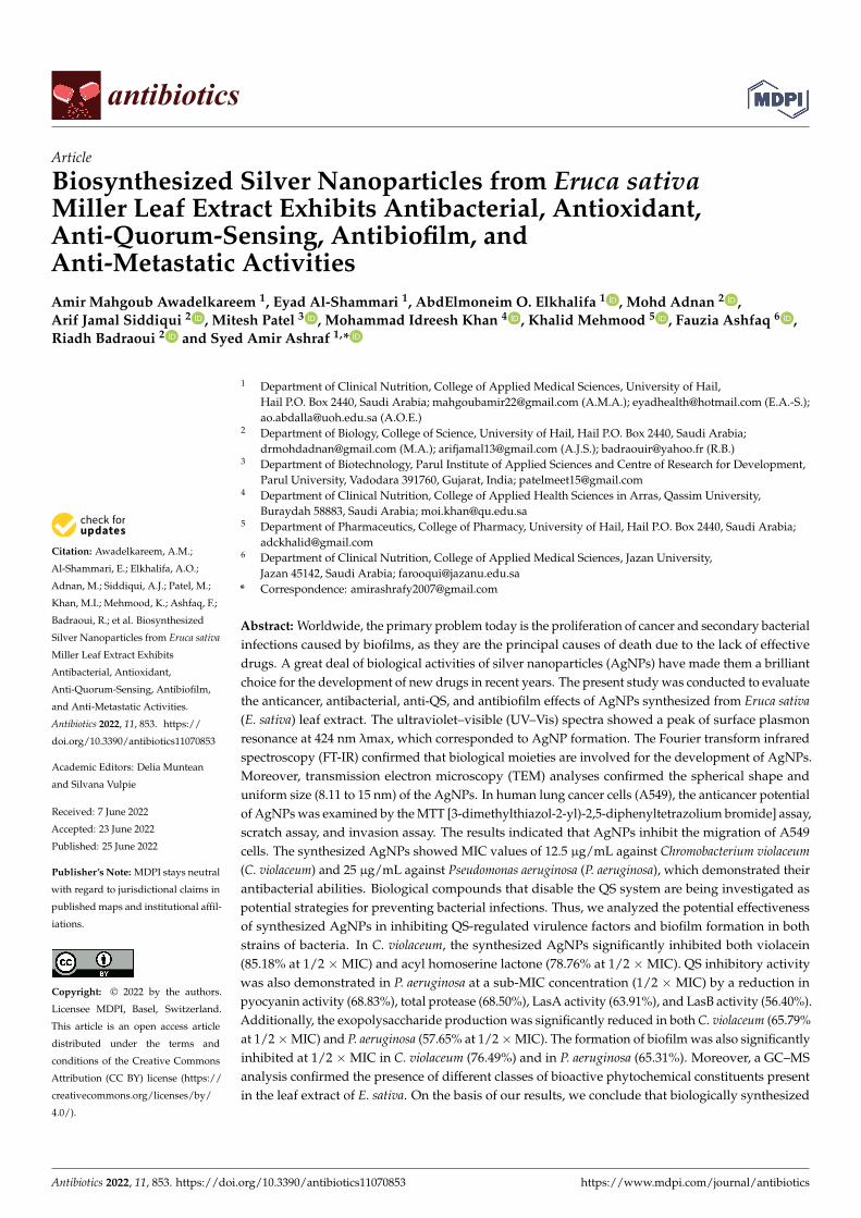
The leaf extract of E. sativa is an inexpensive and readily available plant ingredient.We synthesized the AgNPs and then characterized them for further application in clinical,food and pharmaceutical industries, etc. UV–visible spectroscopy is a significant andextensively used system for determining the formation of AgNPs in aqueous solutions. Inthis study, UV–Vis analysis was used to observe the formation and stability of the AgNPssynthesized from E. sativa leaf extract. A spectroscopic measurement of AgNPs synthe-sized after 24 h revealed a clearly symmetric absorption spectrum with peak maximum at424 nm (Figure 1A). Free electrons in AgNPs contribute to the absorption band appearance,which is caused by their mutual vibration of light wave incidences, thus causing a SurfacePlasmon Resonance (SPR) absorption band. After two weeks of synthesis, spectroscopicmeasurements were also performed and the synthesized NPs showed no noticeable varia-tion in the spectroscopic results, which indicates their stability. This suggests that the E.sativa leaf extract could be acting as a capping agent or a binding agent to provide stabilityto AgNPs.
FT-IR explorations were conducted to investigate the capping ability of the E. sativaleaf extract on AgNP surfaces. Figure 1B shows the FT-IR spectra of pure leaf extract andbiosynthesized AgNPs from E. sativa leaf extract. The FT-IR spectrum of fresh E. sativaextract displayed peaks at around 3346, 2132 and 1636 cm−1, which correspond to the aminegroup’s N-H/O-H vibration stretch (around 3300 cm−1), C ≡ C (around 2100 cm−1) as wellas C = C (around 1635 cm−1). The AgNPs synthesized using E. sativa extract showed peaksat around 3351, 2118 and 1642 cm−1, corresponding to the functional groups of amine N-H/O-H vibration stretch (around 3370 cm−1), C ≡ C stretch (around 2100 cm−1) and amideC = O (around 1640 cm−1). Therefore, the FT-IR spectrum revealed that the functionalgroups -NH, -OH and -C = O were significant in the reduction of Ag+ to Ag0, and thewidening of the peak heights in the spectrum of AgNPs authenticate that the E. sativa leafextract was present on the surface of the AgNPs. It may help to stabilize the developedparticles and prevent accumulation and oozing in the medium by saturating them with theextract from E. sativa leaves.
Antibiotics 2022, 11, 853 4 of 28Antibiotics 2022, 11, 853 4 of 31
Figure 1. Characterization of E. sativa‐leaf‐extract‐capped silver nanoparticles (AgNPs). (A). UV–
visible absorption spectra of synthesized AgNPs. (B). FT‐IR pattern of E. sativa leaf extract and syn‐
thesized AgNPs. (C). Morphological analysis of synthesized AgNPs with variable diameter using
TEM micrograph.
FT‐IR explorations were conducted to investigate the capping ability of the E. sativa
leaf extract on AgNP surfaces. Figure 1B shows the FT‐IR spectra of pure leaf extract and
biosynthesized AgNPs from E. sativa leaf extract. The FT‐IR spectrum of fresh E. sativa
extract displayed peaks at around 3346, 2132 and 1636 cm−1, which correspond to the
amine group’s N‐H/O‐H vibration stretch (around 3300 cm−1), C ≡ C (around 2100 cm−1) as
well as C = C (around 1635 cm−1). The AgNPs synthesized using E. sativa extract showed
peaks at around 3351, 2118 and 1642 cm−1, corresponding to the functional groups of
amine N‐H/ O‐H vibration stretch (around 3370 cm−1), C ≡ C stretch (around 2100 cm−1)
and amide C = O (around 1640 cm−1). Therefore, the FT‐IR spectrum revealed that the
functional groups ‐NH, ‐OH and ‐C = O were significant in the reduction of Ag+ to Ag0,
and the widening of the peak heights in the spectrum of AgNPs authenticate that the E.
sativa leaf extract was present on the surface of the AgNPs. It may help to stabilize the
developed particles and prevent accumulation and oozing in the medium by saturating
them with the extract from E. sativa leaves.
Moreover, TEM images were also analyzed to investigate the detailed morphology
of newly developed AgNPs. Figure 1C shows TEM images of synthesized AgNPs. The
developed particles were found to be of sizes ranging from 8.11 to 15 nm. Overall, all of
these results indicate that the AgNPs were prepared and stabilized successfully.
Figure 1. Characterization of E. sativa-leaf-extract-capped silver nanoparticles (AgNPs). (A) UV–visible absorption spectra of synthesized AgNPs. (B) FT-IR pattern of E. sativa leaf extract andsynthesized AgNPs. (C) Morphological analysis of synthesized AgNPs with variable diameter usingTEM micrograph.
Moreover, TEM images were also analyzed to investigate the detailed morphologyof newly developed AgNPs. Figure 1C shows TEM images of synthesized AgNPs. Thedeveloped particles were found to be of sizes ranging from 8.11 to 15 nm. Overall, all ofthese results indicate that the AgNPs were prepared and stabilized successfully.
2.2. Anticancer Potential of Synthesized AgNPs
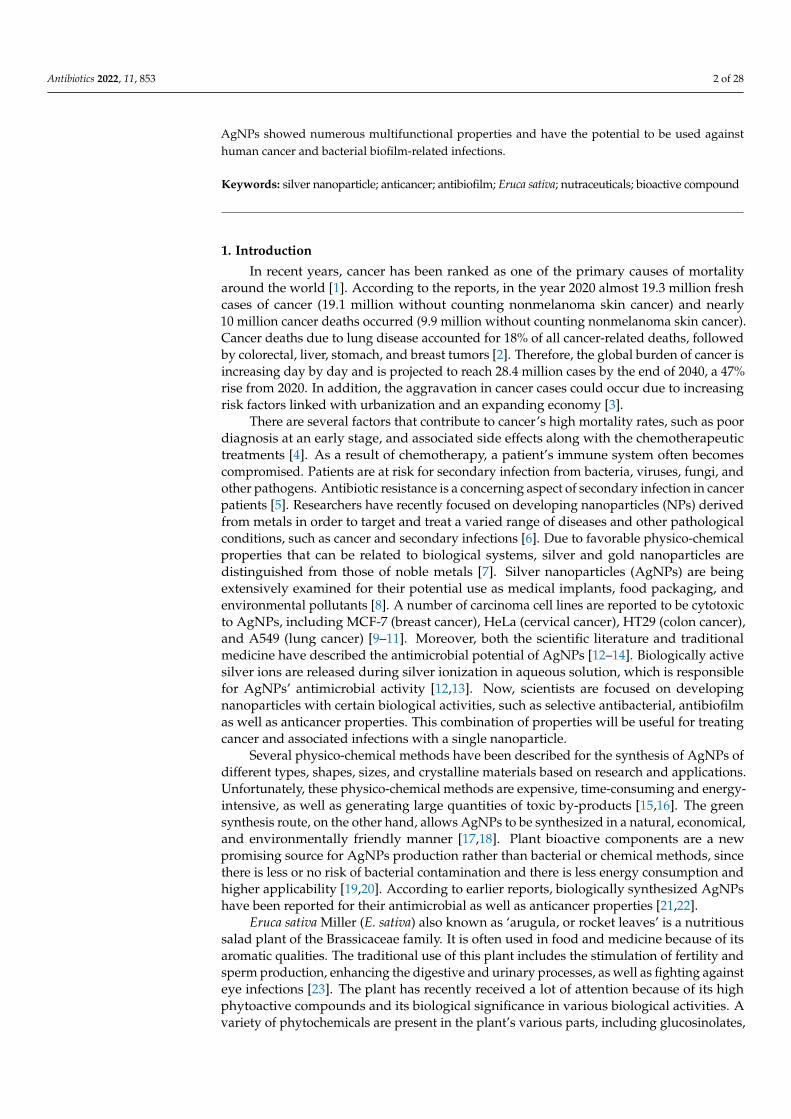
The newly developed AgNPs and their effect on the cytotoxicity of A549 cancercells was evaluated by the MTT assay. We found that the lung cancer cell viability wasconsiderably inhibited in a time- and concentration-dependent manner as a result of thetreatment with different concentrations of AgNPs. A dose–response inhibition curve wasused to determine the values of IC50 after 24 h and the IC50 value was calculated to be25.15 µg/mL (Figure 2).
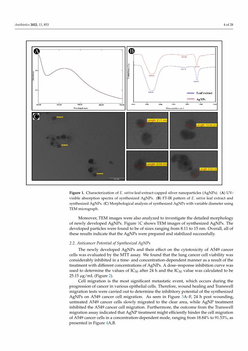
Cell migration is the most significant metastatic event, which occurs during theprogression of cancer in various epithelial cells. Therefore, wound healing and Transwellmigration tests were carried out to determine the inhibitory potential of the synthesizedAgNPs on A549 cancer cell migration. As seen in Figure 3A–F, 24 h post wounding,untreated A549 cancer cells slowly migrated to the clear area, while AgNP treatmentinhibited the A549 cancer cell migration. Furthermore, the outcome from the Transwellmigration assay indicated that AgNP treatment might efficiently hinder the cell migrationof A549 cancer cells in a concentration-dependent mode, ranging from 18.84% to 91.53%, aspresented in Figure 4A,B.
Antibiotics 2022, 11, 853 5 of 28
Antibiotics 2022, 11, 853 5 of 31
2.2. Anticancer Potential of Synthesized AgNPs
The newly developed AgNPs and their effect on the cytotoxicity of A549 cancer cells
was evaluated by the MTT assay. We found that the lung cancer cell viability was consid‐
erably inhibited in a time‐ and concentration‐dependent manner as a result of the treat‐
ment with different concentrations of AgNPs. A dose–response inhibition curve was used
to determine the values of IC50 after 24 h and the IC50 value was calculated to be 25.15
μg/mL (Figure 2).
Figure 2. Cytotoxicity and IC50 (estimated value 25.15 μg/mL) of AgNPs on A549 cancer cells.
Cell migration is the most significant metastatic event, which occurs during the pro‐
gression of cancer in various epithelial cells. Therefore, wound healing and Transwell mi‐
gration tests were carried out to determine the inhibitory potential of the synthesized
AgNPs on A549 cancer cell migration. As seen in Figure 3A–F, 24 h post wounding, un‐
treated A549 cancer cells slowly migrated to the clear area, while AgNP treatment inhib‐
ited the A549 cancer cell migration. Furthermore, the outcome from the Transwell migra‐
tion assay indicated that AgNP treatment might efficiently hinder the cell migration of
A549 cancer cells in a concentration‐dependent mode, ranging from 18.84% to 91.53%, as
presented in Figure 4A,B.
Figure 2. Cytotoxicity and IC50 (estimated value 25.15 µg/mL) of AgNPs on A549 cancer cells.
Antibiotics 2022, 11, 853 6 of 31
Figure 3. Inhibitory effect of synthesized AgNPs on A549 cell migration. (A–C). A scratch was made
onto a monolayer of untreated A549 cells at 0 h. (D–F). Following treatment with different concen‐
trations of AgNPs (2.5, 10 and 25 μg/mL) at 24 h, the migration patterns of A549 cells were recorded
using inverted light microscopy.
Figure 3. Inhibitory effect of synthesized AgNPs on A549 cell migration. (A–C) A scratch wasmade onto a monolayer of untreated A549 cells at 0 h. (D–F) Following treatment with differentconcentrations of AgNPs (2.5, 10 and 25 µg/mL) at 24 h, the migration patterns of A549 cells wererecorded using inverted light microscopy.
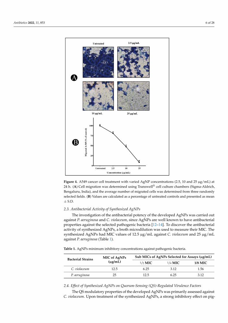
Antibiotics 2022, 11, 853 6 of 28Antibiotics 2022, 11, 853 7 of 31
Figure 4. A549 cancer cell treatment with varied AgNP concentrations (2.5, 10 and 25 μg/mL) at 24
h. (A). Cell migration was determined using Transwell® cell culture chambers (Sigma‐Aldrich, Ben‐
galuru, India), and the average number of migrated cells was determined from three randomly se‐
lected fields. (B). Values are calculated as a percentage of untreated controls and presented as mean
± S.D.
2.3. Antibacterial Activity of Synthesized AgNPs
The investigation of the antibacterial potency of the developed AgNPs was carried
out against P. aeruginosa and C. violaceum, since AgNPs are well known to have antibacte‐
rial properties against the selected pathogenic bacteria [12–14]. To discover the antibacte‐
rial activity of synthesized AgNPs, a broth microdilution was used to measure their MIC.
The synthesized AgNPs had MIC values of 12.5 μg/mL against C. violaceum and 25 μg/mL
against P. aeruginosa (Table 1).
Table 1. AgNPs minimum inhibitory concentrations against pathogenic bacteria.
Bacterial Strains MIC of AgNPs
(μg/mL)
Sub MICs of AgNPs Selected for Assays (μg/mL)
½ MIC ¼ MIC 1/8 MIC
C. violaceum 12.5 6.25 3.12 1.56
P. aeruginosa 25 12.5 6.25 3.12
2.4. Effect of Synthesized AgNPs on Quorum‐Sensing (QS)‐Regulated Virulence Factors
The QS modulatory properties of the developed AgNPs was primarily assessed
against C. violaceum. Upon treatment of the synthesized AgNPs, a strong inhibitory effect
on pigment production was observed at a sub‐MIC level, which indicates the reduction in
Figure 4. A549 cancer cell treatment with varied AgNP concentrations (2.5, 10 and 25 µg/mL) at24 h. (A) Cell migration was determined using Transwell® cell culture chambers (Sigma-Aldrich,Bengaluru, India), and the average number of migrated cells was determined from three randomlyselected fields. (B) Values are calculated as a percentage of untreated controls and presented as mean± S.D.
2.3. Antibacterial Activity of Synthesized AgNPs
The investigation of the antibacterial potency of the developed AgNPs was carried outagainst P. aeruginosa and C. violaceum, since AgNPs are well known to have antibacterialproperties against the selected pathogenic bacteria [12–14]. To discover the antibacterialactivity of synthesized AgNPs, a broth microdilution was used to measure their MIC. Thesynthesized AgNPs had MIC values of 12.5 µg/mL against C. violaceum and 25 µg/mLagainst P. aeruginosa (Table 1).
Table 1. AgNPs minimum inhibitory concentrations against pathogenic bacteria.
Bacterial Strains MIC of AgNPs(µg/mL)
Sub MICs of AgNPs Selected for Assays (µg/mL)
1/2 MIC 1/4 MIC 1/8 MIC
C. violaceum 12.5 6.25 3.12 1.56
P. aeruginosa 25 12.5 6.25 3.12
2.4. Effect of Synthesized AgNPs on Quorum-Sensing (QS)-Regulated Virulence Factors
The QS modulatory properties of the developed AgNPs was primarily assessed againstC. violaceum. Upon treatment of the synthesized AgNPs, a strong inhibitory effect on pig-
Antibiotics 2022, 11, 853 7 of 28
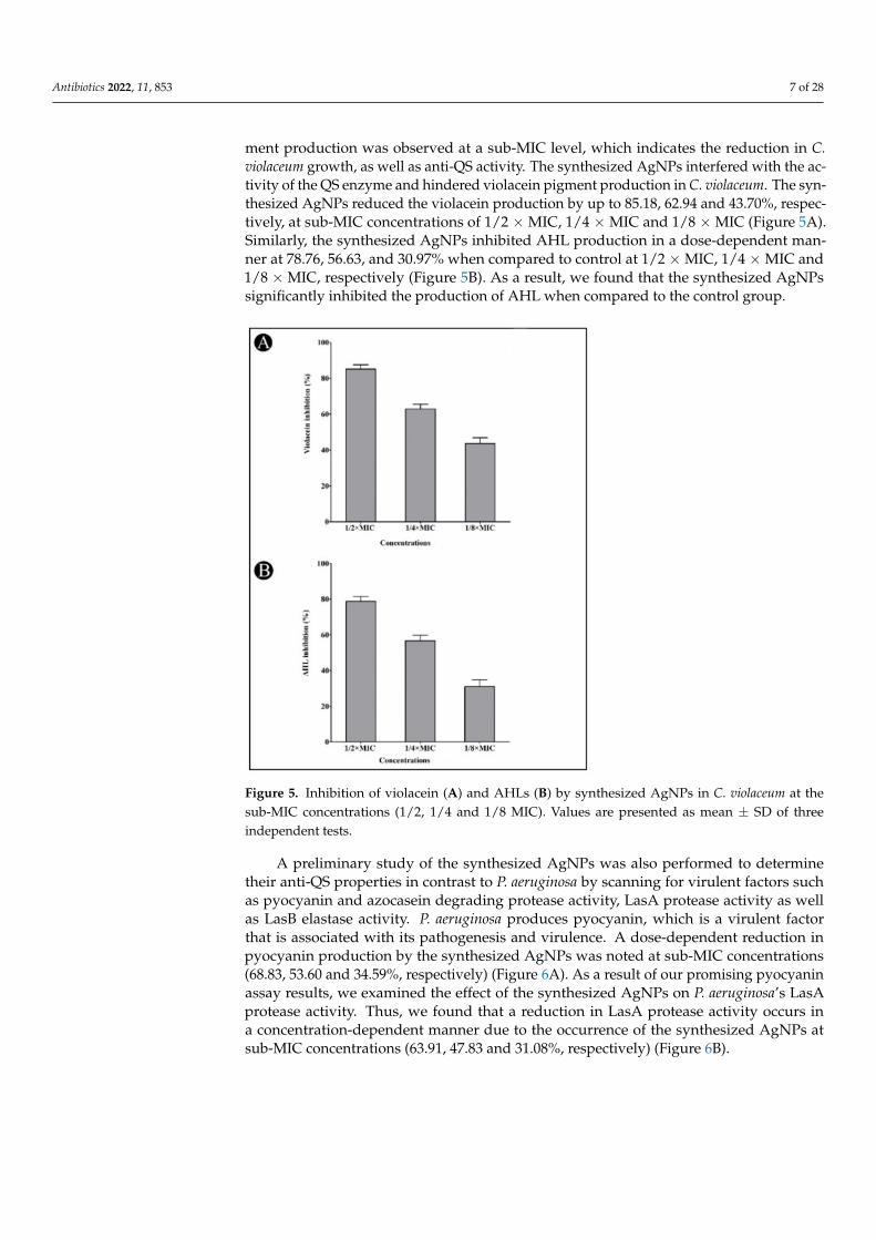
ment production was observed at a sub-MIC level, which indicates the reduction in C.violaceum growth, as well as anti-QS activity. The synthesized AgNPs interfered with the ac-tivity of the QS enzyme and hindered violacein pigment production in C. violaceum. The syn-thesized AgNPs reduced the violacein production by up to 85.18, 62.94 and 43.70%, respec-tively, at sub-MIC concentrations of 1/2 × MIC, 1/4 × MIC and 1/8 × MIC (Figure 5A).Similarly, the synthesized AgNPs inhibited AHL production in a dose-dependent man-ner at 78.76, 56.63, and 30.97% when compared to control at 1/2 × MIC, 1/4 × MIC and1/8 × MIC, respectively (Figure 5B). As a result, we found that the synthesized AgNPssignificantly inhibited the production of AHL when compared to the control group.
Figure 5. Inhibition of violacein (A) and AHLs (B) by synthesized AgNPs in C. violaceum at thesub-MIC concentrations (1/2, 1/4 and 1/8 MIC). Values are presented as mean ± SD of threeindependent tests.
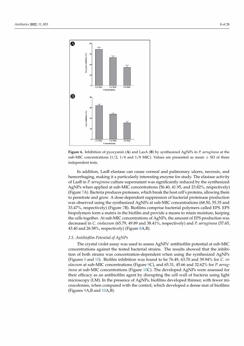
A preliminary study of the synthesized AgNPs was also performed to determinetheir anti-QS properties in contrast to P. aeruginosa by scanning for virulent factors suchas pyocyanin and azocasein degrading protease activity, LasA protease activity as wellas LasB elastase activity. P. aeruginosa produces pyocyanin, which is a virulent factorthat is associated with its pathogenesis and virulence. A dose-dependent reduction inpyocyanin production by the synthesized AgNPs was noted at sub-MIC concentrations(68.83, 53.60 and 34.59%, respectively) (Figure 6A). As a result of our promising pyocyaninassay results, we examined the effect of the synthesized AgNPs on P. aeruginosa’s LasAprotease activity. Thus, we found that a reduction in LasA protease activity occurs ina concentration-dependent manner due to the occurrence of the synthesized AgNPs atsub-MIC concentrations (63.91, 47.83 and 31.08%, respectively) (Figure 6B).
Antibiotics 2022, 11, 853 8 of 28
Figure 6. Inhibition of pyocyanin (A) and LasA (B) by synthesized AgNPs in P. aeruginosa at thesub-MIC concentrations (1/2, 1/4 and 1/8 MIC). Values are presented as mean ± SD of threeindependent tests.
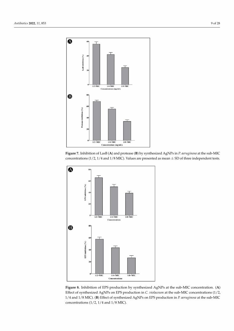
In addition, LasB elastase can cause corneal and pulmonary ulcers, necrosis, andhemorrhaging, making it a particularly interesting enzyme for study. The elastase activityof LasB in P. aeruginosa culture supernatant was significantly reduced by the synthesizedAgNPs when applied at sub-MIC concentrations (56.40, 41.95, and 23.82%, respectively)(Figure 7A). Bacteria produces proteases, which break the host cell’s proteins, allowing themto penetrate and grow. A dose-dependent suppression of bacterial proteinase productionwas observed using the synthesized AgNPs at sub-MIC concentrations (68.50, 55.35 and33.47%, respectively) (Figure 7B). Biofilms comprise bacterial polymers called EPS. EPSbiopolymers form a matrix in the biofilm and provide a means to retain moisture, keepingthe cells together. At sub-MIC concentrations of AgNPs, the amount of EPS production wasdecreased in C. violaceum (65.79, 49.89 and 38.41%, respectively) and P. aeruginosa (57.65,43.40 and 26.58%, respectively) (Figure 8A,B).
2.5. Antibiofilm Potential of AgNPs
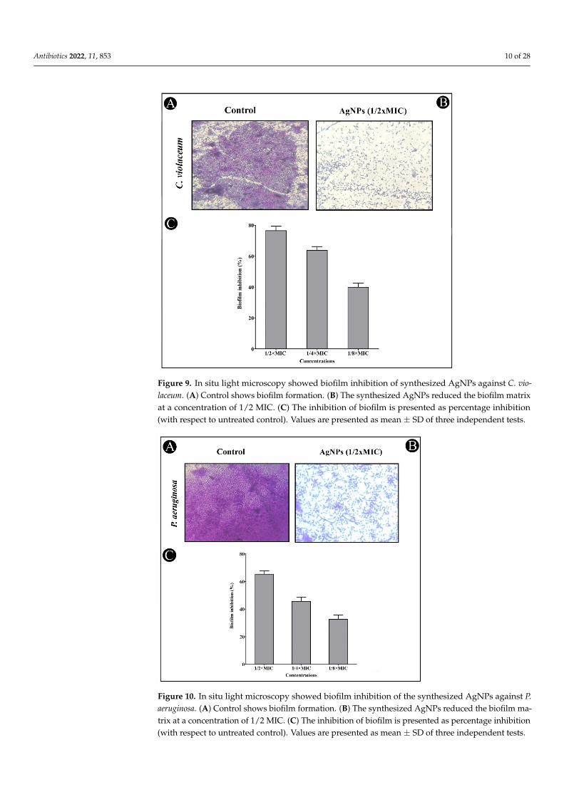
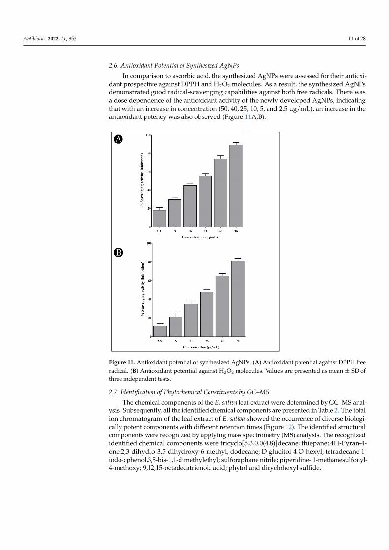
The crystal violet assay was used to assess AgNPs’ antibiofilm potential at sub-MICconcentrations against the tested bacterial strains. The results showed that the inhibi-tion of both strains was concentration-dependent when using the synthesized AgNPs(Figures 9 and 10). Biofilm inhibition was found to be 76.49, 63.70 and 39.94% for C. vi-olaceum at sub-MIC concentrations (Figure 9C), and 65.31, 45.66 and 32.62% for P. aerug-inosa at sub-MIC concentrations (Figure 10C). The developed AgNPs were assessed fortheir efficacy as an antibiofilm agent by disrupting the cell wall of bacteria using lightmicroscopy (LM). In the presence of AgNPs, biofilms developed thinner, with fewer mi-crocolonies, when compared with the control, which developed a dense mat of biofilms(Figures 9A,B and 10A,B).
Antibiotics 2022, 11, 853 9 of 28
Figure 7. Inhibition of LasB (A) and protease (B) by synthesized AgNPs in P. aeruginosa at the sub-MICconcentrations (1/2, 1/4 and 1/8 MIC). Values are presented as mean ± SD of three independent tests.
Figure 8. Inhibition of EPS production by synthesized AgNPs at the sub-MIC concentration. (A)Effect of synthesized AgNPs on EPS production in C. violaceum at the sub-MIC concentrations (1/2,1/4 and 1/8 MIC). (B) Effect of synthesized AgNPs on EPS production in P. aeruginosa at the sub-MICconcentrations (1/2, 1/4 and 1/8 MIC).
Antibiotics 2022, 11, 853 10 of 28
Antibiotics 2022, 11, 853 11 of 31
2.5. Antibiofilm Potential of AgNPs
The crystal violet assay was used to assess AgNPs’ antibiofilm potential at sub‐MIC
concentrations against the tested bacterial strains. The results showed that the inhibition
of both strains was concentration‐dependent when using the synthesized AgNPs (Figures
9 and 10). Biofilm inhibition was found to be 76.49, 63.70 and 39.94% for C. violaceum at
sub‐MIC concentrations (Figure 9C), and 65.31, 45.66 and 32.62% for P. aeruginosa at sub‐
MIC concentrations (Figure 10C). The developed AgNPs were assessed for their efficacy
as an antibiofilm agent by disrupting the cell wall of bacteria using light microscopy (LM).
In the presence of AgNPs, biofilms developed thinner, with fewer microcolonies, when
compared with the control, which developed a dense mat of biofilms (Figures 9A,B and
10A,B).
Figure 9. In situ light microscopy showed biofilm inhibition of synthesized AgNPs against C. vio‐
laceum. (A). Control shows biofilm formation. (B). The synthesized AgNPs reduced the biofilm ma‐
trix at a concentration of 1/2 MIC. (C). The inhibition of biofilm is presented as percentage inhibition
(with respect to untreated control). Values are presented as mean ± SD of three independent tests.
Figure 9. In situ light microscopy showed biofilm inhibition of synthesized AgNPs against C. vio-laceum. (A) Control shows biofilm formation. (B) The synthesized AgNPs reduced the biofilm matrixat a concentration of 1/2 MIC. (C) The inhibition of biofilm is presented as percentage inhibition(with respect to untreated control). Values are presented as mean ± SD of three independent tests.
Antibiotics 2022, 11, 853 12 of 31
Figure 10. In situ light microscopy showed biofilm inhibition of the synthesized AgNPs against P.
aeruginosa. (A). Control shows biofilm formation. (B). The synthesized AgNPs reduced the biofilm
matrix at a concentration of 1/2 MIC. (C). The inhibition of biofilm is presented as percentage inhi‐
bition (with respect to untreated control). Values are presented as mean ± SD of three independent
tests.
2.6. Antioxidant Potential of Synthesized AgNPs
In comparison to ascorbic acid, the synthesized AgNPs were assessed for their anti‐
oxidant prospective against DPPH and H2O2 molecules. As a result, the synthesized
AgNPs demonstrated good radical‐scavenging capabilities against both free radicals.
There was a dose dependence of the antioxidant activity of the newly developed AgNPs,
indicating that with an increase in concentration (50, 40, 25, 10, 5, and 2.5 μg/mL), an in‐
crease in the antioxidant potency was also observed (Figure 11A, B).
Figure 10. In situ light microscopy showed biofilm inhibition of the synthesized AgNPs against P.aeruginosa. (A) Control shows biofilm formation. (B) The synthesized AgNPs reduced the biofilm ma-trix at a concentration of 1/2 MIC. (C) The inhibition of biofilm is presented as percentage inhibition(with respect to untreated control). Values are presented as mean ± SD of three independent tests.
Antibiotics 2022, 11, 853 11 of 28
2.6. Antioxidant Potential of Synthesized AgNPs
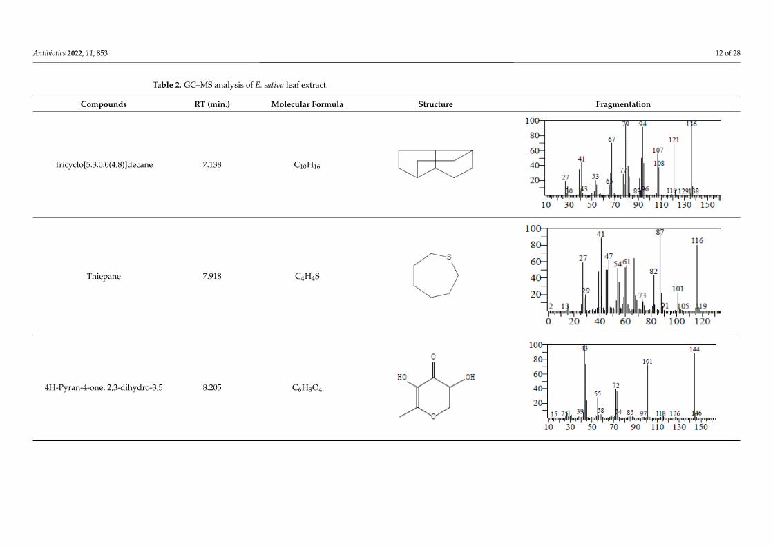
In comparison to ascorbic acid, the synthesized AgNPs were assessed for their antioxi-dant prospective against DPPH and H2O2 molecules. As a result, the synthesized AgNPsdemonstrated good radical-scavenging capabilities against both free radicals. There wasa dose dependence of the antioxidant activity of the newly developed AgNPs, indicatingthat with an increase in concentration (50, 40, 25, 10, 5, and 2.5 µg/mL), an increase in theantioxidant potency was also observed (Figure 11A,B).
Antibiotics 2022, 11, 853 13 of 31
Figure 11. Antioxidant potential of synthesized AgNPs. (A). Antioxidant potential against DPPH
free radical. (B). Antioxidant potential against H2O2 molecules. Values are presented as mean ± SD
of three independent tests.
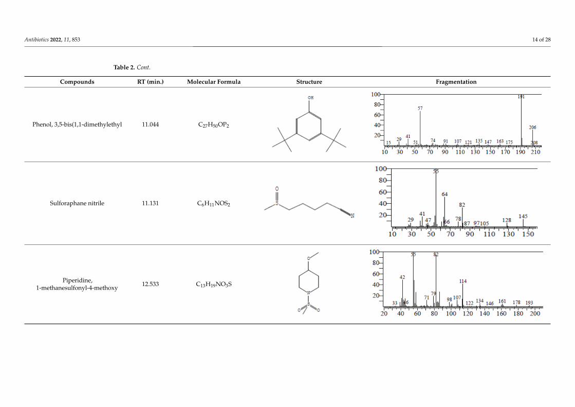
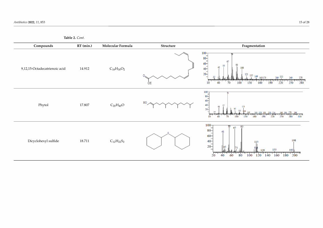
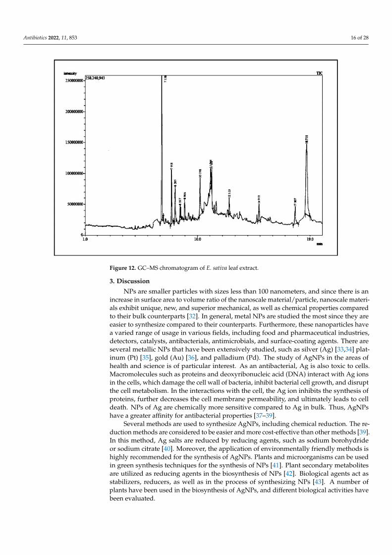
2.7. Identification of Phytochemical Constituents by GC–MS
The chemical components of the E. sativa leaf extract were determined by GC–MS
analysis. Subsequently, all the identified chemical components are presented in Table 2.
The total ion chromatogram of the leaf extract of E. sativa showed the occurrence of diverse
biologically potent components with different retention times (Figure 12). The identified
structural components were recognized by applying mass spectrometry (MS) analysis.
The recognized identified chemical components were tricyclo[5.3.0.0(4,8)]decane;
thiepane; 4H‐Pyran‐4‐one,2,3‐dihydro‐3,5‐dihydroxy‐6‐methyl; dodecane; D‐glucitol‐4‐
O‐hexyl; tetradecane‐1‐iodo‐; phenol,3,5‐bis‐1,1‐dimethylethyl; sulforaphane nitrile; pi‐
peridine‐ 1‐methanesulfonyl‐4‐methoxy; 9,12,15‐octadecatrienoic acid; phytol and dicy‐
clohexyl sulfide.
Figure 11. Antioxidant potential of synthesized AgNPs. (A) Antioxidant potential against DPPH freeradical. (B) Antioxidant potential against H2O2 molecules. Values are presented as mean ± SD ofthree independent tests.
2.7. Identification of Phytochemical Constituents by GC–MS
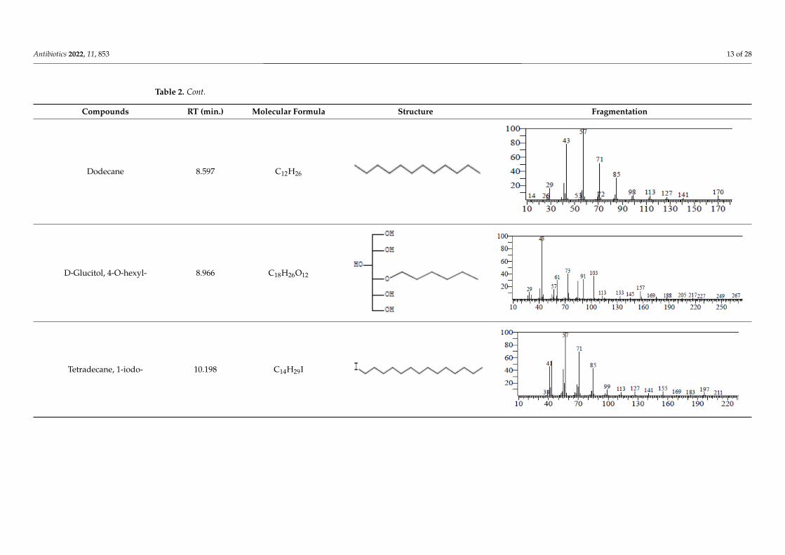
The chemical components of the E. sativa leaf extract were determined by GC–MS anal-ysis. Subsequently, all the identified chemical components are presented in Table 2. The totalion chromatogram of the leaf extract of E. sativa showed the occurrence of diverse biologi-cally potent components with different retention times (Figure 12). The identified structuralcomponents were recognized by applying mass spectrometry (MS) analysis. The recognizedidentified chemical components were tricyclo[5.3.0.0(4,8)]decane; thiepane; 4H-Pyran-4-one,2,3-dihydro-3,5-dihydroxy-6-methyl; dodecane; D-glucitol-4-O-hexyl; tetradecane-1-iodo-; phenol,3,5-bis-1,1-dimethylethyl; sulforaphane nitrile; piperidine- 1-methanesulfonyl-4-methoxy; 9,12,15-octadecatrienoic acid; phytol and dicyclohexyl sulfide.
Antibiotics 2022, 11, 853 12 of 28
Table 2. GC–MS analysis of E. sativa leaf extract.
Compounds RT (min.) Molecular Formula Structure Fragmentation
Tricyclo[5.3.0.0(4,8)]decane 7.138 C10H16
Antibiotics 2022, 11, 853 14 of 31
Antibiotics 2022, 11, 853. https://doi.org/10.3390/antibiotics11070853 www.mdpi.com/journal/antibiotics
Table 2. GC–MS analysis of E. sativa leaf extract.
Compounds RT (min.) Molecular Formula Structure Fragmentation
Tricyclo[5.3.0.0(4,8)]decane 7.138 C10H16
Thiepane 7.918 C4H4S
4H-Pyran-4-one, 2,3-dihydro-3,5
8.205 C6H8O4
Thiepane 7.918 C4H4S
Antibiotics 2022, 11, 853 14 of 31
Antibiotics 2022, 11, 853. https://doi.org/10.3390/antibiotics11070853 www.mdpi.com/journal/antibiotics
Table 2. GC–MS analysis of E. sativa leaf extract.
Compounds RT (min.) Molecular Formula Structure Fragmentation
Tricyclo[5.3.0.0(4,8)]decane 7.138 C10H16
Thiepane 7.918 C4H4S
4H-Pyran-4-one, 2,3-dihydro-3,5
8.205 C6H8O4
4H-Pyran-4-one, 2,3-dihydro-3,5 8.205 C6H8O4
Antibiotics 2022, 11, 853 14 of 31
Antibiotics 2022, 11, 853. https://doi.org/10.3390/antibiotics11070853 www.mdpi.com/journal/antibiotics
Table 2. GC–MS analysis of E. sativa leaf extract.
Compounds RT (min.) Molecular Formula Structure Fragmentation
Tricyclo[5.3.0.0(4,8)]decane 7.138 C10H16
Thiepane 7.918 C4H4S
4H-Pyran-4-one, 2,3-dihydro-3,5
8.205 C6H8O4
Antibiotics 2022, 11, 853 13 of 28
Table 2. Cont.
Compounds RT (min.) Molecular Formula Structure Fragmentation
Dodecane 8.597 C12H26
Antibiotics 2022, 11, 853 15 of 31
Dodecane 8.597 C12H26
D-Glucitol, 4-O-hexyl- 8.966 C18H26O12
Tetradecane, 1-iodo- 10.198 C14H29I
Antibiotics 2022, 11, 853 15 of 31
Dodecane 8.597 C12H26
D-Glucitol, 4-O-hexyl- 8.966 C18H26O12
Tetradecane, 1-iodo- 10.198 C14H29I
D-Glucitol, 4-O-hexyl- 8.966 C18H26O12
Antibiotics 2022, 11, 853 15 of 31
Dodecane 8.597 C12H26
D-Glucitol, 4-O-hexyl- 8.966 C18H26O12
Tetradecane, 1-iodo- 10.198 C14H29I
Antibiotics 2022, 11, 853 15 of 31
Dodecane 8.597 C12H26
D-Glucitol, 4-O-hexyl- 8.966 C18H26O12
Tetradecane, 1-iodo- 10.198 C14H29I
Tetradecane, 1-iodo- 10.198 C14H29I
Antibiotics 2022, 11, 853 15 of 31
Dodecane 8.597 C12H26
D-Glucitol, 4-O-hexyl- 8.966 C18H26O12
Tetradecane, 1-iodo- 10.198 C14H29I
Antibiotics 2022, 11, 853 15 of 31
Dodecane 8.597 C12H26
D-Glucitol, 4-O-hexyl- 8.966 C18H26O12
Tetradecane, 1-iodo- 10.198 C14H29I
Antibiotics 2022, 11, 853 14 of 28
Table 2. Cont.
Compounds RT (min.) Molecular Formula Structure Fragmentation
Phenol, 3,5-bis(1,1-dimethylethyl 11.044 C27H50OP2
Antibiotics 2022, 11, 853 16 of 31
Phenol, 3,5-bis(1,1-di-methylethyl
11.044 C27H50OP2
Sulforaphane nitrile 11.131 C6H11NOS2
Piperidine, 1-methanesulfonyl-4-methoxy
12.533 C13H19NO3S
Antibiotics 2022, 11, 853 16 of 31
Phenol, 3,5-bis(1,1-di-methylethyl
11.044 C27H50OP2
Sulforaphane nitrile 11.131 C6H11NOS2
Piperidine, 1-methanesulfonyl-4-methoxy
12.533 C13H19NO3S
Sulforaphane nitrile 11.131 C6H11NOS2
Antibiotics 2022, 11, 853 16 of 31
Phenol, 3,5-bis(1,1-di-methylethyl
11.044 C27H50OP2
Sulforaphane nitrile 11.131 C6H11NOS2
Piperidine, 1-methanesulfonyl-4-methoxy
12.533 C13H19NO3S
Antibiotics 2022, 11, 853 16 of 31
Phenol, 3,5-bis(1,1-di-methylethyl
11.044 C27H50OP2
Sulforaphane nitrile 11.131 C6H11NOS2
Piperidine, 1-methanesulfonyl-4-methoxy
12.533 C13H19NO3S
Piperidine,1-methanesulfonyl-4-methoxy 12.533 C13H19NO3S
Antibiotics 2022, 11, 853 16 of 31
Phenol, 3,5-bis(1,1-di-methylethyl
11.044 C27H50OP2
Sulforaphane nitrile 11.131 C6H11NOS2
Piperidine, 1-methanesulfonyl-4-methoxy
12.533 C13H19NO3S
Antibiotics 2022, 11, 853 16 of 31
Phenol, 3,5-bis(1,1-di-methylethyl
11.044 C27H50OP2
Sulforaphane nitrile 11.131 C6H11NOS2
Piperidine, 1-methanesulfonyl-4-methoxy
12.533 C13H19NO3S
Antibiotics 2022, 11, 853 15 of 28
Table 2. Cont.
Compounds RT (min.) Molecular Formula Structure Fragmentation
9,12,15-Octadecatrienoic acid 14.912 C18H30O2
Antibiotics 2022, 11, 853 17 of 31
9,12,15-Octadecatrienoic acid 14.912 C18H30O2
Phytol 17.807 C20H40O
Dicyclohexyl sulfide 18.711 C12H22S2
Antibiotics 2022, 11, 853 17 of 31
9,12,15-Octadecatrienoic acid 14.912 C18H30O2
Phytol 17.807 C20H40O
Dicyclohexyl sulfide 18.711 C12H22S2
Phytol 17.807 C20H40O
Antibiotics 2022, 11, 853 17 of 31
9,12,15-Octadecatrienoic acid 14.912 C18H30O2
Phytol 17.807 C20H40O
Dicyclohexyl sulfide 18.711 C12H22S2
Antibiotics 2022, 11, 853 17 of 31
9,12,15-Octadecatrienoic acid 14.912 C18H30O2
Phytol 17.807 C20H40O
Dicyclohexyl sulfide 18.711 C12H22S2
Dicyclohexyl sulfide 18.711 C12H22S2
Antibiotics 2022, 11, 853 17 of 31
9,12,15-Octadecatrienoic acid 14.912 C18H30O2
Phytol 17.807 C20H40O
Dicyclohexyl sulfide 18.711 C12H22S2
Antibiotics 2022, 11, 853 17 of 31
9,12,15-Octadecatrienoic acid 14.912 C18H30O2
Phytol 17.807 C20H40O
Dicyclohexyl sulfide 18.711 C12H22S2
Antibiotics 2022, 11, 853 16 of 28Antibiotics 2022, 11, 853 18 of 31
Figure 12. GC–MS chromatogram of E. sativa leaf extract.
3. Discussion
NPs are smaller particles with sizes less than 100 nanometers, and since there is an
increase in surface area to volume ratio of the nanoscale material/particle, nanoscale ma‐
terials exhibit unique, new, and superior mechanical, as well as chemical properties com‐
pared to their bulk counterparts [32]. In general, metal NPs are studied the most since
they are easier to synthesize compared to their counterparts. Furthermore, these nanopar‐
ticles have a varied range of usage in various fields, including food and pharmaceutical
industries, detectors, catalysts, antibacterials, antimicrobials, and surface‐coating agents.
There are several metallic NPs that have been extensively studied, such as silver (Ag)
[33,34] platinum (Pt) [35], gold (Au) [36], and palladium (Pd). The study of AgNPs in the
areas of health and science is of particular interest. As an antibacterial, Ag is also toxic to
cells. Macromolecules such as proteins and deoxyribonucleic acid (DNA) interact with Ag
ions in the cells, which damage the cell wall of bacteria, inhibit bacterial cell growth, and
disrupt the cell metabolism. In the interactions with the cell, the Ag ion inhibits the syn‐
thesis of proteins, further decreases the cell membrane permeability, and ultimately leads
to cell death. NPs of Ag are chemically more sensitive compared to Ag in bulk. Thus,
AgNPs have a greater affinity for antibacterial properties [37–39].
Several methods are used to synthesize AgNPs, including chemical reduction. The
reduction methods are considered to be easier and more cost‐effective than other methods
[39]. In this method, Ag salts are reduced by reducing agents, such as sodium borohydride
or sodium citrate [40]. Moreover, the application of environmentally friendly methods is
highly recommended for the synthesis of AgNPs. Plants and microorganisms can be used
in green synthesis techniques for the synthesis of NPs [41]. Plant secondary metabolites
are utilized as reducing agents in the biosynthesis of NPs [42]. Biological agents act as
stabilizers, reducers, as well as in the process of synthesizing NPs [43]. A number of plants
have been used in the biosynthesis of AgNPs, and different biological activities have been
evaluated.
Figure 12. GC–MS chromatogram of E. sativa leaf extract.
3. Discussion
NPs are smaller particles with sizes less than 100 nanometers, and since there is anincrease in surface area to volume ratio of the nanoscale material/particle, nanoscale materi-als exhibit unique, new, and superior mechanical, as well as chemical properties comparedto their bulk counterparts [32]. In general, metal NPs are studied the most since they areeasier to synthesize compared to their counterparts. Furthermore, these nanoparticles havea varied range of usage in various fields, including food and pharmaceutical industries,detectors, catalysts, antibacterials, antimicrobials, and surface-coating agents. There areseveral metallic NPs that have been extensively studied, such as silver (Ag) [33,34] plat-inum (Pt) [35], gold (Au) [36], and palladium (Pd). The study of AgNPs in the areas ofhealth and science is of particular interest. As an antibacterial, Ag is also toxic to cells.Macromolecules such as proteins and deoxyribonucleic acid (DNA) interact with Ag ionsin the cells, which damage the cell wall of bacteria, inhibit bacterial cell growth, and disruptthe cell metabolism. In the interactions with the cell, the Ag ion inhibits the synthesis ofproteins, further decreases the cell membrane permeability, and ultimately leads to celldeath. NPs of Ag are chemically more sensitive compared to Ag in bulk. Thus, AgNPshave a greater affinity for antibacterial properties [37–39].
Several methods are used to synthesize AgNPs, including chemical reduction. The re-duction methods are considered to be easier and more cost-effective than other methods [39].In this method, Ag salts are reduced by reducing agents, such as sodium borohydrideor sodium citrate [40]. Moreover, the application of environmentally friendly methods ishighly recommended for the synthesis of AgNPs. Plants and microorganisms can be usedin green synthesis techniques for the synthesis of NPs [41]. Plant secondary metabolitesare utilized as reducing agents in the biosynthesis of NPs [42]. Biological agents act asstabilizers, reducers, as well as in the process of synthesizing NPs [43]. A number ofplants have been used in the biosynthesis of AgNPs, and different biological activities havebeen evaluated.
Antibiotics 2022, 11, 853 17 of 28
At the present time, various types of AgNPs are developed by biological synthesis thatare reported to possess various biological properties such as antimicrobial, antioxidant, an-tiviral and anticancer activities, etc. [44–49]. In spite of the success of many NPs synthesizedusing microorganisms and plants, nanoscience research is still focused on developing newNPs with specified physical, chemical, and biological properties. E. sativa has varieties ofphytochemical constituents and is widely used in traditional medicine due to its excellentbiological properties [50,51]. The present study describes the AgNP synthesis and the char-acterization of the E. sativa leaf extract that may possess biological and economic benefits.Furthermore, synthetic AgNPs have significant anticancer, anti-metastatic, antibacterial,anti-QS, antibiofilm and antioxidant activity.
The results of our study showed that AgNPs were synthesized, and when silver nitratewas exposed to the leaf extract of E. sativa, the alteration in color could be attributed to theformation of NPs, which were also confirmed by UV–Vis spectroscopy. As a result of itsSPR properties, the biosynthesized AgNPs exhibited a strong band of absorption at 424 nm.There was a direct correlation between the amount of extract and the incubation periodin this case, and it was likely due to the stimulation of longitudinal plasmon vibrations aswell as the reduction of the AgNO3 ions [52,53]. The analysis of the FT-IR spectrum wasperformed to determine the key factors that contribute to the reduction of silver ions (Ag+)into AgNPs (Ag0) in the aqueous extract of E. sativa leaves. Comparing the absorbancepeak of the AgNPs with the control leaf extract, a change in absorbance peak intensity wasrecorded at different points. The spectra of the leaf-extract-based AgNPs revealed a range ofabsorption bands between 1642 and 3351cm−1. Accordingly, this suggests the biomolecule’spresence and its capability of reducing and stabilizing silver ions (Ag+) into AgNPs (Ag0)present in aqueous leaf extracts. It was found that the FT-IR exploration revealed a numberof absorption peaks, specifically those associated with the N-H stretching vibration, whichindicates strong hydrogen bonds, and C-O extension vibrations, which are associatedwith ketones, carboxylic acids and aldehydes, which are related to Ag ion reduction, thatresulted in the stabilization of the NPs by oxidizing the hydroxyl radical [54,55]. Based onthe analysis of images obtained by TEM of the biosynthesized AgNPs, it appears that mostparticles were crystalline in nature and nearly spherical in shape. A few clusters of AgNPswere observed in our study, suggesting that they might be responsible for the particle sizevariation. Upon closer examination, it is easy to determine that the clusters are the result ofindividual particles clustering together. The TEM analyses of previous studies reporteddifferent sizes of spherical AgNPs containing bio-organic compounds [56,57].
Presently, cancer is still a life-threatening disease around the world despite all theprogress in oncology [58]. The disease starts locally, but it could spread to other portionsof the body through migration, invasion, and metastasis [59]. The process of metastasisinvolves a number of complex mechanisms, which include the extrication, gathering andmotility of cancer cells, followed by their adhesion to endothelial cells and their growthat different sites [60]. In cancer, metastasis is the dominant cause of death because ofresistance to apoptosis and cytotoxic agents. The rate of mortality and morbidity inmetastatic cancer patients is high, because current chemotherapy agents do not havethe ability to selectively and effectively kill the cancer cells without damaging healthycells at the sites of metastasis [61]. Thus, around the world, metastatic disease remains acrucial clinical challenge in cancer treatment [61]. For cancer treatment, radiation therapy,chemotherapy and surgery are currently considered to be the most common methods. Itis well-known that these treatments have a range of side effects on human health. As aconsequence, metastasis is found to be the most challenging obstacle to successful cancermanagement [60].
The newly developed AgNPs were also examined for their anticancer and anti-metastatic potentials against lung cancer cells (A549) in the present study. The AgNPsshowed significant cytotoxicity when tested against malignant A549 cells with an IC50value of 25.15 µg/mL. Additionally, we tested the AgNPs’ effect on the migration of cellsin a wound-healing assay, since migration is a key step in the development of cancer and
Antibiotics 2022, 11, 853 18 of 28
metastasis. AgNPs inhibited the migration of cancer cells towards the wound in a signifi-cant way. The present study discovered that AgNPs could inhibit cell migration, a highlyimportant process during the early phases of wound healing. AgNPs were also found toobstruct the invasion of malignant cancer cells. Thus, in this study, we found that AgNPsinhibited the metastatic process in A549 lung cancer cells in vitro using wound-healing andinvasion assays.
We further analyzed the potency of our biosynthesized AgNPs with E. sativa extracttargeting the biofilms and related virulence factors to confirm their activities to a broaderscope. Around 65% of all bacterial infections can be attributed to biofilms [62], and as aresult of the resistance of bacteria to various antibiotics, biofilm-related infections are oftendifficult to treat. In order to prevent the formation of biofilms, it is crucial to identify noveland effective molecules [63]. The use of antibiotics in treating biofilm-related infections isinsufficient; however, understanding the mechanism of biofilm formation will facilitatethe strategic control and treatment of biofilm infections [64]. The inhibition of biofilmsis another way to regulate the surface-adhered bacteria’s growth and survival. Manybacterial behaviors, including virulence and biofilm formation, are influenced by a cascadeof signaling reactions known as QS [65]. Bacterial pathogenicity and virulence can bereduced by obstructing the QS cascades. As a result of communicating through signalmolecules, bacteria at a certain density are believed to begin expressing virulence genes,which enable the production of virulence factors. Therefore, antimicrobial therapy cannow be aimed at blocking bacterial communication [66]. Antimicrobial compounds withQS inhibitory effects are new-generation antimicrobials [67]. In the fight against biofilms,AgNPs offer a new horizon due to their excellent selective and specific mechanisms ofaction. Considering that AgNPs synthesized from plants have a natural effect and are lesstoxic than anti-QS agents, they are ideal for combating pathogenic bacterial biofilms [64].For the purpose of searching for a new method or molecule to prevent biofilm formation,we investigated AgNPs’ anti-QS and antibiofilm activity against the model pathogenicorganisms C. violaceum and P. aeruginosa [57].
The purple pigment (violacein) present in C. violaceum makes it an ideal indicatororganism for the screening of QS inhibitors. The reduction in violacein pigment productionas a result of AgNPs interference with QS was evaluated in this study. Within C. violaceum,violacein synthesis is controlled by the CviR QS system. Consequently, any change in theamount of C. violaceum pigment is interpreted as a direct sign of QS hindrance. A numberof natural products have also been reported to inhibit the production of violacein by C.violaceum [68,69]. Additionally, AgNPs inhibited AHL synthesis, indicating that AgNPsinterfered with QS by inhibiting the production of AHL molecules. Based on our study ofviolacein and AHL inhibition, we found that AgNPs are most active. Therefore, we screenedfurther AgNPs against P. aeruginosa to determine whether they have broad-spectrum QSactivity, since the QS system of P. aeruginosa is different from C. violaceum.
A variety of QS-mediated phenotypes of P. aeruginosa were investigated using AgNPs,including azocaseinolytic activity, pyocyanin synthesis, LasA protease activity, and LasBelastase activity [70]. It has been established that virulence factors play a key role in theinvasion of bacteria and their proliferation within the host population [70]. The patho-genesis of P. aeruginosa is caused by the secretion of proteases by this organism [70]. Inthe azocasein degrading assay, AgNPs were found to reduce the protease production ofP. aeruginosa at concentrations below the MIC. Two QS systems are found in P. aeruginosa(LasIR and RhIIR). The first QS system (Las) occurs through the formation of diffusiblesignal molecules 3O-C12-HSL due to a synthase encoded by the LasI gene, which is furtheractivated by LasR (transcriptional activator) to activate many virulence genes (LasA andLasB). Several virulent enzymes are produced by these genes, including alkaline protease,LasA protease, and LasB elastase.
P. aeruginosa’s second QS system (Rhl) synthesizes the diffusible signal moleculesC4-HSL via RhlI, which further associates with RhlR and activates the expression systemfor pyocyanins. The LasIR gene encodes proteins that play an important role in the
Antibiotics 2022, 11, 853 19 of 28
pathogenesis of P. aeruginosa [71]. In this study, we tested different sub-MIC concentrationsof AgNPs in order to determine if they inhibited all of the QS-associated proteins andfactors in a dose-dependent manner. A third and distinct virulence factor, pyocyanin,which is produced in QS, is also important and very different. Pyocyanin has been welldocumented as having a role in the pathogenesis of many diseases, particularly cysticfibrosis [72]. Our results are consistent with those found in the literature, where othernatural products have also been reported to inhibit QS-associated proteins and factors of P.aeruginosa to varying degrees [73].
Synthesized AgNPs scavenge a significant amount of radicals in the antioxidant assay.A possible reason for the higher antioxidant activity of the synthesized AgNPs may lie inthe presence of phytochemical components in the leaf extract and silver ions, which couldresult in antioxidant activity proceeding simultaneously through hydrogen atom transfer(HAT) and single electron transfer (SET) [74]. Thus, the antioxidant abilities of AgNPsrecommend their use as natural antioxidants in preventing degenerative diseases causedby oxidative stress.
The medicinal value of Ag has been widely recognized since the ancient times [75].The use of plant-based bio-constituents is currently a highly effective and eco-friendlymethod for manufacturing monodisperse, colloidal, homogeneous AgNP solutions. Therehave been several studies involving the synthesis of AgNPs from plant leaves, includingAcalypha indica [76], Ficus benghalensis [76,77], and Mulberry [77,78], as well as from seedextracts of a number of plants, including Macrotyloma uniflorum [79], Jatropha curcas [80],Illicium verum [80,81], and Pistacia atlantica [75,81]. In recent years, there has been a rise inthe use of nanomaterials for the advancement of diagnostic tools such as biosensing andbioimaging. The application of NPs in clinical imaging modalities could affect the biologicalfactors in the microenvironment and be able to detect changes in the microenvironmentand the smaller stage of growth of cancer or tumor cells [82]. AgNPs have also been shownto damage cells by interfering with the integrity of cell membranes, causing oxidativestress and apoptosis [83]. Therefore, in this study the antimicrobial, antibiofilm, anti-QS,antioxidant and anticancer activities of biologically synthesized AgNPs were reported. Inaddition, as per our literature survey, we did not find a single report of biosynthesizedAgNPs that exhibited such a varied potential, which demonstrates their applicability invarious biomedical applications.
4. Materials and Methods4.1. Plant Extract Preparation and Green Synthesis of Nanoparticles
Fresh leaves of E. sativa were collected from the Bapalal Vaidya Botanical ResearchCentre, Veer Narmad South Gujarat University, Gujarat, India. Furthermore, the obtainedplant material was cleaned and washed three to four times with running tap water andonce with sterile distilled water (D/W). A total of 20 g of fresh leaves were finely choppedand simmered in 100 mL of distilled water for 1 h at 60 ◦C. Following the boiling process,the mixture was cooled and strained using Whatman filter paper number 1. After filtration,the suspension was collected and stored at 4 ◦C for further use as a reducer, stabilizer, andcapping agent. AgNPs were prepared by mixing 5 mL of leaf extract of E. sativa with 45 mLof AgNO3 solution (0.1 M) and incubating at room temperature for a bio-reduction processwith vigorous stirring. The transformation of color from transparent yellow to dark browncan be seen as a sign that AgNPs are forming.
4.2. Biophysical Characterization of Synthesized AgNPs
As a first step, spectrophotometric analysis was performed on the synthesized AgNPs.The reaction mixture was centrifuged at 10,000 rpm for 10 min and the obtained pelletwas re-suspended in sterile D/W, which was scanned using a UV–visible spectrum witha resolution of 1 nm between 300 and 700 nm. FT-IR (Perkin-Elmer, Waltham, USA) wasapplied to observe the interaction between E. sativa leaf extract and AgNPs. FT-IR spectrawere noted in the wave range of 500 to 4000 cm−1. TEM was also applied to quantify the size
Antibiotics 2022, 11, 853 20 of 28
and shape of the NPs. The synthesized AgNPs (1 mL) were stained with phosphotungisticacid solution (0.5%), placed over copper grids, dried, and images were taken using TEMequipped with a CCD camera (Tecnai 20, Philips, Eindhoven, Netherlands).
4.3. Cytotoxic Assay (MTT Assay)
The newly developed AgNPs were tested against the human lung cancer cells (A549)for their cytotoxicity potential. In flasks (25 cm2), cancer cells were grown in Dulbecco’sModified Eagle’s Medium (DMEM) (MP Biomedicals, Eschwege, Germany) with 10% FetalBovine Serum (FBS) and 10,000 units/mL penicillin and 5 mg/mL streptomycin antibioticsolution (Hi-Media, Bengaluru, India) at 37 ◦C in atmosphere humidified with 5% CO2.When the cells reached 80% confluence, they were seeded at a density of 104 cells per well in96-well plates and incubated in the same conditions as above. A hemocytometer was usedto investigate the viability of the cells after staining with Trypan Blue (Hi-Media, Bengaluru,India) (0.4%). Afterward, cells were exposed to different concentrations of synthesizedAgNPs (50 µg/mL, 40 µg/mL, 25 µg/mL, 10 µg/mL, 5 µg/mL and 2.5 µg/mL) for 24 h.The media containing AgNPs was aspirated from the plate after it was removed from theincubator. Afterwards, a medium of 200 µL containing MTT reagent (10%) (MP Biomedicals,Eschwege, Germany) was added to each well to obtain a final concentration of 0.5 mg/mL,and the plates were incubated at 37 ◦C for an additional 3 h under humidified atmosphere(5% CO2). Following this, the medium was removed and 100 µL of dimethyl sulfoxide(DMSO) (Merck, Darmstadt, Germany) was added to dissolve the formazan crystals. Theabsorbance of the amount of formazan crystal was measured at 570 nm and 630 nm with anELISA reader (EL10A, Biobase, China). The percentage of growth inhibition was tabulatedafter deducting the background and blank, and the percent growth inhibition (IC50) wasdetermined by the dose–response curve for the respective cell line. The positive controlused for this assay was 5-Flourouracil (5-FU) [84].
4.4. Wound-Healing Assay
Using wound-healing assays, the effect of synthesized AgNPs on the cell viability ofA549 cancer cells was studied. The assay was performed using monolayers of cells grownin 6-well plates. Cells were plated at a concentration of 1 × 10⁶ cells/mL for the finalvolume of 3 mL. In the central region of the culture, an injury line was made using a sterile1 mL pipette tip. An inverted microscope was used to photograph three random viewsalong each scraped line. Afterward, the cells were treated with different concentrations ofAgNPs (2.5, 10 and 25 µg/mL) for another 24 h and later-on a set of images was recorded.Cell migration and wound healing are indicated by a reduction in the scraped area [85].
4.5. Transwell Migration Assay
A cell culture Transwell insert (8 µm pore size, 24-well format, Himedia®, Bengaluru,India) was used to seed 1 × 10⁶ cells in serum-free media; afterwards, cells were treatedwith different concentrations of AgNPs (25, 10 and 2.5µg/mL) for 24 h. The lower chamberwas filled with 10% FBS. By using a cotton swab, cells that had not moved through thepores were removed after 10 h of incubation. Cells, which were pierced, were treatedwith methanol and stained with crystal violet (0.1%). Furthermore, the counting of cellnumbers was performed in terms of mean numbers of three randomly selected fields underan inverted microscope. Afterwards, we quantified the cell numbers that had penetratedthe cell membrane [86].
4.6. Screening of Antibacterial Activity of Synthesized AgNPs4.6.1. Bacterial Strains and Growth Conditions
Synthesized AgNPs were tested for antibacterial properties against the two bacterialstrains P. aeruginosa (MTCC 2488) and C. violaceum (MTCC 2656). Both bacterial strainswere grown and maintained using sterile Luria Bertani (LB) broth (HiMedia®, Bengaluru,India) at 30 ◦C in a shaking condition (120 rpm) for 24 h.
Antibiotics 2022, 11, 853 21 of 28
4.6.2. Determination of Minimum Inhibitory Concentration (MIC)
For the determination of MIC for the newly developed AgNPs, they were tested againstthe selected bacterial strains, and a broth dilution method was used [87]. The inoculumswere prepared from an active culture of bacteria in LB broth. Dilution of synthesized AgNPswas performed in two-fold ranging from 50 to 1.56 µg/mL and added to 96-well plates(100 µL each well). Furthermore, 20 µL of each bacterial strain culture (108 CFU/mL) wereincubated in their respective well for 24 h at 37 ◦C. Afterwards, MICs were determined asthe concentration at which observable growth was inhibited. The negative control was onlycomprised of media, while the positive control was only comprised of bacteria inoculatedwithout any synthesized AgNPs.
4.7. Quorum Sensing Inhibitory Activity in C. violaceum4.7.1. Evaluation of Anti-Quorum-Sensing (QS) Activity
For the evaluation of the anti-QS properties of the synthesized AgNPs, a well-diffusionassay was conducted using C. violaceum. Overnight-grown bacteria were incubated overthe LB Petri plates and wells were made by using a gel puncture. In the next step, 60 µLof synthesized AgNPs (50 µg/mL) were inoculated into each punctured gel well and theplates were incubated for 24 h at 37 ◦C. On the next day, a zone of clearance was detectedthat exhibited anti-QS effects against the selected bacterial strains [88].
4.7.2. Violacein Inhibition Assay
Spectrophotometric measurements of C. violaceum produced violacein pigment wereperformed in the absence, as well as in the presence of synthesized AgNPs [89]. Briefly,16–18 h (OD600 nm = 0.1) grown cultures were placed into Erlenmeyer flasks filled withLB broth along with the presence or absence of synthesized AgNPs (sub-MIC—1/2, 1/4and 1/8 MIC) and incubated for 24 h at 28 ◦C. Once incubation was completed, colonies ofbacterial cells were collected via centrifugation (10,000 rpm for 10 min) and dissolved in1 mL of DMSO. Afterwards, centrifugation was again carried out under the same conditionsthat were used in the previous step to remove dead cells, and the absorbance of solubleviolacein was observed at 585 nm. Analyzing the OD at 585 nm, a comparison between theamount of treated C. violaceum and the untreated control was carried out. The magnitudeof the decline in violacein production in presence of synthesized AgNPs was determinedby using the formula presented below
Violacein inhibition (%) = [Control (OD) − Treated (OD)/Control (OD)] × 100
4.7.3. Quantification of Acyl Homoserine Lactones (AHLs)
The AHLs of C. violaceum treated with or without synthesized AgNPs were evaluatedusing the Taghadosi et al. (2015) method with slight modifications [90]. First, 20 µL ofC. violaceum culture were mixed with 180 µL of LB medium either with (sub-MIC—1/2,1/4 and 1/8 MIC) or without synthesized AgNPs, and were incubated at 30 ◦C for 24 hunder shaking conditions. After incubation, the cell supernatants were transferred to a newtube and equal volumes of acidified ethyl acetate (0.5% glacial acetic acid) were added andmixed. A rotary evaporator at 45 ◦C was then used to dry the organic layer collected fromcontrol and treatment tubes. In order to re-use the residues, the residues were suspendedin 20% acidified ethyl acetate and kept under refrigeration at −20 ◦C. Residues from thetreated and control experiments were placed in the wells and mixed with equal volumes of(2 M) hydroxylamine/NaOH (3.5 M). Following this, equal volumes of ferric chloride (4 MHCl (10%)) and 95% ethanol were also added, and then the absorbance of the samples wasrecorded at 520 nm.
Antibiotics 2022, 11, 853 22 of 28
4.8. Quorum-Sensing Inhibitory Activity in P. aeruginosa4.8.1. Quantitative Analysis of Pyocyanin Production in P. aeruginosa
Extracted pyocyanins from the culture supernatants of P. aeruginosa were obtainedaccording to the method referred by Essar et al. (1990) [91], and were treated with orwithout synthesized AgNPs. Firstly, the 5 mL supernatant of P. aeruginosa treated with(sub-MIC—1/2, 1/4 and 1/8 MIC) or without synthesized AgNPs was extracted with 3 mLchloroform and then re-extraction was performed using HCl of 0.2 M (1 mL). Afterwards,the obtained solution was poured into a glass cuvette for measuring the absorbance at520 nm.
4.8.2. LasA Staphylolytic Assay
A boiled S. aureus cell suspension was lysed with P. aeruginosa culture supernatant toevaluate the LasA protease activity [71]. A culture of S. aureus cells (106 CFU/mL) that wasgrown overnight was centrifuged at 8000 rpm for 5 min to collect the cell pellet. Collectedcell pellets were suspended in 0.02 M Tris–HCl (pH 8.5) and boiled for 10 min. After boiling,the cell suspension was diluted with the same buffer to make an optical density of 0.8 at595 nm. Then, the cell-free culture supernatants of P. aeruginosa, cultivated with sub-MICconcentrations of (1/2, 1/4 and 1/8 MIC) or without synthesized AgNPs, were added todiluted S. aureus suspensions (9:1). In the subsequent step, the solution was poured into acuvette for measuring the absorbance at 595 nm.
4.8.3. LasB Elastase Assay
LasB elastase activity was measured as per the method described by Adonizio et al.(2008) [70] to determine the elastolytic activity. A culture of P. aeruginosa was incubatedwith synthesized AgNPs (sub-MIC—1/2, 1/4 and 1/8 MIC). The treated and non-treatedcultures were incubated in 900 µL elastin Congo Red buffer (100 mM Tris, 1mM CaCl2,pH 7.5) holding 20 mg of elastin Congo Red (Sigma, Bengaluru, India). A centrifuge wasused to remove the insoluble Congo Red elastin after 3 h at 37 ◦C in a shaker incubator.Afterwards, the collected supernatant absorbance was recorded at 495 nm. The LB mediumwithout synthesized AgNPs was used as a negative control.
4.8.4. Azocasein Assay for Proteolytic Activity
The proteolytic activity of P. aeruginosa supernatants cultured with (sub-MIC—1/2,1/4 and 1/8 MIC) or without synthesized AgNPs was determined by applying the methodreferred by Kessler et al. (1993) [71]. Afterwards, 150 µL of culture supernatants wereadded to 1 mL (0.3%) of azocasein (Sigma, St. Louis, USA) in 0.05 M Tris–HCl and 0.5 mMCaCl2 (pH 7.5), and the incubation of supernatants was performed at 37 ◦C for 15 min. Tostop the reaction, 0.5 mL of 10% trichloroacetic acid was added. After centrifugation, thesupernatant was collected and absorbance was measured at 400 nm.
4.8.5. Extraction and Estimation of Exopolysaccharides (EPS)
Both C. violaceum and P. aeruginosa bacterial strains were grown in the presence ofsynthesized AgNPs (sub-MIC—1/2, 1/4, and 1/8 MIC) and then centrifuged and filter-sterilized. EPS was precipitated overnight at 4 ◦C via the addition of chilled absoluteethanol to the supernatant [92].
4.9. Antibiofilm Assay
The antibiotic potential of synthetic AgNPs at sub-MIC were evaluated using thecrystal violet staining method [93]. Bacterial cell cultures incubated in 96-well microtiterplates using glucose (0.2%)-supplemented LB (100 µL) of each test strain (108 CFU/mL)and the synthesized AgNPs were incubated at 37 ◦C for 24 h. Afterwards, planktonic cellswere carefully removed from the wells immediately after incubation and washed withPBS (200 µL). In order to visualize biofilms, attached cells were stained for 30 min withcrystal violet (0.1%) and incubated at 37 ◦C. Then, PBS was used to wash off excess dye,
Antibiotics 2022, 11, 853 23 of 28
and 200 µL of 95% ethanol was used to fix the plates and the absorbance of samples weremeasured using a spectrophotometer at 470 nm.
In Situ Visualization of Biofilms
To assess the biofilms formed on glass cover slips by tested strains, we referred to amethod designated by Musthafa et al. (2010) [94]. First, 24-well microtiter plates consistingof 1 × 1 cm cover slips were inoculated with 500 µL of the selected cultures in glucose(0.2%)-supplemented LB with the concentrations of 108 CFU/mL. The same well wastreated with 500 µL of synthesized AgNPs (sub-MIC). After 24 h of incubation at 37 ◦C,glass cover slips containing biofilms were gently removed and washed with PBS. Biofilmswere stained with 0.1% crystal violet and observed under LM with a magnification of 40×(Axioscope A1, ZEISS, Jena, Germany).
4.10. Determination of DPPH Free-Radical-Scavenging Activity
The ability of synthesized AgNPs to scavenge radicals was measured in terms of theirantioxidant activity against DPPH. Different concentrations of synthesized AgNPs, i.e.,50, 40, 25, 10, 5, 2.5 µg/mL were mixed with 2 mL of 6 × 10⁻⁵ M of DPPH solution inDMSO. Tubes were incubated in the dark for up to an hour. Afterwards, sample absorbancewas measured at 517 nm. DPPH solution without AgNPs was used as a control. DMSOaqueous solutions and ascorbic acid were used as a blank and standard, respectively [50].The DPPH radical-scavenging ability was determined using the equation below:
DPPH scavenging activity (%) = (A0 − A1)/A0 × 100
where, A0 = absorbance of the control; A1 = absorbance of the sample.
4.11. Determination of Hydrogen Peroxide Scavenging Activity
The hydrogen peroxide (H2O2) scavenging activity of synthesized AgNPs was mea-sured using the method presented by Ruch et al. [95]. Each of the different concentrationsof AgNPs (50, 40, 25, 10, 5 and 2.5 µg/mL) was mixed with an H2O2 solution (2 mM, 1 mL)prepared in phosphate buffer (0.1 M, pH 7.4) and incubated for 10 min at room temperature.In order to determine the absorbance at 230 nm, phosphate buffer without H2O2 was usedas a blank and ascorbic acid solution was used as a positive control. The following formulawas used to calculate how much H2O2 was scavenged:
% inhibition = (A0 − A1)/A0) × 100
where A0 is the absorbance of the control; A1 is the absorbance of the extract/standard.
4.12. Gas Chromatography-Mass Spectrophotometry (GC–MS) Analysis
In order to analyze the E. sativa leaf extract by GC–MS, we used a Shimadzu NexisGC-2030 Gas Chromatograph (GC) coupled with a QP2020 NX Mass Spectrometer. Forthe separation of the analytes, the column temperature was adjusted to 50 ◦C for 3 min,raised at a frequency of 10 ◦C per minute up to 270 ◦C, and finally raised to 300 ◦C for10 min. The system was filled with 10 µL of sample and the carrier gas that was used washelium. To determine the probable composition of the leaf extract, the peaks obtained fromthe GC–MS separation were compared with the NIST database [96].
5. Conclusions
The development of a reliable and eco-friendly method for producing metallic nanopar-ticles is a crucial aspect of nanotechnology. Currently, nanoparticles are regarded asfundamental components of nanotechnology. The physiochemical properties of silvernanoparticles make them a very attractive substance to be used in the fields of biologyand medicine. Using a natural and low-cost biological reduction agent in conjunction withleaf extracts of E. sativa, we were able to demonstrate in the present study that a green
Antibiotics 2022, 11, 853 24 of 28
nano-chemistry methodology can be effectively and efficiently utilized to produce metalnanostructures, thus eluding the usage of toxic solvents and the creation of waste. As testedby MTT, the synthesized AgNPs displayed significant anticancer activity against A549cancer cells. The scratch assay and invasion assay also demonstrated the inhibitory effecton cancer cell migration. In addition, these AgNPs exhibited excellent antibacterial, anti-QS,and antibiofilm properties against C. violaceum and P. aeruginosa. The prepared AgNPsshowed anti-QS activity against C. violaceum by inhibiting violacein and acyl homoserinelactone and against P. aeruginosa via a reduction in pyocyanin activity, total protease, LasAactivity, and LasB activity. Furthermore, there was a significant decline in EPS productionin the presence of AgNPs. The initial attachment and maturation of biofilms are based onthese factors. Therefore, it is possible to further evaluate the AgNPs synthesized in thisstudy for their anticancer and antibiofilm activities and optimize them for industrial scaleproduction. It would be worthwhile to carry out more research on the efficacy and doseresponse of E. sativa AgNPs for use in clinical applications. Further, these AgNPs can beused in developing nutraceuticals, drugs and fabricating medical devices such as catheters,probes, etc. that resist colonization by pathogenic bacteria resistant to antibiotics.
Author Contributions: Conceptualization, A.M.A. and S.A.A.; methodology, M.P., R.B., E.A.-S. andA.O.E.; validation, A.J.S. and M.A.; formal analysis, F.A., K.M. and S.A.A.; investigation, A.J.S., S.A.A.and A.M.A.; data curation, E.A.-S., S.A.A., R.B. and M.I.K.; writing—original draft preparation, M.P.,S.A.A. and M.A.; writing—review and editing, S.A.A., K.M., A.J.S. and M.A.; visualization, M.P.,E.A.-S. and A.O.E.; supervision, M.A., S.A.A., E.A.-S. and A.M.A.; project administration, S.A.A. andM.A. All authors have read and agreed to the published version of the manuscript.
Funding: This research has been funded by Scientific Research Deanship at University of Hail-SaudiArabia through project number RG-20151.
Data Availability Statement: All data generated or analyzed during this study are included inthis article.
Conflicts of Interest: The authors declare no conflict of interest. The funders had no role in the designof the study; in the collection, analyses, or interpretation of data; in the writing of the manuscript, orin the decision to publish the results.
References1. Bray, F.; Laversanne, M.; Weiderpass, E. The ever-increasing importance of cancer as a leading cause of premature death
worldwide. Cancer 2021, 127, 3029–3030. [CrossRef] [PubMed]2. World Health Organization. Global Health Estimates 2020: Deaths by Cause, Age, Sex, by Country andby Region, 2000–2019; WHO:
Geneva, Switzerland, 2020.3. Sung, H.; Ferlay, J.; Siegel, R.L. Global Cancer Statistics 2020: GLOBOCAN Estimates of Incidence and Mortality Worldwide for
36 Cancers in 185 Countries. CA Cancer J. Clin. 2021, 71, 209–249. [CrossRef] [PubMed]4. Siegel, R.L.; Miller, K.D.; Jemal, A. Cancer Statistics, 2017. CA Cancer J. Clin. 2017, 67, 7–30. [CrossRef] [PubMed]5. Muteeb, G.; Rehman, M.T.; Ali, S.Z.; Al-Shahrani, A.M.; Kamal, M.A.; Ashraf, G.M. Phage Display Technique: A Novel Medicinal
Approach to Overcome an tibiotic Resistance by Using Peptide-Based Inhibitors Against β-Lactamases. Curr. Drug Metab. 2017,18, 90–95. [CrossRef] [PubMed]
6. Oves, M.; Aslam, M.; Rauf, M.A.; Qayyum, S.; Qari, H.A.; Khan, M.S.; Alam, M.Z.; Tabrez, S.; Pugazhendhi, A.; Ismail, I.M.I.Antimicrobial and anticancer activities of silver nanoparticles synthesized from the root hair extract of Phoenix dactylifera. Mater.Sci. Eng. C Mater. Biol. Appl. 2018, 89, 429–443. [CrossRef] [PubMed]
7. Mody, V.V.; Siwale, R.; Singh, A.; Mody, H.R. Introduction to metallic nanoparticles. J. Pharm. Bioallied Sci. 2010, 2, 282–289.[CrossRef]
8. Calderón-Jiménez, B.; Johnson, M.E.; Montoro Bustos, A.R.; Murphy, K.E.; Winchester, M.R.; Vega Baudrit, J.R. Silver Nanoparti-cles: Technological Advances, Societal Impacts, and Metrological Challenges. Front. Chem. 2017, 5, 6. [CrossRef]
9. Gengan, R.M.; Anand, K.; Phulukdaree, A.; Chuturgoon, A. A549 lung cell line activity of biosynthesized silver nanoparticlesusing Albizia adianthifolia leaf. Colloids Surfaces. B Biointerfaces 2013, 105, 87–91. [CrossRef]
10. Jeyaraj, M.; Rajesh, M.; Arun, R.; MubarakAli, D.; Sathishkumar, G.; Sivanandhan, G.; Dev, G.K.; Manickavasagam, M.;Premkumar, K.; Thajuddin, N.; et al. An investigation on the cytotoxicity and caspase-mediated apoptotic effect of biologicallysynthesized silver nanoparticles using Podophyllum hexandrum on human cervical carcinoma cells. Colloids Surfaces. B Biointerfaces2013, 102, 708–717. [CrossRef]
Antibiotics 2022, 11, 853 25 of 28
11. Sanpui, P.; Chattopadhyay, A.; Ghosh, S.S. Induction of apoptosis in cancer cells at low silver nanoparticle concentrations usingchitosan nanocarrier. ACS Appl. Mater. Interfaces 2011, 3, 218–228. [CrossRef]
12. Choi, O.; Deng, K.K.; Kim, N.J.; Ross, L., Jr.; Surampalli, R.Y.; Hu, Z. The inhibitory effects of silver nanoparticles, silver ions, andsilver chloride colloids on microbial growth. Water Res. 2008, 42, 3066–3074. [CrossRef] [PubMed]
13. Rai, M.; Yadav, A.; Gade, A. Silver nanoparticles as a new generation of antimicrobials. Biotechnol. Adv. 2009, 27, 76–83. [CrossRef]14. Zhang, Y.; Yang, D.; Kong, Y.; Wang, X.; Ginoble Pandoli, O.; Gao, G. Synergetic Antibacterial Effects of Silver Nanoparticles@Aloe
Vera Prepared via a Green Method. Nano Biomed. Eng. 2010, 2, 252–257. [CrossRef]15. Mishra, S.; Singh, B.R.; Naqvi, A.H.; Singh, H.B. Potential of biosynthesized silver nanoparticles using Stenotrophomonas sp.
BHU-S7 (MTCC 5978) for management of soil-borne and foliar phytopathogens. Sci. Rep. 2017, 7, 45154. [CrossRef] [PubMed]16. Bagheri, H.; Banihashemi, S. Sol-gel-based silver nanoparticles-doped silica—Polydiphenylamine nanocomposite for micro-solid-
phase extraction. Anal. Chim. Acta 2015, 886, 56–65. [CrossRef] [PubMed]17. Ueno, S.; Nakashima, K.; Sakamoto, Y.; Wada, S. Synthesis of Silver-Strontium Titanate Hybrid Nanoparticles by Sol-Gel-
Hydrothermal Method. Nanomaterials 2015, 5, 386–397. [CrossRef] [PubMed]18. Ali, K.; Ahmed, B.; Dwivedi, S.; Saquib, Q.; Al-Khedhairy, A.A.; Musarrat, J. Microwave Accelerated Green Synthesis of Stable
Silver Nanoparticles with Eucalyptus globulus Leaf Extract and Their Antibacterial and Antibiofilm Activity on Clinical Isolates.PLoS ONE 2015, 10, e0131178. [CrossRef]
19. Maddinedi, S.B.; Mandal, B.K.; Maddili, S.K. Biofabrication of size controllable silver nanoparticles—A green approach. J.Photochem. Photobiol. B Biol. 2017, 167, 236–241. [CrossRef]
20. Azmath, P.; Baker, S.; Rakshith, D.; Satish, S. Corrigendum to “Mycosynthesis of silver nanoparticles bearing antibacterialactivity”. Saudi Pharm. J. 2017, 25, 148. [CrossRef]
21. Venkatesan, J.; Kim, S.K.; Shim, M.S. Antimicrobial, Antioxidant, and Anticancer Activities of Biosynthesized Silver NanoparticlesUsing Marine Algae Ecklonia cava. Nanomaterials 2016, 6, 235. [CrossRef]
22. Kanjikar, A.P.; Hugar, A.L.; Londonkar, R.L. Characterization of phyto-nanoparticles from Ficus krishnae for their antibacterial andanticancer activities. Drug Dev. Ind. Pharm. 2018, 44, 377–384. [CrossRef] [PubMed]
23. Gulfraz, M.; Sadiq, A.; Tariq, H.; Imran, M.; Qureshi, R. Phytochemical analysis and antibacterial activity of Eruca sativa seed. Pak.J. Bot. 2011, 43, 1351–1359.
24. Di Gioia, F.; Avato, P.; Serio, F.; Argentieri, M.P. Glucosinolate profile of Eruca sativa, Diplotaxis tenuifolia and Diplotaxis erucoidesgrown in soil and soilless systems. J. Food Compos. Anal. 2018, 69, 197–204. [CrossRef]
25. Alqasoumi, S.; Al-Sohaibani, M.; Al-Howiriny, T.; Al-Yahya, M.; Rafatullah, S. Rocket “Eruca sativa”: A salad herb with potentialgastric anti-ulcer activity. World J. Gastroenterol. 2009, 15, 1958–1965. [CrossRef]
26. Zuhair, T. Phytochemicals Screening by GC/MS and Determination of Some Flavonol in Cultivated Iraqi Eruca sativa DriedLeaves Extract and its Biological Activity as Antioxidant. Int. J. Pharmacogn. Phytochem. Res. 2016, 8, 1722–1730.
27. Nurzynska-Wierdak, R. Nutritional and energetic value of Eruca sativa Mill. leaves. Acta Sci. Pol. Hortorum Cultus 2015, 14,191–199.
28. Jirovetz, L.; Smith, D.; Buchbauer, G. Aroma compound analysis of Eruca sativa (Brassicaceae) SPME headspace leaf samplesusing GC, GC-MS, and olfactometry. J. Agric. Food Chem. 2002, 50, 4643–4646. [CrossRef]
29. Nazif, N.M.; Habib, A.A.E.; Tawfik, W.A.M.; Hassan, R.A. Chemical composition and cytotoxic activity of Eruca sativa L. Seedscultivated in Egypt. Asian J. Chem. 2010, 22, 2407–2416.
30. Hussein, S.A. Phytochemical Study of Vsome Medicinal Compounds Present in Hedera helix L. Plant Cultivated in Iraq. Master’sThesis, Baghdad University, Baghdad, Iraq, 2014.
31. Alaraidh, I.A.; Ibrahim, M.M.; El-Gaaly, G.A. Evaluation of Green Synthesis of Ag Nanoparticles Using Eruca sativa and Spinaciaoleracea Leaf Extracts and Their Antimicrobial Activity. Iran. J. Biotechnol. 2014, 12, 50–55. [CrossRef]
32. Cushing, B.L.; Kolesnichenko, V.L.; O’Connor, C.J. Recent Advances in the Liquid-Phase Syntheses of Inorganic Nanoparticles.Chem. Rev. 2004, 104, 3893–3946. [CrossRef]
33. Ajitha, B.; Ashok Kumar Reddy, Y.; Sreedhara Reddy, P. Green synthesis and characterization of silver nanoparticles using Lantanacamara leaf extract. Mater. Sci. Eng. C Mater. Biol. Appl. 2015, 49, 373–381. [CrossRef] [PubMed]
34. Feng, A.; Wu, S.; Senyuan, C.; Zhang, H.; Shao, W.; Xiao, Z. Synthesis of Silver Nanoparticles with Tunable Morphologies via aReverse Nano-Emulsion Route. Mater. Trans. 2013, 54, 1145–1148. [CrossRef]
35. Aritonang, H.F.; Onggo, D.; Ciptati, C.; Radiman, C.L. Synthesis of Platinum Nanoparticles from K2PtCl4 Solution Using BacterialCellulose Matrix. J. Nanoparticles 2014, 2014, 285954. [CrossRef]
36. Dos Santos, M.M.; Queiroz, M.J.; Baptista, P.V. Enhancement of antibiotic effect via gold:silver-alloy nanoparticles. J. NanoparticleRes. 2012, 14, 859. [CrossRef]
37. Vazquez-Muñoz, R.; Borrego, B.; Juárez-Moreno, K.; García-García, M.; Mota Morales, J.D.; Bogdanchikova, N.; Huerta-Saquero,A. Toxicity of silver nanoparticles in biological systems: Does the complexity of biological systems matter? Toxicol. Lett. 2017, 276,11–20. [CrossRef]
38. Kim, J.; Kwon, S.; Ostler, E. Antimicrobial effect of silver-impregnated cellulose: Potential for antimicrobial therapy. J. Biol. Eng.2009, 3, 20. [CrossRef] [PubMed]
39. Zielinska, A.; Skwarek, E.; Zaleska, A.; Gazda, M.; Hupka, J. Preparation of silver nanoparticles with controlled particle size.Procedia Chem. 2009, 1, 1560–1566. [CrossRef]
Antibiotics 2022, 11, 853 26 of 28
40. Sondi, I.; Salopek-Sondi, B. Silver nanoparticles as antimicrobial agent: A case study on E. coli as a model for Gram-negativebacteria. J. Colloid Interface Sci. 2004, 275, 177–182. [CrossRef]
41. Mulfinger, L.; Solomon, S.; Bahadory, M.; Jeyarajasingam, A.; Rutkowsky, S.; Boritz, C. Synthesis and Study of Silver Nanoparticles.J. Chem. Educ. 2007, 84, 322. [CrossRef]
42. Jirovetz, L.; Buchbauer, G.; Shafi, M.P.; Leela, N.K. Analysis of the essential oils of the leaves, stems, rhizomes and roots of themedicinal plant Alpinia galanga from southern India. Acta Pharm. 2003, 53, 73–81.
43. Chandran, S.P.; Chaudhary, M.; Pasricha, R.; Ahmad, A.; Sastry, M. Synthesis of gold nanotriangles and silver nanoparticles usingAloe vera plant extract. Biotechnol. Prog. 2006, 22, 577–583. [CrossRef] [PubMed]
44. Kim, J.S.; Kuk, E.; Yu, K.N.; Kim, J.-H.; Park, S.J.; Lee, H.J.; Kim, S.H.; Park, Y.K.; Park, Y.H.; Hwang, C.-Y.; et al. Antimicrobialeffects of silver nanoparticles. Nanomed. Nanotechnol. Biol. Med. 2007, 3, 95–101. [CrossRef] [PubMed]
45. Rogers, J.V.; Parkinson, C.V.; Choi, Y.W.; Speshock, J.L.; Hussain, S.M. A Preliminary Assessment of Silver Nanoparticle Inhibitionof Monkeypox Virus Plaque Formation. Nanoscale Res. Lett. 2008, 3, 129–133. [CrossRef]
46. Galdiero, S.; Falanga, A.; Vitiello, M.; Cantisani, M.; Marra, V.; Galdiero, M. Silver nanoparticles as potential antiviral agents.Molecules 2011, 16, 8894–8918. [CrossRef] [PubMed]
47. Oves, M.; Khan, M.S.; Zaidi, A.; Ahmed, A.S.; Ahmed, F.; Ahmad, E.; Sherwani, A.; Owais, M.; Azam, A. Antibacterial andcytotoxic efficacy of extracellular silver nanoparticles biofabricated from chromium reducing novel OS4 strain of Stenotrophomonasmaltophilia. PLoS ONE 2013, 8, e59140. [CrossRef] [PubMed]
48. Bhuyan, T.; Mishra, K.; Khanuja, M.; Prasad, R.; Varma, A. Biosynthesis of zinc oxide nanoparticles from Azadirachta indica forantibacterial and photocatalytic applications. Mater. Sci. Semicond. Process. 2015, 32, 55–61. [CrossRef]
49. Aziz, N.; Pandey, R.; Barman, I.; Prasad, R. Leveraging the Attributes of Mucor hiemalis-Derived Silver Nanoparticles for aSynergistic Broad-Spectrum Antimicrobial Platform. Front. Microbiol. 2016, 7, 1984. [CrossRef]
50. Awadelkareem, A.M.; Al-Shammari, E.; Elkhalifa, A.E.O. Phytochemical and In Silico ADME/Tox Analysis of Eruca sativa Extractwith Antioxidant, Antibacterial and Anticancer Potential against Caco-2 and HCT-116 Colorectal Carcinoma Cell Lines. Molecules2022, 27, 1409. [CrossRef]
51. Awadelkareem, A.M.; Al-Shammari, E.; Elkhalifa, A.O. Anti-Adhesion and Antibiofilm Activity of Eruca sativa Miller ExtractTargeting Cell Adhesion Proteins of Food-Borne Bacteria as a Potential Mechanism: Combined In Vitro-In Silico Approach. Plants2022, 11, 610. [CrossRef]
52. Kumar, C.G.; Mamidyala, S.K. Extracellular synthesis of silver nanoparticles using culture supernatant of Pseudomonas aeruginosa.Colloids Surfaces. B Biointerfaces 2011, 84, 462–466. [CrossRef]
53. Yamal, G.; Sharmila, P.; Rao, K.S.; Pardha-Saradhi, P. Inbuilt Potential of YEM Medium and Its Constituents to Generate Ag/Ag2ONanoparticles. PLoS ONE 2013, 8, e61750. [CrossRef]
54. Selvi, B.; Madhavan, J.; Santhanam, A. Cytotoxic effect of silver nanoparticles synthesized from Padina tetrastromatica on breastcancer cell line. Adv. Nat. Sci. Nanosci. Nanotechnol. 2016, 7, 035015. [CrossRef]
55. Manikandan, R.; Beulaja, M.; Thiagarajan, R.; Palanisamy, S.; Goutham, G.; Koodalingam, A.; Prabhu, N.M.; Kannapiran, E.;Basu, M.J.; Arulvasu, C.; et al. Biosynthesis of silver nanoparticles using aqueous extract of Phyllanthus acidus L. fruits andcharacterization of its anti-inflammatory effect against H2O2 exposed rat peritoneal macrophages. Process Biochem. 2017, 55,172–181. [CrossRef]
56. Elbeshehy, E.K.; Elazzazy, A.M.; Aggelis, G. Silver nanoparticles synthesis mediated by new isolates of Bacillus spp., nanoparticlecharacterization and their activity against Bean Yellow Mosaic Virus and human pathogens. Front. Microbiol. 2015, 6, 453.[CrossRef] [PubMed]
57. Mahmoud, W.M.; Abdelmoneim, T.S.; Elazzazy, A.M. The Impact of Silver Nanoparticles Produced by Bacillus pumilus asAntimicrobial and Nematicide. Front. Microbiol. 2016, 7, 1746. [CrossRef] [PubMed]
58. Elkhalifa, A.E.O.; Al-Shammari, E.; Alam, M.J.; Alcantara, J.C.; Khan, M.A.; Eltoum, N.E.; Ashraf, S.A. Okra-Derived DietaryCarotenoid Lutein against Breast Cancer, with an Approach towards Developing a Nutraceutical Product: A Meta-Analysis Study.JPRI 2021, 33, 135–142. [CrossRef]
59. Coghlin, C.; Murray, G.I. Current and emerging concepts in tumour metastasis. J. Pathol. 2010, 222, 1–15. [CrossRef]60. Lee, Y.C.; Lin, H.H.; Hsu, C.H.; Wang, C.J.; Chiang, T.A.; Chen, J.H. Inhibitory effects of andrographolide on migration and
invasion in human non-small cell lung cancer A549 cells via down-regulation of PI3K/Akt signaling pathway. Eur. J. Pharmacol.2010, 632, 23–32. [CrossRef]
61. Liotta, L.A. An attractive force in metastasis. Nature 2001, 410, 24–25. [CrossRef]62. Jamal, M.; Ahmad, W.; Andleeb, S.; Jalil, F.; Imran, M.; Nawaz, M.A.; Hussain, T.; Ali, M.; Rafiq, M.; Kamil, M.A. Bacterial biofilm
and associated infections. J. Chin. Med. Assoc. JCMA 2018, 81, 7–11. [CrossRef]63. Adnan, M.; Patel, M.; Deshpande, S.; Alreshidi, M.; Siddiqui, A.J.; Reddy, M.N.; Emira, N.; De Feo, V. Effect of Adiantum philip-
pense Extract on Biofilm Formation, Adhesion with Its Antibacterial Activities against Foodborne Pathogens, and Characterizationof Bioactive Metabolites: An in vitro-in silico Approach. Front. Microbiol. 2020, 11, 823. [CrossRef] [PubMed]
64. Barzegari, A.; Kheyrolahzadeh, K.; Hosseiniyan Khatibi, S.M.; Sharifi, S.; Memar, M.Y.; Zununi Vahed, S. The Battle of Probioticsand Their Derivatives against Biofilms. Infect. Drug Resist. 2020, 13, 659–672. [CrossRef] [PubMed]
65. Upadhyay, A.; Upadhyaya, I.; Kollanoor-Johny, A.; Venkitanarayanan, K. Antibiofilm effect of plant derived antimicrobials onListeria monocytogenes. Food Microbiol. 2013, 36, 79–89. [CrossRef] [PubMed]
Antibiotics 2022, 11, 853 27 of 28
66. Onbas, T.; Osmanagaoglu, O.; Kiran, F. Potential Properties of Lactobacillus plantarum F-10 as a Bio-control Strategy for WoundInfections. Probiotics Antimicrob. Proteins 2019, 11, 1110–1123. [CrossRef]
67. García-Contreras, R.; Nuñez-López, L.; Jasso-Chávez, R.; Kwan, B.W.; Belmont, J.A.; Rangel-Vega, A.; Maeda, T.; Wood, T.K.Quorum sensing enhancement of the stress response promotes resistance to quorum quenching and prevents social cheating.ISME J. 2015, 9, 115–125. [CrossRef]
68. Sybiya Vasantha Packiavathy, I.A.; Agilandeswari, P.; Musthafa, K.S.; Karutha Pandian, S.; Veera Ravi, A. Antibiofilm and quorumsensing inhibitory potential of Cuminum cyminum and its secondary metabolite methyl eugenol against Gram negative bacterialpathogens. Food Res. Int. 2012, 45, 85–92. [CrossRef]
69. Husain, F.M.; Ahmad, I. Doxycycline interferes with quorum sensing-mediated virulence factors and biofilm formation ingram-negative bacteria. World J. Microbiol. Biotechnol. 2013, 29, 949–957. [CrossRef]
70. Adonizio, A.; Kong, K.F.; Mathee, K. Inhibition of quorum sensing-controlled virulence factor production in Pseudomonasaeruginosa by South Florida plant extracts. Antimicrob. Agents Chemother. 2008, 52, 198–203. [CrossRef]
71. Kessler, E.; Safrin, M.; Olson, J.C.; Ohman, D.E. Secreted LasA of Pseudomonas aeruginosa is a staphylolytic protease. J. Biol. Chem.1993, 268, 7503–7508. [CrossRef]
72. Winstanley, C.; Fothergill, J.L. The role of quorum sensing in chronic cystic fibrosis Pseudomonas aeruginosa infections. FEMSMicrobiol. Lett. 2009, 290, 1–9. [CrossRef]
73. Vattem, D.A.; Mihalik, K.; Crixell, S.H.; McLean, R.J. Dietary phytochemicals as quorum sensing inhibitors. Fitoterapia 2007, 78,302–310. [CrossRef] [PubMed]
74. Falagas, M.E.; Makris, G.C. Probiotic bacteria and biosurfactants for nosocomial infection control: A hypothesis. J. Hosp. Infect.2009, 71, 301–306. [CrossRef] [PubMed]
75. Sadeghi, B.; Rostami, A.; Momeni, S.S. Facile green synthesis of silver nanoparticles using seed aqueous extract of Pistacia atlanticaand its antibacterial activity. Spectrochim. Acta. Part A Mol. Biomol. Spectrosc. 2015, 134, 326–332. [CrossRef] [PubMed]
76. Krishnaraj, C.; Jagan, E.G.; Rajasekar, S.; Selvakumar, P.; Kalaichelvan, P.T.; Mohan, N. Synthesis of silver nanoparticles usingAcalypha indica leaf extracts and its antibacterial activity against water borne pathogens. Colloids Surfaces. B Biointerfaces 2010, 76,50–56. [CrossRef] [PubMed]
77. Saxena, A.; Tripathi, R.M.; Zafar, F.; Singh, P. Green synthesis of silver nanoparticles using aqueous solution of Ficus benghalensisleaf extract and characterization of their antibacterial activity. Mater. Lett. 2012, 67, 91–94. [CrossRef]
78. Awwad, A.; Salem, N. Green Synthesis of Silver Nanoparticles by Mulberry Leaves Extract. J. Nanosci. Nanotechnol. 2012, 2,125–128. [CrossRef]
79. Vidhu, V.K.; Aromal, S.A.; Philip, D. Green synthesis of silver nanoparticles using Macrotyloma uniflorum. Spectrochim. Acta. PartA Mol. Biomol. Spectrosc. 2011, 83, 392–397. [CrossRef]
80. Bar, H.; Bhui, D.K.; Sahoo, G.P.; Sarkar, P.; Pyne, S.; Misra, A. Green synthesis of silver nanoparticles using seed extract of Jatrophacurcas. Colloids Surf. A Physicochem. Eng. Asp. 2009, 348, 212–216. [CrossRef]
81. Luna, C.; Chávez, V.H.; Barriga-Castro, E.D.; Núñez, N.O.; Mendoza-Reséndez, R. Biosynthesis of silver fine particles andparticles decorated with nanoparticles using the extract of Illicium verum (star anise) seeds. Spectrochim. Acta. Part A Mol. Biomol.Spectrosc. 2015, 141, 43–50. [CrossRef]
82. Da Cruz Paula, A. Characterization of Different Breast Cancer Stem Cell Phenotypes in Proliferative, Pre-malignant and NeoplasticLesions of the Breast: Associations with Breast Cancer Behavior and Progression. Ph.D. Thesis, Universidade do Porto, Porto,Portugal, 2016.
83. El-Naggar, N.E.-A.; Hussein, M.H.; El-Sawah, A.A. Bio-fabrication of silver nanoparticles by phycocyanin, characterization,in vitro anticancer activity against breast cancer cell line and in vivo cytotxicity. Sci. Rep. 2017, 7, 10844. [CrossRef]
84. Reddy, M.N.; Adnan, M.; Alreshidi, M.M.; Saeed, M.; Patel, M. Evaluation of Anticancer, Antibacterial and Antioxidant Propertiesof a Medicinally Treasured Fern Tectaria coadunata with its Phytoconstituents Analysis by HR-LCMS. Anti-Cancer Agents Med.Chem. 2020, 20, 1845–1856. [CrossRef] [PubMed]
85. Adnan, M.; Siddiqui, A.J. Deciphering the Molecular Mechanism Responsible for Efficiently Inhibiting Metastasis of HumanNon-Small Cell Lung and Colorectal Cancer Cells Targeting the Matrix Metalloproteinases by Selaginella repanda. Plants 2021, 10,979. [CrossRef] [PubMed]
86. Wu, J.-G.; Ma, L.; Lin, S.-H.; Wu, Y.-B.; Yi, J.; Yang, B.-J.; Wu, J.-Z.; Wong, K.-H. Anticancer and anti-angiogenic activities of extractfrom Actinidia eriantha Benth root. J. Ethnopharmacol. 2017, 203, 1–10. [CrossRef]
87. Wiegand, I.; Hilpert, K.; Hancock, R.E. Agar and broth dilution methods to determine the minimal inhibitory concentration (MIC)of antimicrobial substances. Nat. Protoc. 2008, 3, 163–175. [CrossRef] [PubMed]
88. Husain, F.M.; Ahmad, I.; Al-Thubiani, A.S.; Abulreesh, H.H.; AlHazza, I.M.; Aqil, F. Leaf Extracts of Mangifera indica L. InhibitQuorum Sensing—Regulated Production of Virulence Factors and Biofilm in Test Bacteria. Front. Microbiol. 2017, 8, 727. [CrossRef][PubMed]
89. Blosser, R.S.; Gray, K.M. Extraction of violacein from Chromobacterium violaceum provides a new quantitative bioassay for N-acylhomoserine lactone autoinducers. J. Microbiol. Methods 2000, 40, 47–55. [CrossRef]
90. Taghadosi, R.; Shakibaie, M.R.; Masoumi, S. Biochemical detection of N-Acyl homoserine lactone from biofilm-forminguropathogenic Escherichia coli isolated from urinary tract infection samples. Rep. Biochem. Mol. Biol. 2015, 3, 56–61.

Antibiotics 2022, 11, 853 28 of 28
91. Essar, D.W.; Eberly, L.; Hadero, A.; Crawford, I.P. Identification and characterization of genes for a second anthranilate synthasein Pseudomonas aeruginosa: Interchangeability of the two anthranilate synthases and evolutionary implications. J. Bacteriol. 1990,172, 884–900. [CrossRef]
92. Huston, A.L.; Methe, B.; Deming, J.W. Purification, characterization, and sequencing of an extracellular cold-active aminopep-tidase produced by marine psychrophile Colwellia psychrerythraea strain 34H. Appl. Environ. Microbiol. 2004, 70, 3321–3328.[CrossRef]
93. Venkatramanan, M.; Sankar Ganesh, P.; Senthil, R.; Akshay, J.; Veera Ravi, A.; Langeswaran, K.; Vadivelu, J.; Nagarajan, S.;Rajendran, K.; Shankar, E.M. Inhibition of Quorum Sensing and Biofilm Formation in Chromobacterium violaceum by Fruit Extractsof Passiflora edulis. ACS Omega 2020, 5, 25605–25616. [CrossRef]
94. Musthafa, K.S.; Ravi, A.V.; Annapoorani, A.; Packiavathy, I.S.; Pandian, S.K. Evaluation of anti-quorum-sensing activity of edibleplants and fruits through inhibition of the N-acyl-homoserine lactone system in Chromobacterium violaceum and Pseudomonasaeruginosa. Chemotherapy 2010, 56, 333–339. [CrossRef] [PubMed]
95. Ruch, R.J.; Cheng, S.J.; Klaunig, J.E. Prevention of cytotoxicity and inhibition of intercellular communication by antioxidantcatechins isolated from Chinese green tea. Carcinogenesis 1989, 10, 1003–1008. [CrossRef] [PubMed]
96. Ashraf, S.A.; Al-Shammari, E.; Hussain, T.; Tajuddin, S.; Panda, B.P. In-vitro antimicrobial activity and identification of bioactivecomponents using GC-MS of commercially available essential oils in Saudi Arabia. J. Food Sci. Technol. 2017, 54, 3948–3958.[CrossRef] [PubMed]
Related Documents


